Adrenal gland: Individual cells or small groups of normally appearing adrenocortical cells can occasionally show a weak to moderate p16 immunostaining.
Aorta, media
Appendix, mucosa: Few individual cells or small groups of cells with weak to moderate p16 immunostaining can occasionally occur in gastrointestinal epithelium.

Appendix, muscular wall
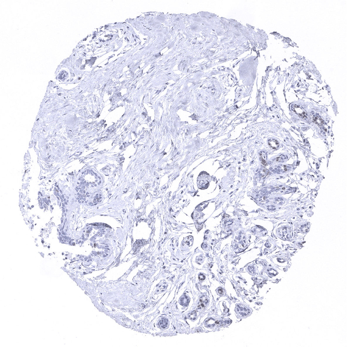
Breast: Few individual luminal epithelial cells with weak to moderate p16 immunostaining can occasionally occur.
Bronchus, mucosa
Cerebellum (molecular layer)
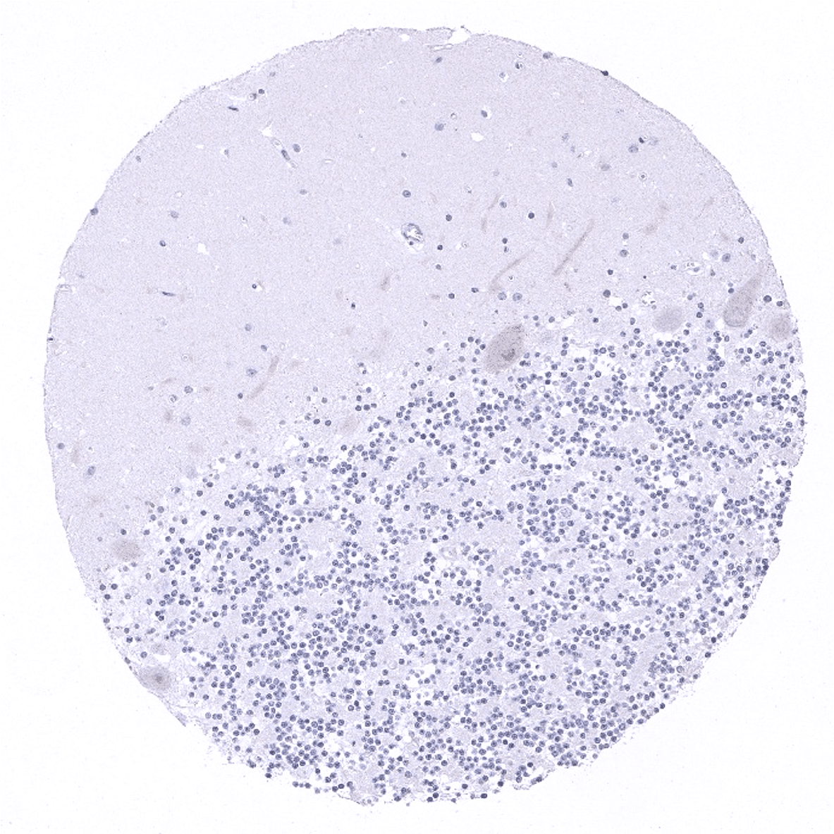
Cerebellum (molecular layer, Purkinje cell layer, granule cell layer)
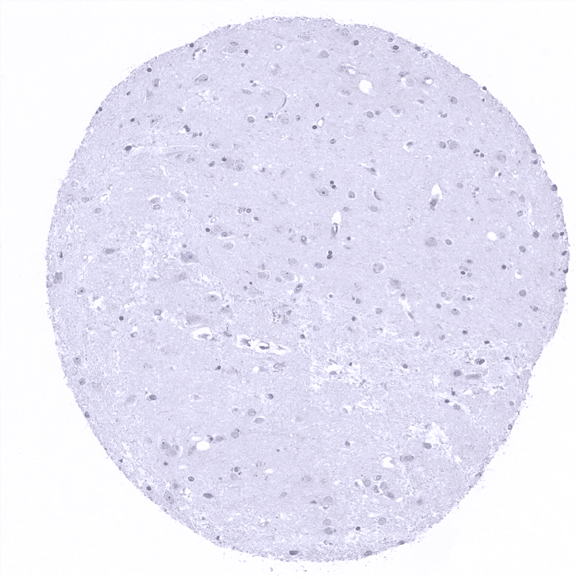
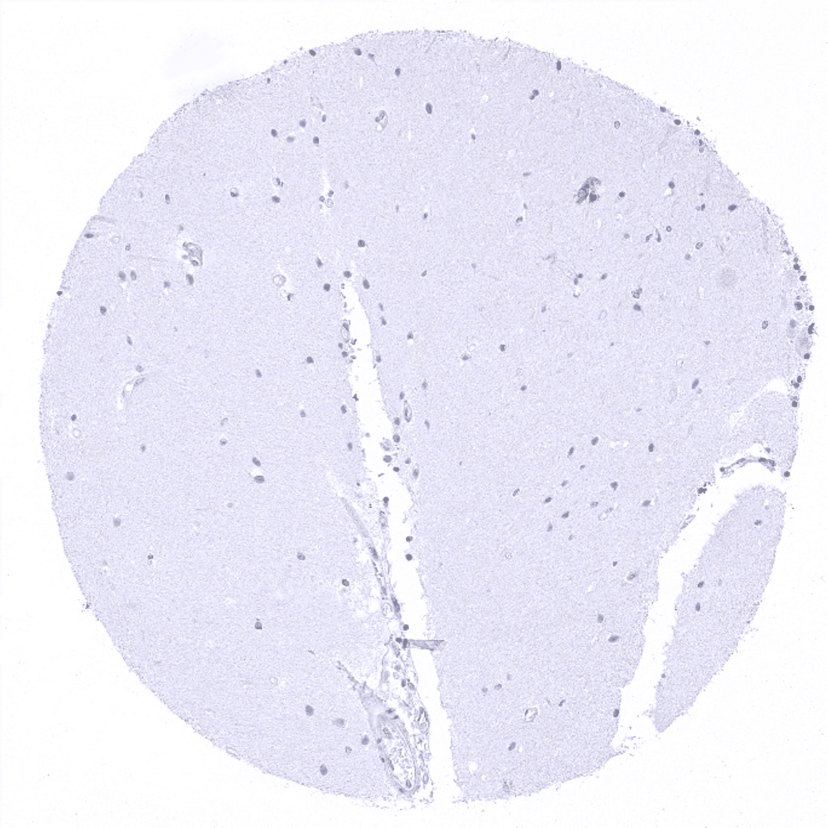
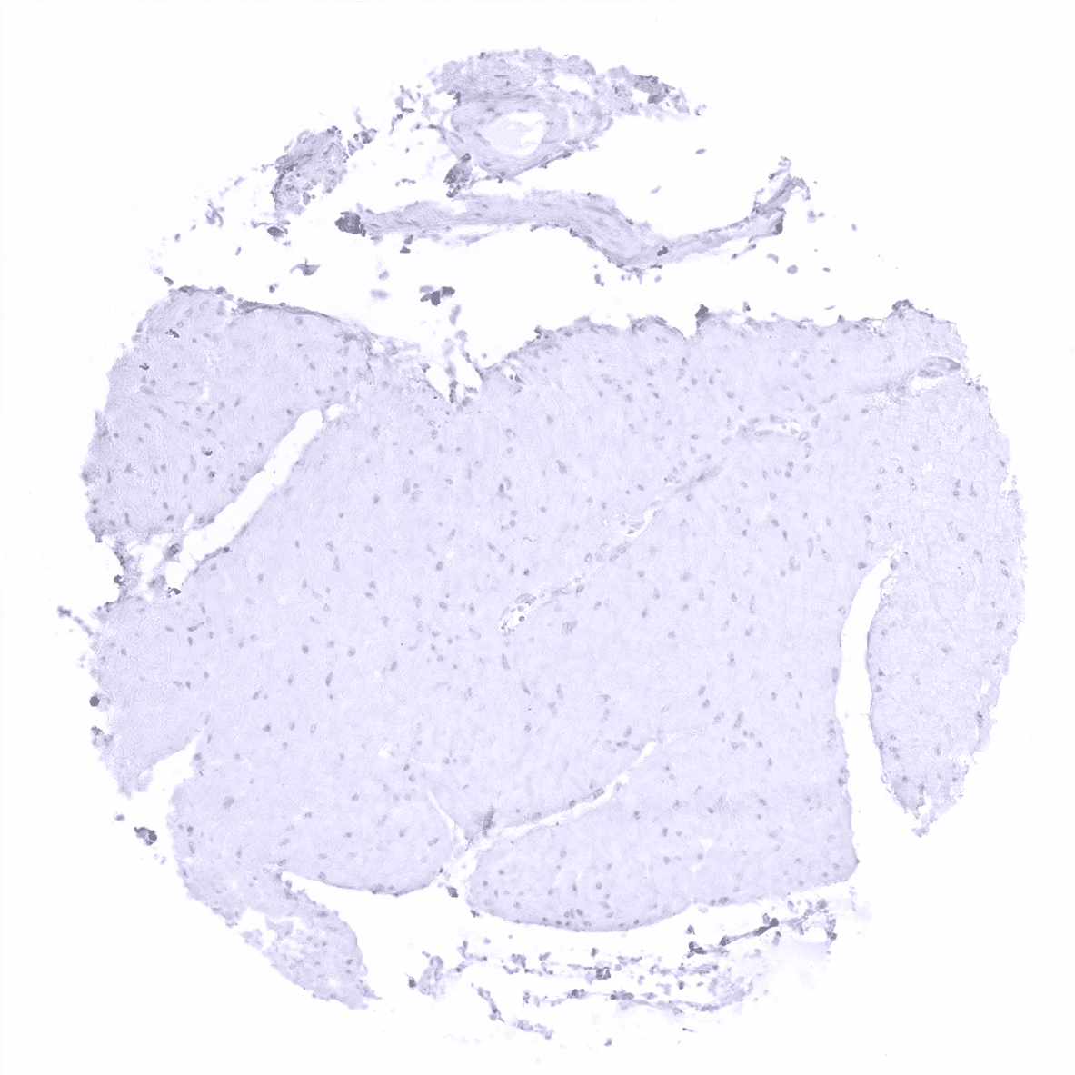
Cerebrum, grey matter
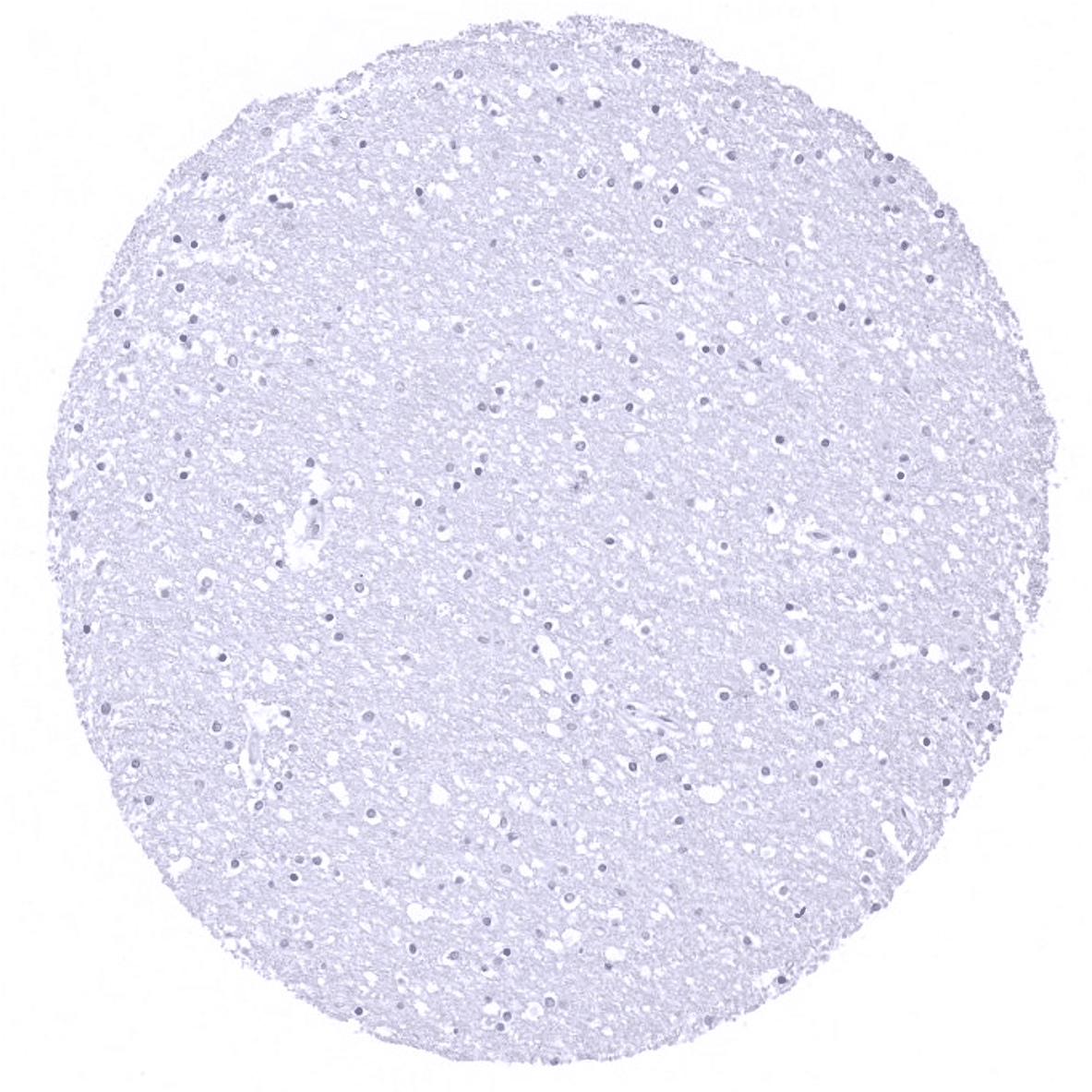
Cerebrum, white matter
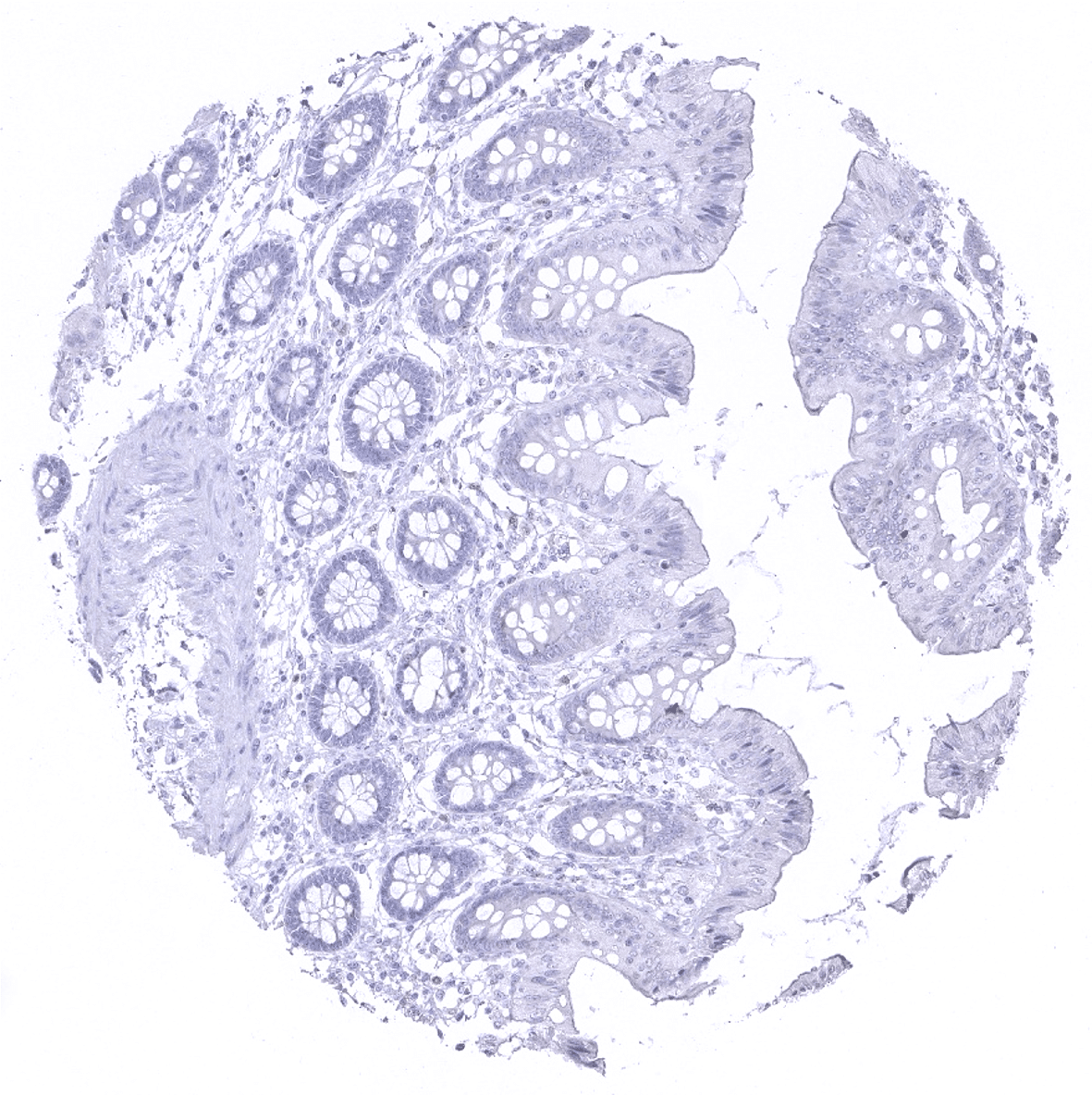
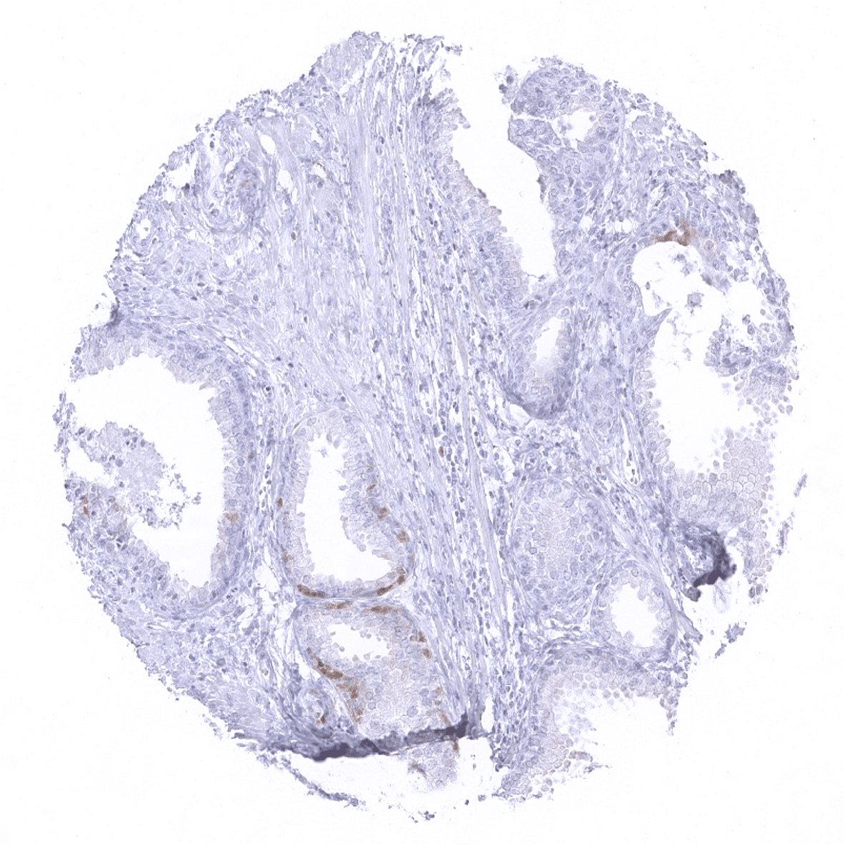
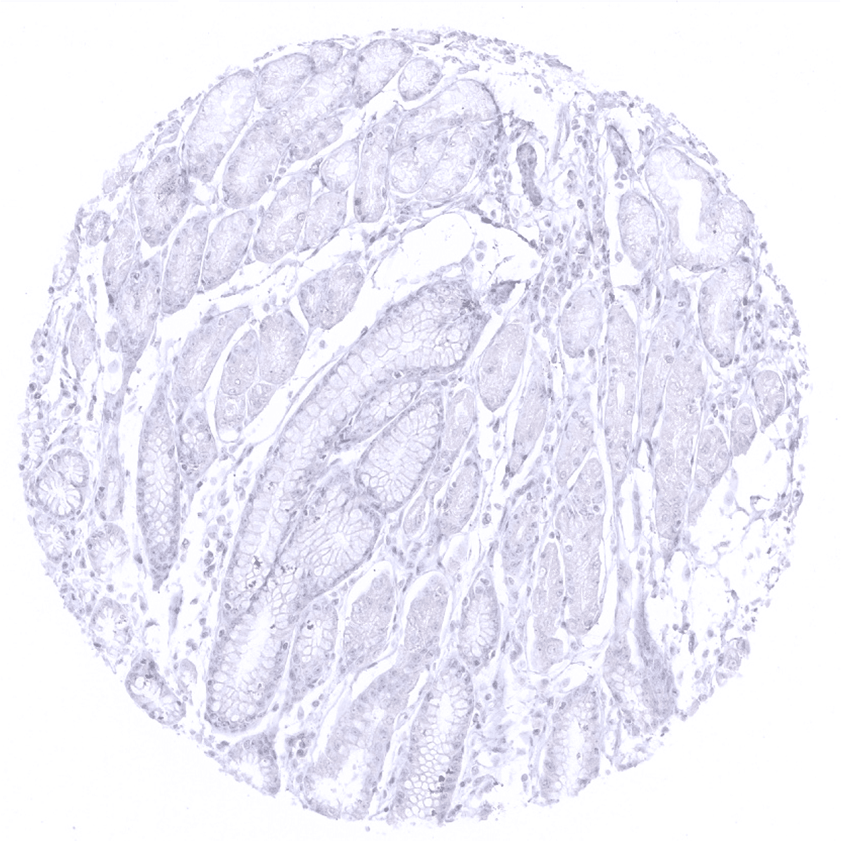
Colon descendens, mucosa

Colon descendens, muscular wall

Duodenum, Brunner gland
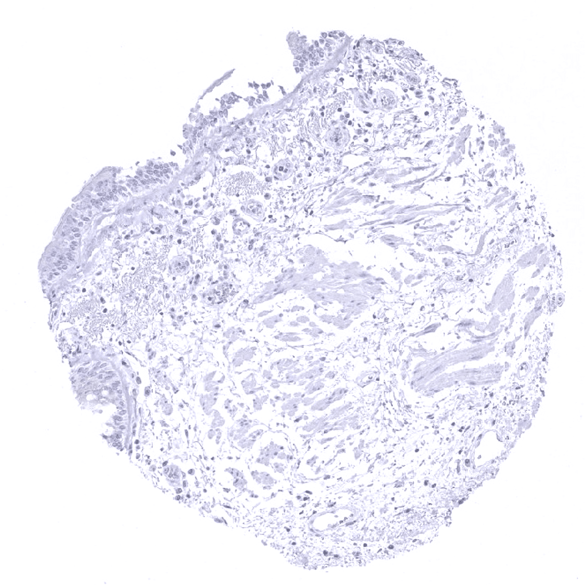
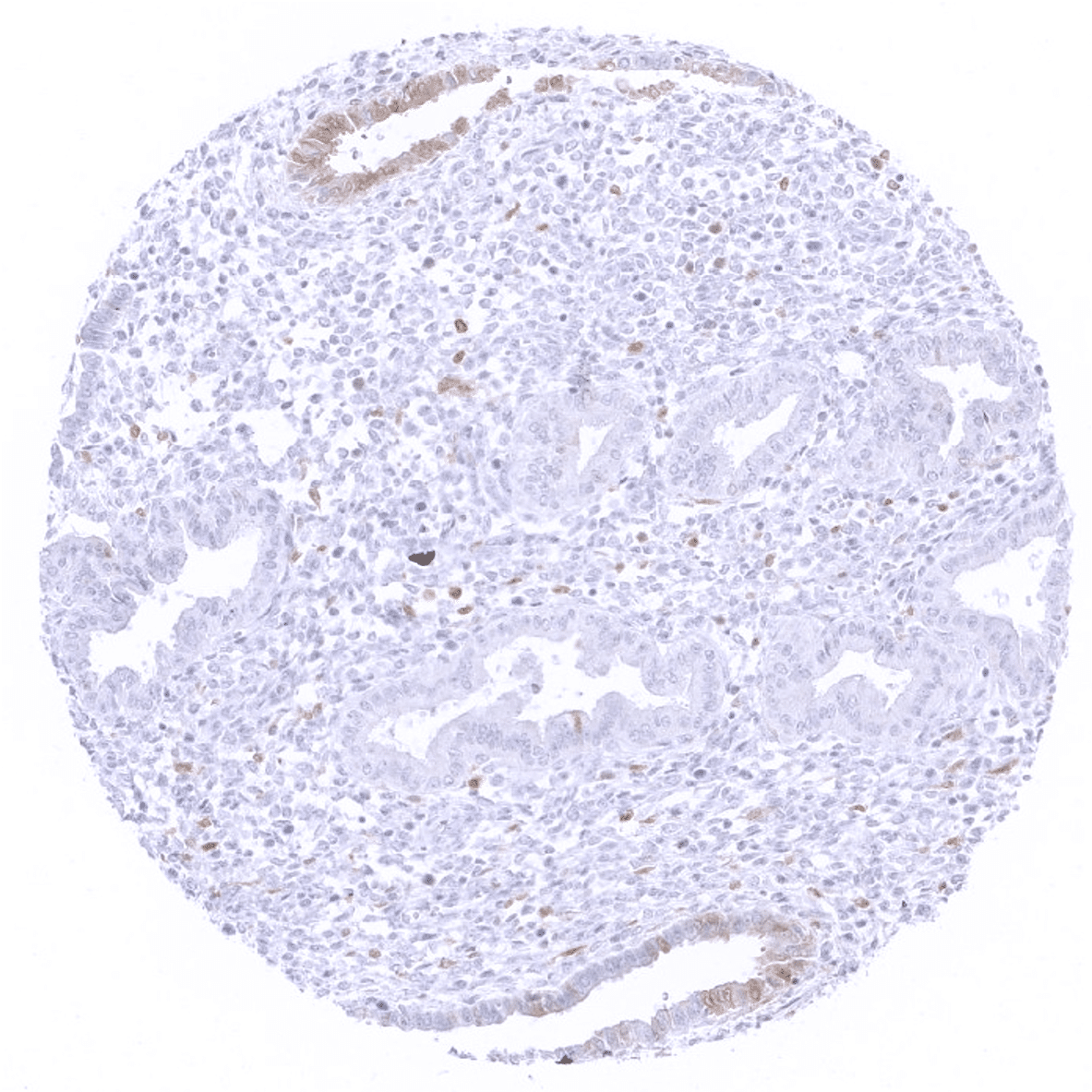
Duodenum, mucosa: Few individual cells with weak to moderate p16 immunostaining can occasionally occur in gastrointestinal epithelium.
Ectocervix

Endocervix
Endometrium, proliferation
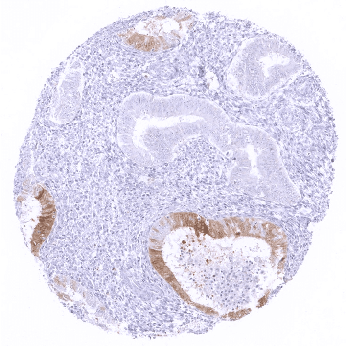
Endometrium, proliferation: Some glands with weak to moderate p16 immunostaining in the majority of cells can be seen in the histologically normal endometrium (not in all cases).
Endometrium, secretion: Some glands with weak to moderate p16 immunostaining in the majority of cells is often seen in the histologically normal endometrium.

Epididymis

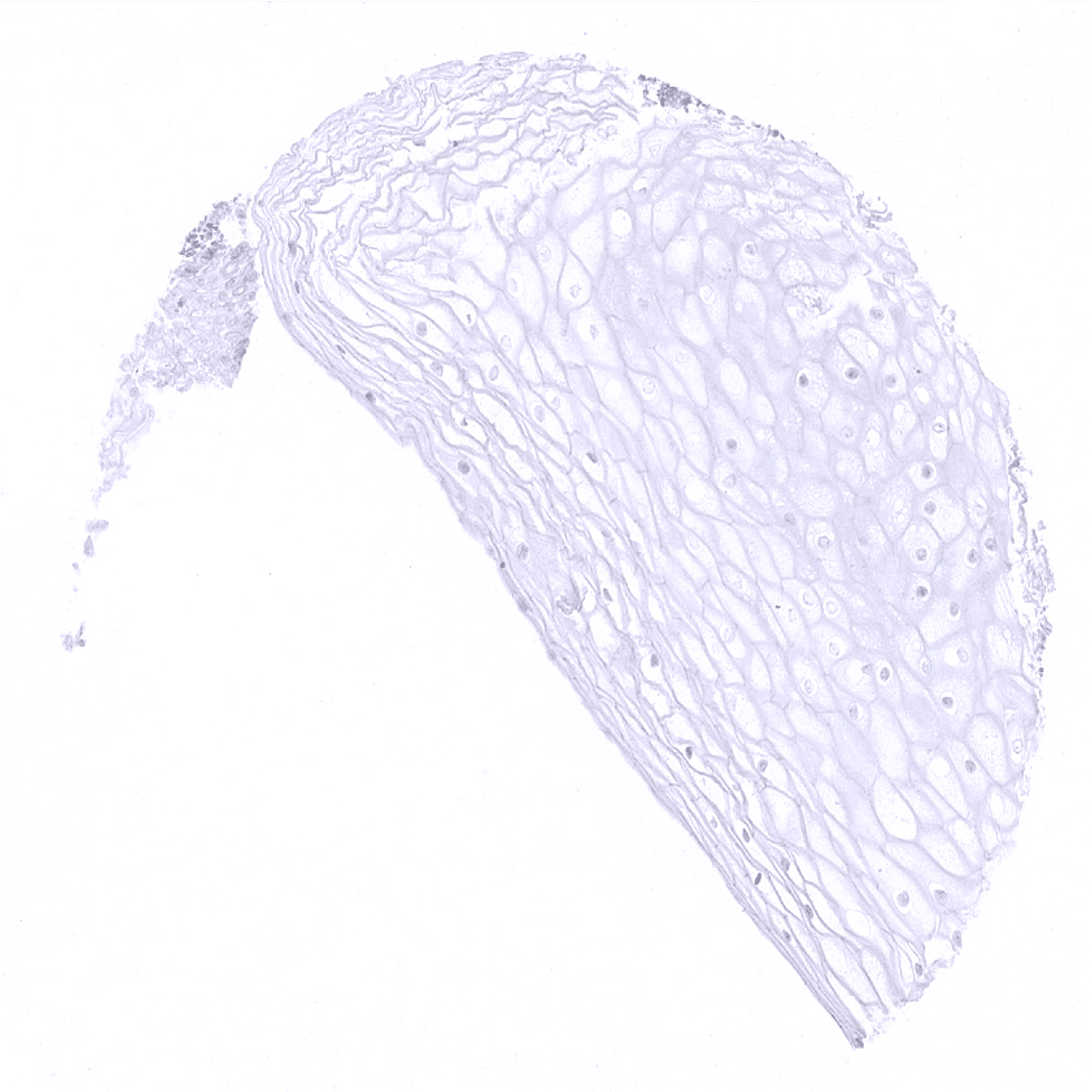
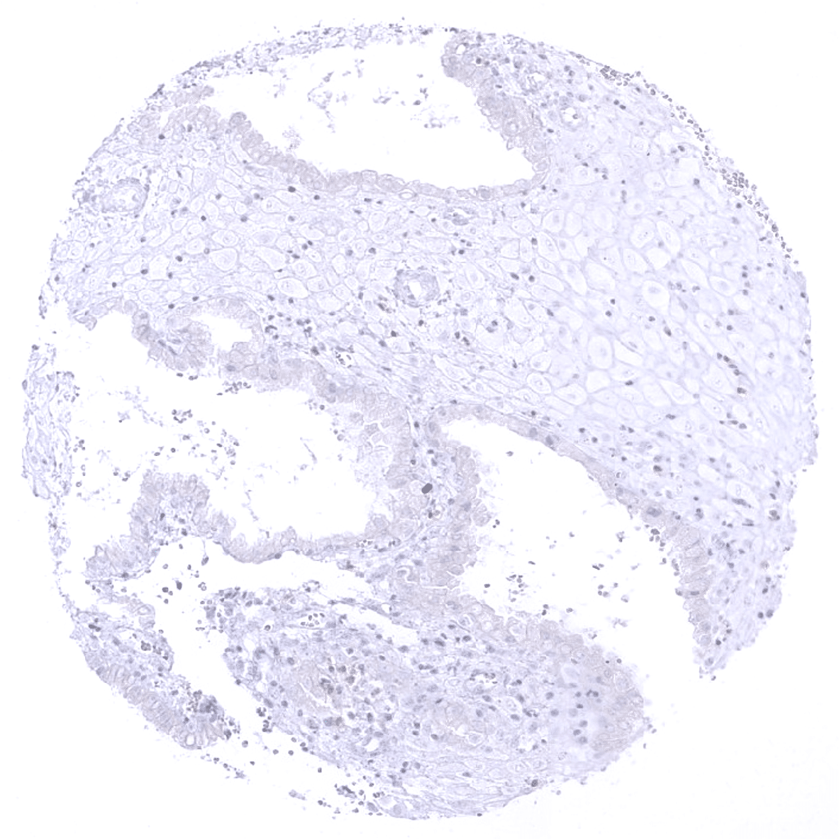
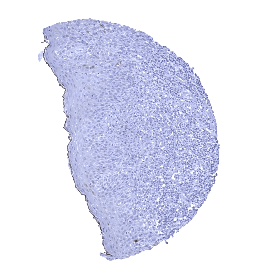
Esophagus, squamous epithelium
Fallopian tube, mucosa
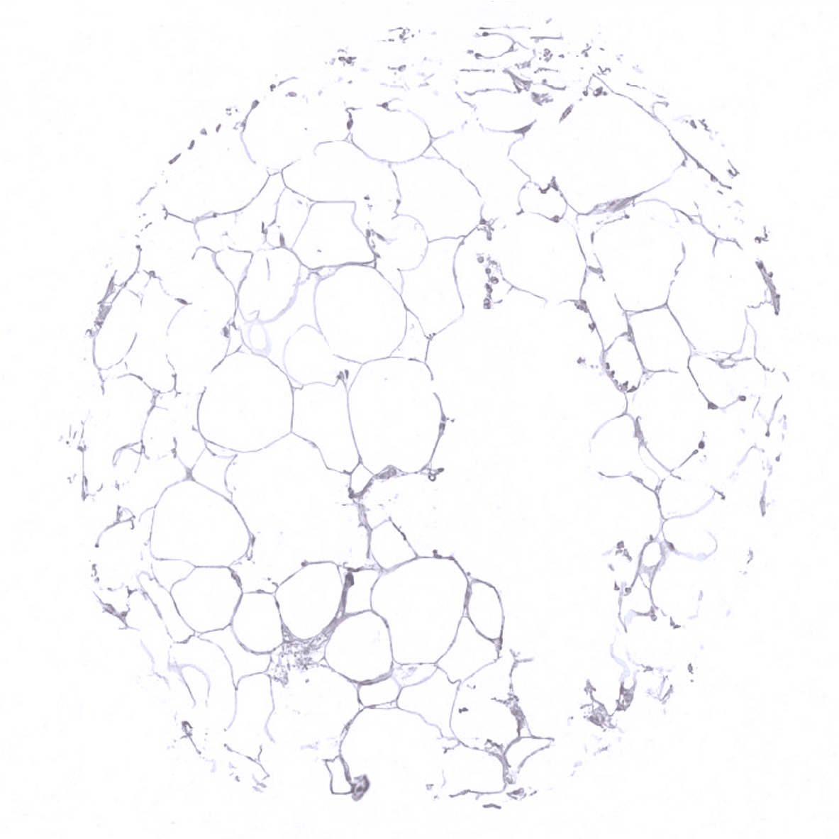
Fat

Gallbladder, epithelium
Heart
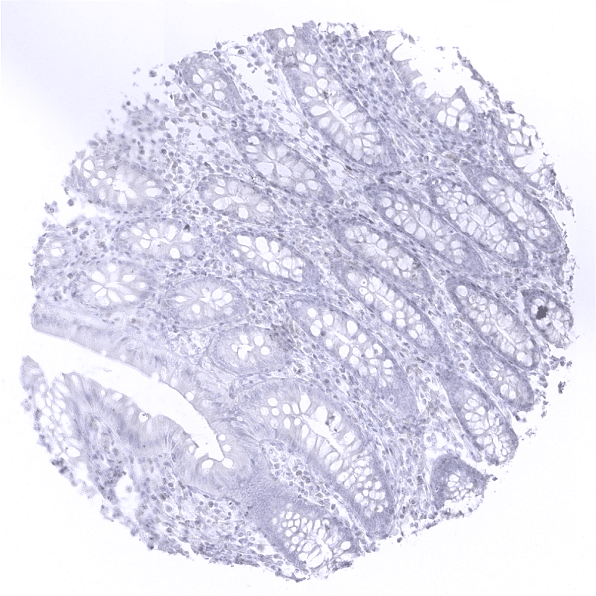
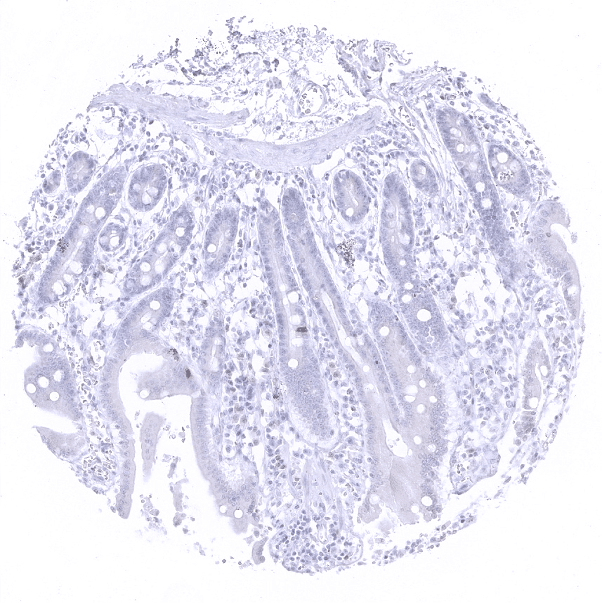
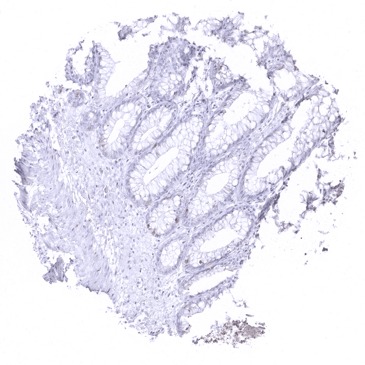
Ileum, mucosa
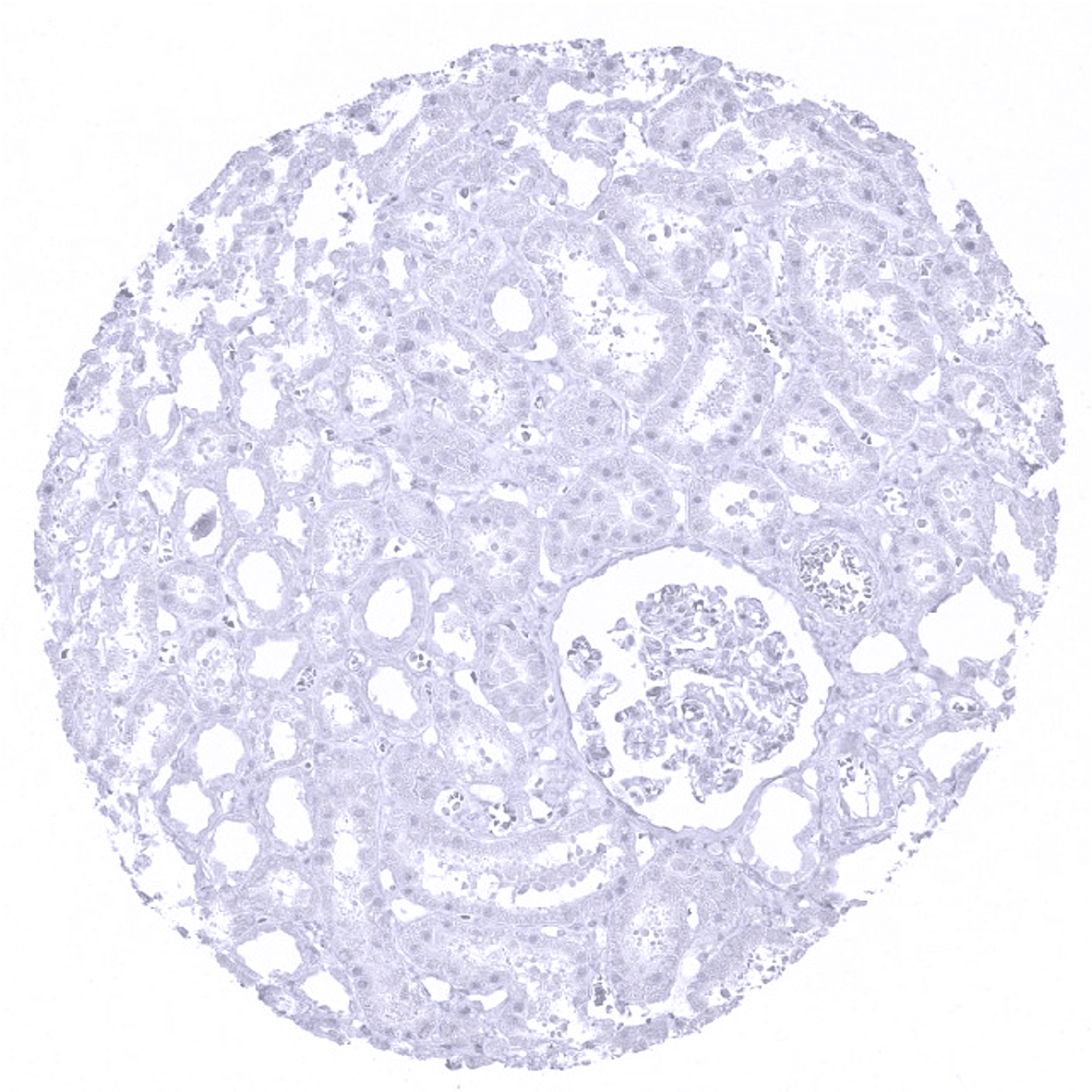
Kidney, cortex
Kidney, medulla
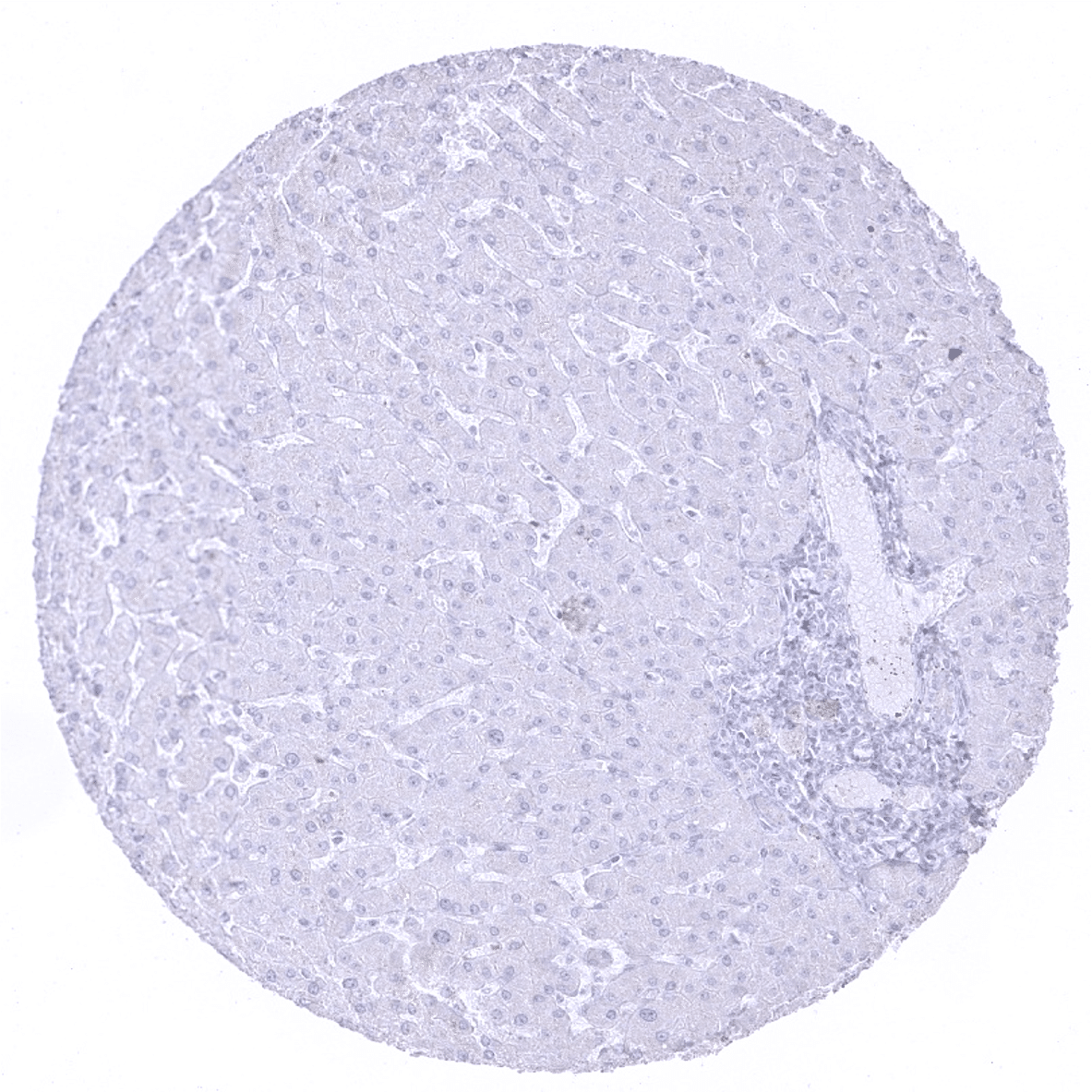
Liver
Lung
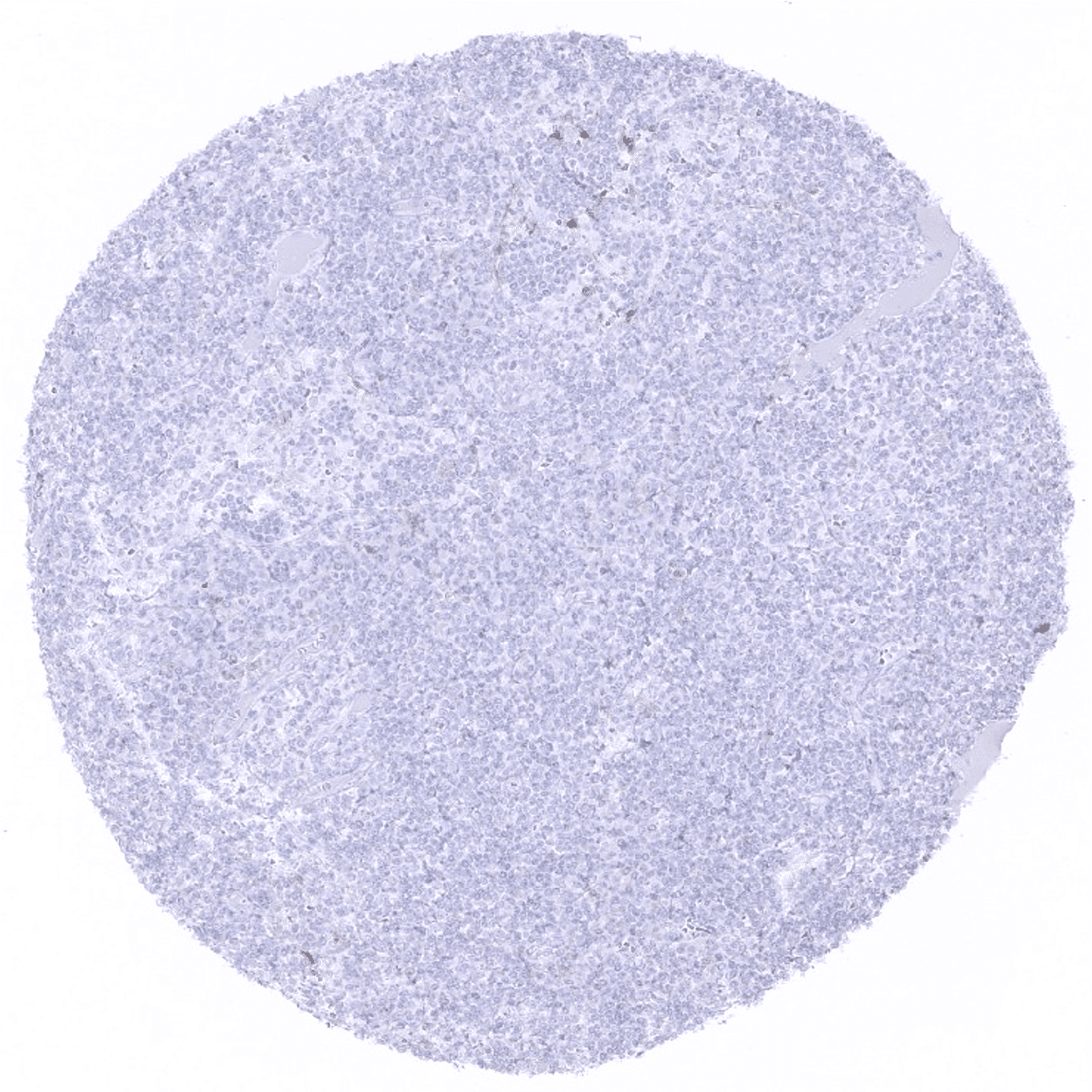
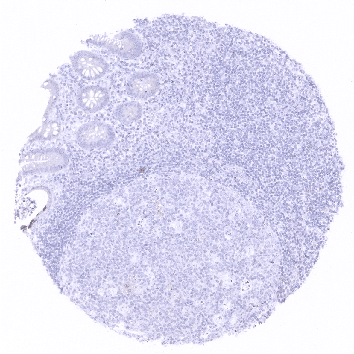
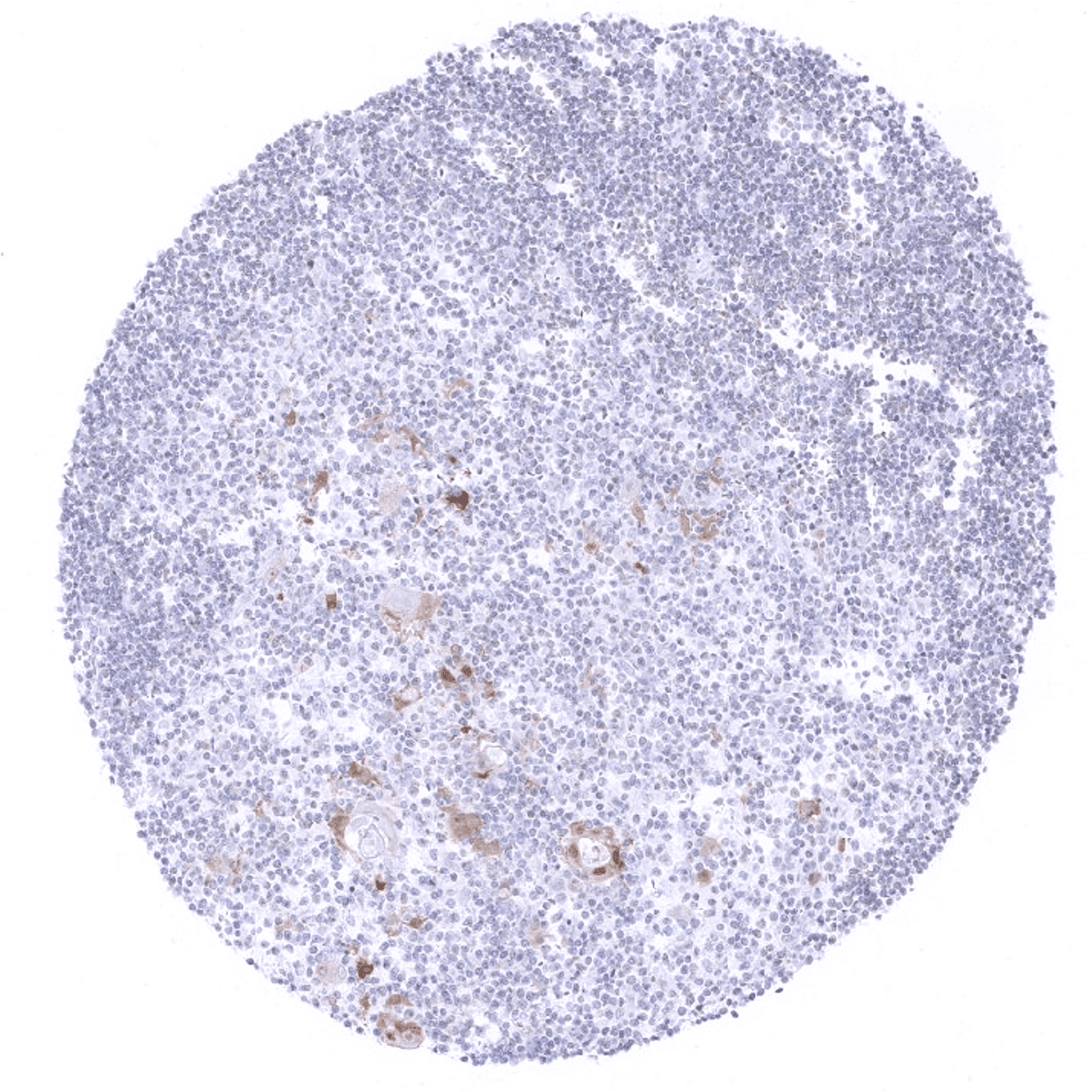
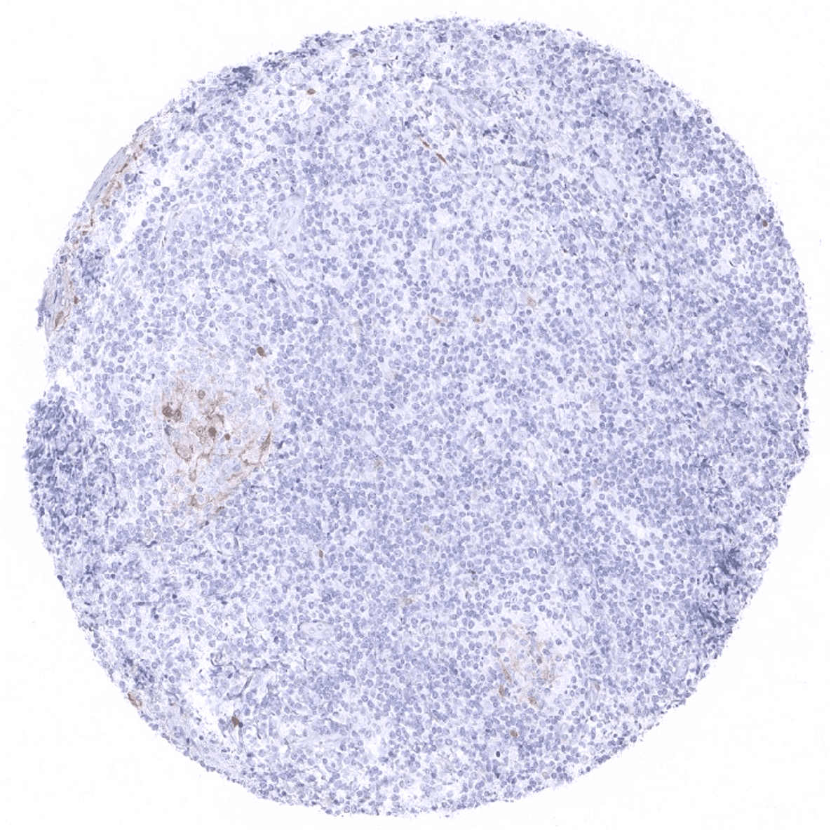
Lymph node
Ovary, stroma

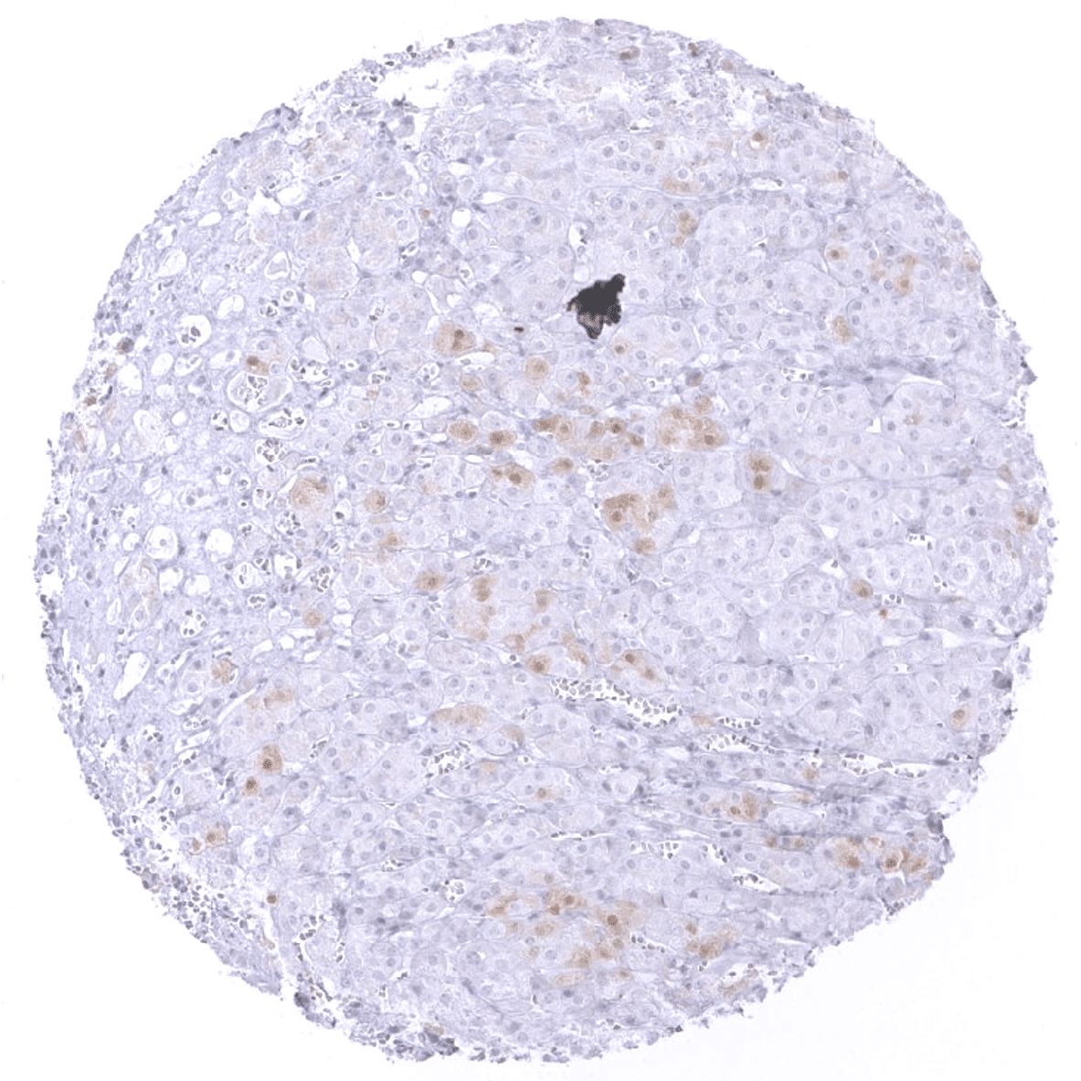
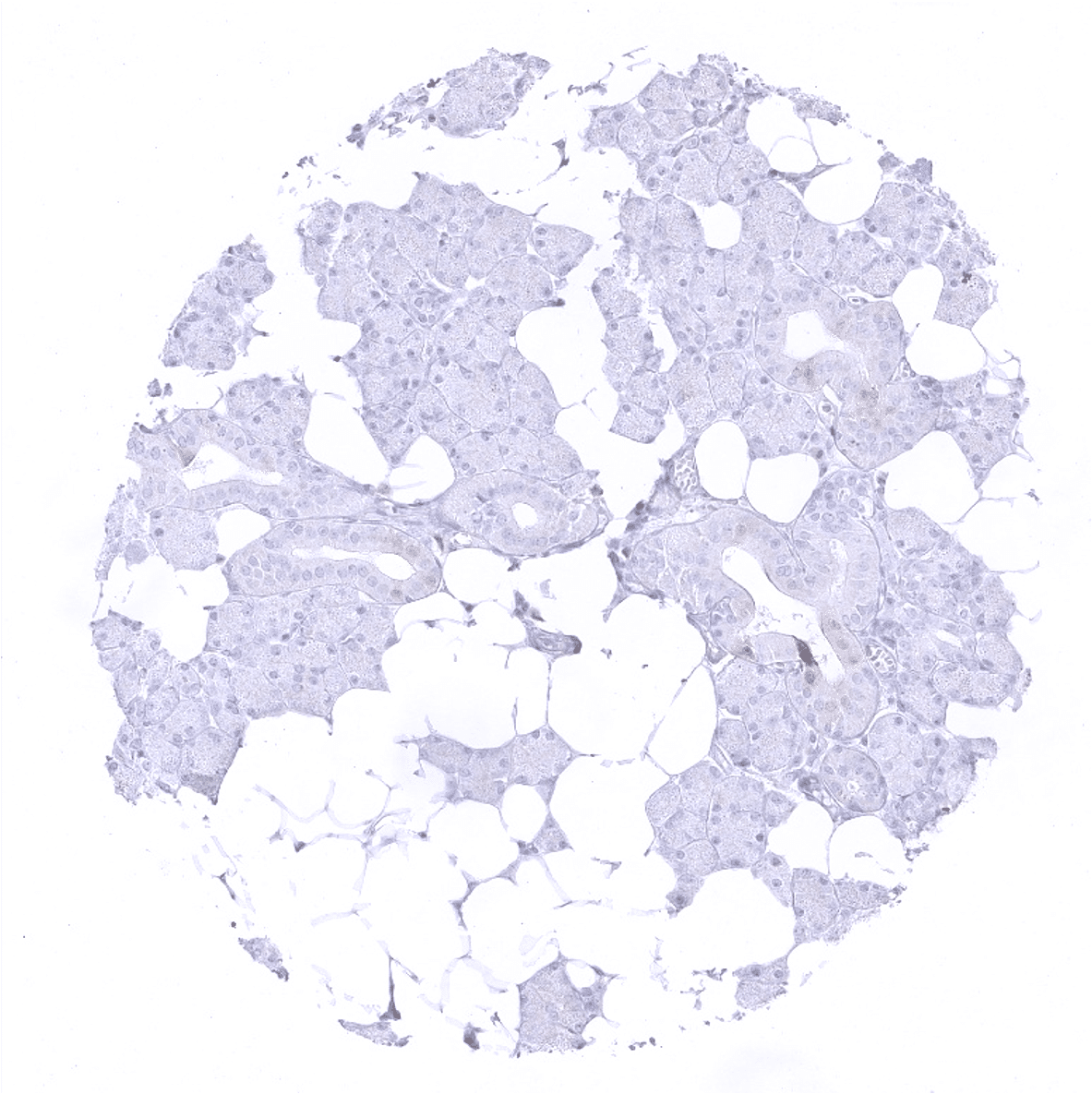
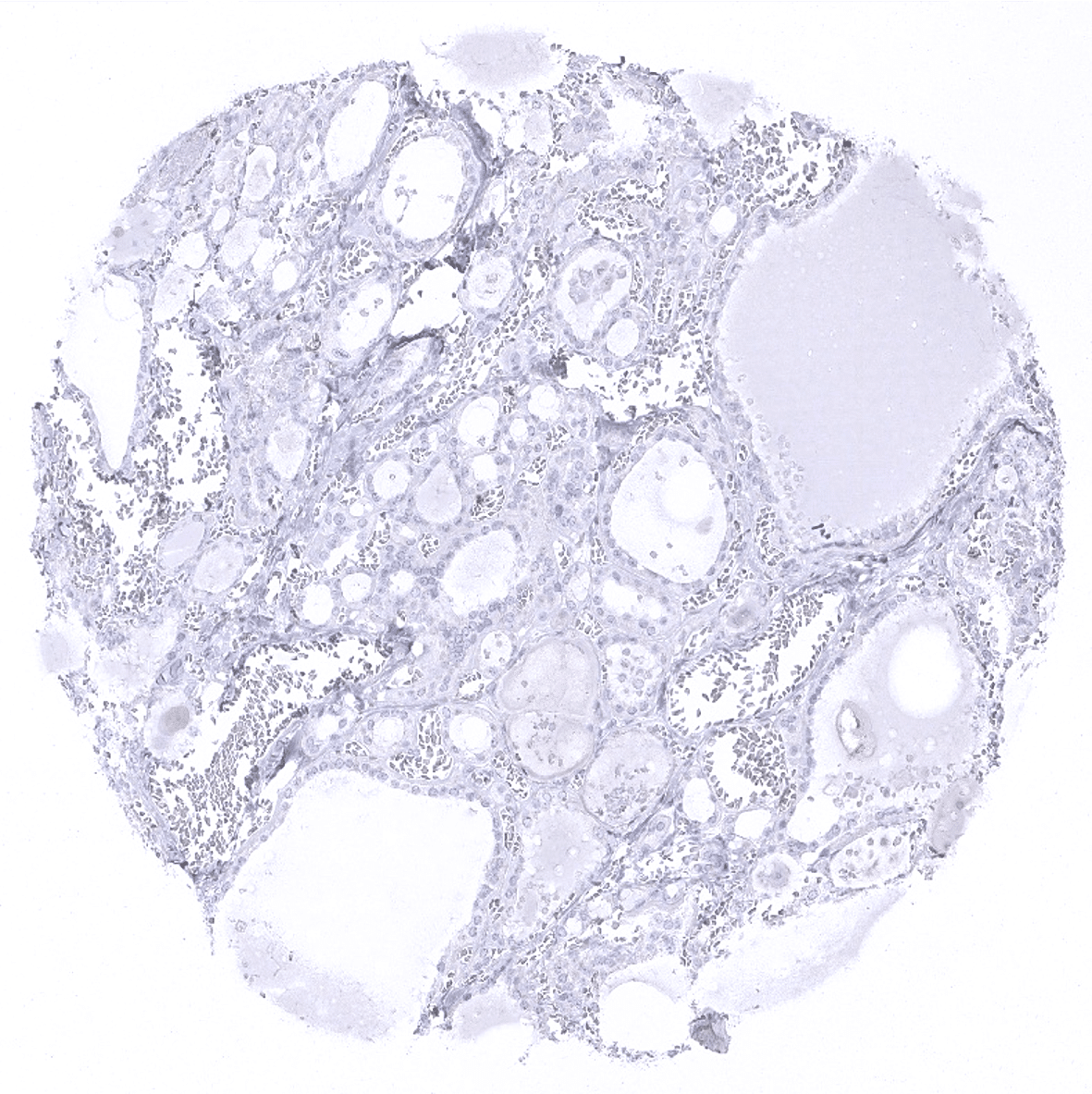
Pancreas- A fraction of cells of islets of Langerhans show a moderate p16 staining
Parathyroid
Parotid gland

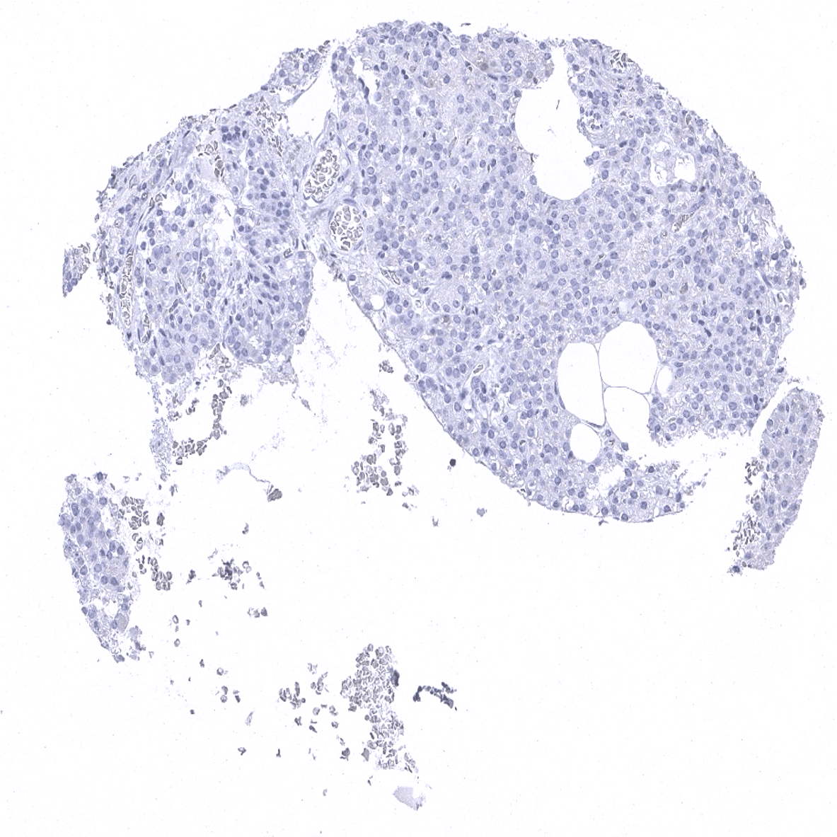
Pituitary gland, anterior lobe: A subset of cells of the adenohypophysis regularly show a moderate to strong p16 immunostaining.
Pregnant uterus (decidua)
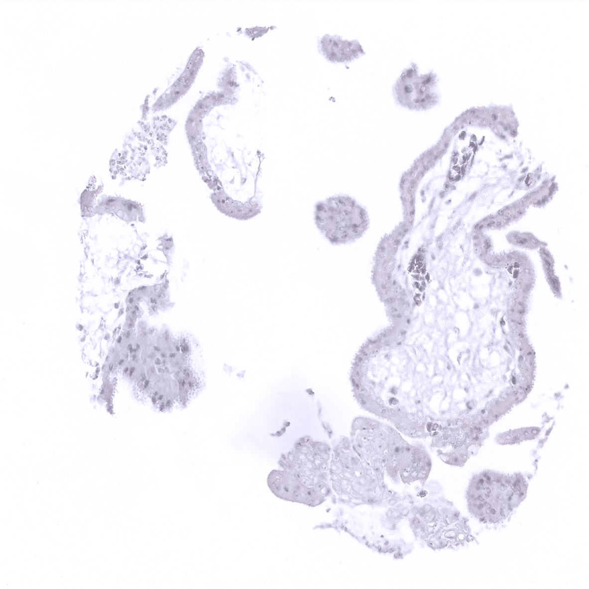
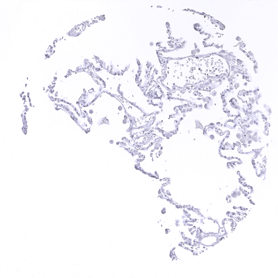
Placenta, early
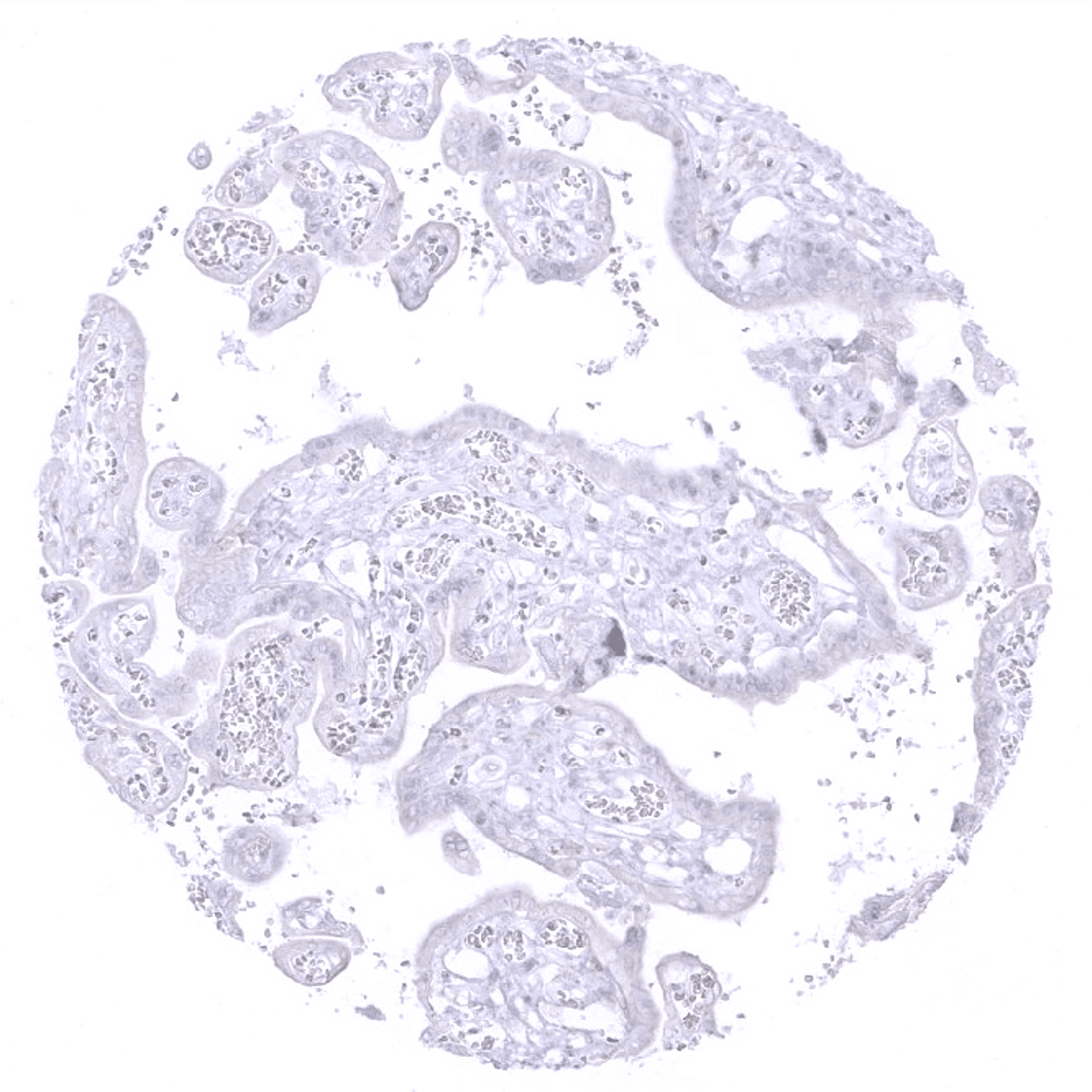
Placenta, mature

Placenta, mature: A weak to moderate p16 immunostaining can occur in stroma cells of the placenta (only in individual cases)

Placenta (amnion and chorion)
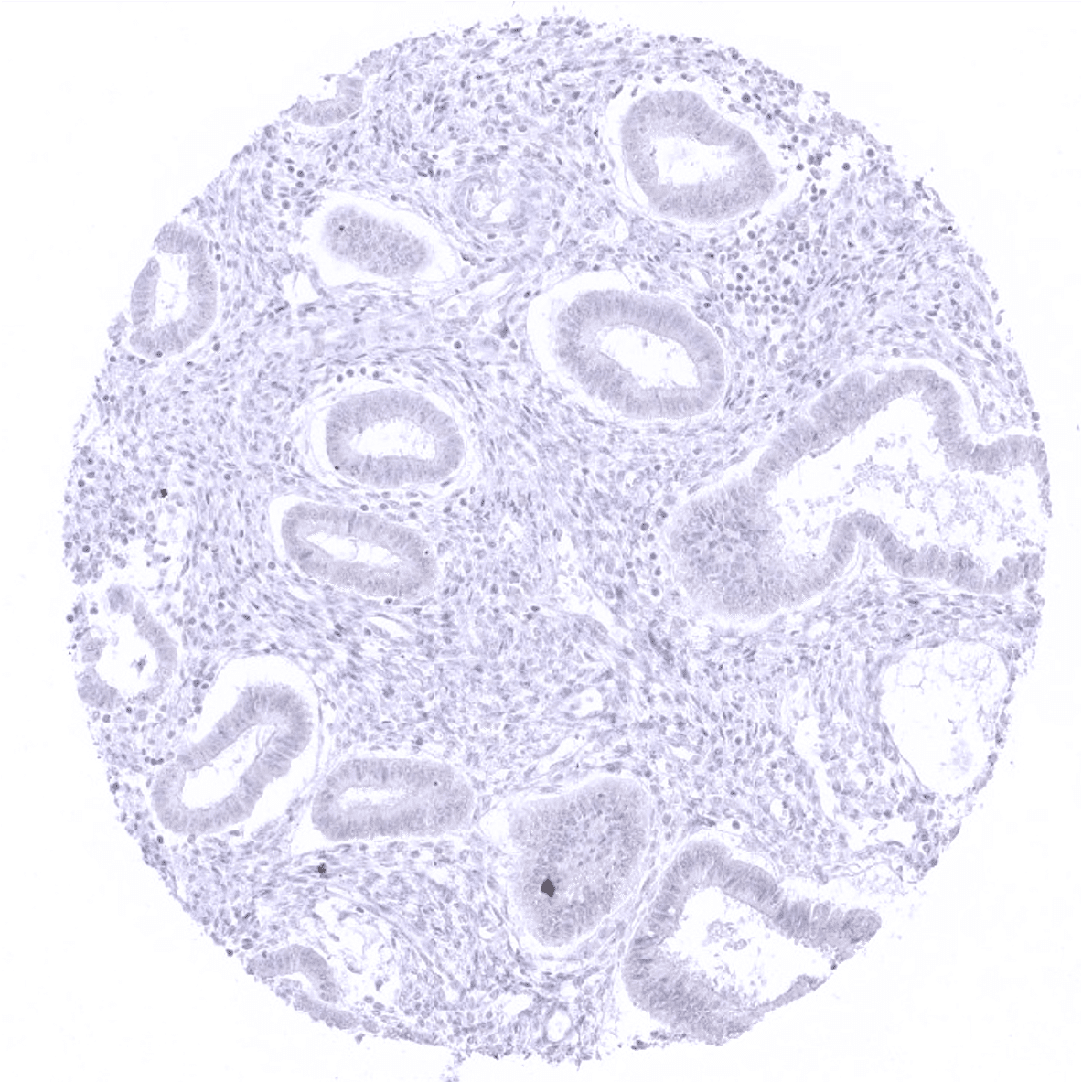
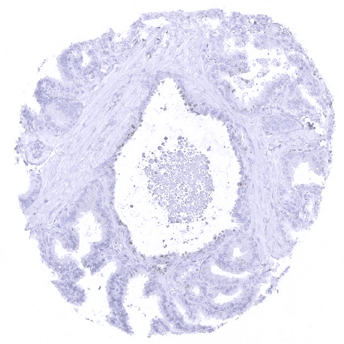
Prostate: Basal cells can show a weak to moderate p16 staining.
Rectum, mucosa
Seminal vesicle: Occasional coloration of the seminal vesicle epithelial cells is due to pigmentation (no true immunostaining).

Sinus paranasales: Few individual cells or groups of cells with weak to moderate p16 immunostaining can occasionally occur in respiratory epithelium.

Skin

Spleen
Stomach, antrum

Stomach, corpus

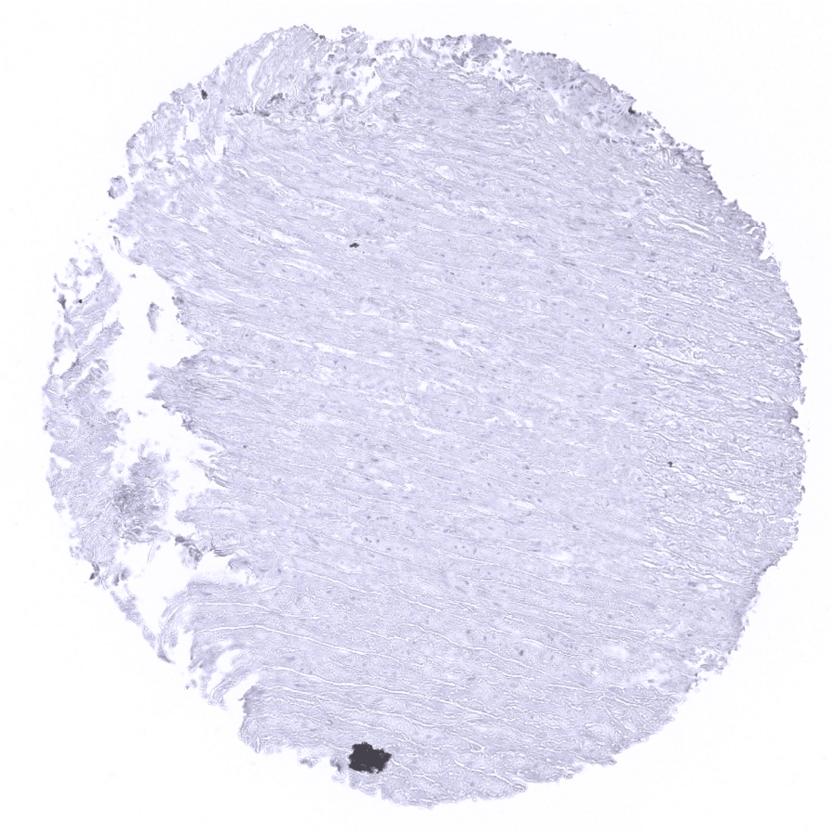
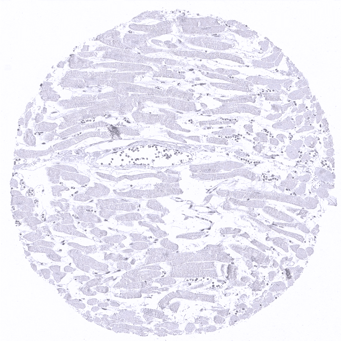
Striated muscle
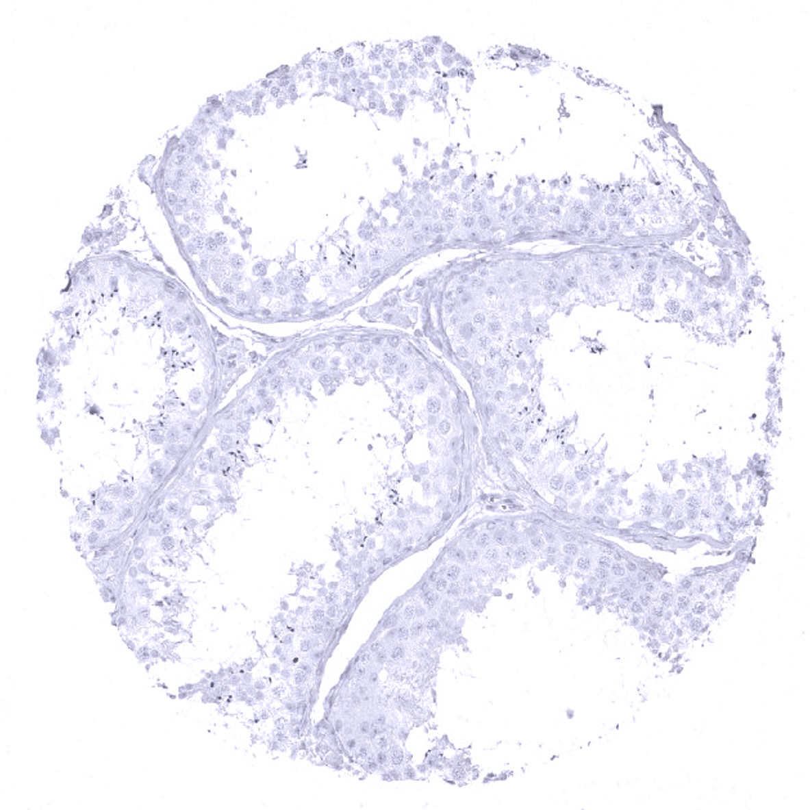
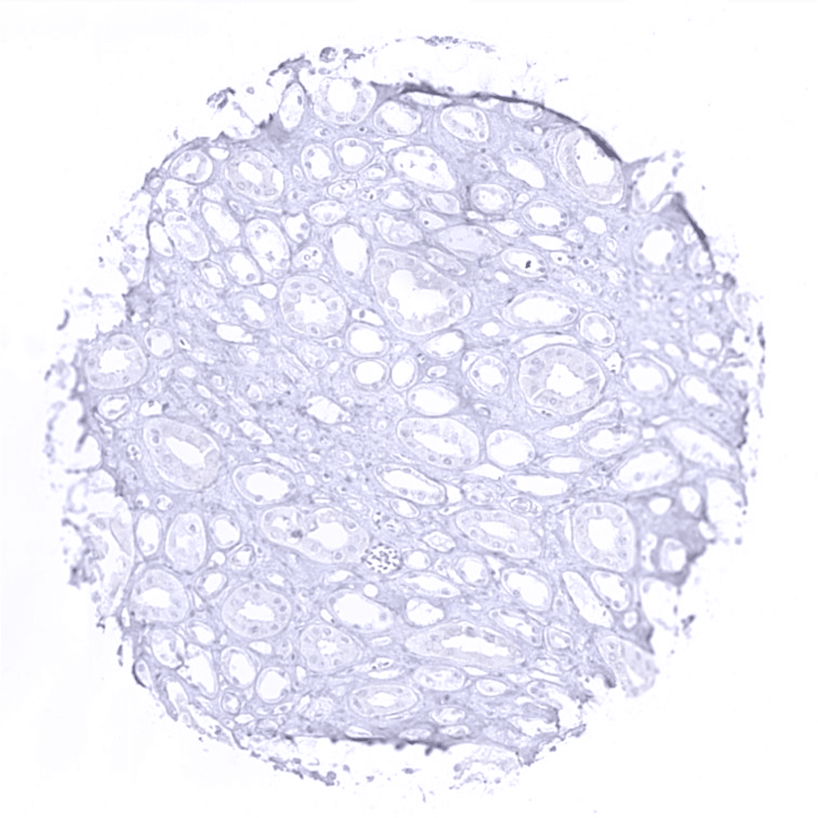
Testis
Thymus: Some elements of corpuscles of Hassall's stain p16 positive.
Thyroid gland
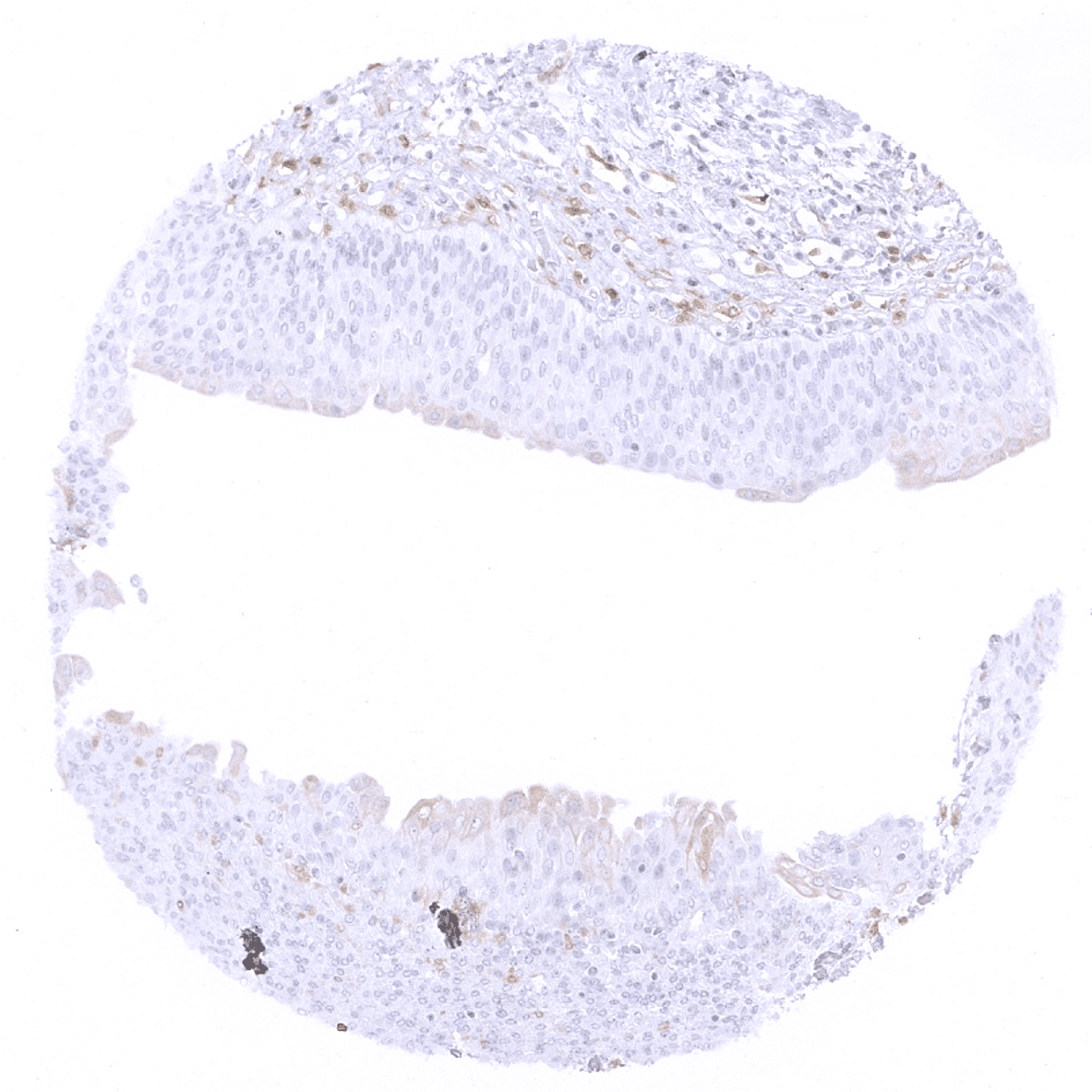
Tonsil: individual cells or small groups of normally appearing squamous epithelial cells of the crypts can occasionally show weak to moderate p16 immunostaining.
Tonsil, surface epithelium: Individual cells or small groups of normally appearing cells with weak to moderate p16 immunostaining can occasionally occur in the surface epithelium.
Urinary bladder, muscular wall
Urinary bladder, urothelium: Occasional cells or small groups of normally appearing cells with weak to moderate p16 immunostaining can occur in urothelium (here: mostly umbrella cells). Very rarely, this also applies to few stroma cells such as in this case.

Uterus, myometrium