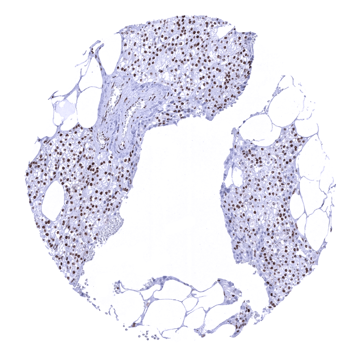
Adrenal gland
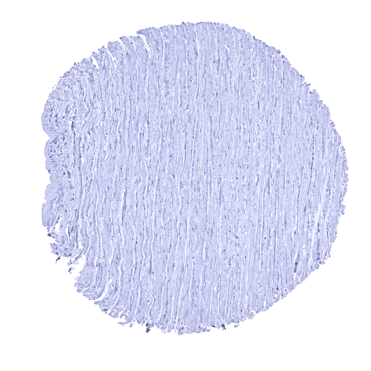
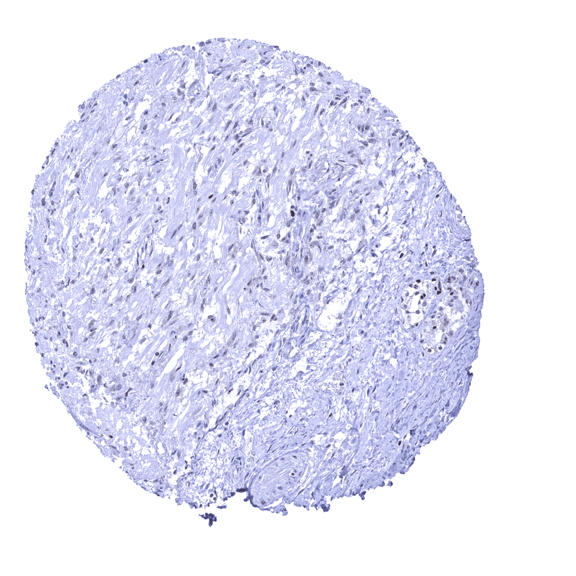
Aorta, media

Appendix, mucosa
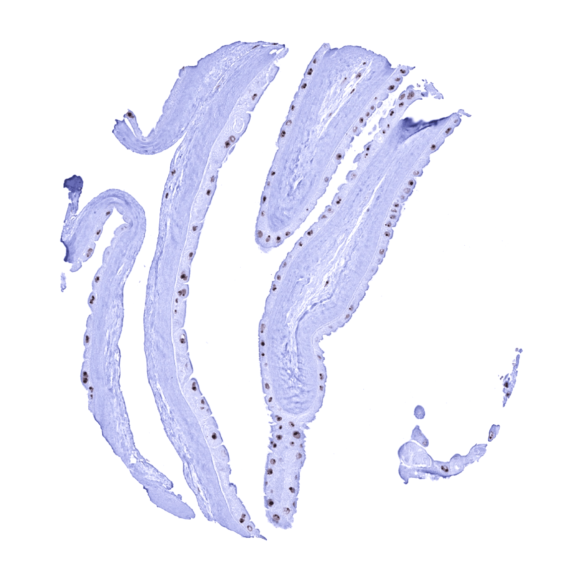
Appendix, muscular wall - The entire nuclei of smooth muscle cells are nucleolin positive but the immunostaining is most distinct in the nucleolus.
Bone marrow
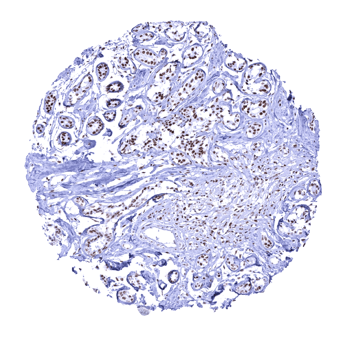
Breast
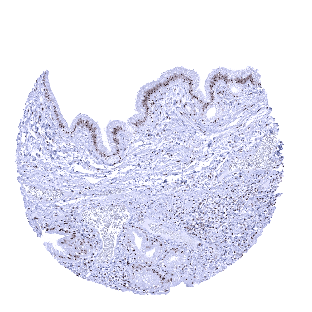
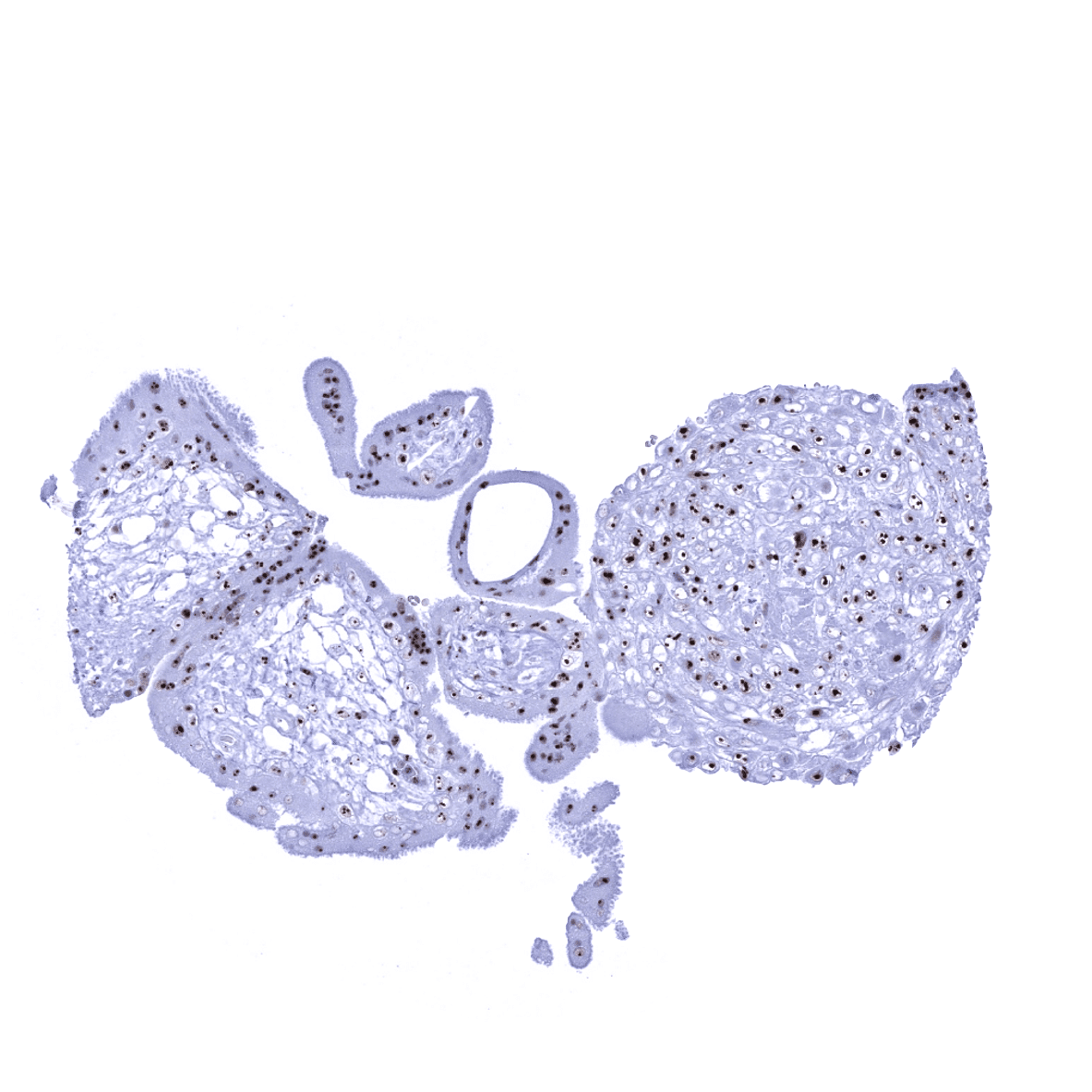
Bronchus, mucosa - In the respiratory epithelium, nucleolin staining is more prominent in basal and goblet cells than in the ciliated cells. The focus of the immunostaining to the nucleoli becomes most visible in the less stained ciliated cells, however.
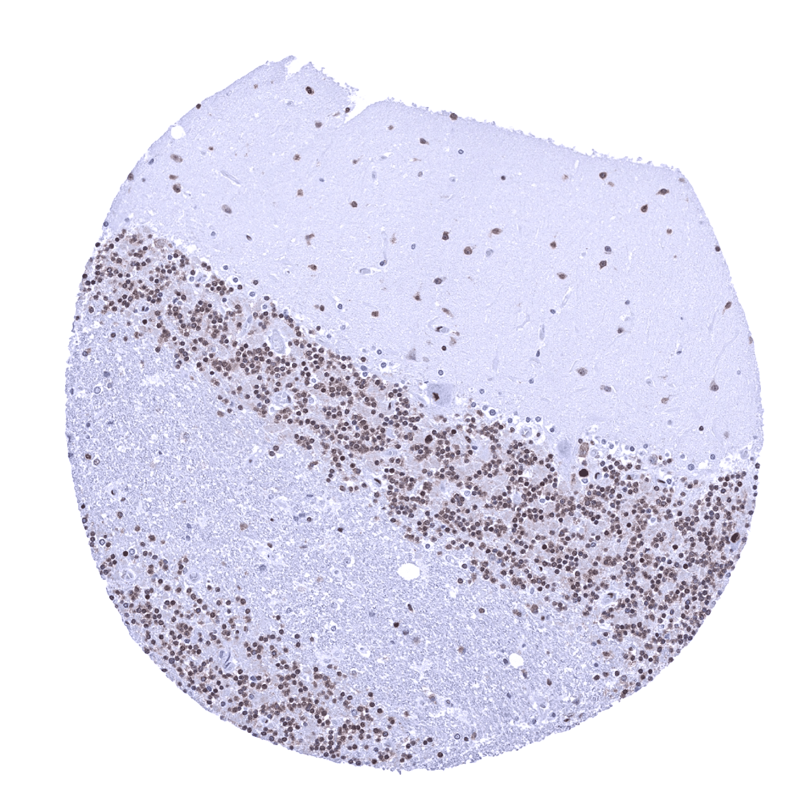
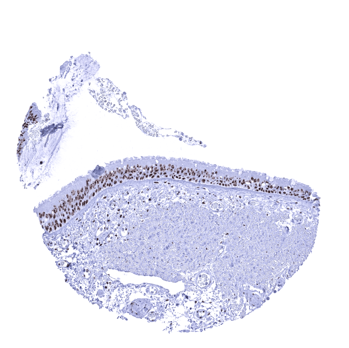
Cerebellum (molecular layer, Purkinje cell layer, granule cell layer, white matter)
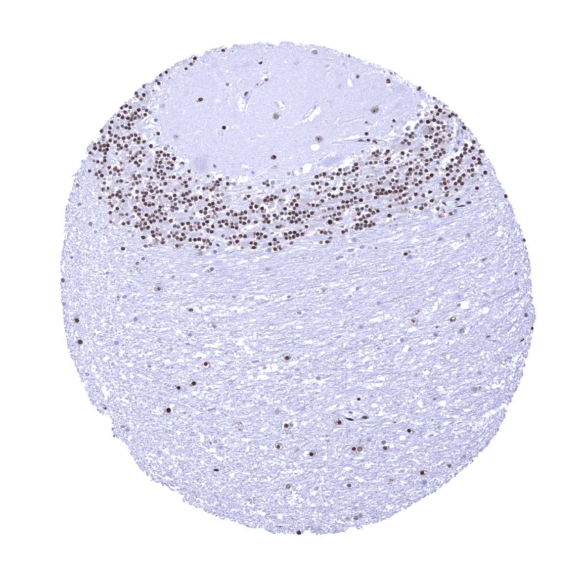
Cerebellum (molecular, Purkinje cell, granule cell layers; white matter)
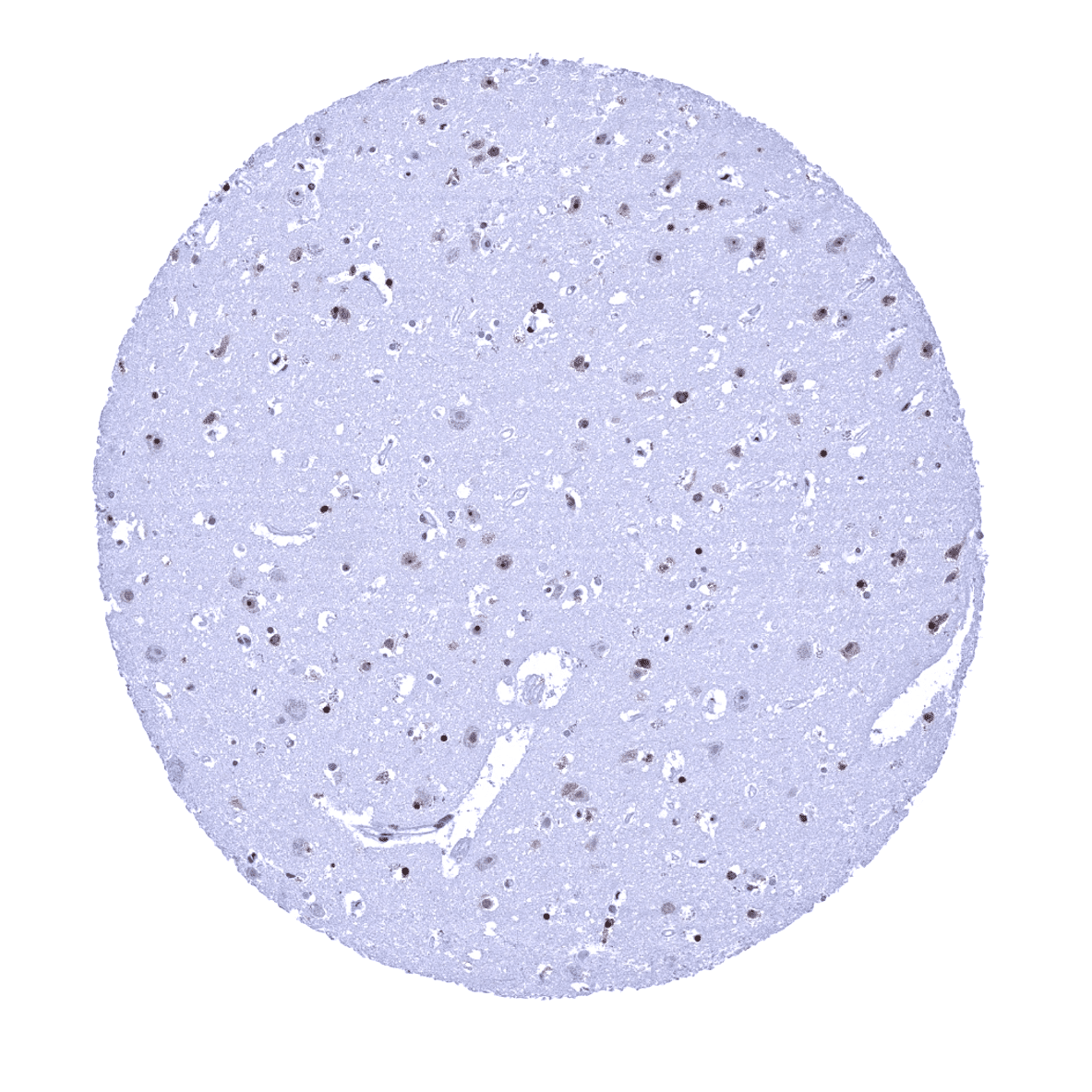
Cerebrum, grey matter
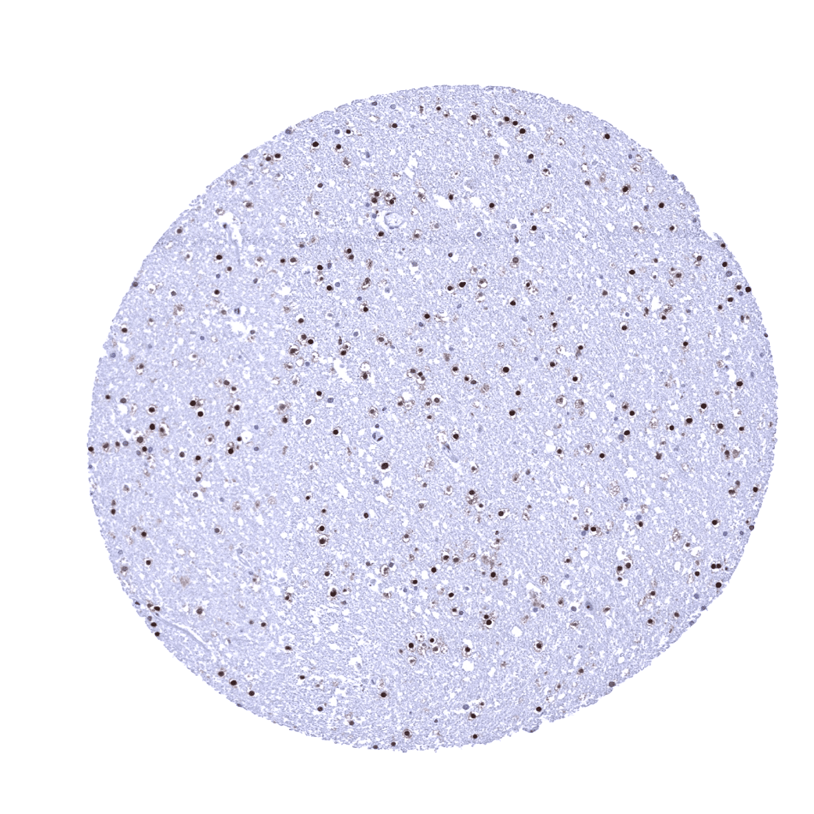
Cerebrum, white matter
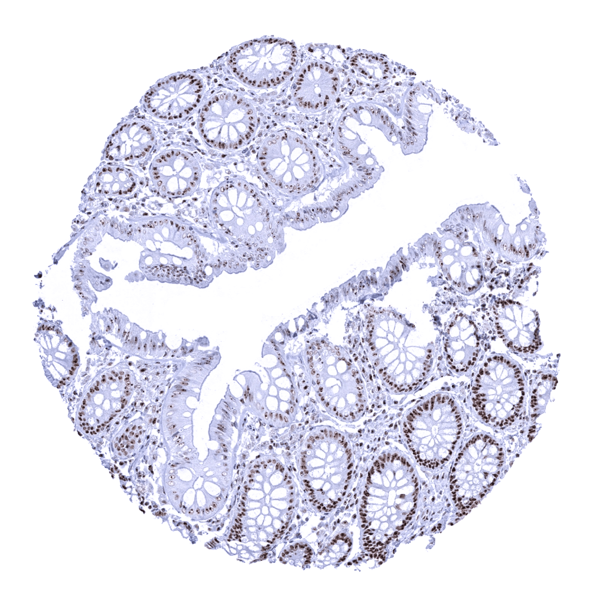
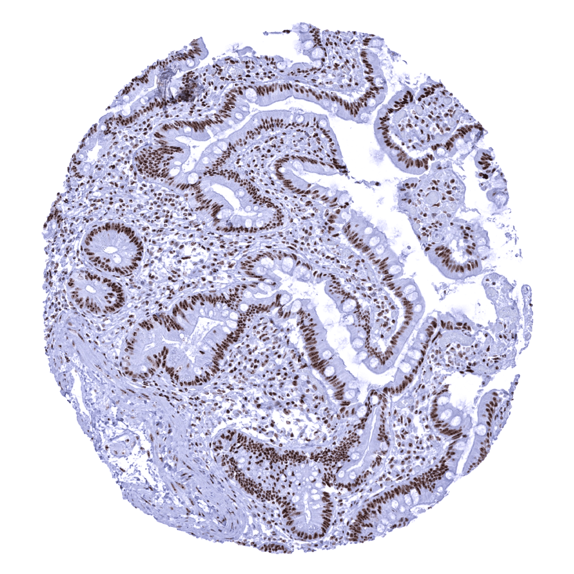
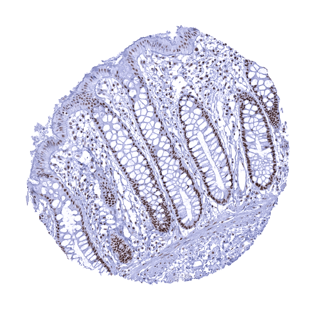
Colon descendens, mucosa - In the colon, nucleolin staining is more prominent in the proliferating cells in the crypt base than in the less proliferative superficial cell layers. The focus of the immunostaining to the nucleoli becomes most visible in the less stained superficial cells, however.
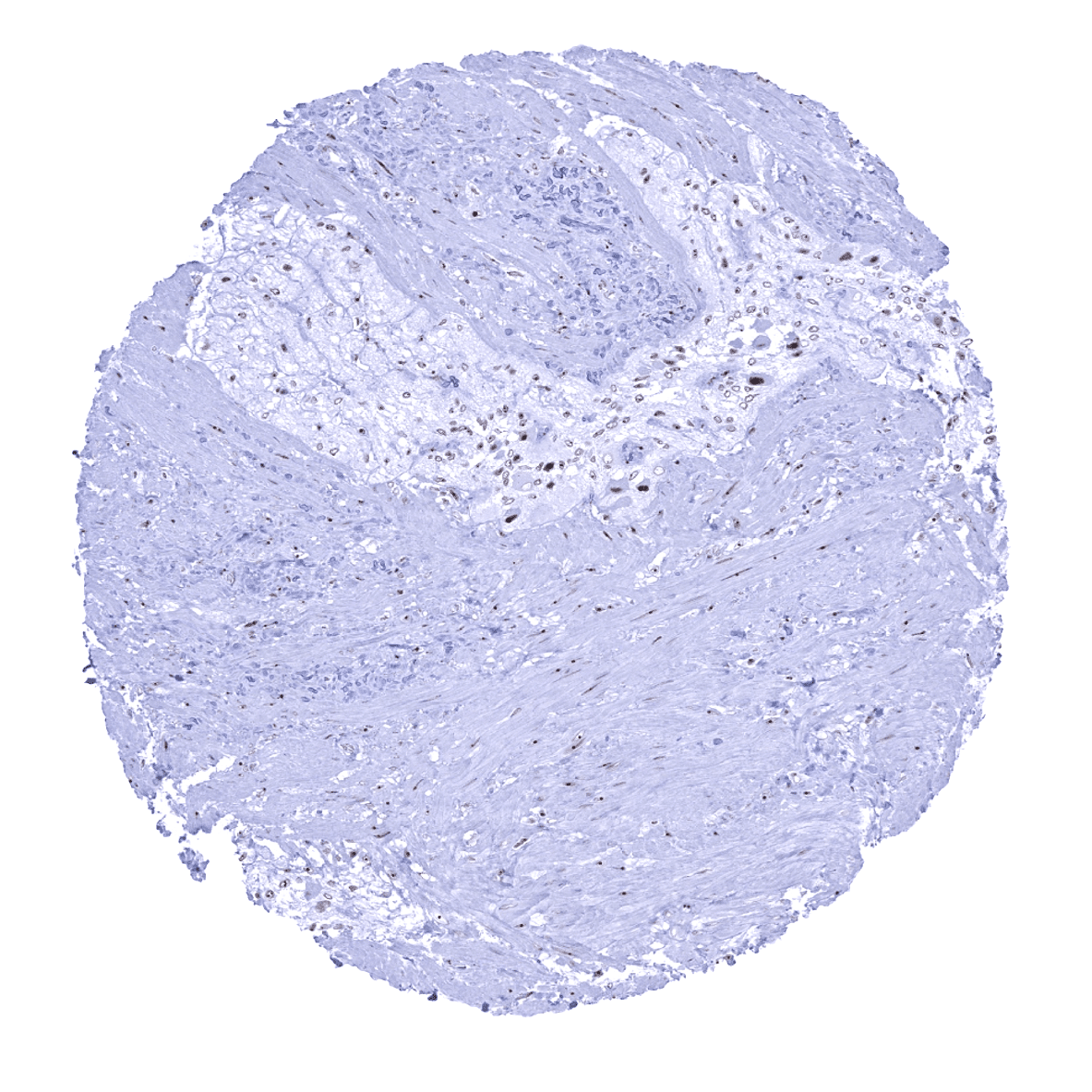
Colon descendens, muscular wall
Duodenum, Brunner gland
Duodenum, mucosa

Epididymis
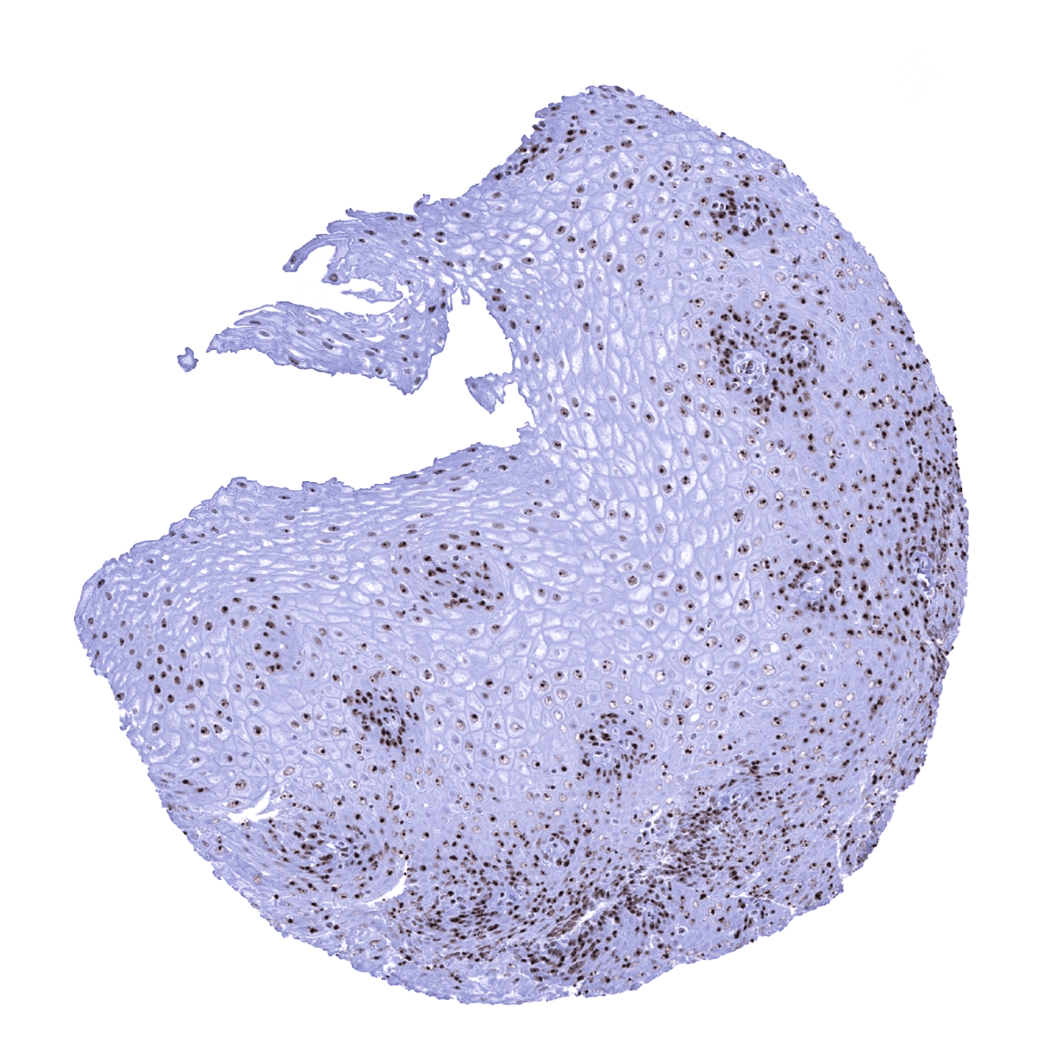
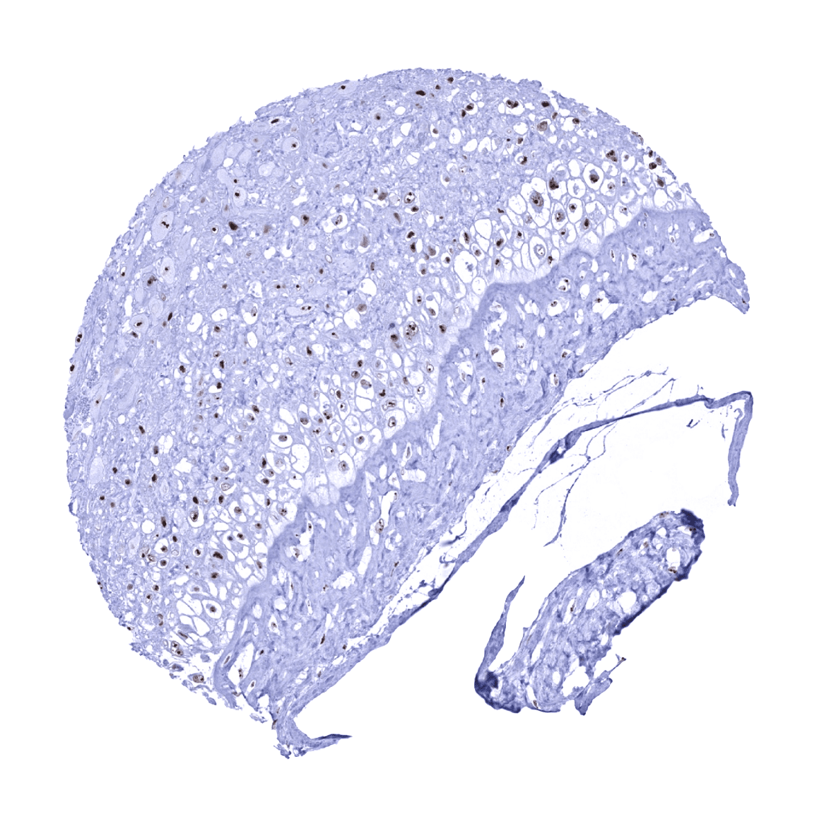
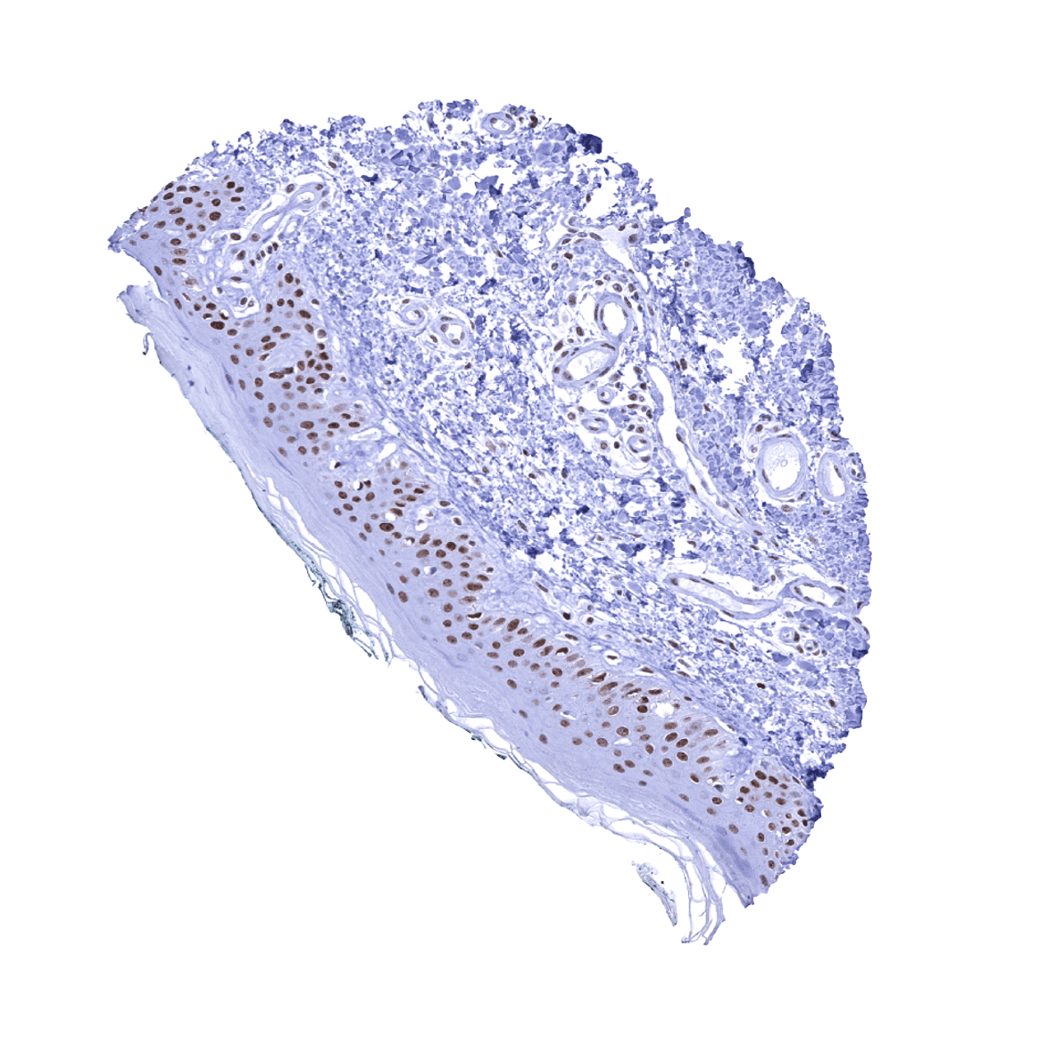
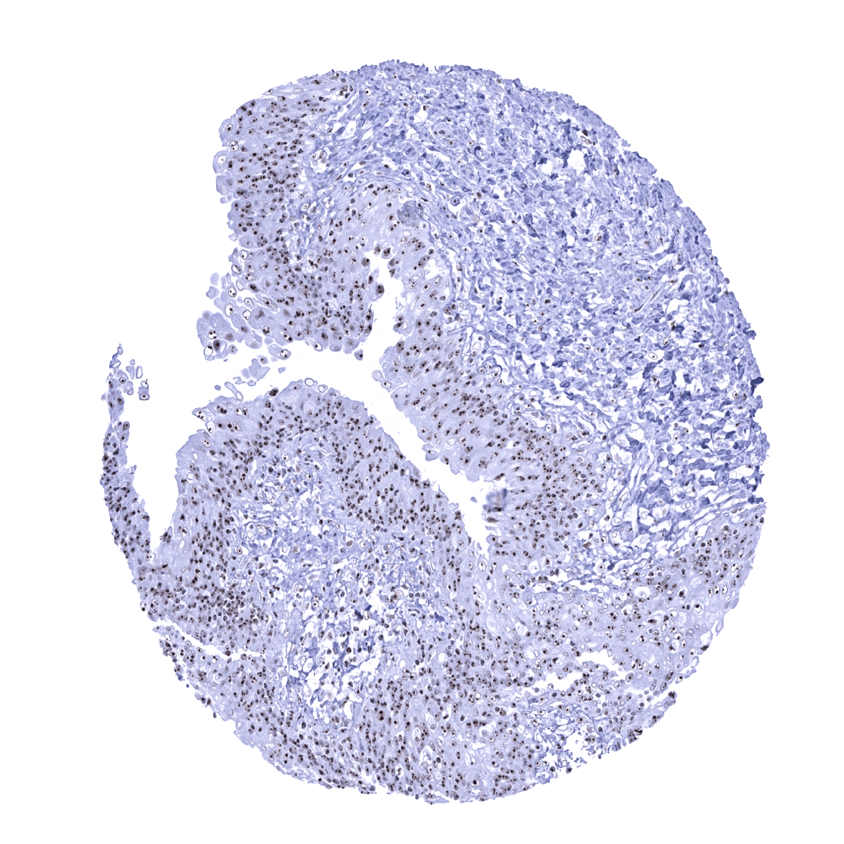
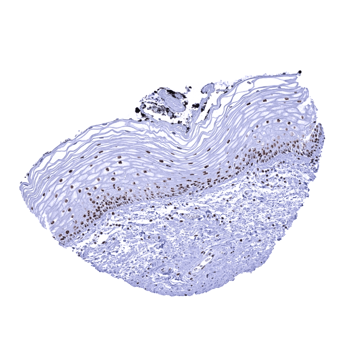
Esophagus, squamous epithelium -The entire nuclei of epithelial cells are positive but the nucleolin immunostaining is often most distinct in the nucleoli.
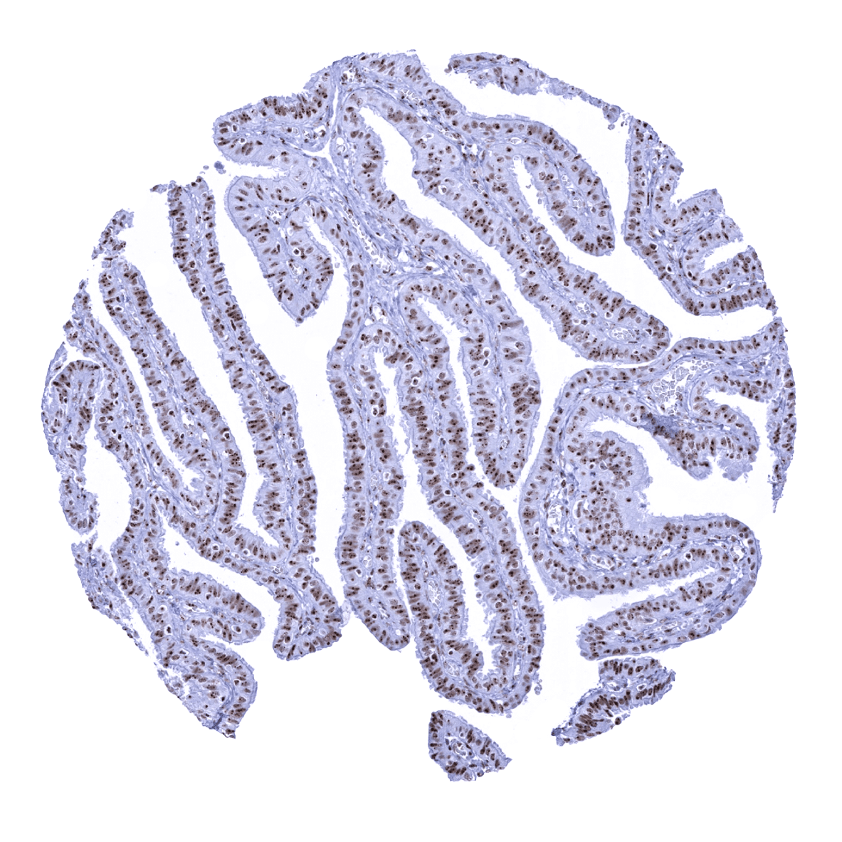
Fallopian tube, mucosa - The entire nuclei of epithelial cells are nucleolin positive but the immunostaining is most distinct in the nucleoli.

Fat
Gallbladder, epithelium - The entire nuclei of epithelial cells are nucleolin positive but the immunostaining is most distinct in the nucleoli.
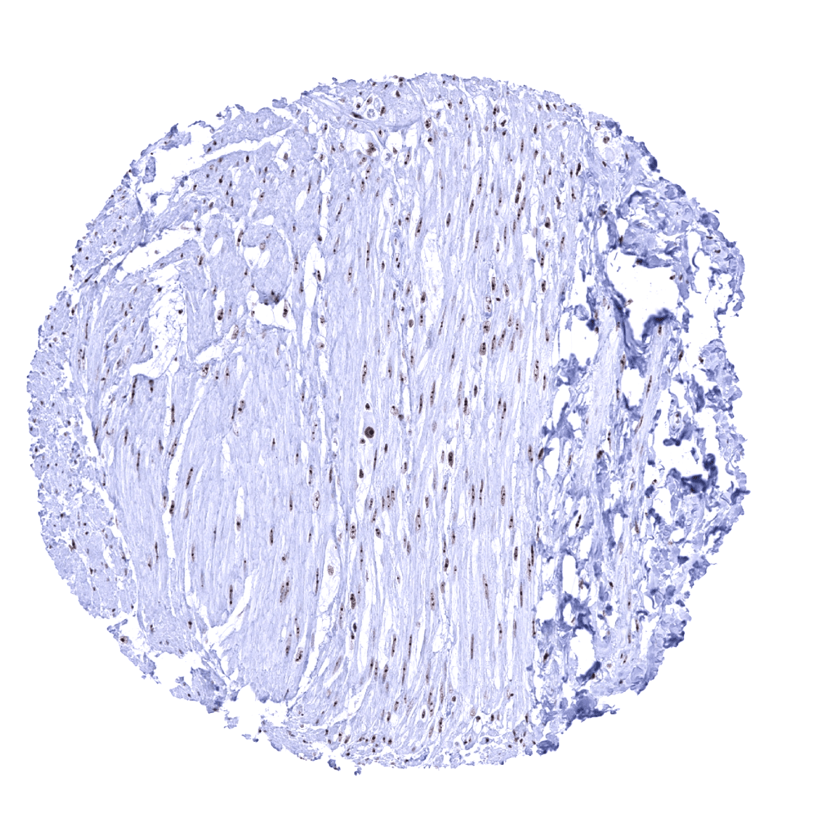
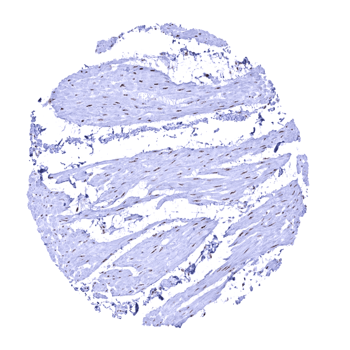
Heart muscle
Ileum, mucosa
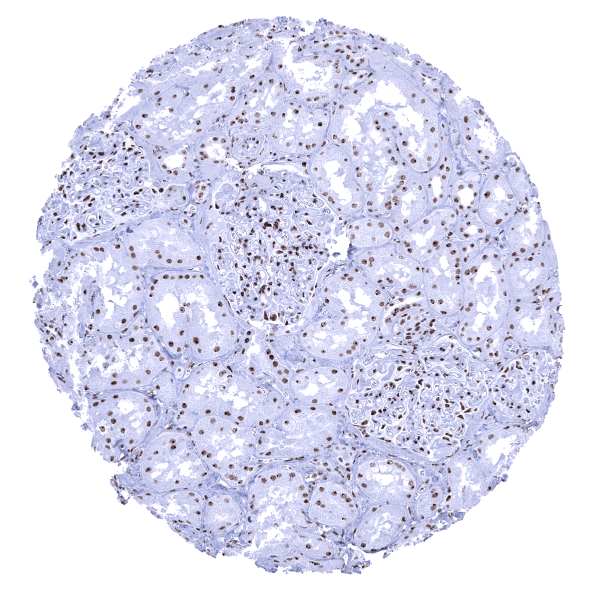
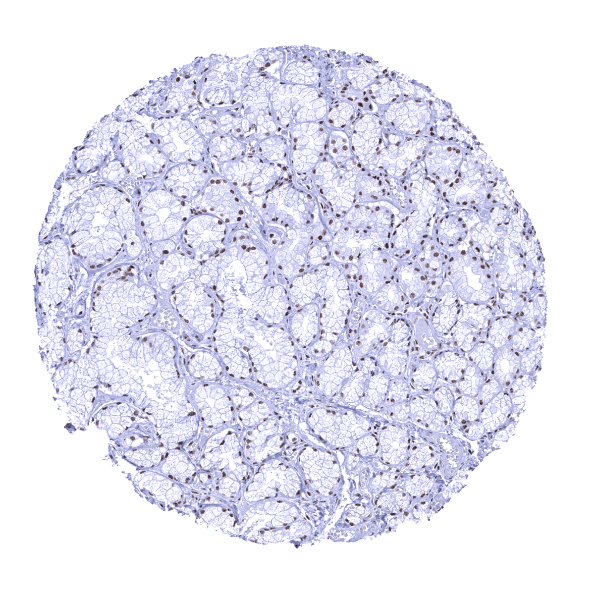
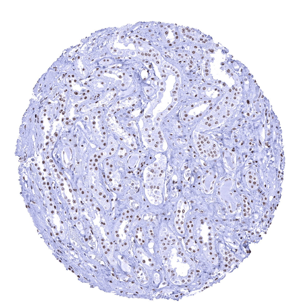
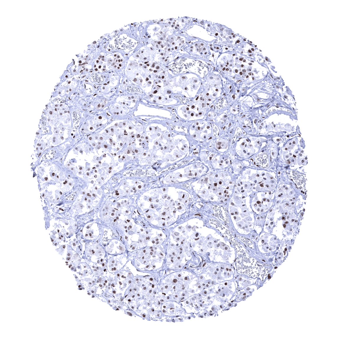
Kidney, cortex
Kidney, medulla
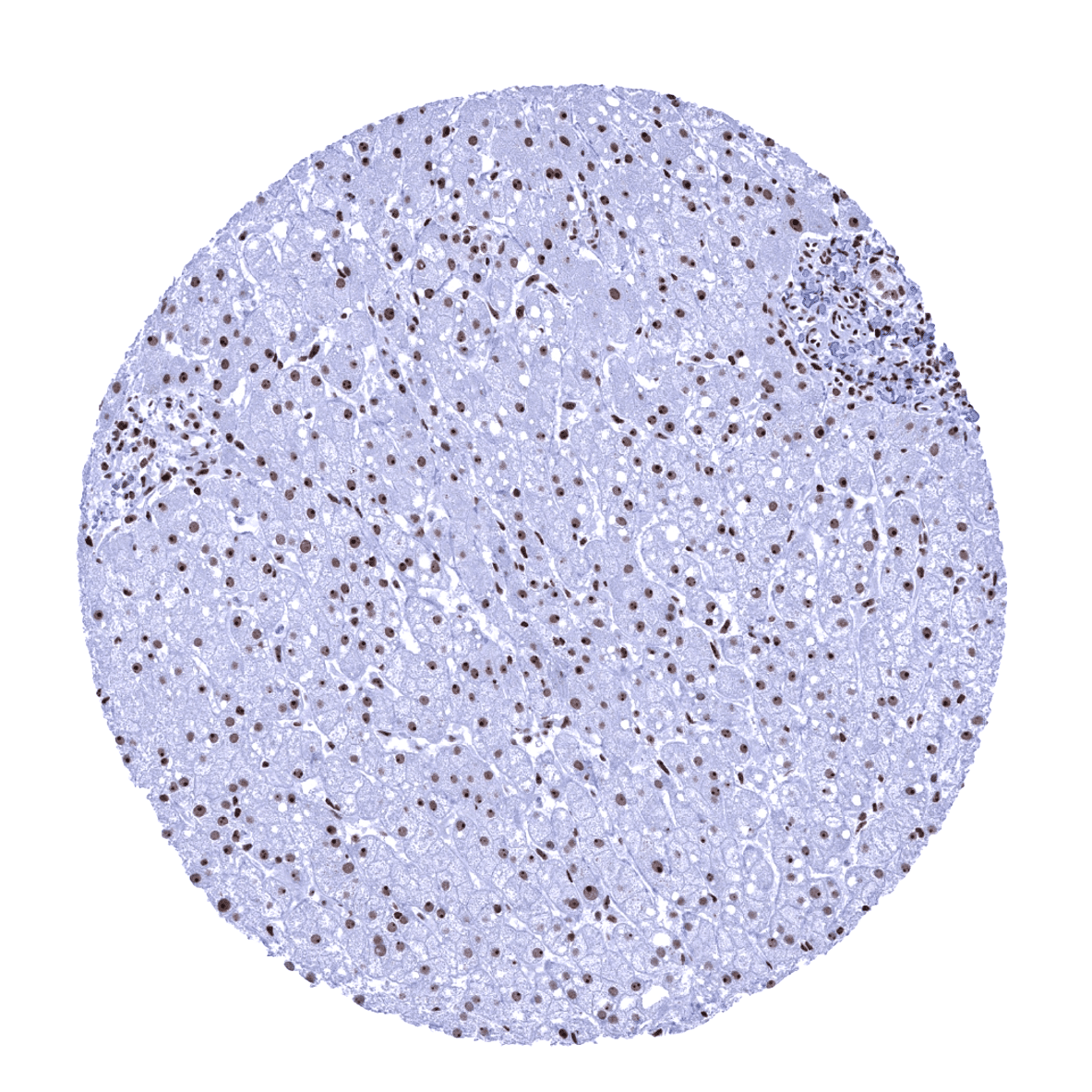
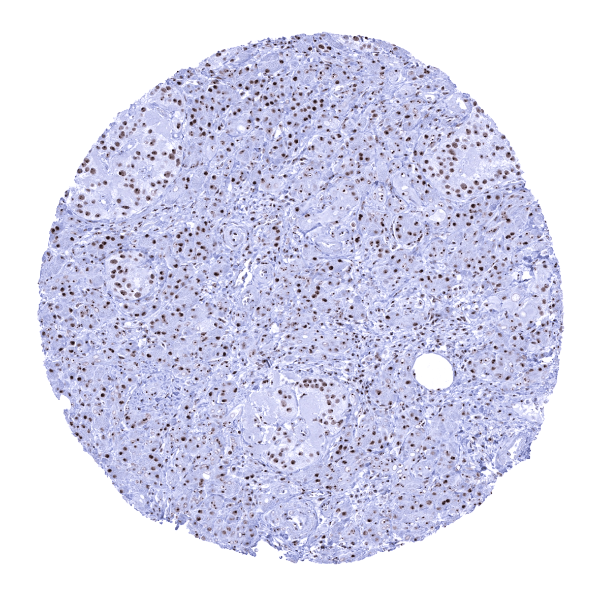
Liver

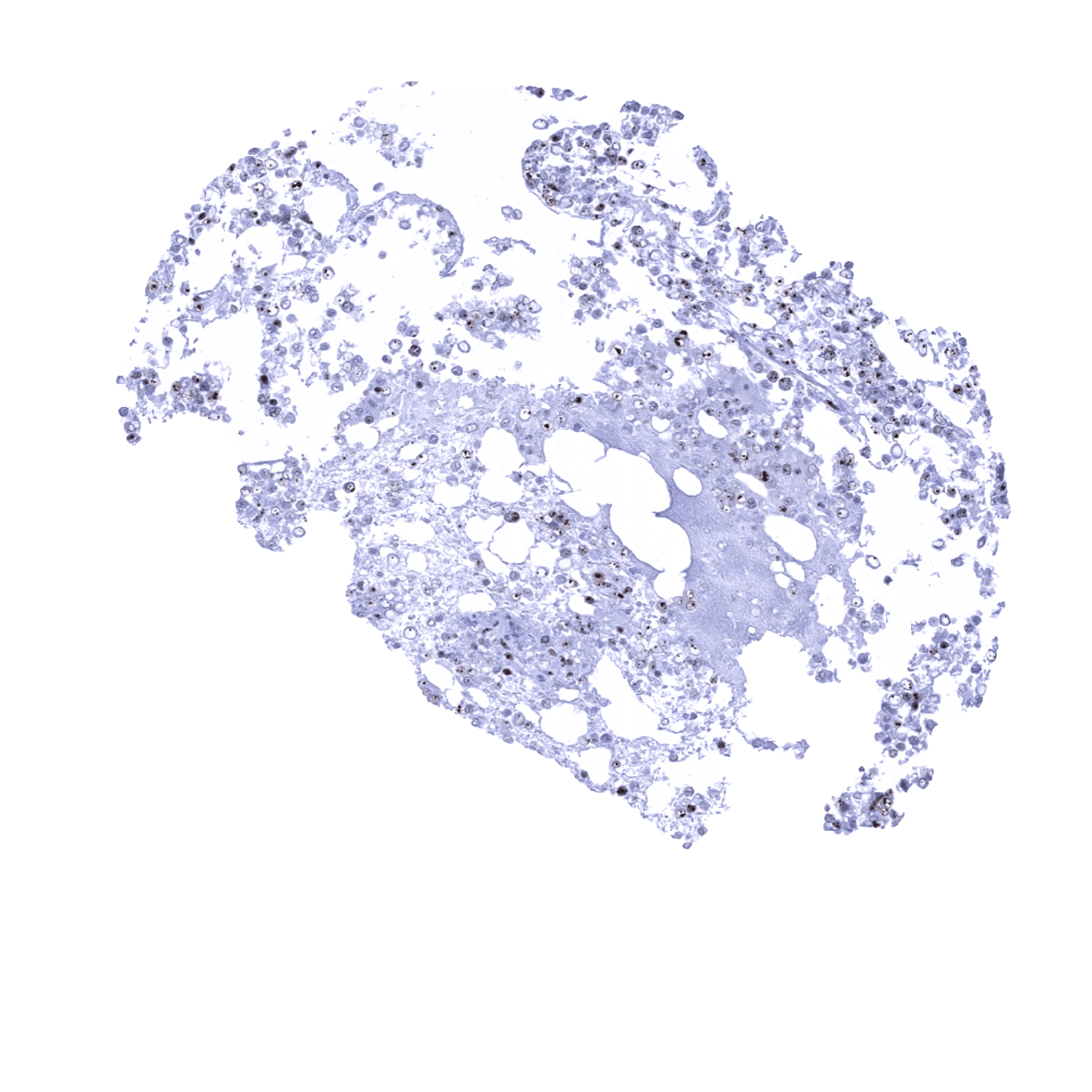
Lung
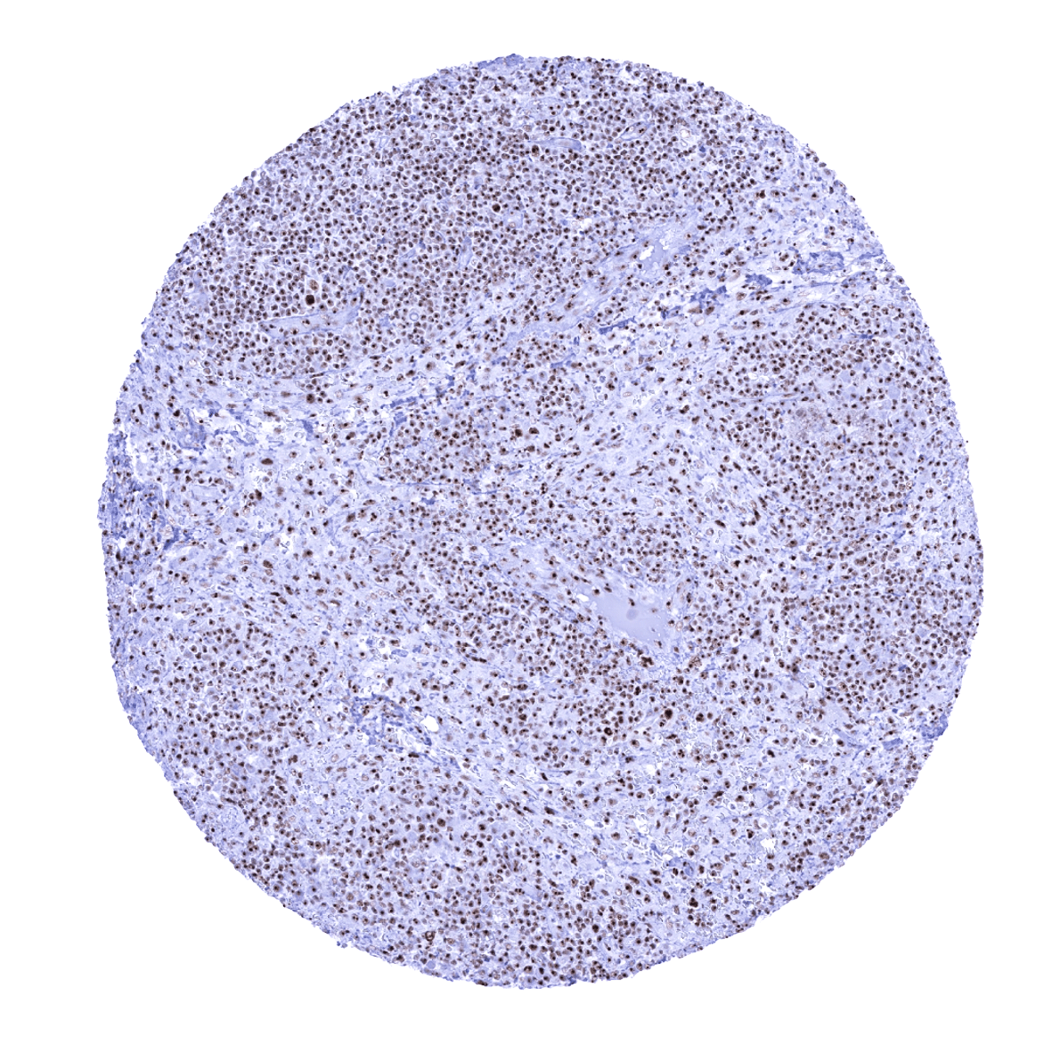
Lymph node
Ovary, stroma
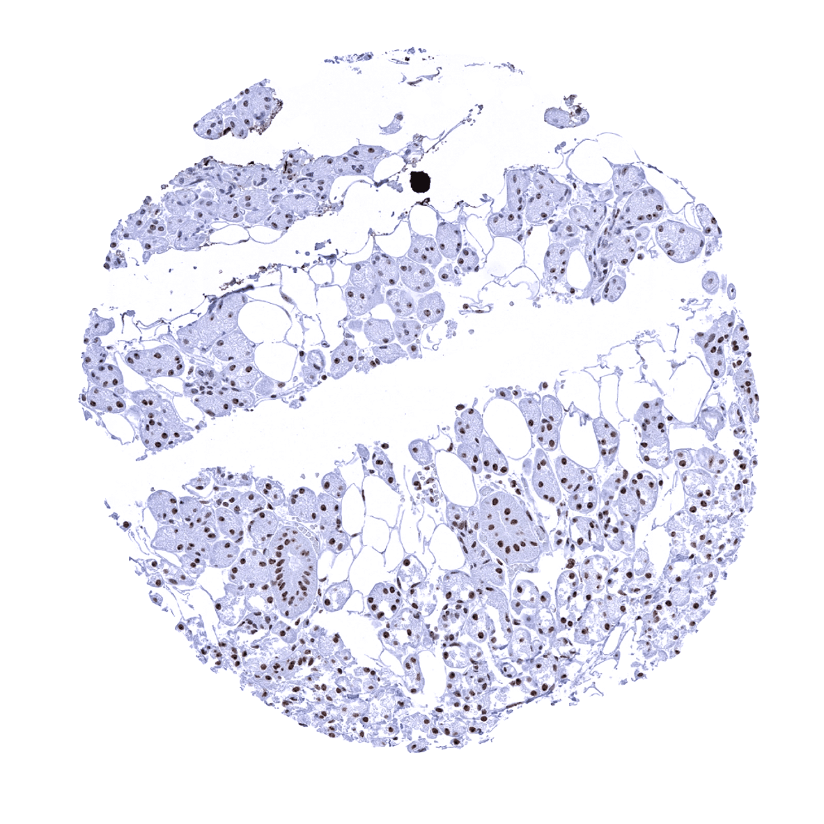
Pancreas
Parathyroid
Parotid gland
Pituitary gland, anterior lobe
Pituitary gland, posterior lobe
Placenta (amnion) - The entire nuclei of amnion cells are positive but the nucleolin immunostaining is often most prominent in the nucleoli.
Placenta (chorion)
Placenta, early
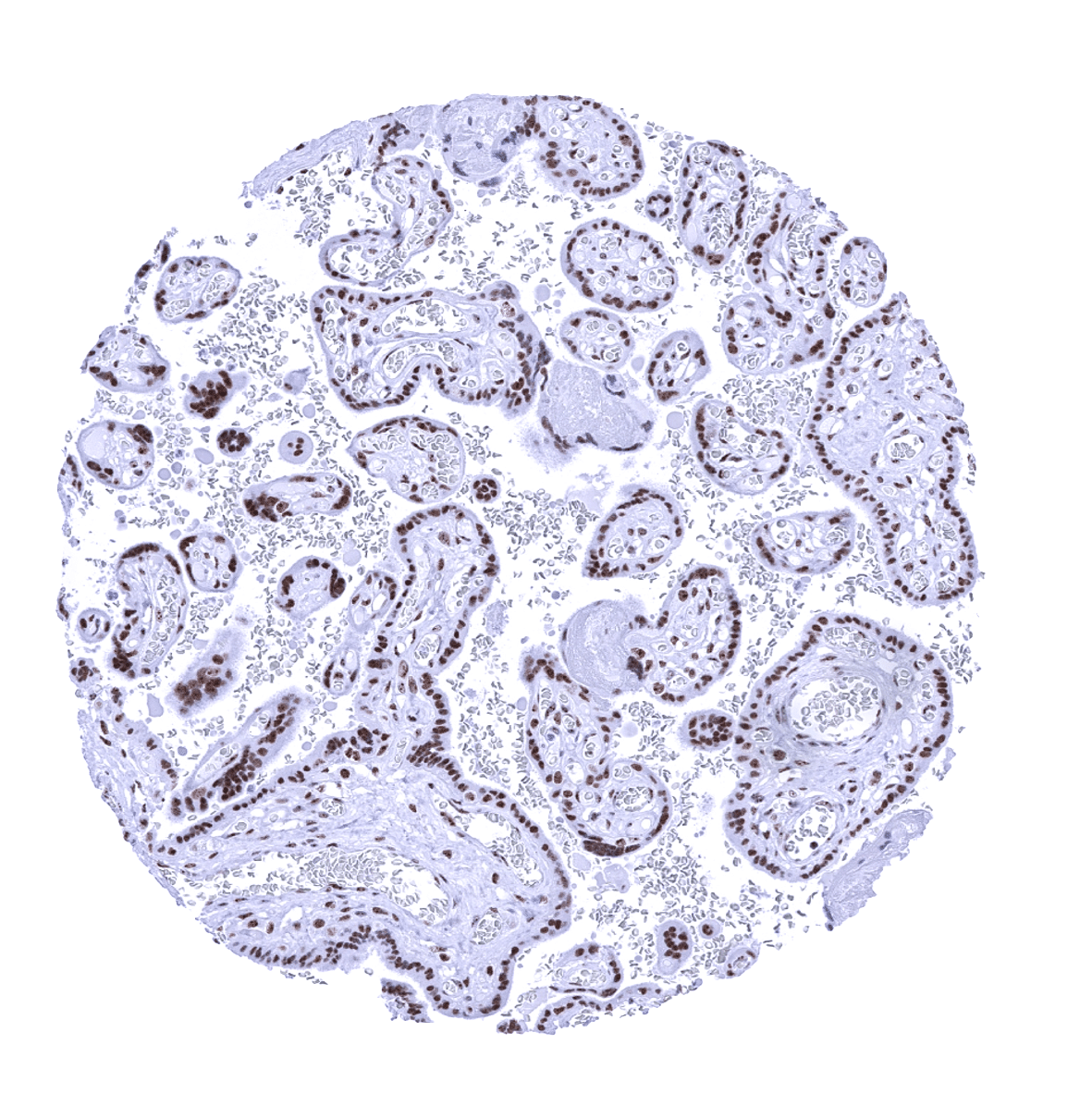
Placenta, mature
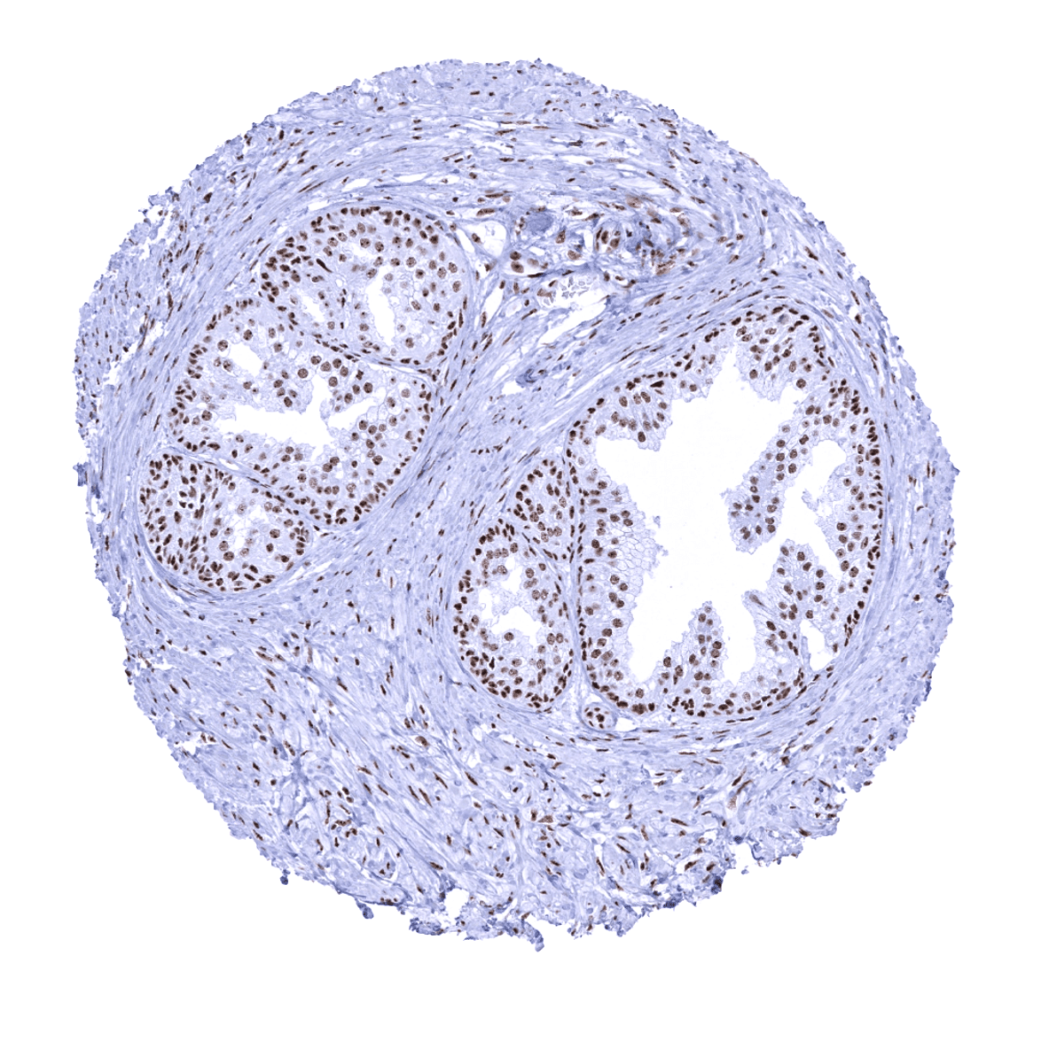
Prostate
Rectum, mucosa - In the rectum, nucleolin staining is more prominent in the proliferating cells in the crypt base than in the less proliferative superficial cell layers. The focus of the immunostaining to the nucleoli becomes most visible in the less stained superficial cells, however.

Seminal vesicle
Sinus paranasales - In this sample of respiratory epithelium, nucleolin staining is particularly prominent in the nucleoli of epithelial (and also inflammatory) cells.

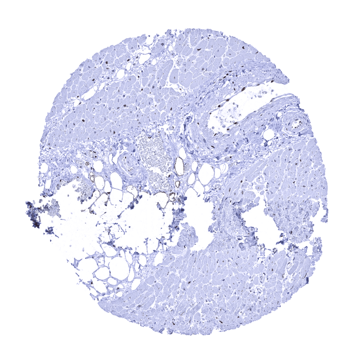
Skeletal muscle
Skin

Spleen

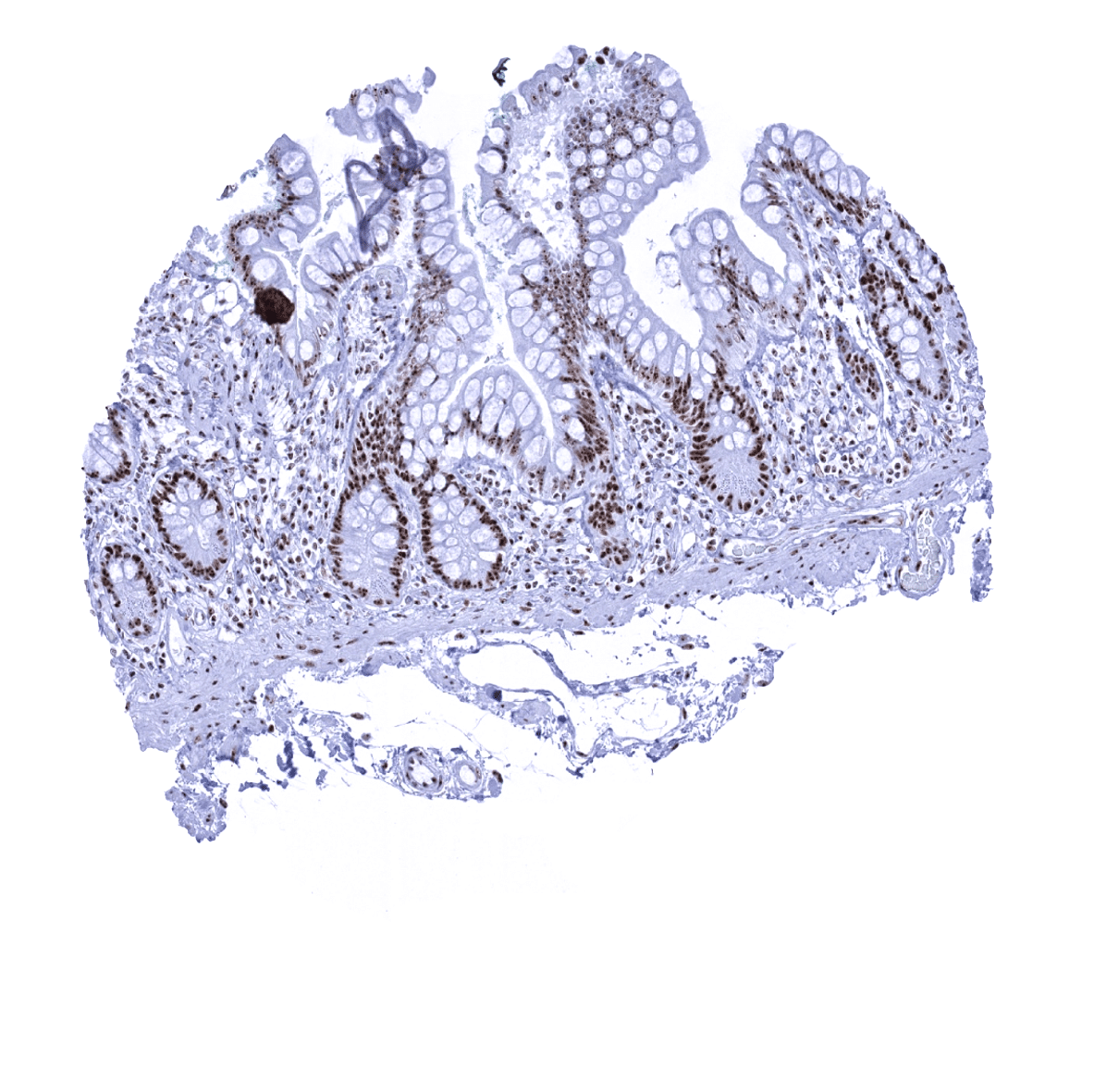
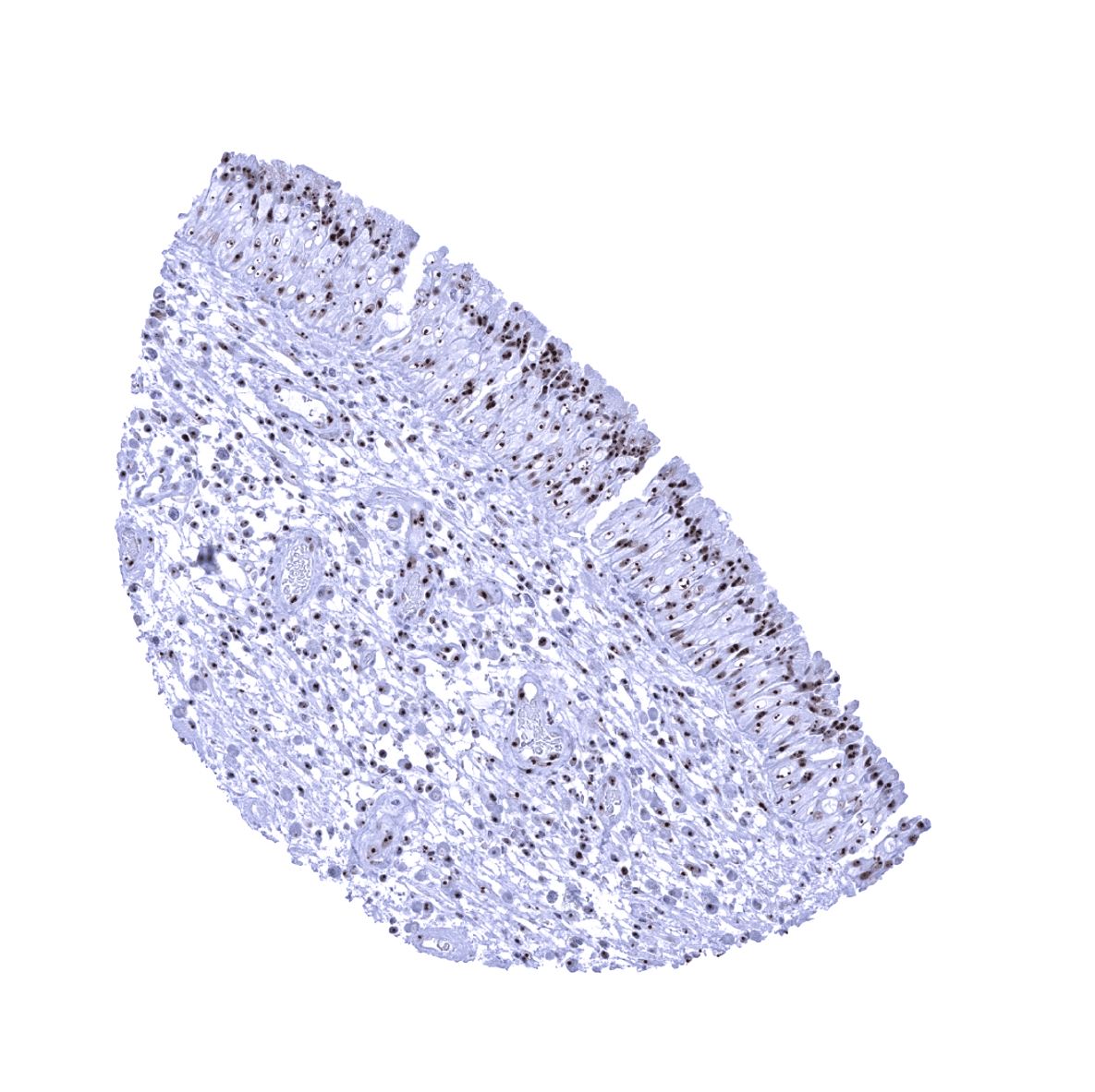
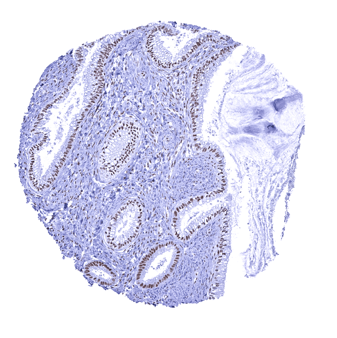
Stomach, antrum - In the gastric mucosa, nucleolin staining is more prominent in the proliferating neck cells than in the less proliferative glands. The focus of the immunostaining to the nucleoli becomes most visible in the glands, however.

Stomach, corpus
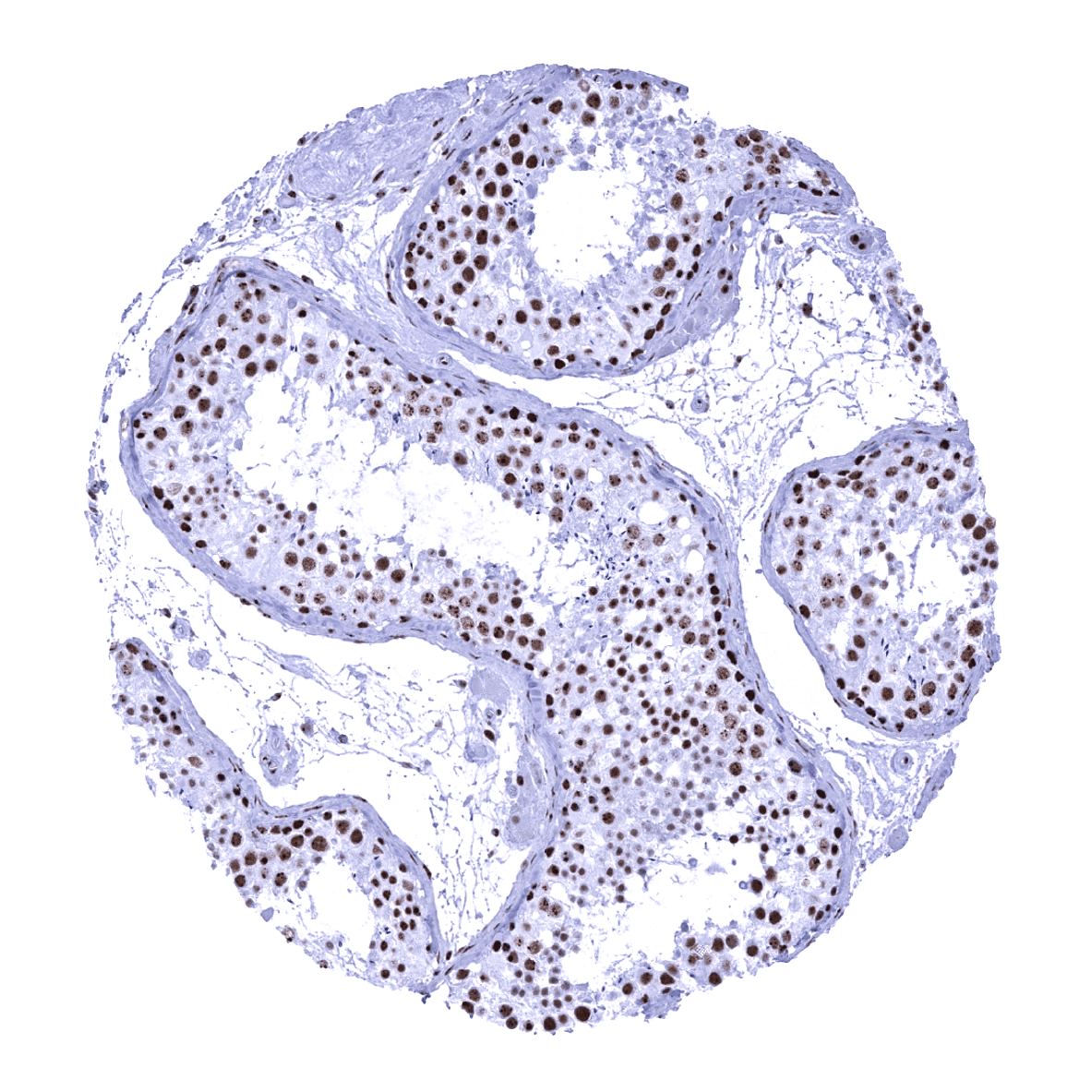
Testis
Thymus
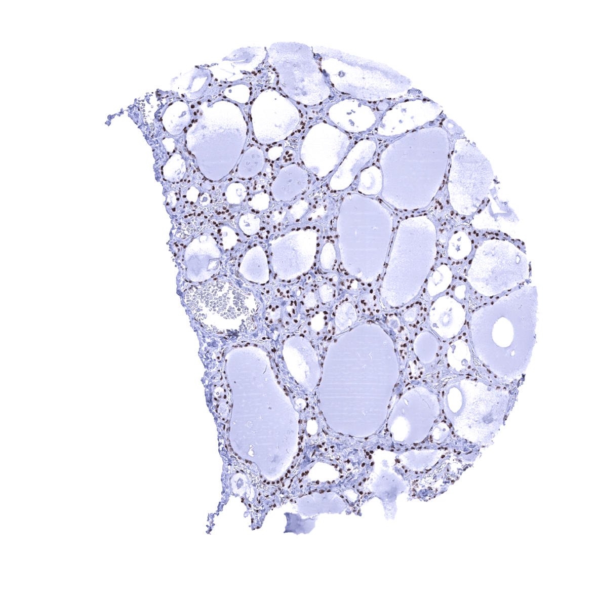
Thyroid gland
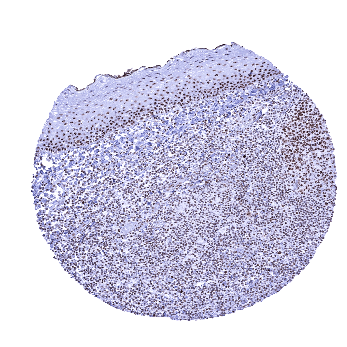
Tonsil, surface epithelium - In the surface epithelium, nucleolin staining is more prominent in the basal and suprabasal cells than in the superficial cell layers. The entire nuclei of epithelial cells are positive but the nucleolin immunostaining is often most distinct in the nucleoli.
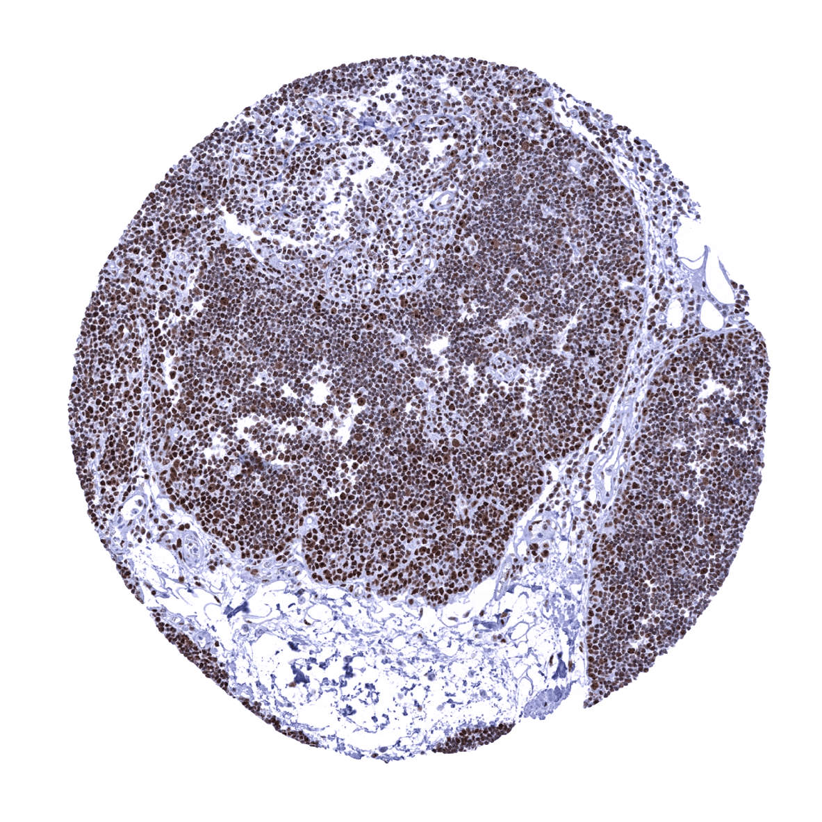
Tonsil
Urinary bladder, muscular wall - The entire nuclei of smooth muscle cells are nucleolin positive but the immunostaining is most distinct in the nucleoli.
Urinary bladder, urothelium - Nucleolin immunostaining is most prominent in the nucleoli of urothelial cells.
Uterus, ectocervix
Uterus, endocervix
Uterus, endometrium (pregnancy)
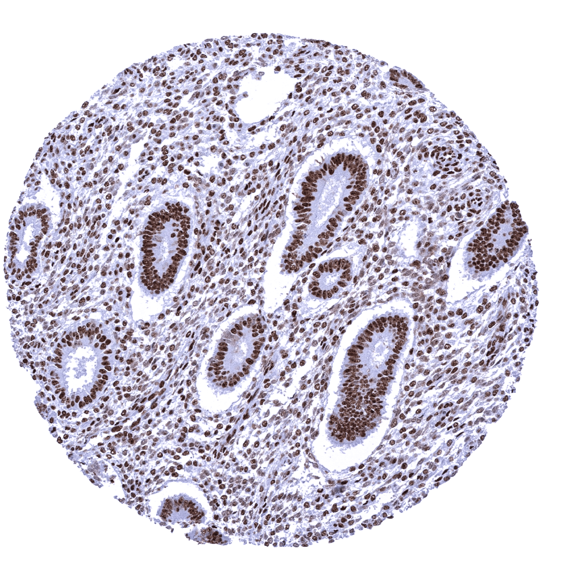
Uterus, endometrium (proliferation) - Strong diffuse nucleolin immunostaining of the entire nuclei in all cells.

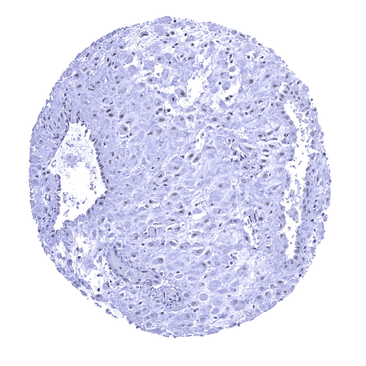
Uterus, endometrium (secretion) - In this sample of endometrium, nucleolin staining is particularly prominent in the nucleoli of epithelial (and also inflammatory) cells.
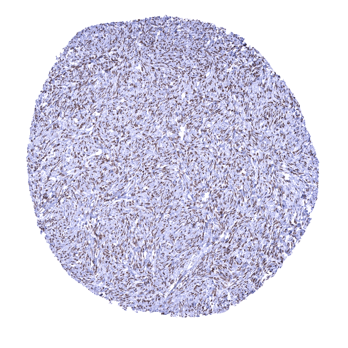
Uterus, myometrium