Adrenal gland
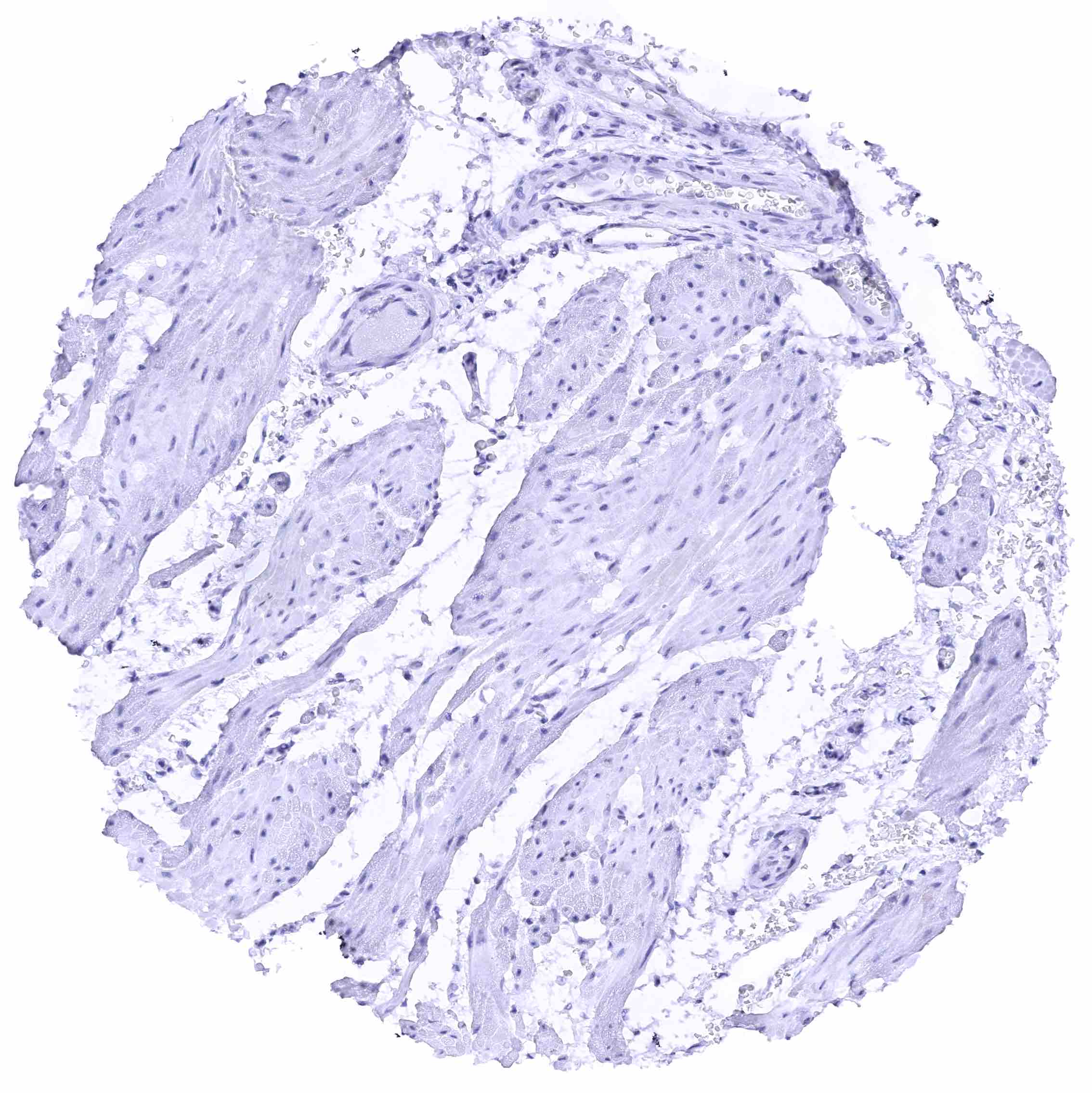
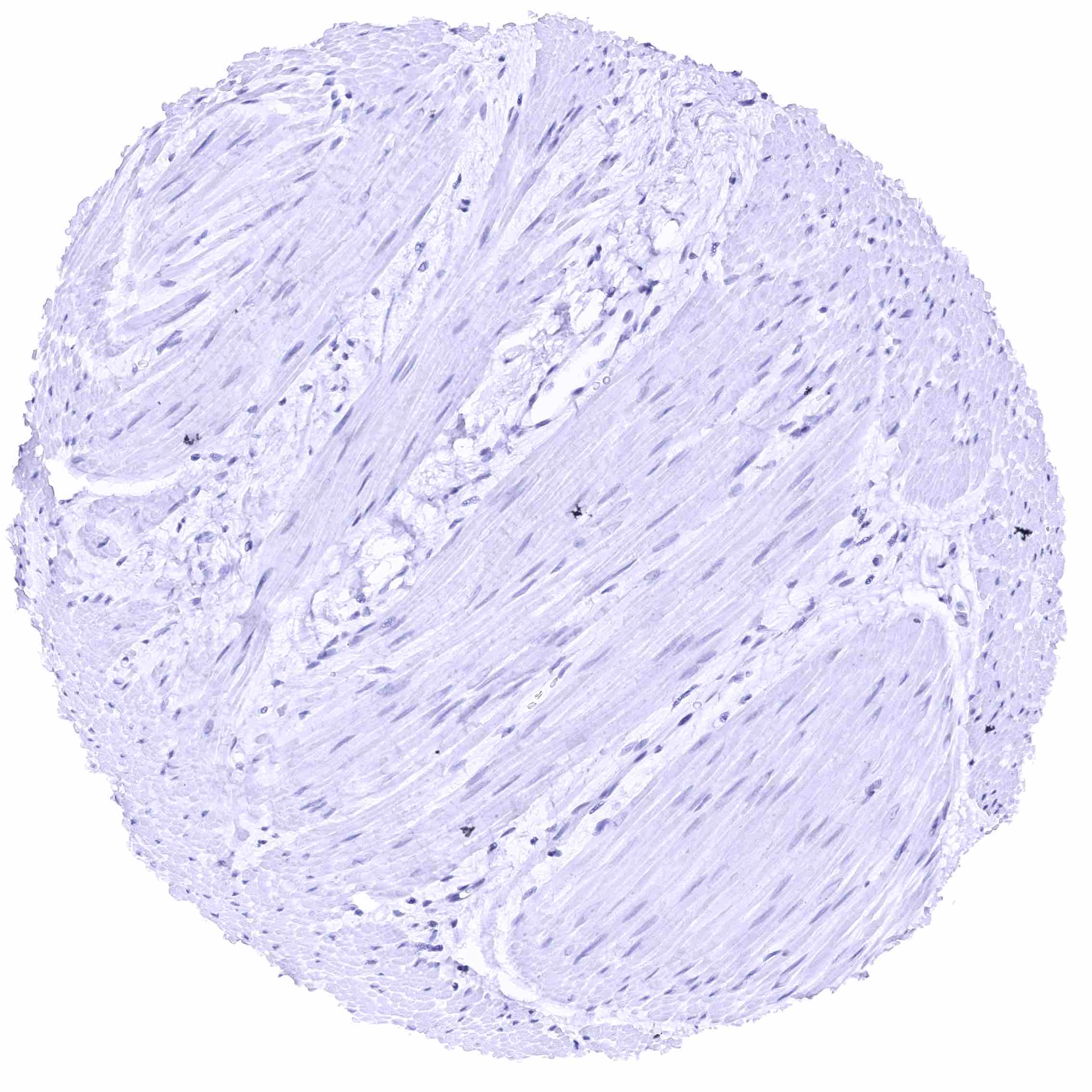
Aorta, media
Appendix, mucosa
Appendix, muscular wall
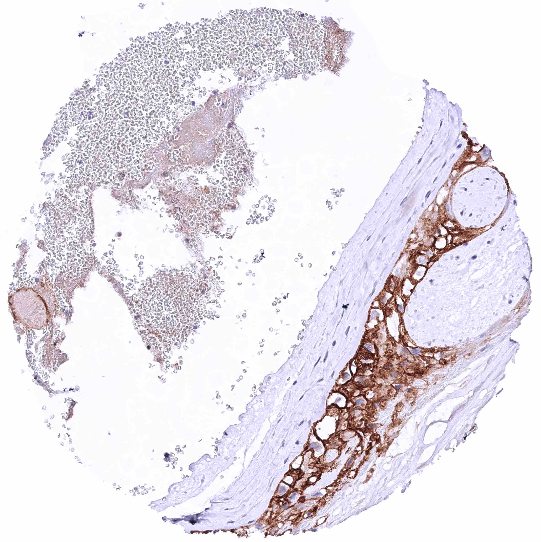
Bone marrow
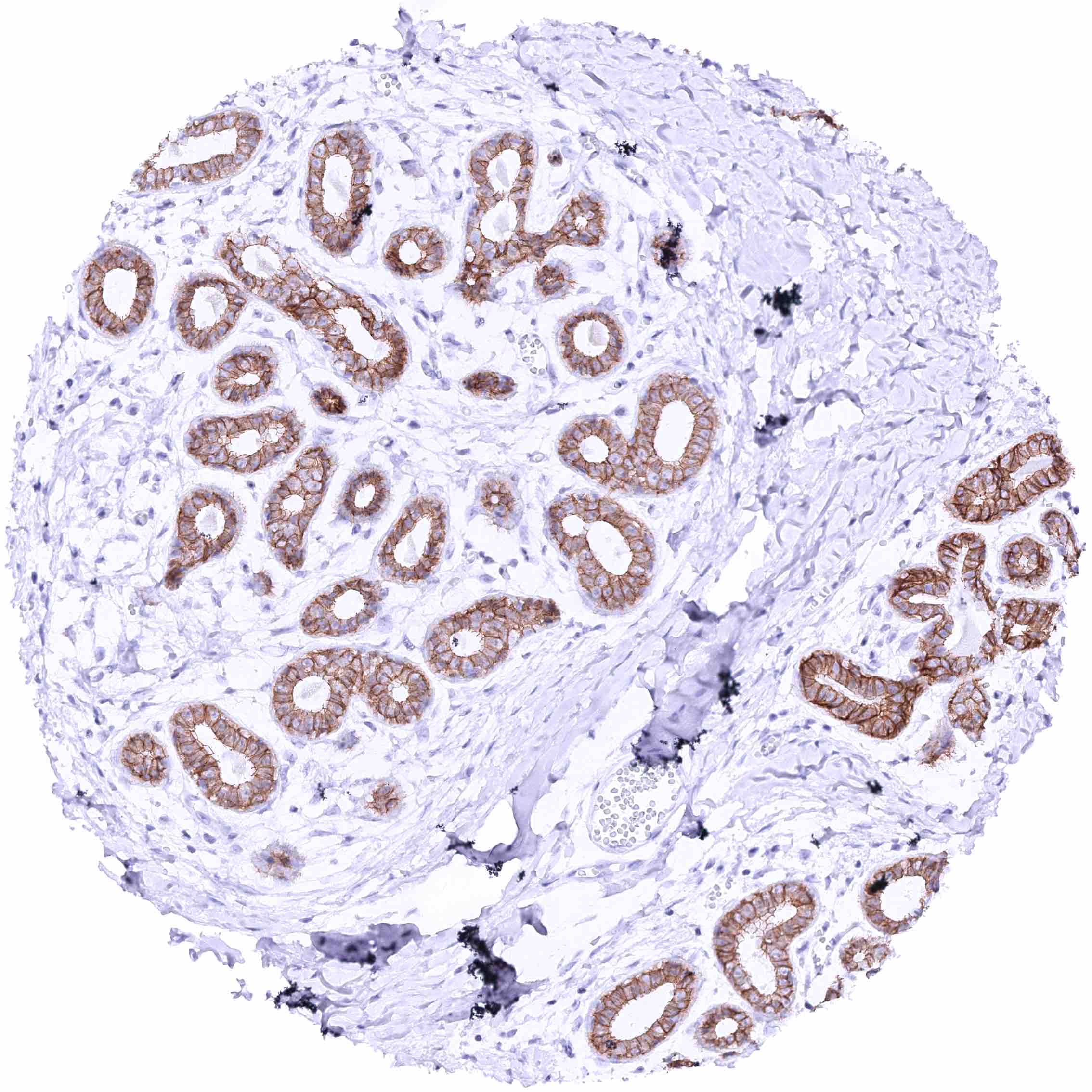
Breast – Strong membranous nectin-4 staining of luminal epithelial cells while staining is markedly less intense or absent in basal cells.
Bronchus, mucosa
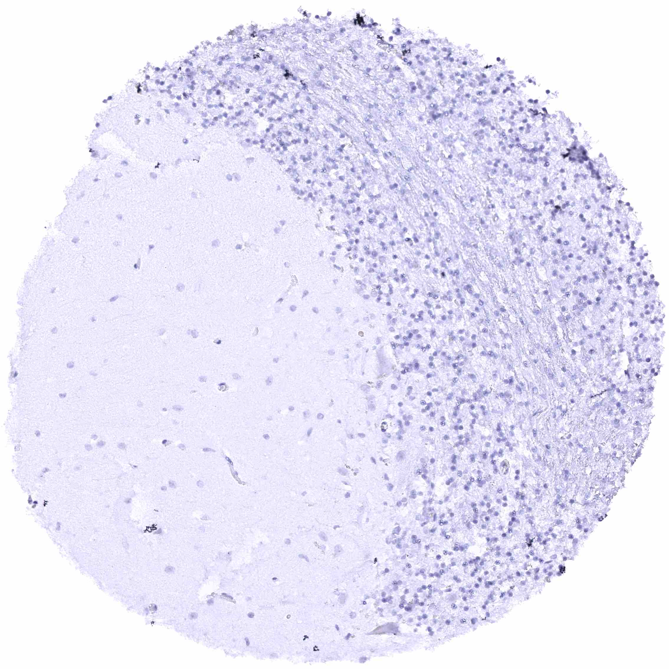
Cerebellum (molecular layer, Purkinje cell layer, granule cell layer)
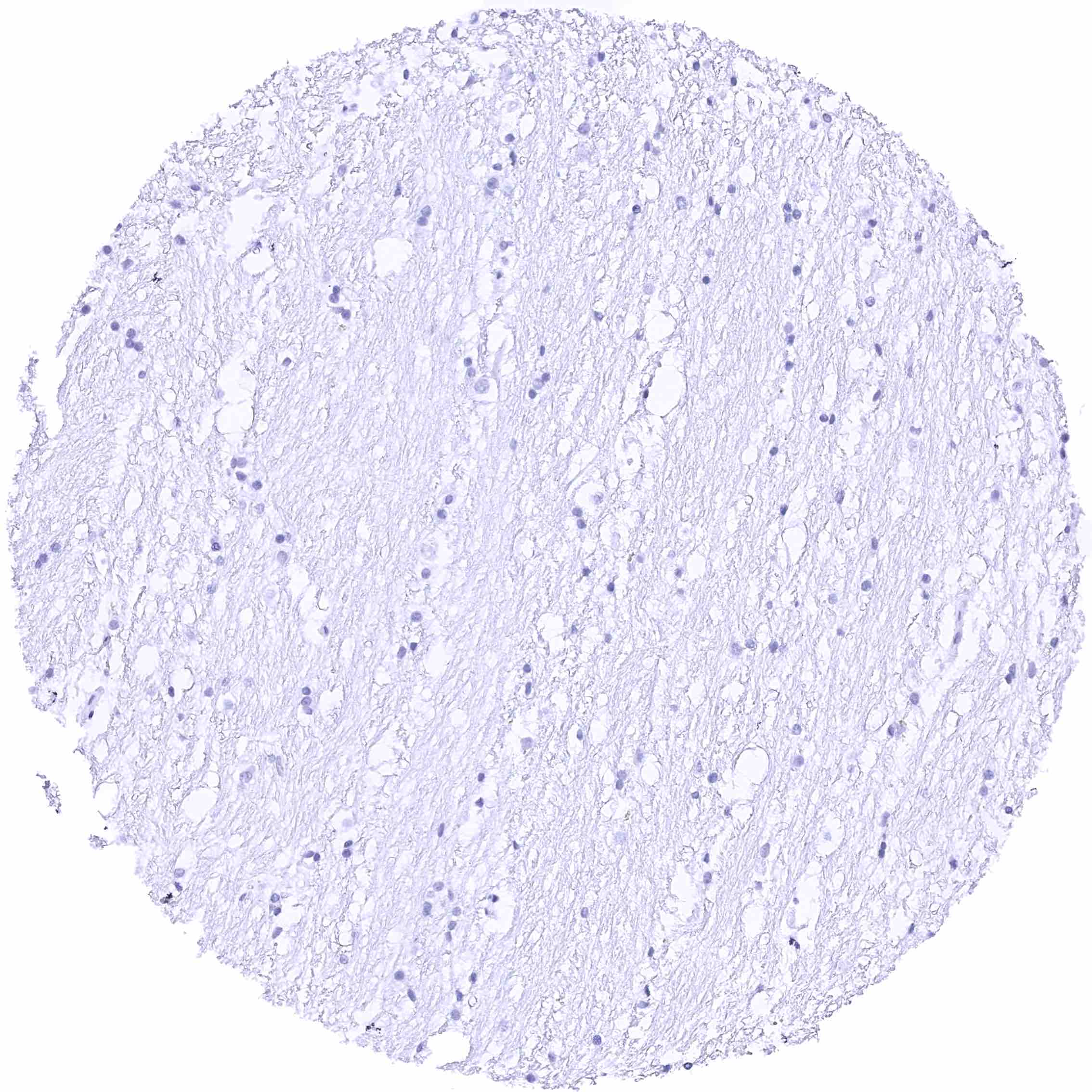
Cerebellum (white matter)
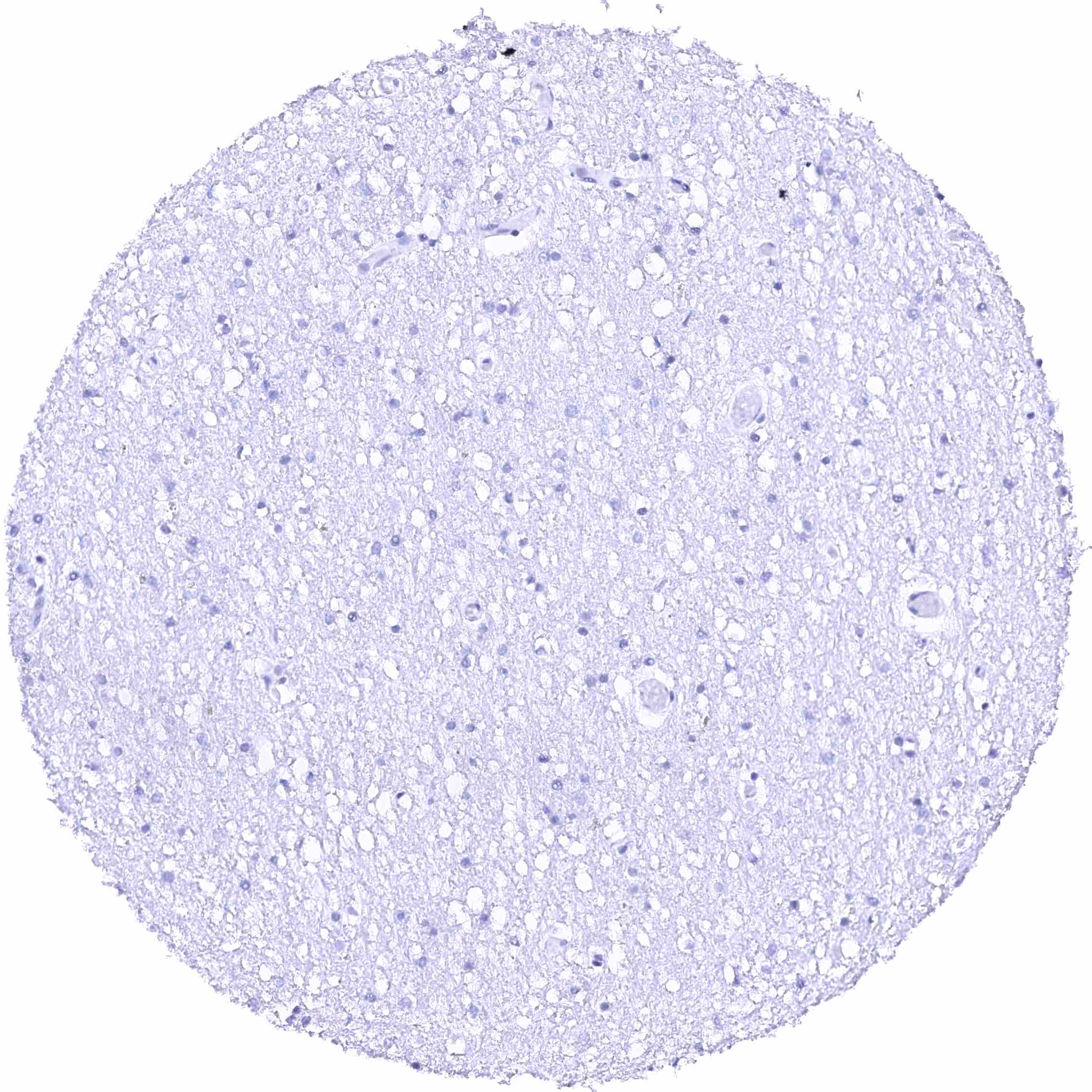
Cerebrum (grey matter)
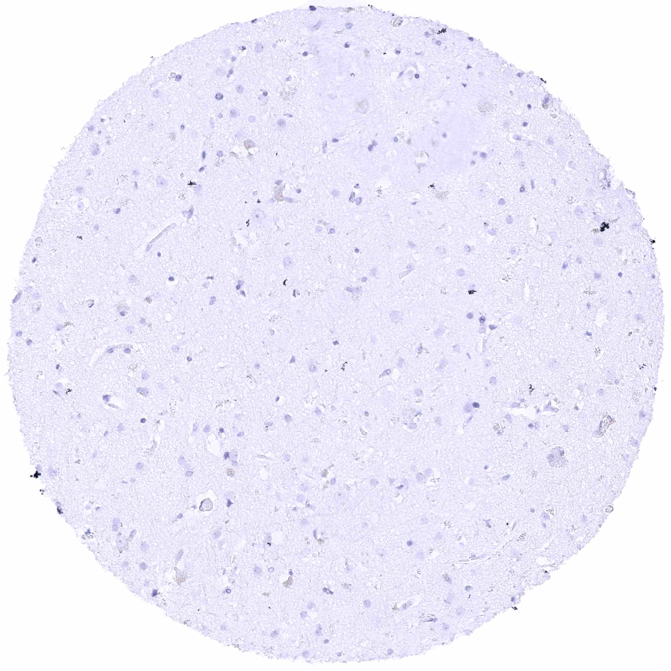
Cerebrum (white matter)
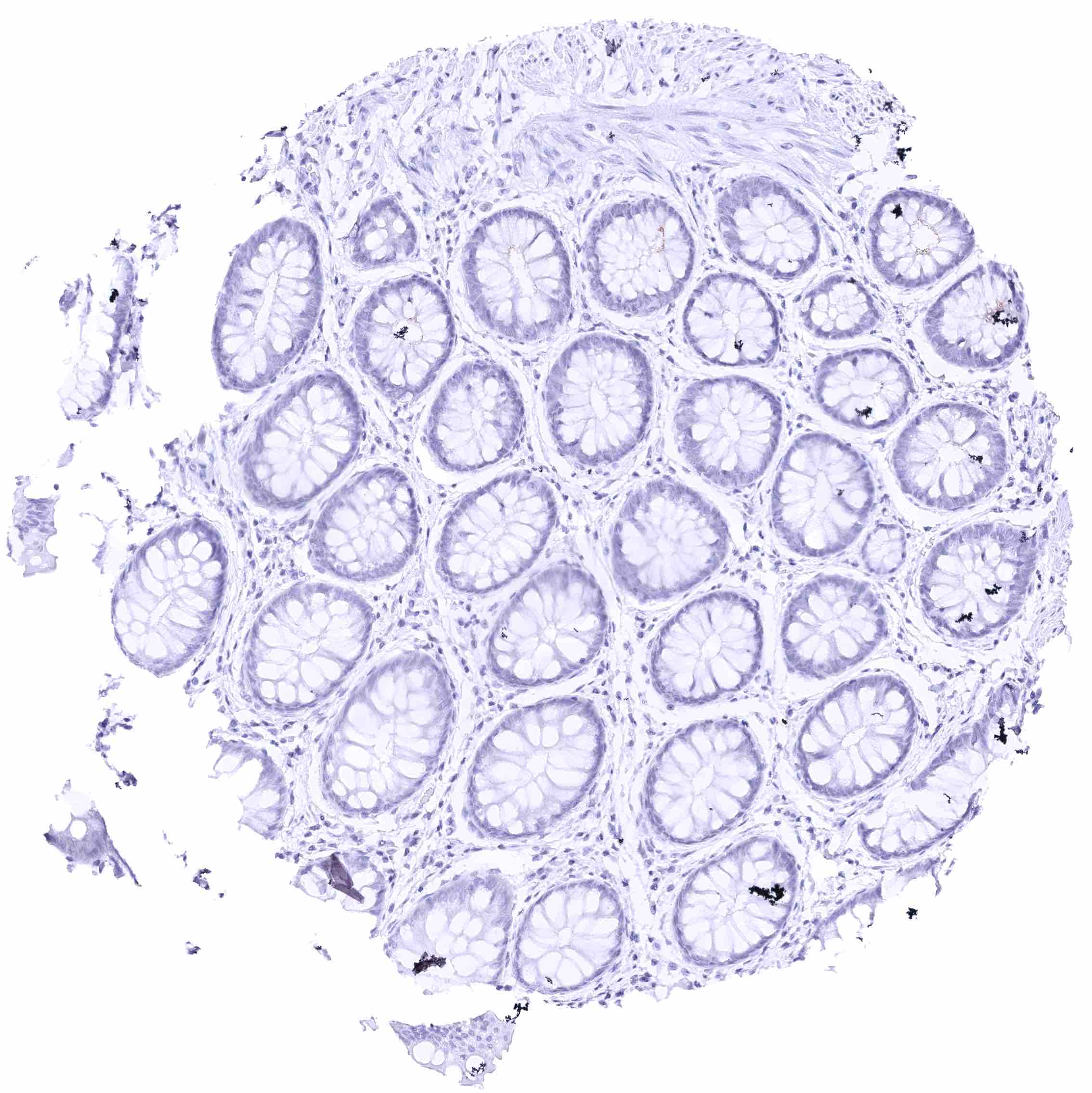
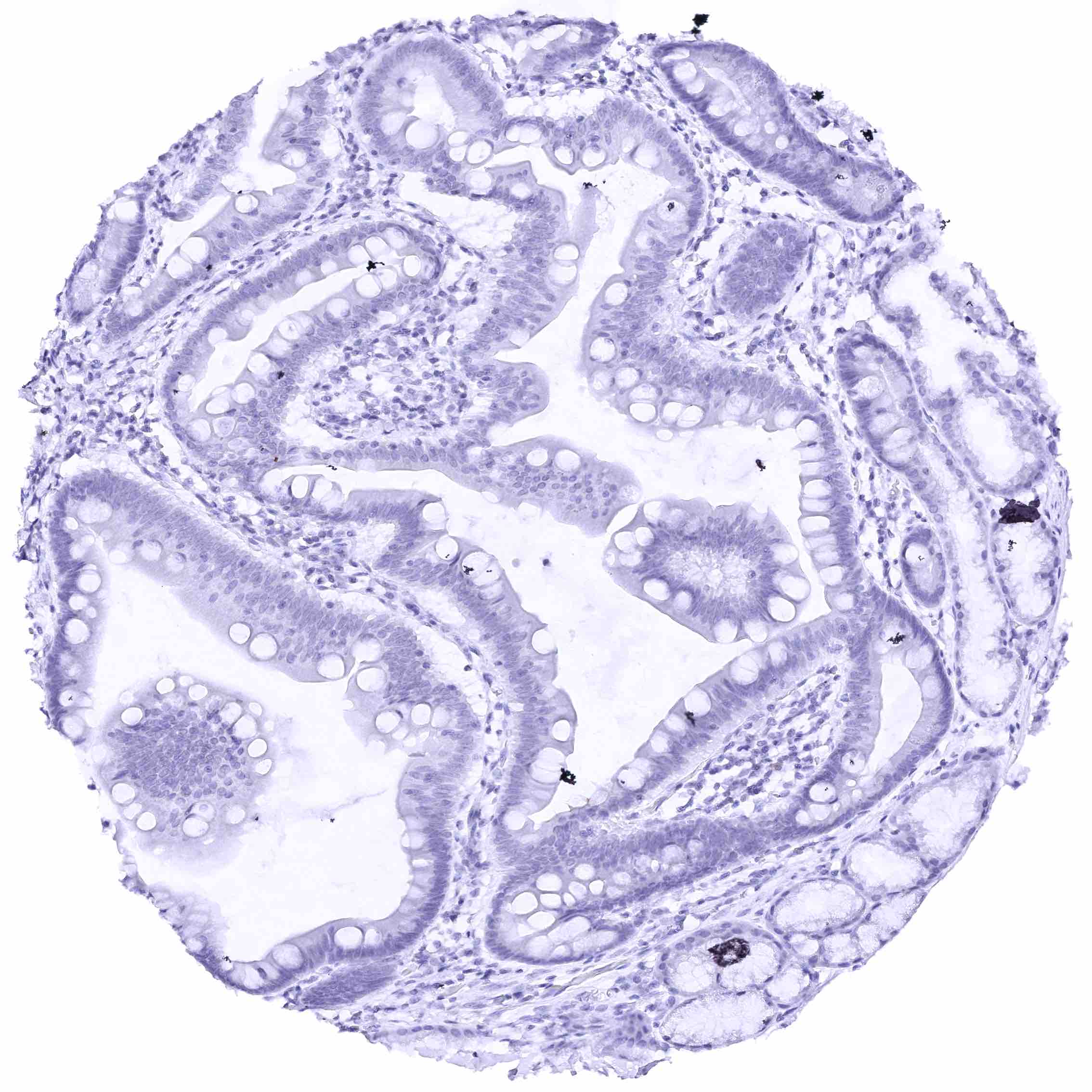
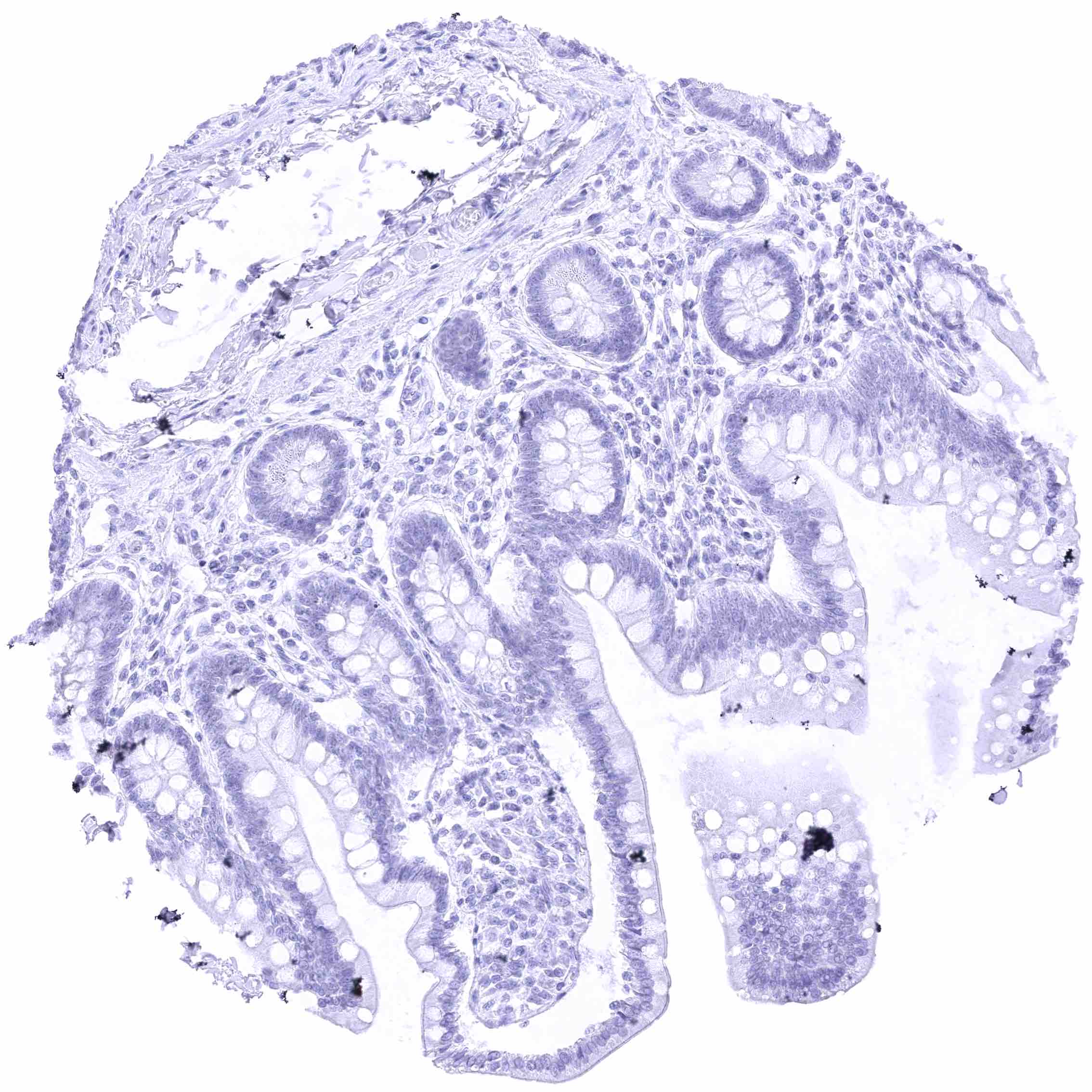
Colon descendens, mucosa
Colon descendens, muscular wall
Duodenum, Brunner gland
Duodenum, mucosa (2)
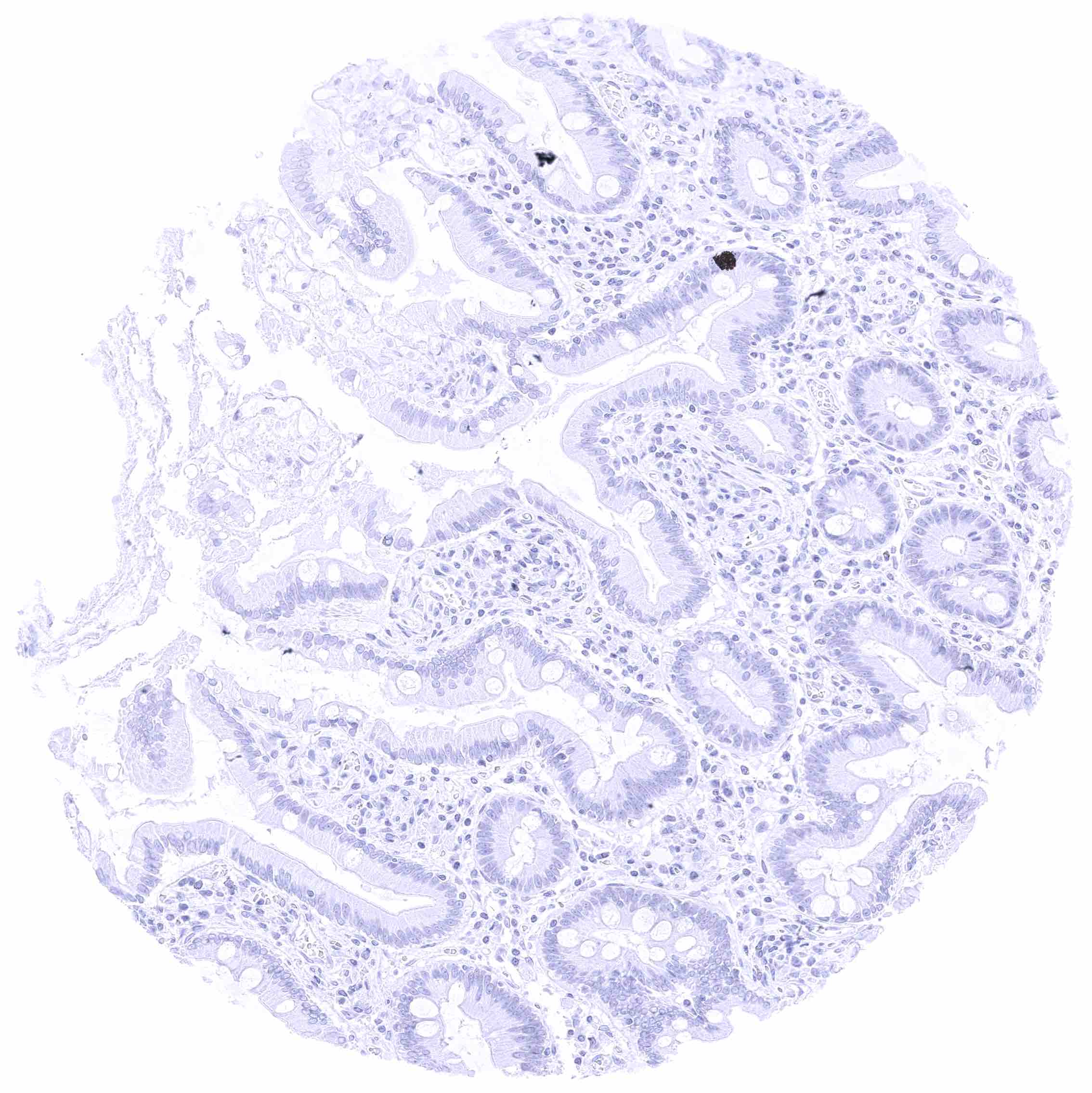
Duodenum, mucosa
Epididymis, Caput – Weak to moderate membranous nectin-4 staining of apical and sometimes also lateral membranes of tall columnar cells and of membranes of basal cells. .jpeg
Esophagus, muscular wall
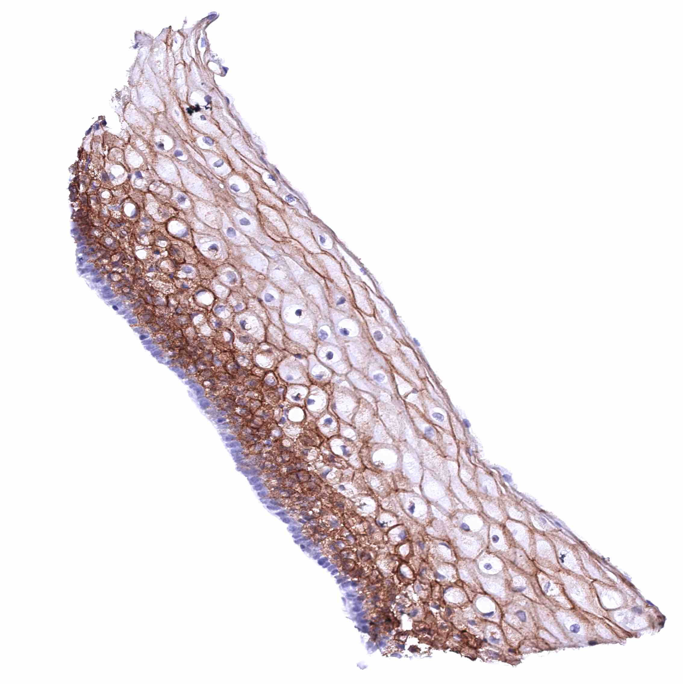
Esophagus, squamous epithelium – Nectin-4 staining of squamous epithelium is strong in the suprabasal cell layers while the staining intensity decreases markedly towards the superficial cell layers where cells are often negative. Basal cells are nectin-4 negative.
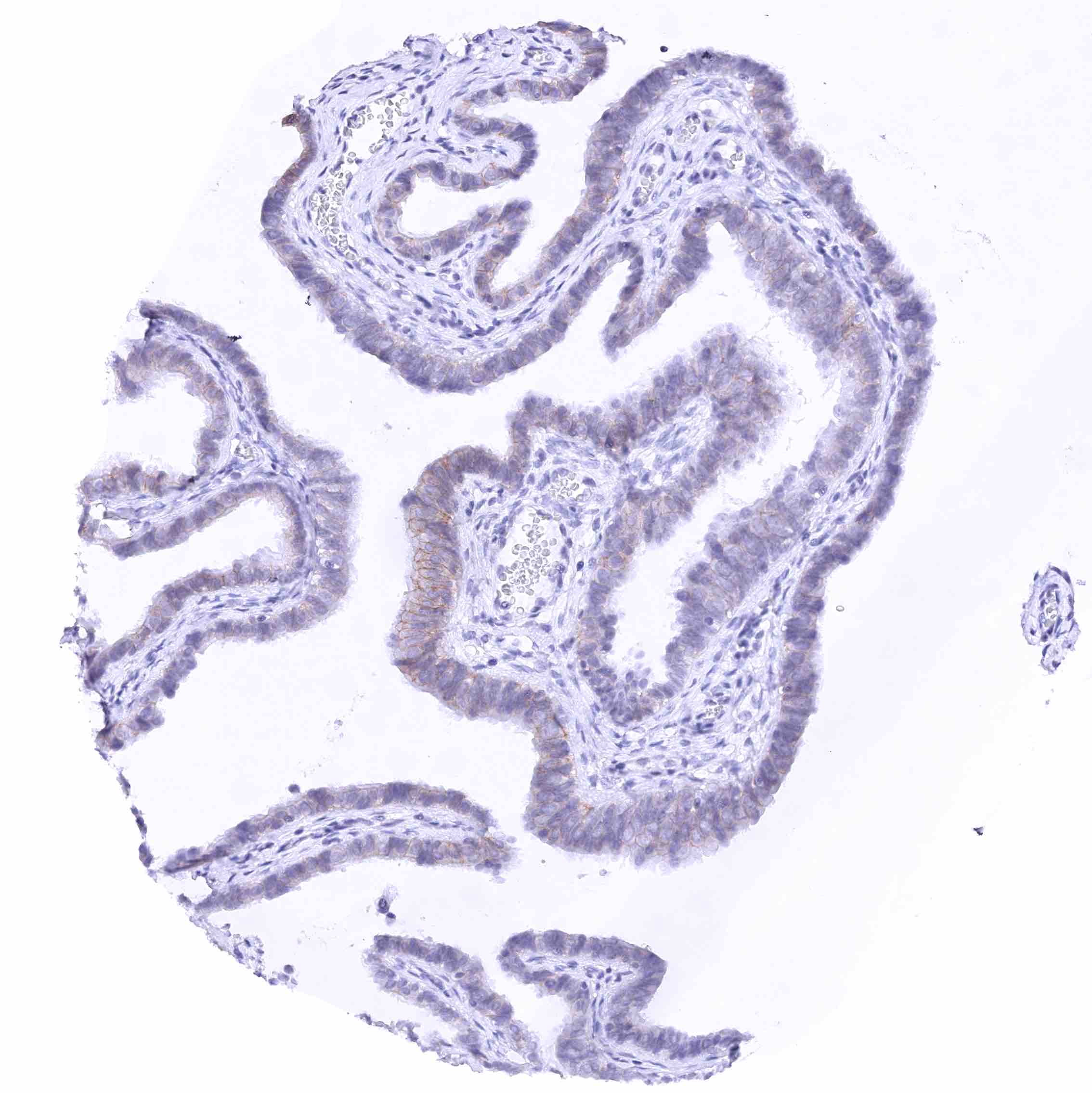
Fallopian tube, mucosa – Weak to moderate membranous nectin-4 staining of a subset of epithelial cells (most prominent at the lateral membranes).
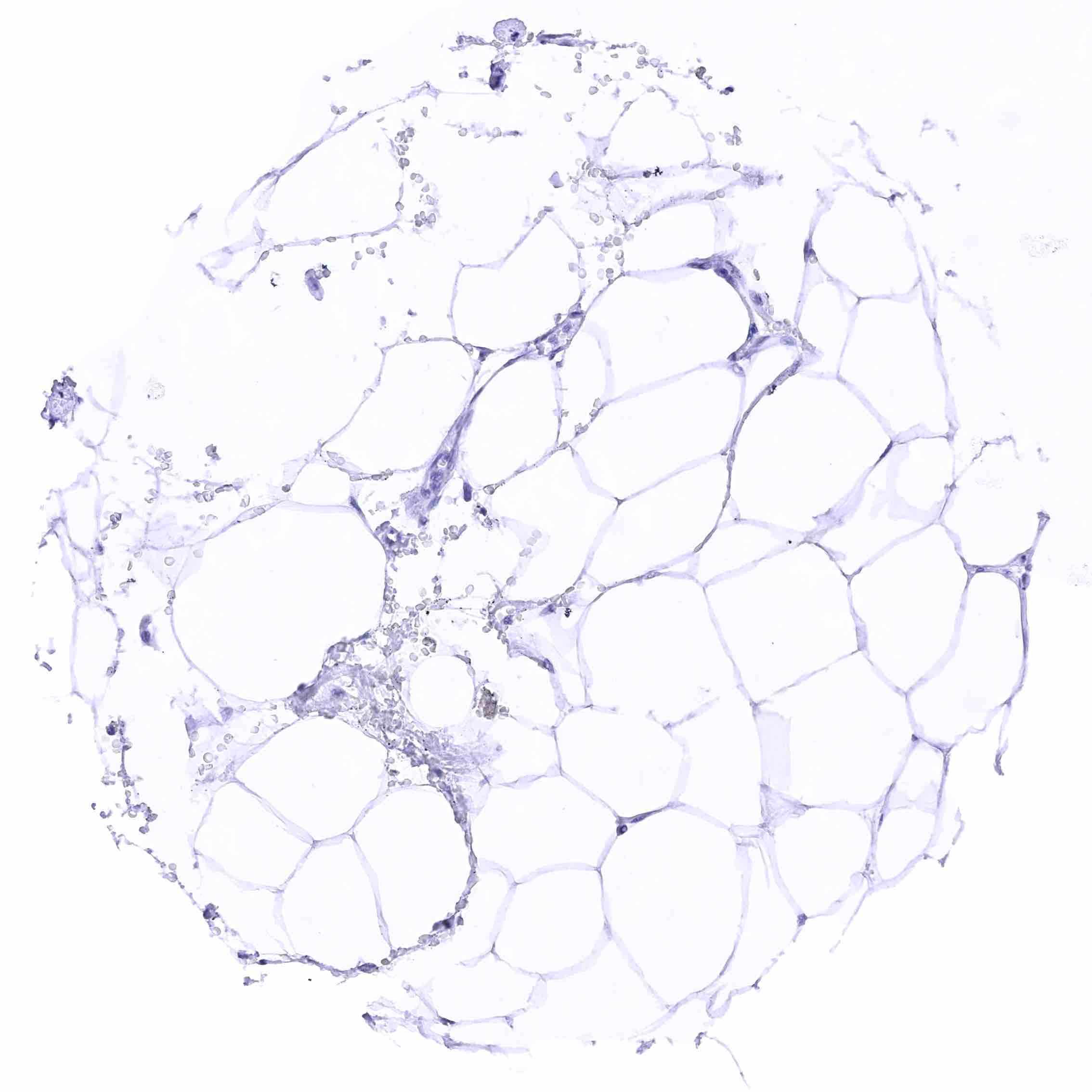
Fat
Gallbladder, epithelium – Weak to moderate membranous nectin-4 staining of many epithelial cells (most prominent at the lateral membranes).
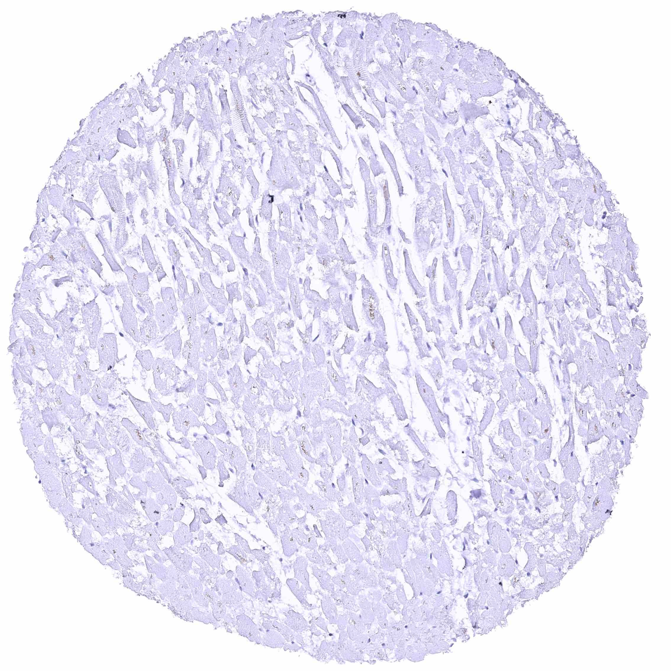
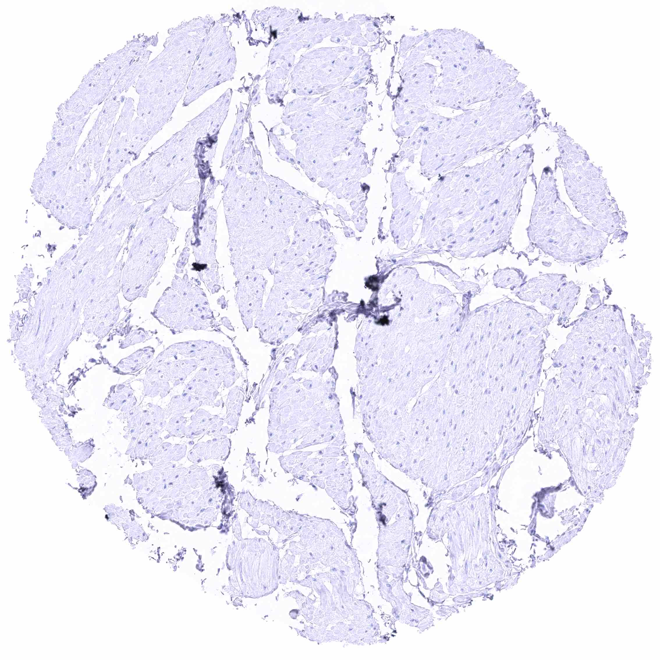
Heart muscle
Ileum, mucosa.jpeg
Ileum, muscular wall
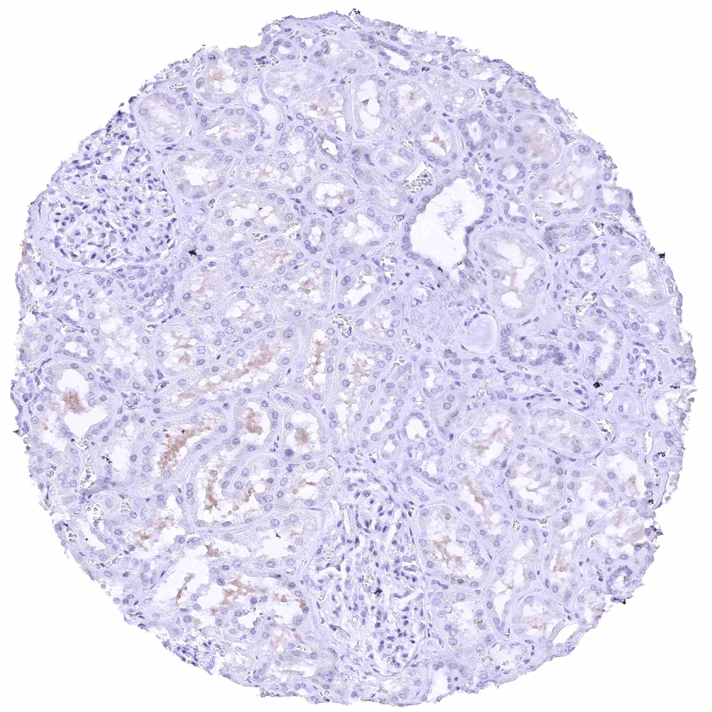
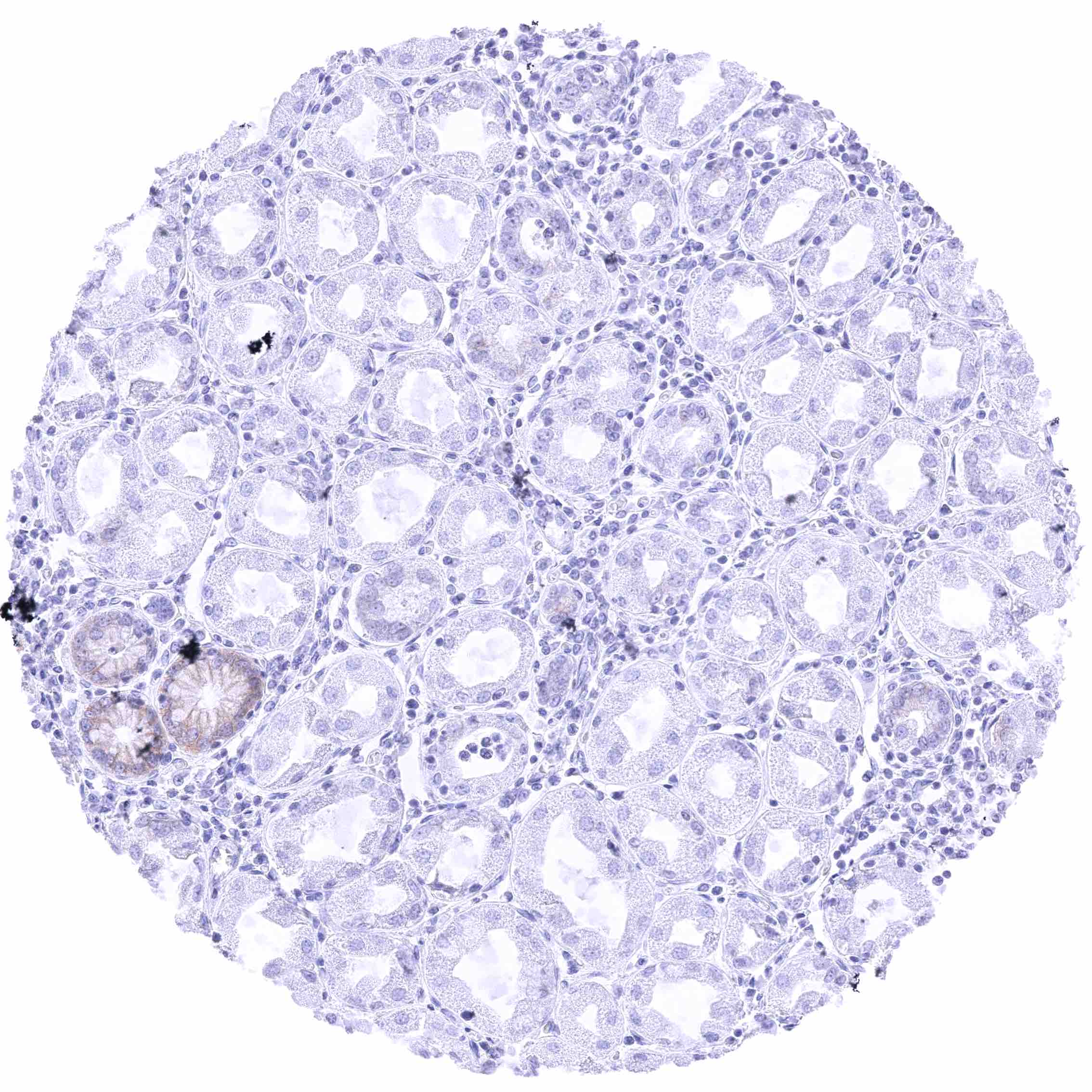
Kidney, cortex – Weak to moderate membranous nectin-4 staining of some flat tubular structures (distal tubuli vs. atrophic tubuli vs. collecting ducts).
Kidney, medulla
Kidney, pelvis, urothelium – Distinct membranous nectin-4 staining of most urothelial cells. .jpeg
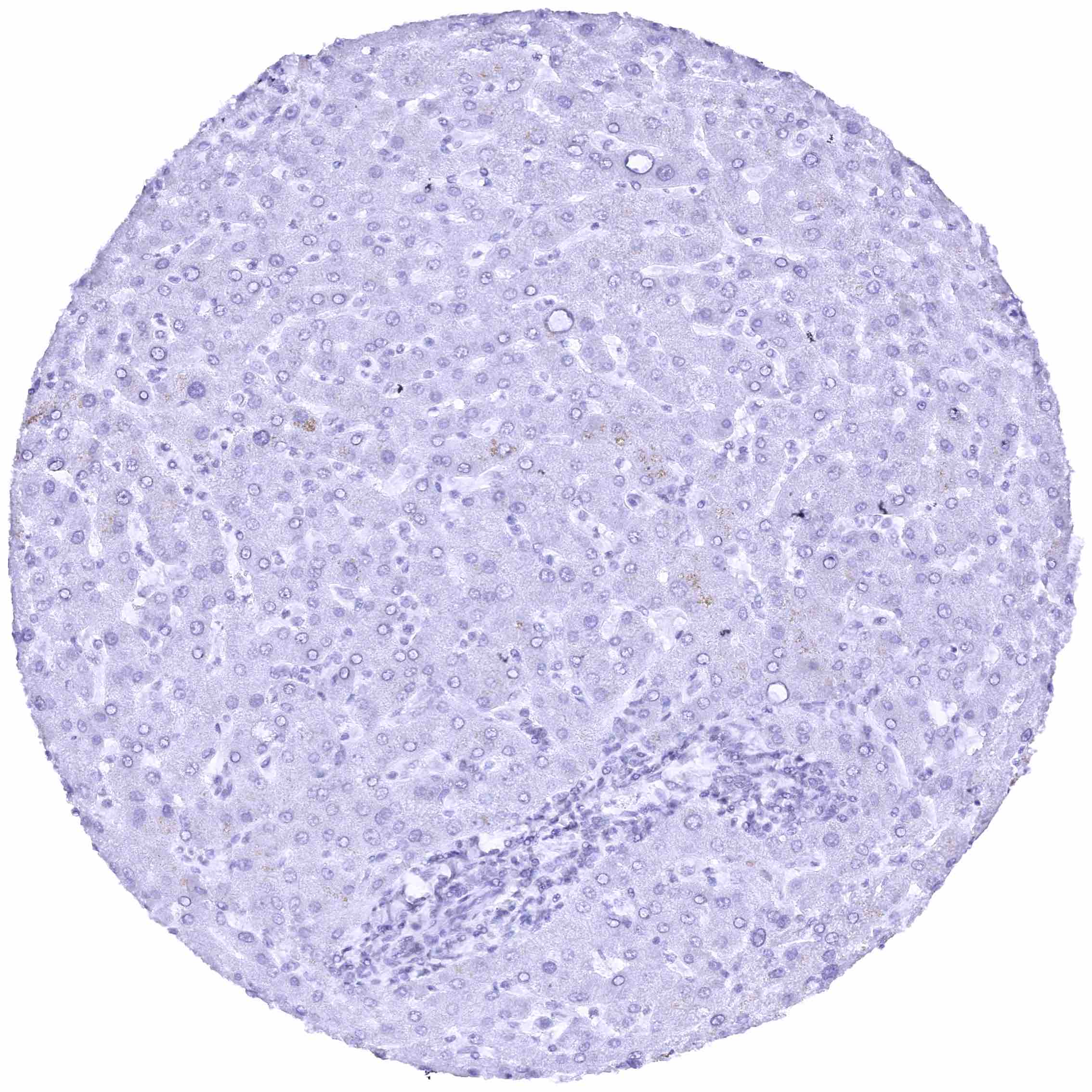
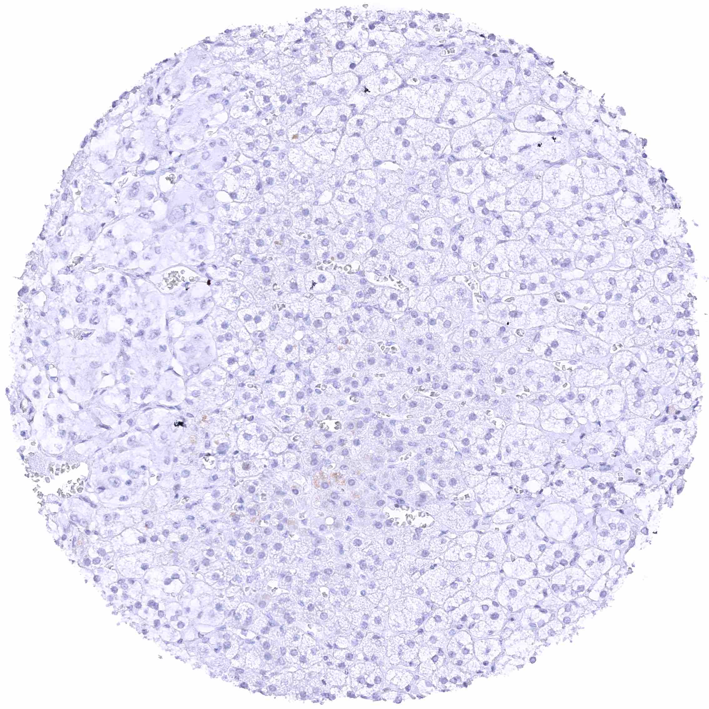
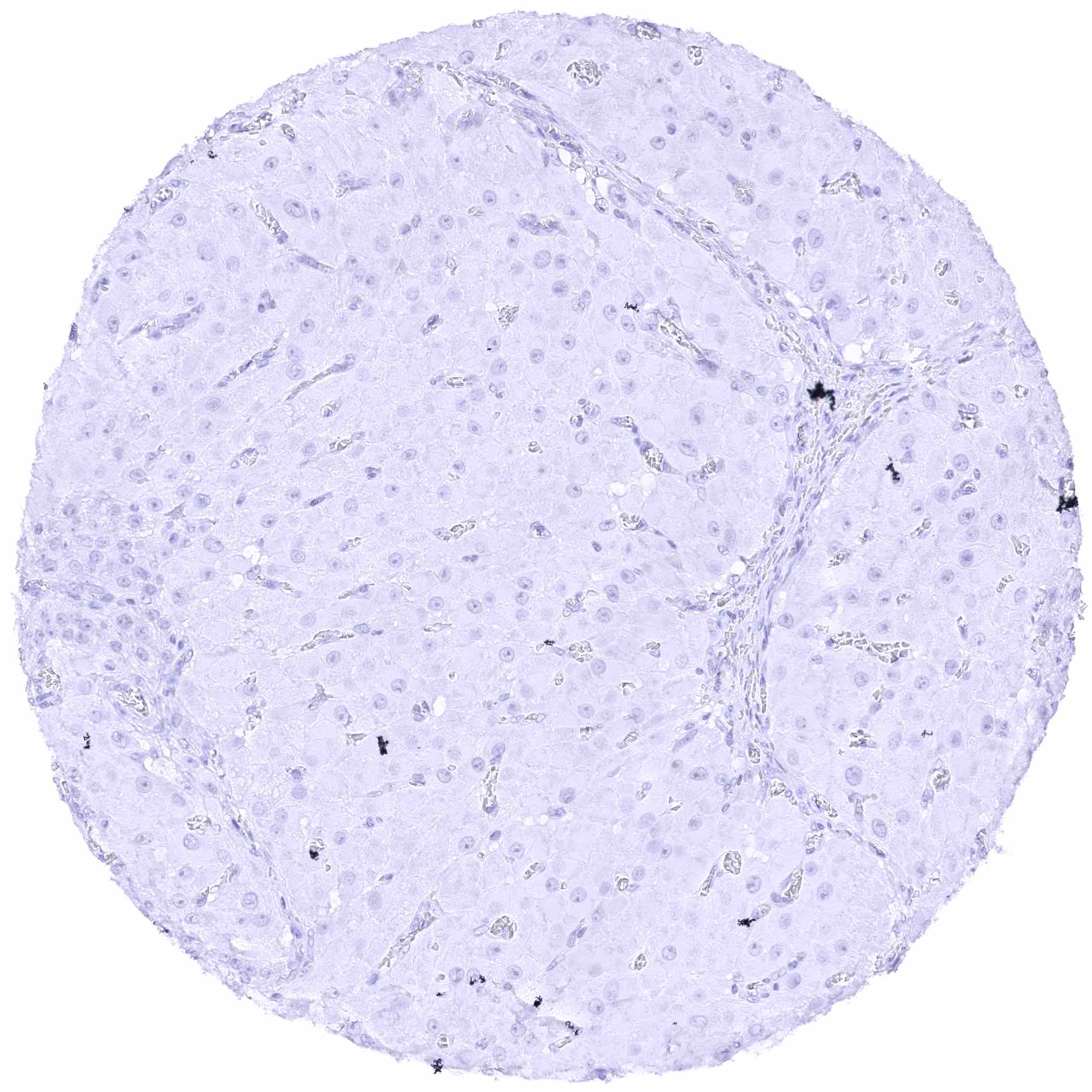
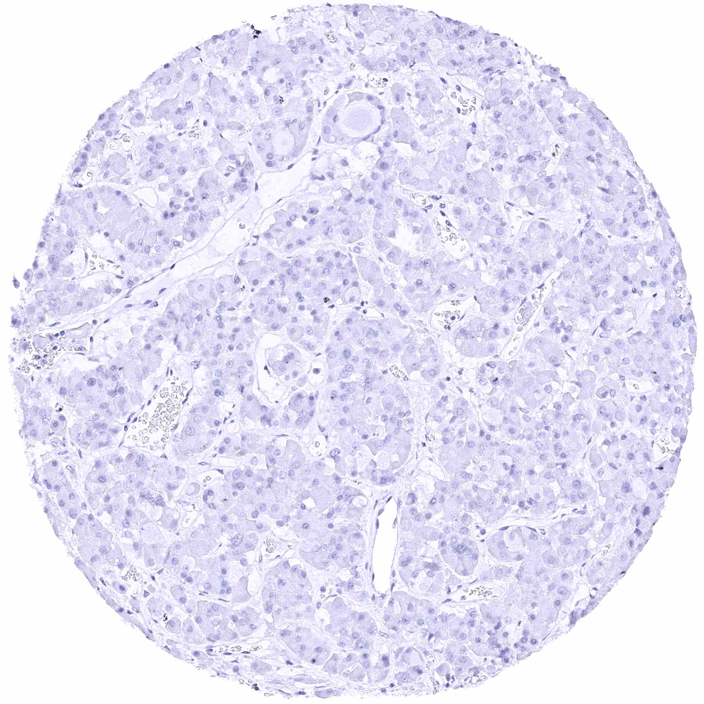
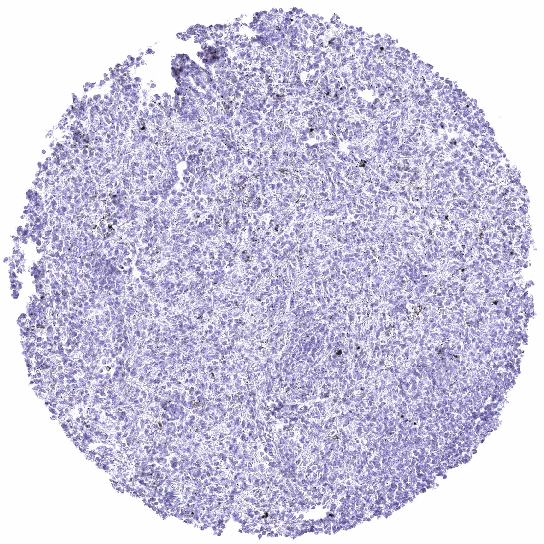
Liver
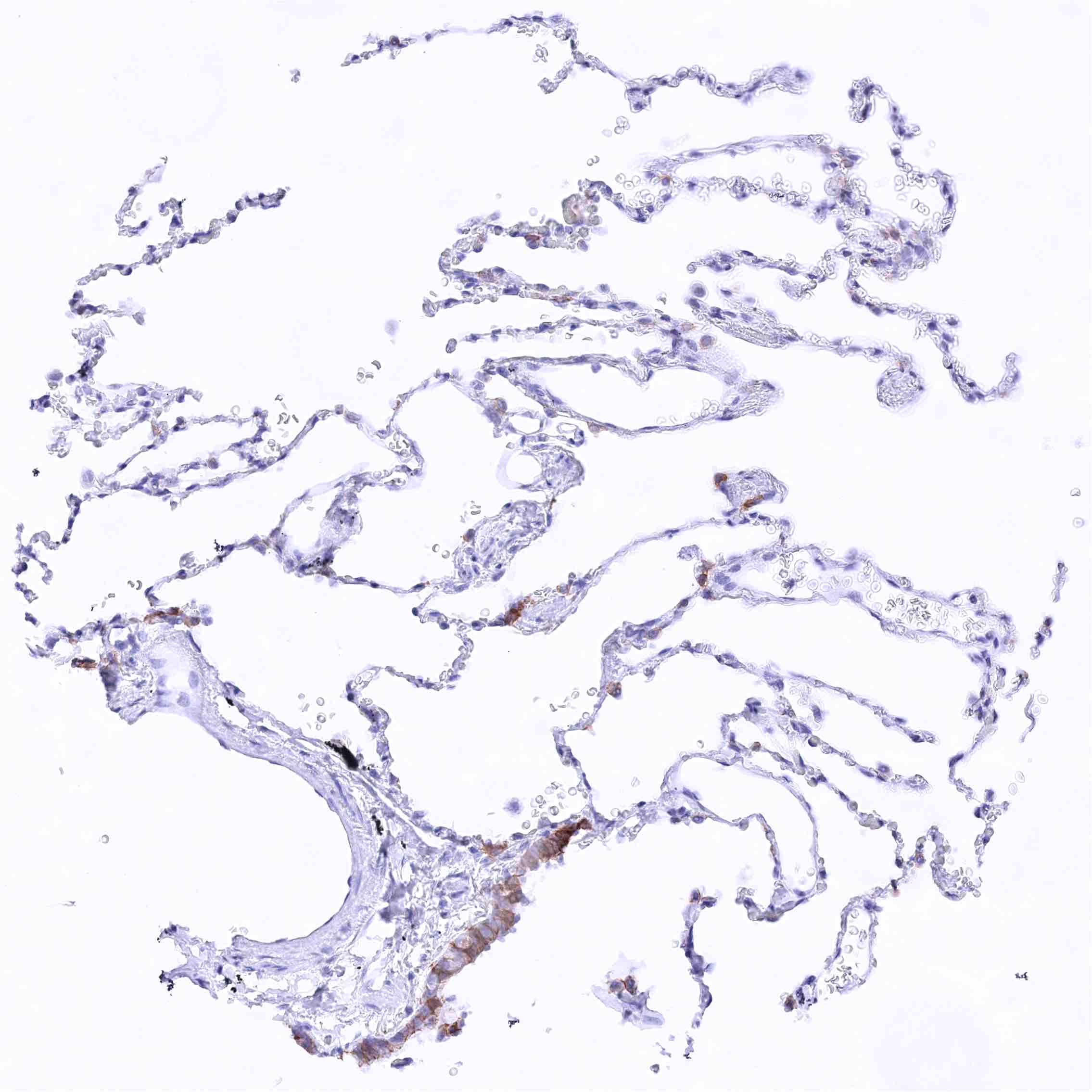
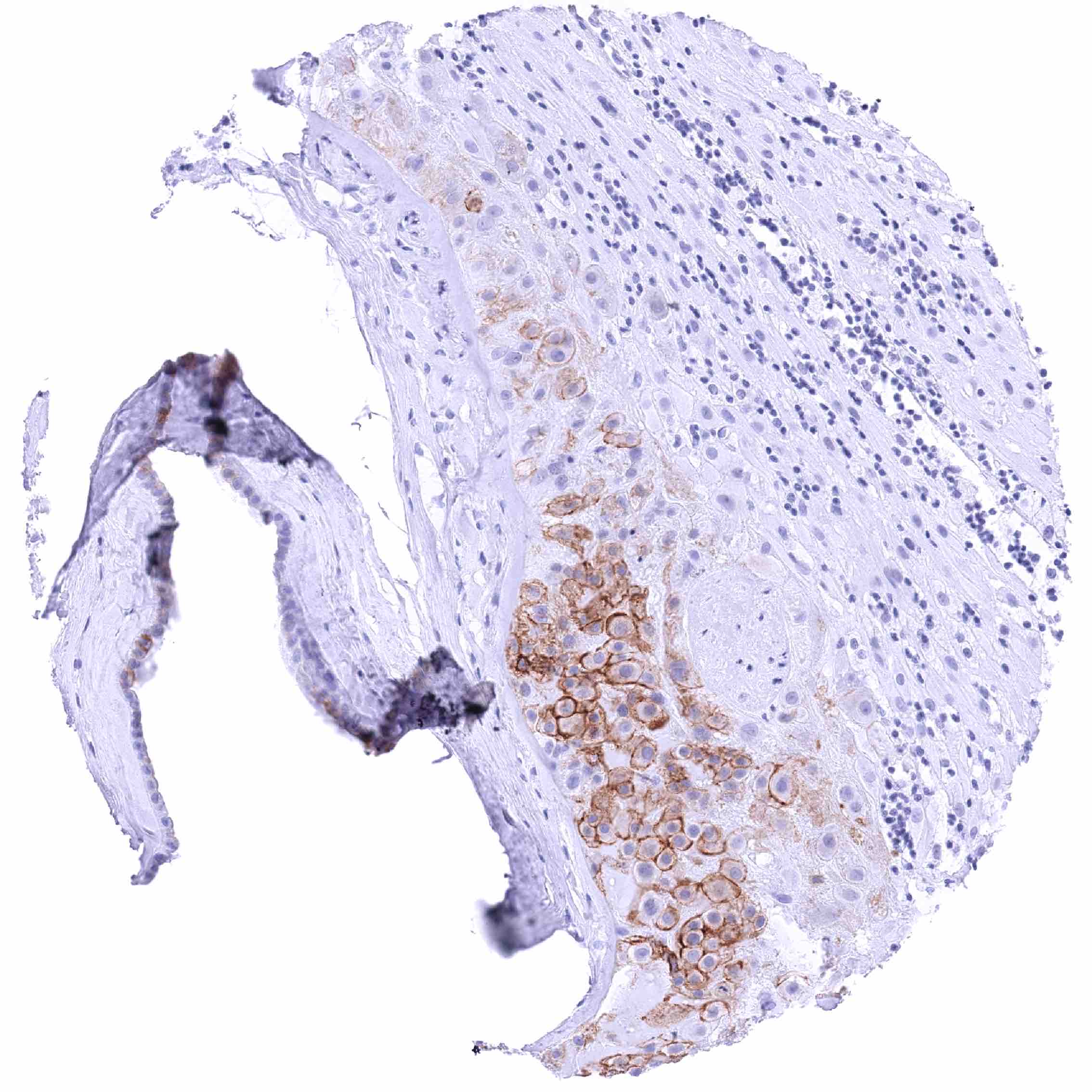
Lung – Moderate to strong membranous nectin-4 staining of at least a fraction of bronchiolar epithelial cells. Pneumocytes are nectin-4 negative.
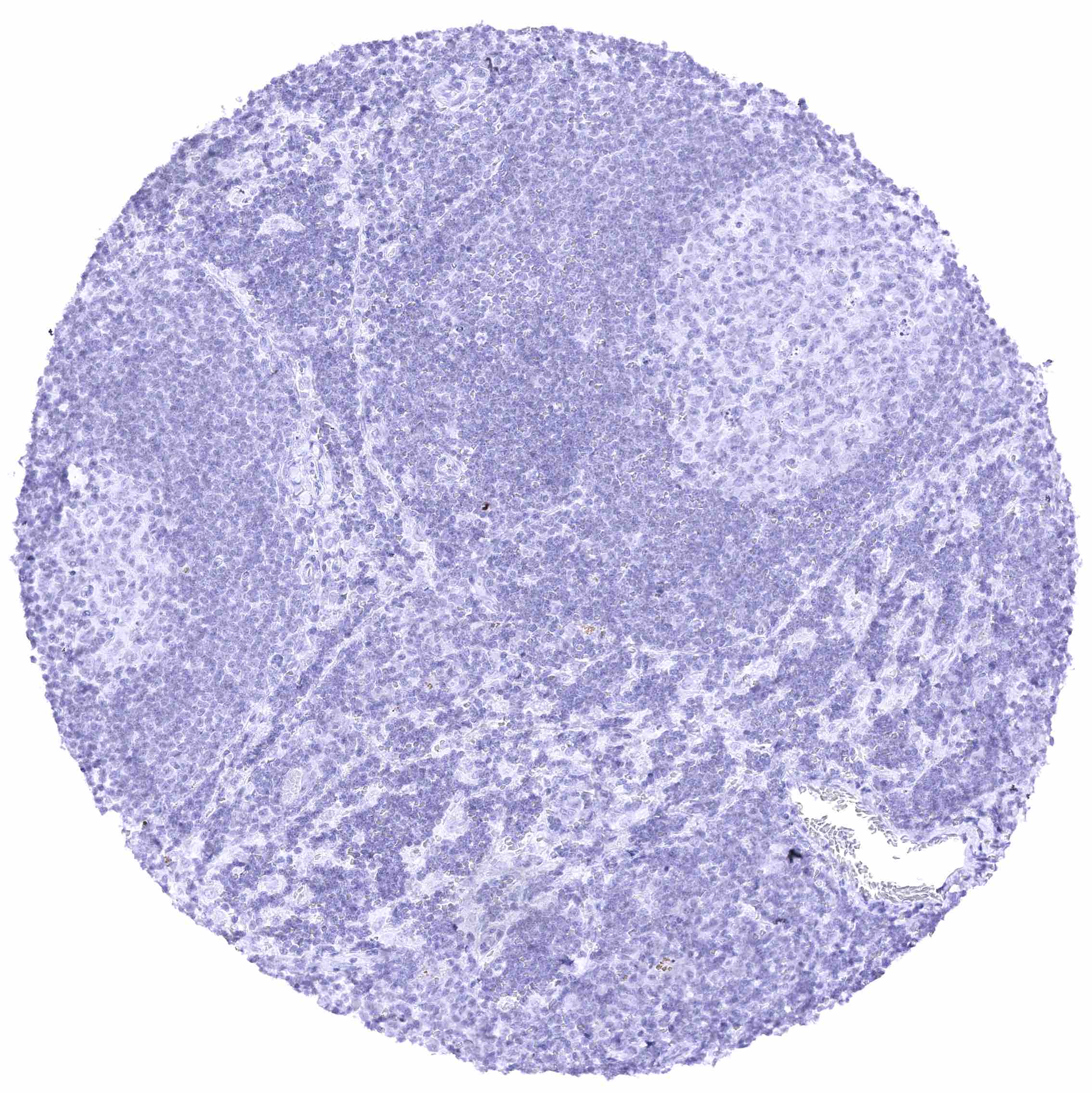
Lymph node
Ovary, corpus luteum
Ovary, stroma.jpeg
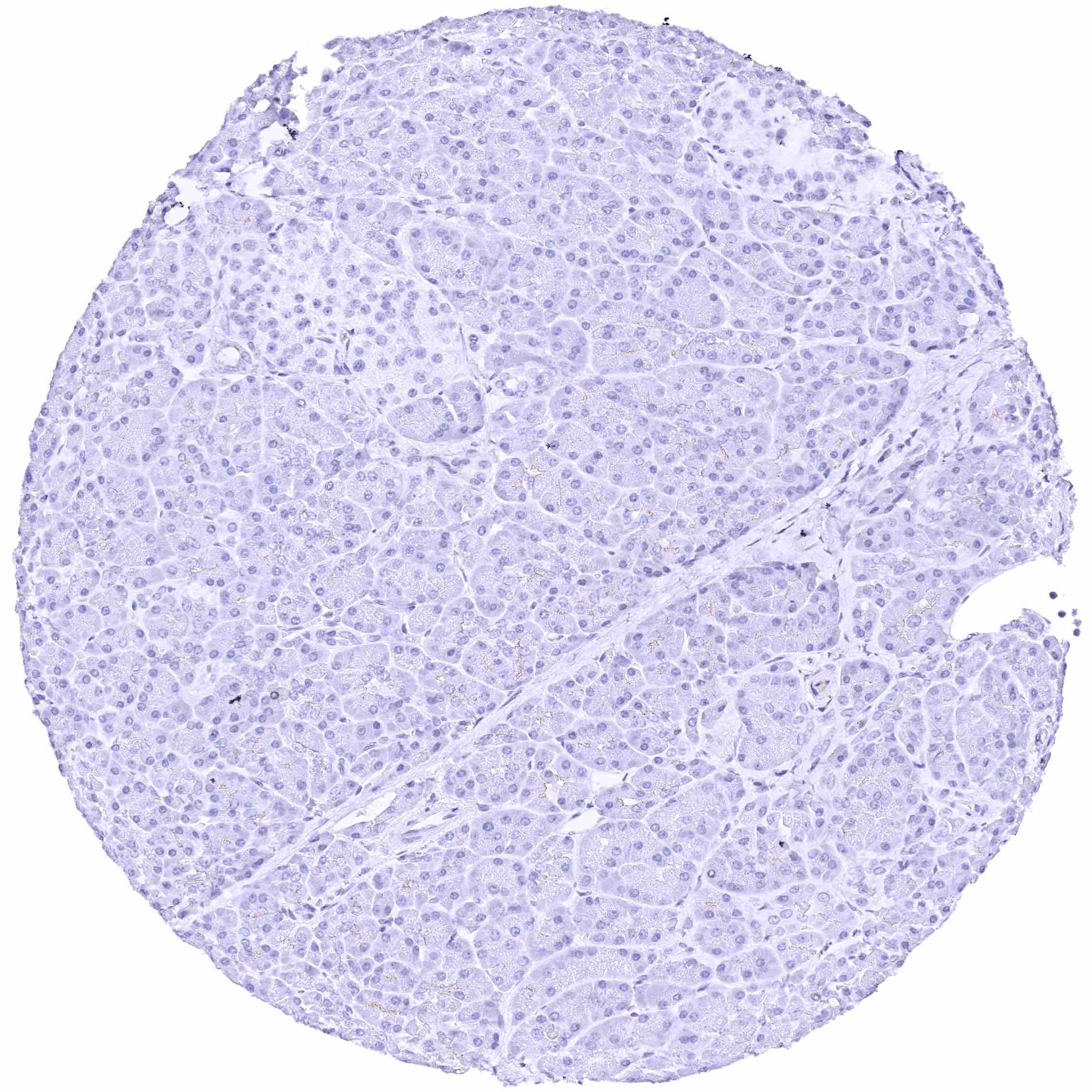
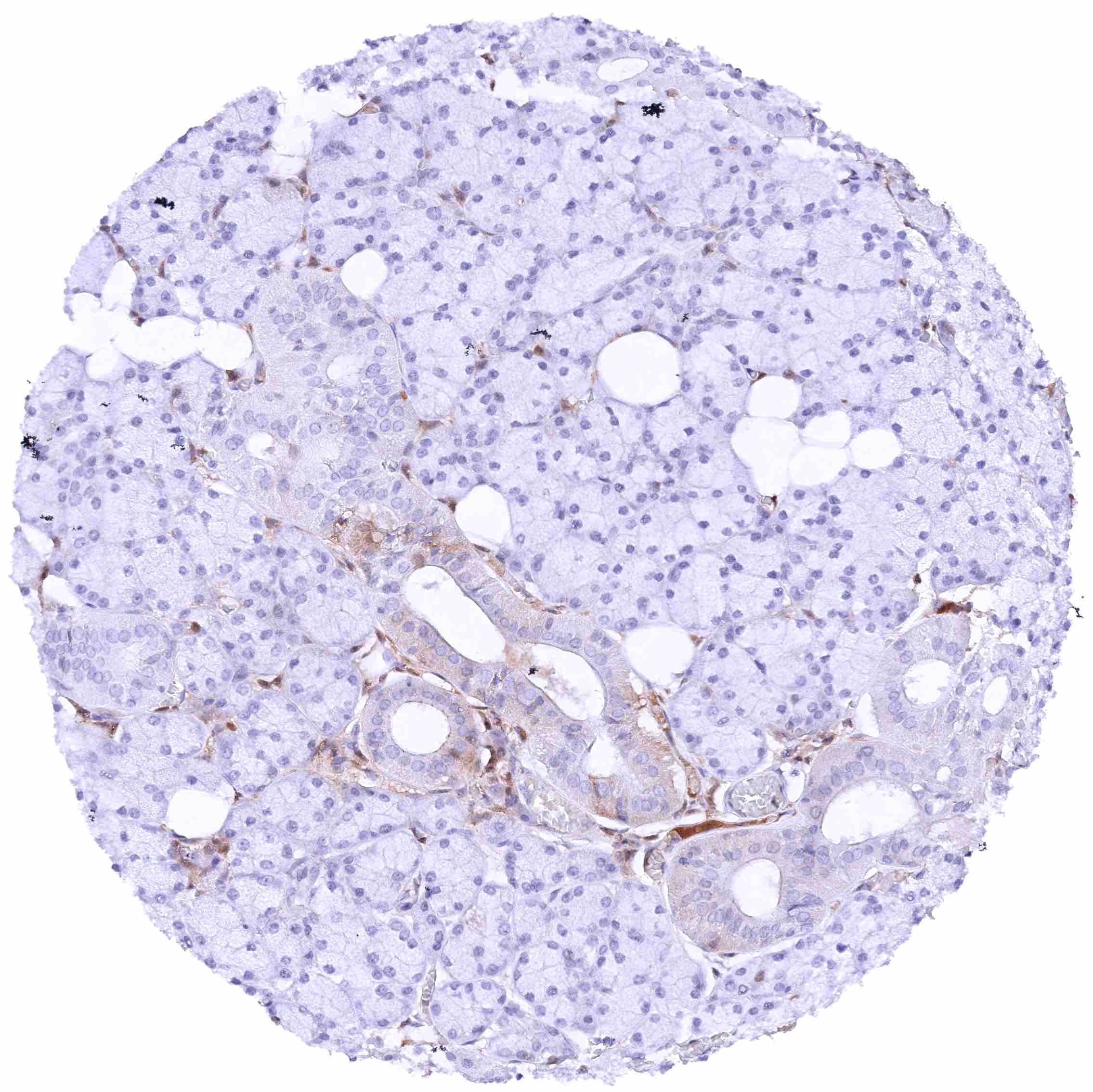
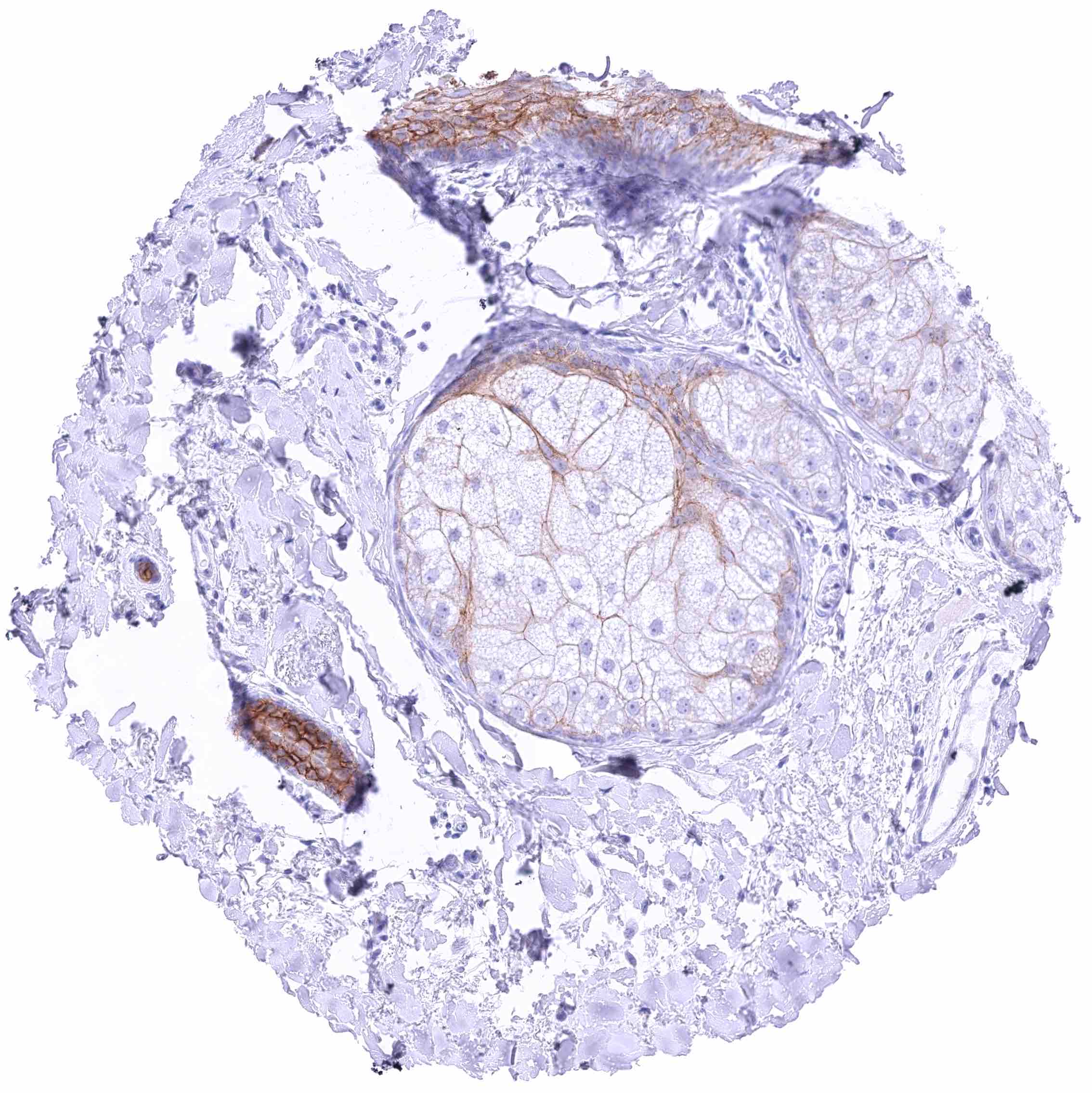
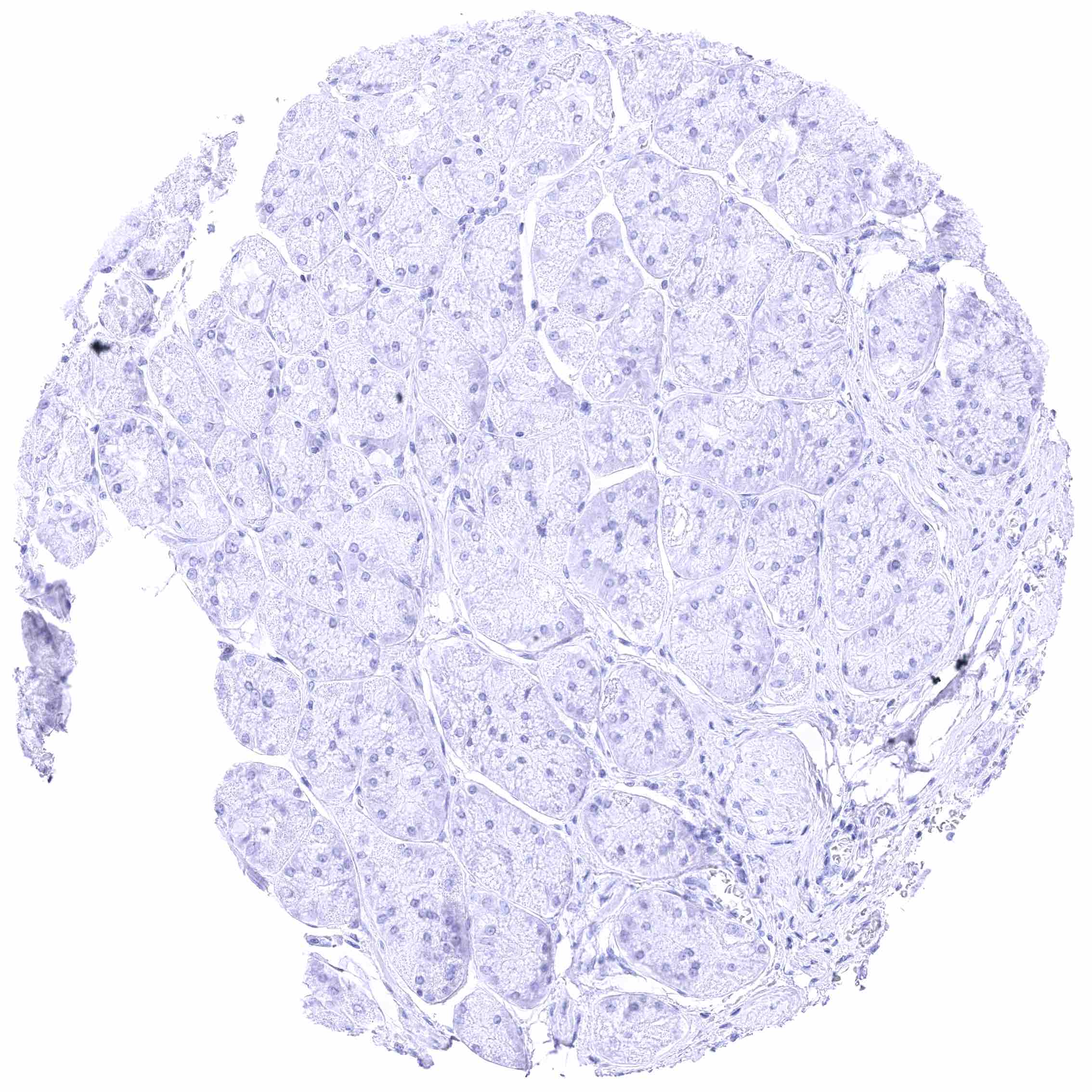
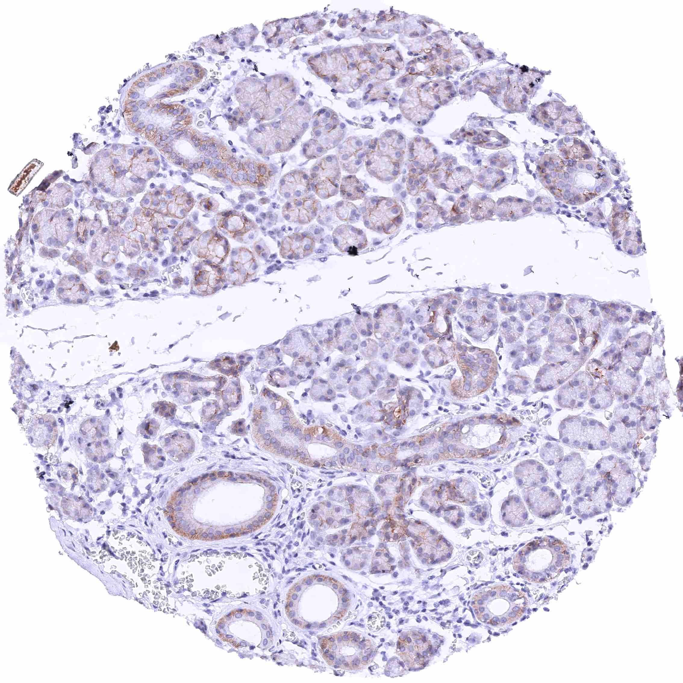
Pancreas – Faint nectin-4 staining of luminal membranes of some acinar cells and-or intercalated ducts. Islet cells are nectin-4 negative.
Parathyroid gland
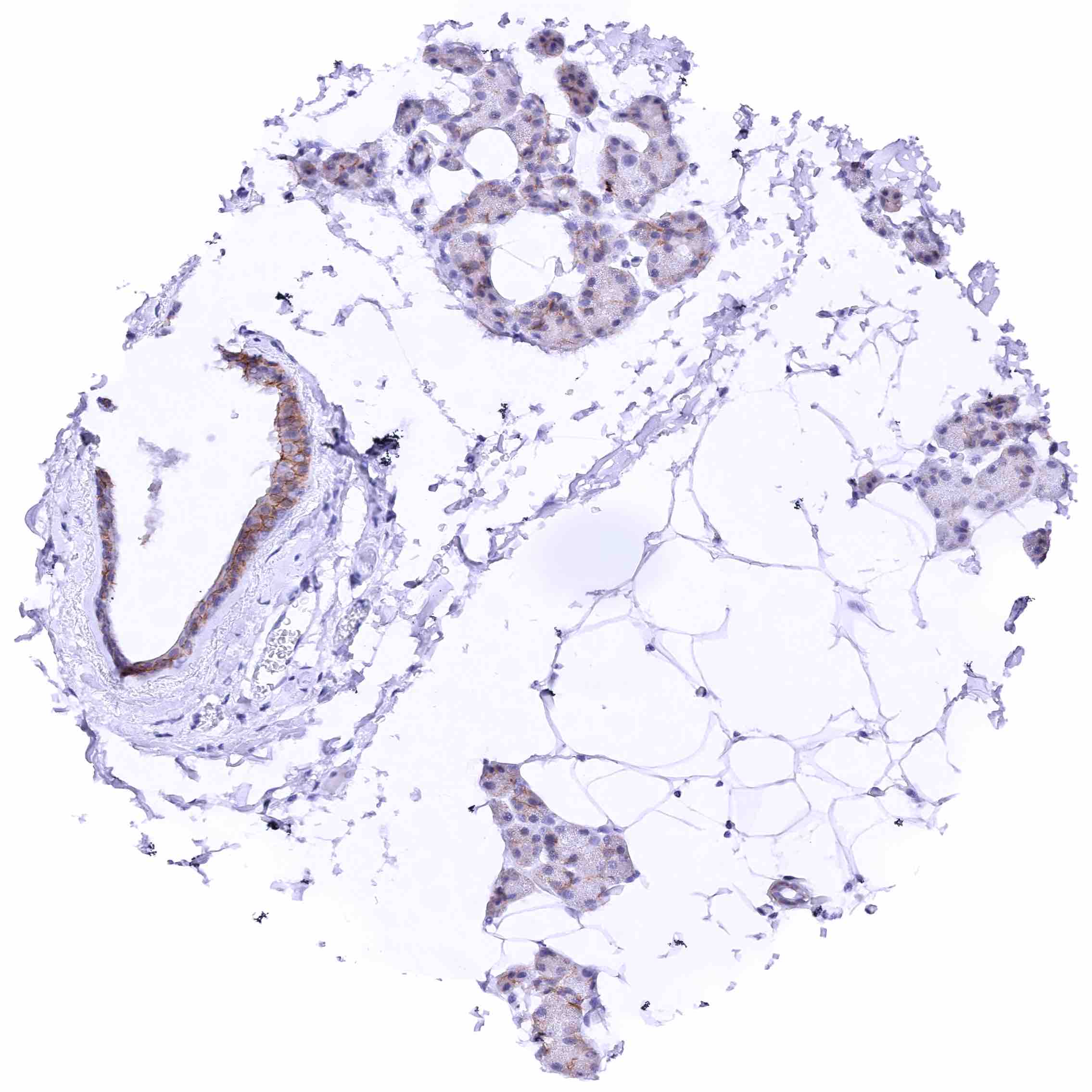
Parotid gland – Moderate membranous nectin-4 staining of subsets of excretory duct cells while staining of glandular cells is only faint. .jpeg
Parotid gland – Strong nectin-4 staining of a subset of myoepithelial cells along glands and excretory ducts. Weak to moderate membranous nectin-4 staining of some excretory duct cells while glandular cells are largely negative.
Pituitary gland, anterior lobe
Pituitary gland, posterior lobe
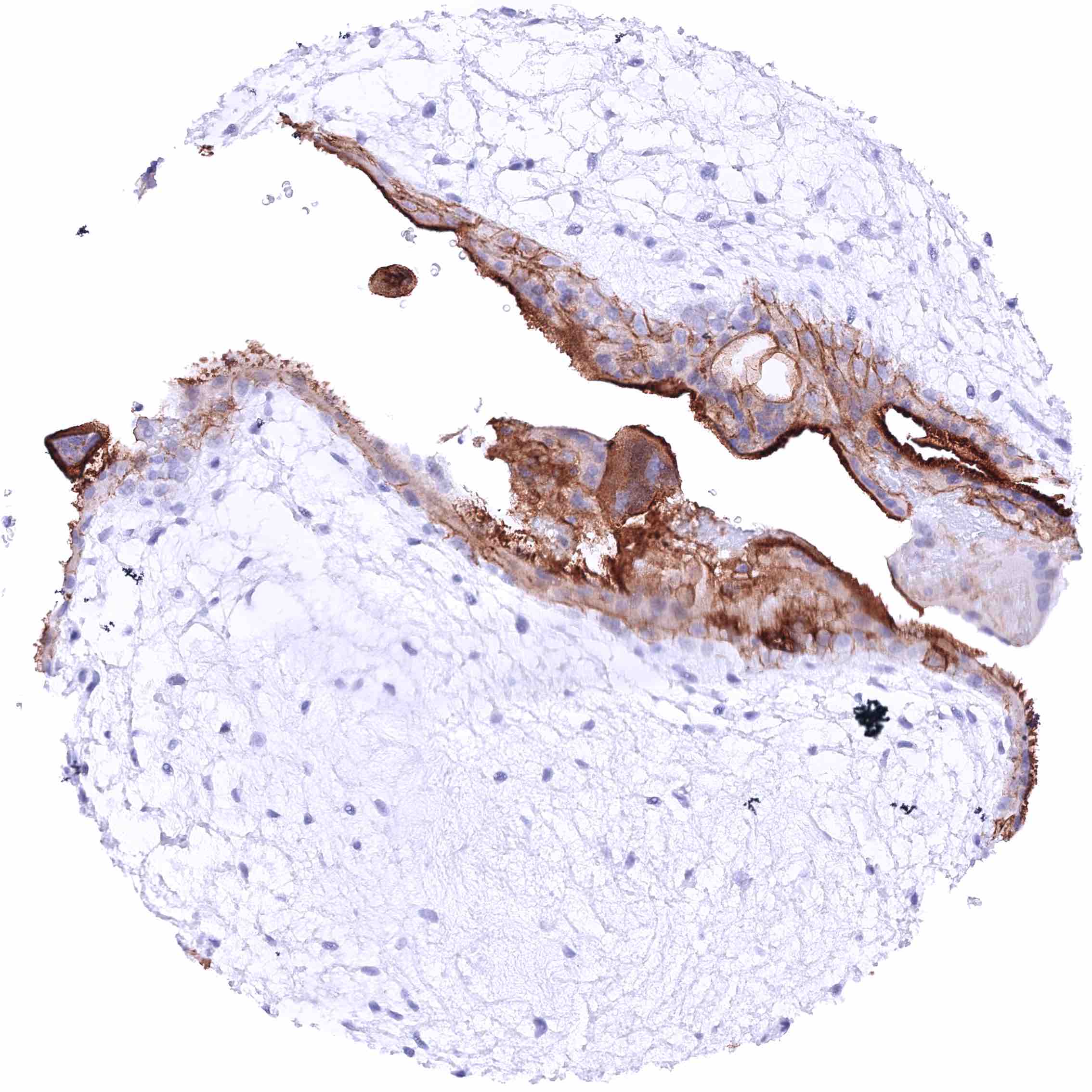
Placenta (amnion and chorion) – Moderate membranous nectin-4 staining of a subset of amnion cells, especially at the apical membranes. Distinctmembranous nectin-4 positivity of only a subset of chorion cells in this sample.
Placenta (chorion) – Strong membranous nectin-4 positivity of all chorion cells.
Placenta, early – Very intense nectin-4 staining of the apical surface membrane of the syncytiotrophoblast. Moderate membranous nectin-4 staining of cytotrophoblast cells. .jpeg
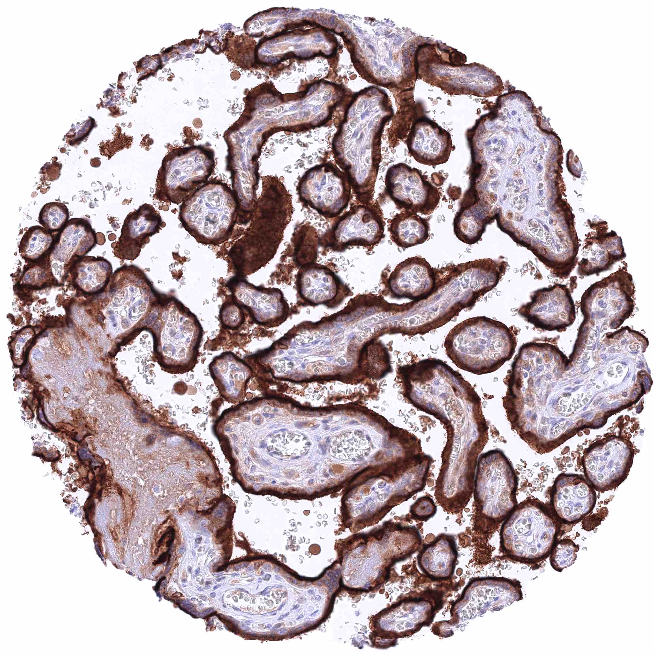
Placenta, mature – Very intense nectin-4 staining of the apical surface membrane of the syncytiotrophoblast.
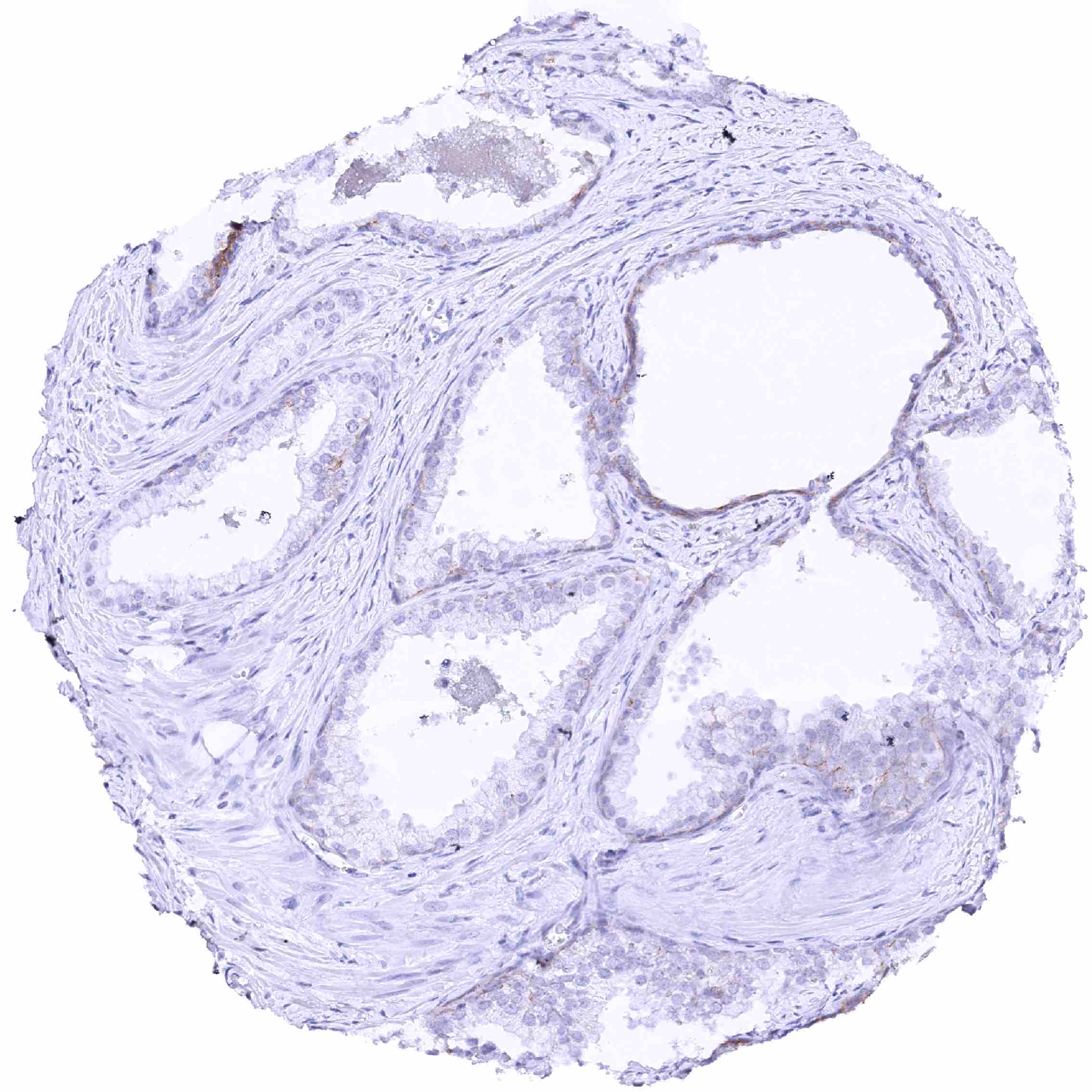
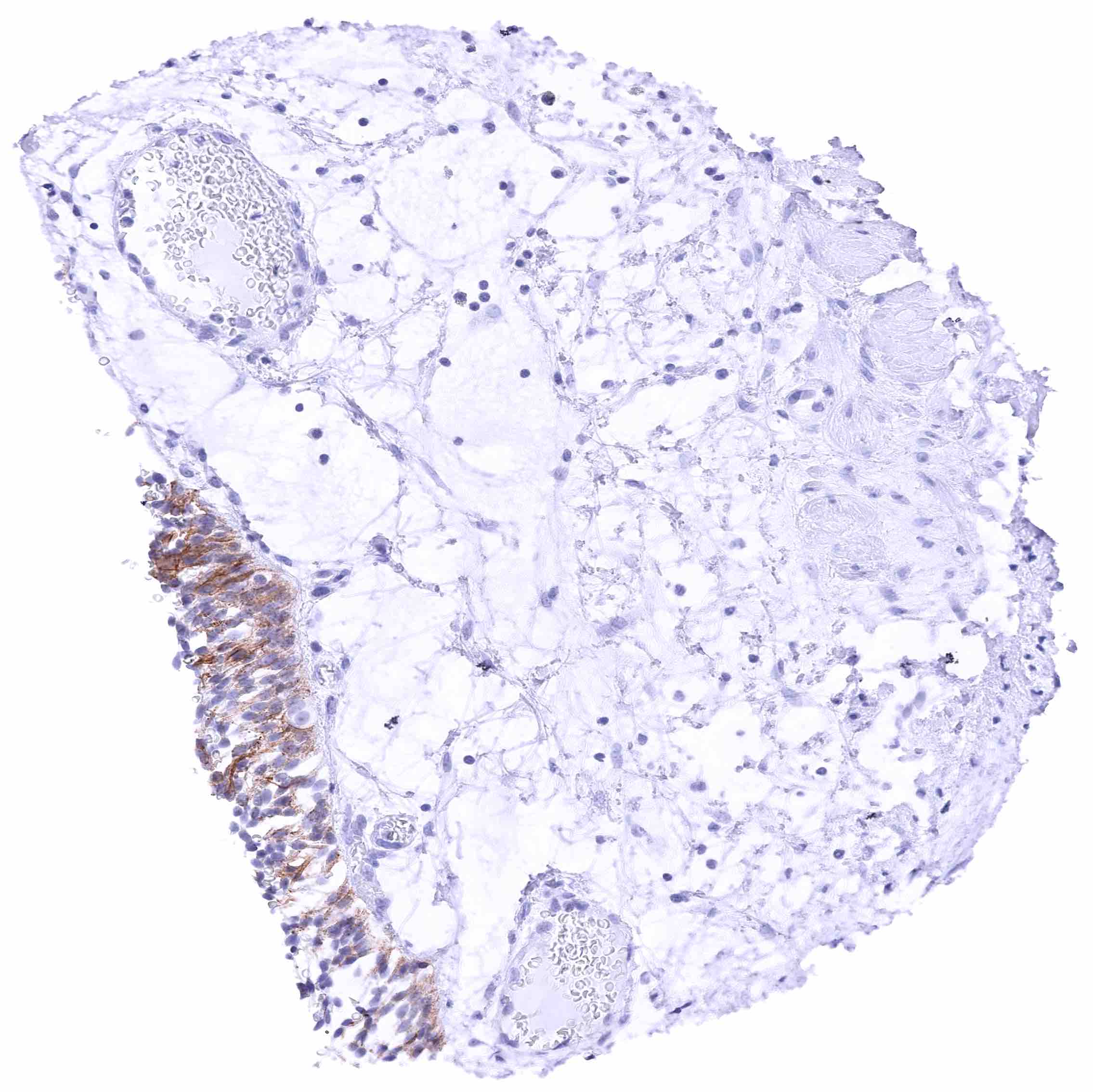
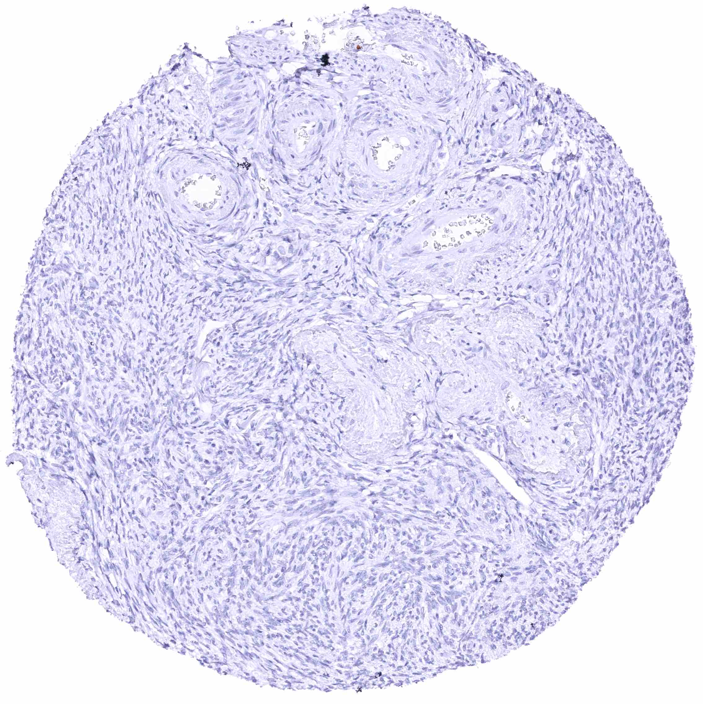
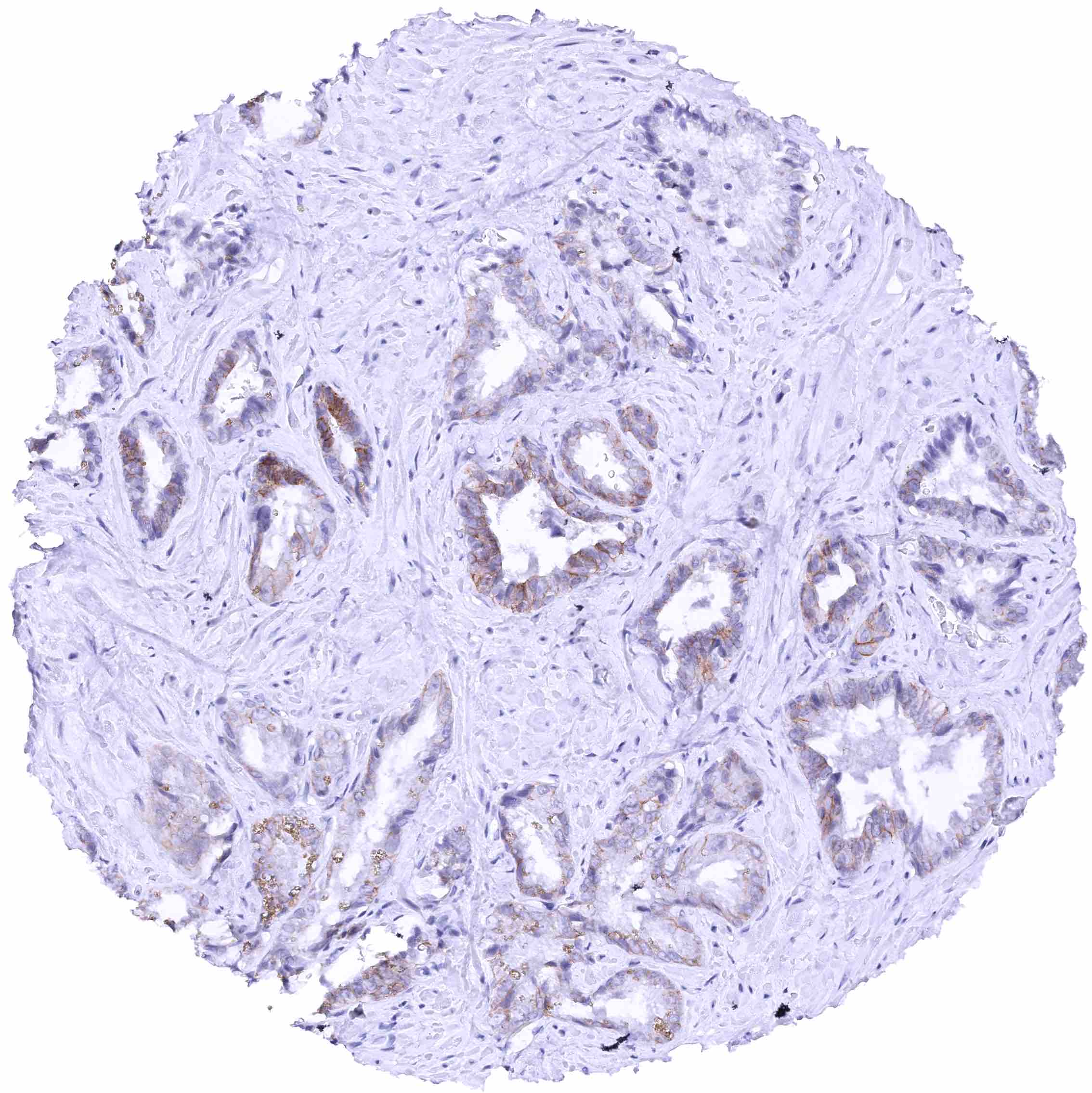
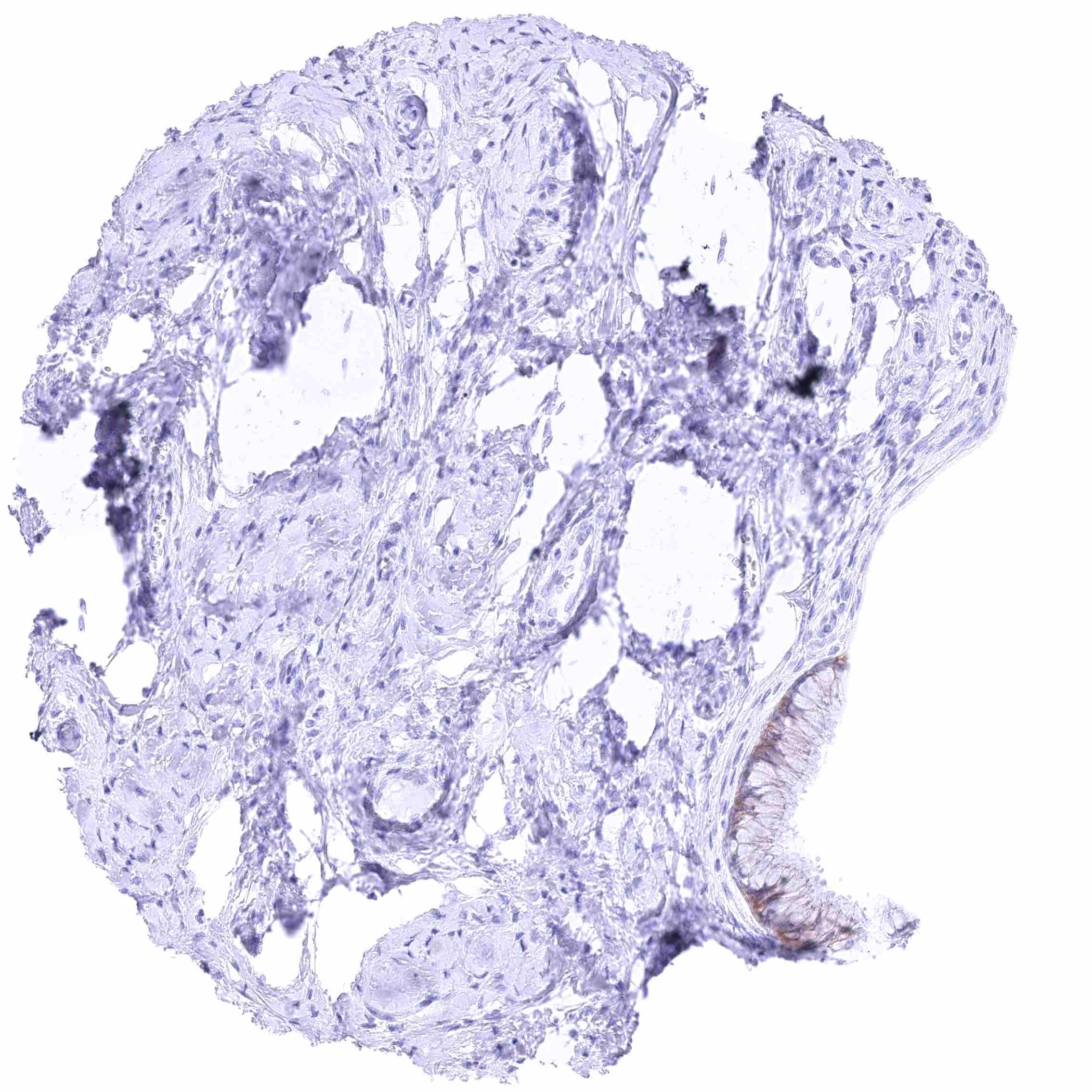
Prostate – Focal weak to moderate membranous nectin-4 staining of acinar cells, especially in an area with glandular atrophy.
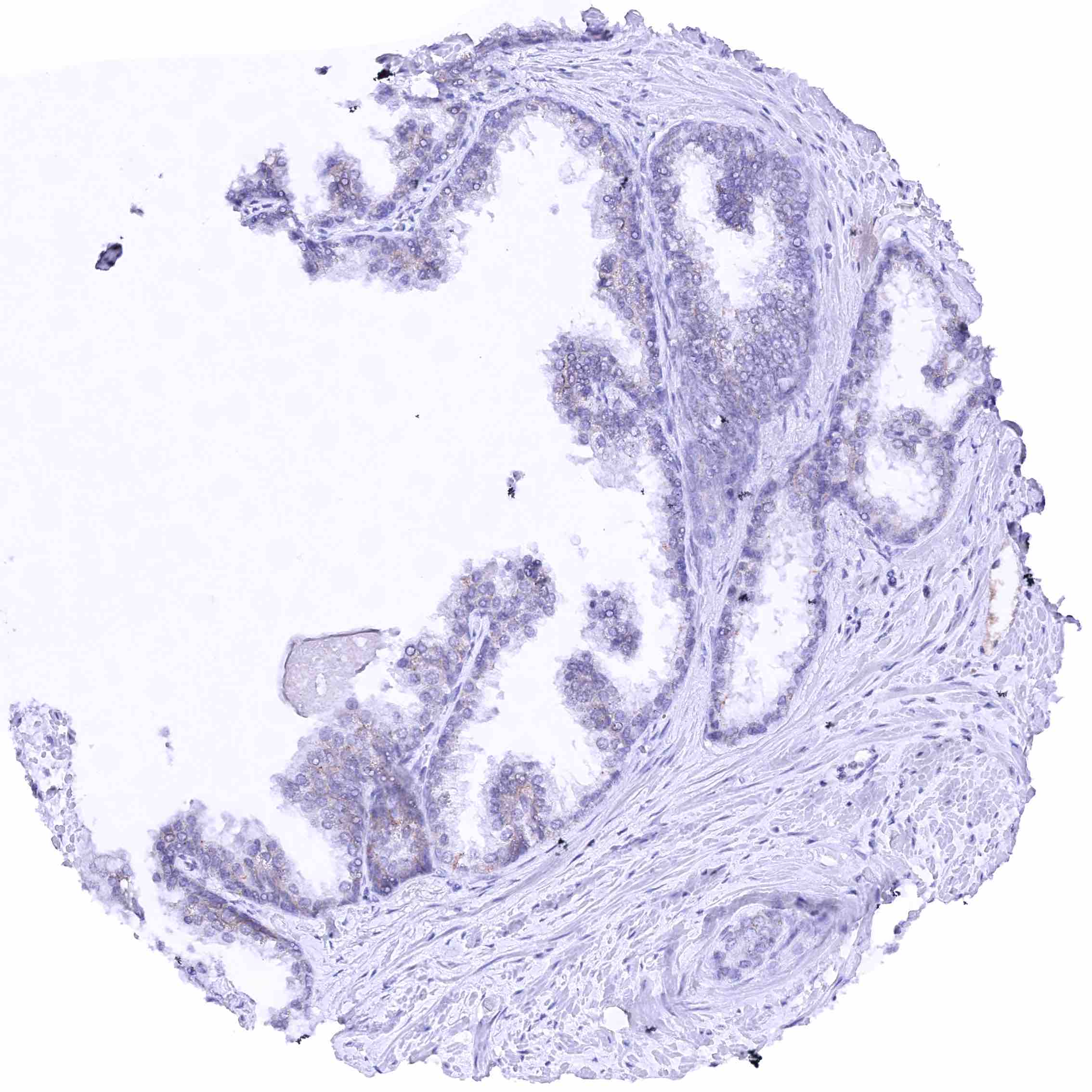
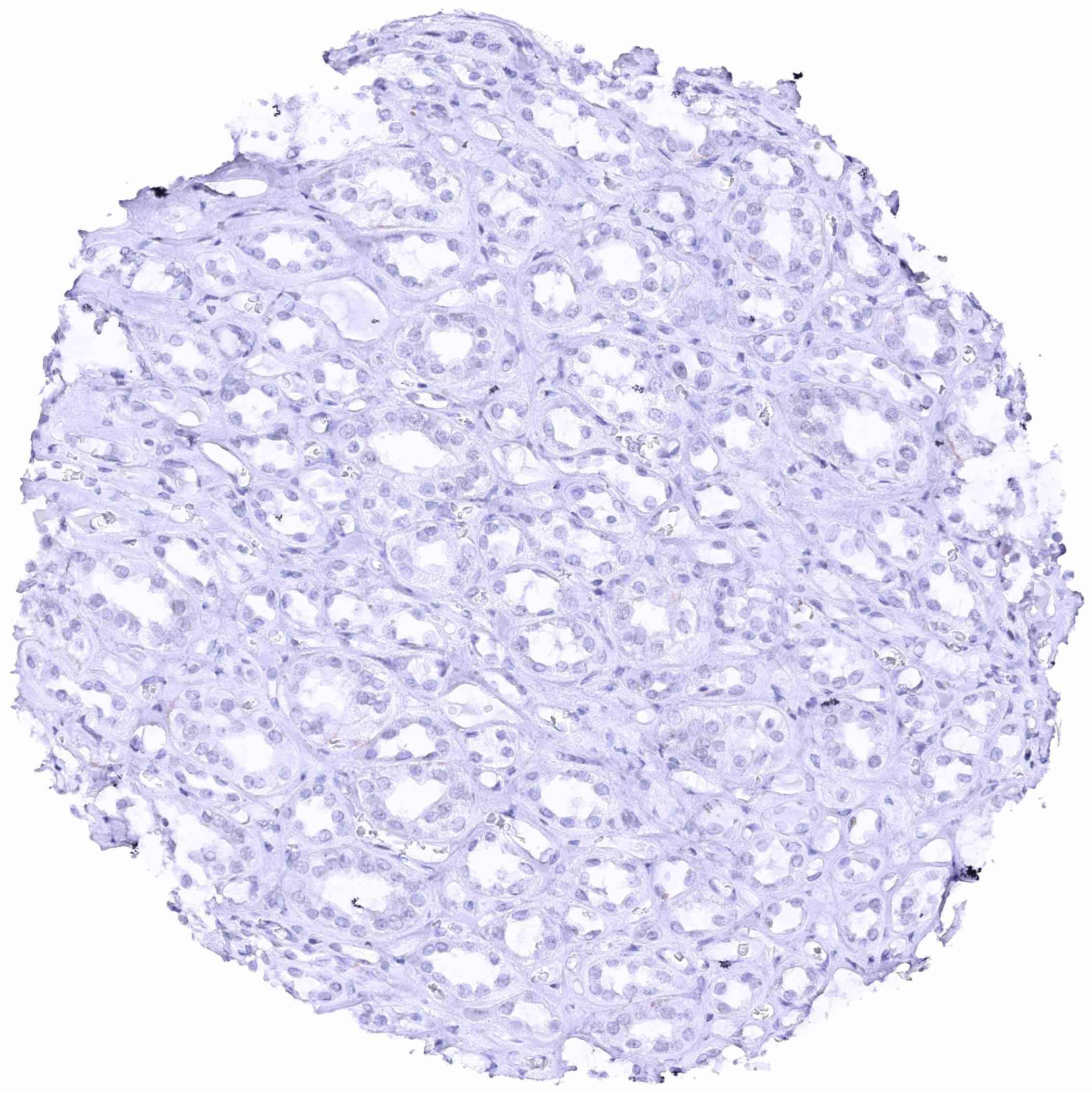
Prostate
Rectum, mucosa
Seminal vesicle – Weak to moderate membranous nectin-4 staining of a subset of epithelial cells.
Sinus paranasales – Weak to moderate membranous nectin-4 staining of ciliated cells (most prominent at the lateral membranes) of the respiratory epithelium.
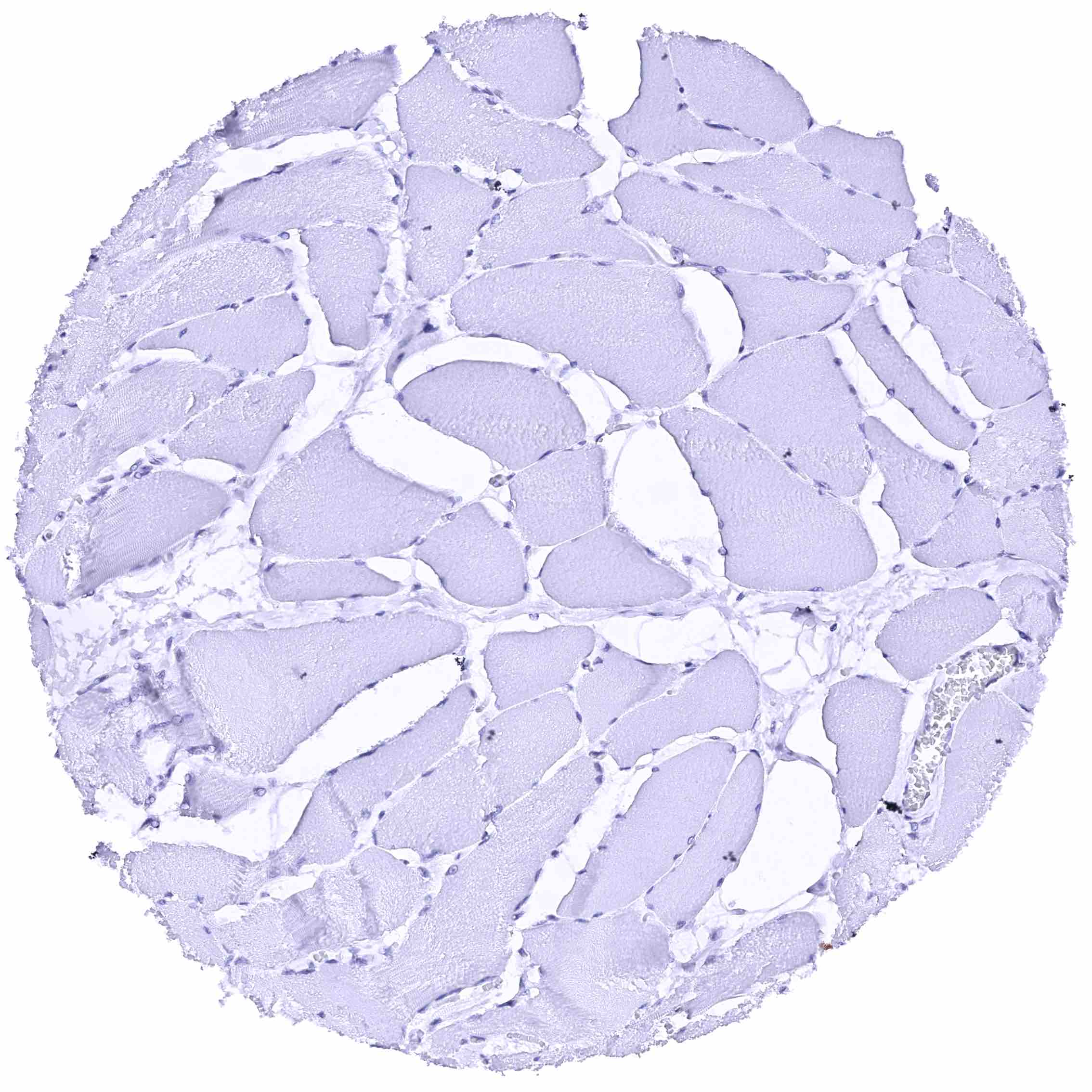
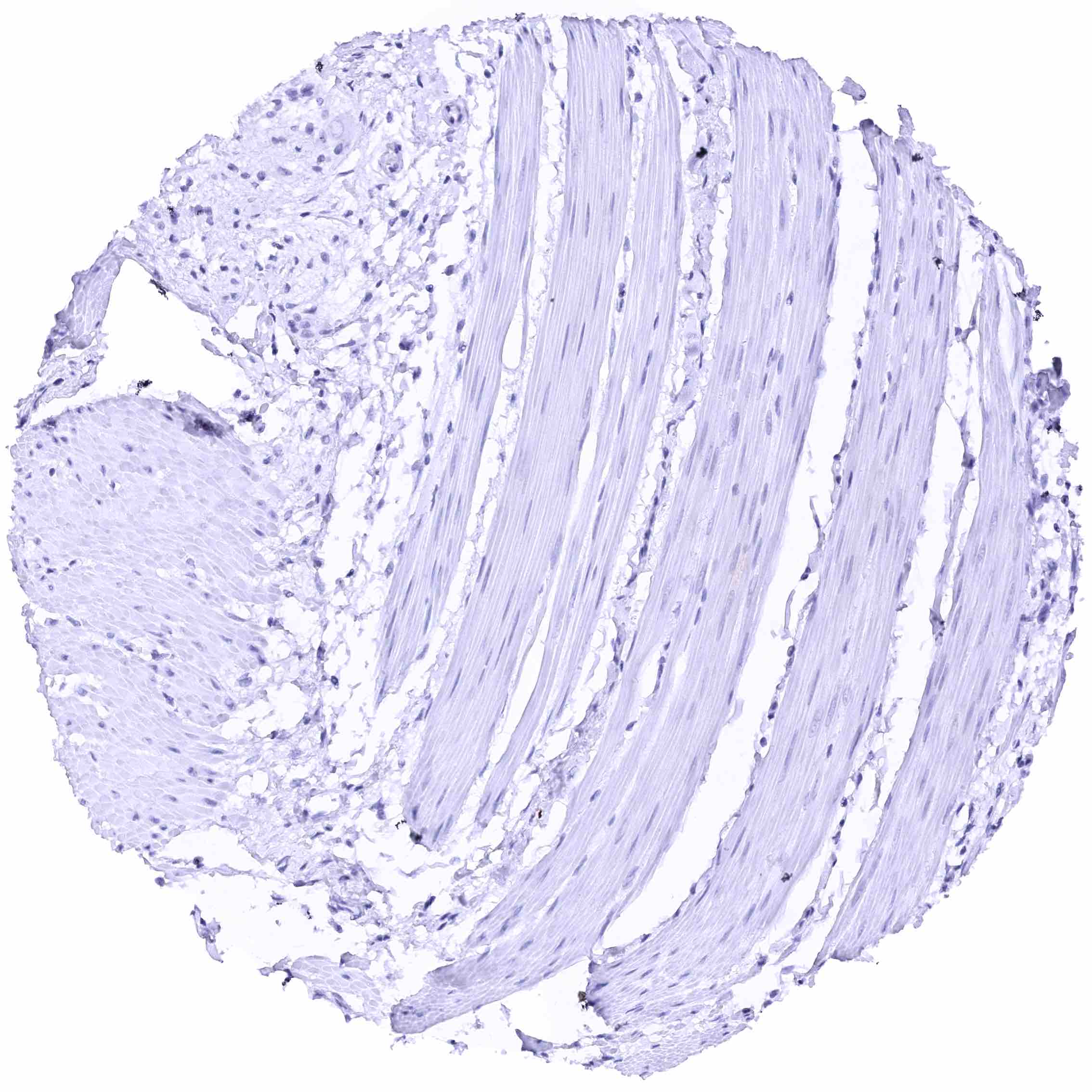
Skeletal muscle

Skin – Moderate to strong membranous nectin-4 staining of suprabasaland intermediate cell layers. The nectin-4 staining intensity decreases markedly towards the superficial cell layers which are often nectin-4 negative. Basal cells are nectin-4 negative.
Skin, sebaceous glands – Weak to moderate membranous nectin-4 staining of sebaceous cells. Moderate membranous nectin-4 staining of peripheral germinative cells. .jpeg
Spleen
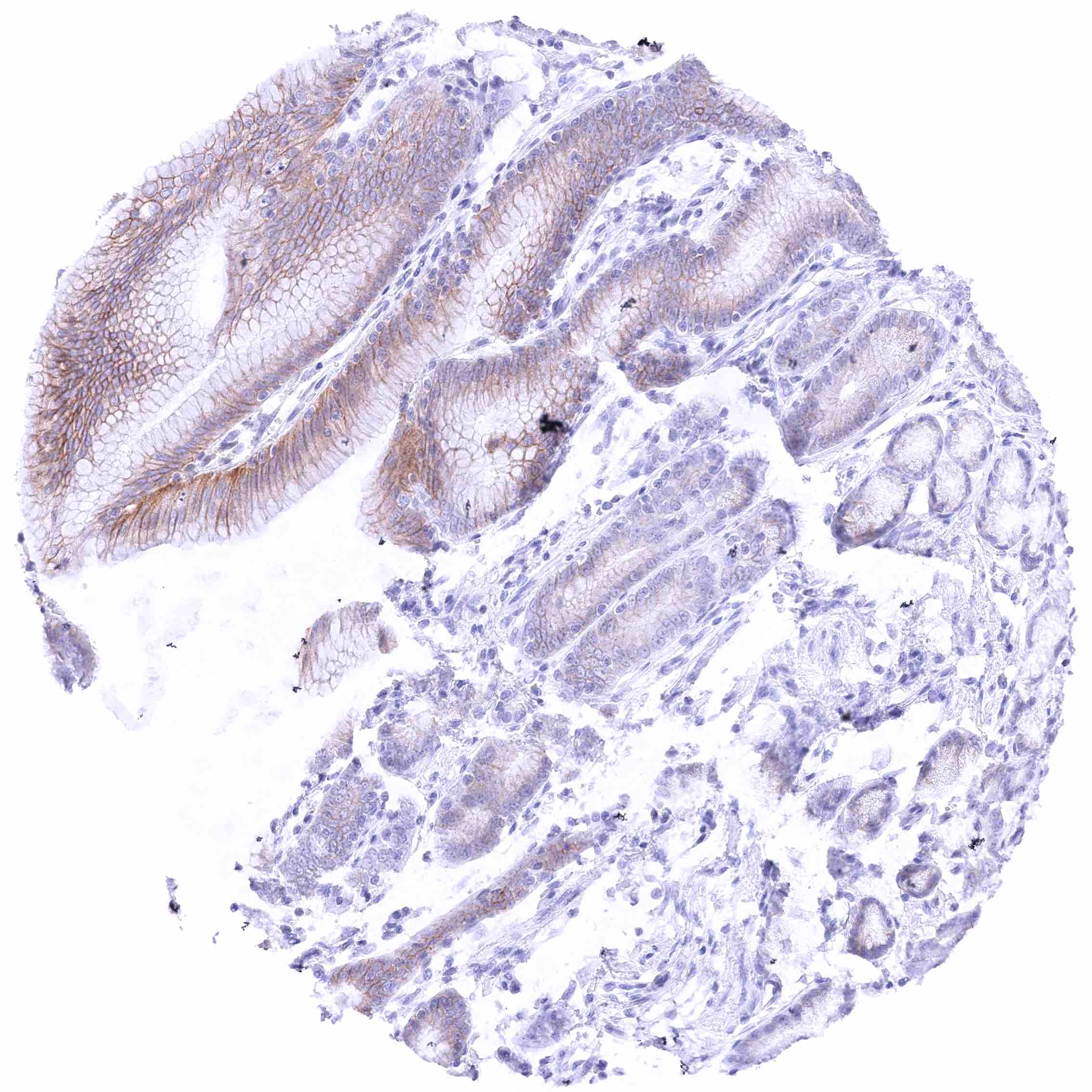
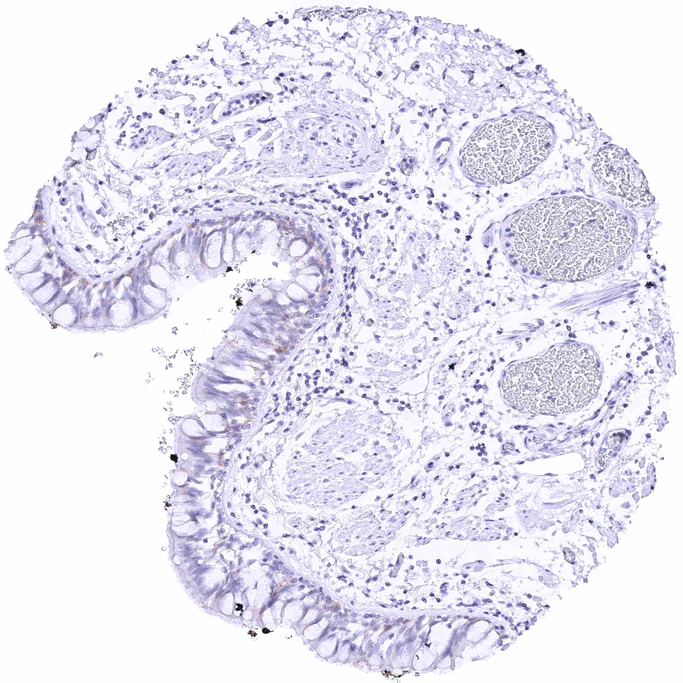
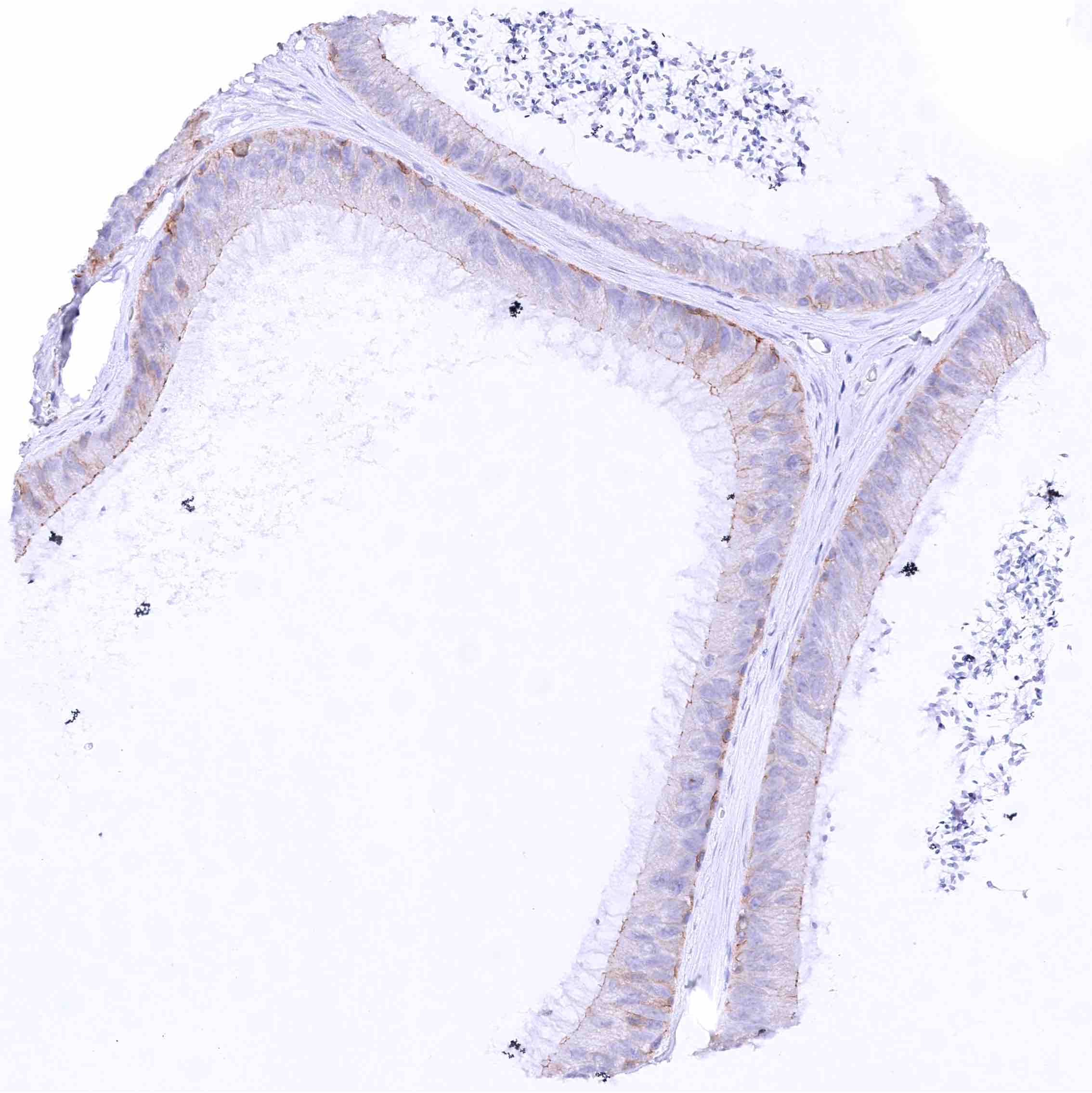
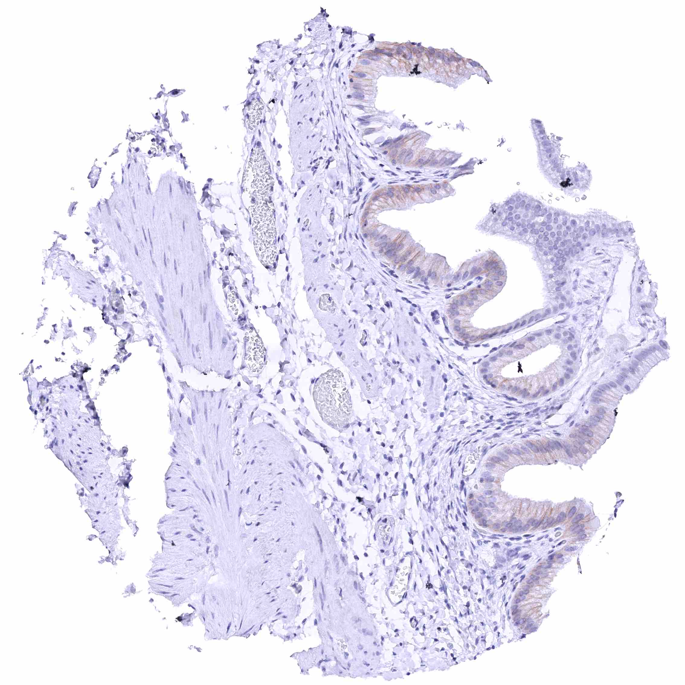
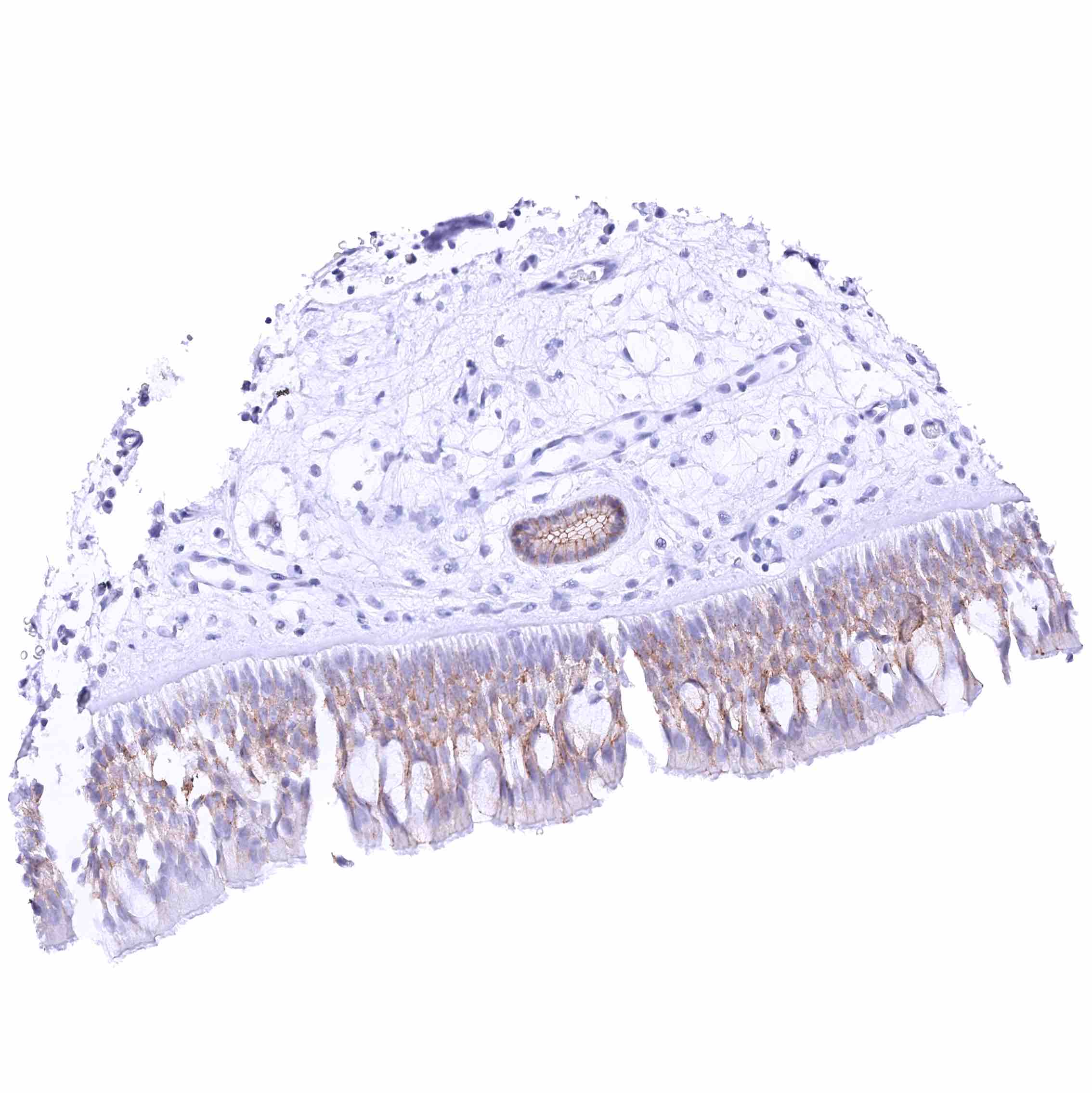
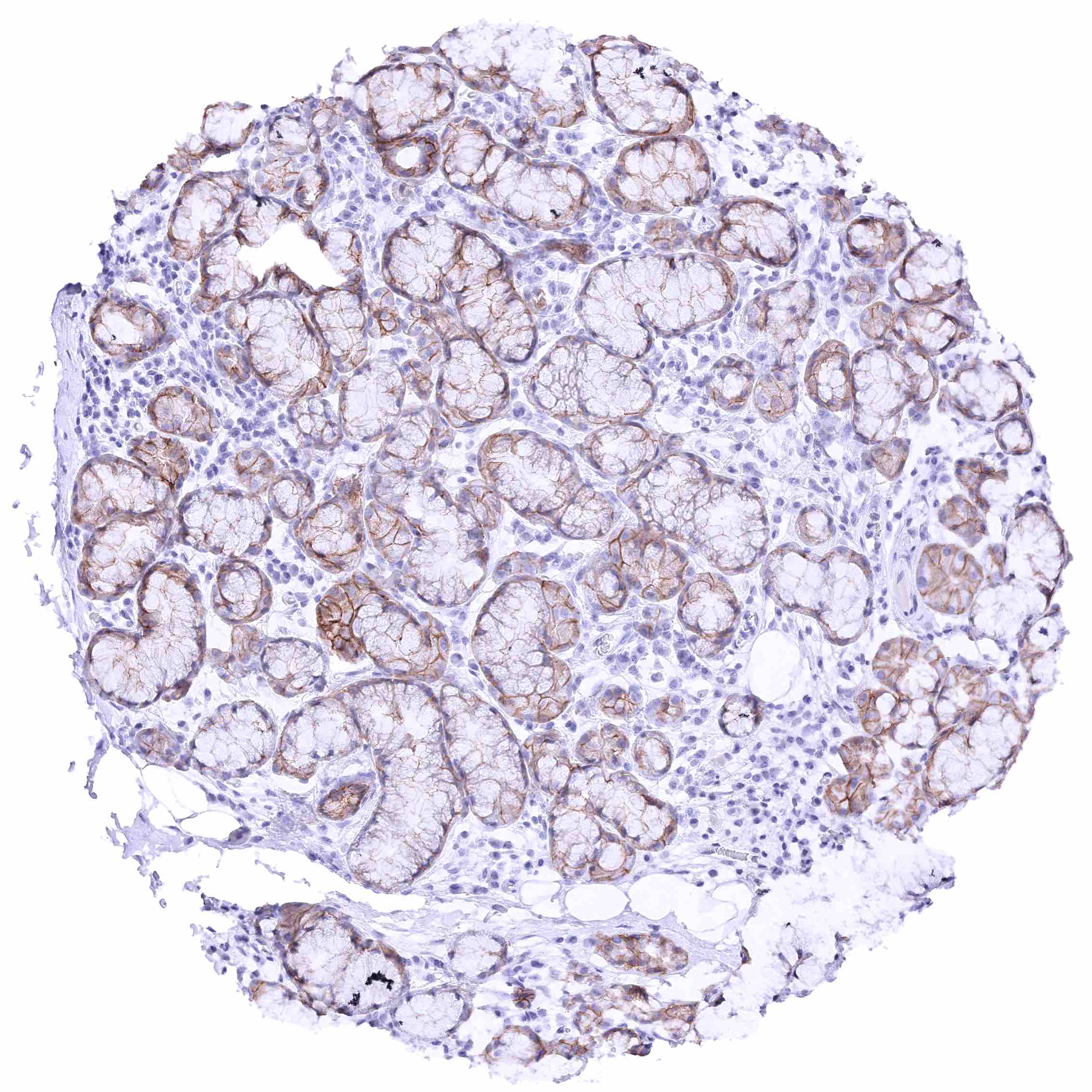
Stomach, antrum – Moderate membranous nectin-4 staining of surface epithelial cells (most prominent at the lateral membranes) while gastric glands show much less or absent staining.
Stomach, antrum.jpeg
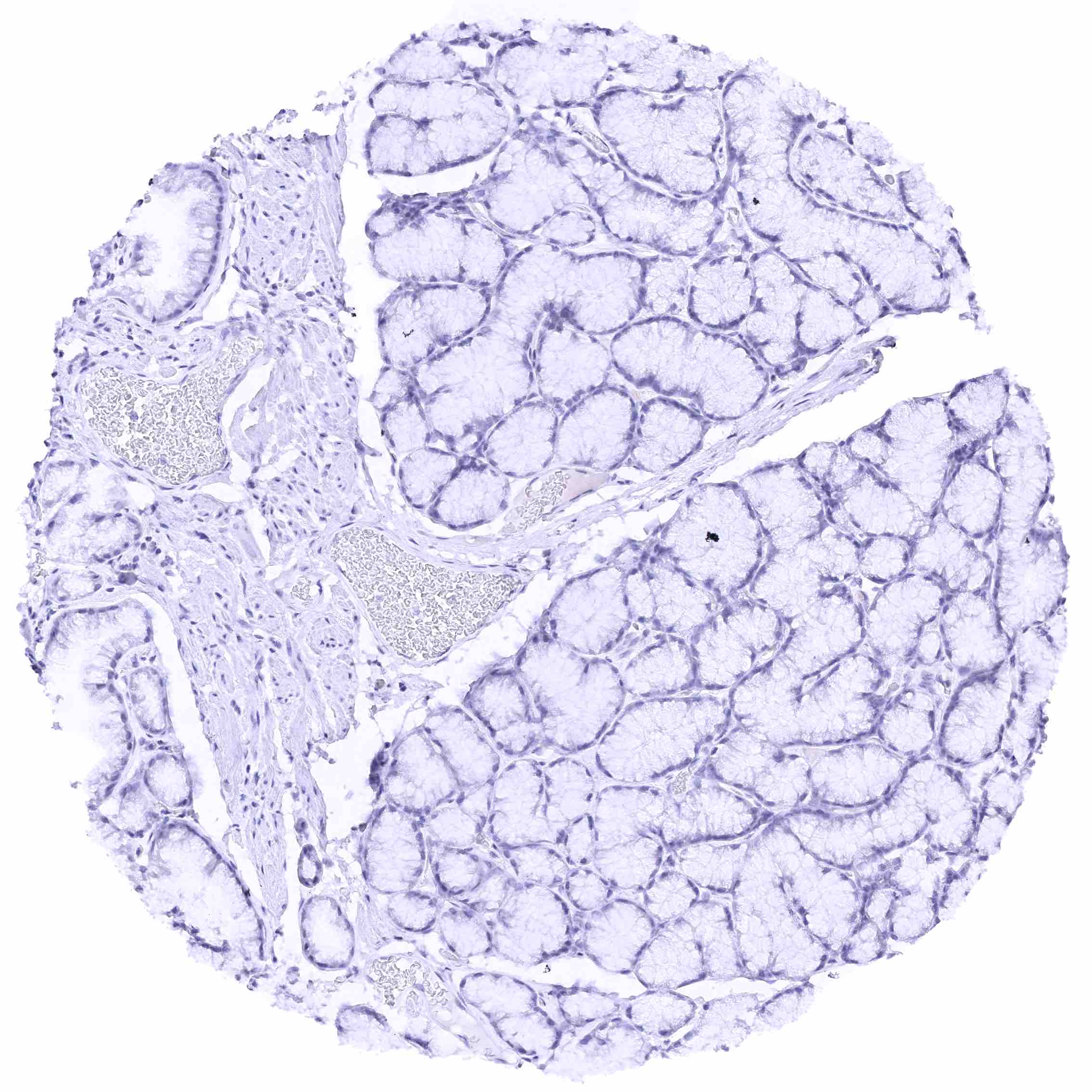
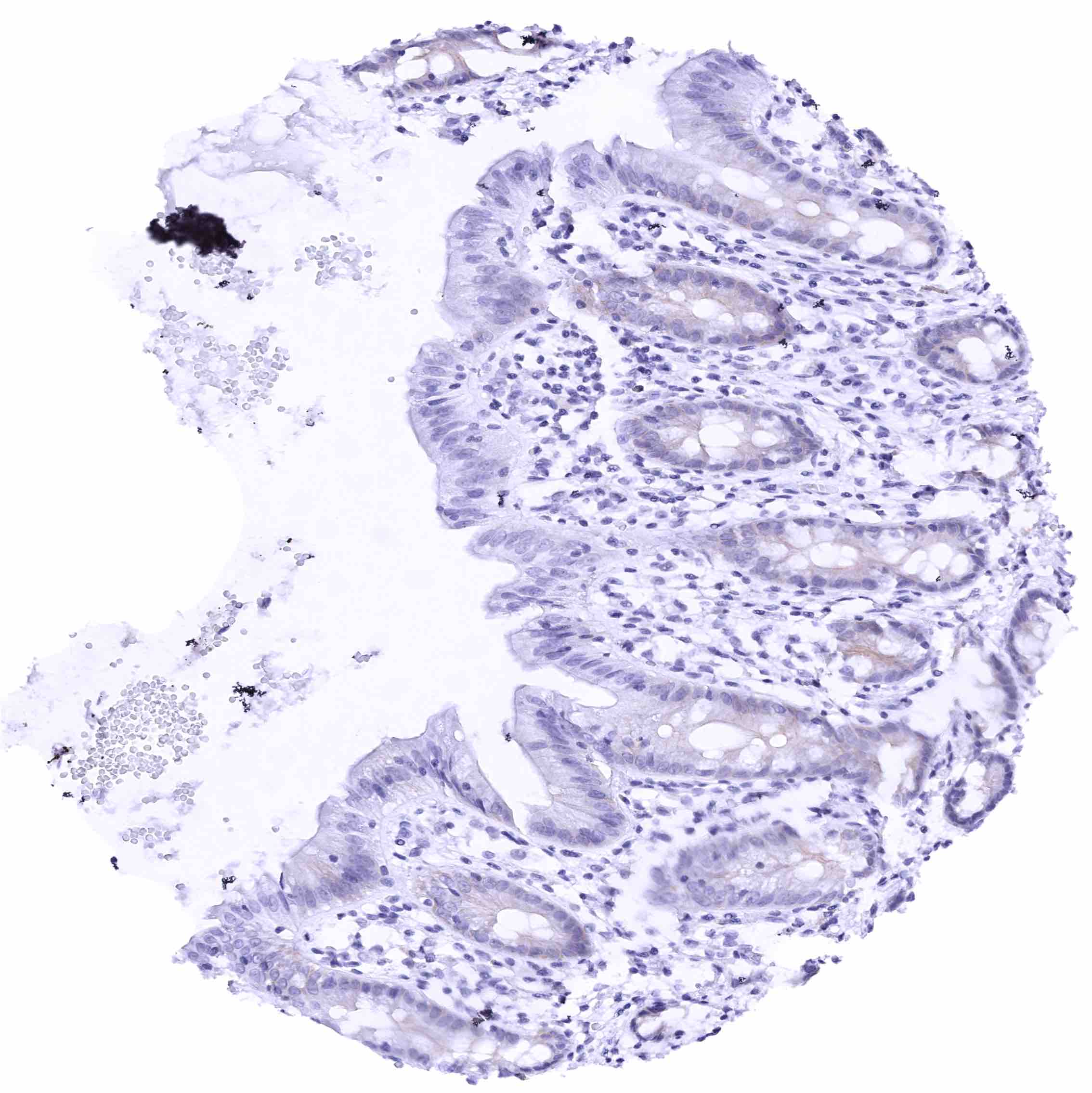
Stomach, corpus – Gastric glands are largely nectin-4 negative.
Stomach, muscular wall
Sublingual gland – Moderate (to strong) membranous nectin-4 staining of a large fraction of glandular and myoepithelial cells. .jpeg
Submandibular gland – Moderate membranous nectin-4 staining of subsets of excretory duct cells while staining of glandular cells is focal and rather weak.
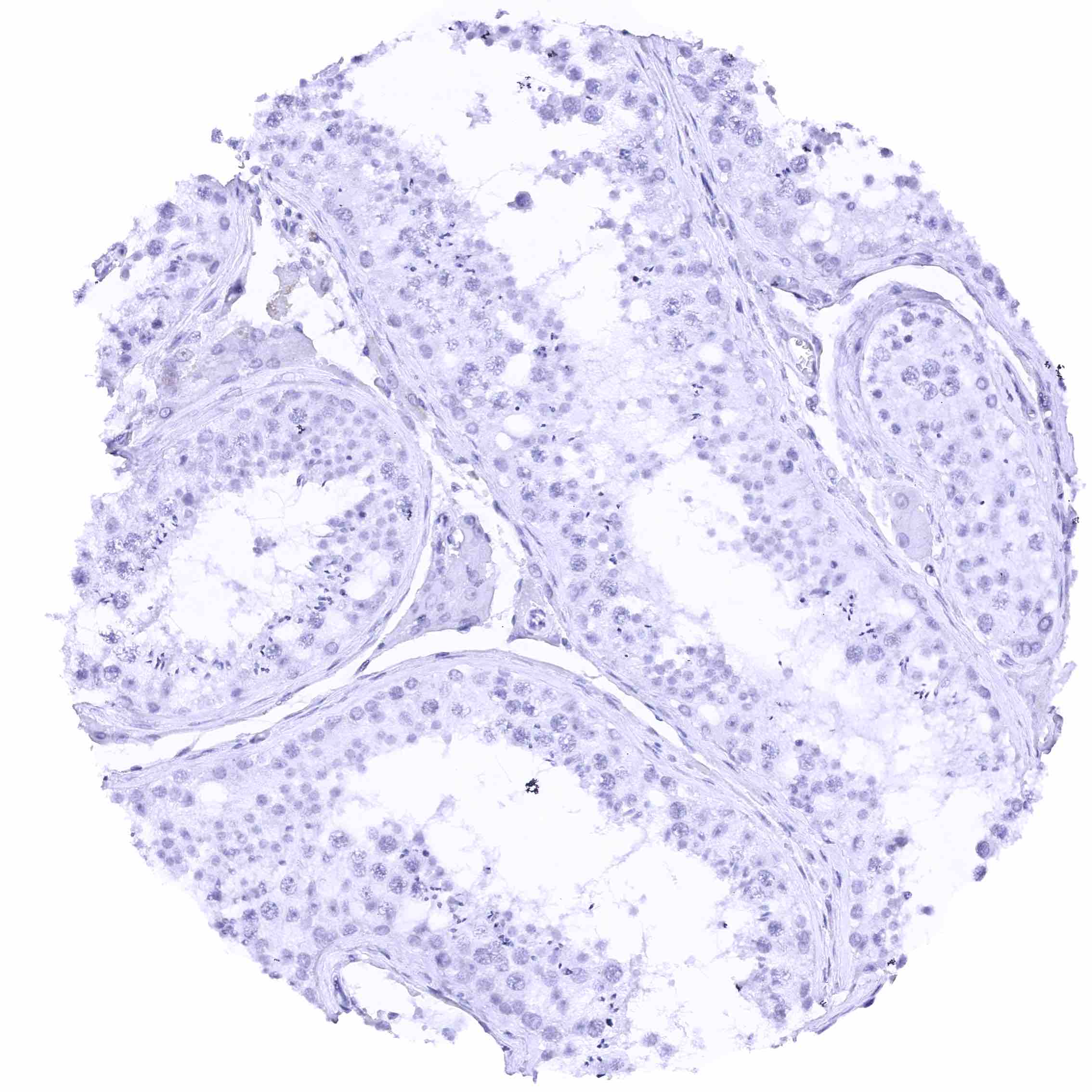
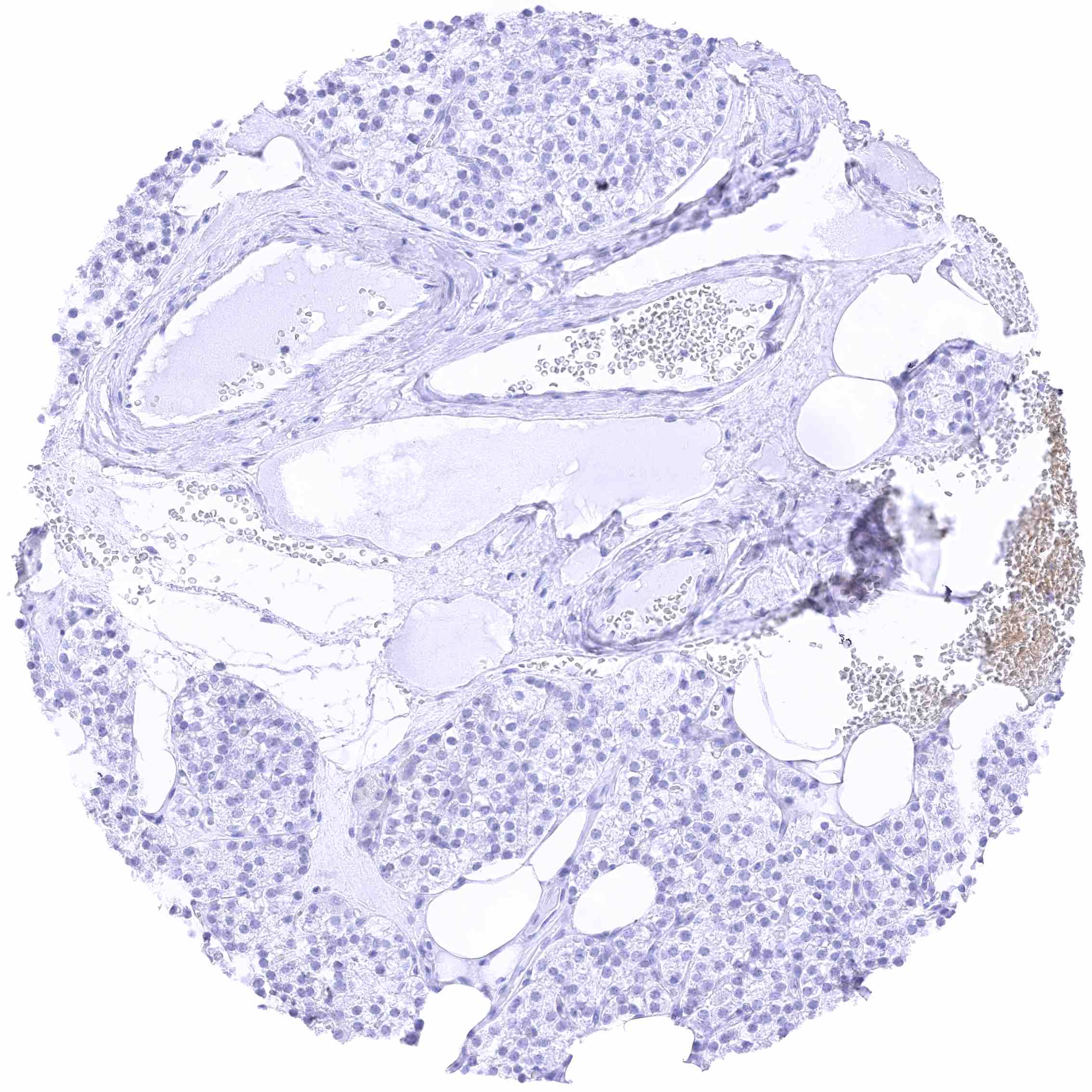
Testis
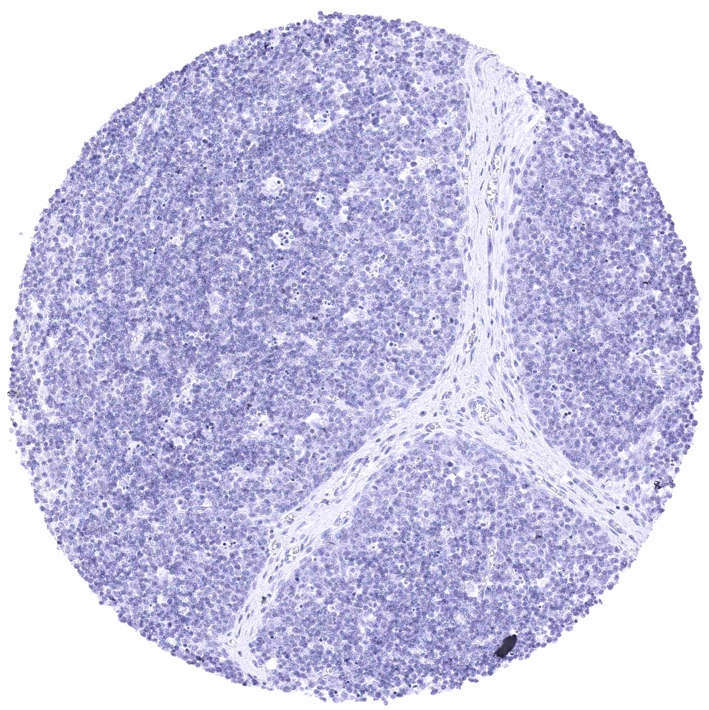
Thymus
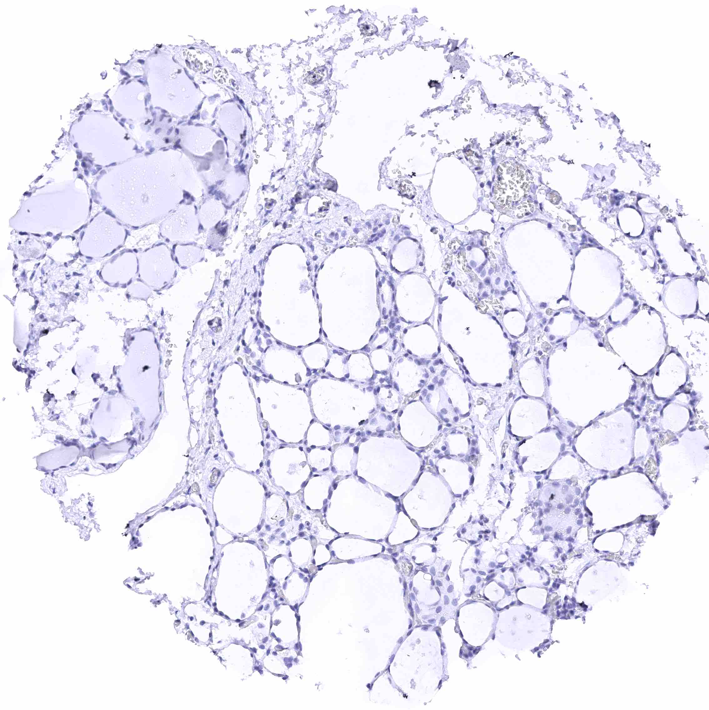
Thyroid gland
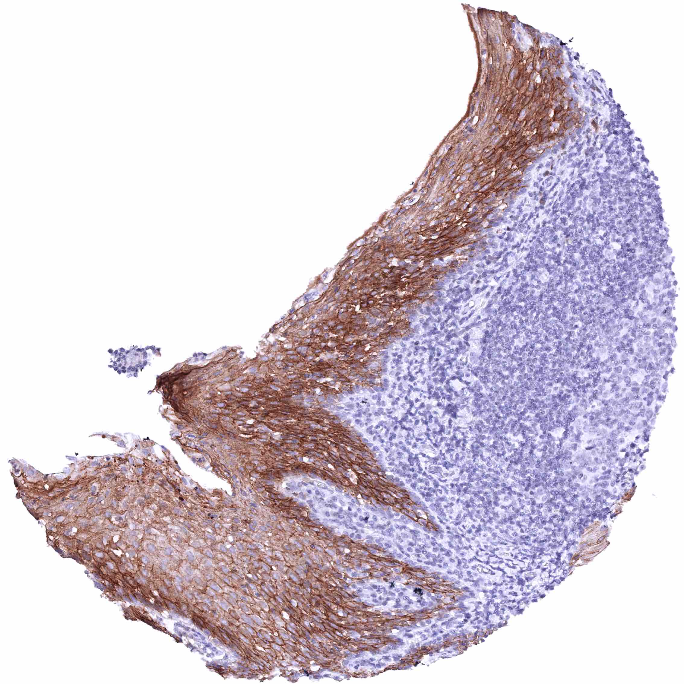
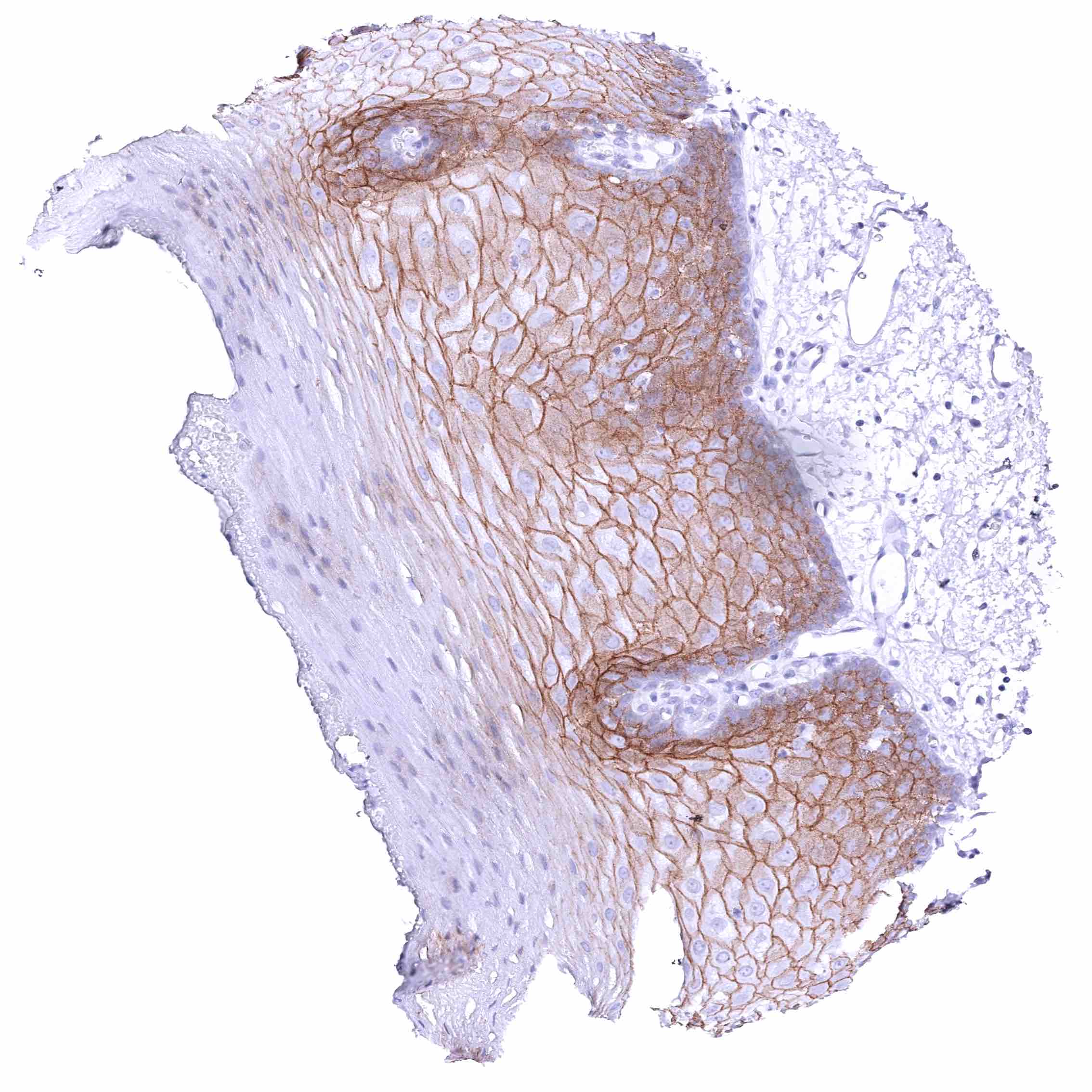
Tonsil, surface epithelium – Strong membranous nectin-4 staining of squamous epithelium. Staining is most intense in the suprabasal cell layers while the staining intensity decreases gradually towards the surface. Basal cells (and lymphocytes) are nectin-4 negative.
Urinary bladder, muscular wall
Urinary bladder, urothelium – Strong membranous nectin-4 staining of most urothelial cells. Only cells of the basal cell layer are always nectin-4 negative.
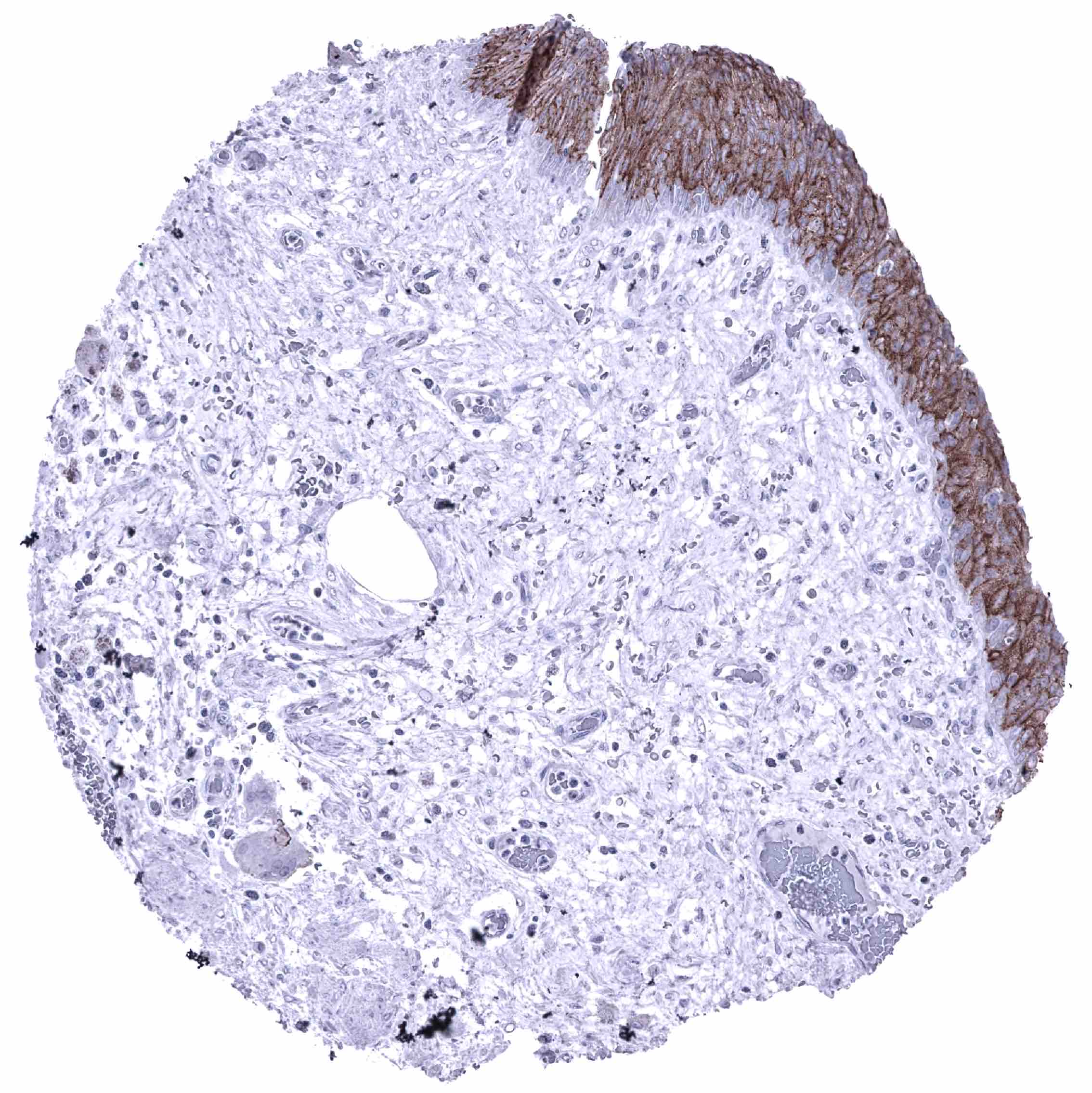
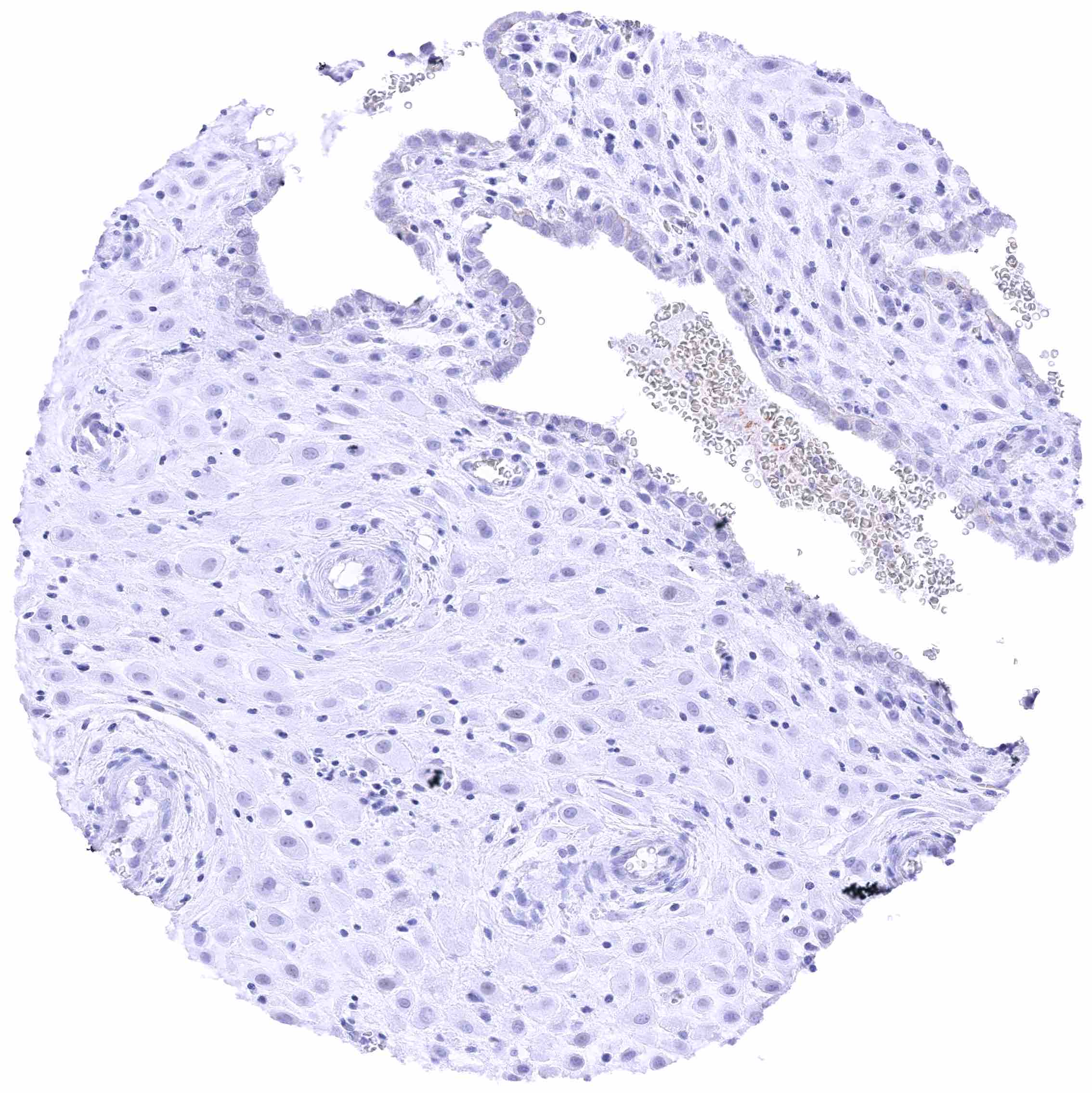
Uterus, ectocervix – Nectin-4 staining of squamous epithelium is most intense in the suprabasal cell layers while the staining intensity decreases gradually towards the superficial cell layers. Basal cells are nectin-4 negative.
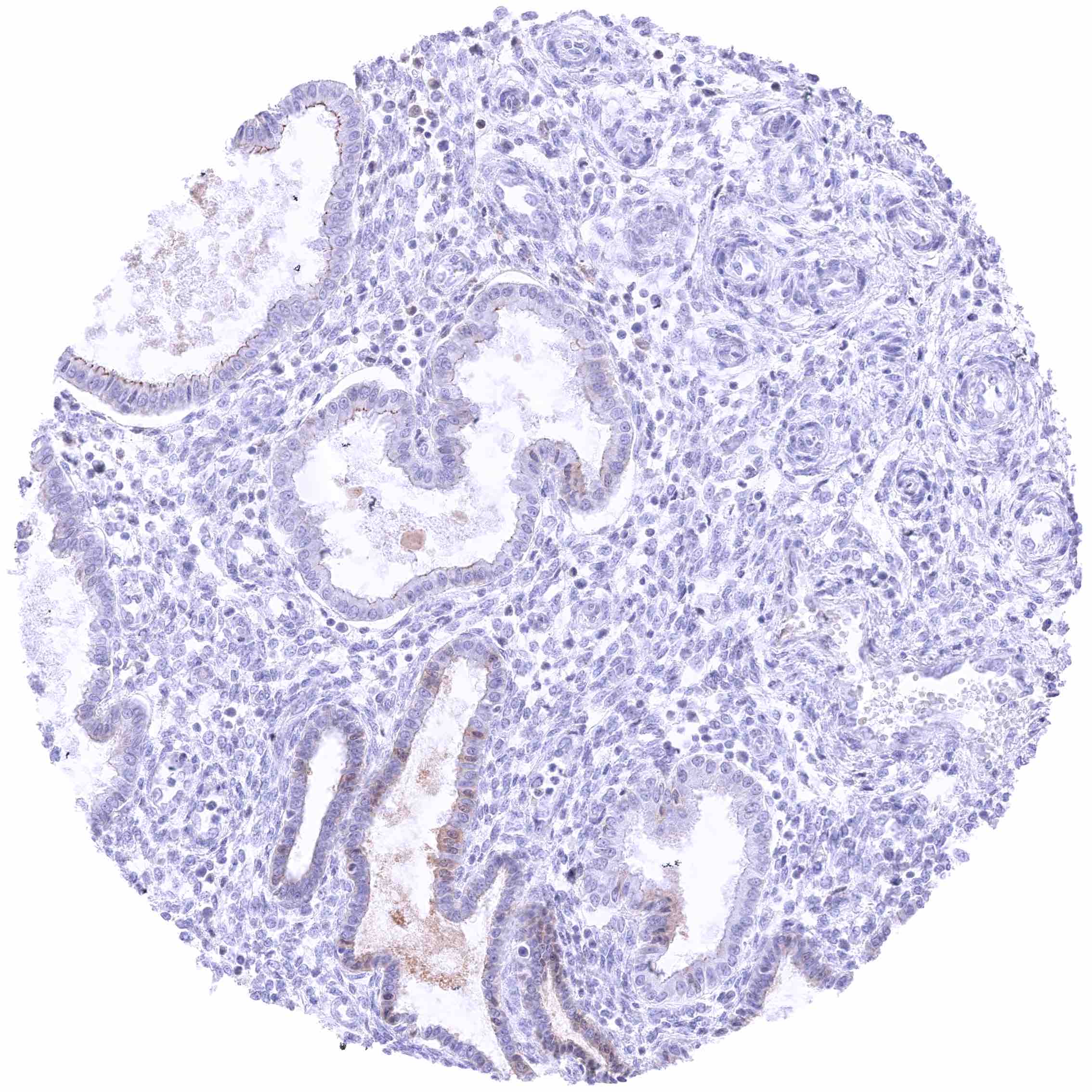
Uterus, endocervix – Distinct membranous, baso-lateral nectin-4 staining of endocervical glands.
Uterus, endometrium (pregnancy)
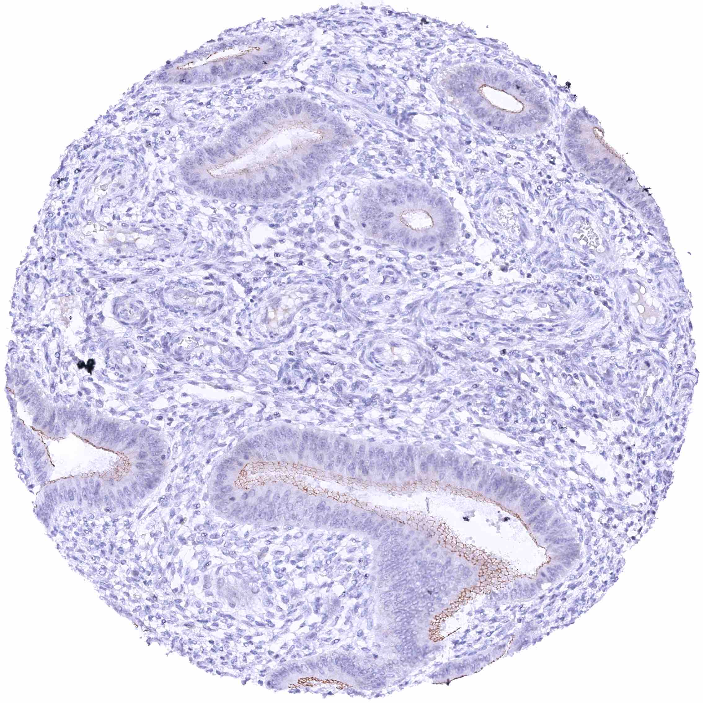
Uterus, endometrium (proliferation) – Weak to moderate nectin-4 staining of apical membranes of endometrial glands.
Uterus, endometrium (secretion) – Weak to moderate nectin-4 staining of apical membranes of endometrial glands.