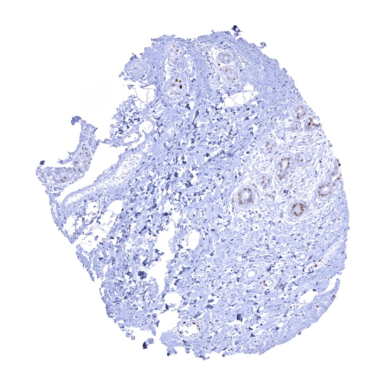
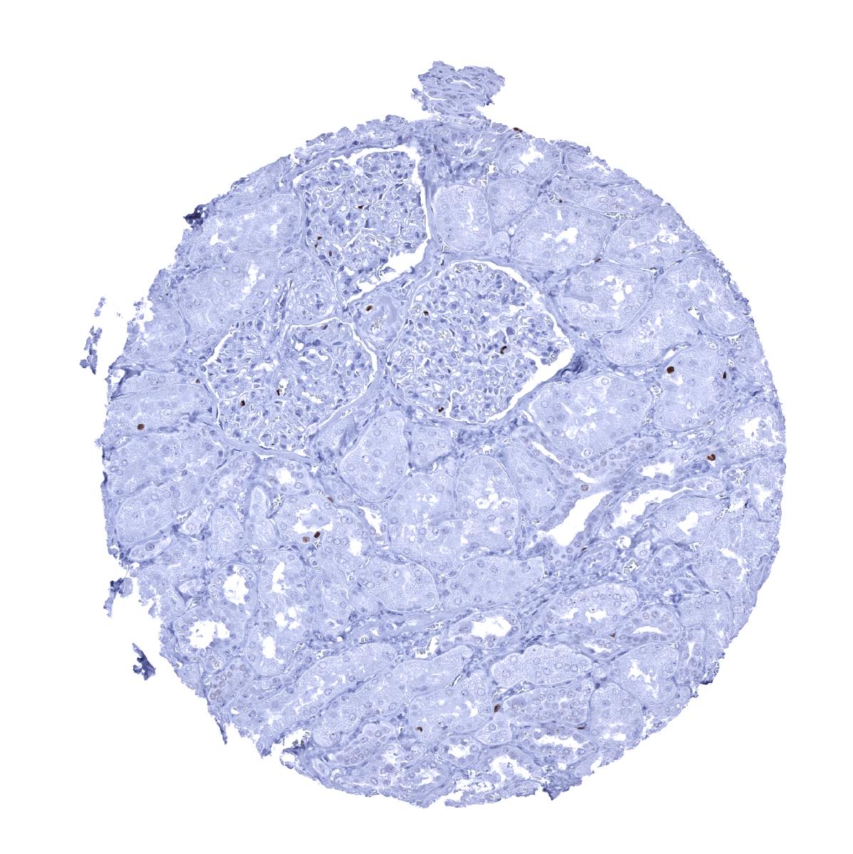
Adrenal gland - A variable MCM7 staining occurs in a small fraction of adrenocortical cells
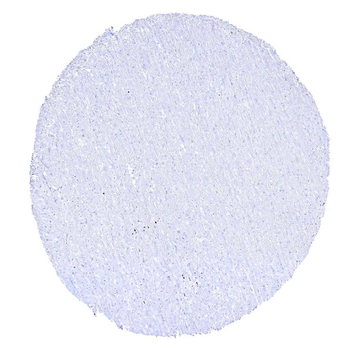
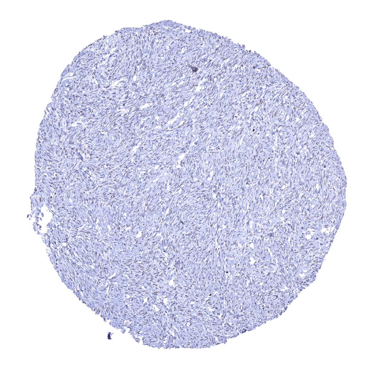
Aorta, media
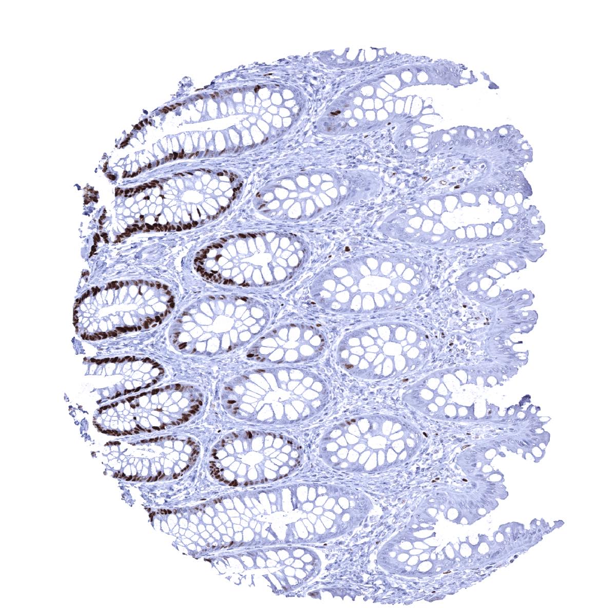
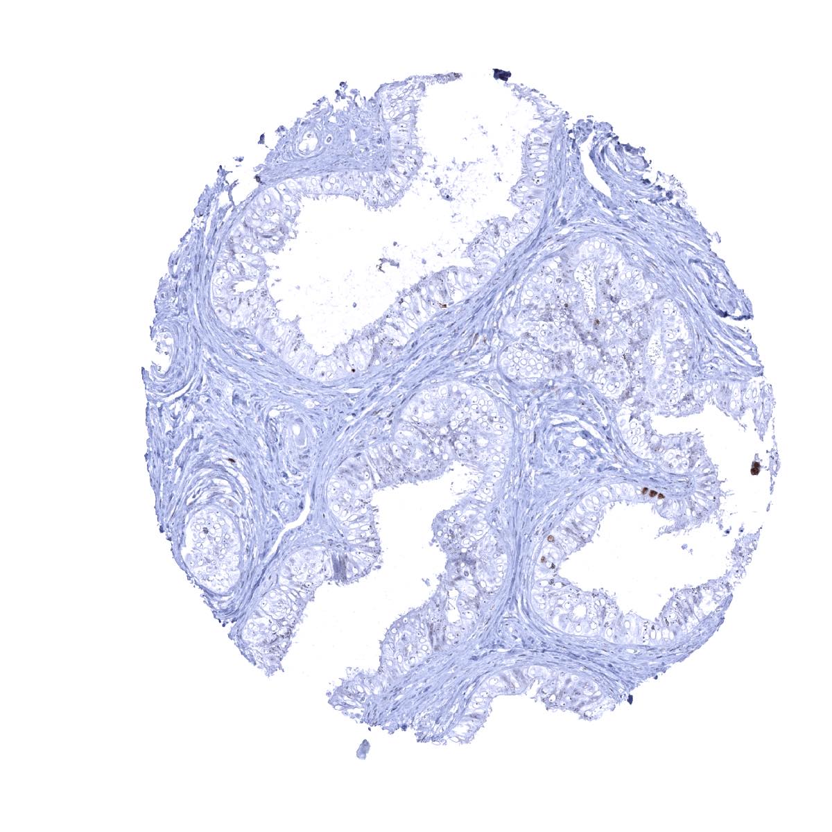
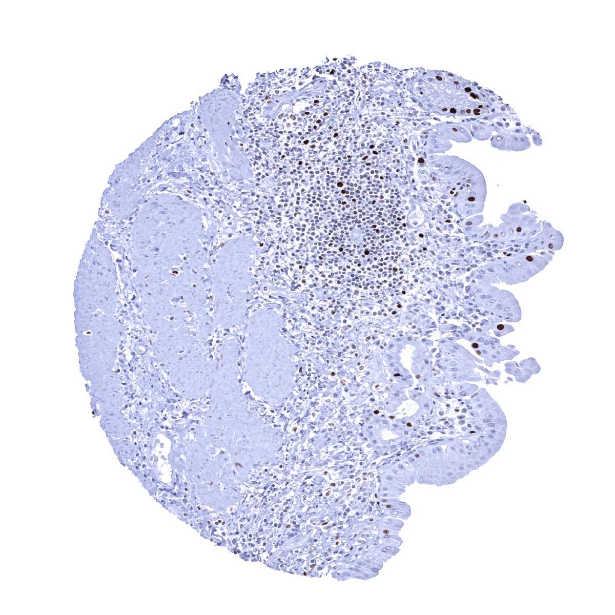
Appendix, mucosa - MCM7 immunostaining predominates in in epithelial cells of the crypts. Some lymphocytes are also positive
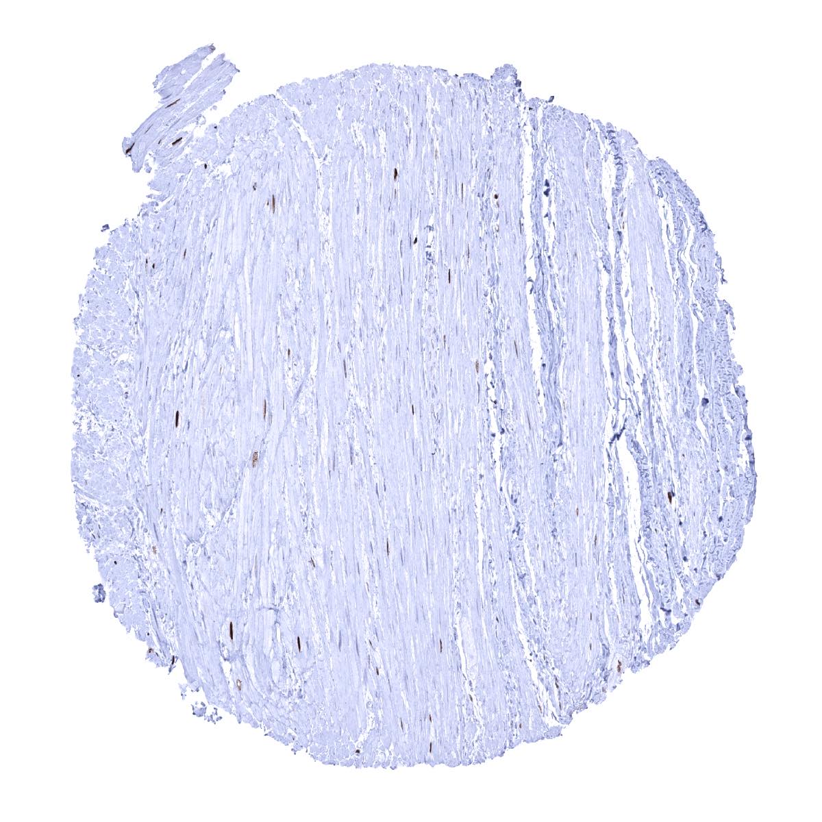
Appendix, muscular wall
Breast - A weak to moderate MCM7 staining occurs in many luminal cells
Bronchus, mucosa - MCM7 staining occurs in a fraction of (mostly basal) respiratory epithelial cells
Cerebellum (molecular layer, Purkinje cell layer, granule cell layer)

Cerebellum (white matter)
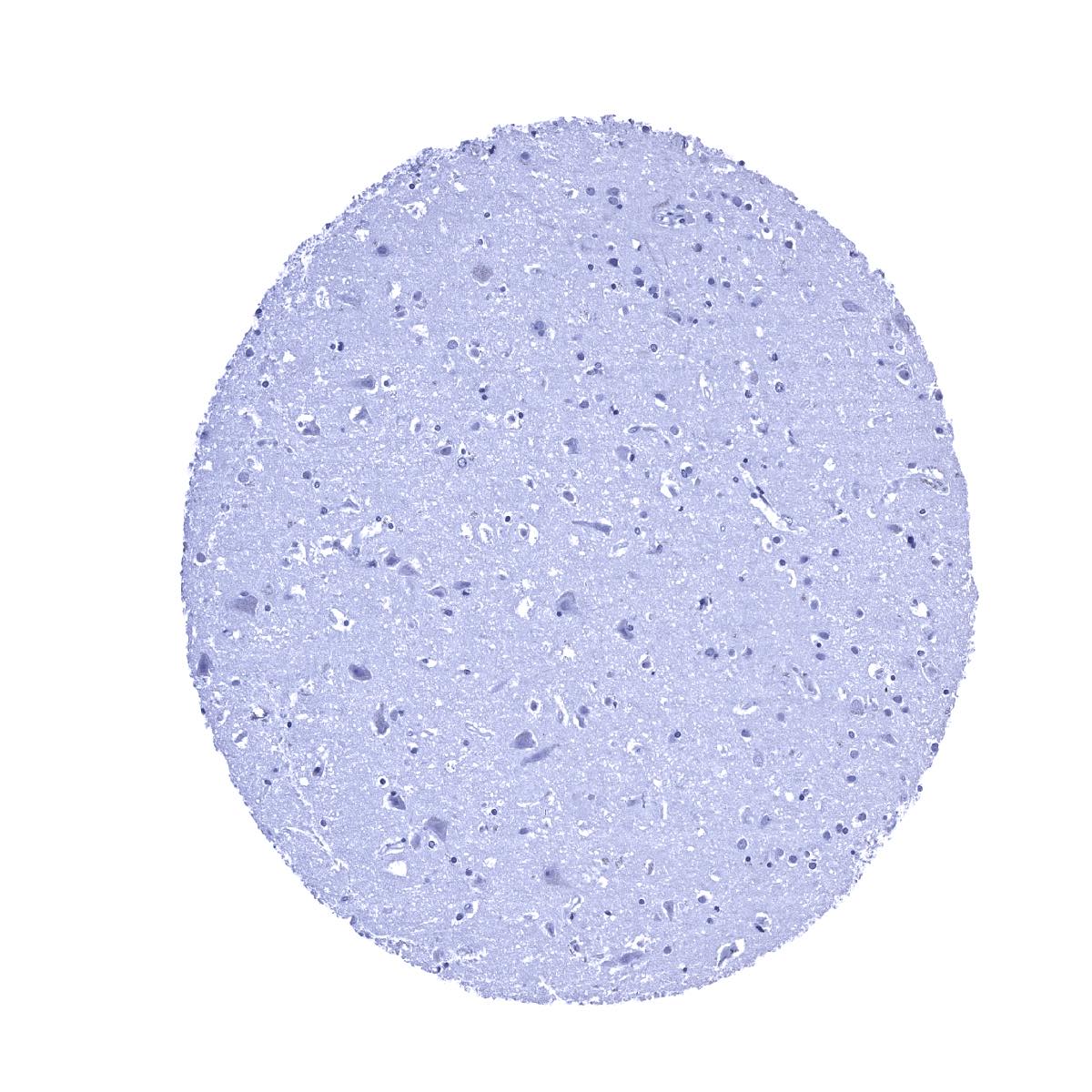
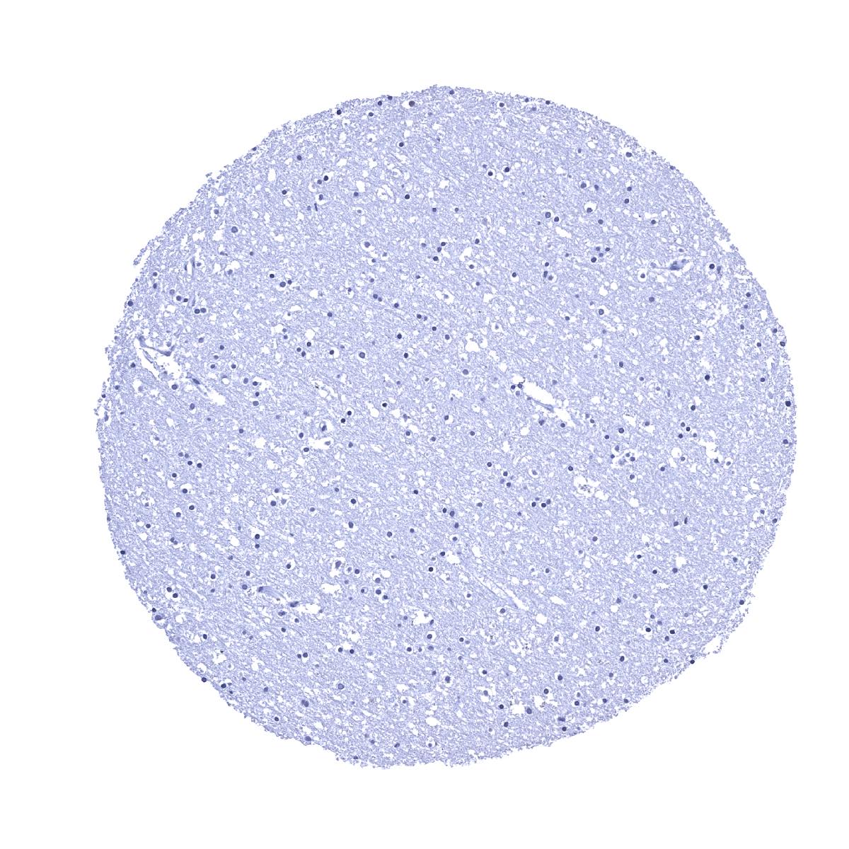
Cerebrum, grey matter
Cerebrum, white matter

Colon descendens, muscular wall
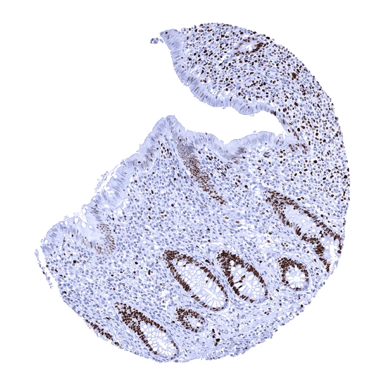
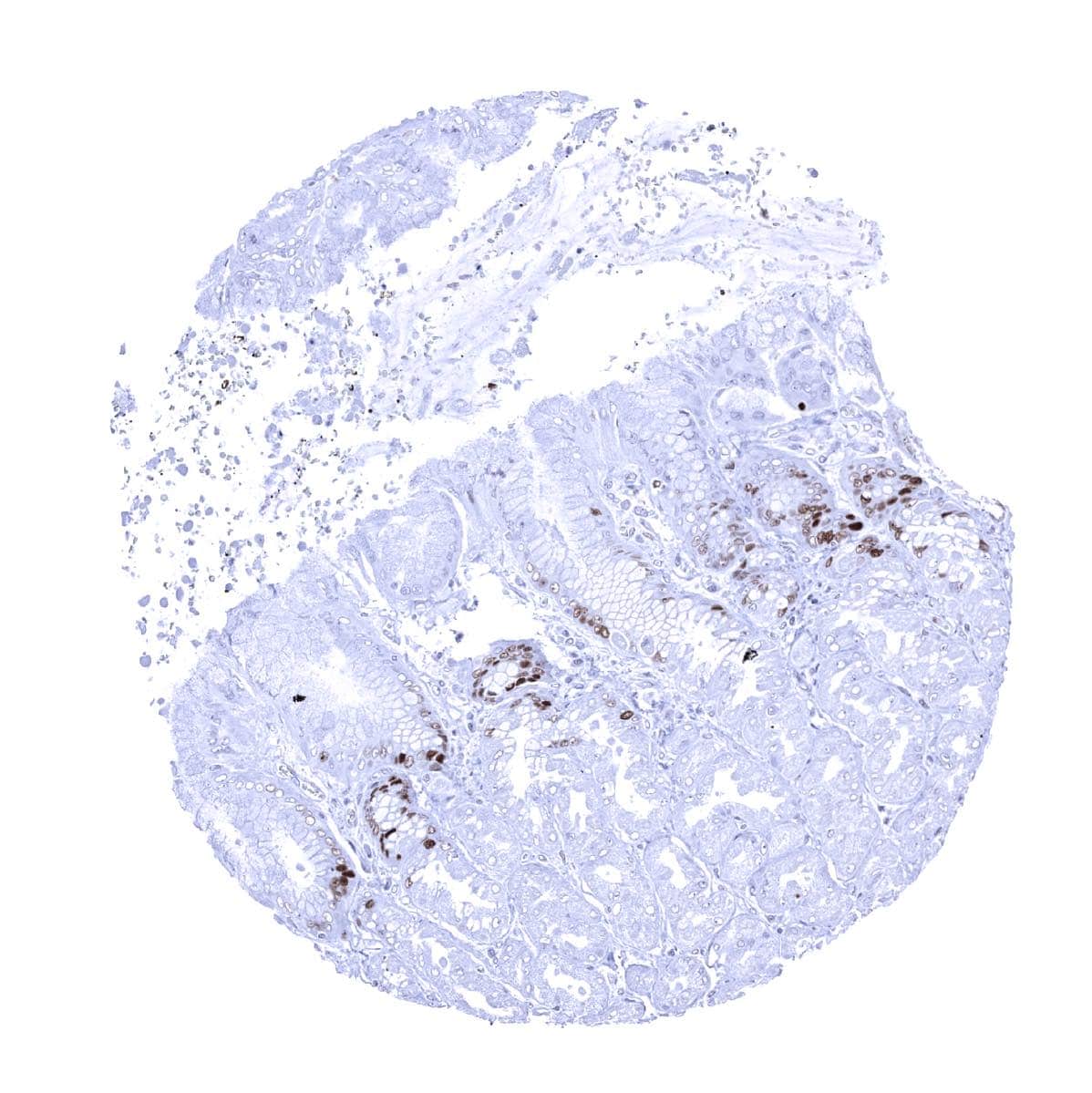
Colon, descendes - MCM7 staining predominates in epithelial cells of the crypts
Duodenum, Brunner gland
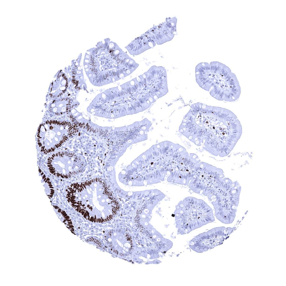
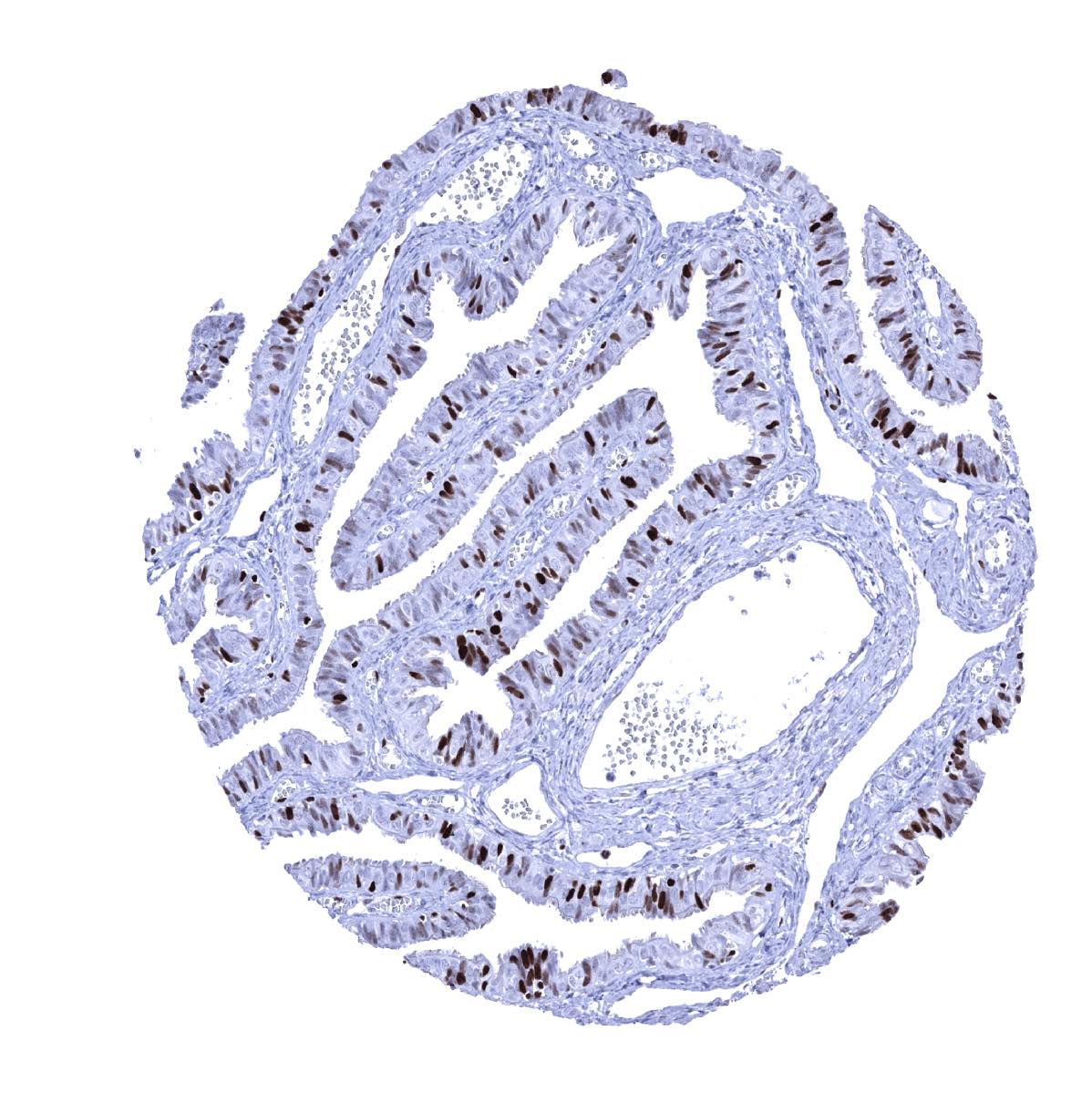
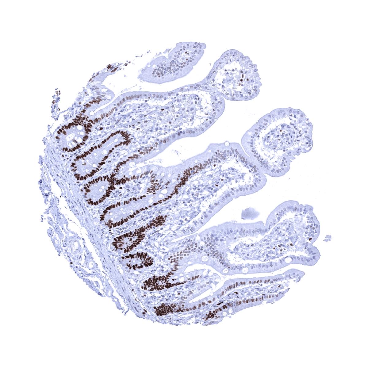
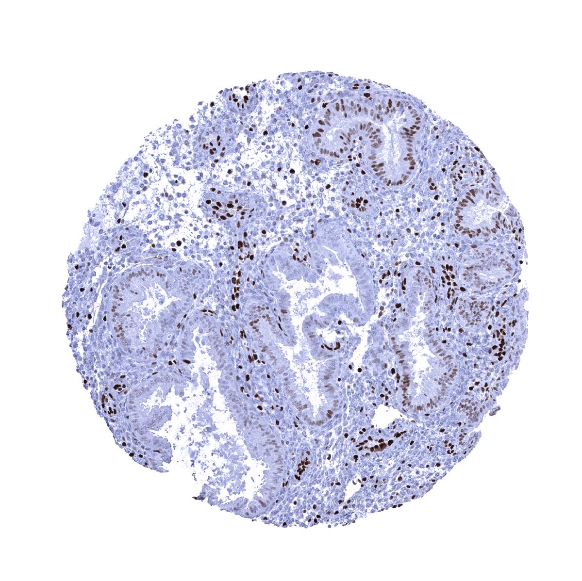
Duodenum, mucosa - MCM7 immunostaining predominates in epithelial cells of the crypts
Epididymis - Only few cells show a weak to moderate MCM7 staining
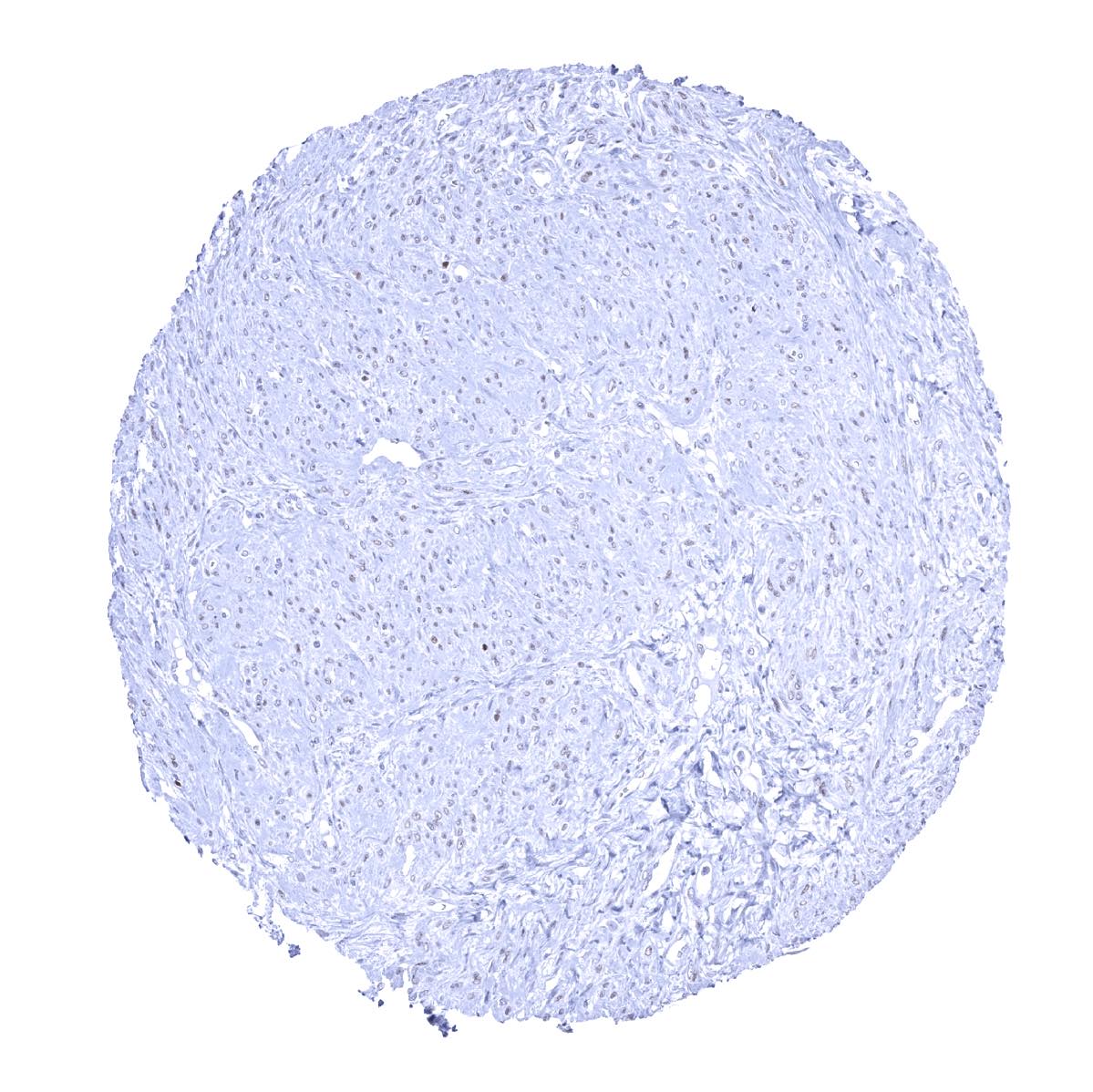
Esophagus, muscular wall - Faint MCM7 immunostaining in some smooth muscle cells
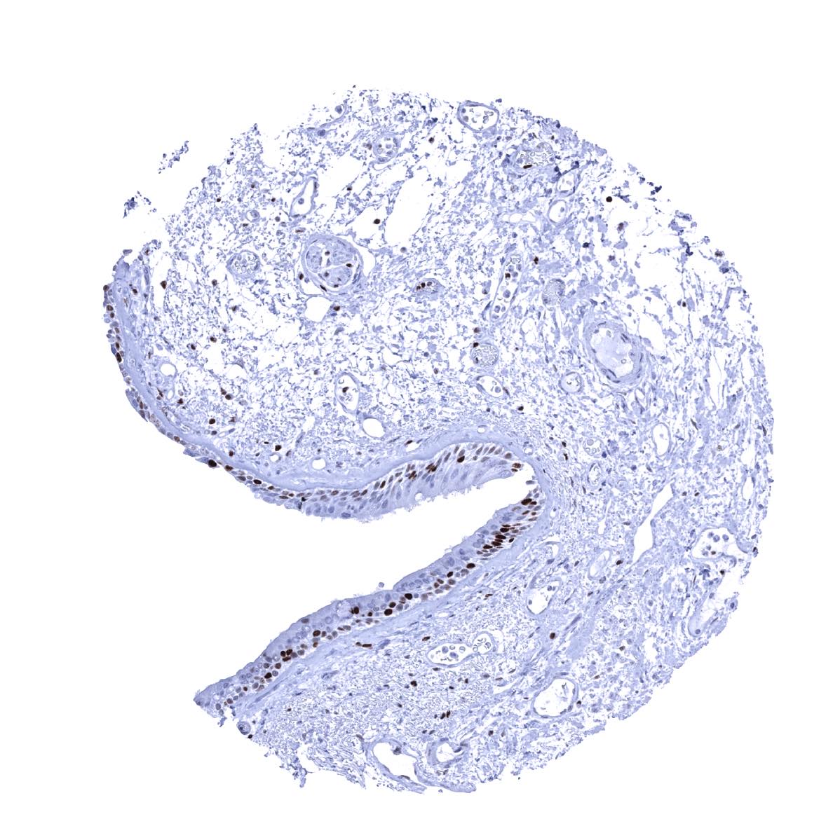
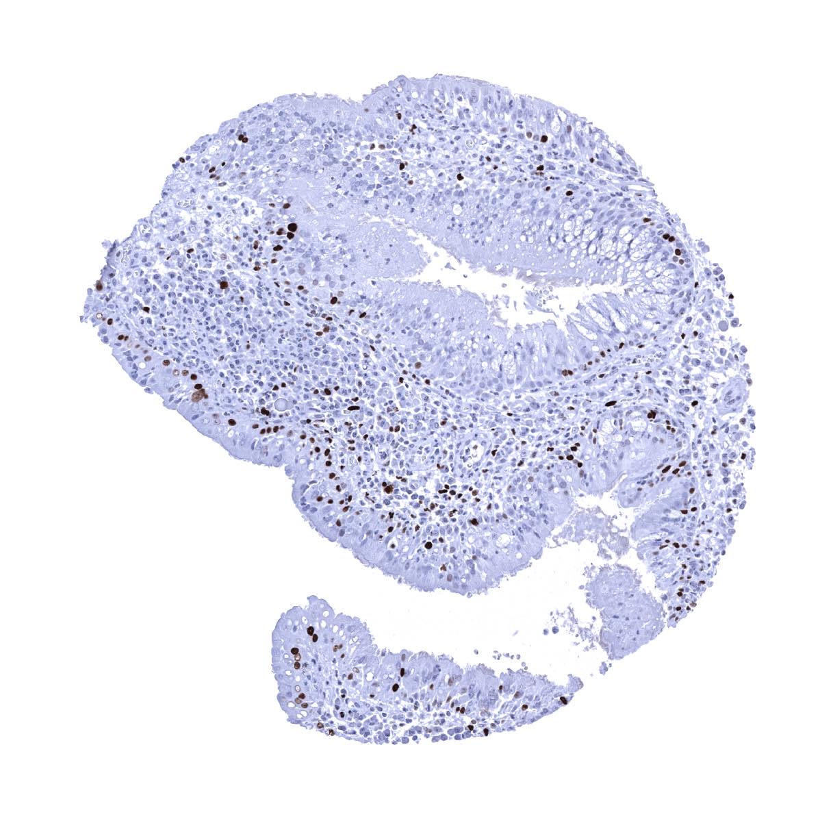
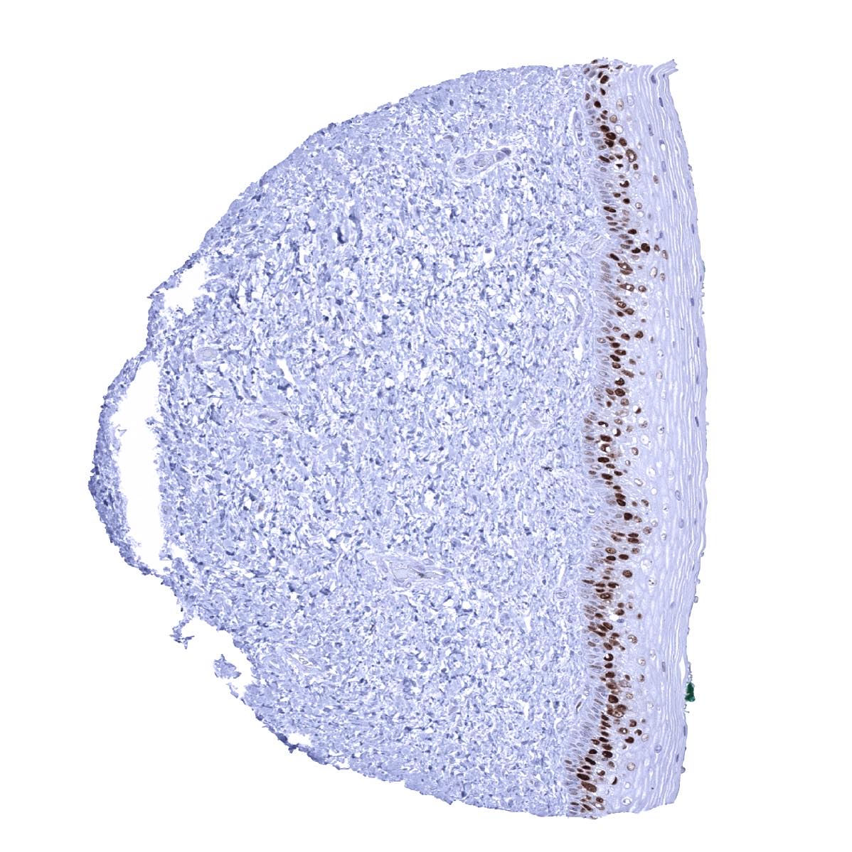
Esophagus, squamous epithelium - Esophageal squamous epithelium shows a moderate to strong MCM7 staining of suprabasal and basal cells
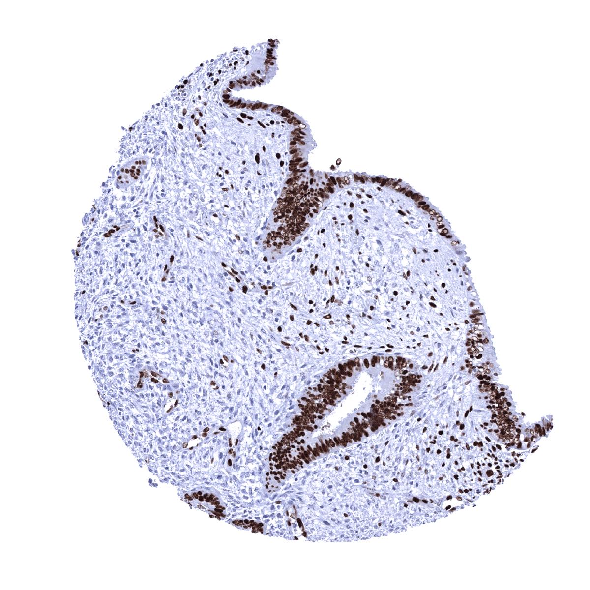
Fallopian tube, mucosa - MCM7 staining in a significant fraction of epithelial cells
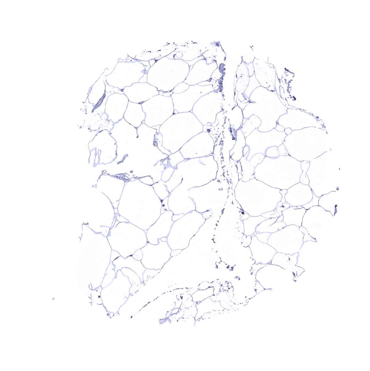
Fat
Gallbladder, epithelium - Few MCM7 positive cells occur in the gallbladder epithelium
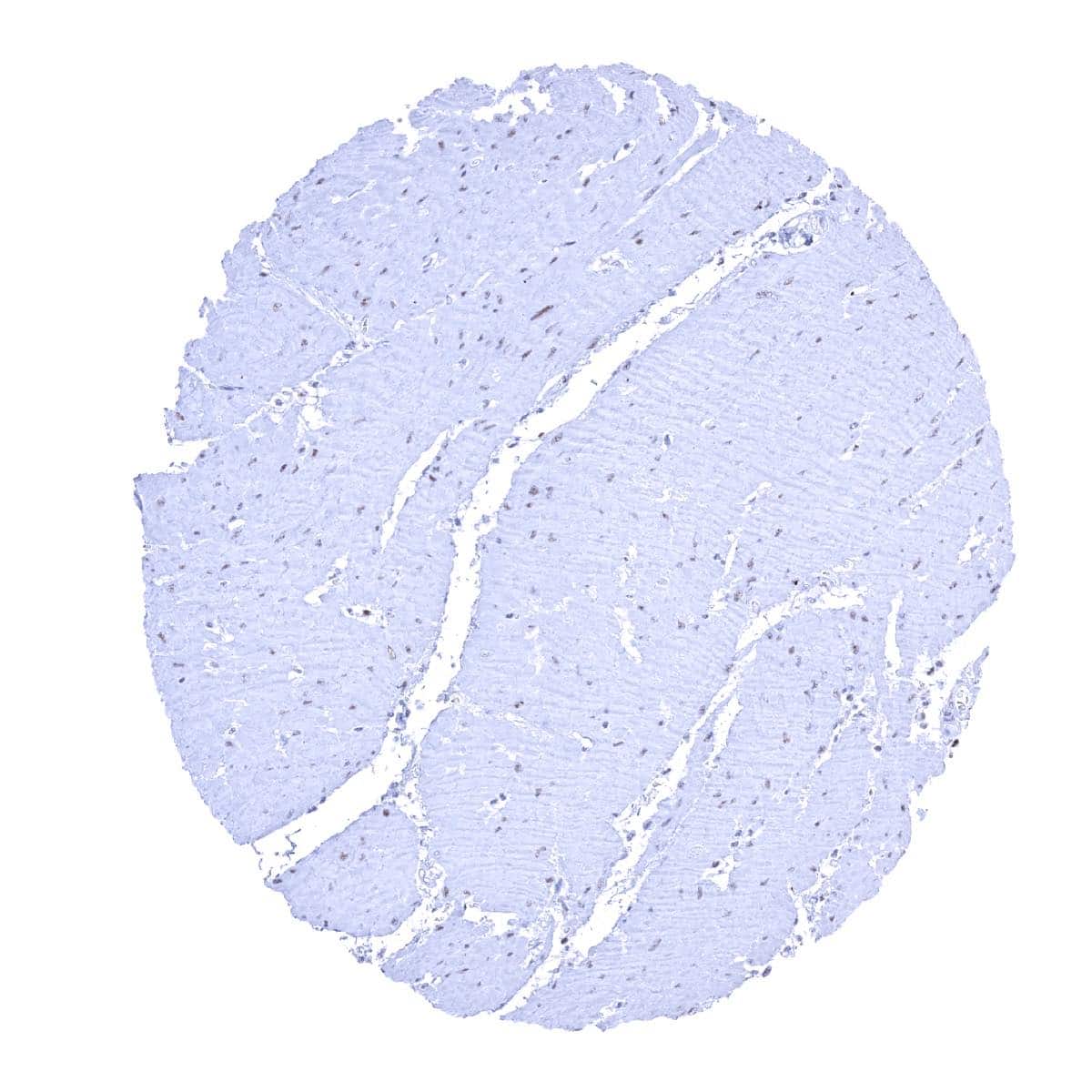
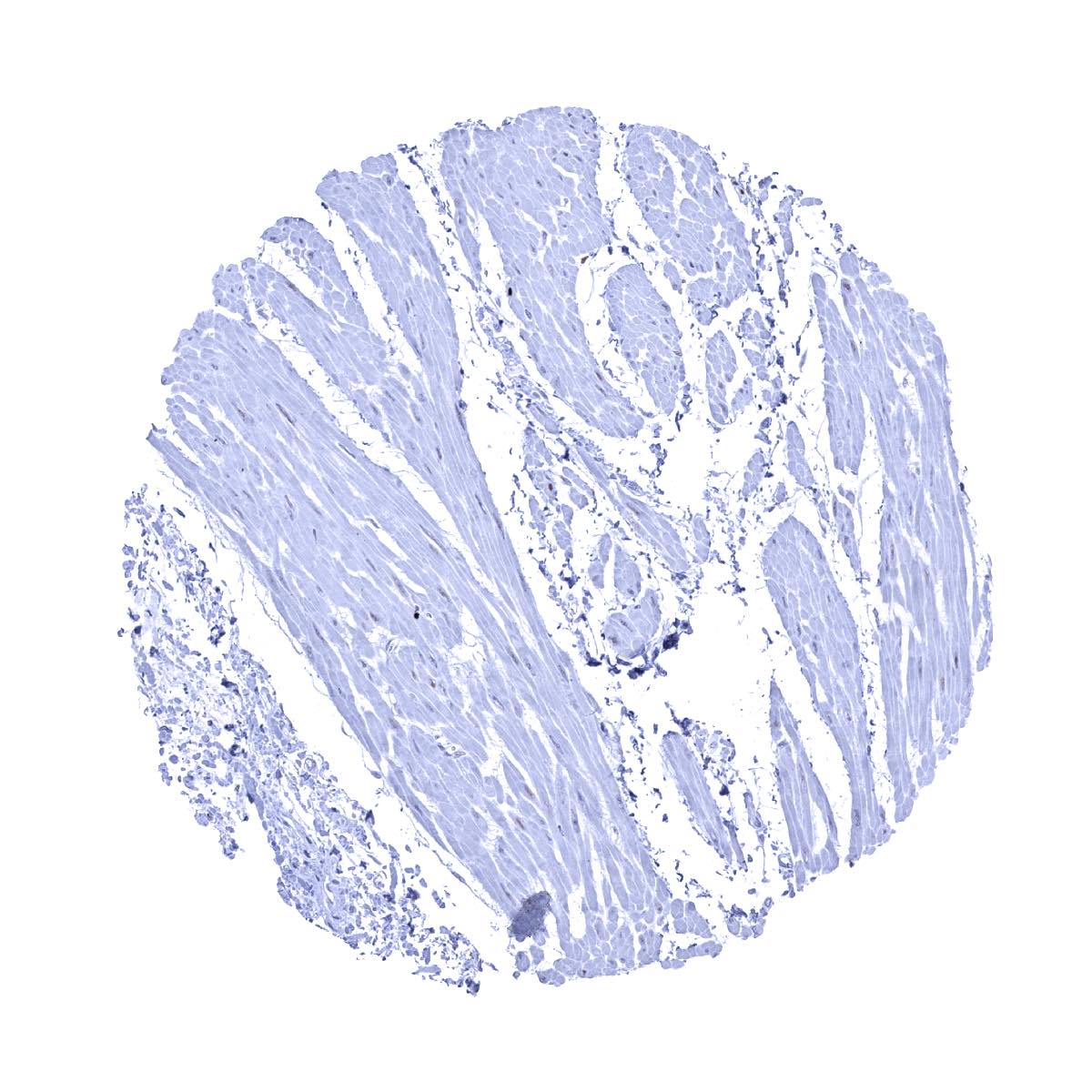
Heart muscle - Faint MCM7 staining in some muscle cells
Ileum, mucosa - MCM7 staining predominates in epithelial cells of the crypts
Kidney, cortex - Only few cells are MCM7 positive
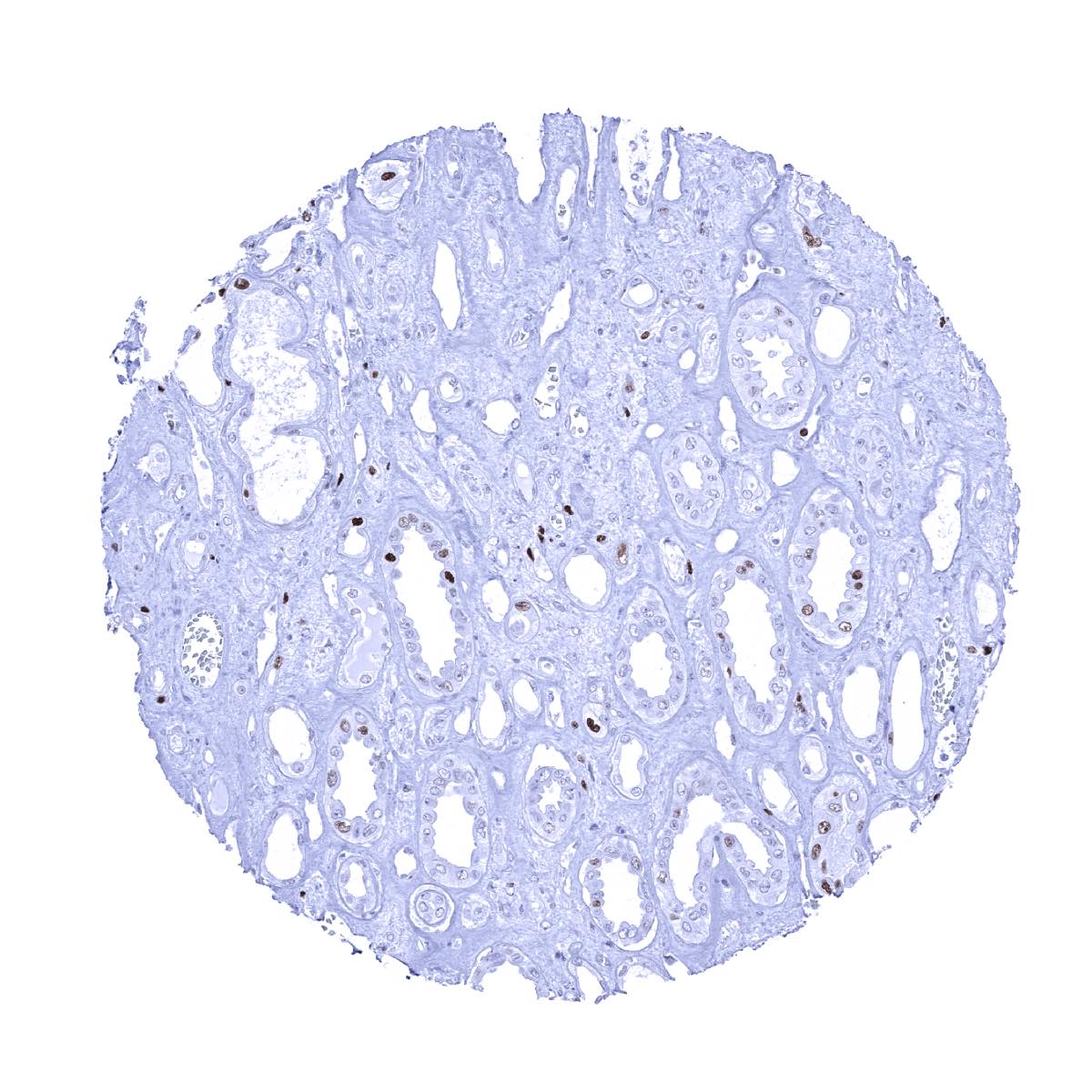
Kidney, medulla
Kidney, pelvis (muscular wall) - Faint MCM7 immunostaining in some smooth muscle cells
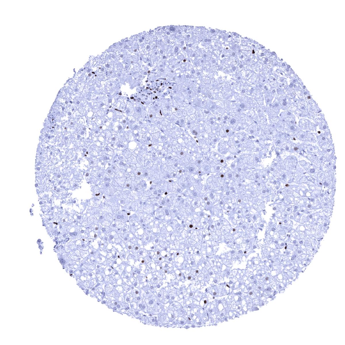
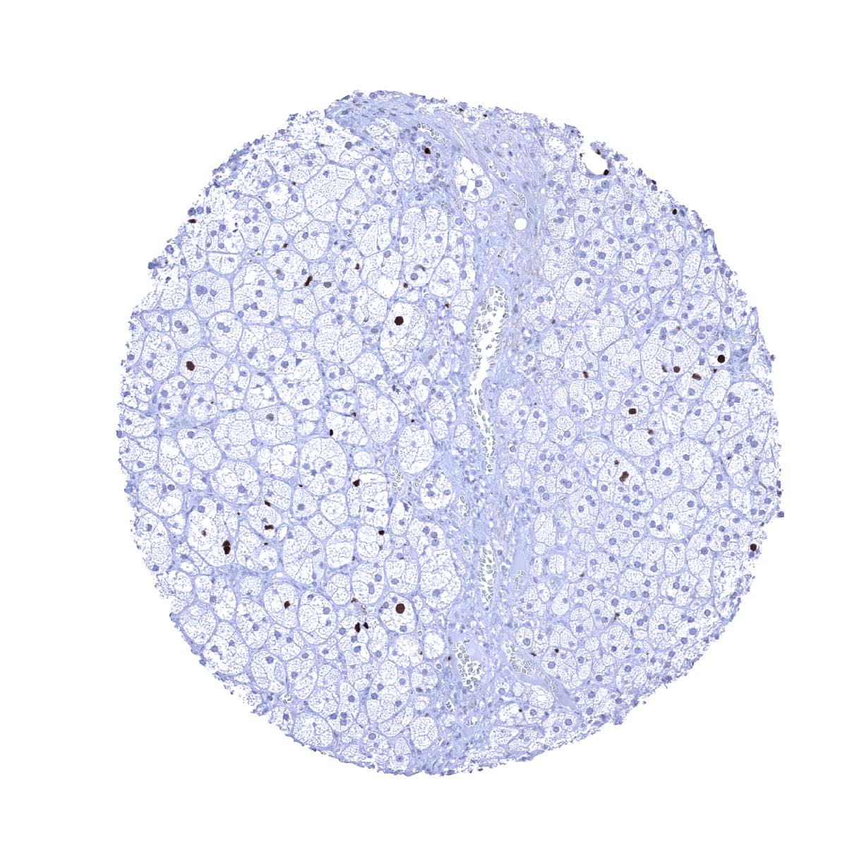
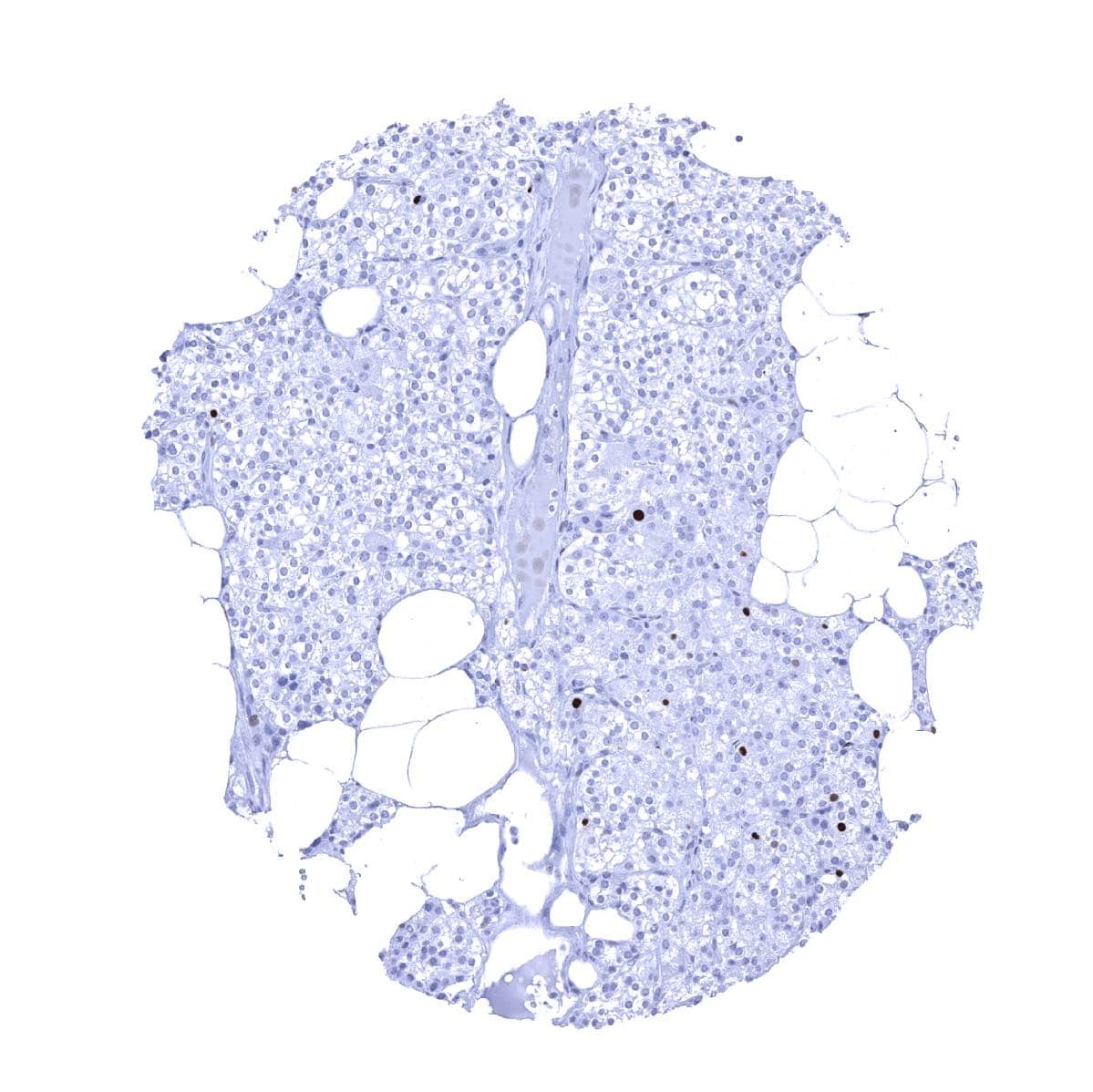
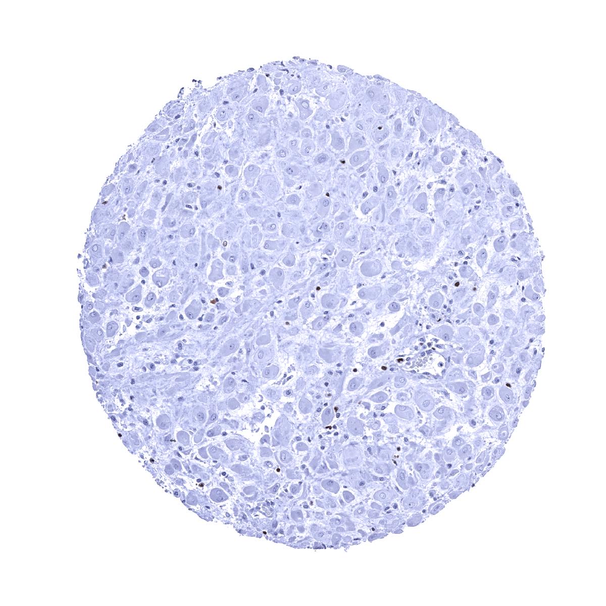
Liver - MCM7 staining of variable intensity in a small fraction of hepatocytes
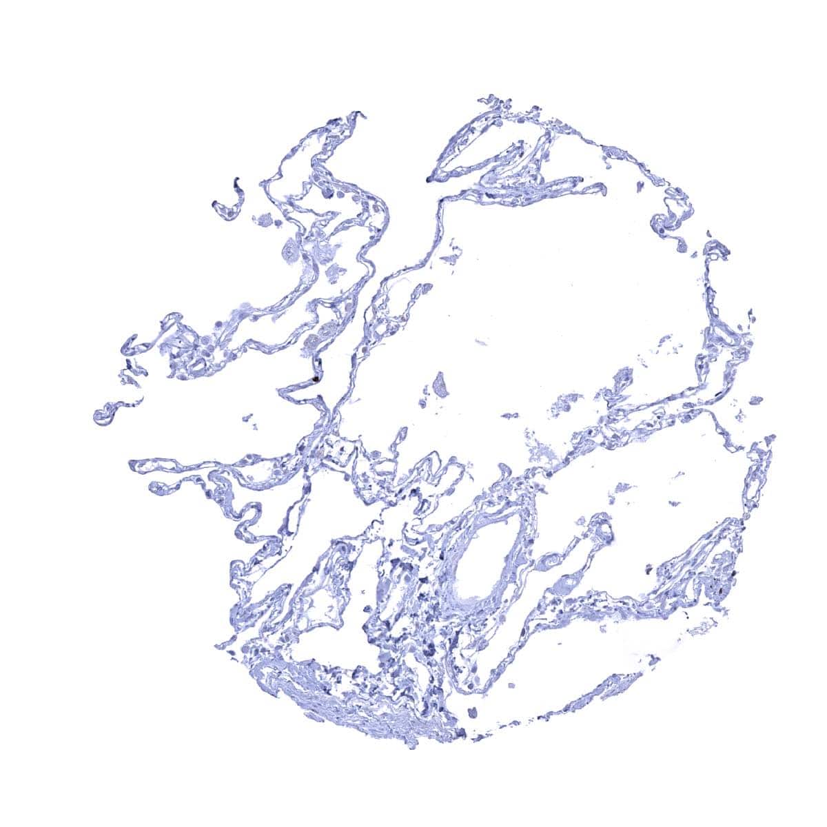
Lung
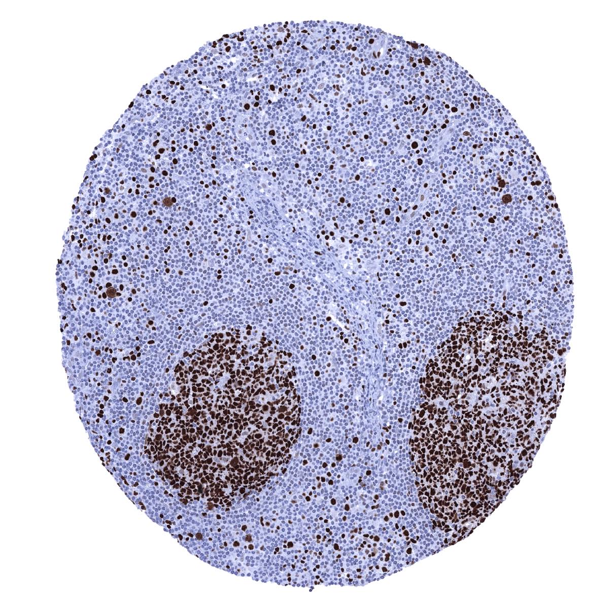
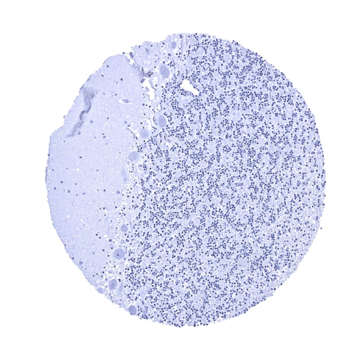
Lymph node - Many lymphocytes are MCM7 positive. A particularly strong MCM7 staining occurs in most cells of germinal centres and in scattered individual cells of the interfollicular zone
Ovary, stroma
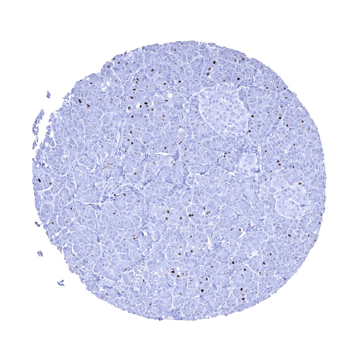
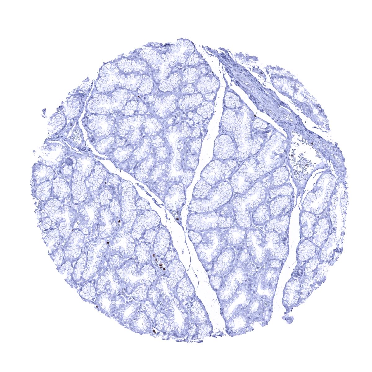
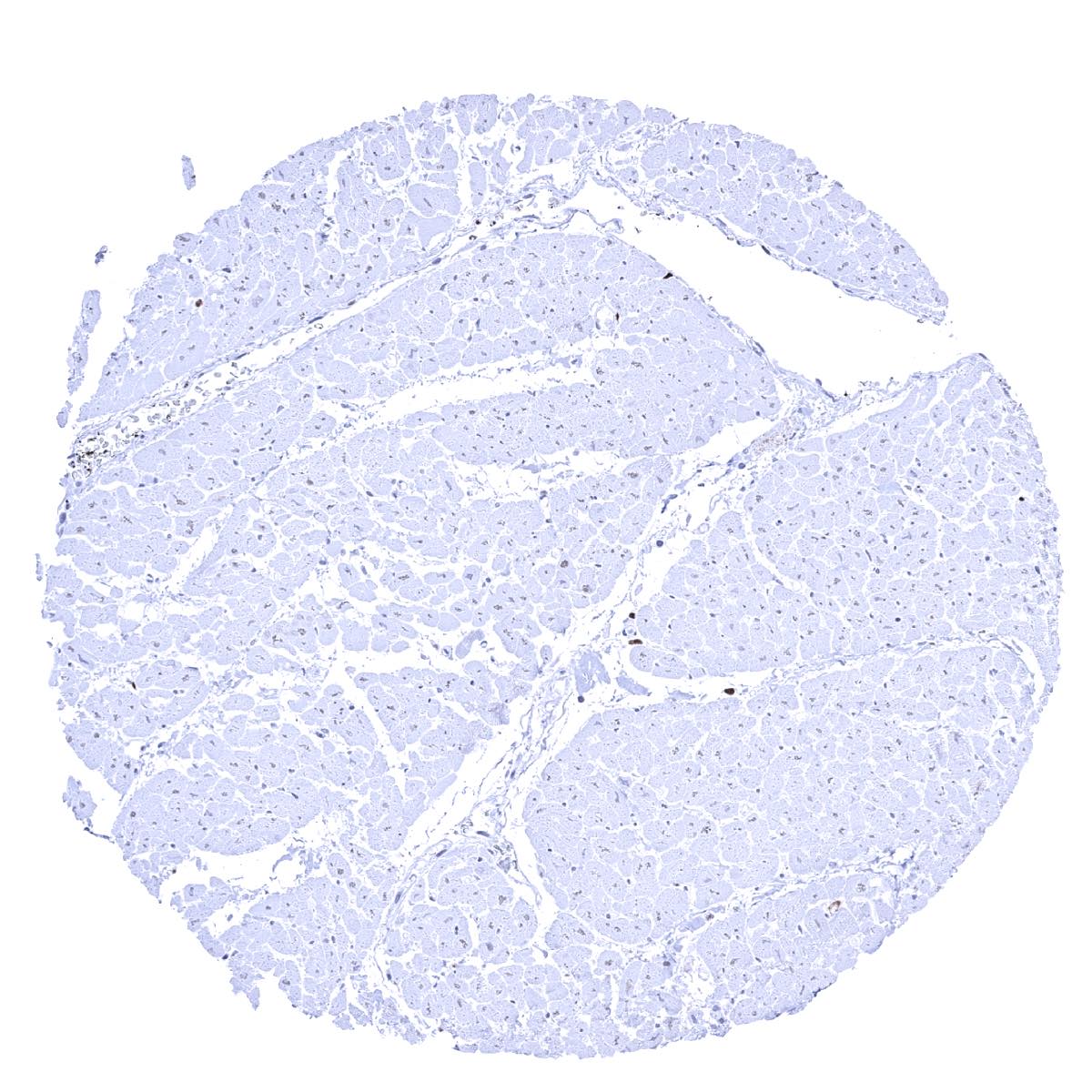
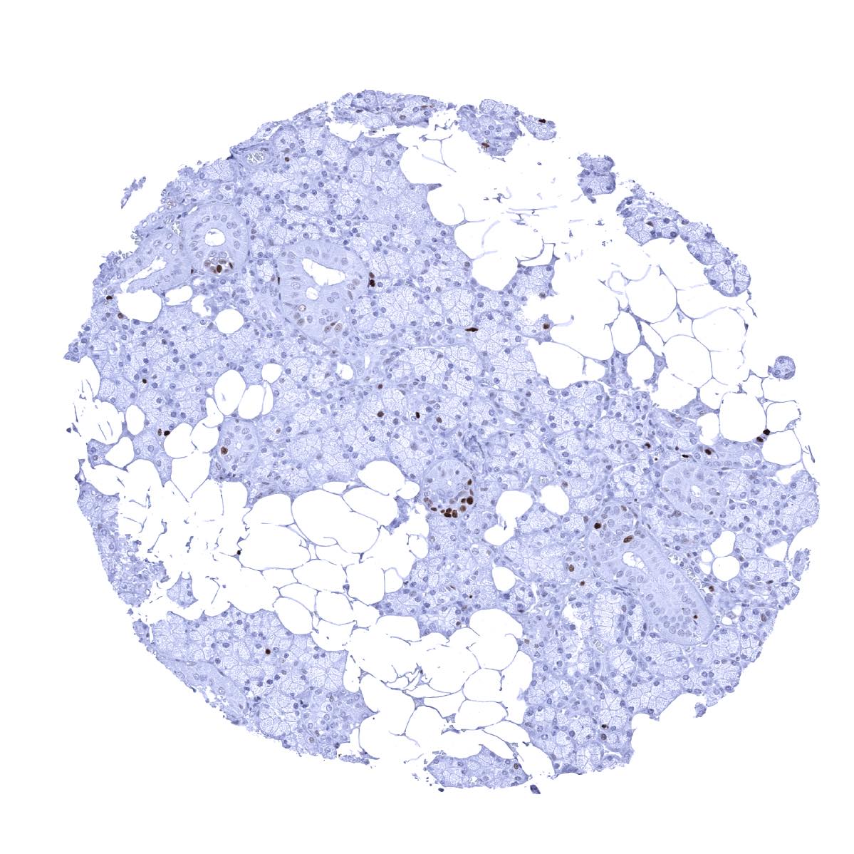
Pancreas - MCM7 staining occurs in a small fraction of pancreatic epithelial cells. The rate of positive cells is particularly low in islets of Langerhans
Parathyroid gland - MCM7 staining in a small fraction of parathyroideal cells
Parotid gland
Pituitary gland, anterior lobe
Pituitary gland, posterior lobe

Placenta (amnion and chorion)

Placenta, early - MCM7 staining in many cells of the cytotrophoblast and a fraction of stroma cells
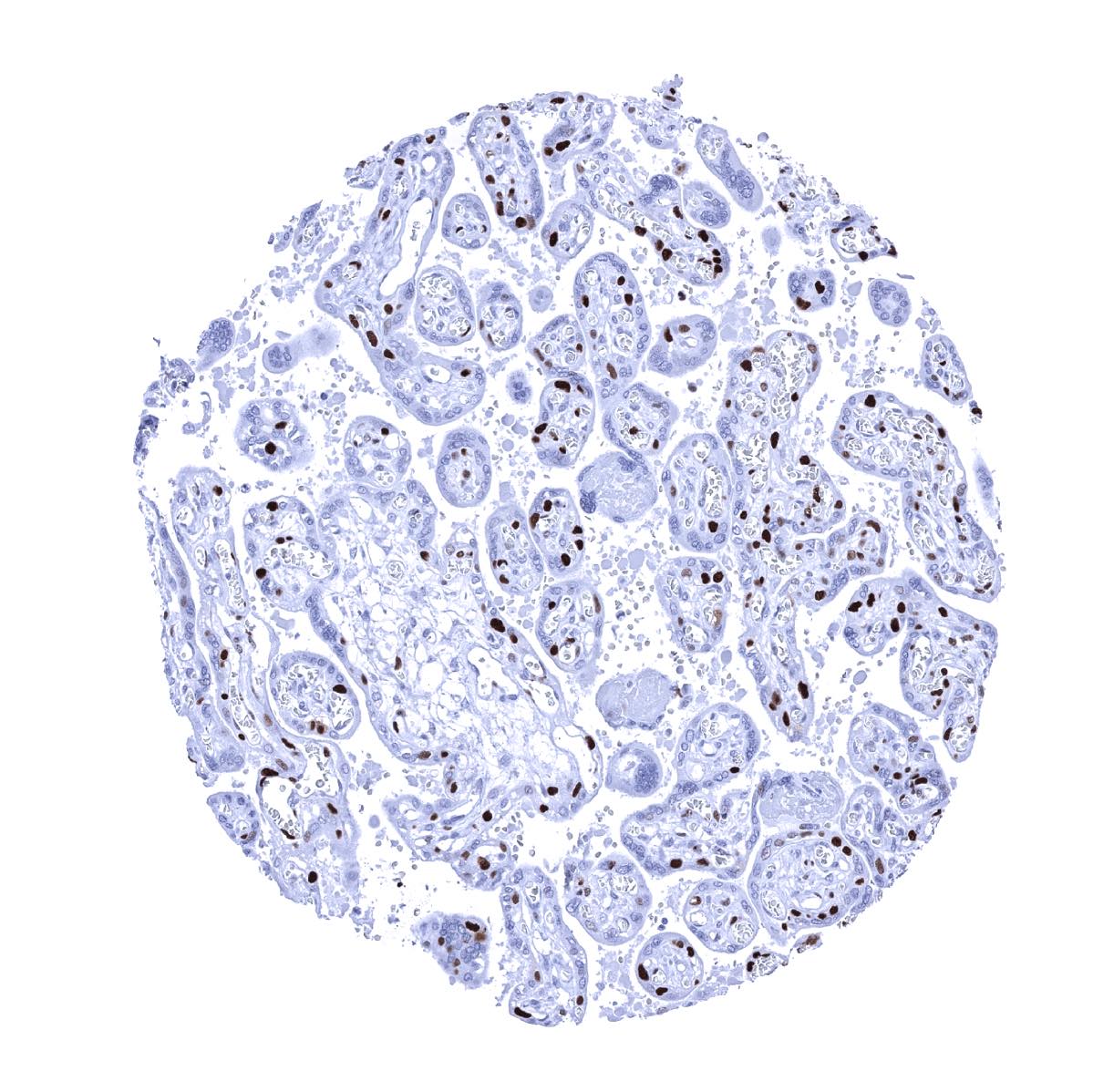
Placenta, mature - Cells of the cytotrophoblast and also some stroma cells show strong MCM7 staining

Prostate - MCM7 staining is more common in basal than in acinar epithelial cells
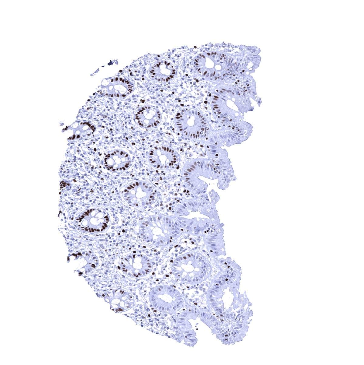
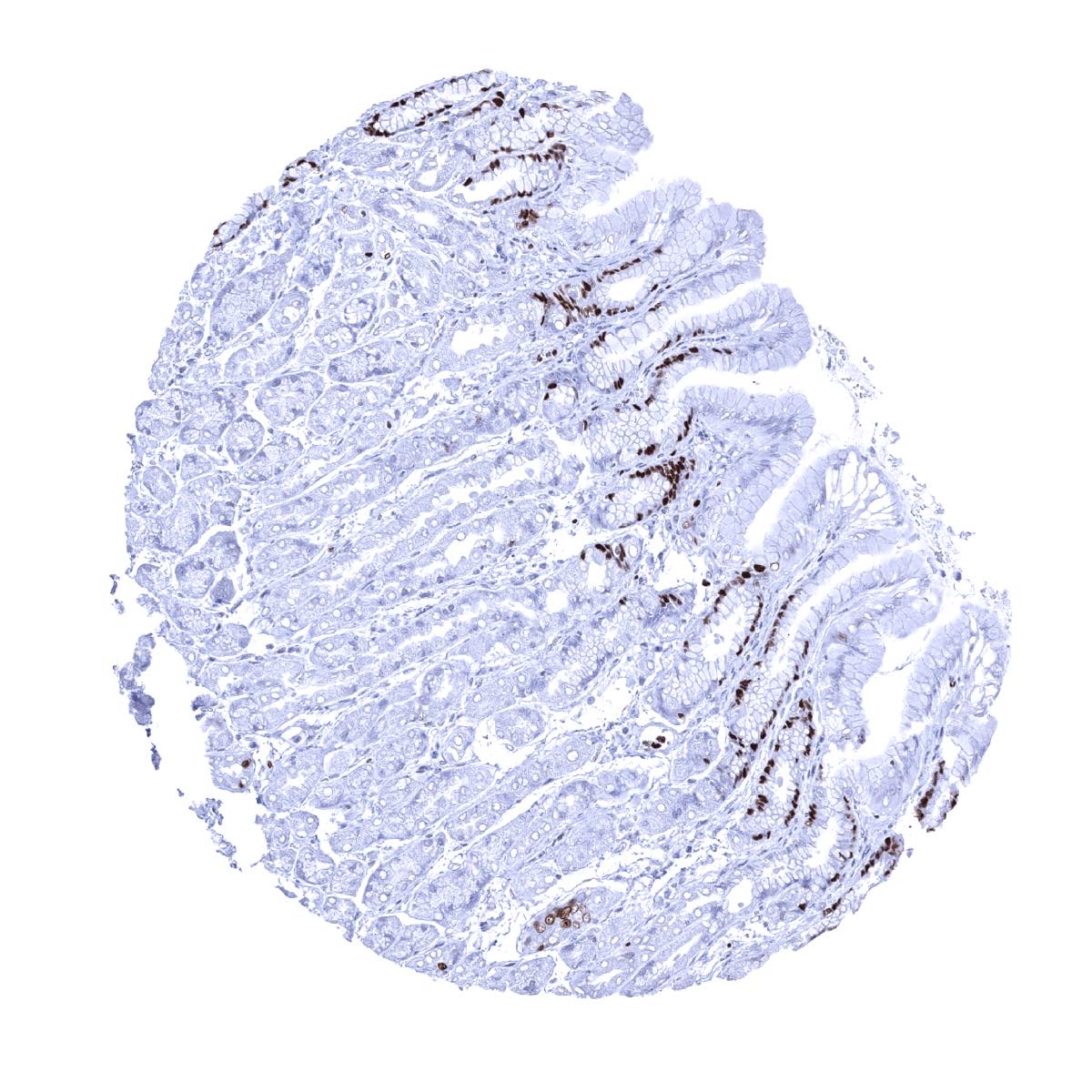
Rectum, mucosa - MCM7 immunostaining predominates in in epithelial cells of the crypts. Some lymphocytes are also positive

Seminal vesicle
Sinus paranasales - MCM7 staining occurs in a fraction of (mostly basal) respiratory epithelial cells

Skeletal muscle - Significant MCM7 staining in a fraction of skeletal muscle cells

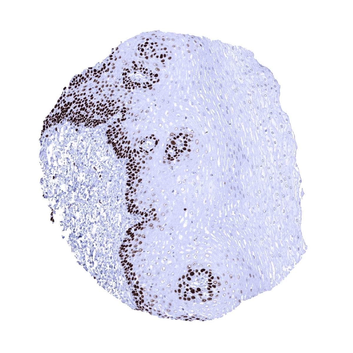
Skin - In the squamous epithelium of the skin, suprabasal and (weaker) basal cell layers show a distinct MCM7 immunostaining
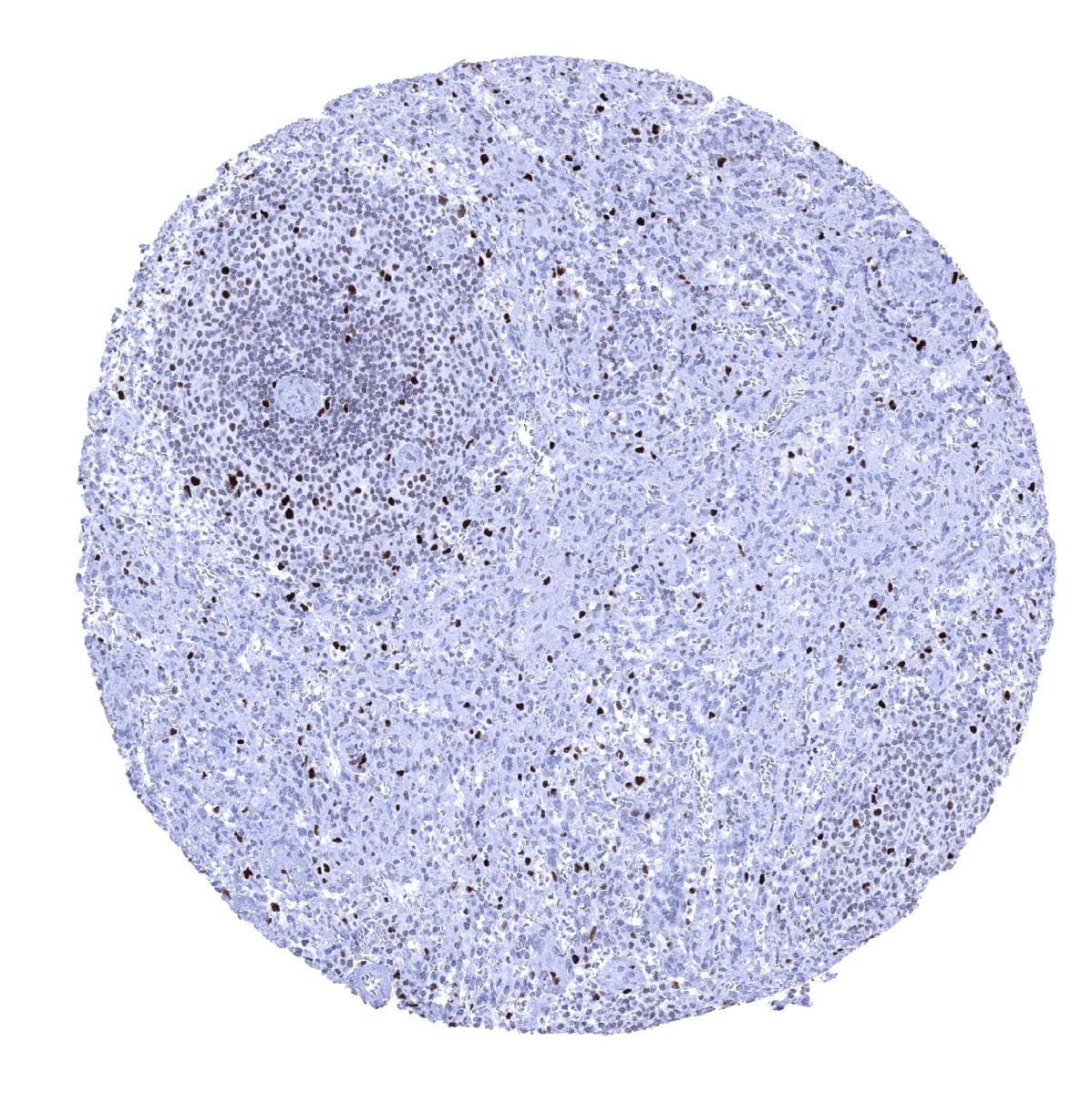
Spleen - Most lymphocytes of the white pulp are MCM7 positive. A strong MCM7 positivity occurs in scattered cells of all compartments
Stomach, antrum - Strong MCM7 immunostaining in many mucous neck cells
Stomach, corpus - Strong MCM7 immunostaining in many mucous neck cells
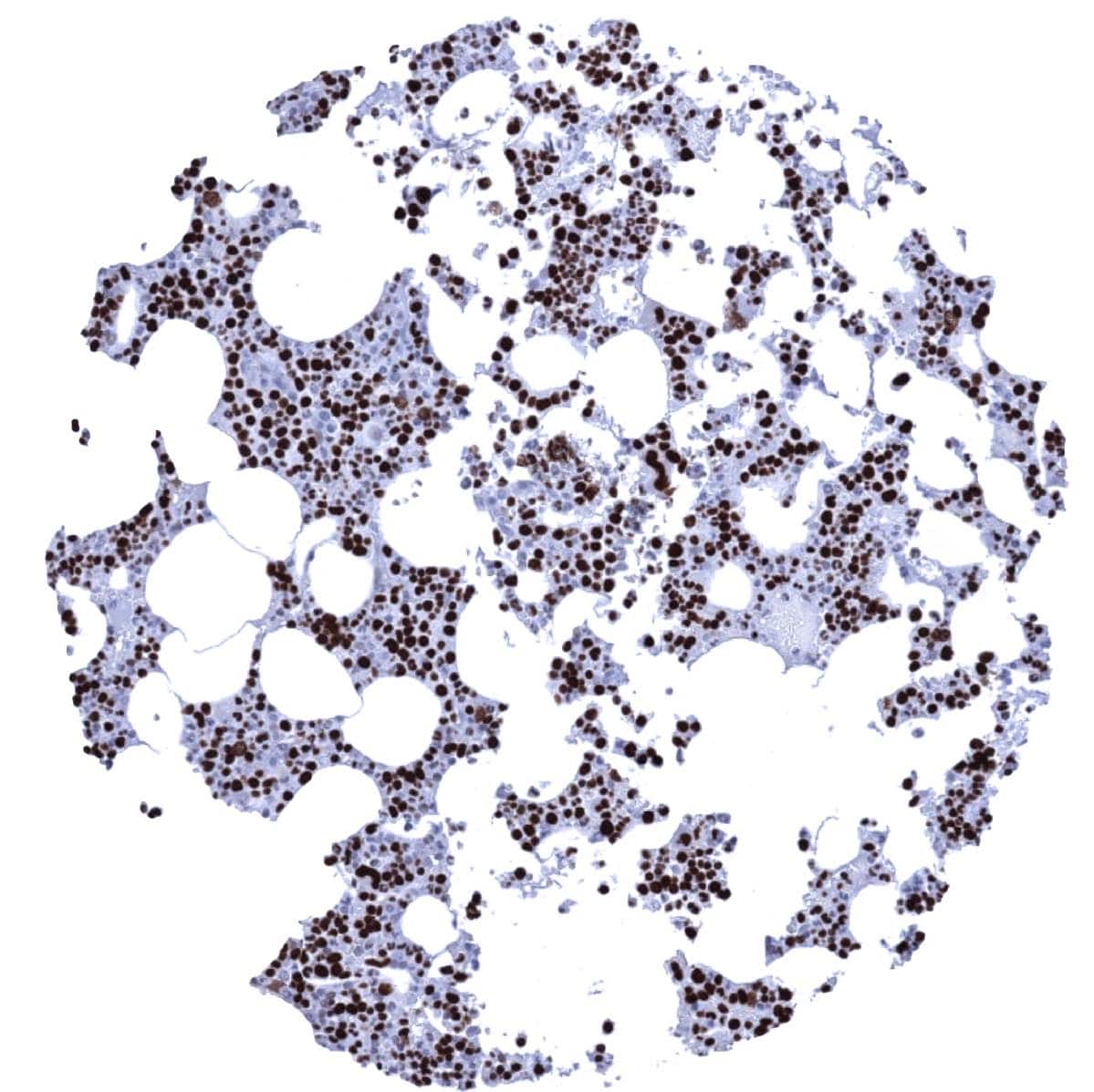
Strong MCM7 immunostaining in the majority of bone marrow cells
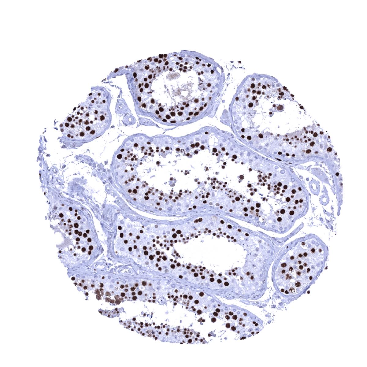
Testis - Most spermatocytes show strong MCM7 positivity but mature sperms and probably also spermatogonia are MCM7 negative

Thymus - A strong MCM7 positivity occurs in most cells of the thymic cortex
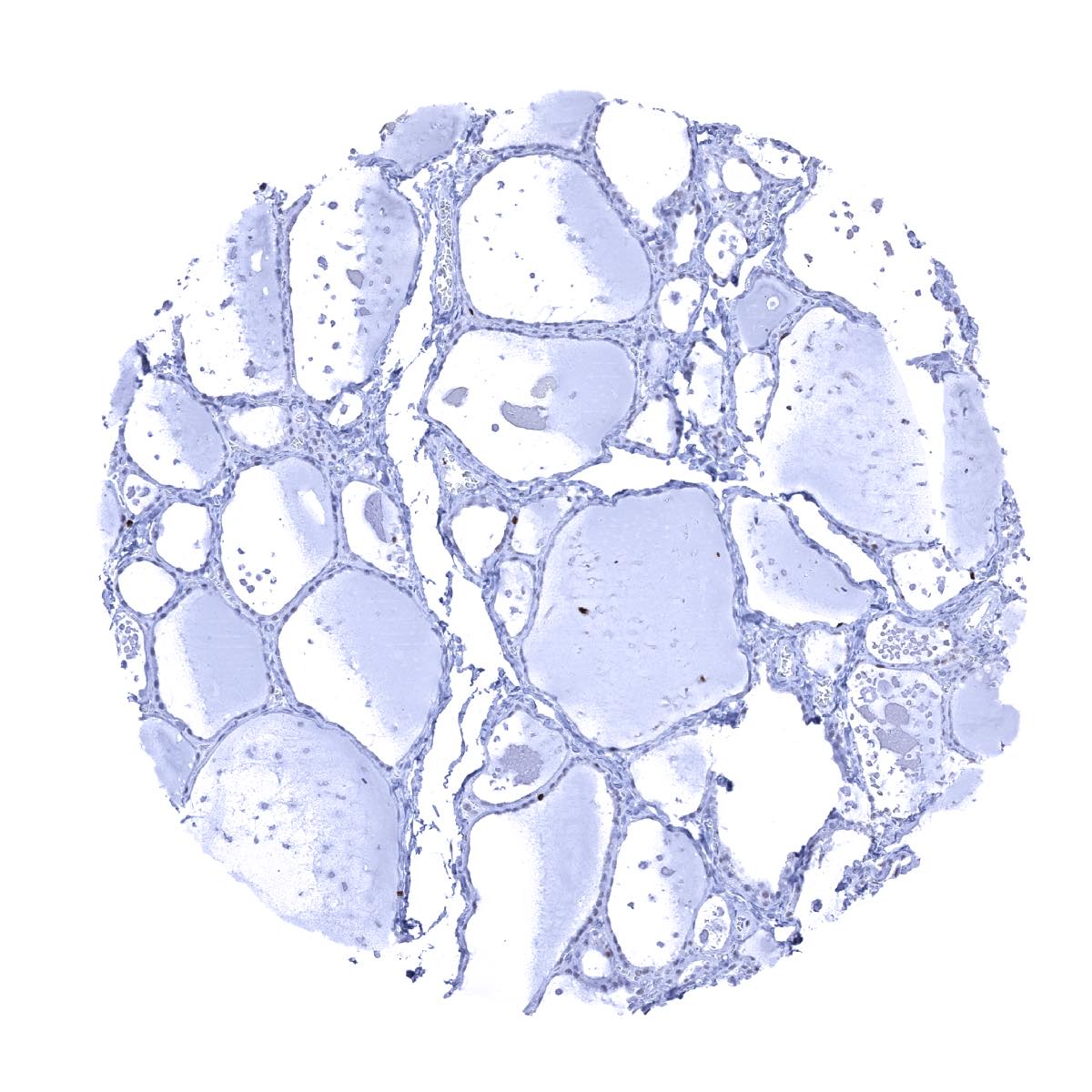
Thyroid gland
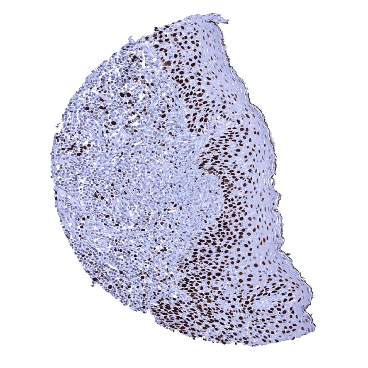
Tonsil, surface epithelium - Moderate to strong MCM7 staining of suprabasal and basal squamous epithelial cells. A fraction of lymphocytes are also MCM7 positive
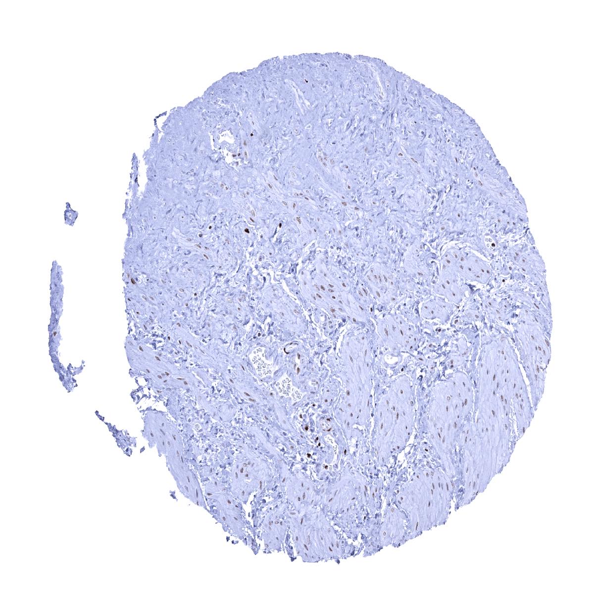
Urinary bladder, muscular wall
Urinary bladder, urothelium - Only few (suprabasal) urothelial cells show a MCM7 staining
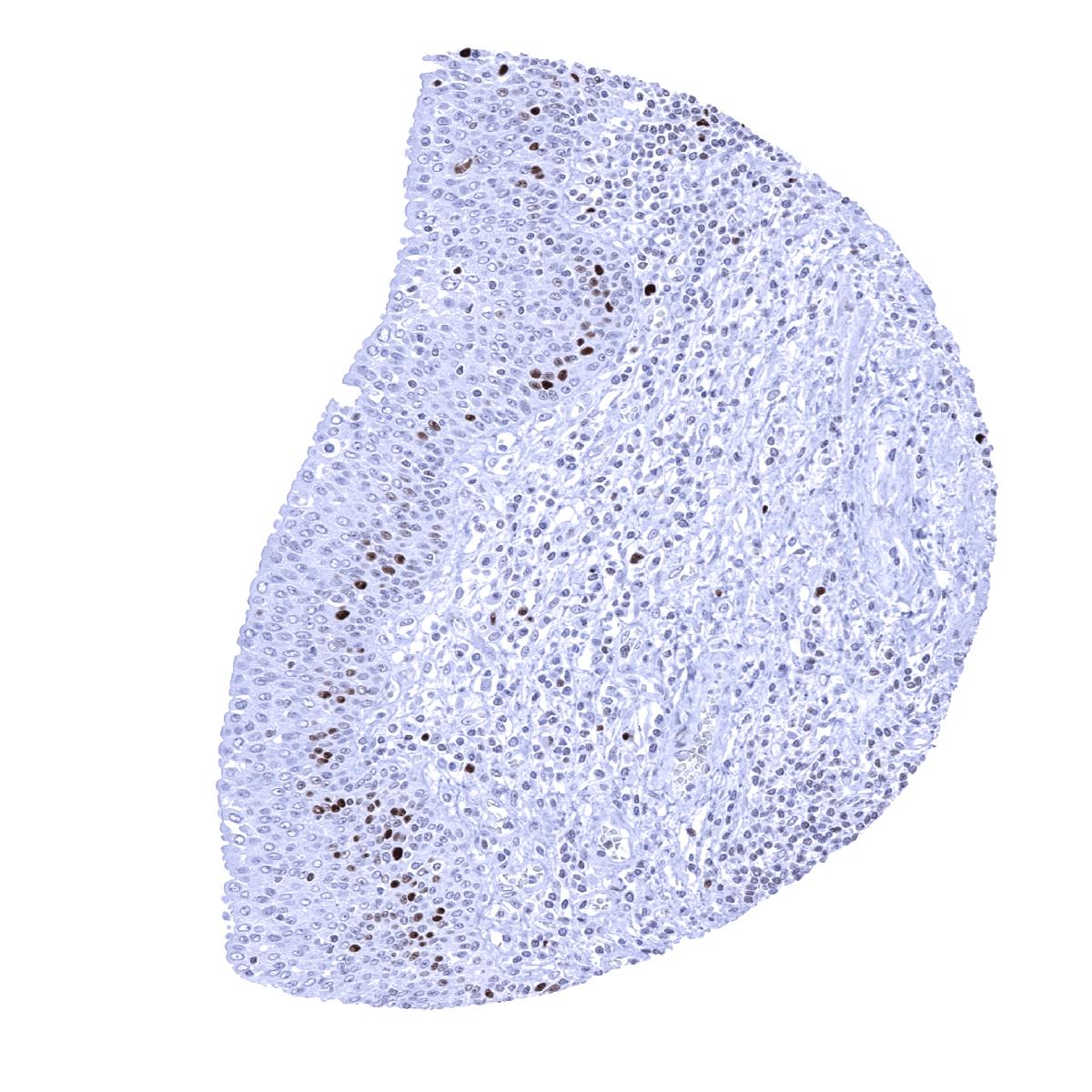
Uterus, ectocervix - The squamous epithelium shows a moderate MCM7 immunostaining of (mostly) suprabasal cells

Uterus, endocervix - Only few epithelial cells show MCM7 staining
Uterus, endometrium (pregnancy) - MCM7 staining is rare in decidua cells
Uterus, endometrium (proliferation) - Almost all epithelial cells and a fraction of stromal cells are MCM7 positive
Uterus, endometrium (secretion) - The fraction of MCM7 positive epithelial cells is lower in the secretion phase
Uterus, myometrium - Faint MCM7 immunostaining in some muscle cells