Adrenal gland
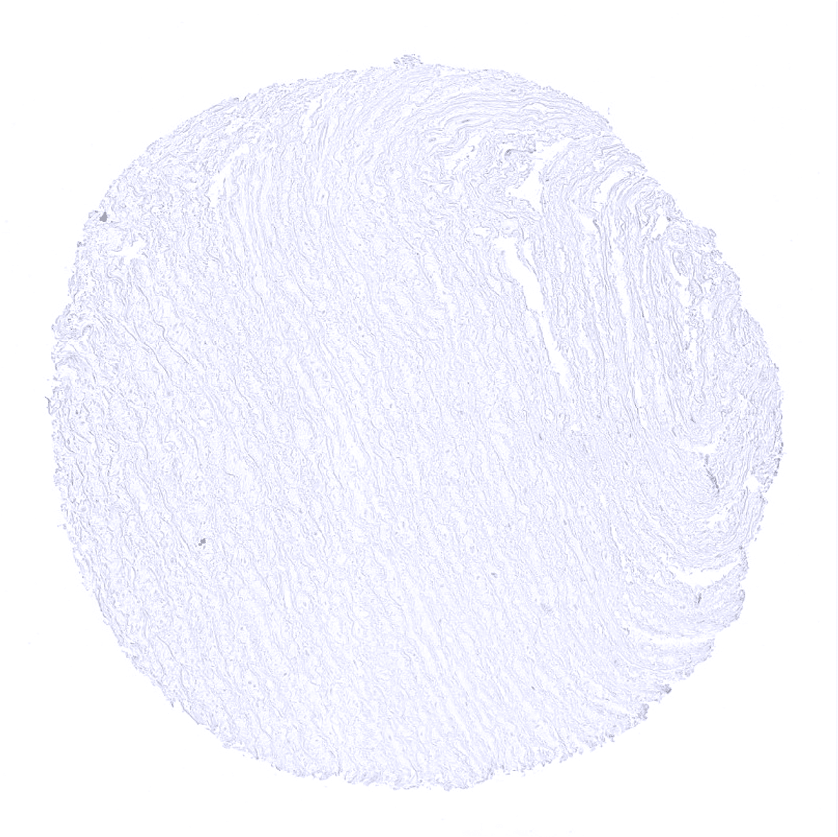
Aorta, media
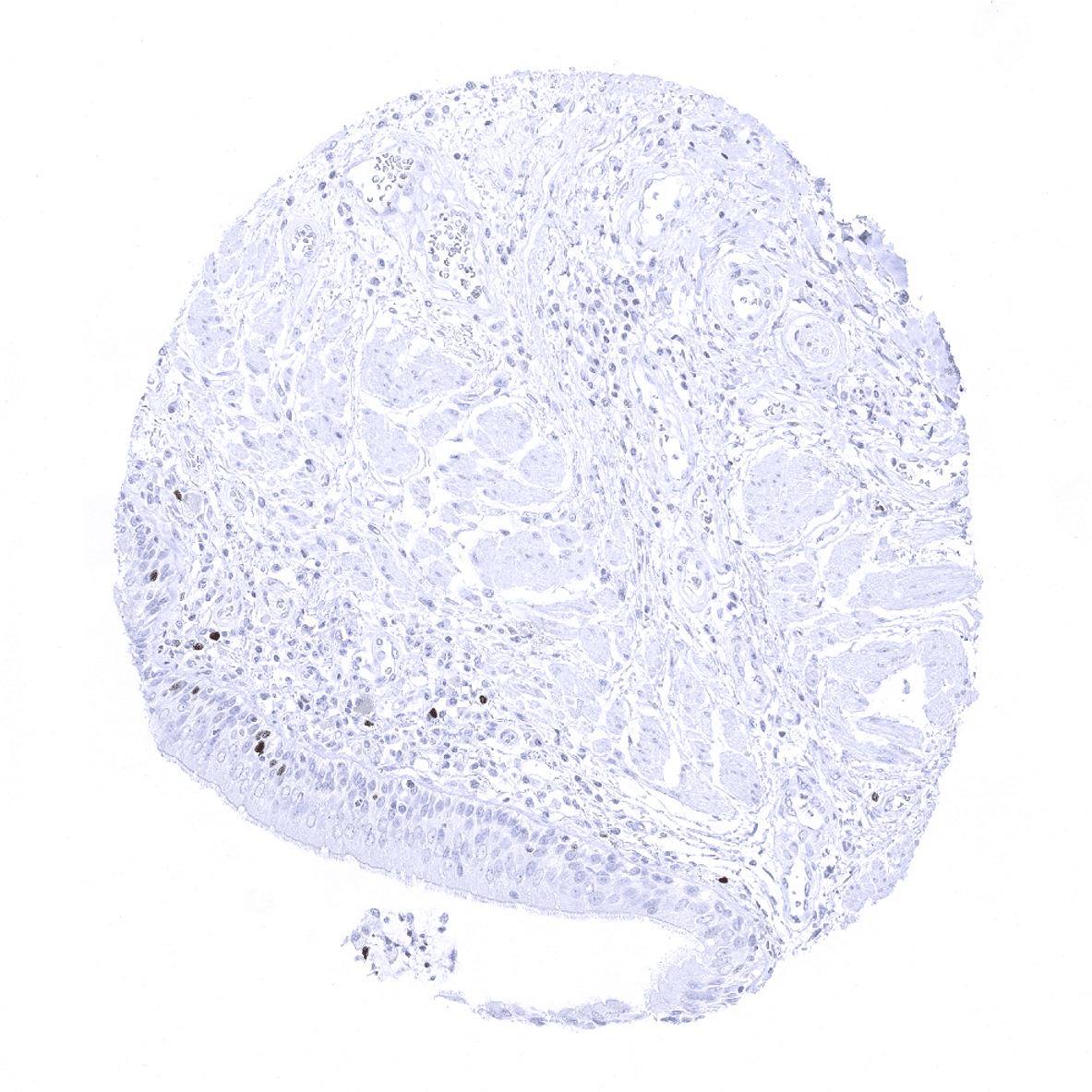
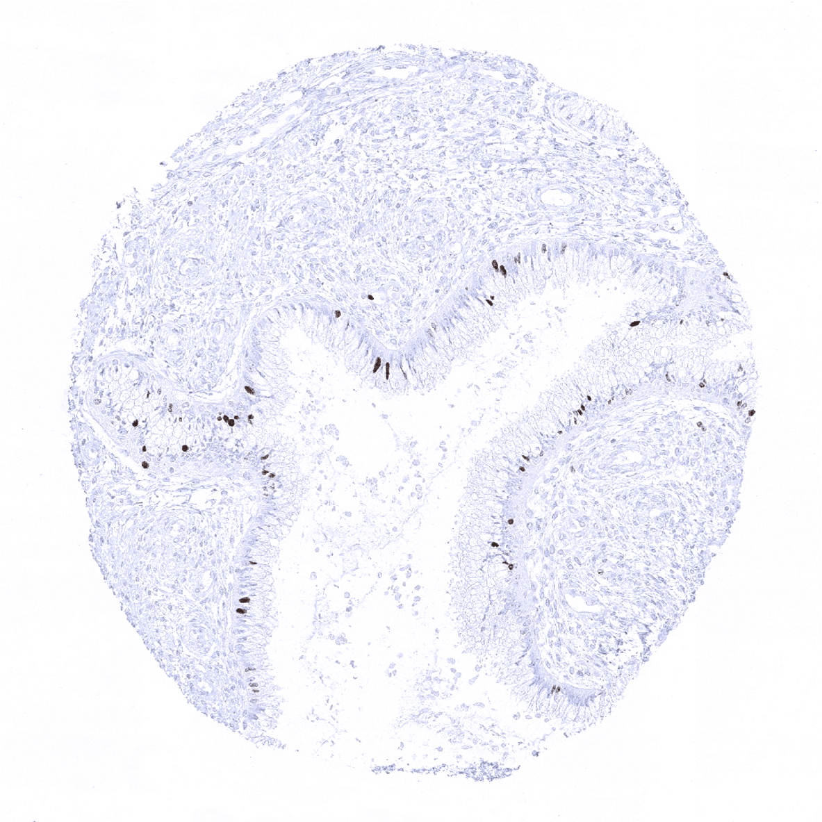
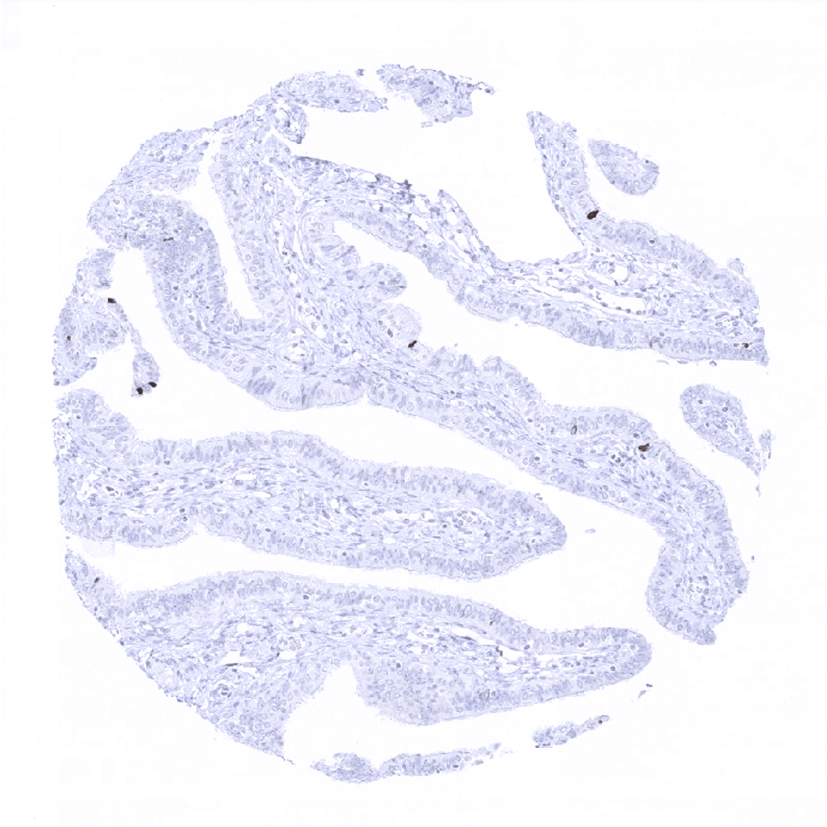
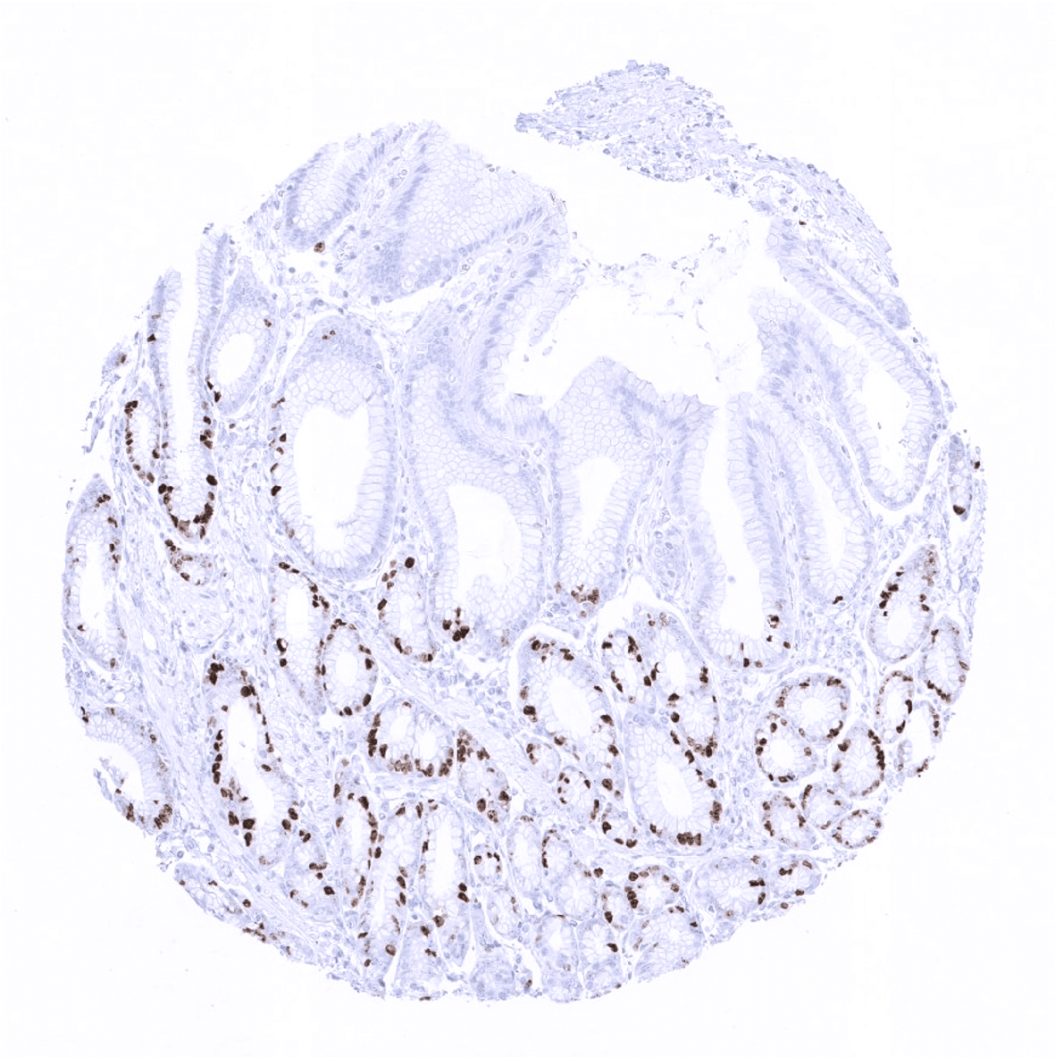
Appendix, mucosa
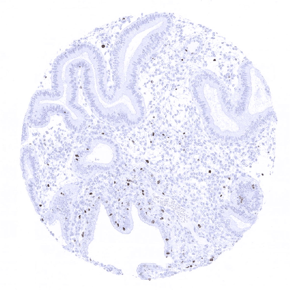
Appendix, muscular wall

Bone marrow

Breast
Bronchus, mucosa
Cerebellum, cortex (Stratum moleculare)

Cerebellum, grey (Stratum neuronorum)
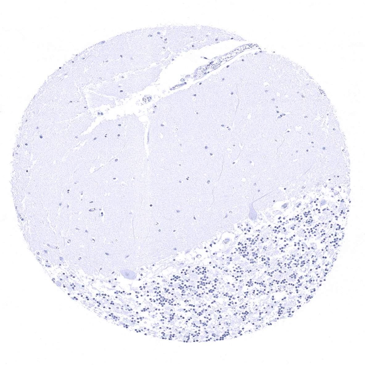
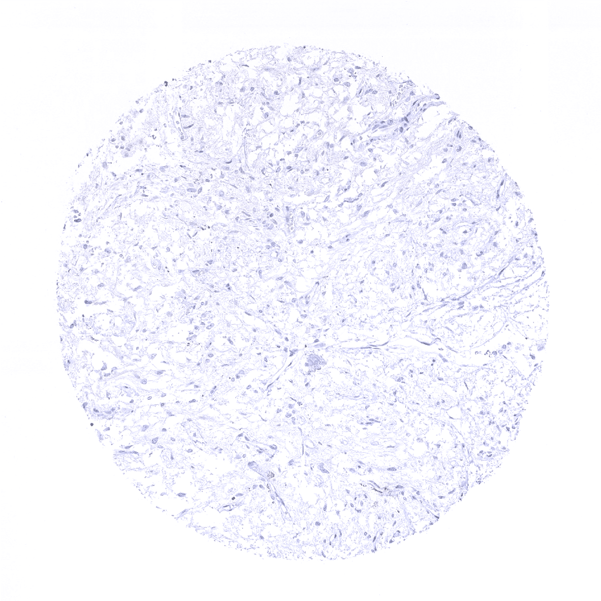
Cerebrum, grey

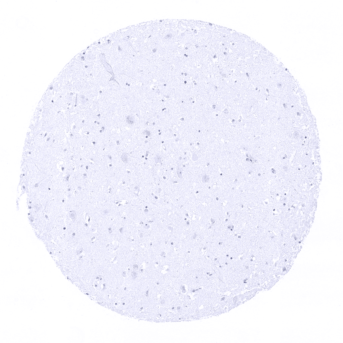
Cerebrum, white
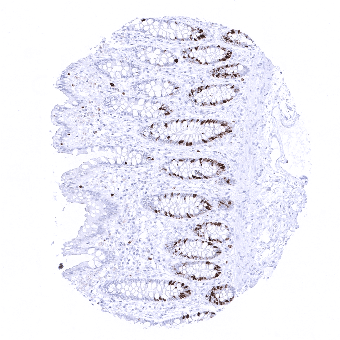
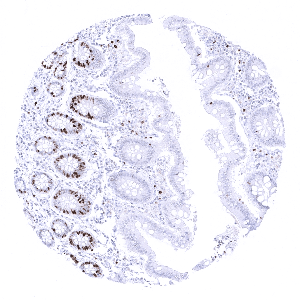
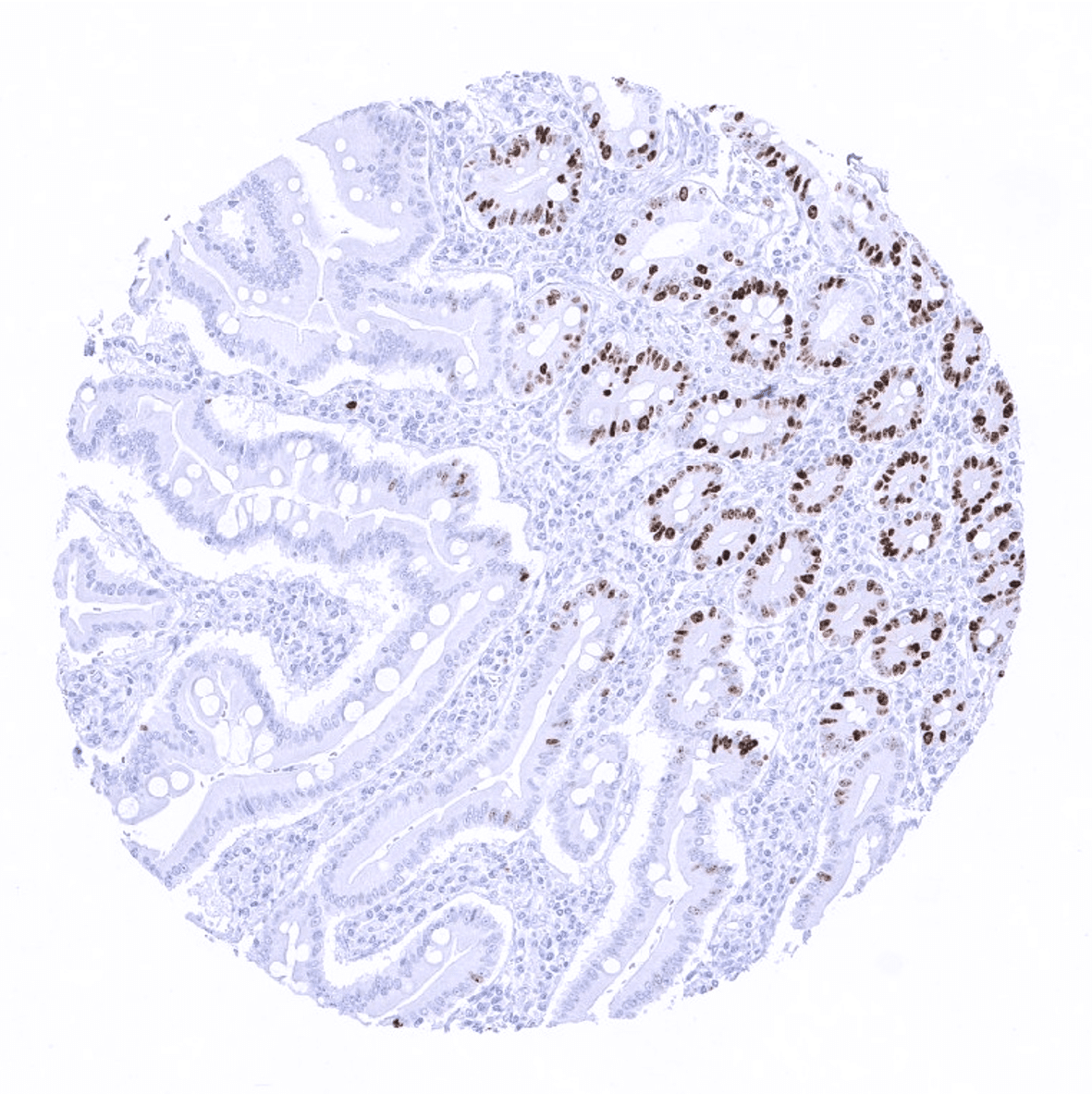
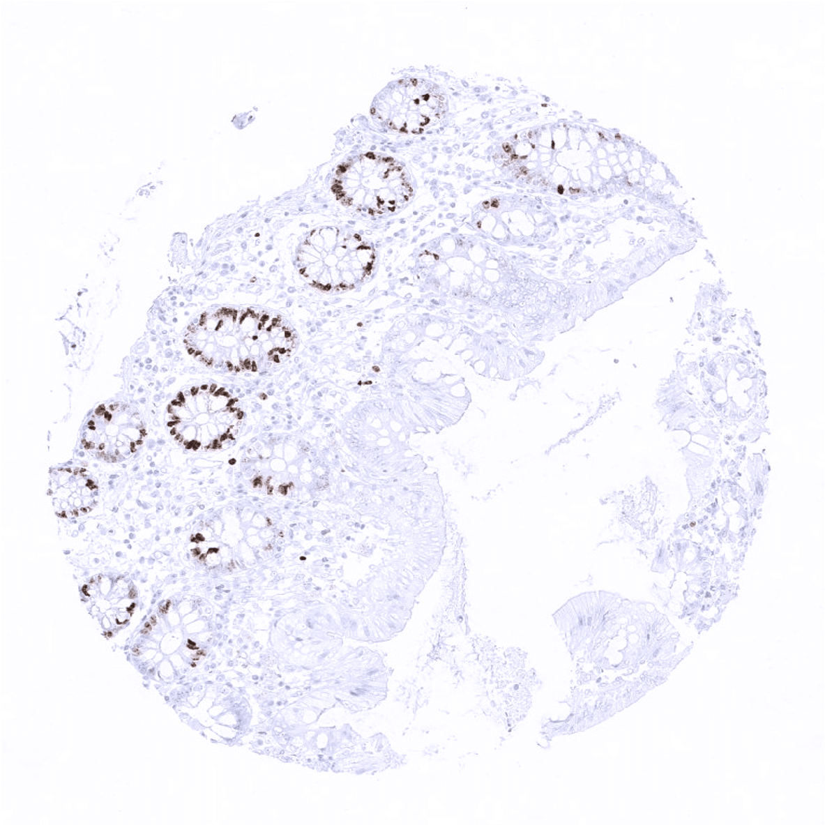
Colon descendens, mucosa
Colon descendens, muscular wall
Duodenum, Brunner gland
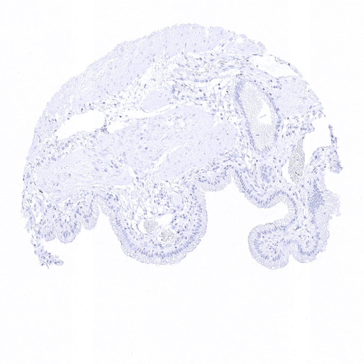
Duodenum, mucosa
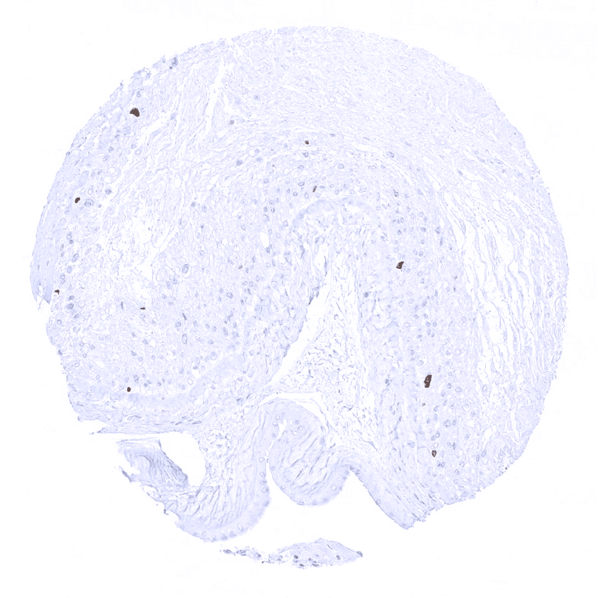
Ectocervix
Endocervix

Endometrium, proliferation
Endometrium, secretion
Epididymis
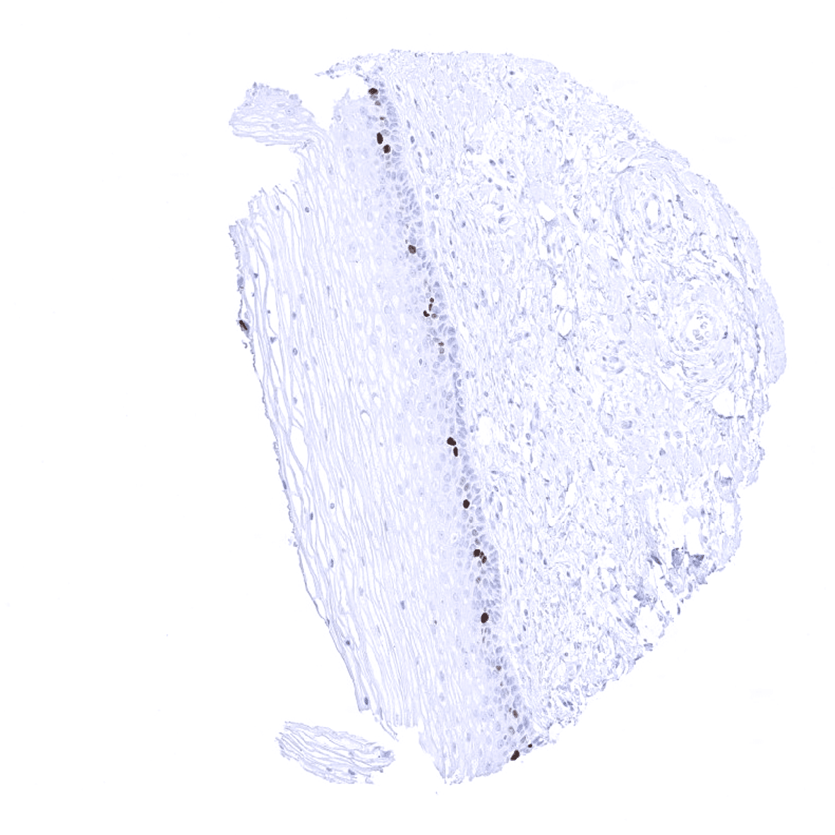
Esophagus, squamous epithelium
Fallopian tube, mucosa

Fat
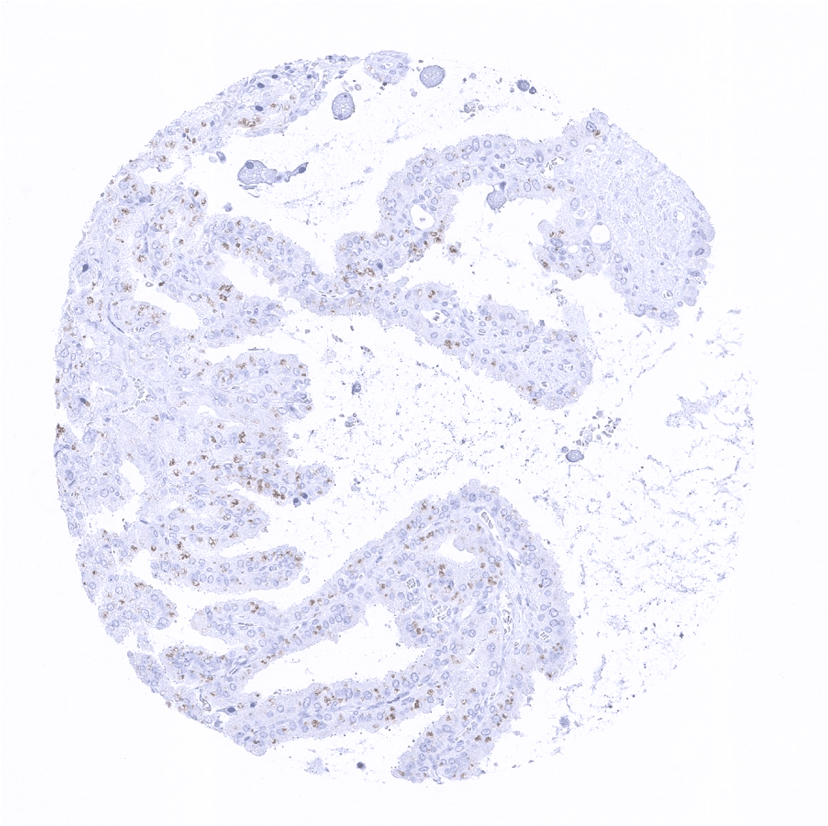
Gallbladder, epithelium
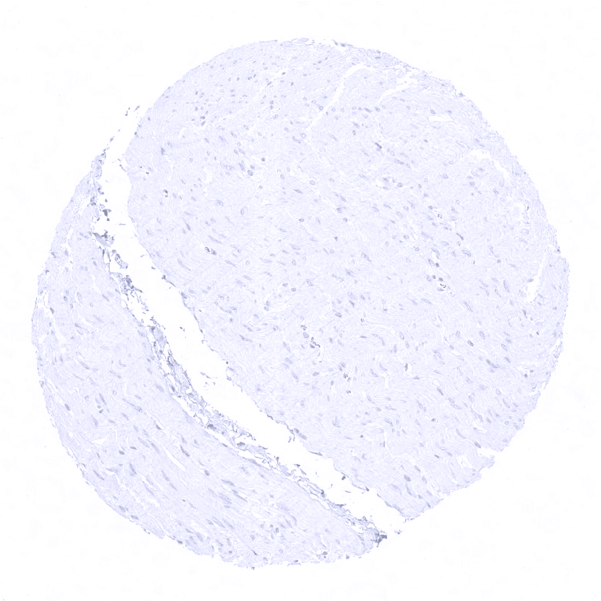
Heart
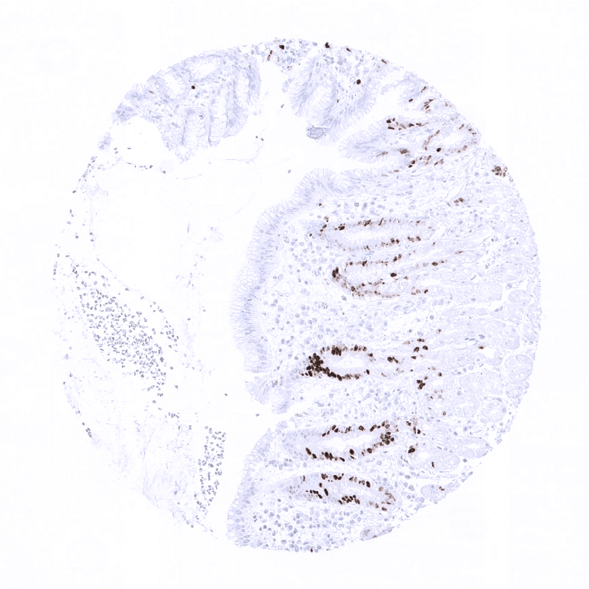
Ileum, mucosa
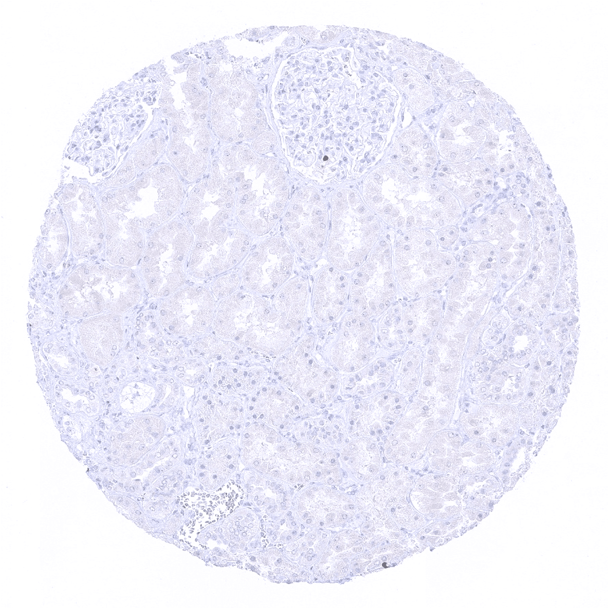
Kidney, cortex
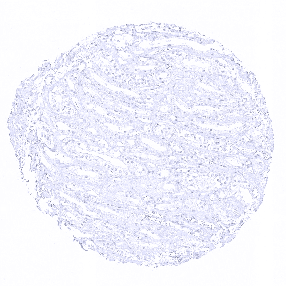
Kidney, medulla
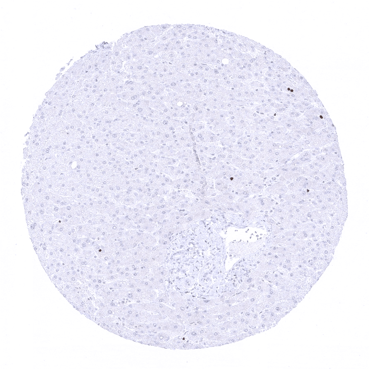
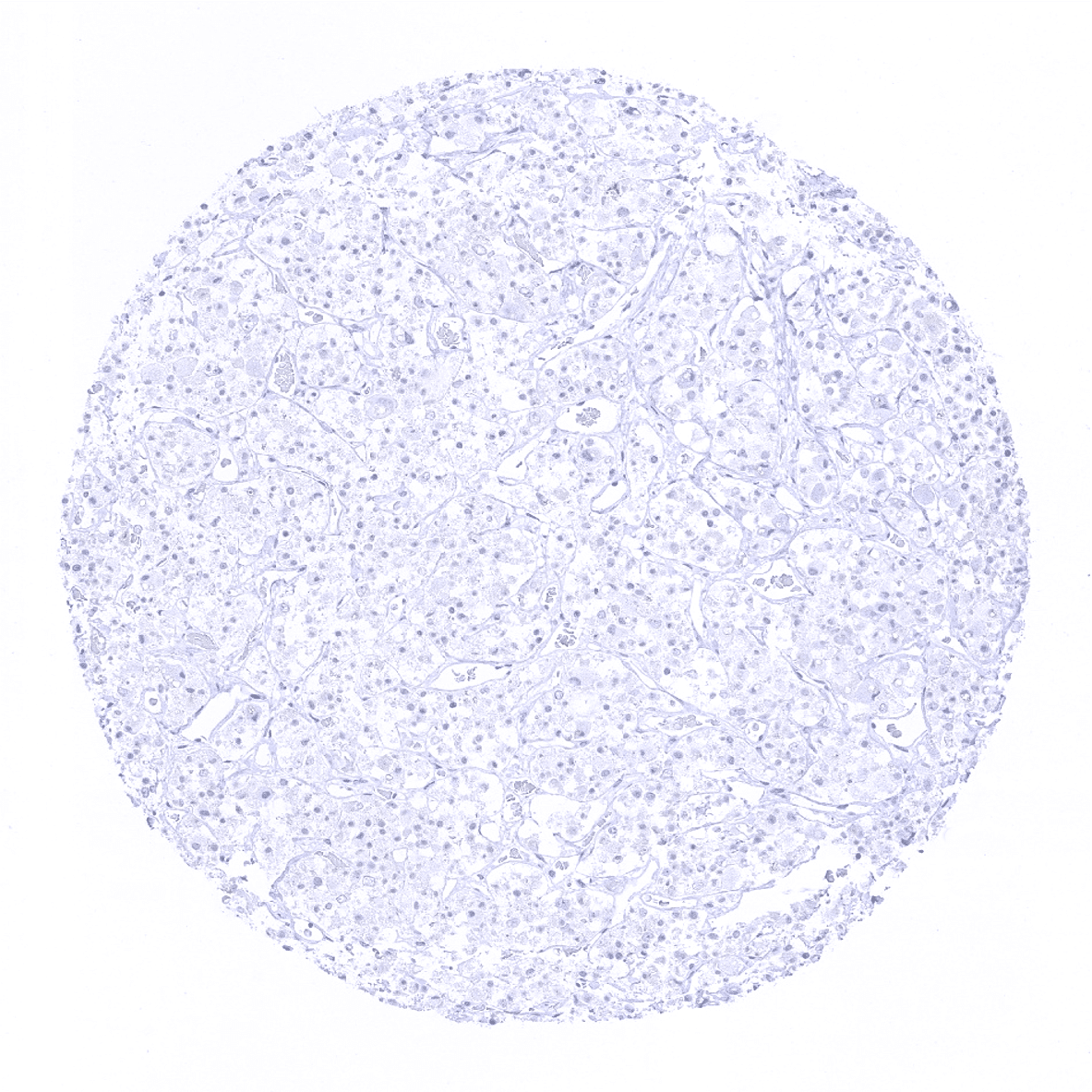
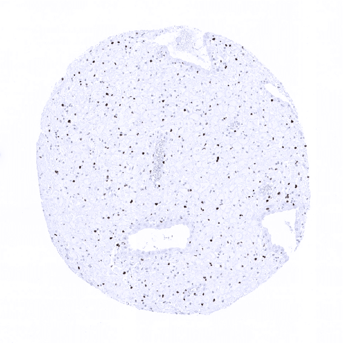
Liver-The liver represents an ideal normal tissue control for Ki-67 staining quality. Less than 1% of the hepatocytes should stain and cytoplasmatic staining should be absent.

Lung

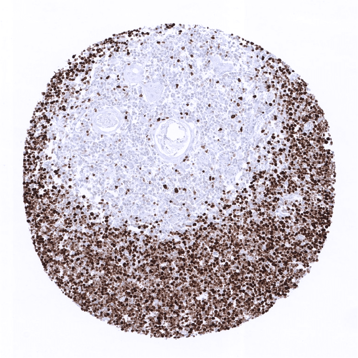
Lymph node
Ovary, stroma

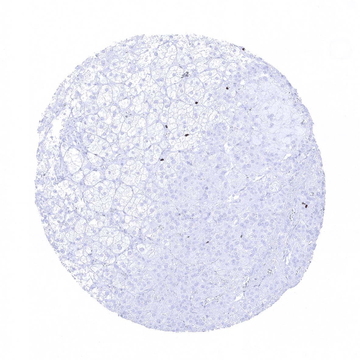
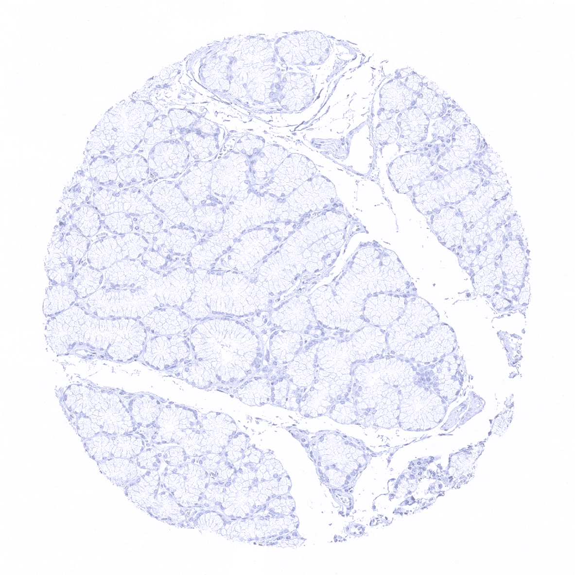
Pancreas

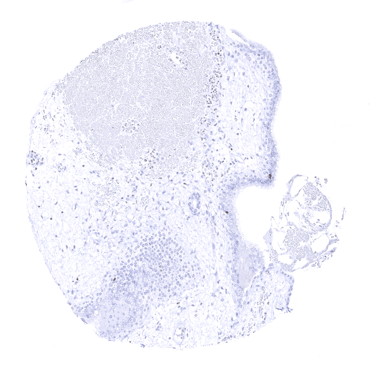
Parathyroid

Parotid gland
Pituitary, anterior lobe
Pituitary, posterior lobe_ infundibulum
Placenta early, decidua
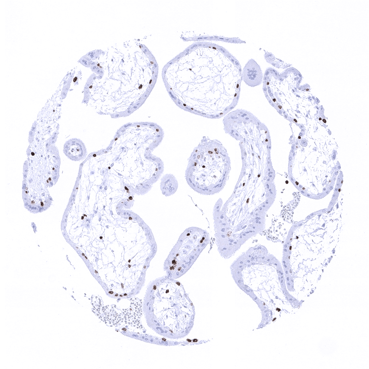
Placenta, early
Placenta, mature, amnion and chorion

Prostate
Seminal vesicle
Sinus paranasales
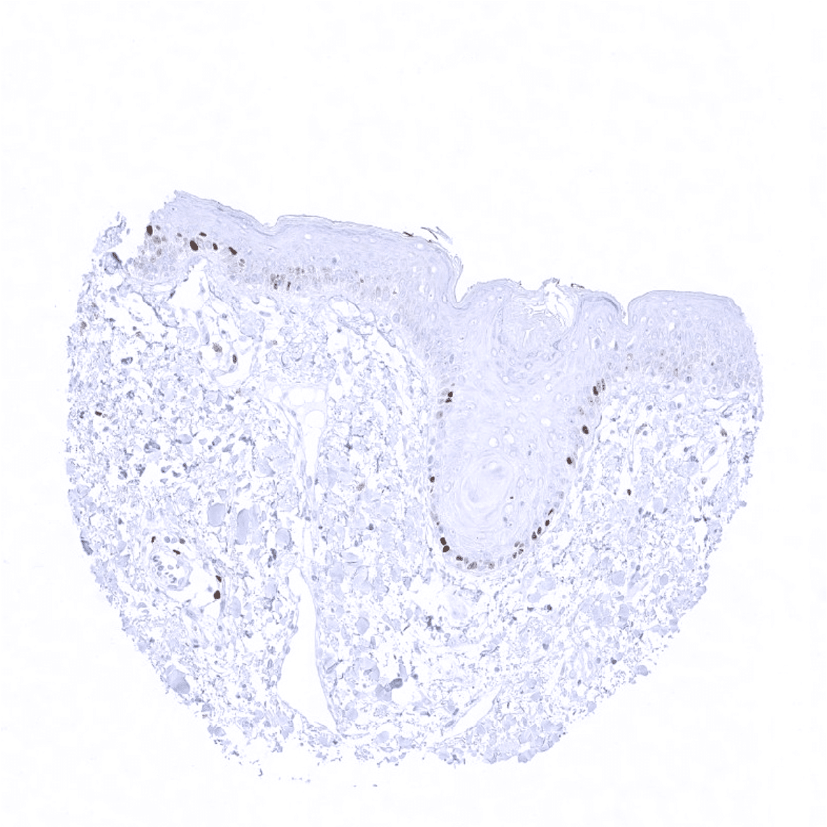
Skin
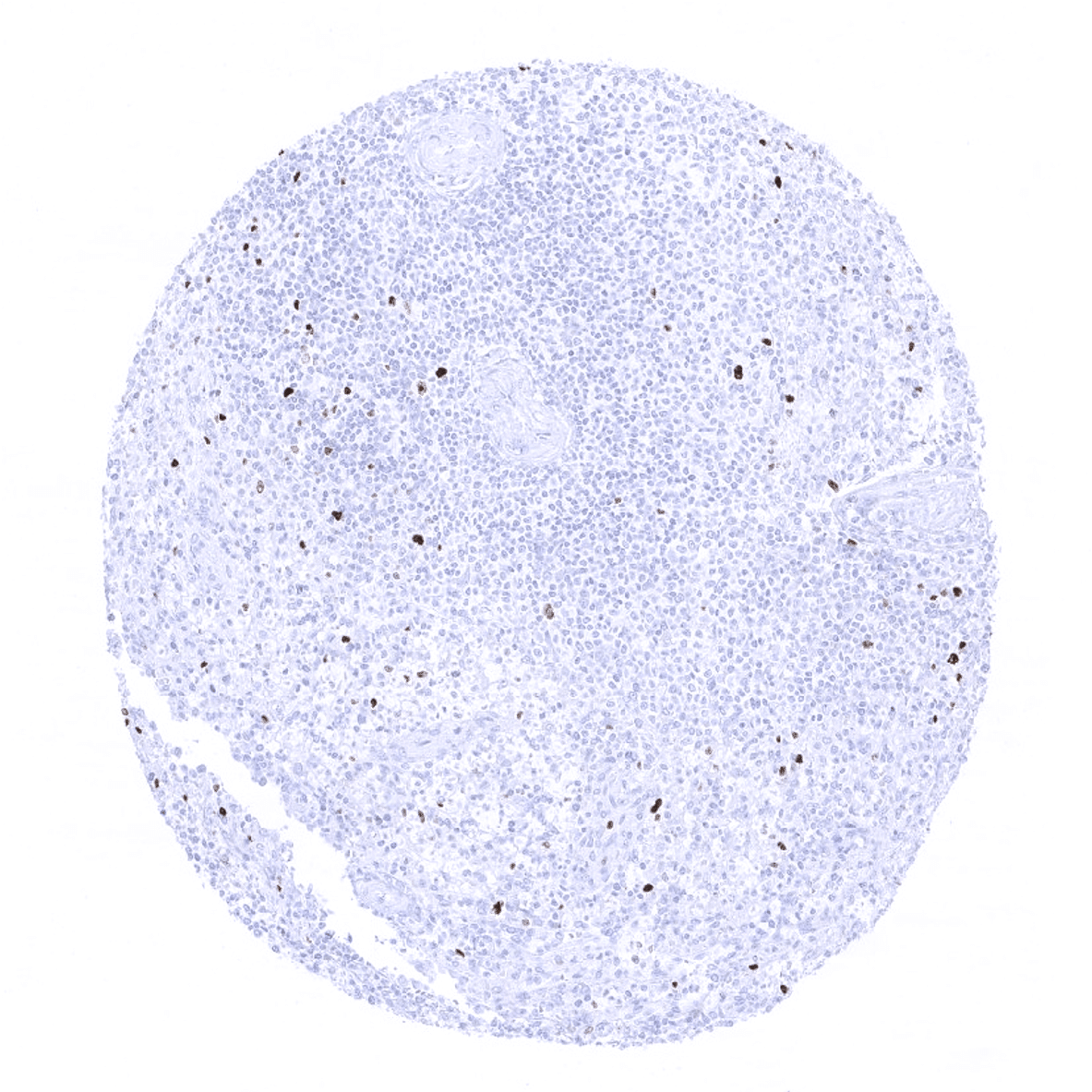
Spleen
Stomach, antrum
Stomach, corpus

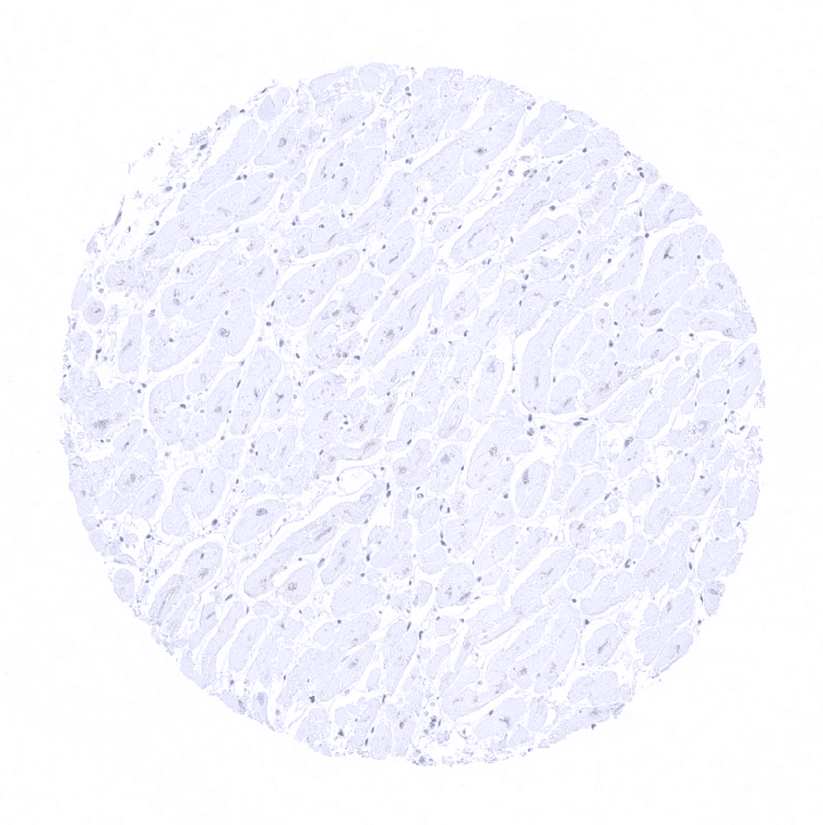
Striated muscle
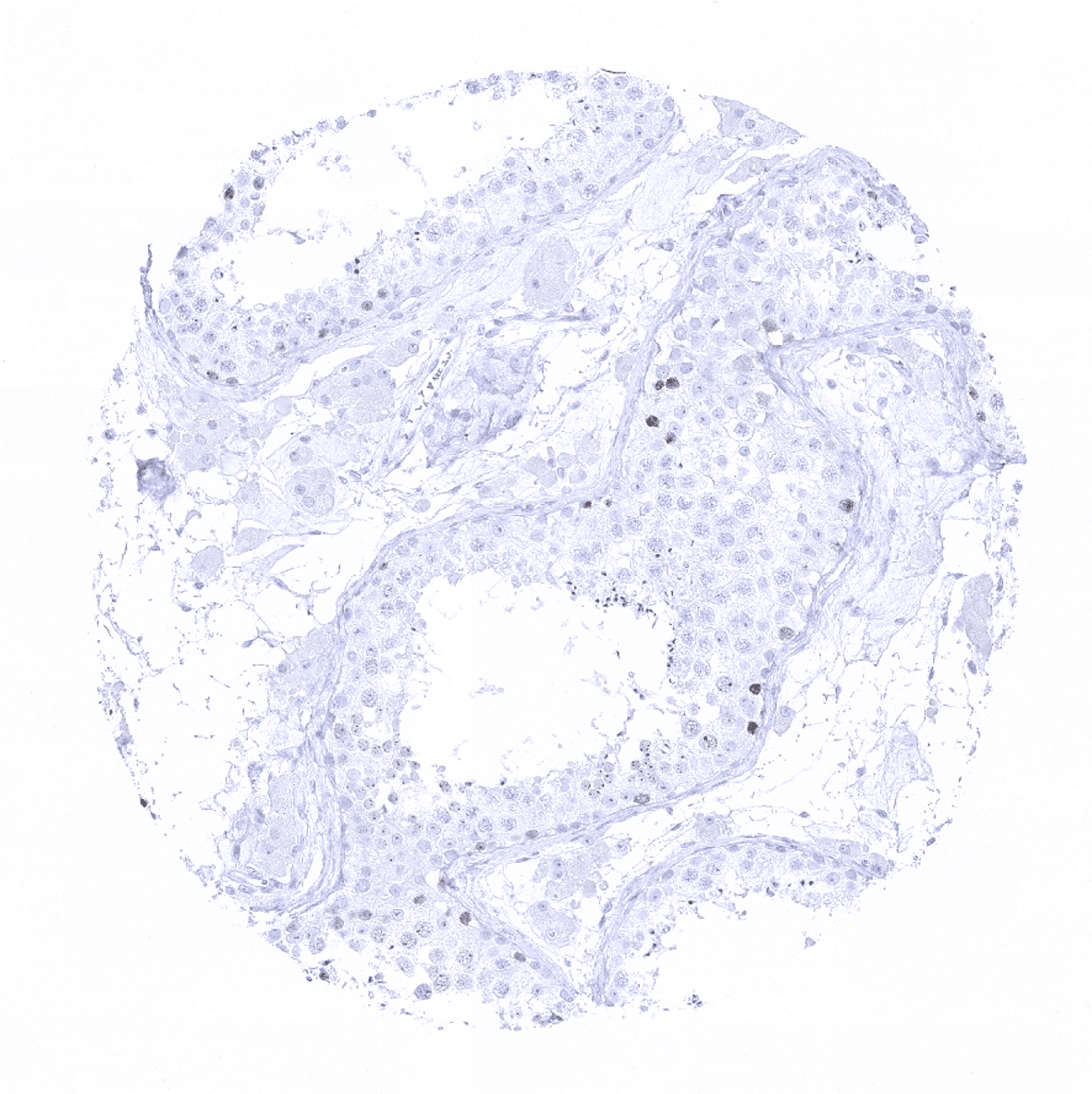
Testis
Thymus- the highest proliferative activity (Ki-67 LI) in normal tissues is seen in the thymic cortex

Thyroid gland
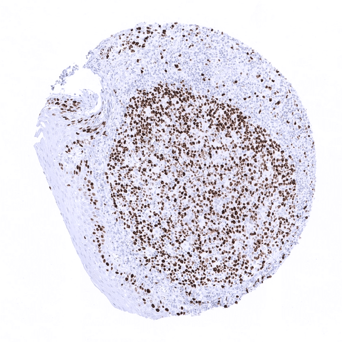
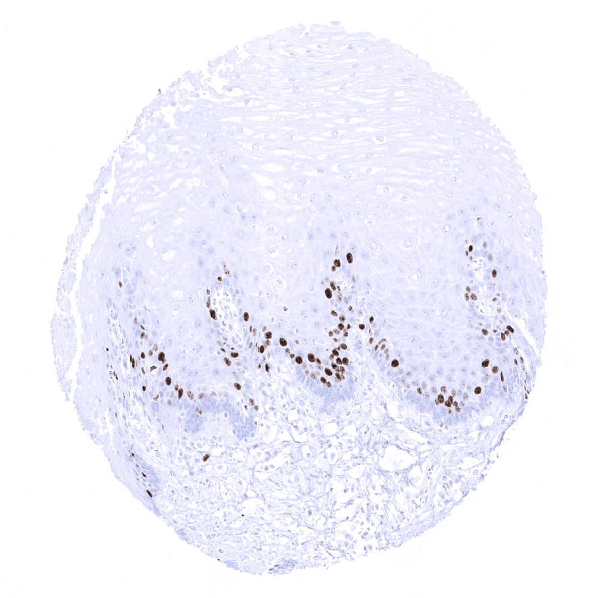
Tonsil: In the tonsil, Ki67 stained cells are preferential seen in germinal centre and in supra basal cells of the surface epithelium.

Tonsil

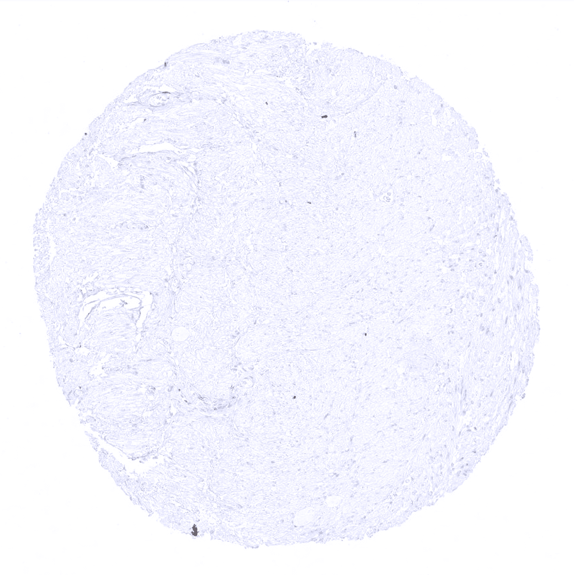
Urinary bladder, muscular wall
Urinary bladder, urothelium
Uterus, myometrium