Adrenal gland - HLA-DRB1 staining in scattered inflammatory cells and in endothelial cells (HLA-DRB1 immunohistochemistry)
Aorta, media
Appendix, mucosa - Strong HLA-DRB1 staining of mucosal inflammatory cells. Superfical epithelial cells also show a weak focal positivity (HLA-DRB1 immunohistochemistry)

Appendix, muscular wall - HLA-DRB1 staining in dispersed inflammatory cells (HLA-DRB1 immunohistochemistry)

Bone marrow - HLA-DRB1 staining of few scattered inflammatory cells (HLA-DRB1 immunohistochemistry)
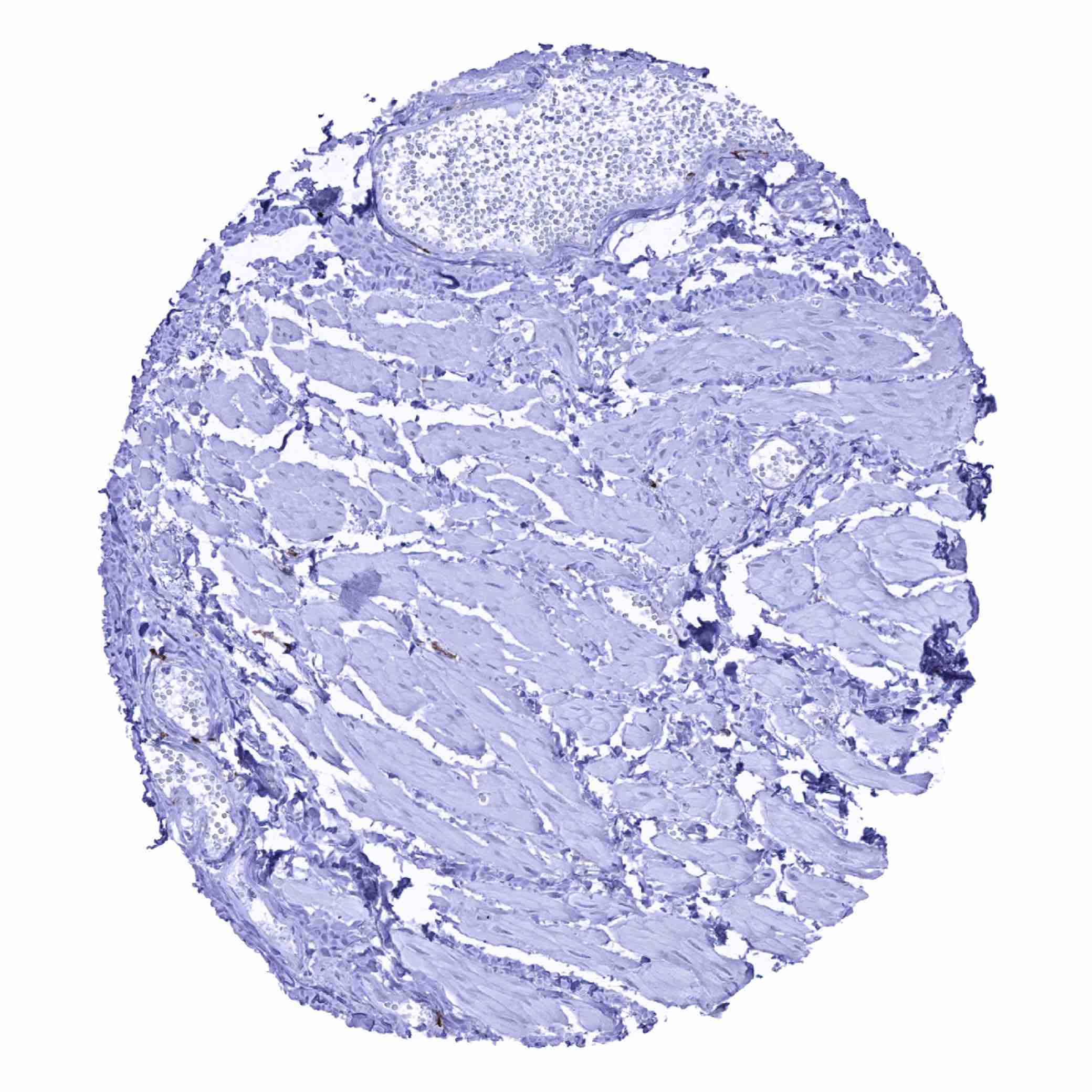
Breast – HLA-DRB1 staining in scattered inflammatory cells (HLA-DRB1 immunohistochemistry)
Bronchus, glands - Strong membranous HLA-DRB1 staining in a group of glandular epithelial cells (HLA-DRB1 immunohistochemistry)

Bronchus, mucosa - HLA-DRB1 staining in scattered inflammatory cells while epithelial cells are unstained in this sample (HLA-DRB1 immunohistochemistry)
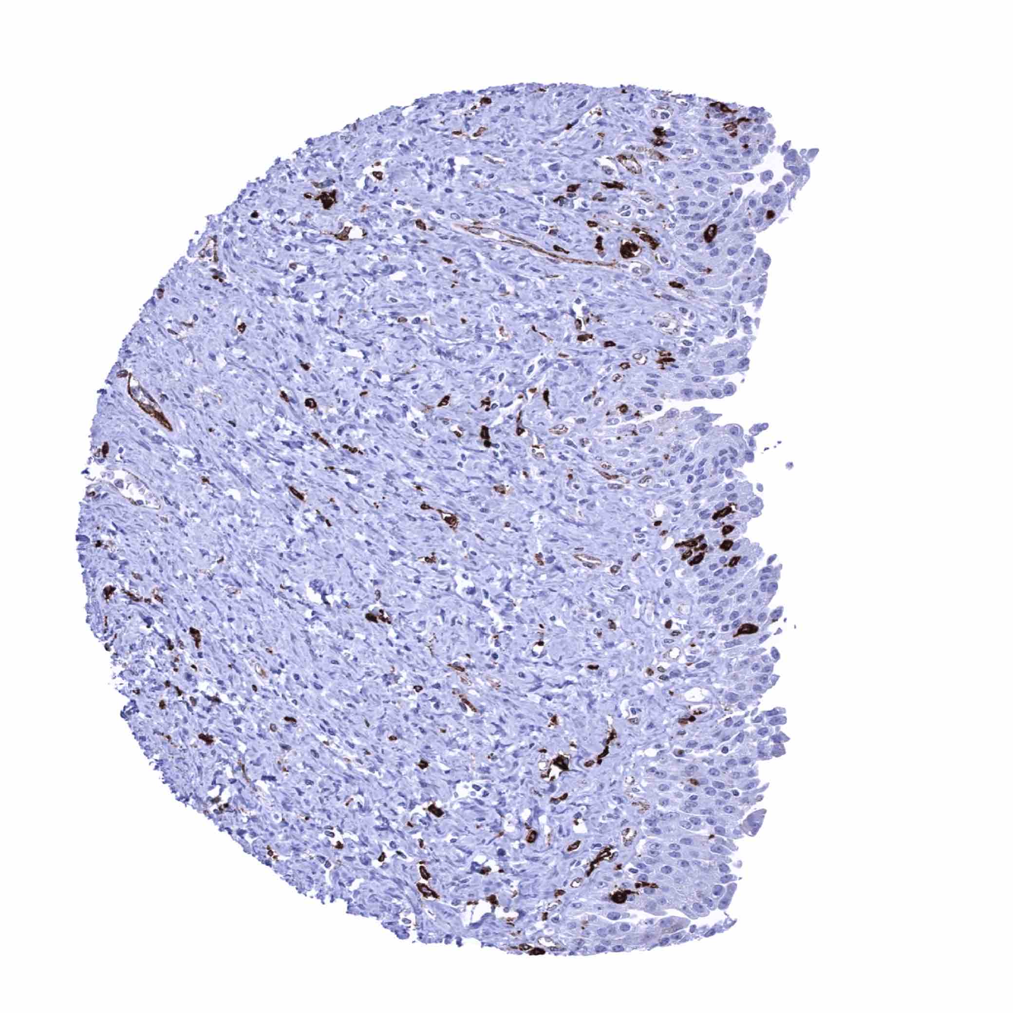
Bronchus, mucosa - Significant HLA-DRB1 staining of epithelial cells in this sample (HLA-DRB1 immunohistochemistry)

Cerebellum (molecular layer, Purkinje cell layer, granule cell layer, white matter)
Cerebellum, grey (Stratum neuronorum)

Cerebrum, grey matter
Cerebrum, white matter

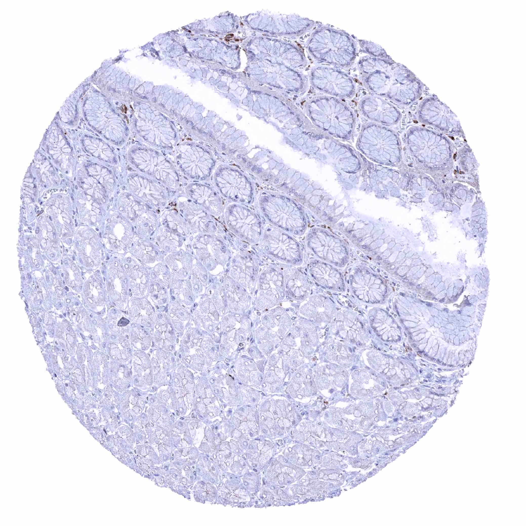
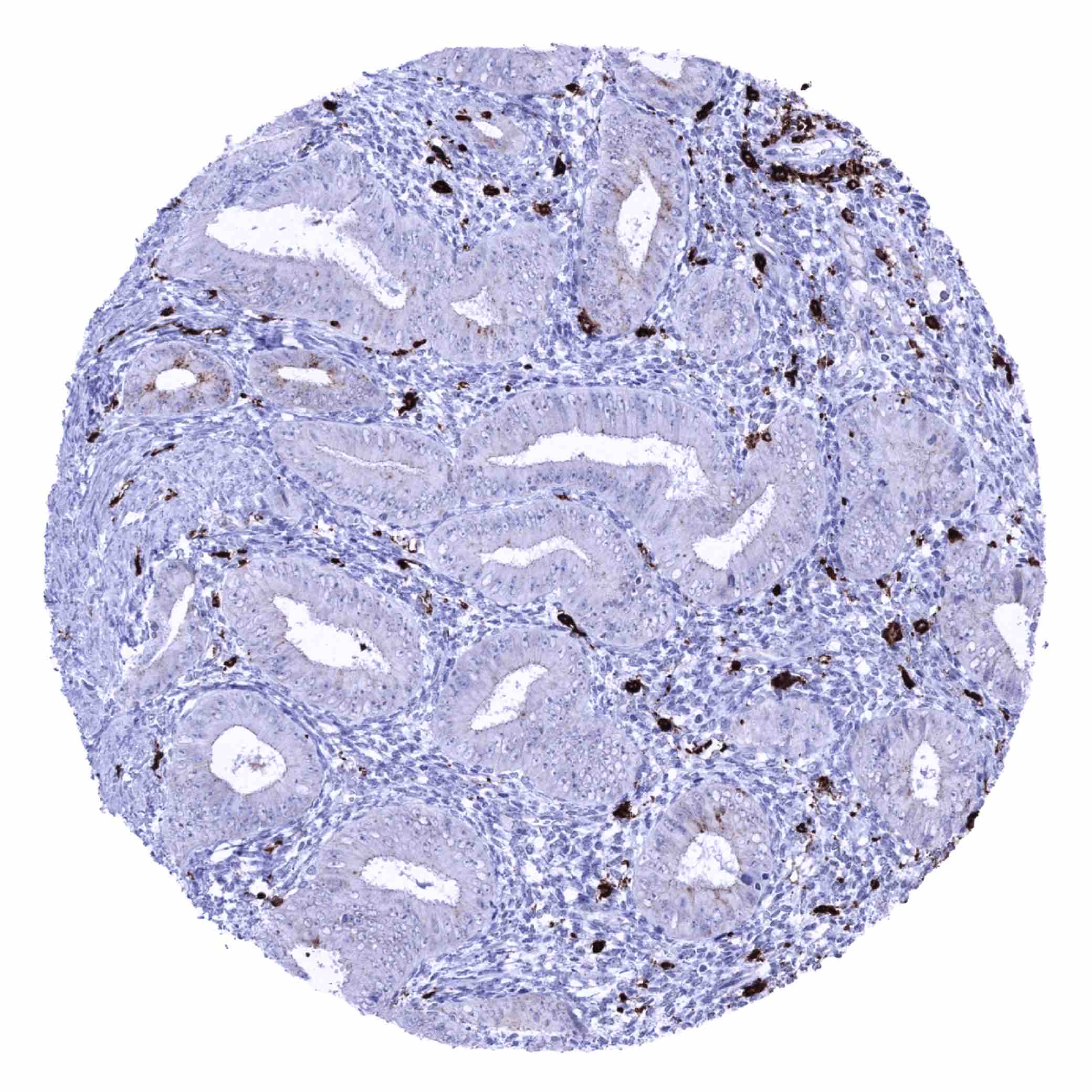
Colon descendens, mucosa - Strong HLA-DRB1 staining of mucosal inflammatory cells (HLA-DRB1 immunohistochemistry)
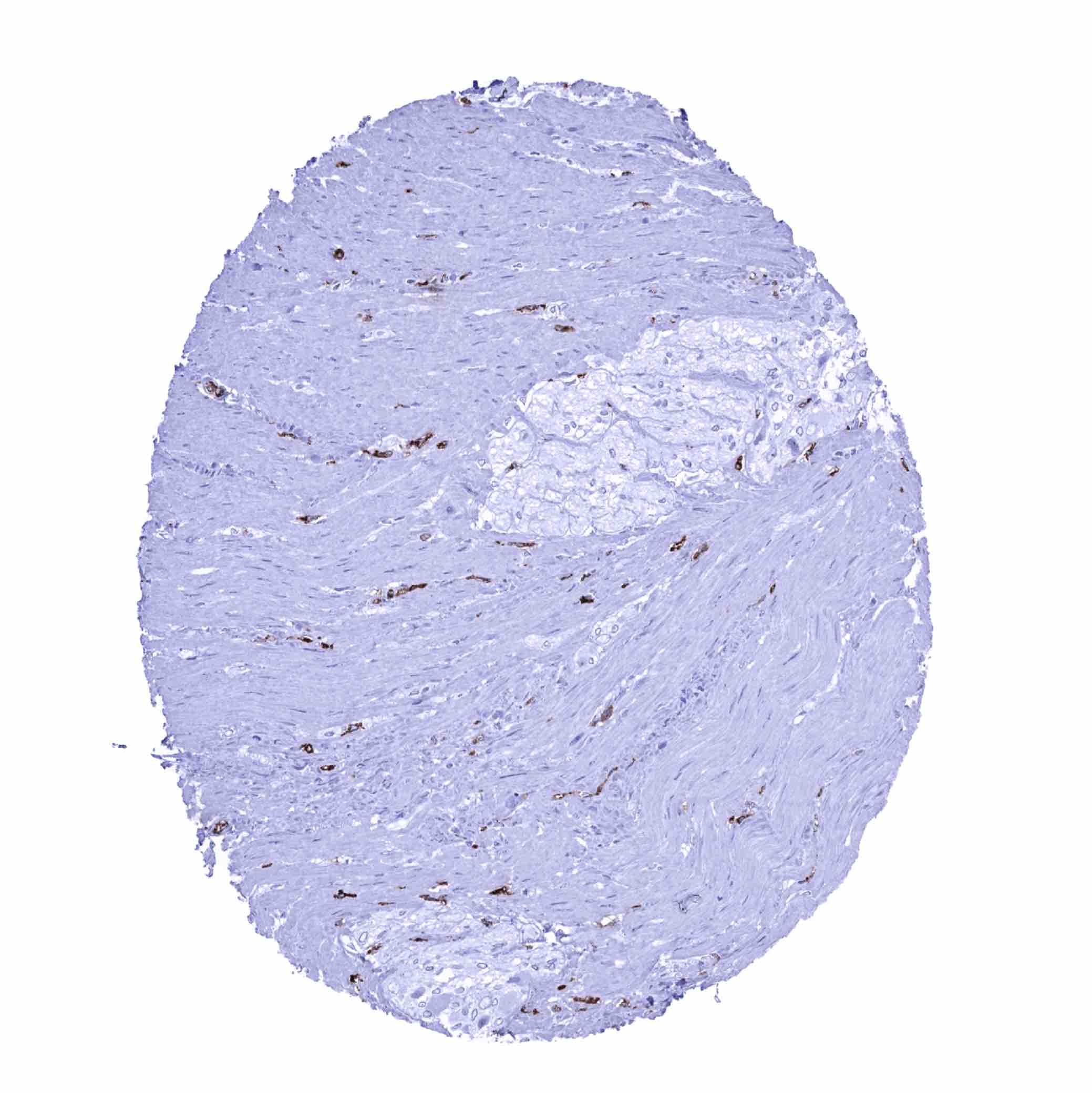
Colon descendens, muscular wall - HLA-DRB1 staining in dispersed inflammatory cells (HLA-DRB1 immunohistochemistry)

Colon descendens, muscular wall
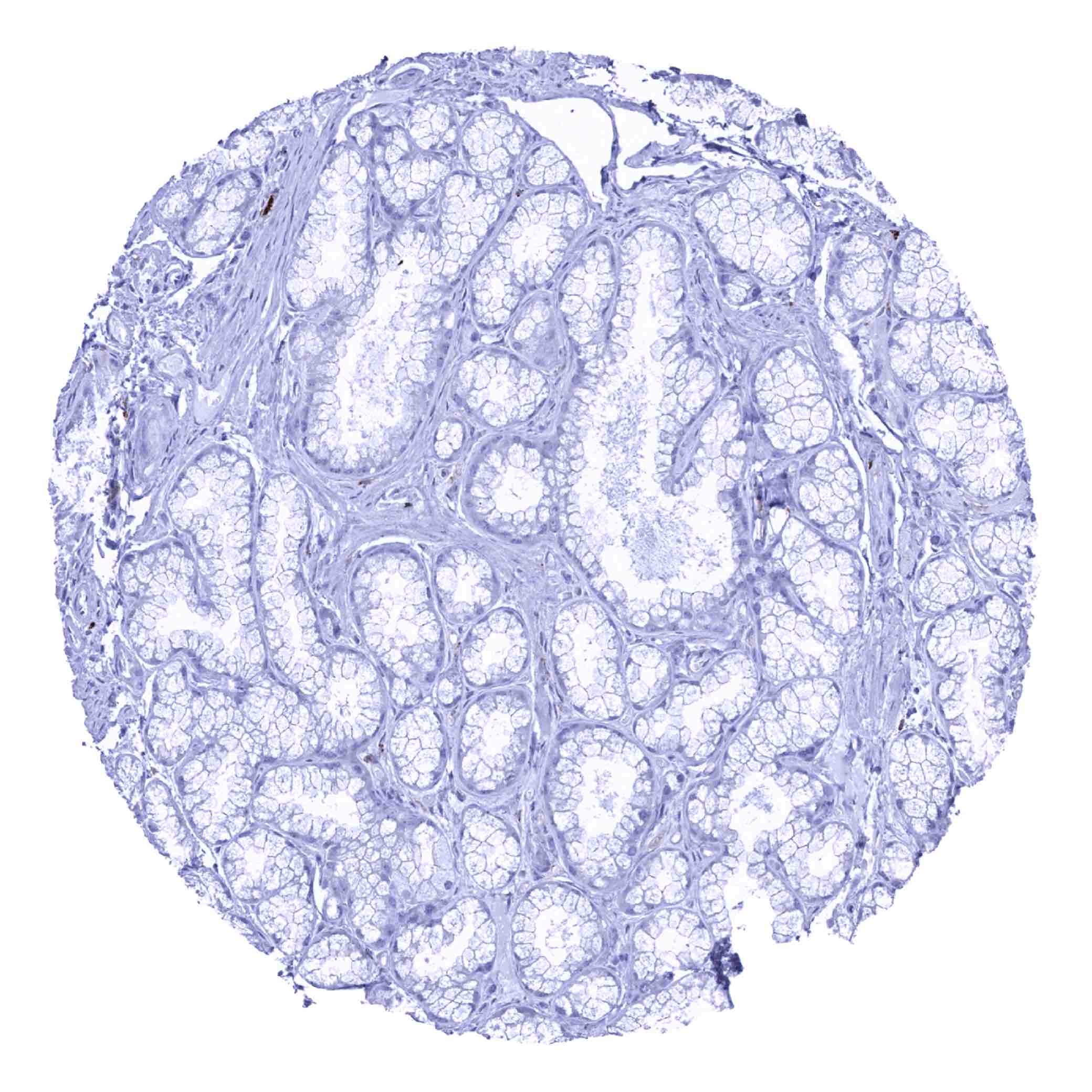
Duodenum, Brunner gland
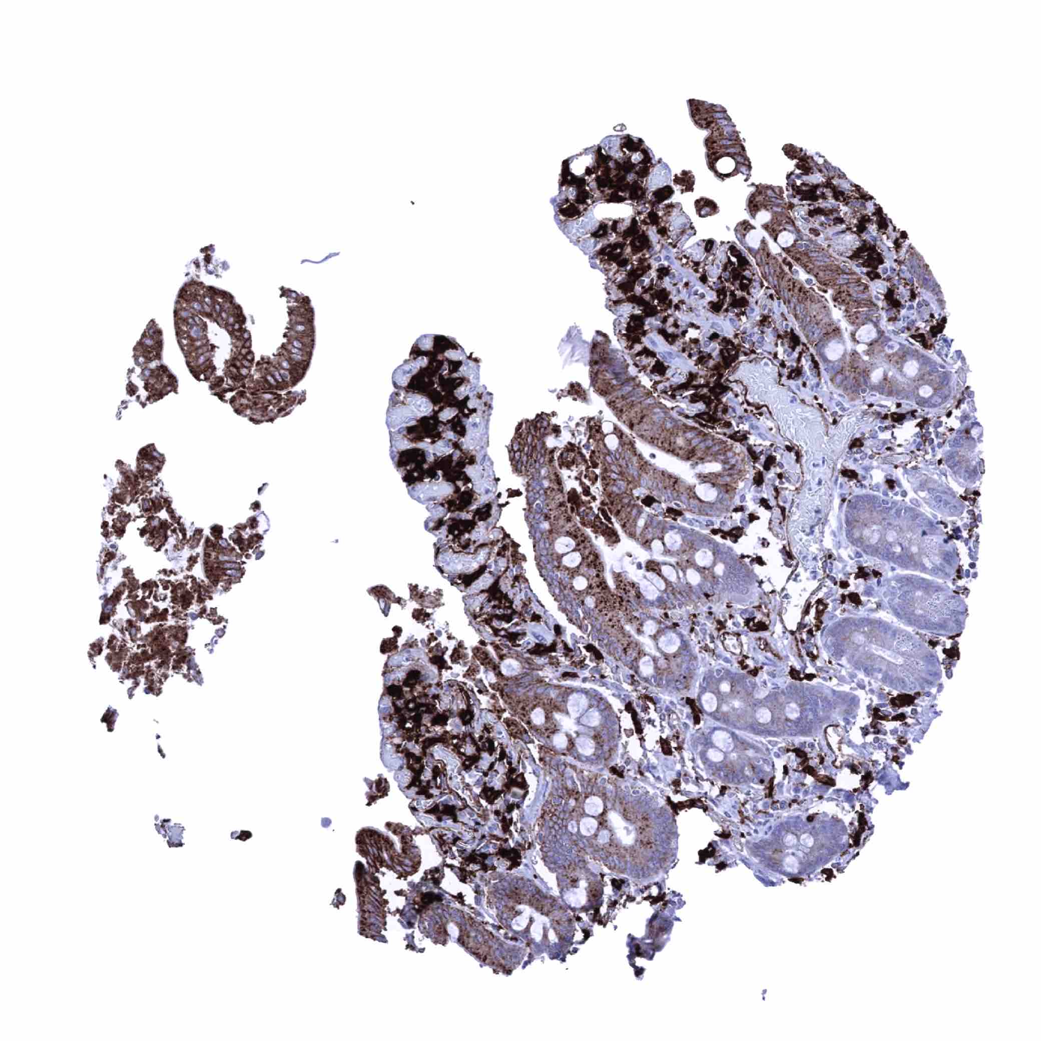
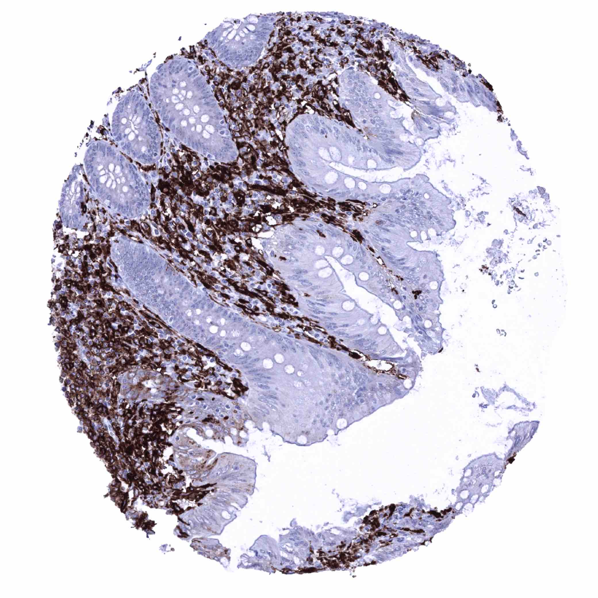
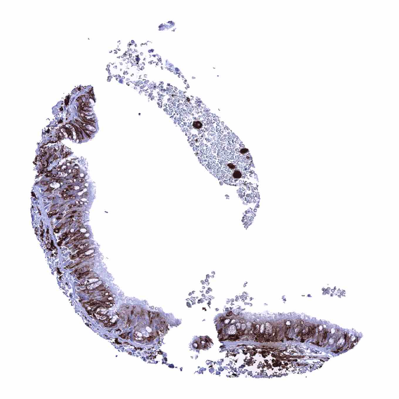
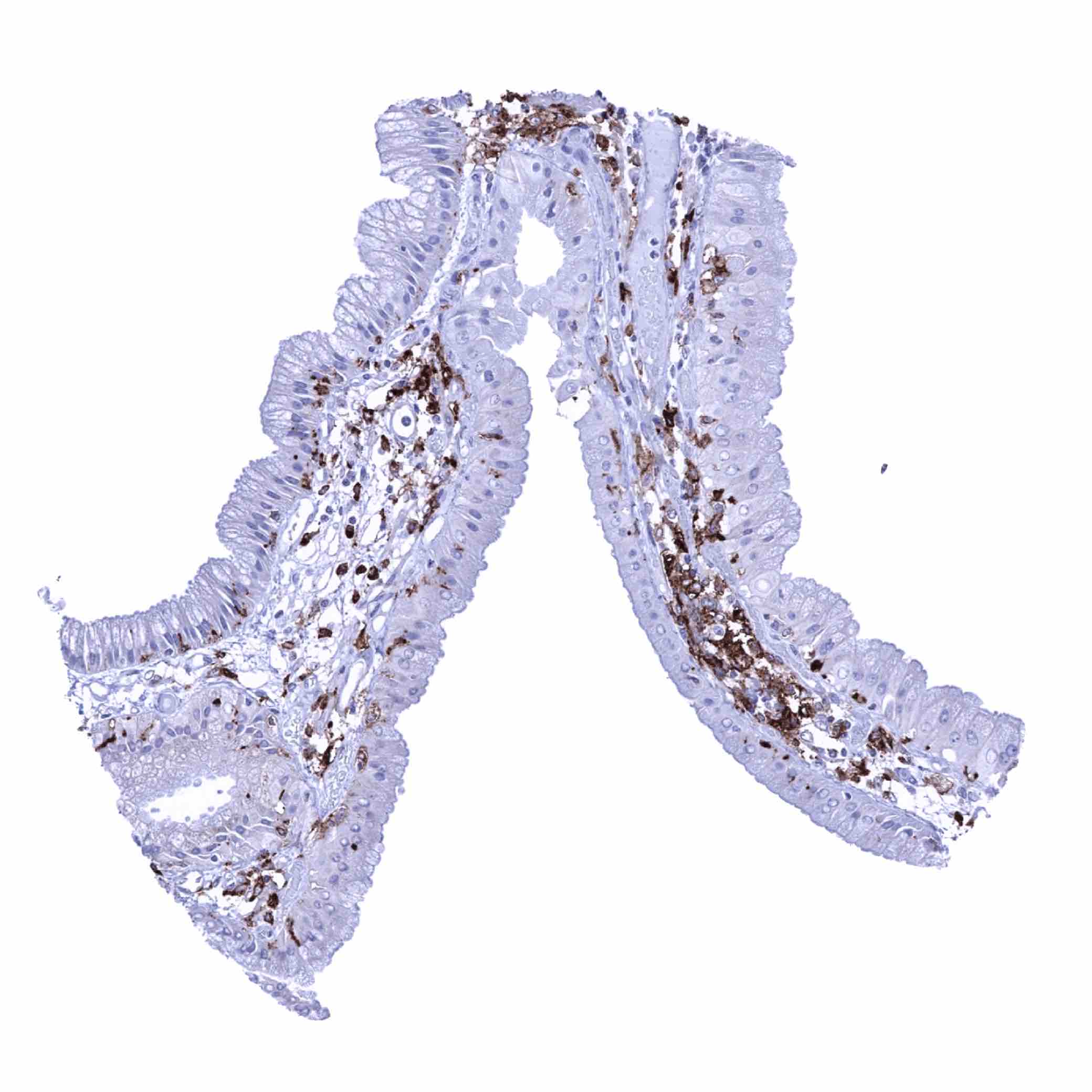
Duodenum, mucosa - Intense HLA-DRB1 staining of macrophages and possibly other inflammatory cells. Superfical epithelial cells also exhibit a positive staining (HLA-DRB1 immunohistochemistry)
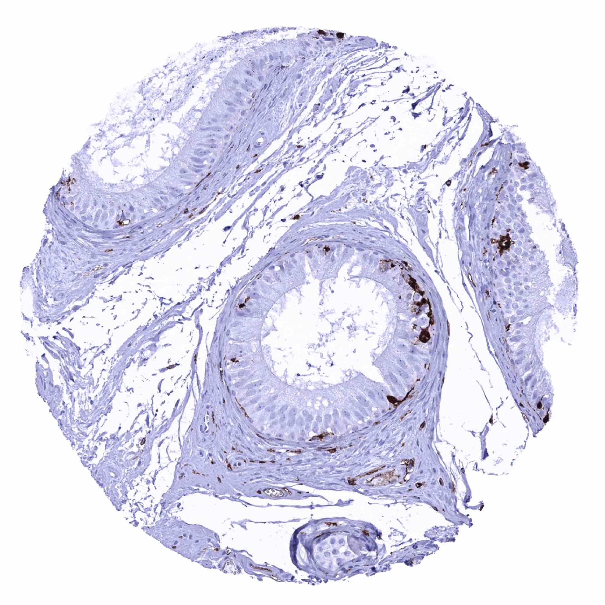
Epididymis - HLA-DRB1 staining in scattered inflammatory cells (HLA-DRB1 immunohistochemistry)
Esophagus, squamous epithelium - HLA-DRB1 staining of scattered inflammatory cells (HLA-DRB1 immunohistochemistry)
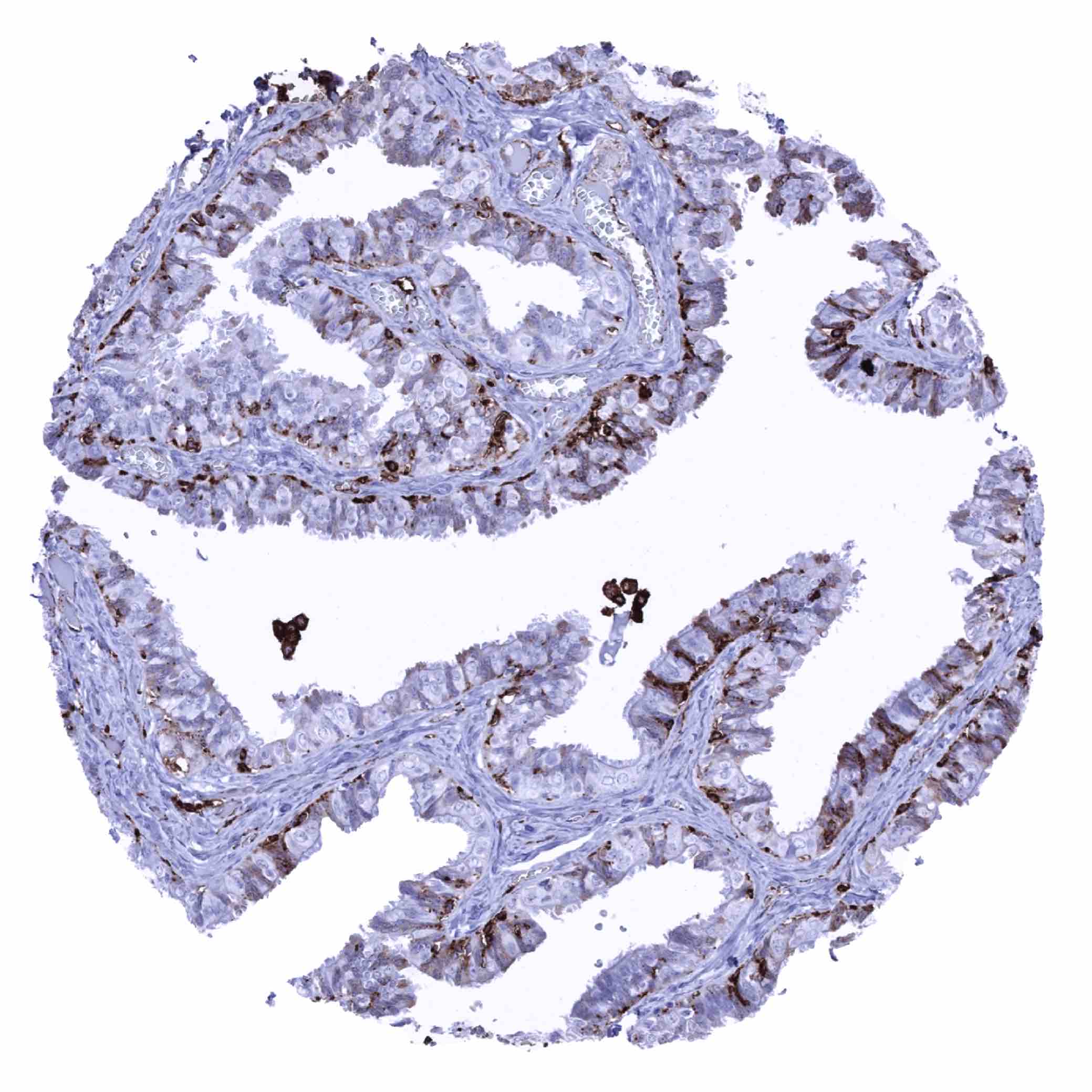
Fallopian tube, mucosa - HLA-DRB1 staining in a fraction of epithelial cells and in few inflammatory cells in this sample (HLA-DRB1 immunohistochemistry)

Fallopian tube, mucosa - HLA-DRB1 staining in restricted to few inflammatory cells in this sample (HLA-DRB1 immunohistochemistry)
Fat
Gallbladder, epithelium - HLA-DRB1 staining of scattered inflammatory cells (HLA-DRB1 immunohistochemistry)

Heart muscle

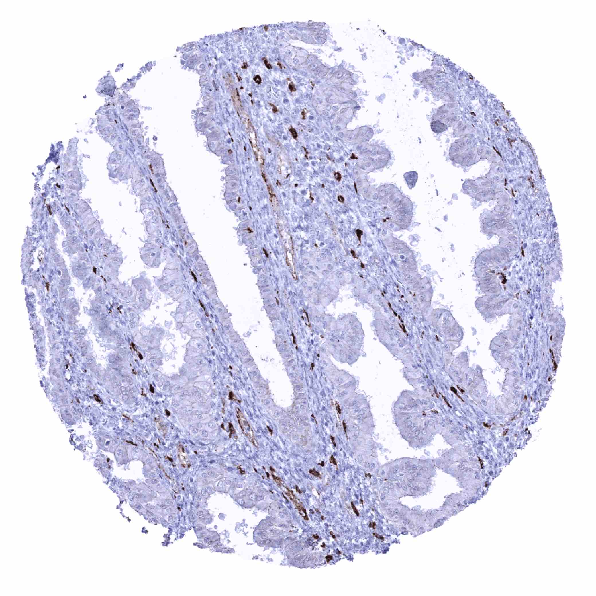
Ileum, mucosa - Intense HLA-DRB1 staining of macrophages and possibly other inflammatory cells. Superfical epithelial cells also exhibit a positive staining (HLA-DRB1 immunohistochemistry)
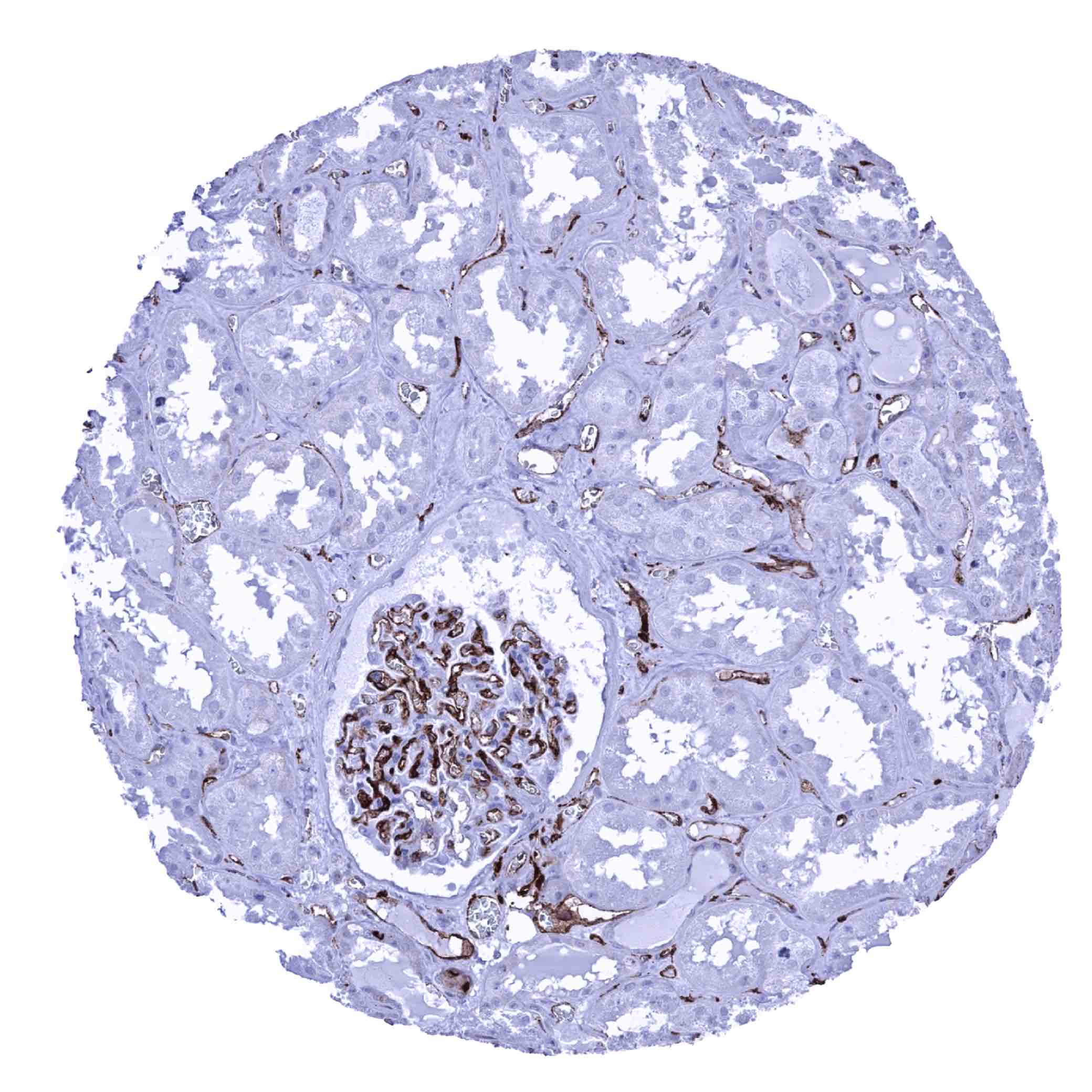
Kidney, cortex - HLA-DRB1 staining is limited to endothelial cells including glomerular endothelium in the non-inflamed kidney (HLA-DRB1 immunohistochemistry)
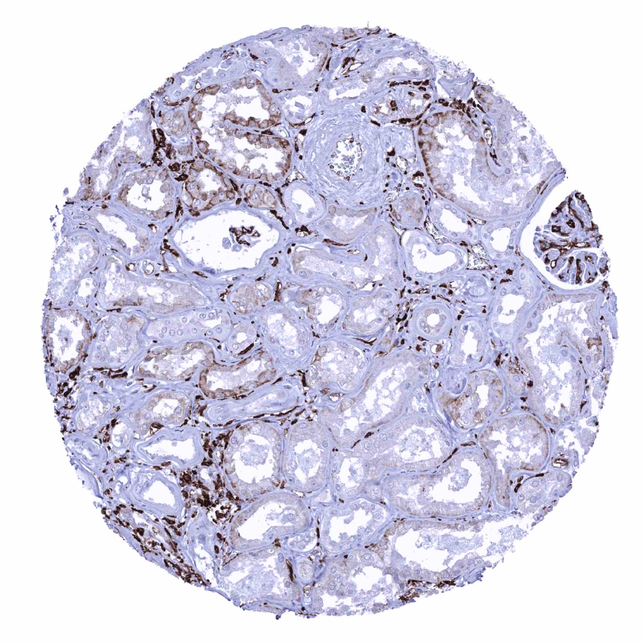
Kidney, cortex - In the inflamed kidney, HLA-DRB1 staining is seen in endothelial cells, inflammatory cells and in tubulus cells adjacent to inflammation (HLA-DRB1 immunohistochemistry)

Kidney, medulla - HLA-DRB1 staining is largely lacking in this sample (HLA-DRB1 immunohistochemistry)
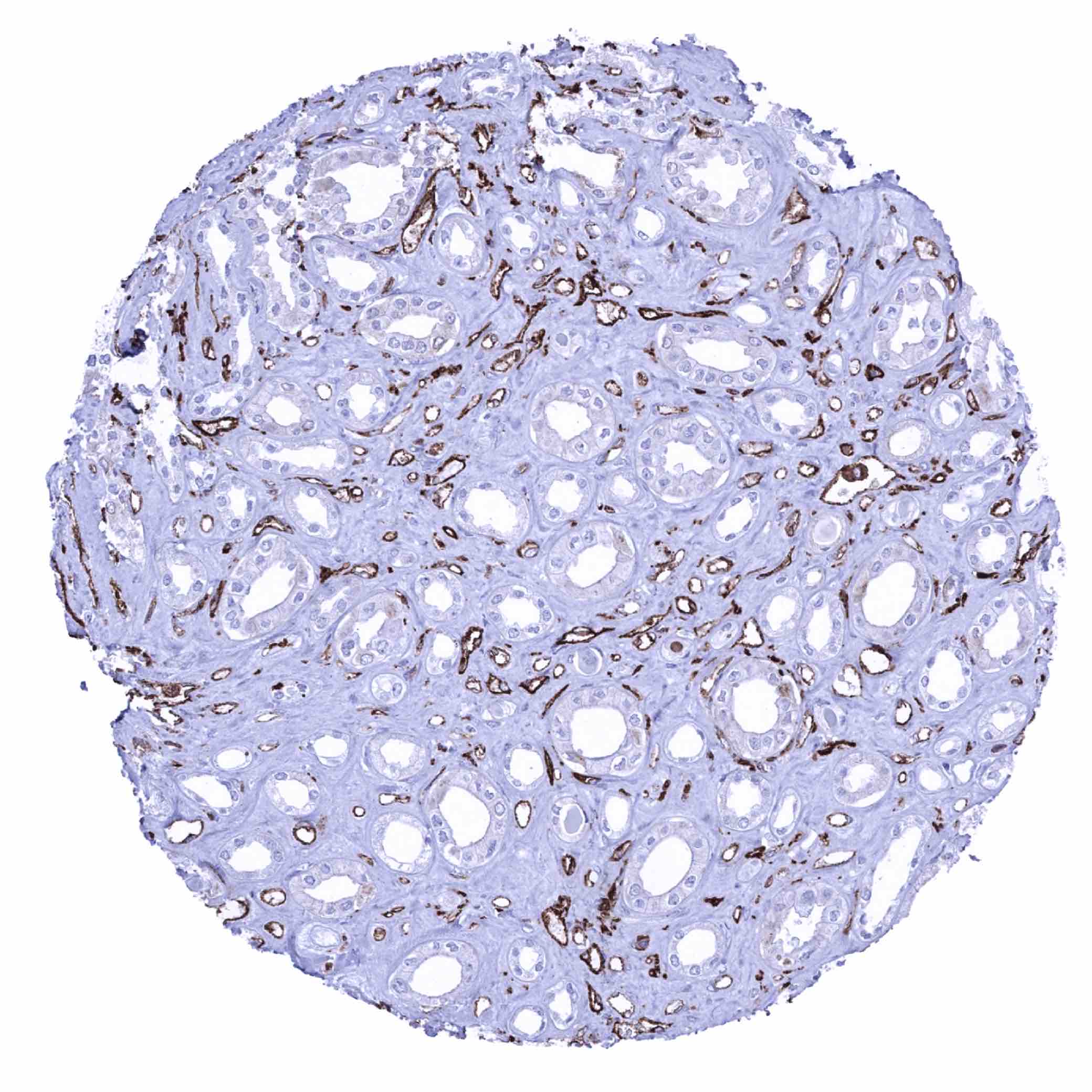
Kidney, medulla - Intense HLA-DRB1 staining in all endothelial cells in this sample (HLA-DRB1 immunohistochemistry)
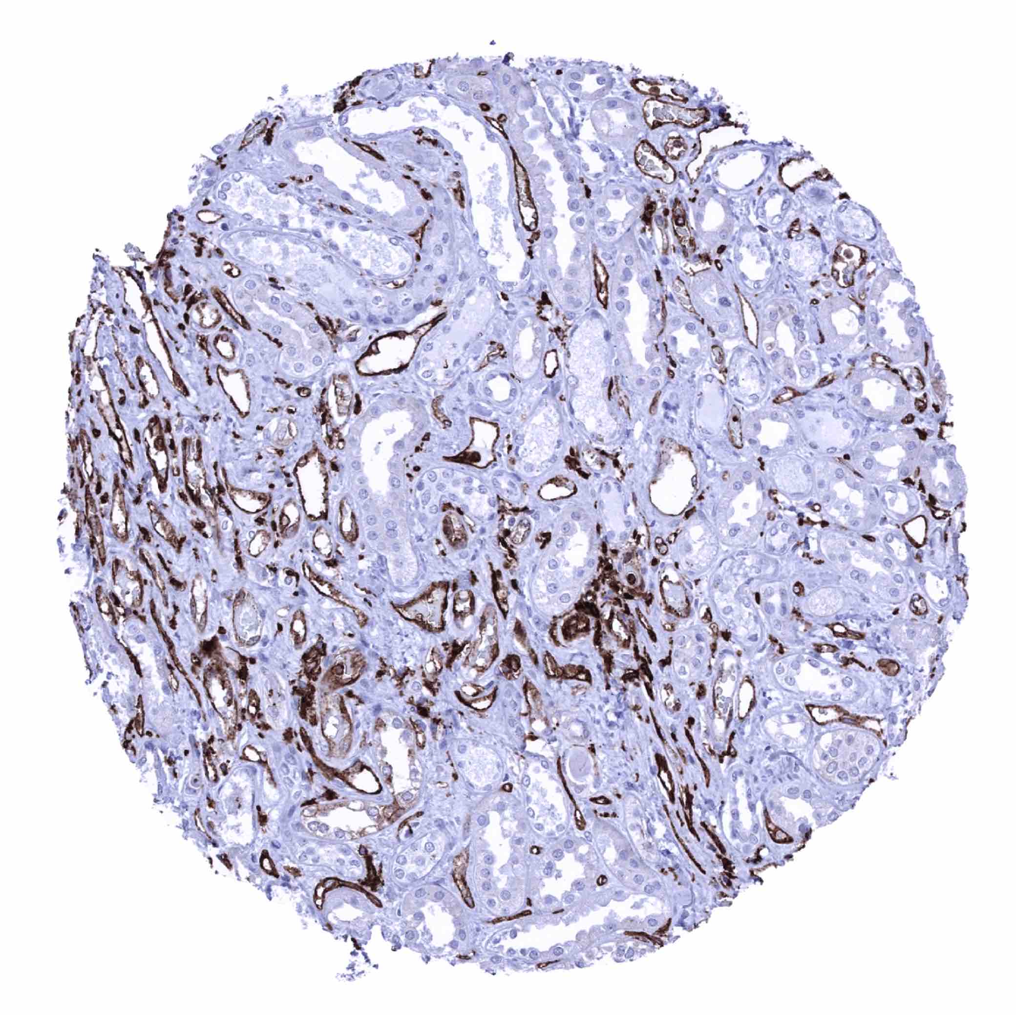
Kidney, medulla - Intense HLA-DRB1 staining in endothelial cells in this sample (HLA-DRB1 immunohistochemistry)

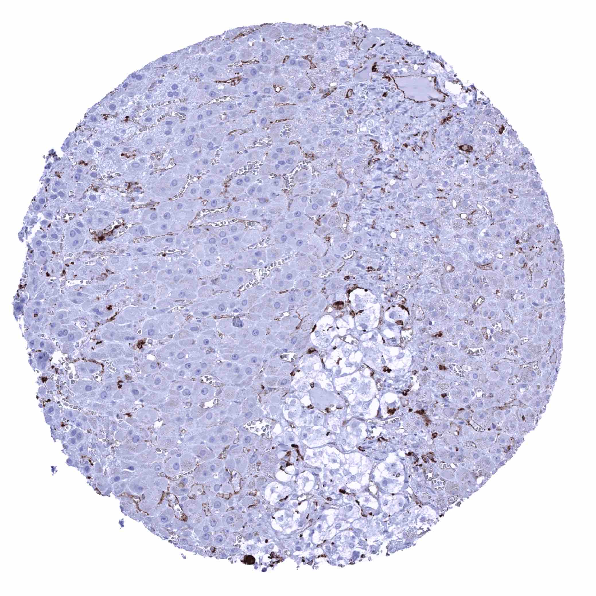
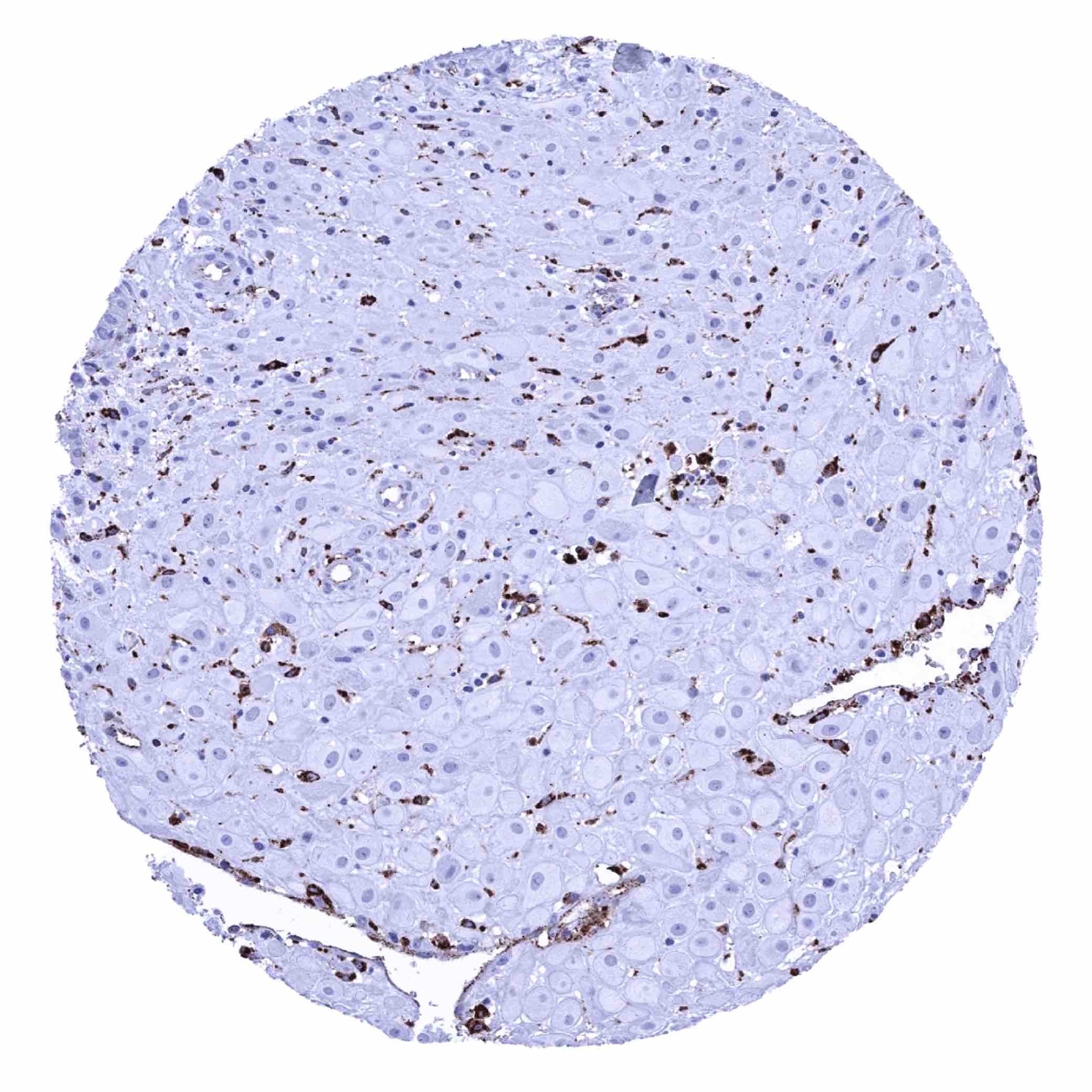
Liver - Intense HLA-DRB1 staining of inflammatory cells and of Kupffer cells (HLA-DRB1 immunohistochemistry)
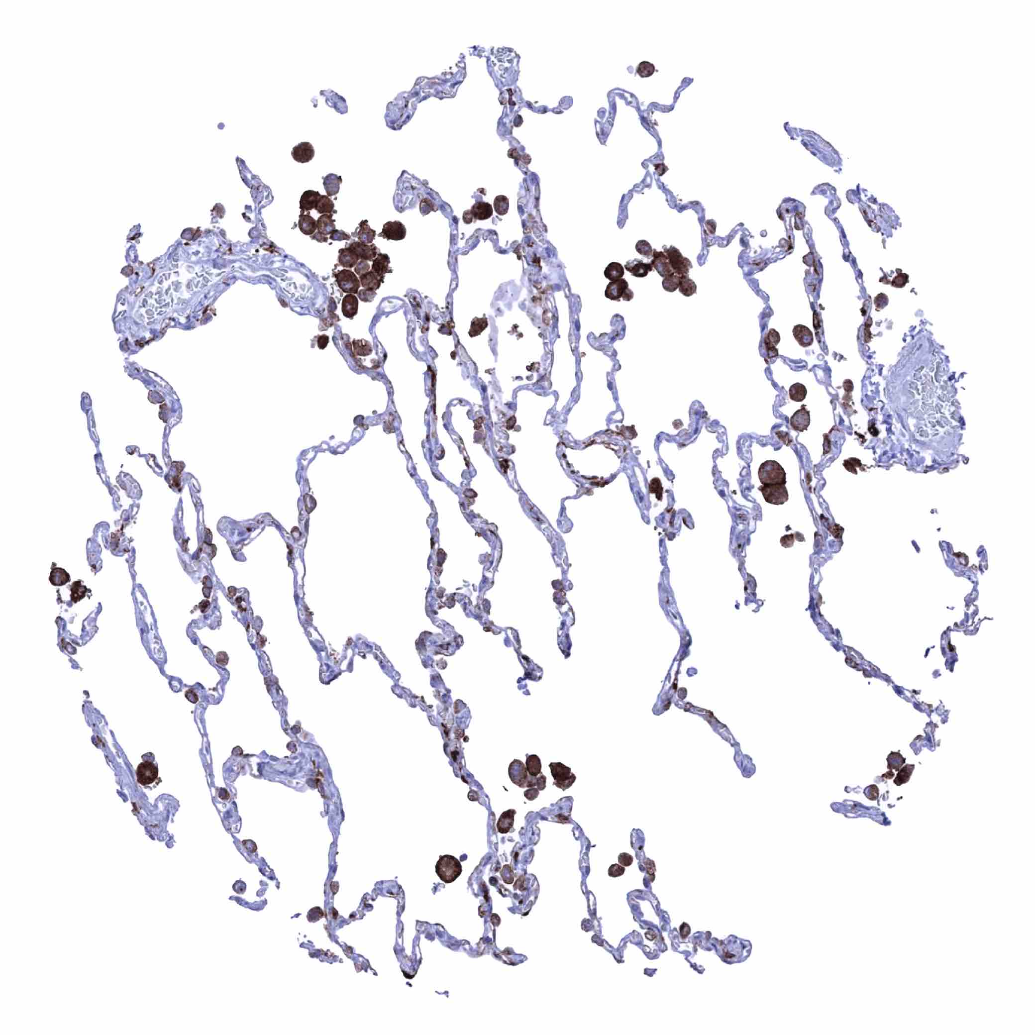
Lung – Strong HLA-DRB1 staining in alveolar macrophages (HLA-DRB1 immunohistochemistry)
Lymph node - HLA-DRB1 staining macrophages, dendritic cells and B-lymphocytes. Some endothelial cells also stain positive (HLA-DRB1 immunohistochemistry)
Ovary, stroma
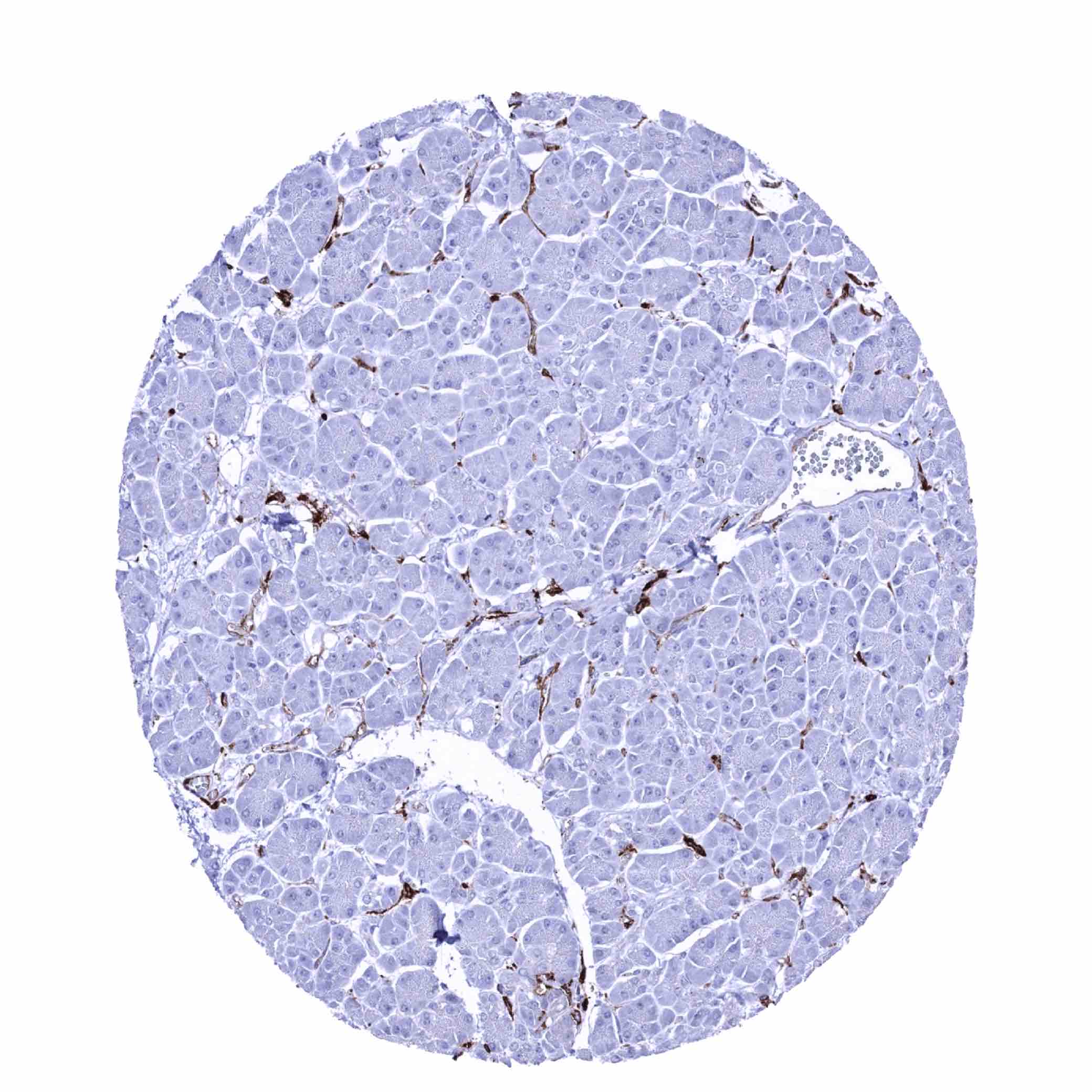
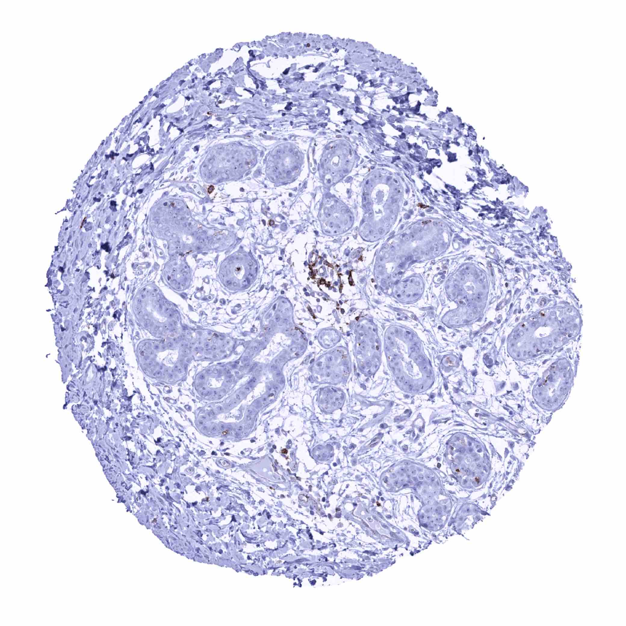
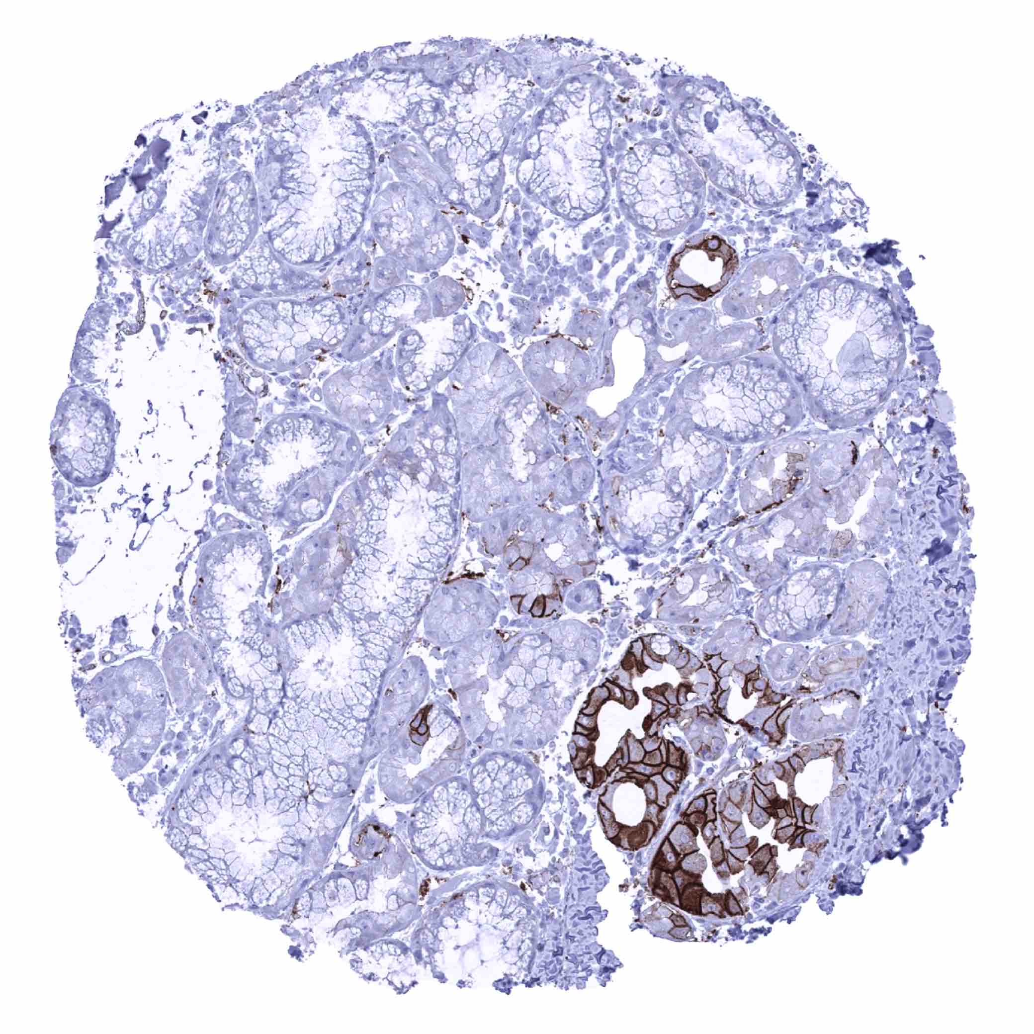
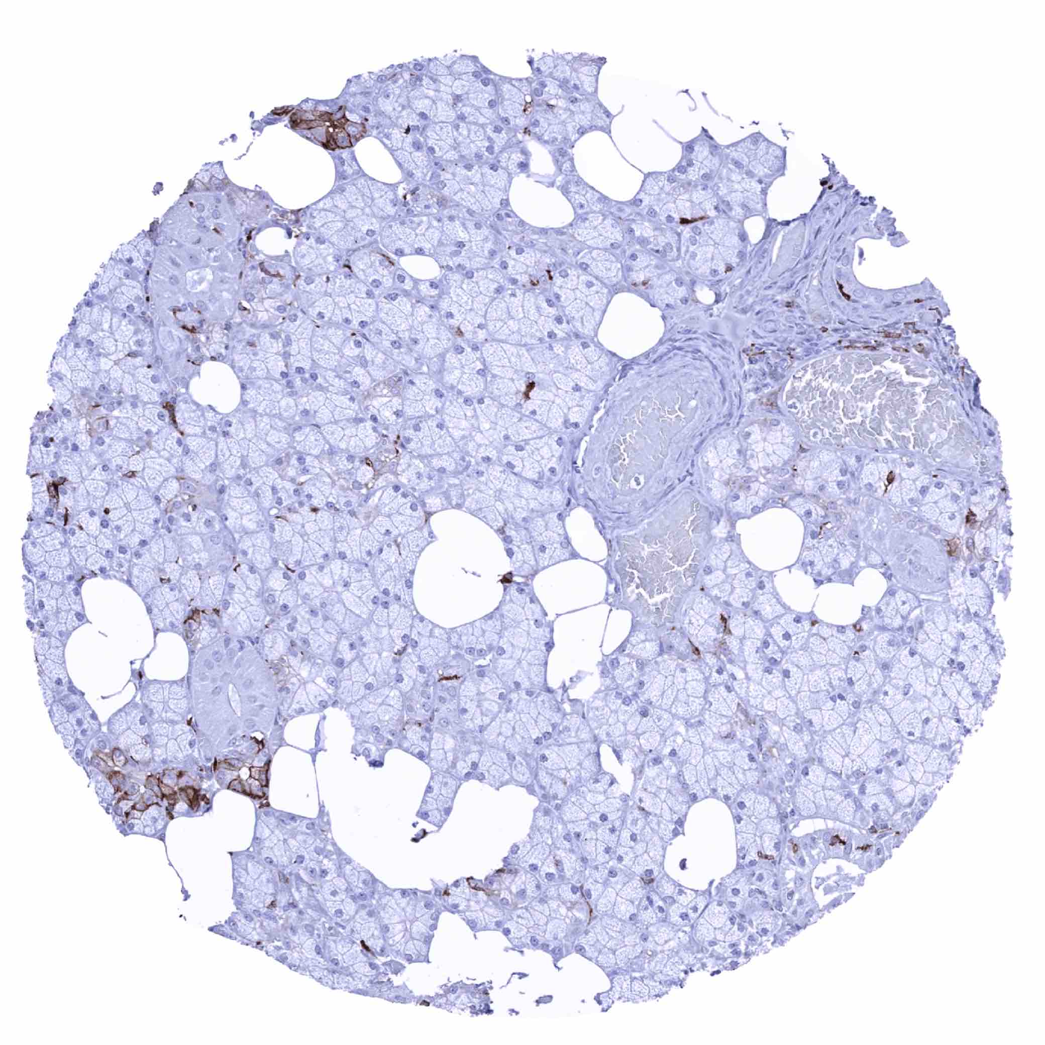
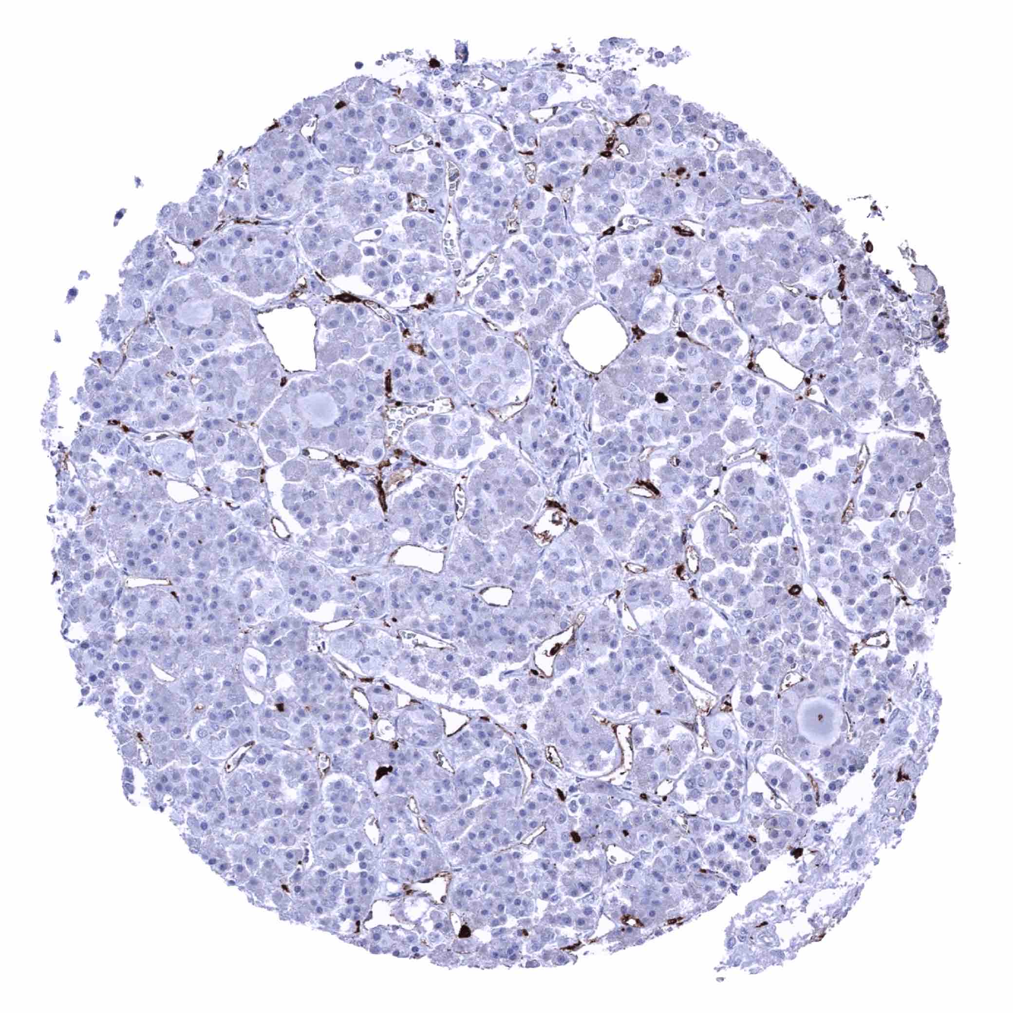
Pancreas - Distinct HLA-DRB1 positivity of inflammatory cells and endothelial cells of small capillaries (HLA-DRB1 immunohistochemistry)
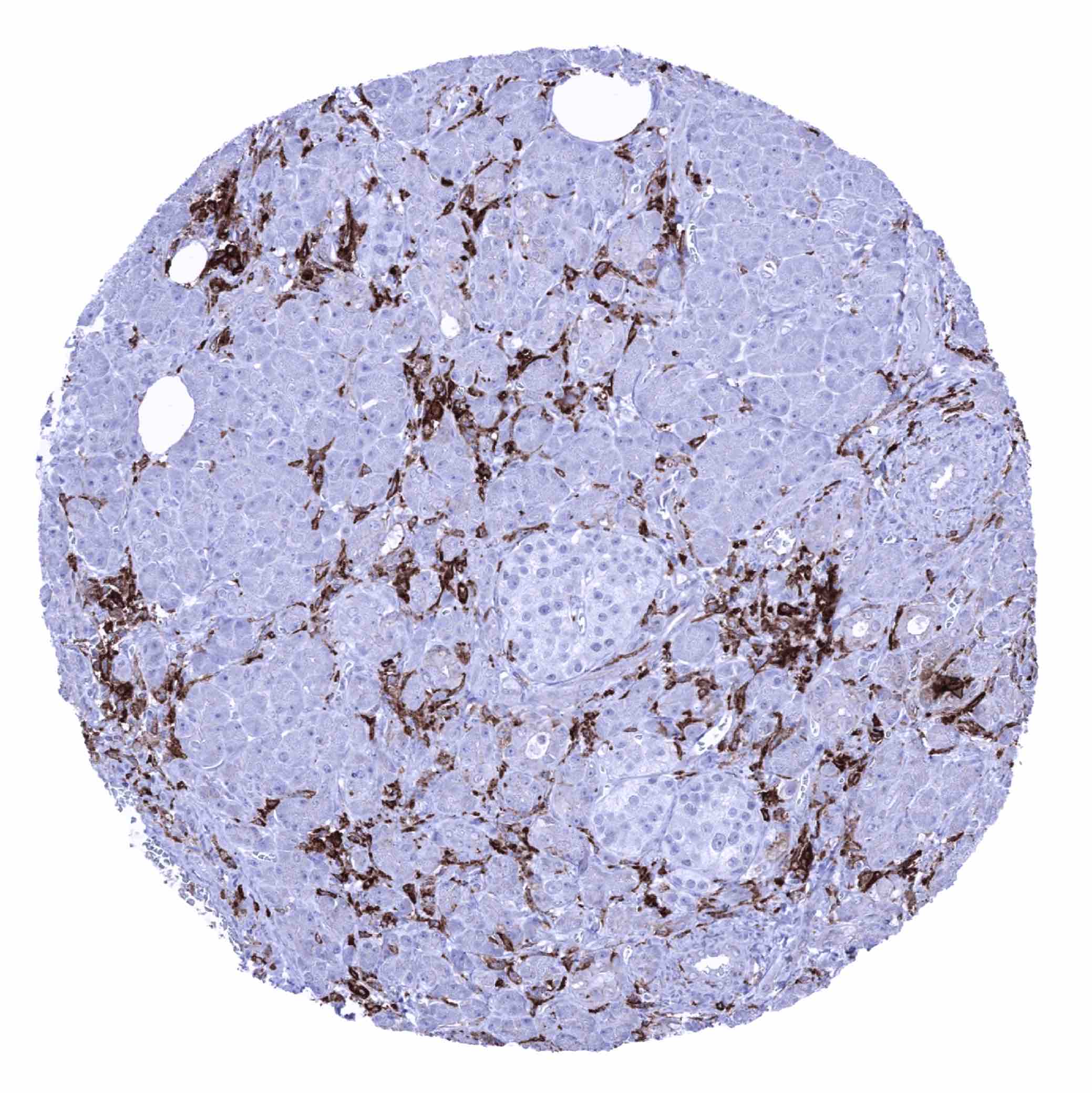
Pancreas - Strong HLA-DRB1 staining of inflammatory cells. Few acinar epithelial cells also show a weak focal positivity (HLA-DRB1 immunohistochemistry)
Parathyroid gland
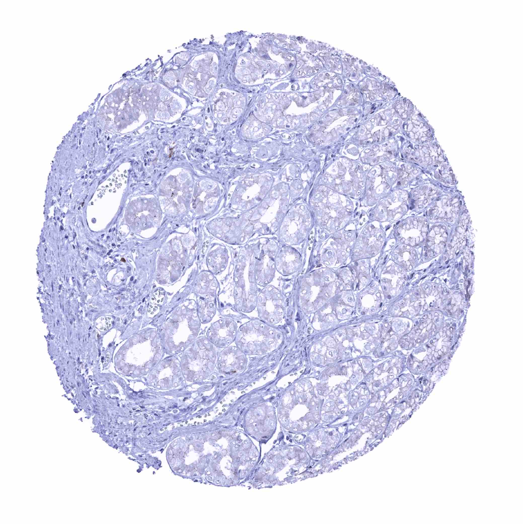
Parotid gland - Distinct HLA-DRB1 staining of few epithelial cells. Some scattered inflammatory cells are also positive (HLA-DRB1 immunohistochemistry)
Pituitary gland, anterior lobe - Significant HLA-DRB1 staining in endothelial cells (HLA-DRB1 immunohistochemistry)

Pituitary gland, posterior lobe

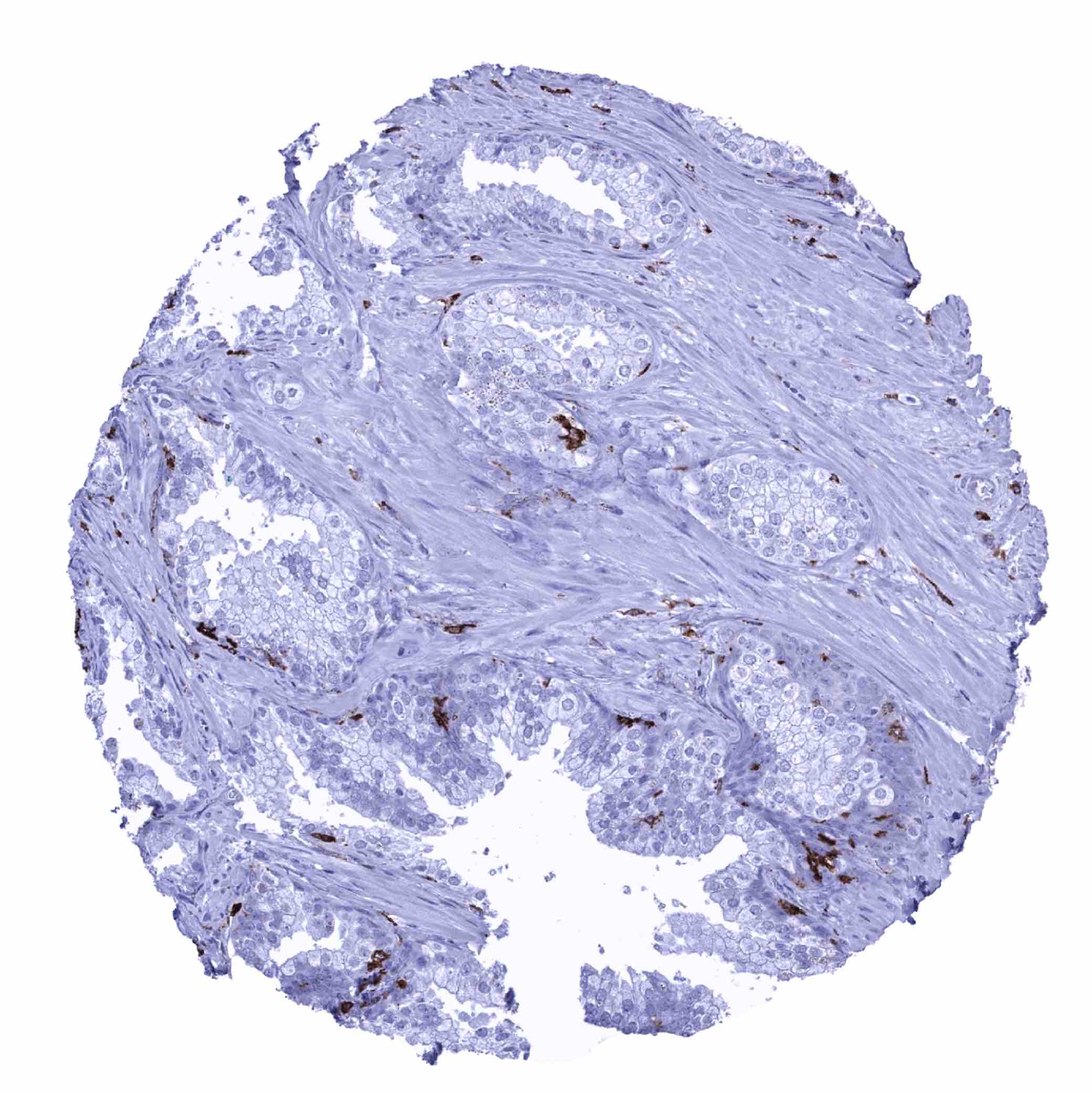
Placenta (amnion and chorion) - HLA-DRB1 staining of scattered inflammatory cells (HLA-DRB1 immunohistochemistry)

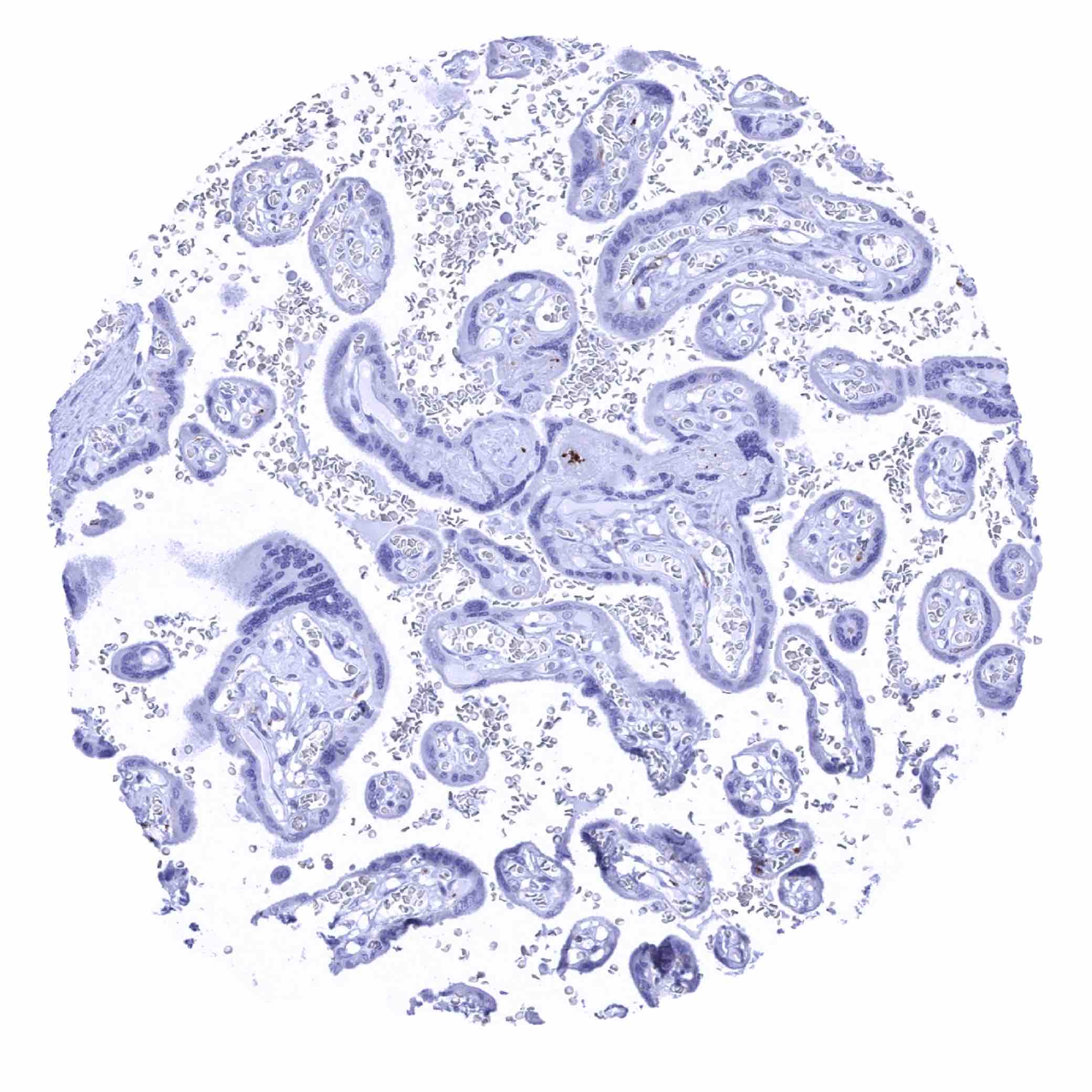
Placenta (amnion and chorion)

Placenta, early
Placenta, mature
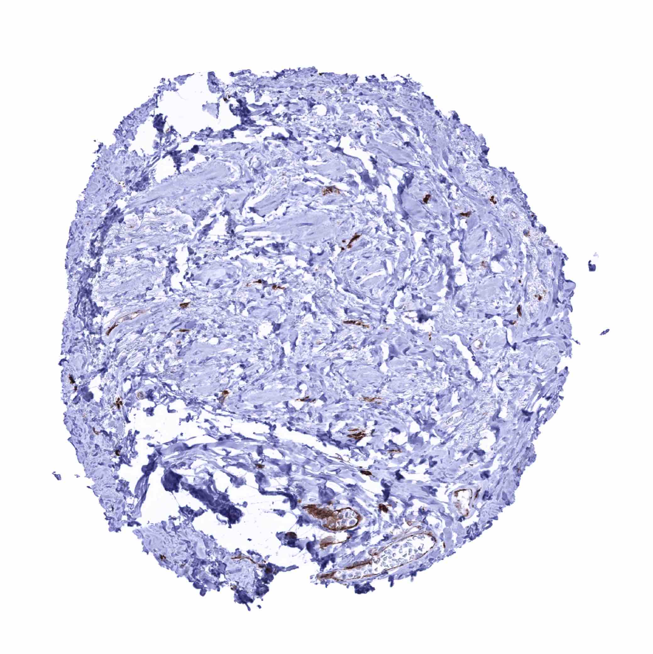
Prostate - HLA-DRB1 staining in scattered inflammatory cells (HLA-DRB1 immunohistochemistry)

Rectum, mucosa
Seminal vesicle - HLA-DRB1 staining in scattered inflammatory cells and in some endothelial cells of small capillaries (HLA-DRB1 immunohistochemistry)
Sinus paranasales - HLA-DRB1 staining in scattered inflammatory cells (HLA-DRB1 immunohistochemistry)
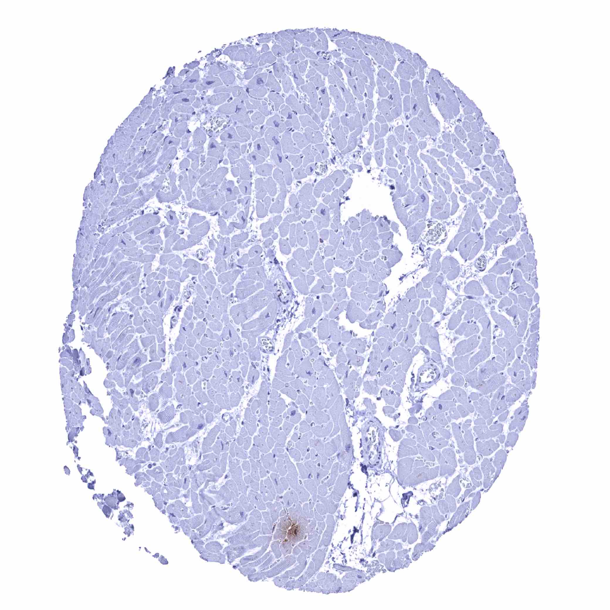
Skeletal muscle
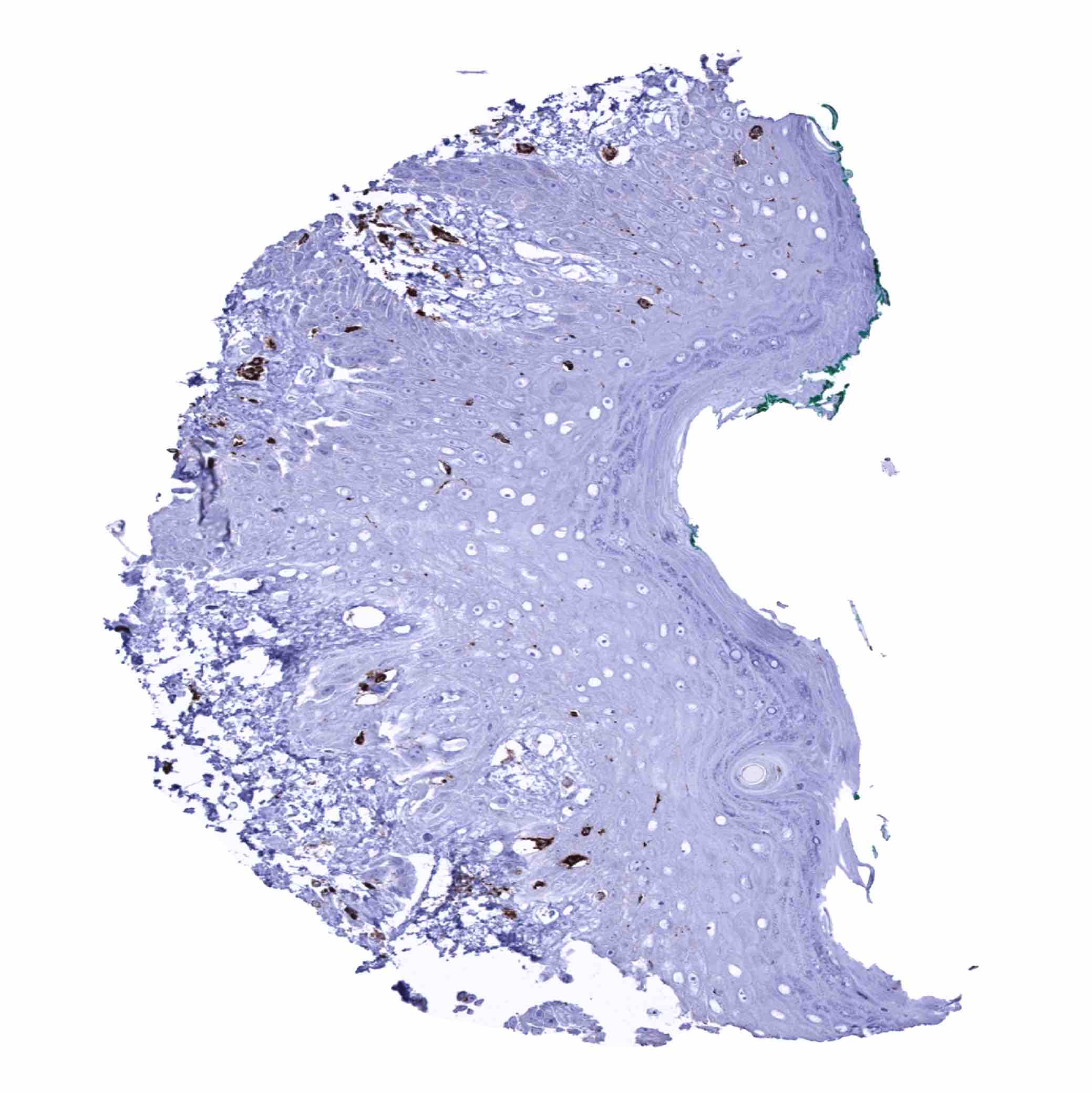
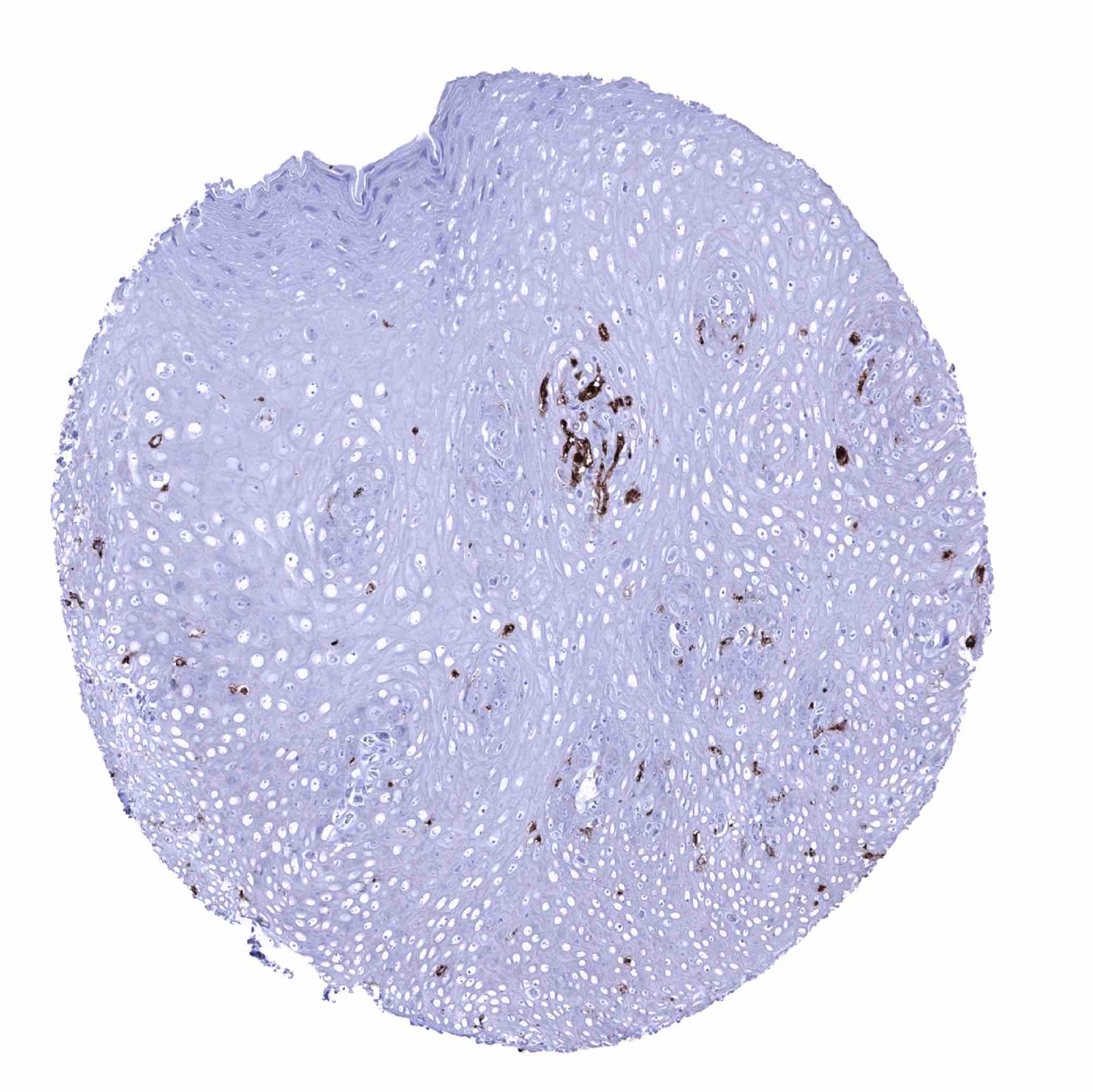
Skin - HLA-DRB1 staining of scattered inflammatory cells (HLA-DRB1 immunohistochemistry)
Spleen - HLA-DRB1 staining of lymphocytes and macrophages (HLA-DRB1 immunohistochemistry)
Stomach, antrum
Stomach, corpus - HLA-DRB1 staining of scattered inflammatory cells (HLA-DRB1 immunohistochemistry)

Sublingual gland - HLA-DRB1 staining is largely limited to endothelial cells and few inflammatory cells in this sample (HLA-DRB1 immunohistochemistry)

Sublingual gland - Intense HLA-DRB1 staining in most epithelial cells. Endothelium and some discernible inflammatory cells are also positive in this sample (HLA-DRB1 immunohistochemistry)
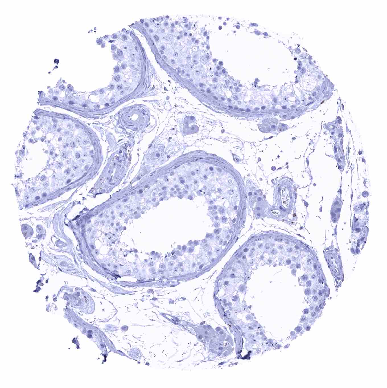
Testis

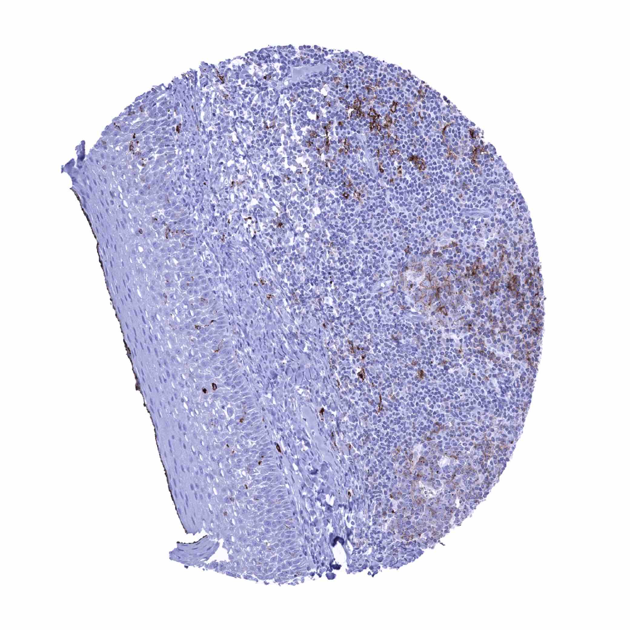
Thymus - HLA-DRB1 staining of a subset of cells. Most thymocytes remain negative (HLA-DRB1 immunohistochemistry)

Thyroid gland
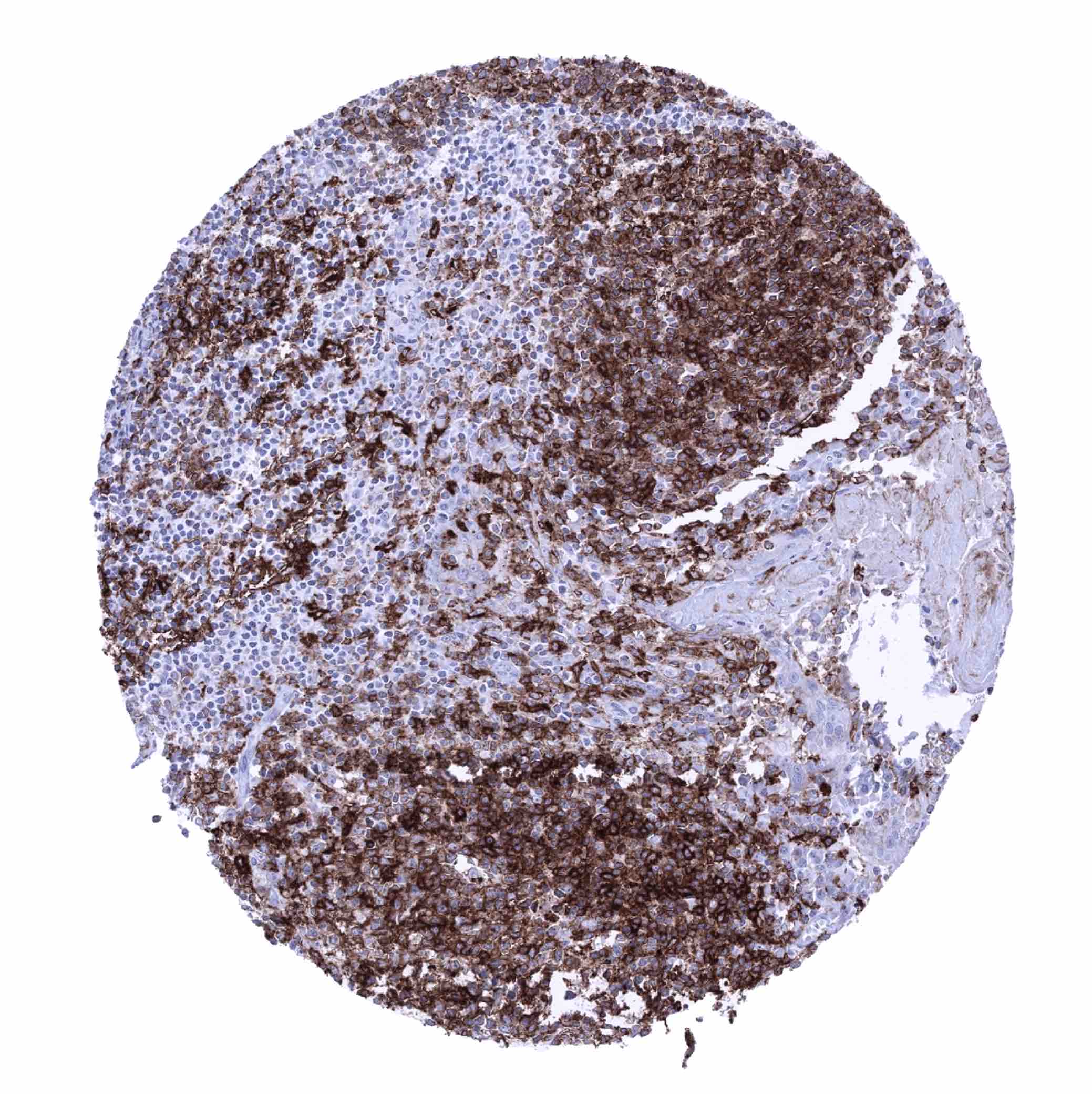
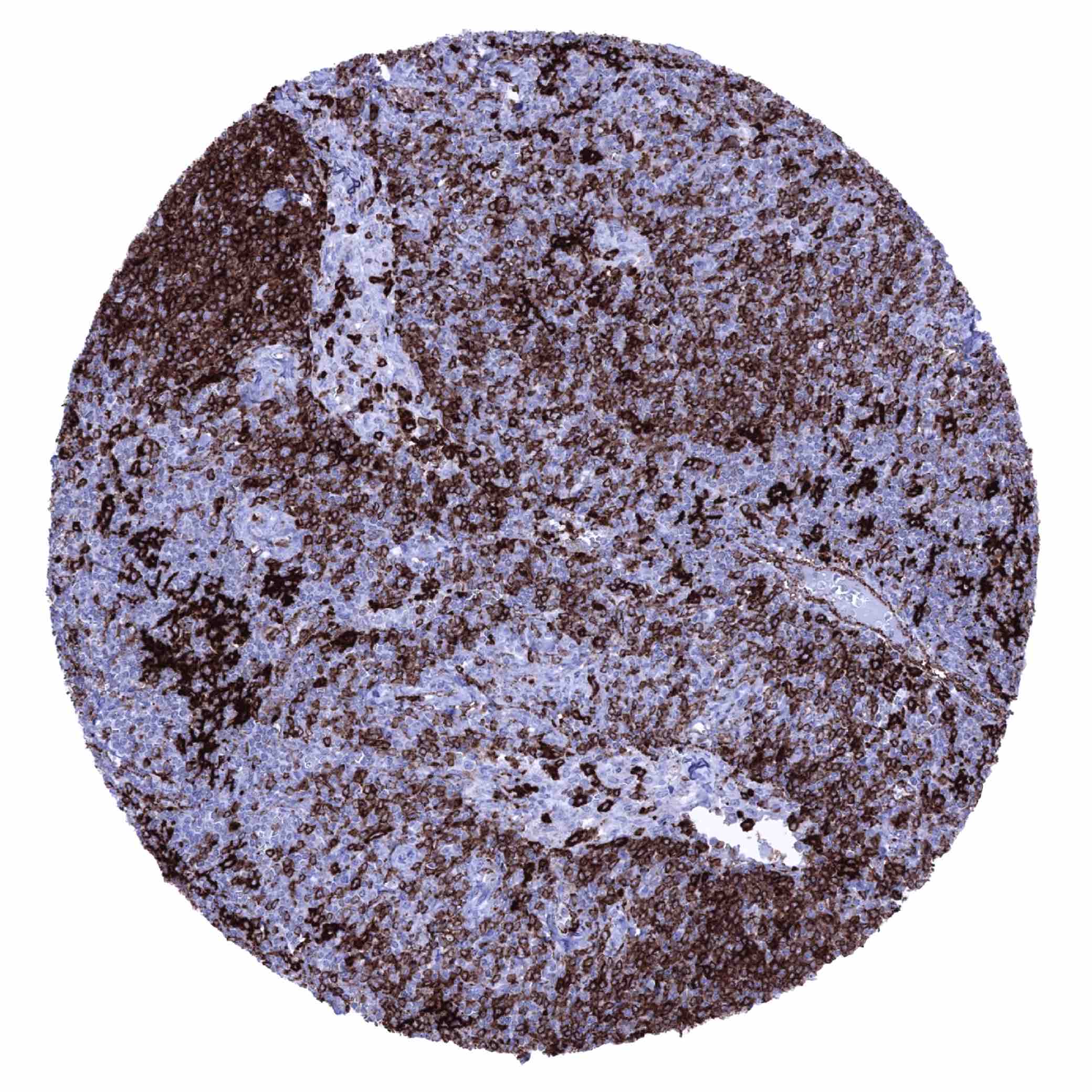
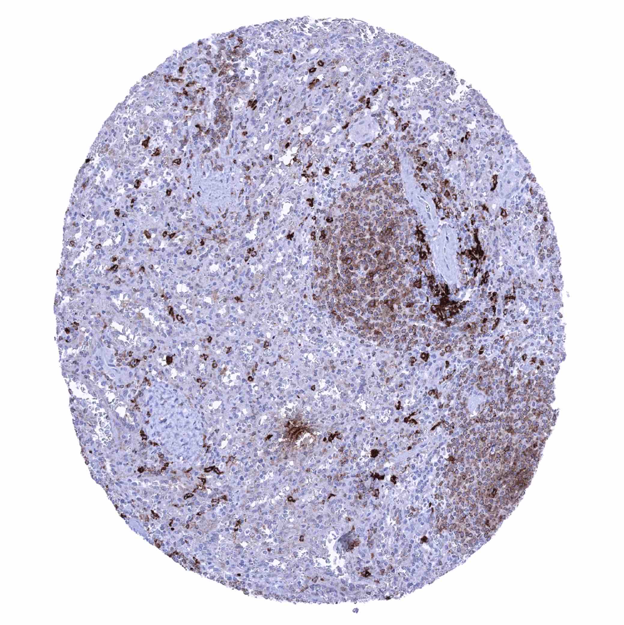
Tonsil - HLA-DRB1 staining of a large fraction of lymphocytes (HLA-DRB1 immunohistochemistry)
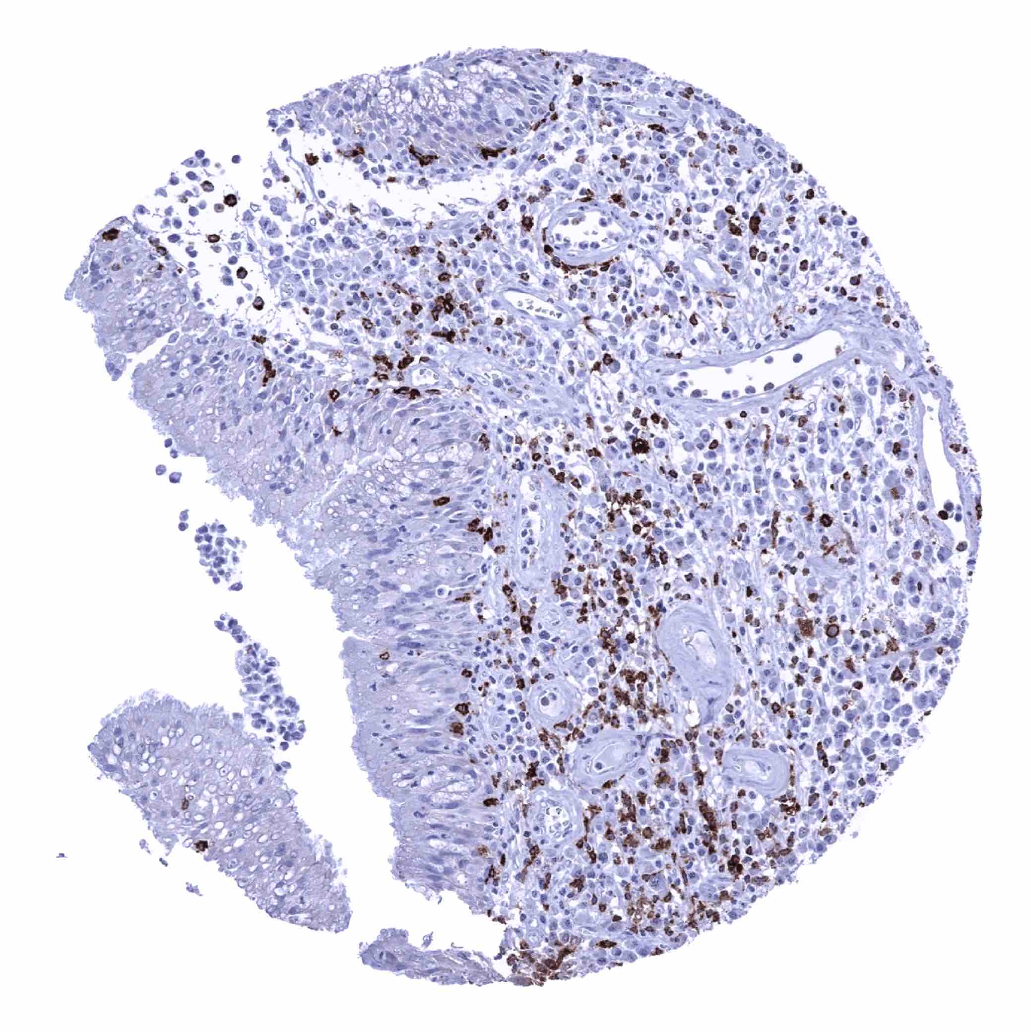
Tonsil, surface epithelium - HLA-DRB1 staining of some inflammatory cells, mainly macrophages and B-lymphocytes (HLA-DRB1 immunohistochemistry)
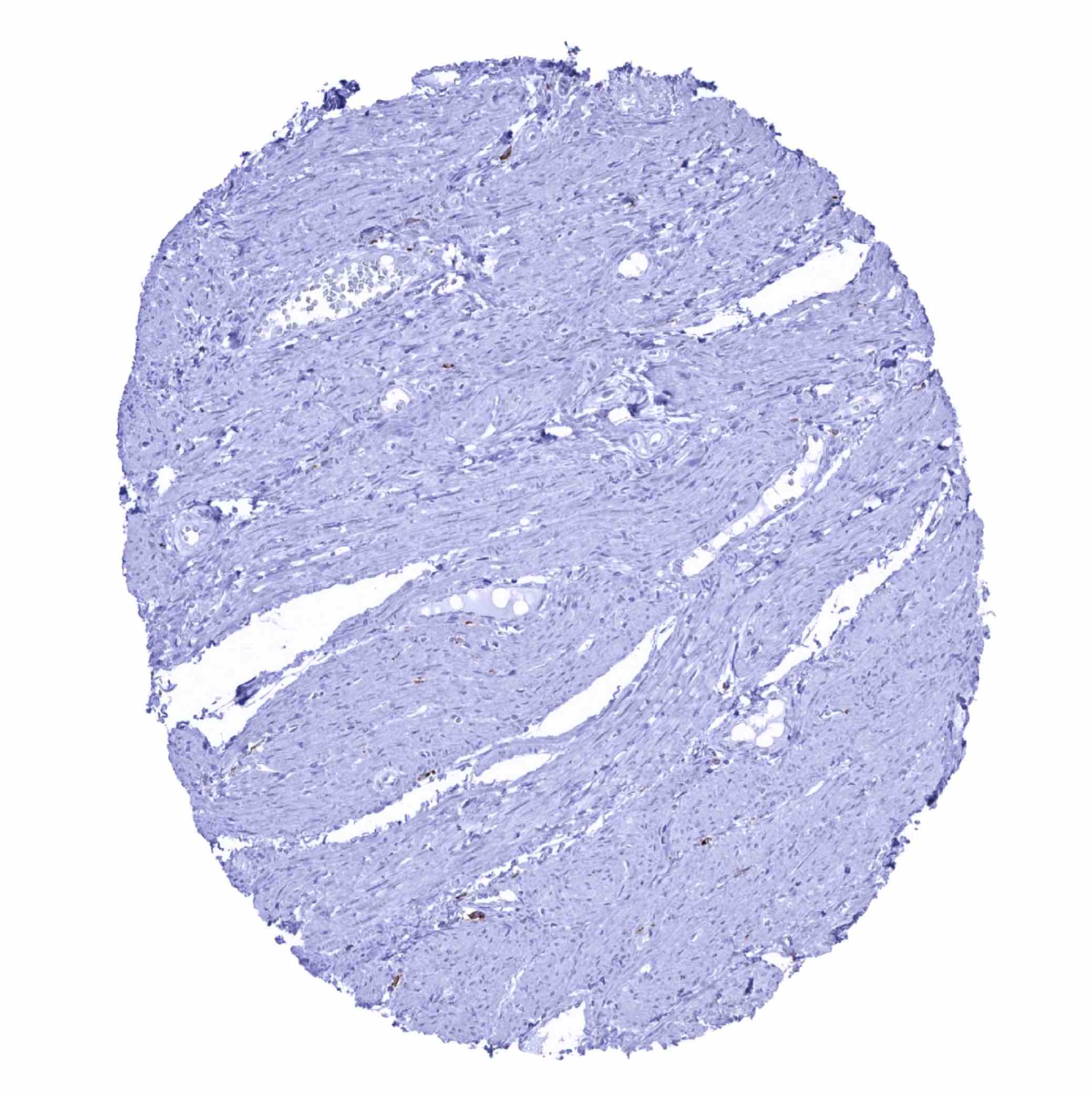
Urinary bladder, muscular wall
Urinary bladder, muscular wall (2)
Urinary bladder, urothelium - HLA-DRB1 staining of scattered inflammatory cells and of endothelial cells in blood vessels (HLA-DRB1 immunohistochemistry)

Uterus, ectocervix - HLA-DRB1 staining of scattered inflammatory cells (HLA-DRB1 immunohistochemistry)

Uterus, endocervix - HLA-DRB1 staining in scattered inflammatory cells, endothelial cells and in some epithelial cells (HLA-DRB1 immunohistochemistry)
Uterus, endometrium (pregnancy) - Strong HLA-DRB1 staining in scattered inflammatory cells (HLA-DRB1 immunohistochemistry)
Uterus, endometrium (proliferation) - HLA-DRB1 staining in scattered inflammatory cells and - focally - in some epithelial cells (HLA-DRB1 immunohistochemistry)
Uterus, endometrium (secretion) - HLA-DRB1 staining in scattered inflammatory cells and in endothelial cells (HLA-DRB1 immunohistochemistry)
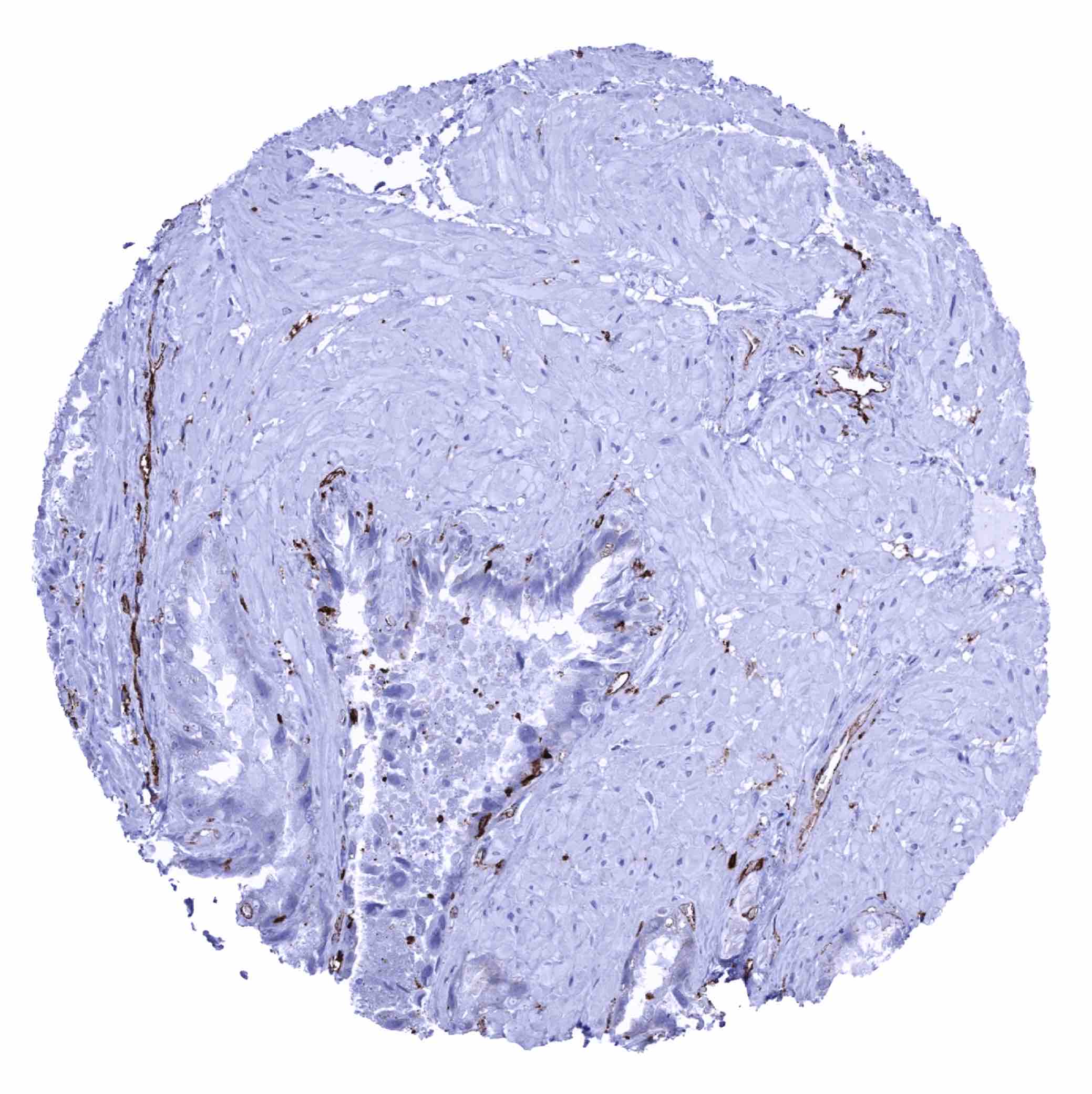
Uterus, myometrium