
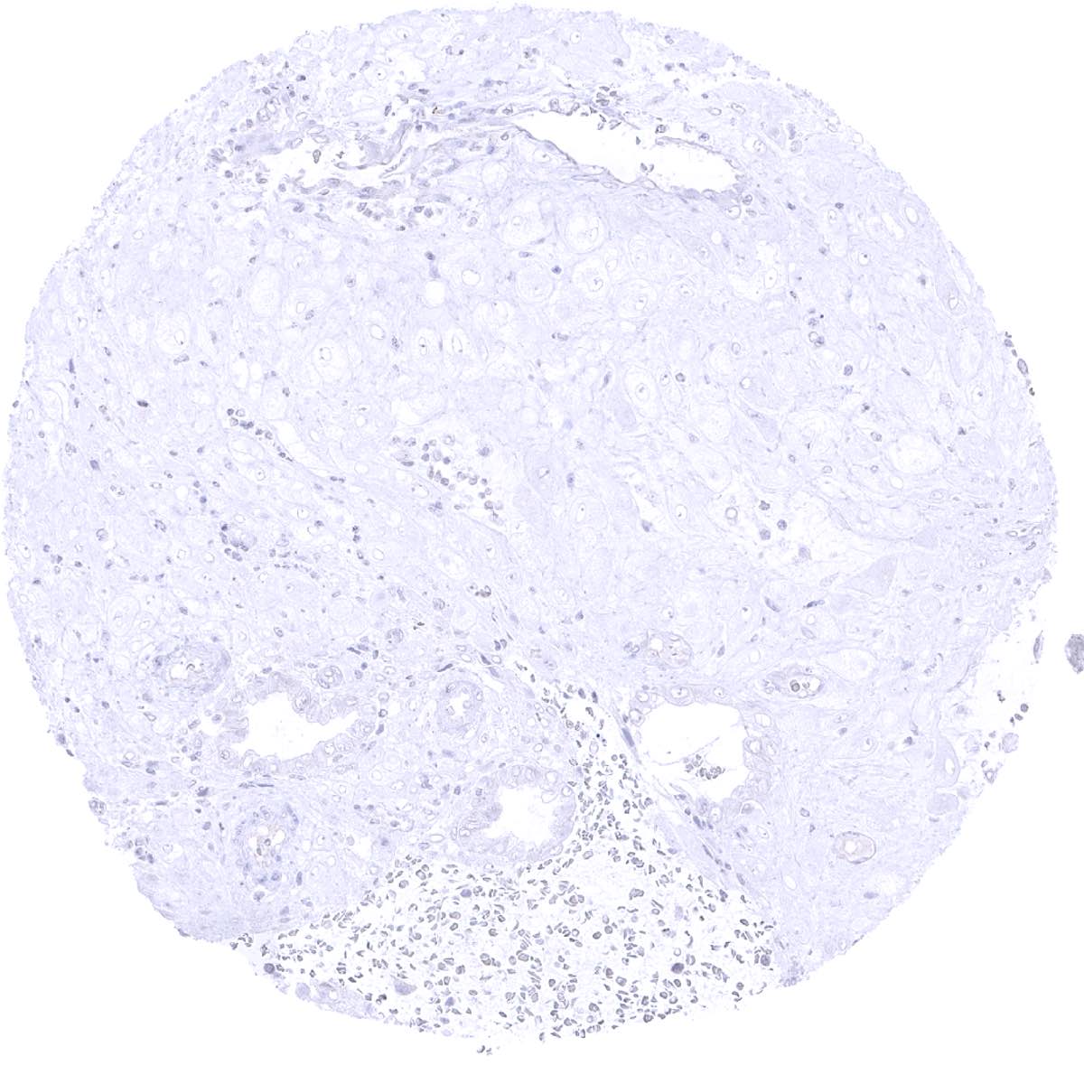
Adrenal gland

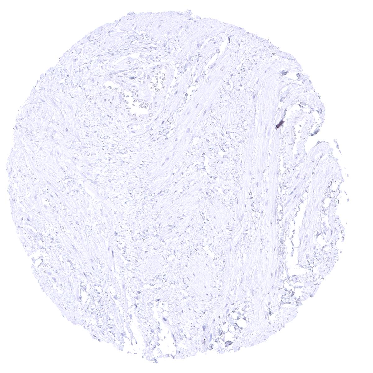
Aorta, media

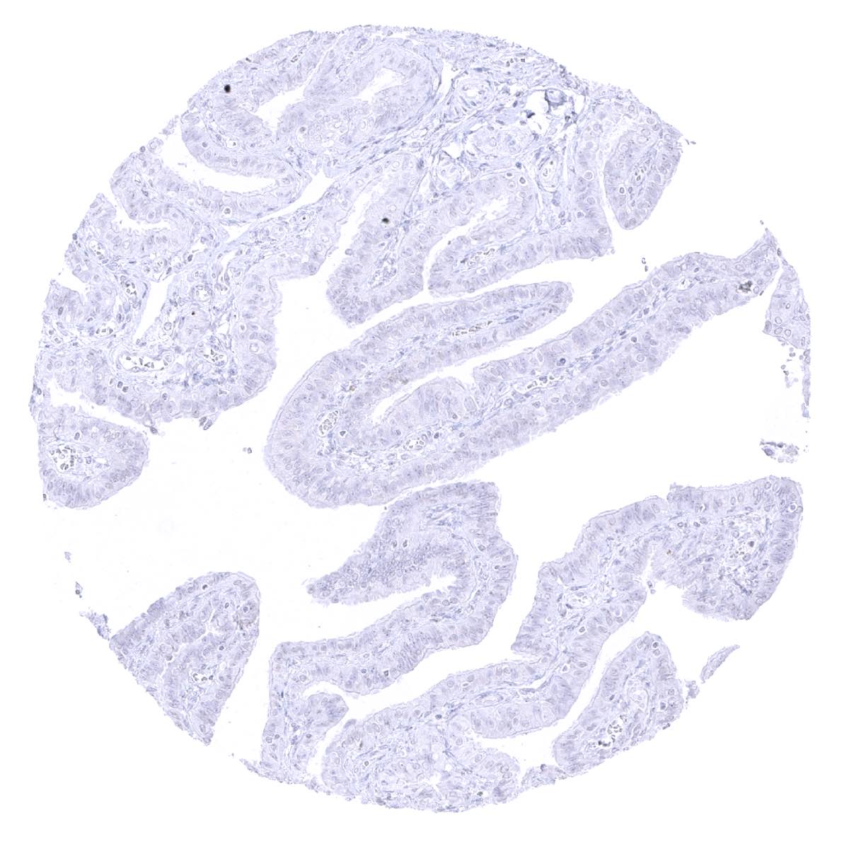
Appendix, mucosa - A very faint staining can be seen at the basolateral membranes of the superficial layers of appendical epithelium.

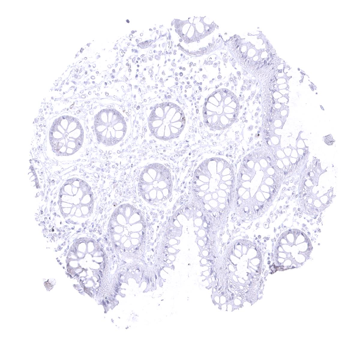
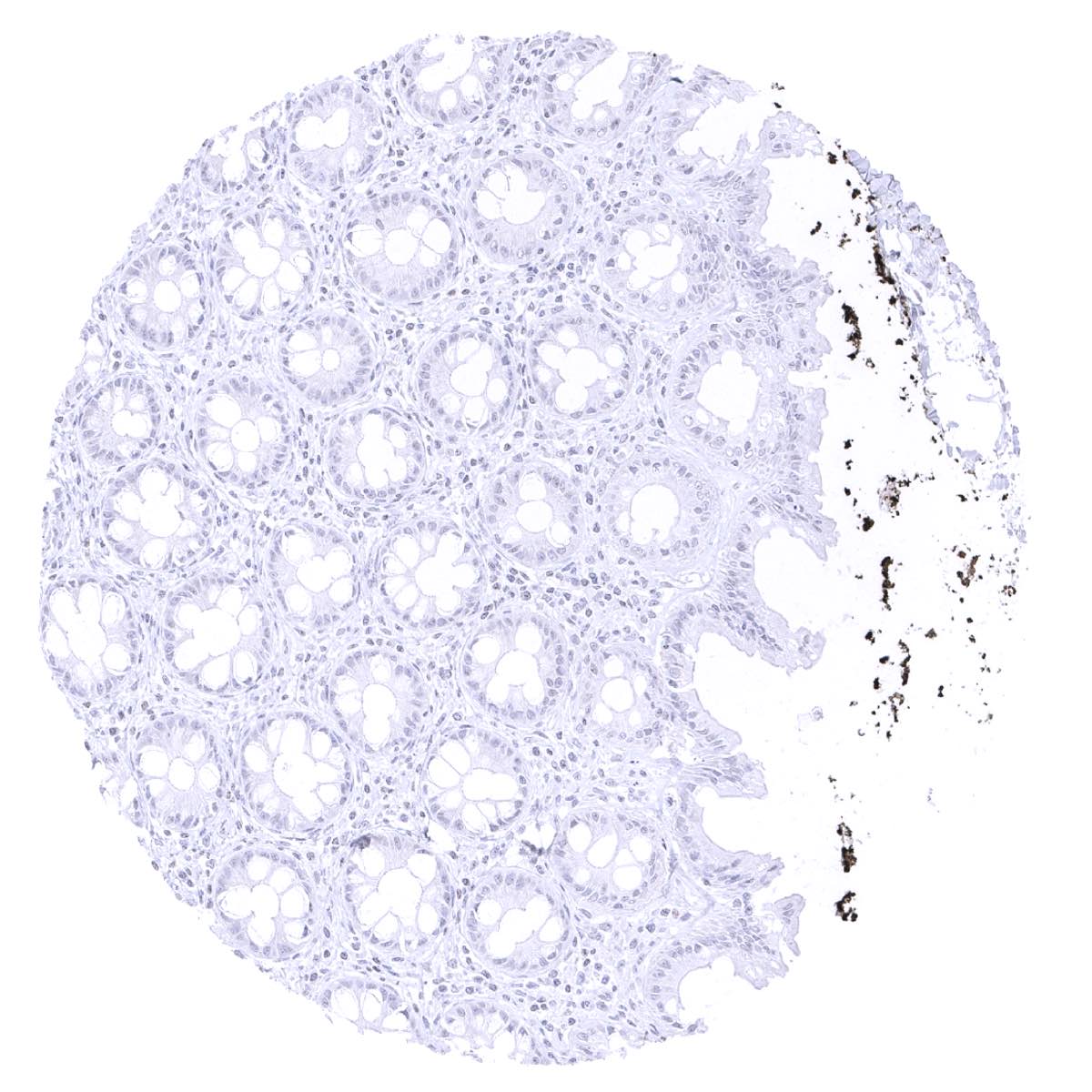
Appendix, mucosa

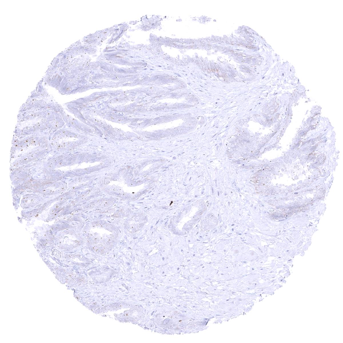
Appendix, muscular wall
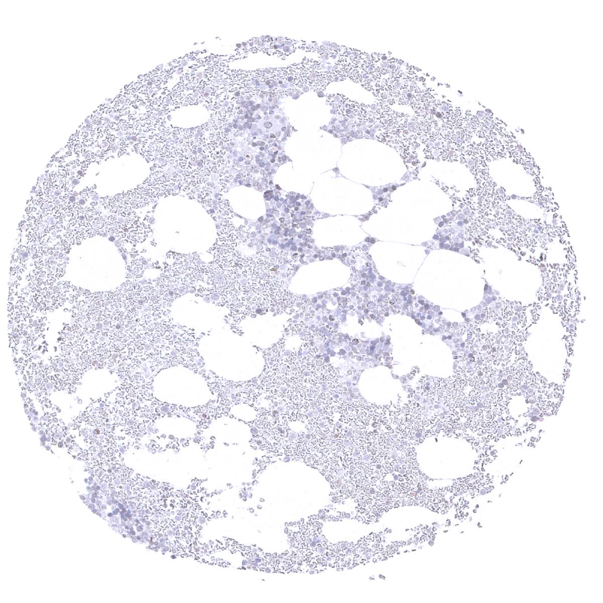
Bone marrow
Breast - HER2 staining is lacking in normal breast in case of appropriate formalin fixation.

Bronchus, mucosa
Cerebellum, cortex (Stratum moleculare) (1)

Cerebellum, cortex (Stratum moleculare) (2)
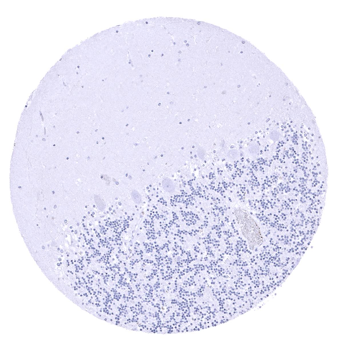
Cerebellum, grey (Stratum neuronorum)
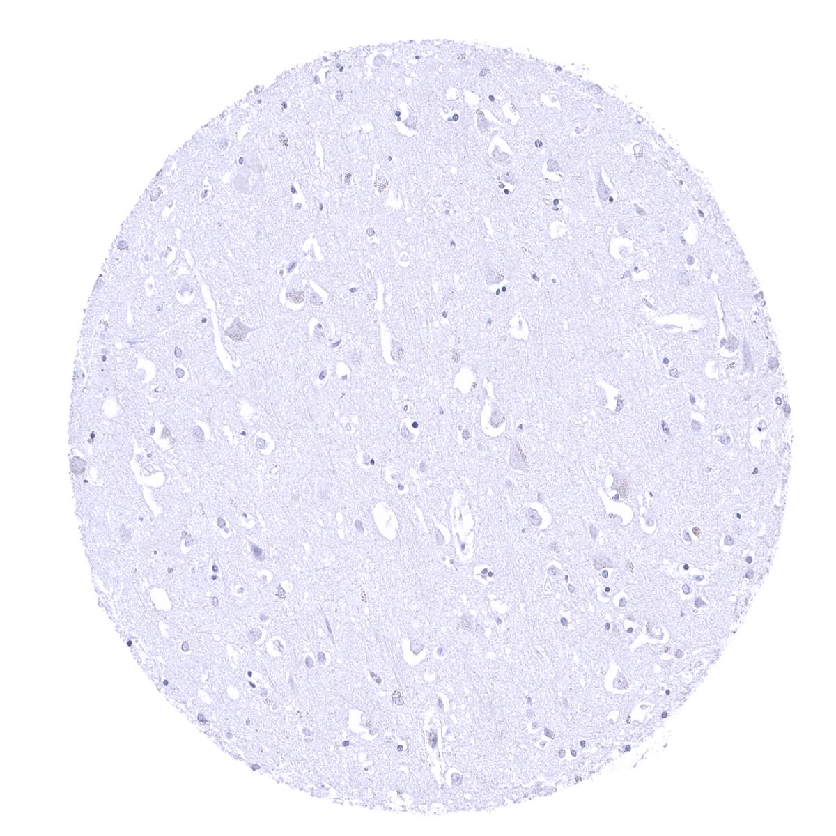
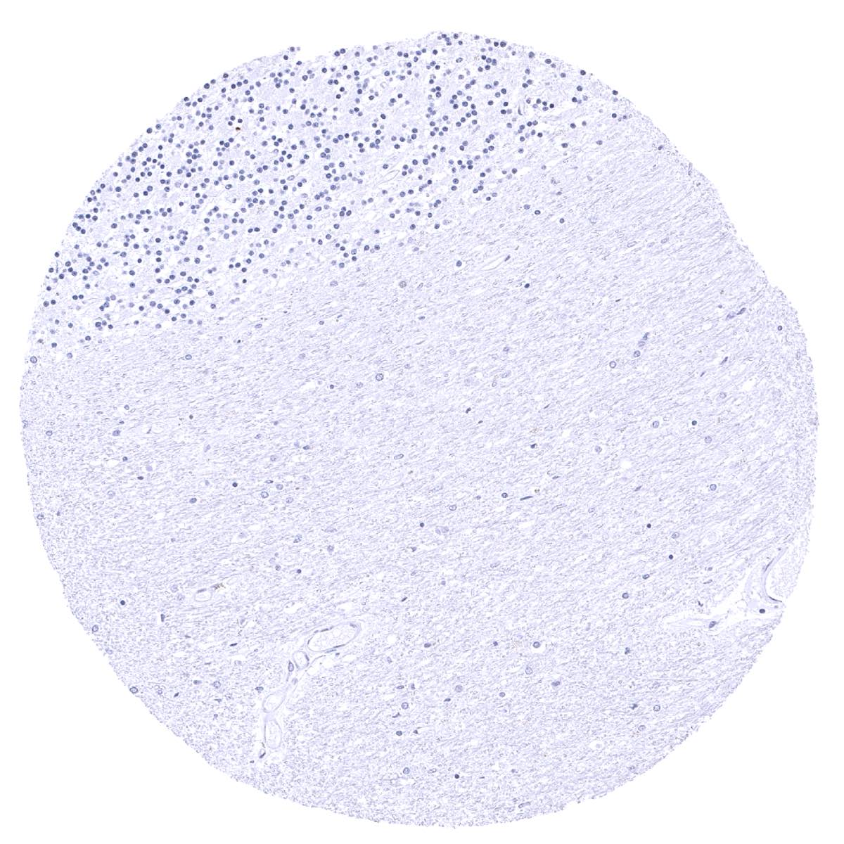
Cerebrum, grey
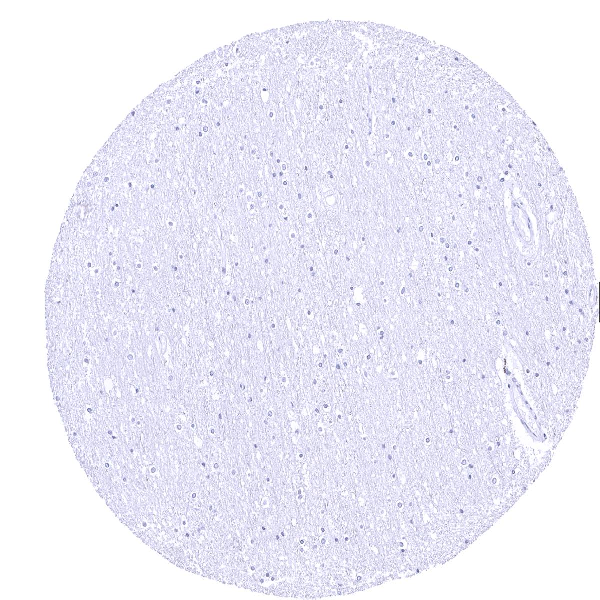
Cerebrum, white
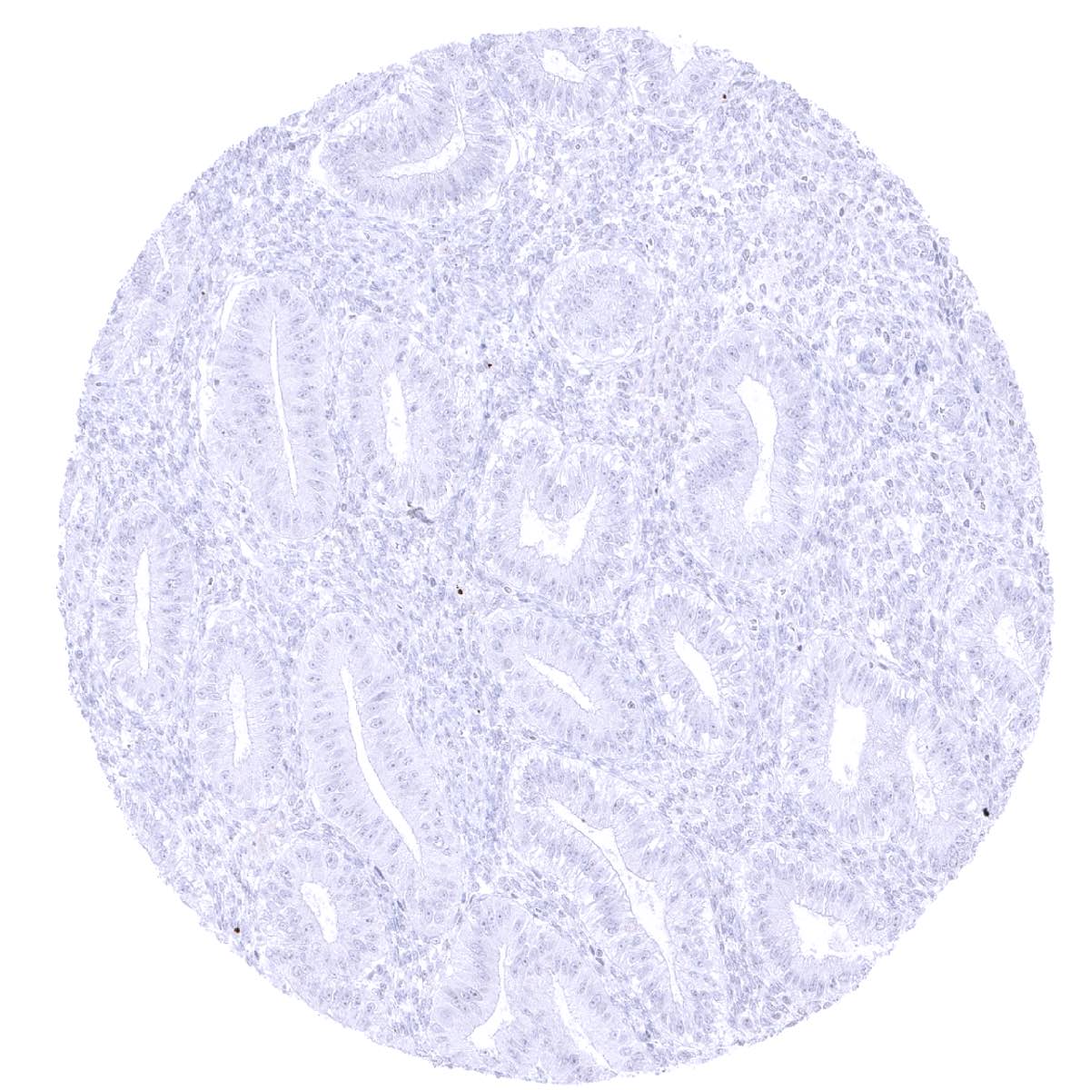
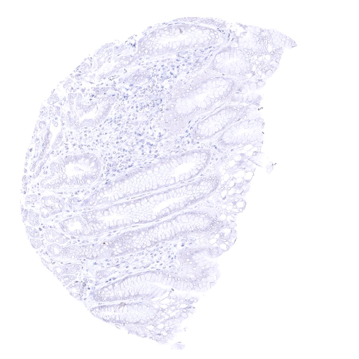
Colon descendens, mucosa

Colon descendens, muscular wall

Duodenum, Brunner gland
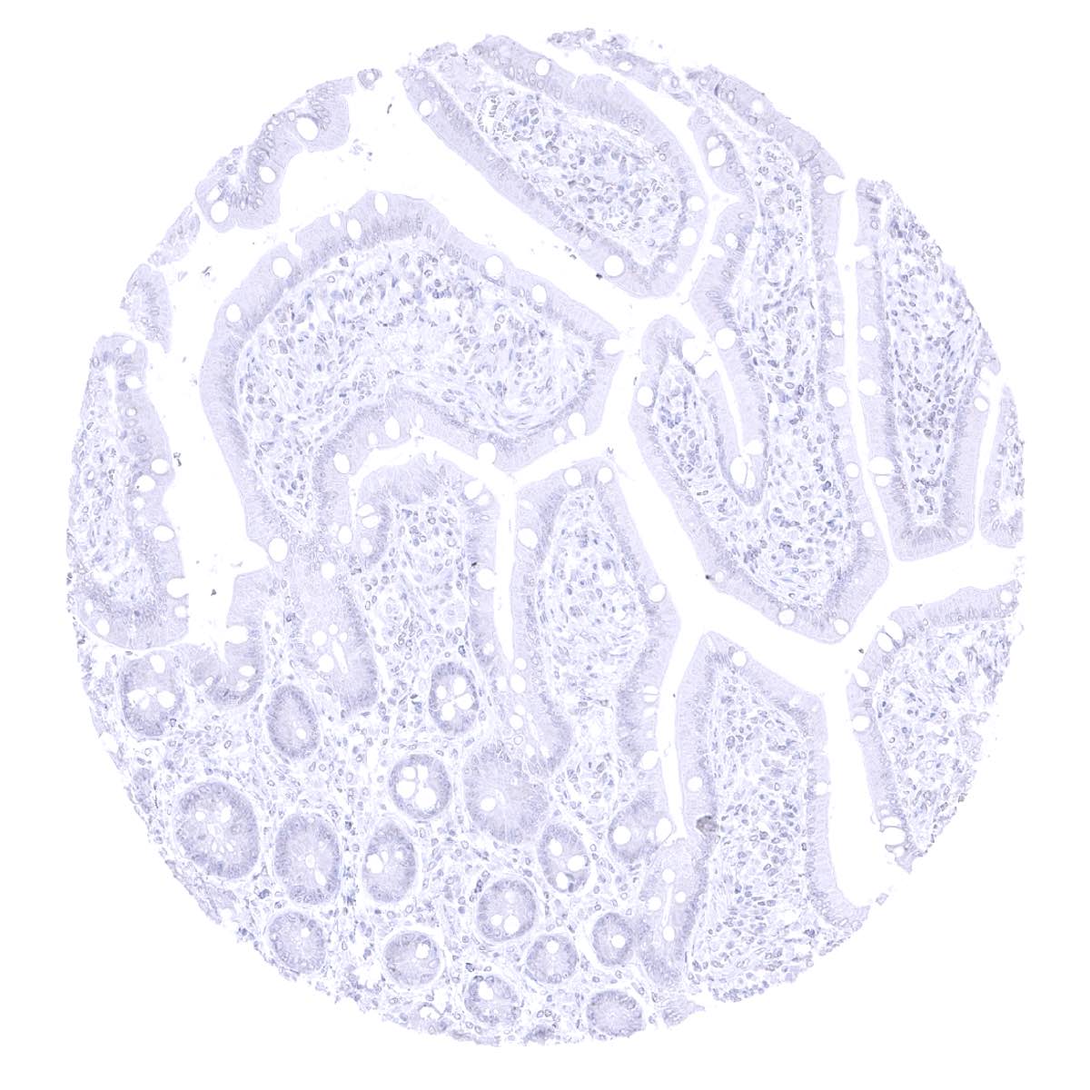
Duodenum, mucosa
Ectocervix

Endocervix
Endometrium, proliferation
Endometrium, secretion
Epididymis
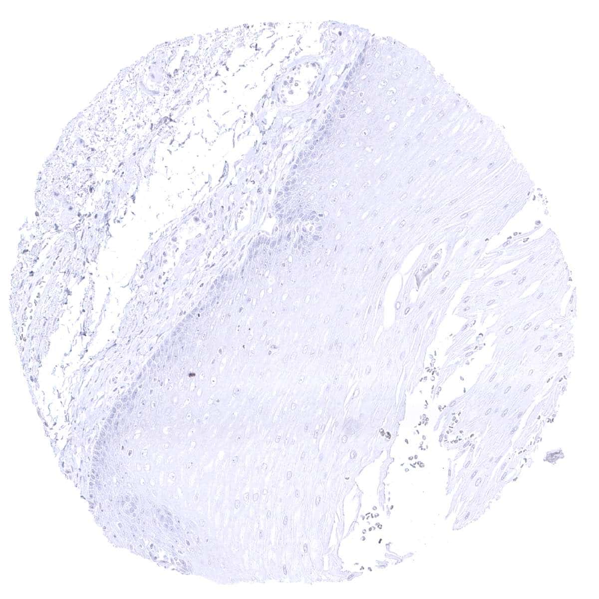
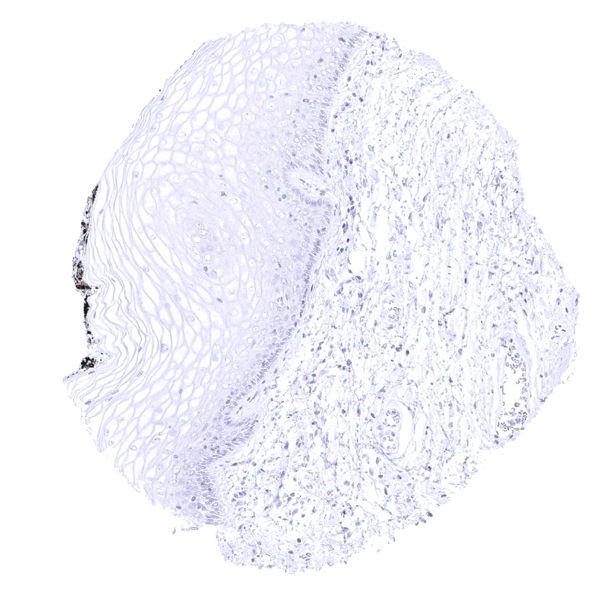
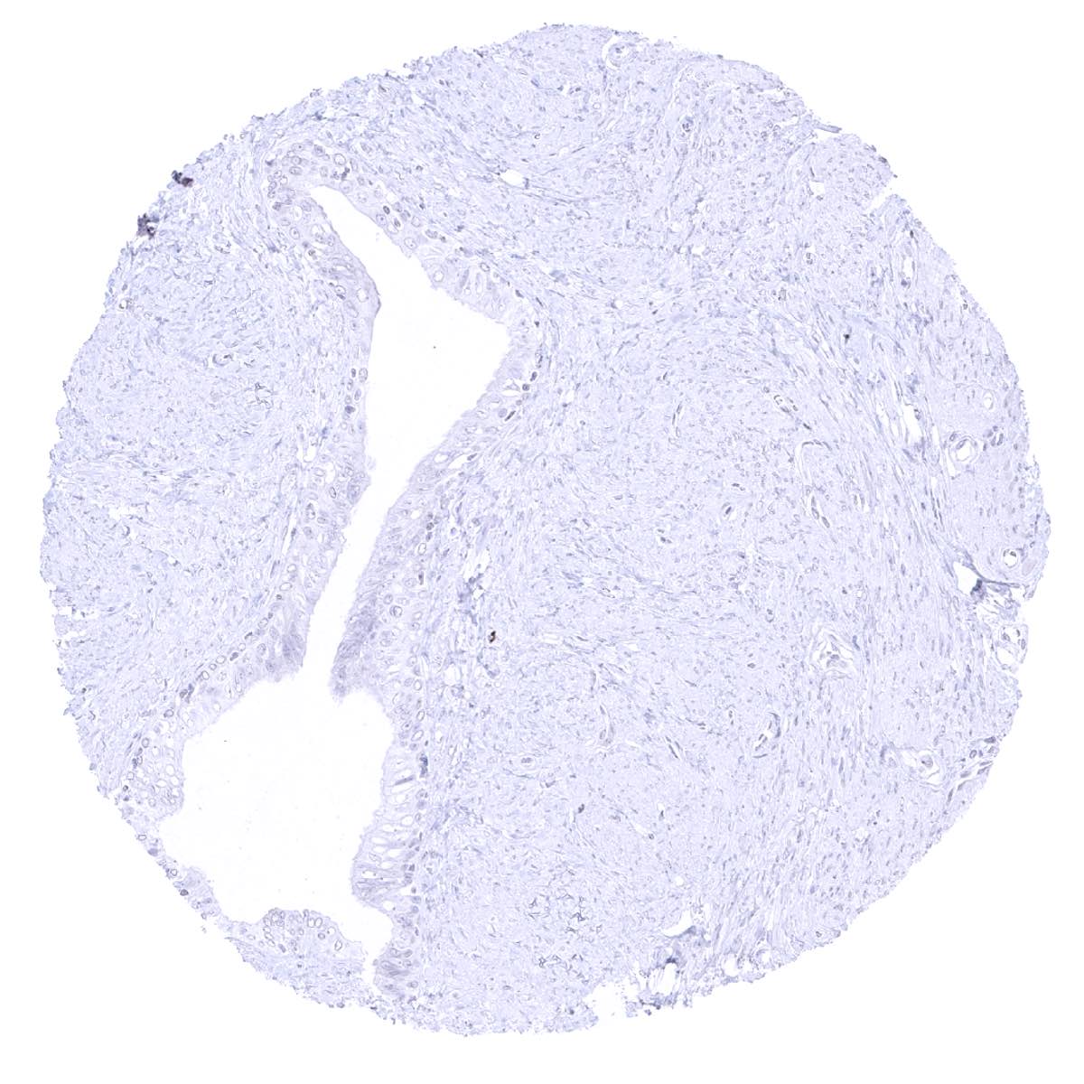
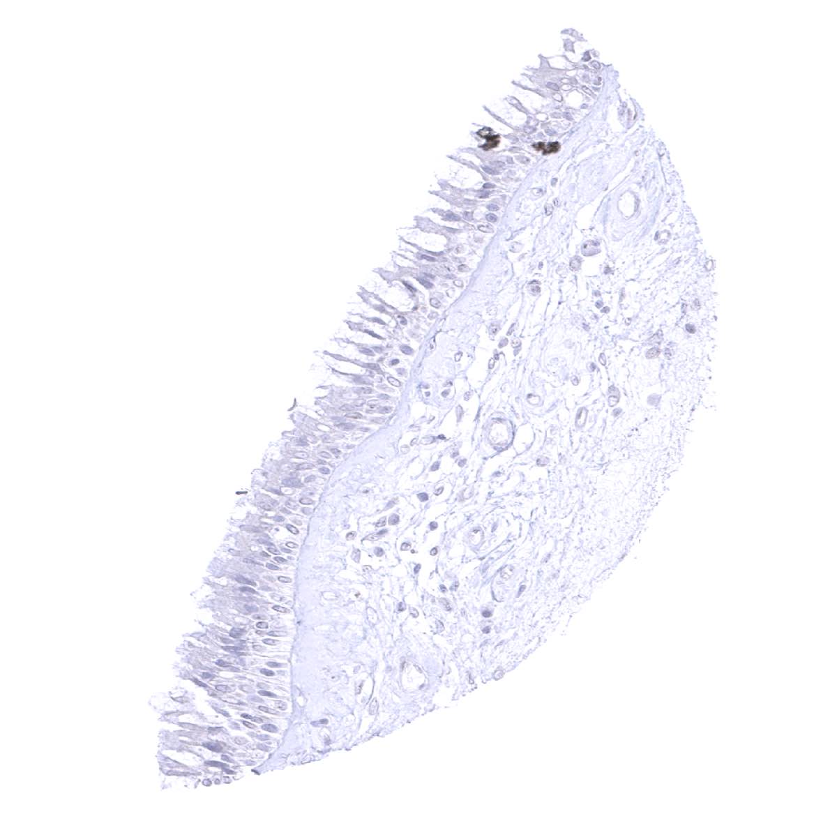
Esophagus, squamous epithelium
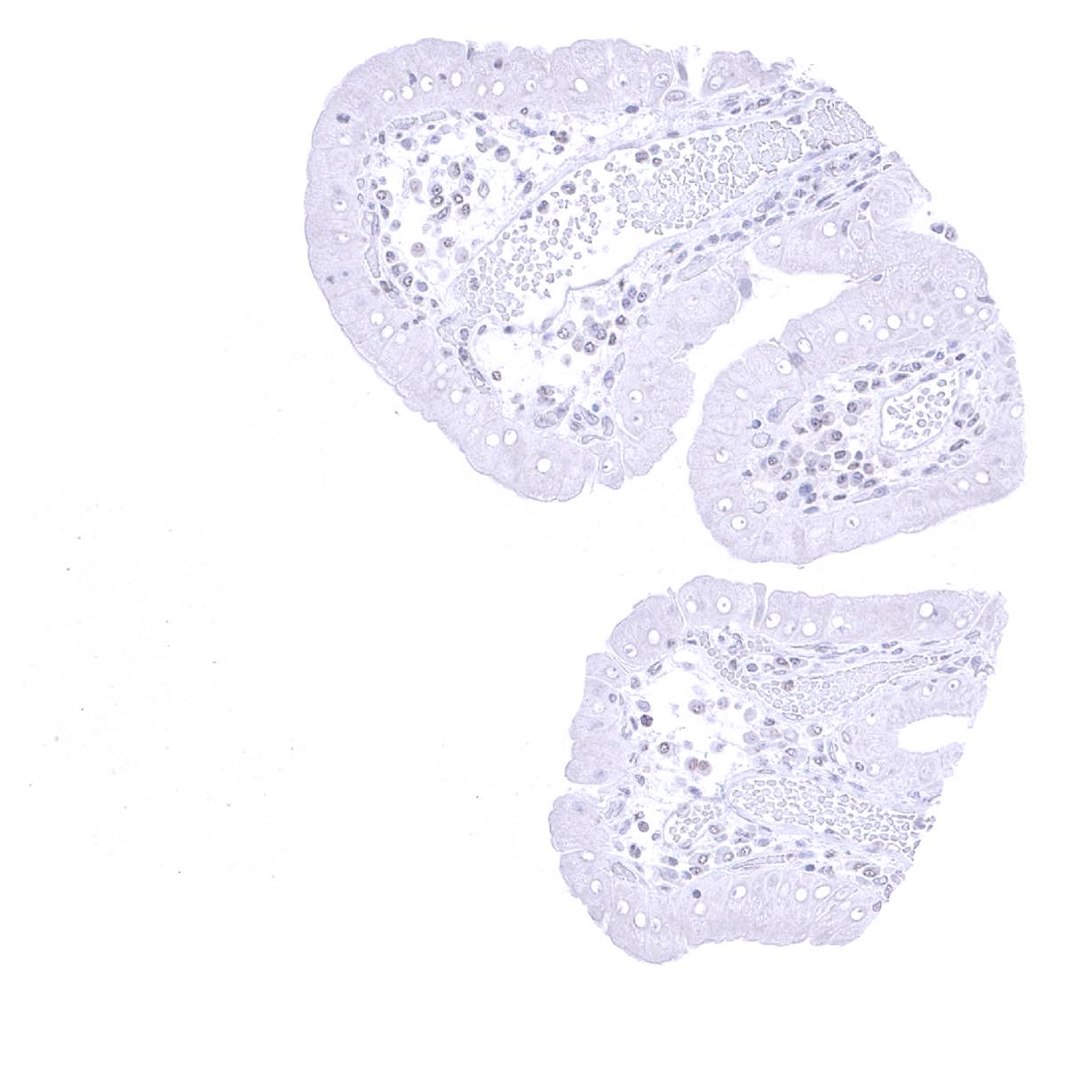
Fallopian tube, mucosa
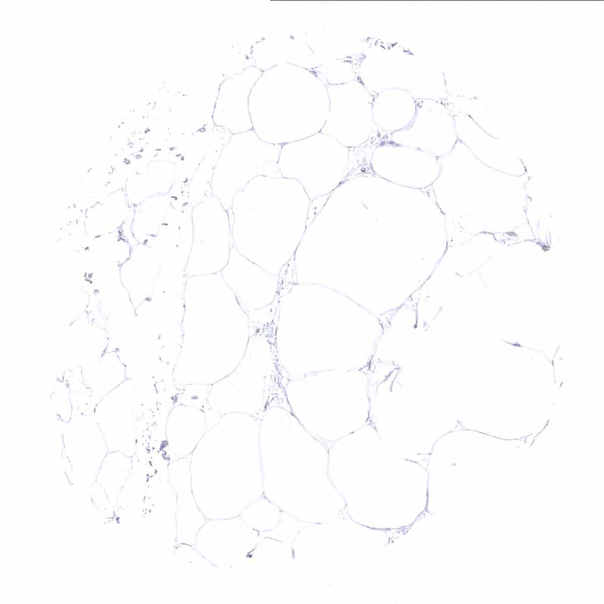
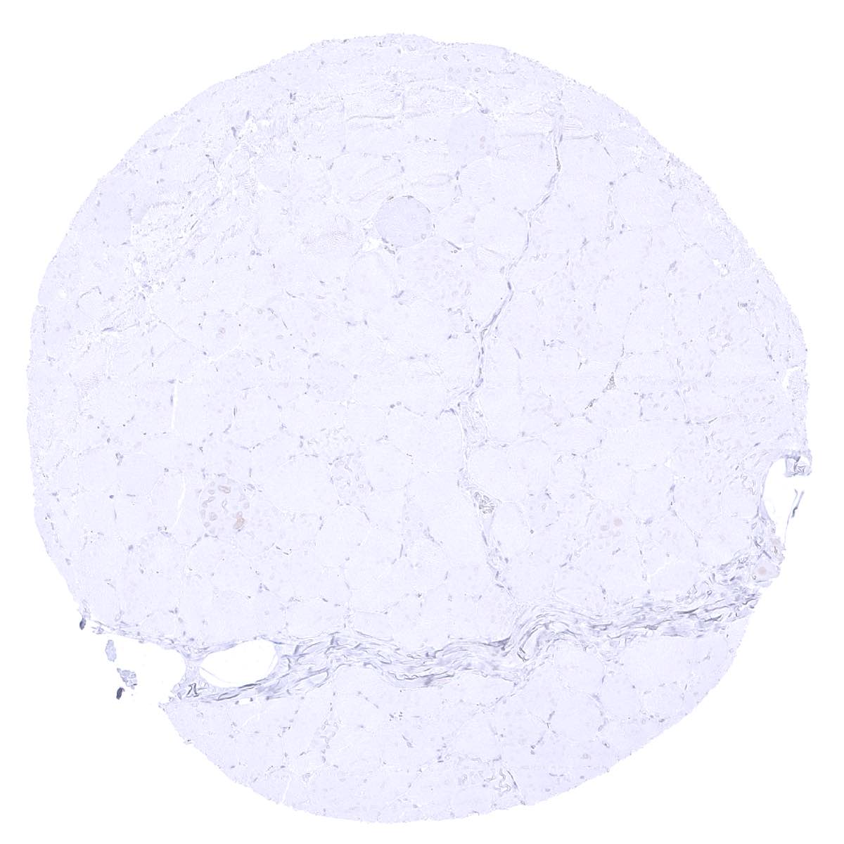
Fat
Gallbladder, epithelium
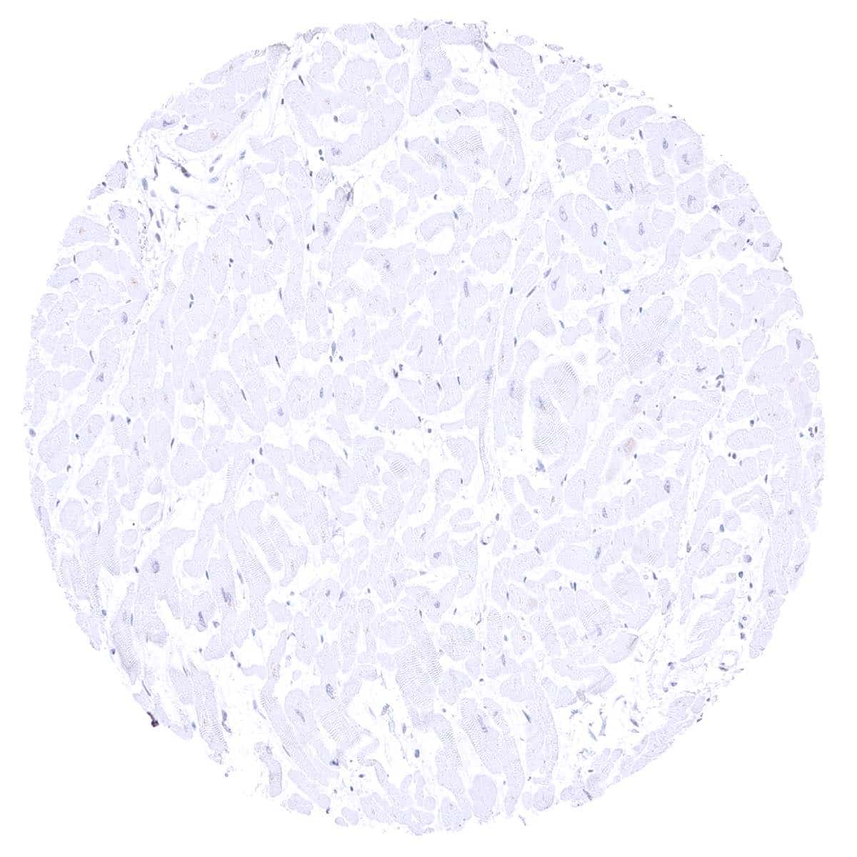
Heart
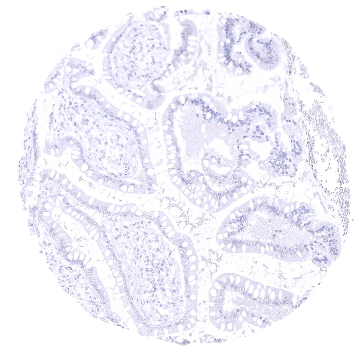
Ileum, mucosa

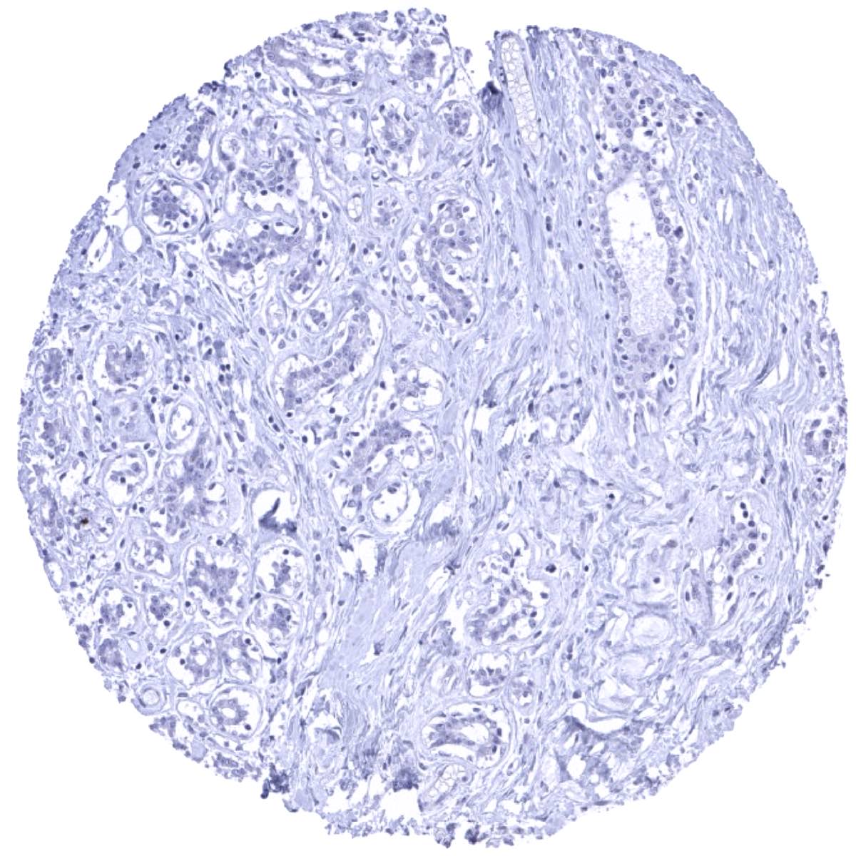
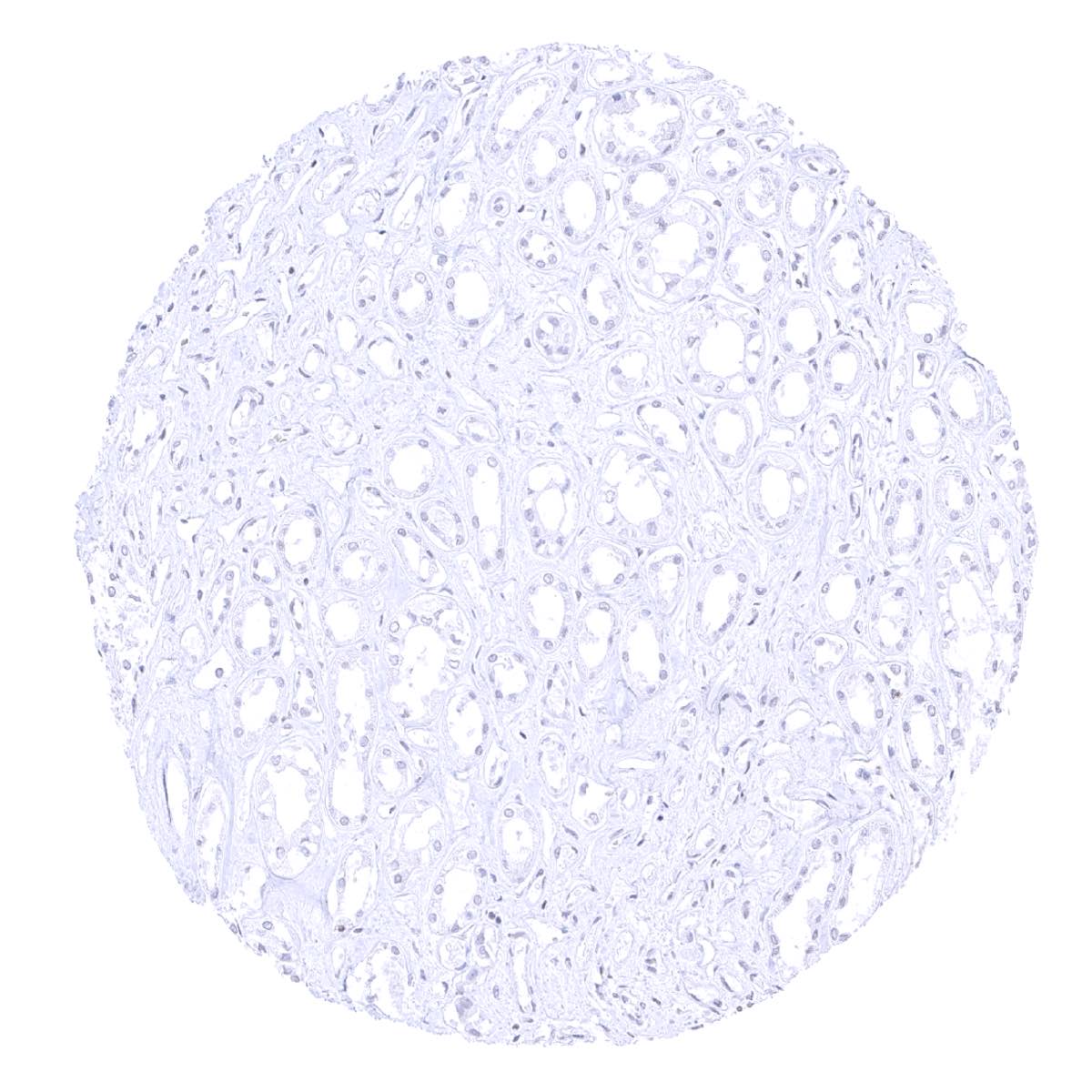
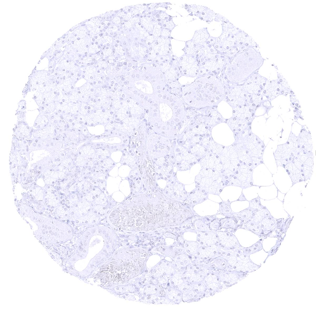
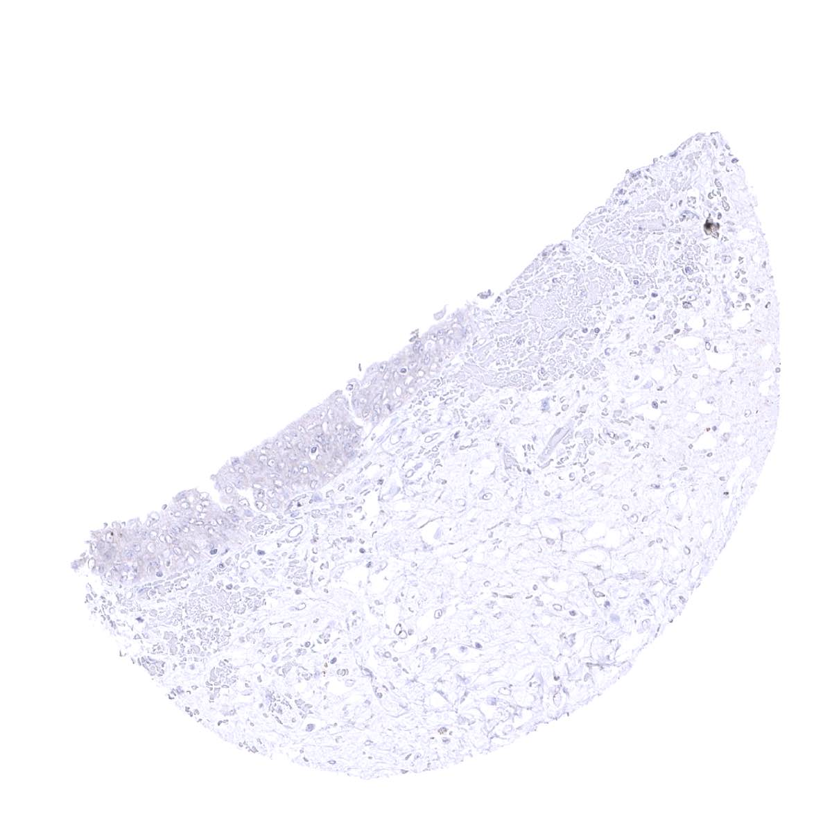
Kidney, cortex
Kidney, medulla

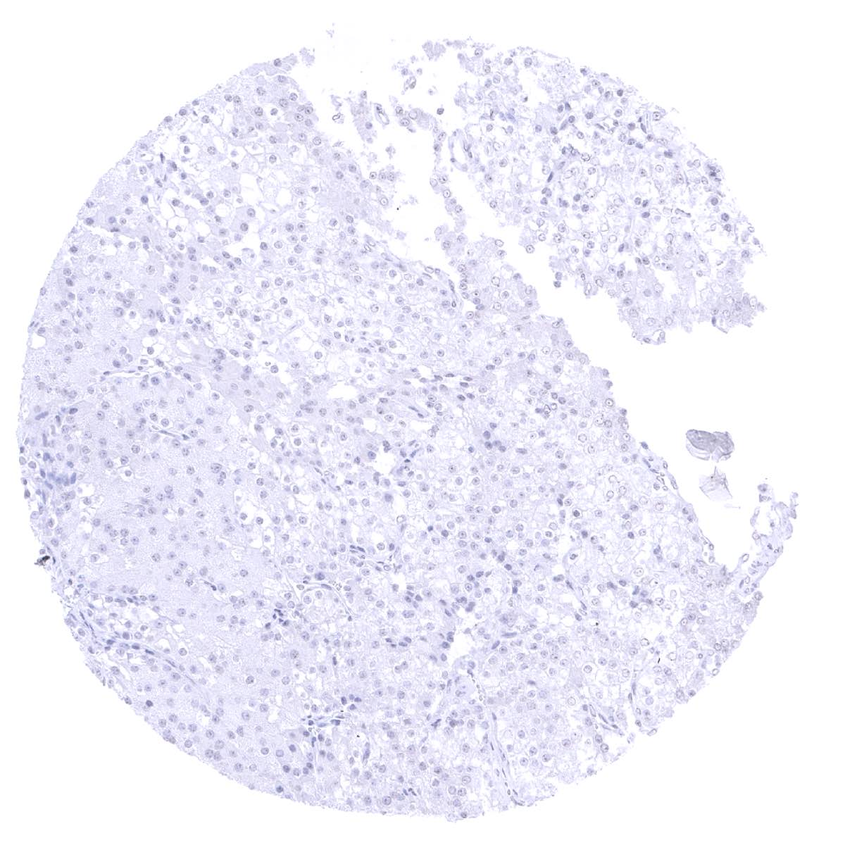
Liver

Lung
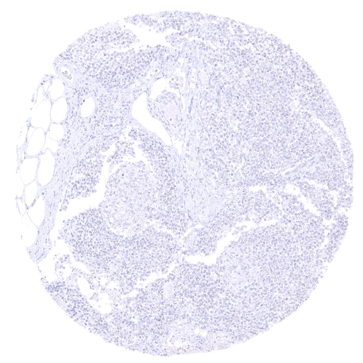
Lymph node

Ovary, stroma
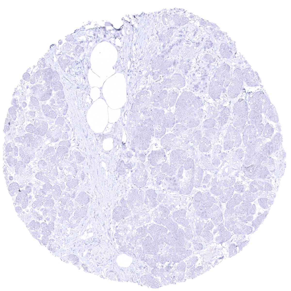
Pancreas
Parathyroid
Parotid gland

Pituitary, anterior lobe

Pituitary, posterior lobe- infundibulum
Placenta early, decidua
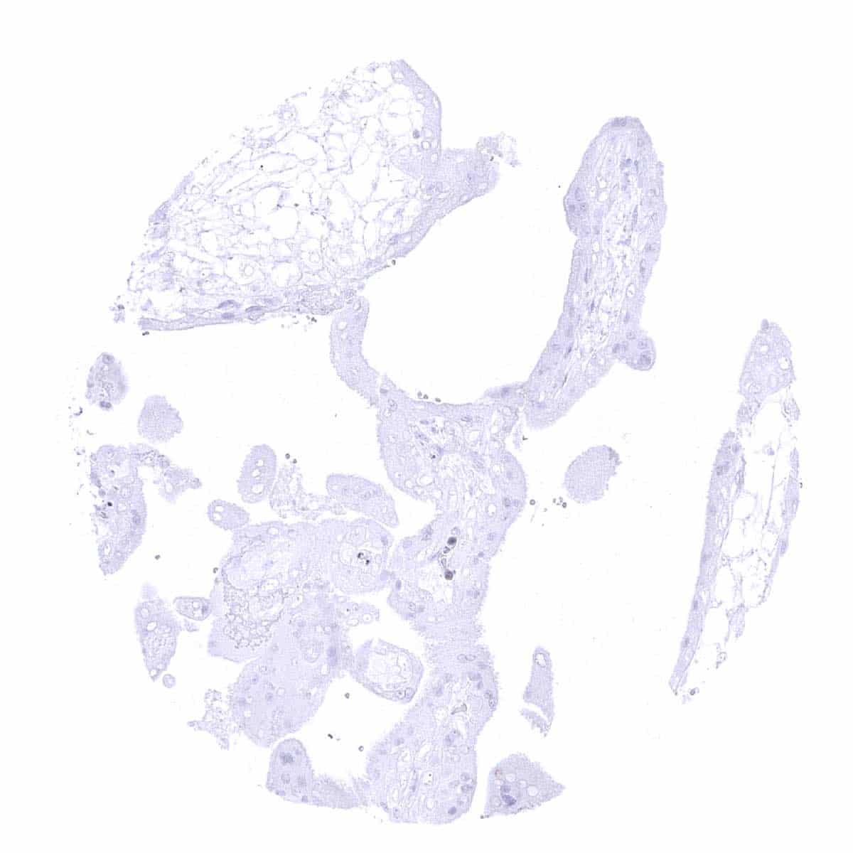
Placenta, early
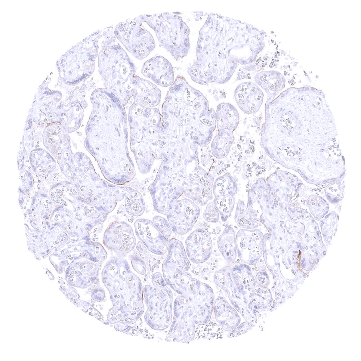
Placenta, mature - A weak to moderate HER2 staining occurs at the surface membrane of the mature placenta.
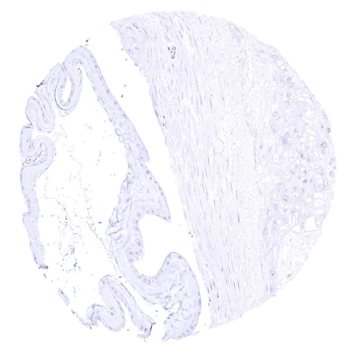
Placenta, mature, amnion and chorion

Prostate - A faint staining can be seen in basal cells of prostate glands, especially if they are atrophic.
Rectum, mucosa
Seminal vesicle - A faint staining can be seen in a fraction of basal cells of seminal vesicles.
Sinus paranasales
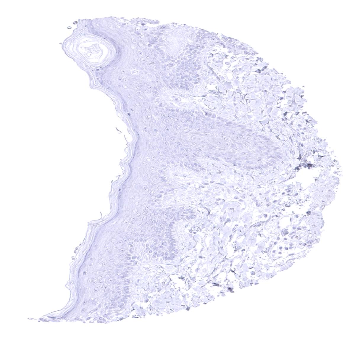
Skin, hairfollicel and sebaceous glands

Spleen

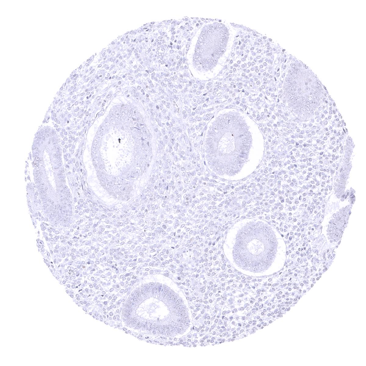
Stomach, antrum
Stomach, corpus
Striated muscle
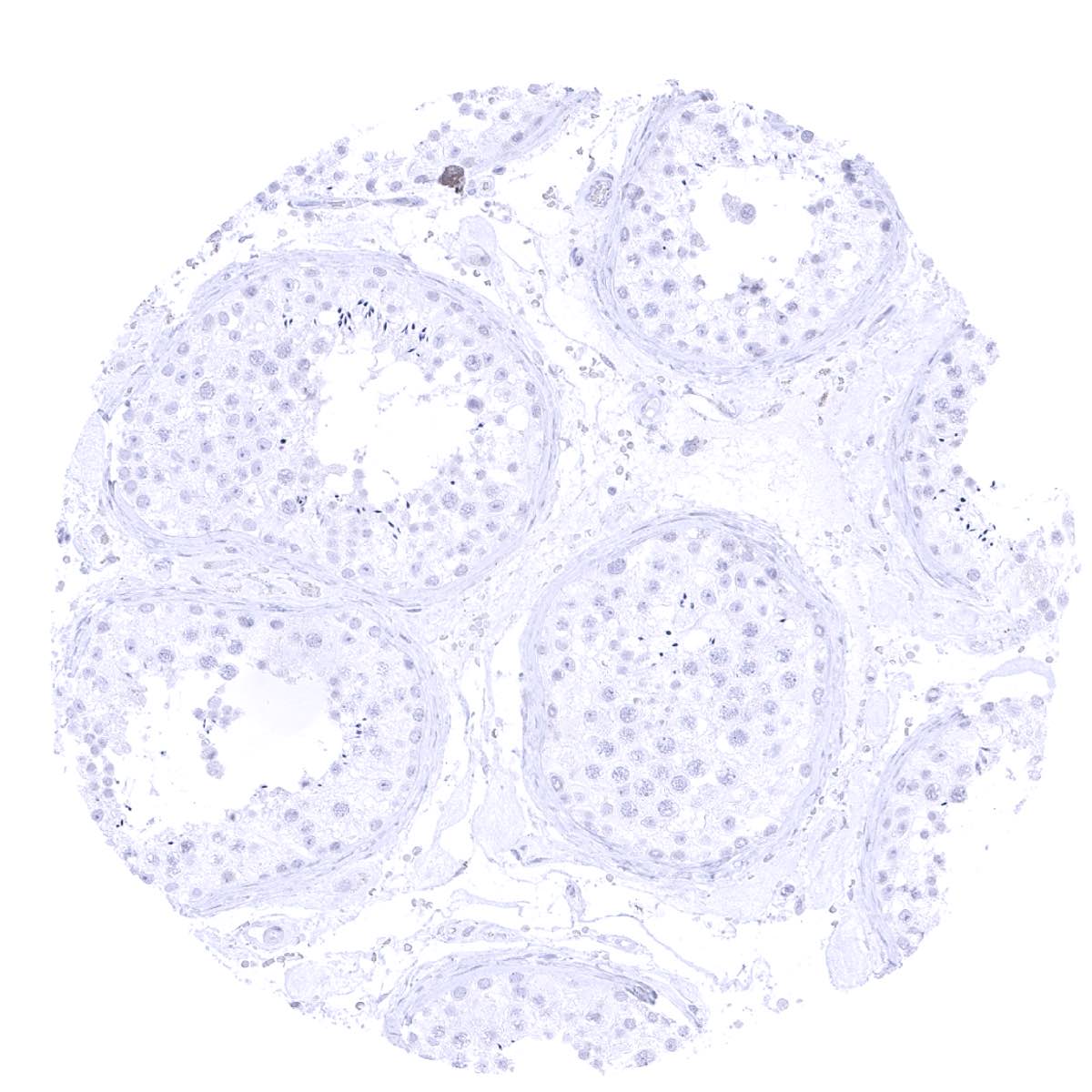
Testis
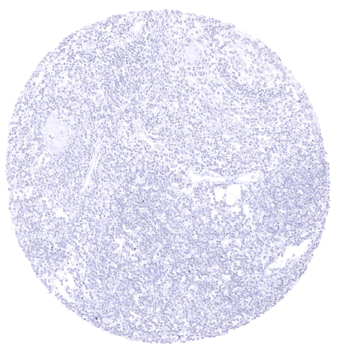
Thymus
Tonsil - A very faint staining of a fraction of epithelial cells occurs in tonsil crypts.

Tonsil, surface epithelium Tyroid gland
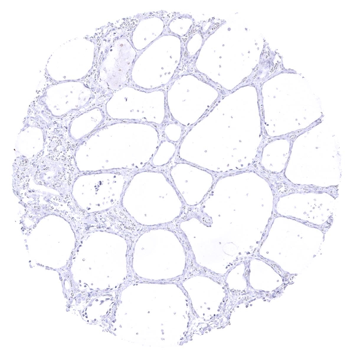
Thyroid gland
Urinary bladder, muscular wall
Urinary bladder, urothelium

Uterus, myometrium