Adrenal gland – Weak to moderate nuclear GATA3 positivity of a subset of adrenocortical cells.

Aorta, endothelium
Appendix, mucosa – Nuclear GATA3 positivity of many lymphocytes. Faint cytoplasmic GATA3 staining of some epithelial cells.
Appendix, muscular wall
Bone marrow
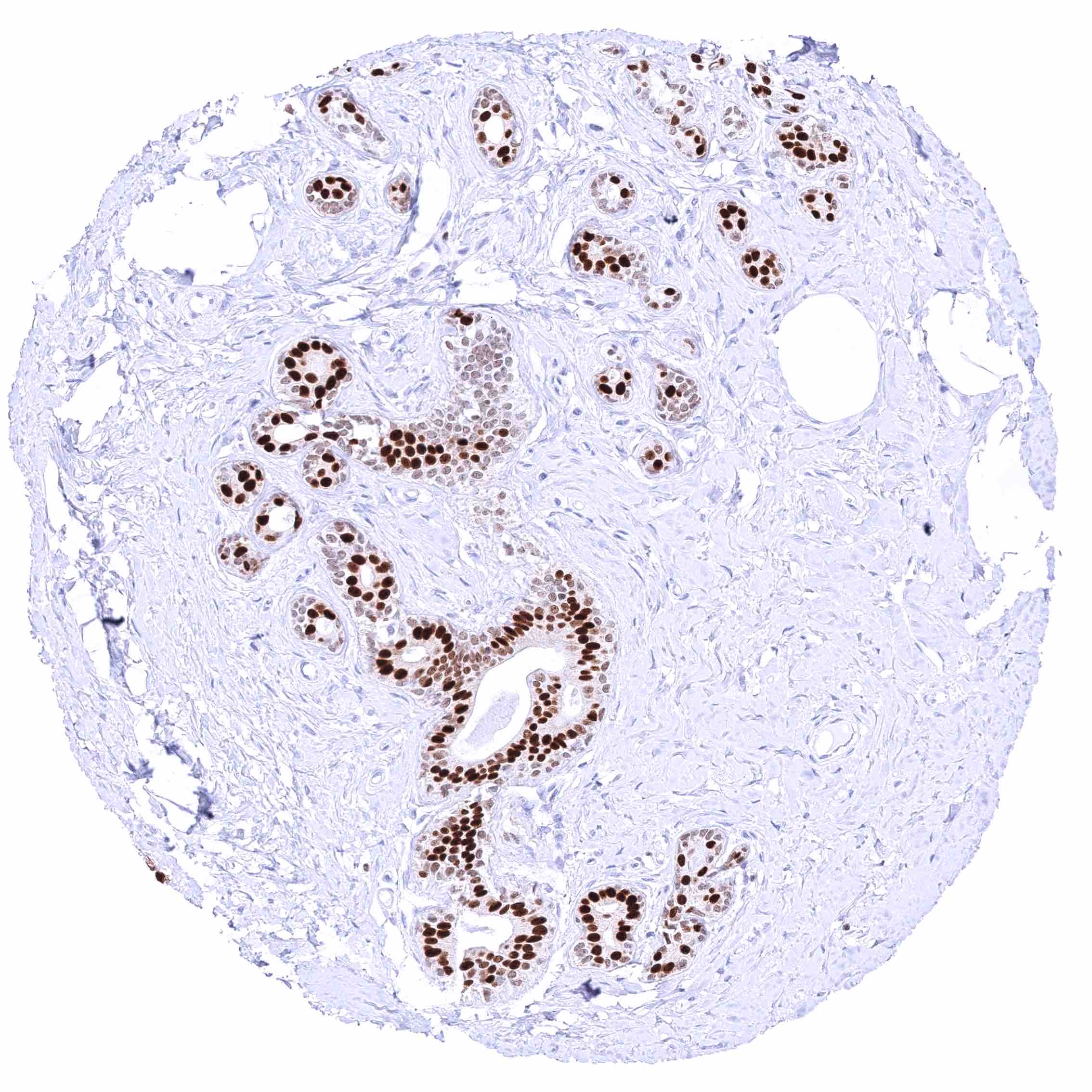
Breast – Moderate to strong GATA3 staining of luminal cells while myoepithelial cells remain GATA3 negative.
Bronchus, mucosa – Granular cytoplasmic GATA3 positivity of most respiratory epithelial cells.
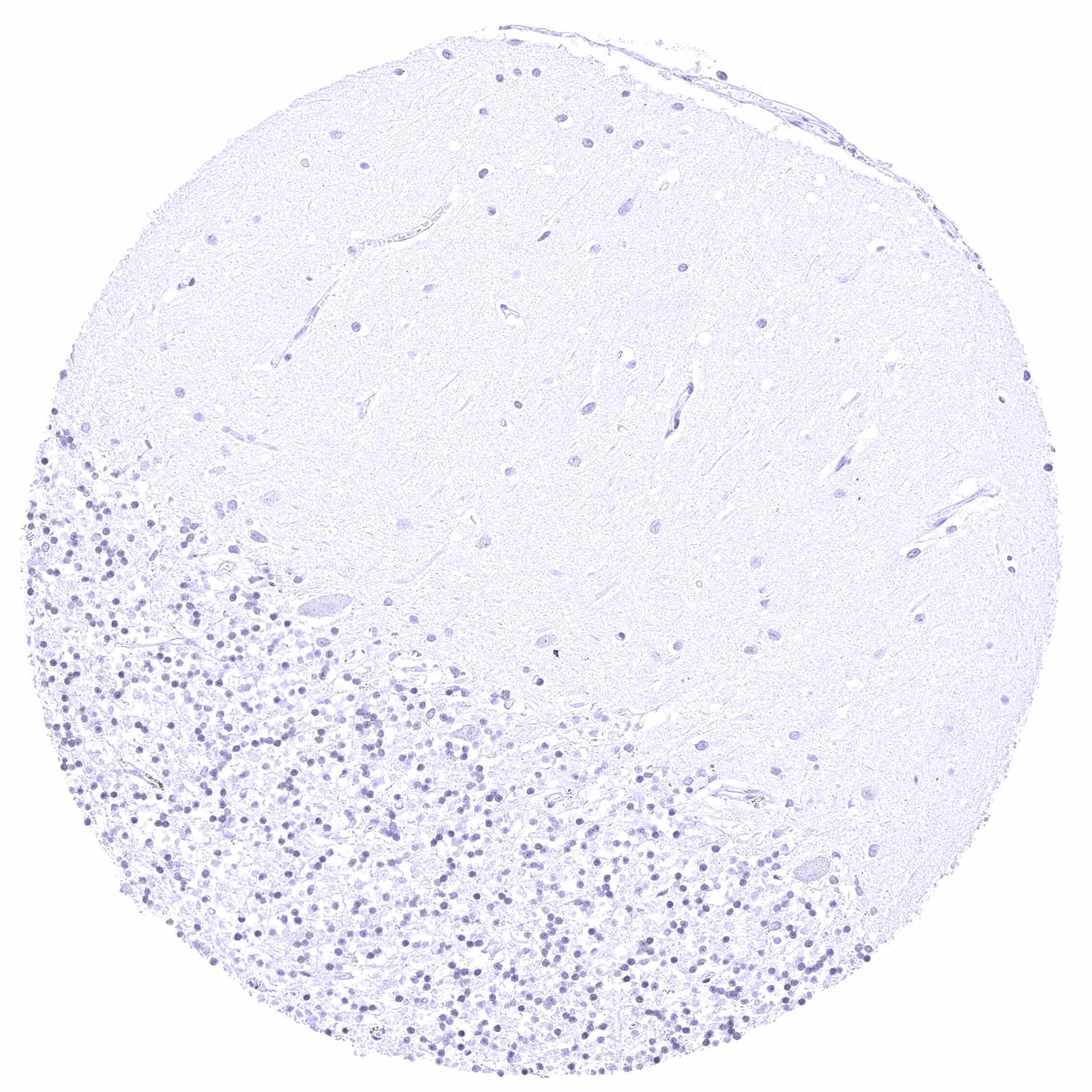
Cerebellum (molecular layer, Purkinje cell layer, granule cell layer)

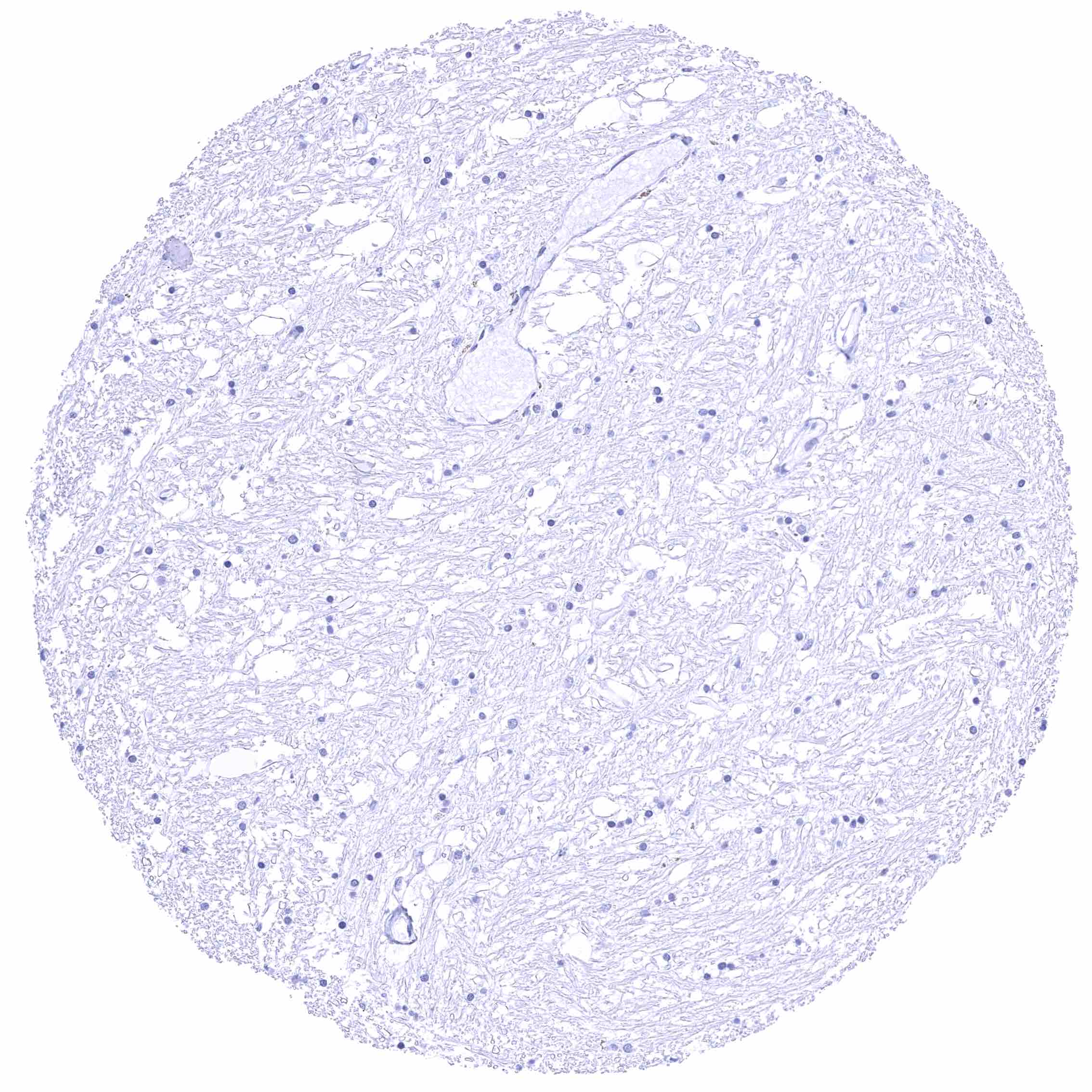
Cerebellum (white matter)

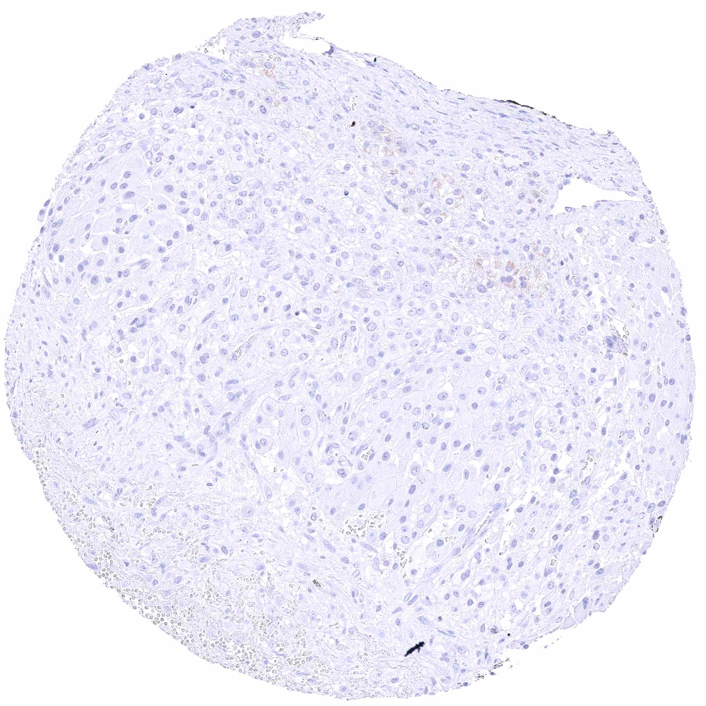
Cerebrum (grey matter)
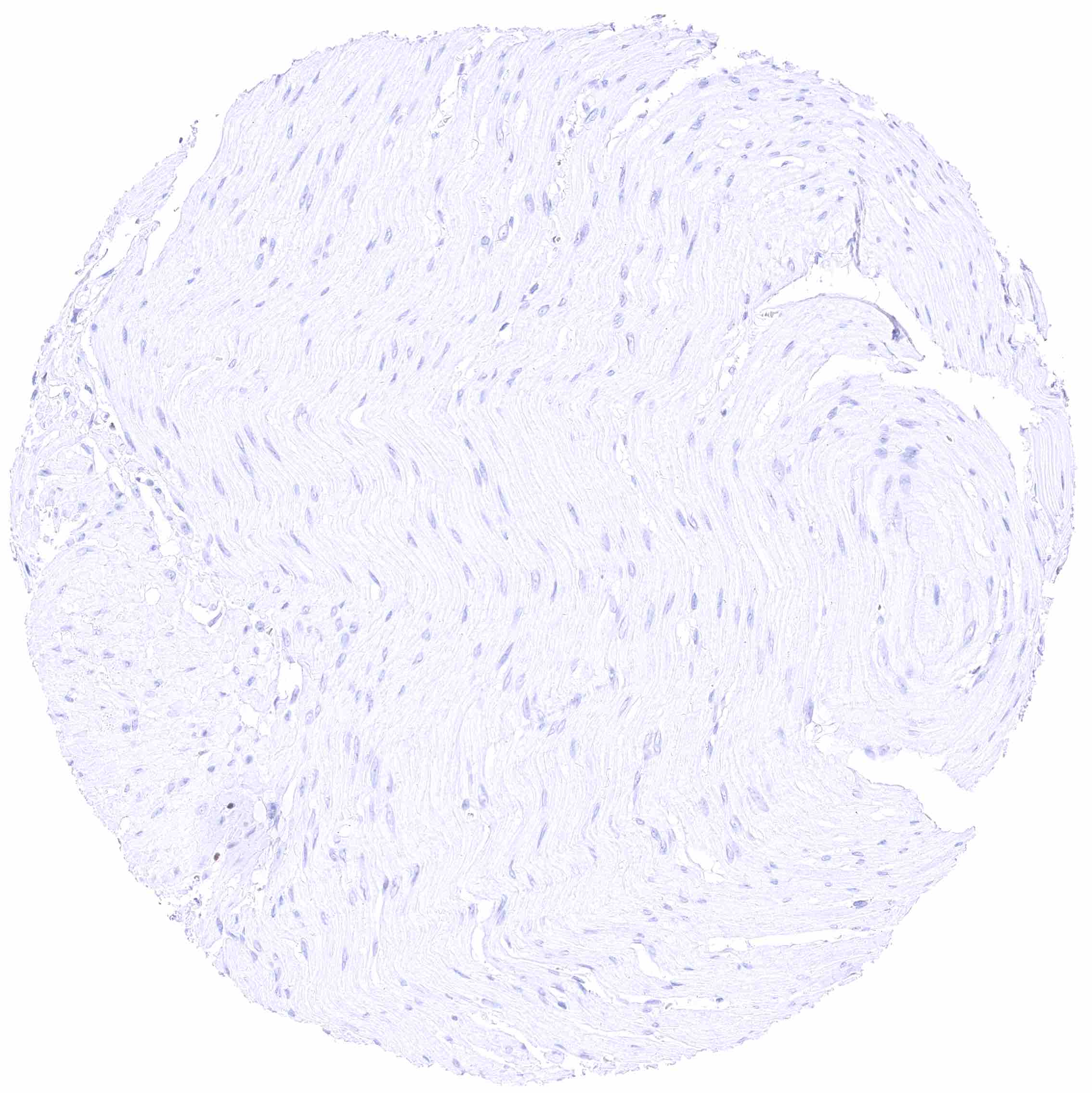
Cerebrum (white matter)
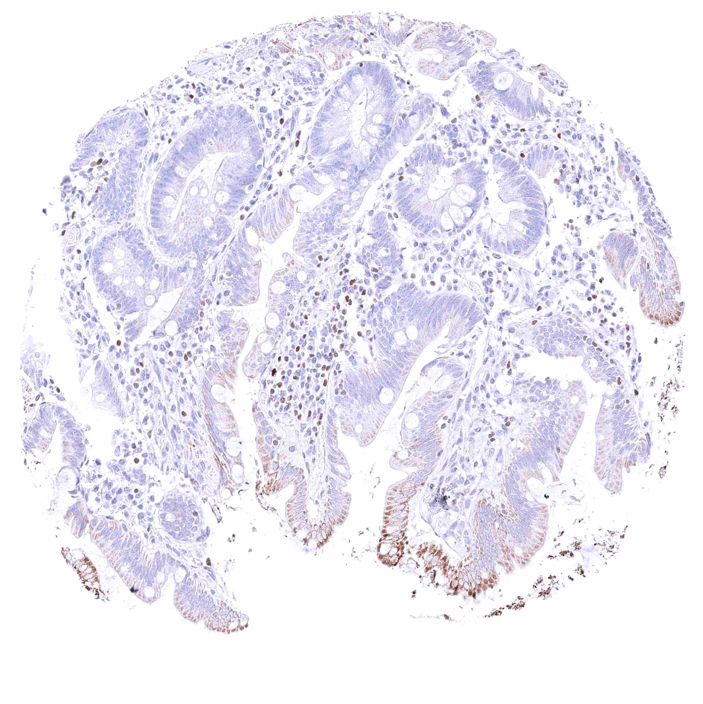
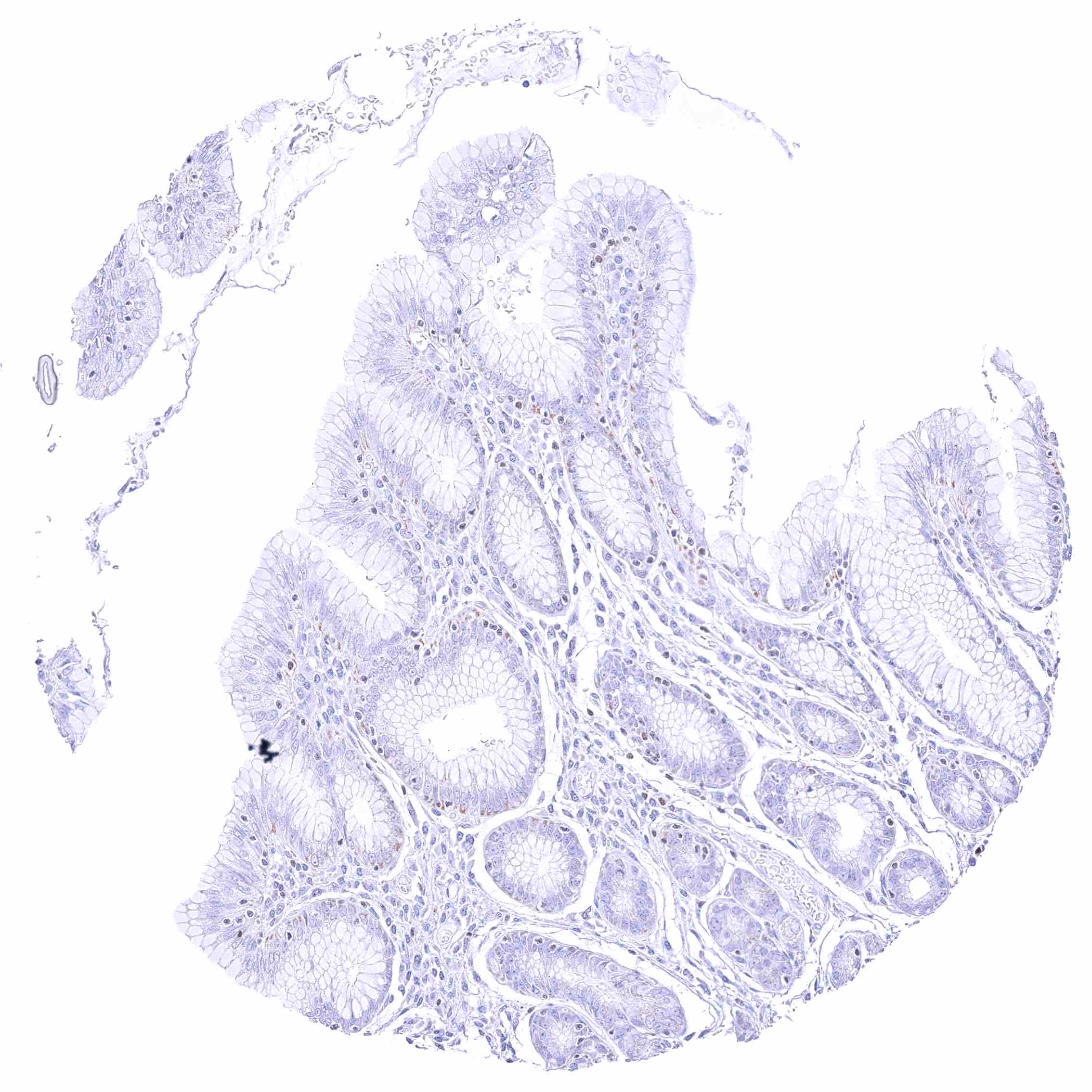
Colon descendens, mucosa – Nuclear GATA3 positivity of few lymphocytes. Faint cytoplasmic GATA3 positivity of some epithelial cells.
Colon descendens, muscular wall
Duodenum, Brunner gland
Duodenum, mucosa – Granular cytoplasmic GATA3 staining of endometrial epithelial cells.
Epididymis (Caput) – Moderate to strong, nuclear GATA3 immunostaining of tall columnar cells nd of basal cells.
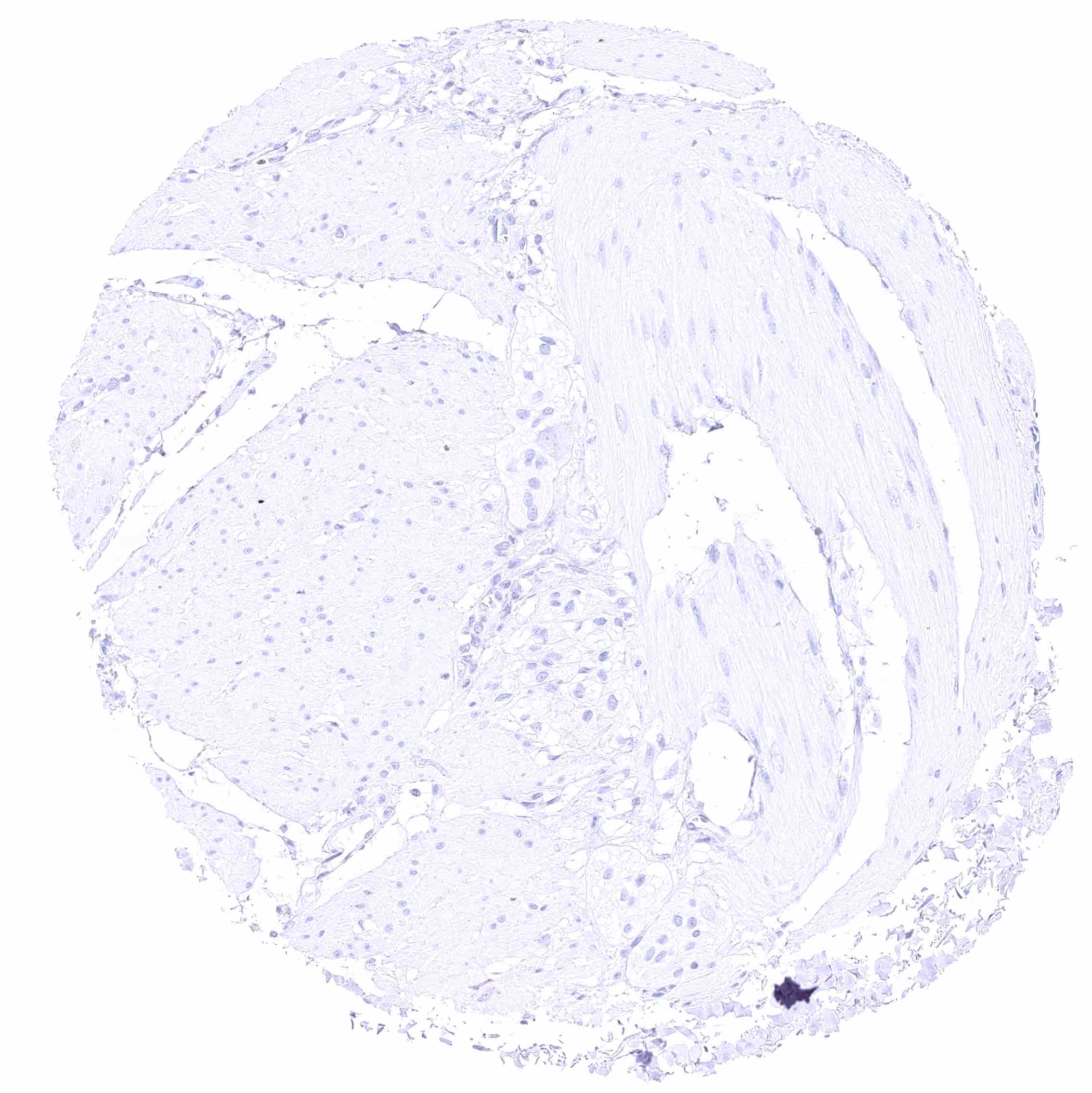
Esophagus, muscular wall
Esophagus, squamous epithelium – Weak nuclear GATA3 positivity of few lymphocytes.
Fallopian tube, mucosa
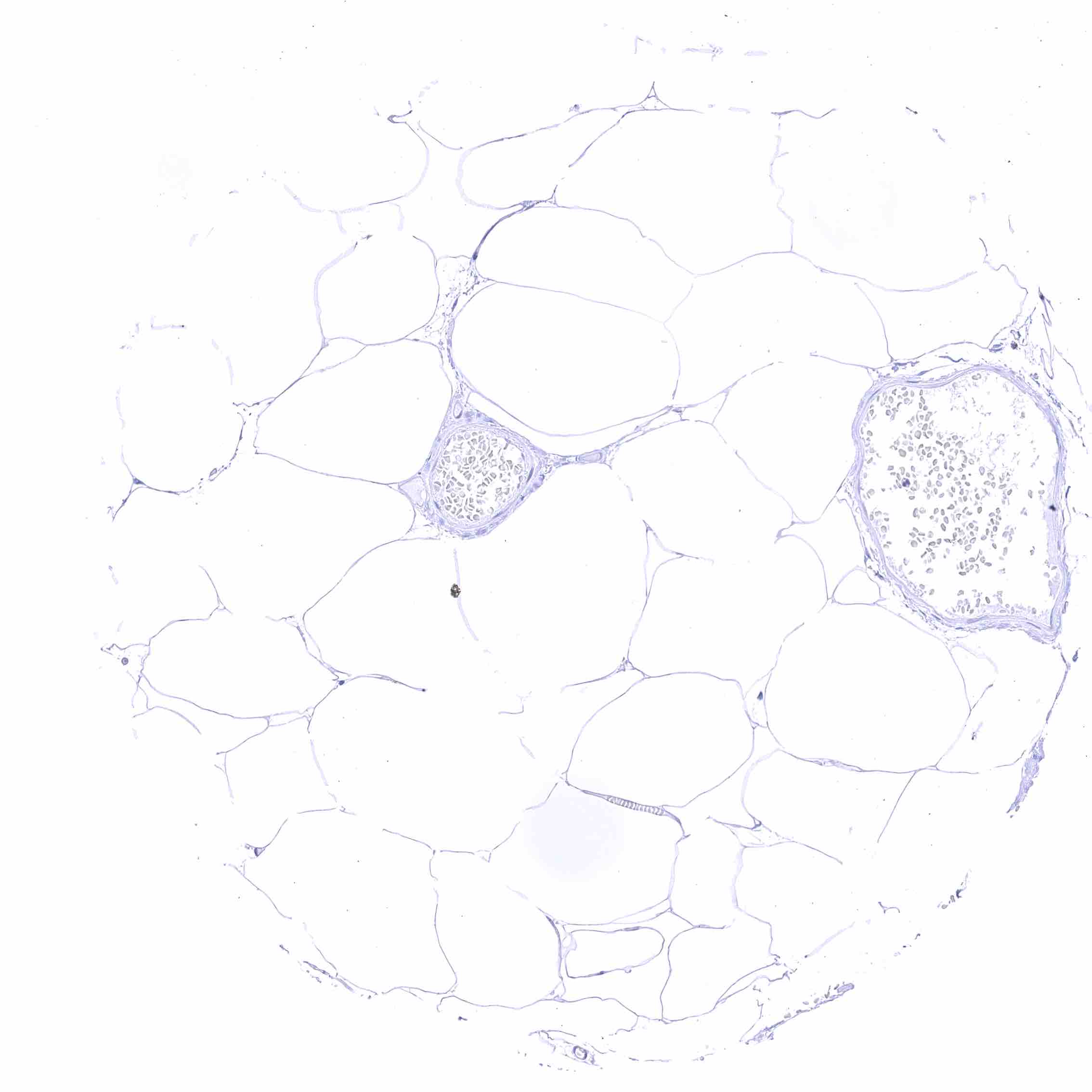
Fat
Gallbladder, epithelium – Faint cytoplasmic GATA3 positivity of some epithelial cells.

Heart muscle
Ileum, mucosa – Nuclear GATA3 positivity of few lymphocytes.
Ileum, muscular wall
Kidney, cortex – Moderate to strong, nuclear GATA3 positivity of collecting ducts, some cells of distal tubuli, and of podocytes.
Kidney, medulla – Moderate to strong, nuclear GATA3 positivity of some collecting ducts.
Kidney, pelvis, urothelium – Strong nuclear GATA3 positivity of all urothelial cells.
Liver – Nuclear GATA3 positivity of several lymphocytes in a portal field.
Lung – Nuclear GATA3 staining of few lymphocytes.
Lymph node – Nuclear GATA3 positivity of numerous lymphocytes.
Ovary, corpus luteum
Ovary, follicular cyst
Ovary, stroma
Pancreas – Nuclear GATA3 staining of several lymphocytes.
Parathyroid gland – Strong nuclear GATA3 positivity of all epithelial cells. .jpeg
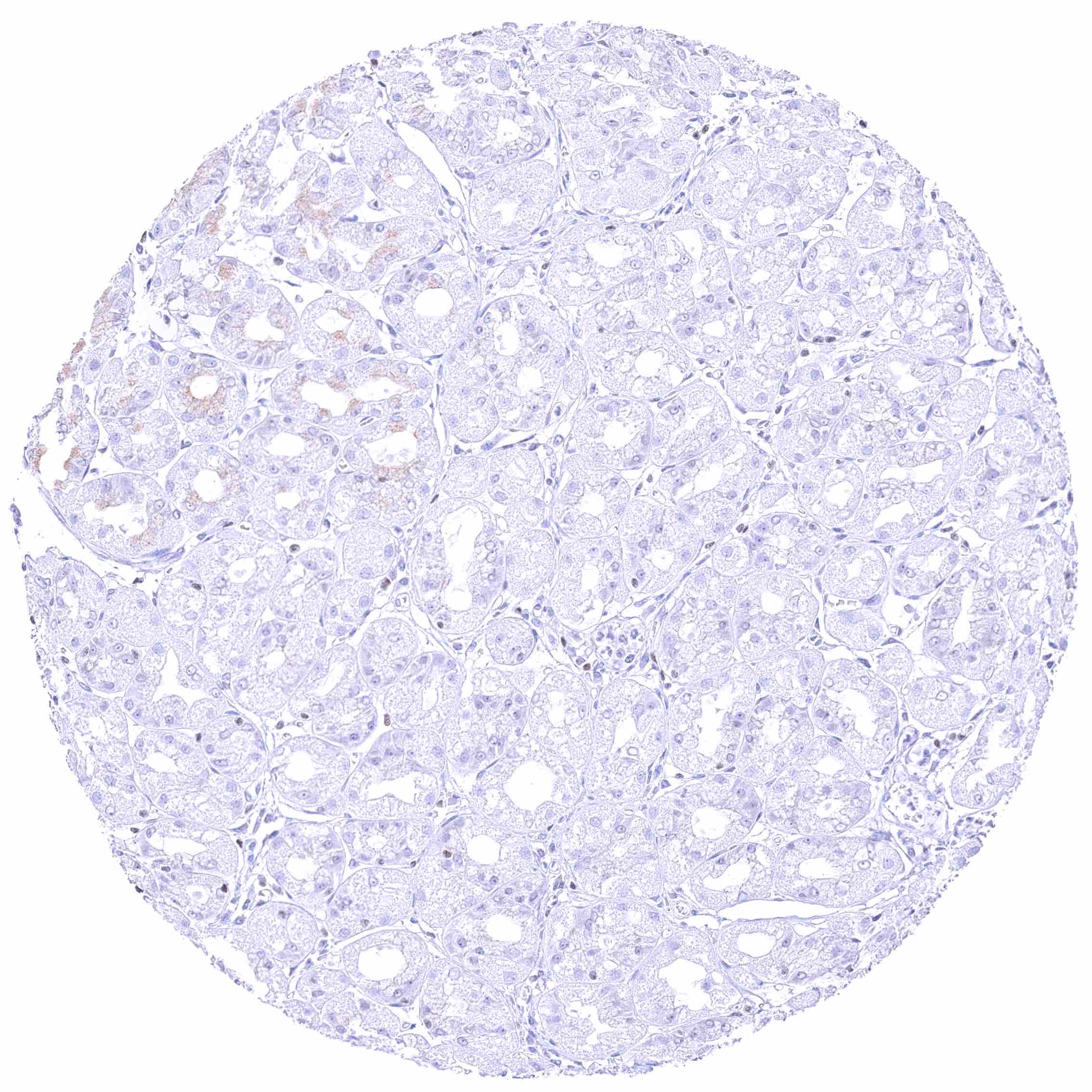
Parotid gland – Faint nuclear GATA3 positivity of few epithelial cells.
Parotid gland – Weak to moderate, nuclear GATA3 positivity of glandular cells.
Pituitary gland, anterior lobe
Pituitary gland, posterior lobe
Placenta (amnion and chorion) – Strong nuclear GATA3 staining of chorion cells while amnion cells only show weak positivity.
Placenta, early – Strong nuclear GATA3 positivity of trophoblastic cells.
Placenta, mature – Strong nuclear GATA3 positivity of trophoblastic cells.
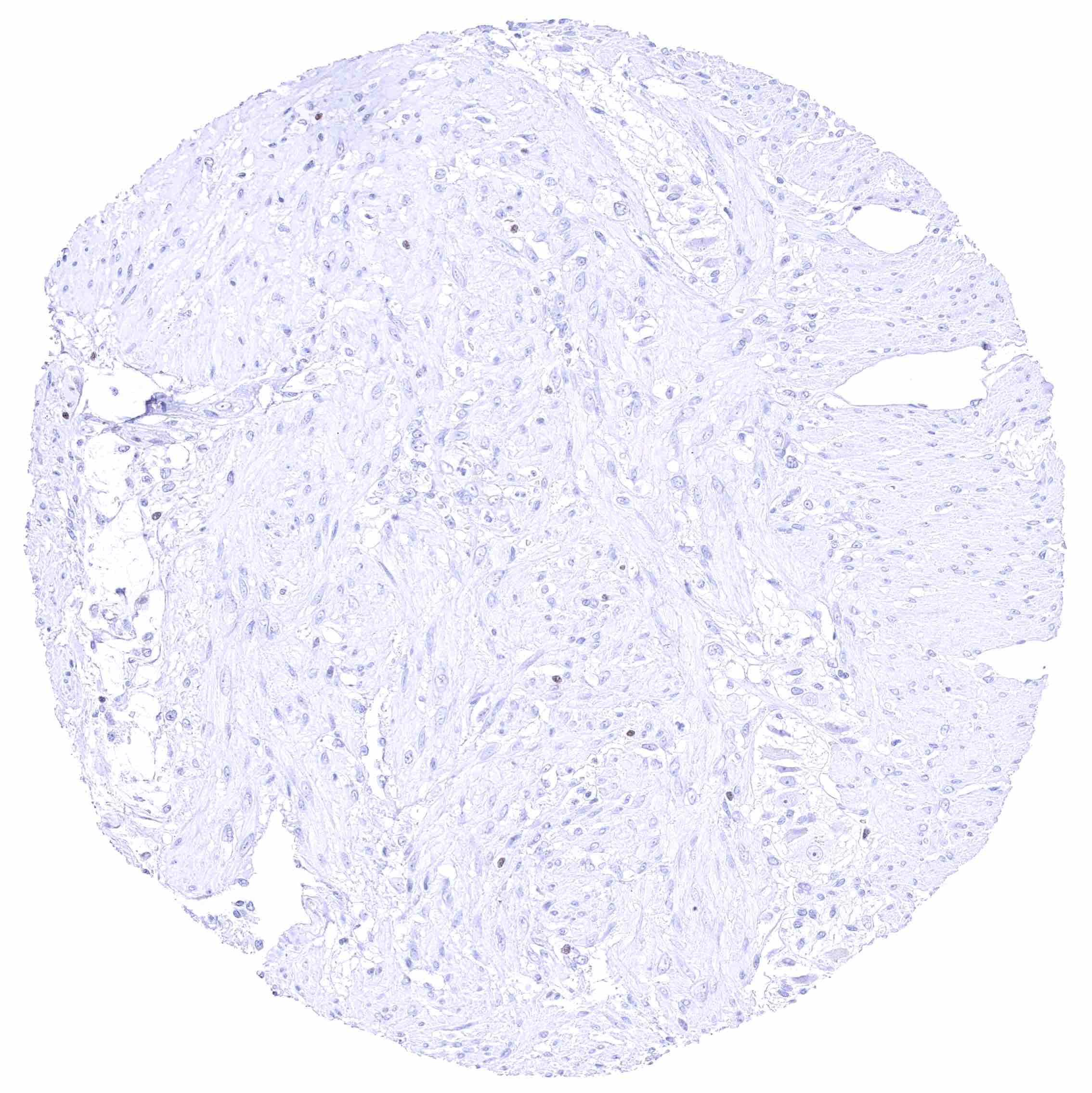
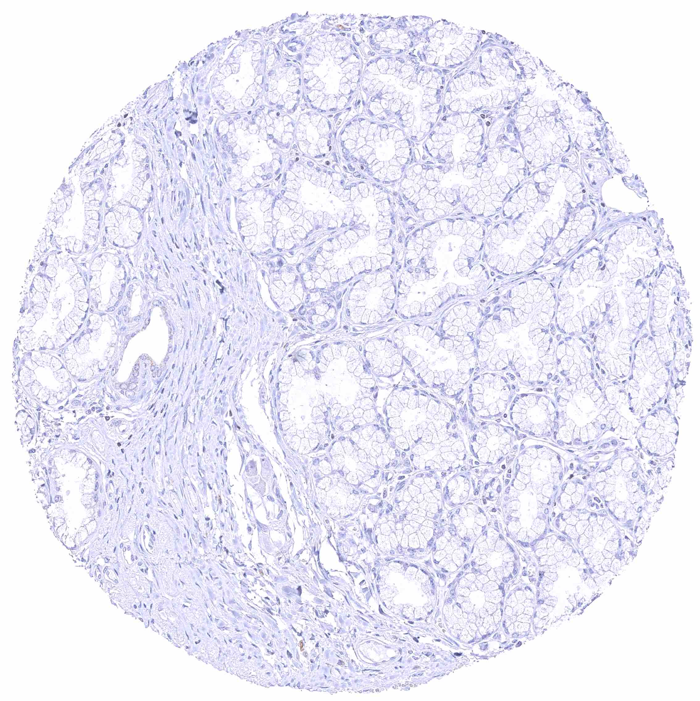
Prostate – Weak to moderate, nuclear GATA3 staining of basal cells. .jpeg
Rectum, mucosa – Nuclear GATA3 positivity of many lymphocytes.
Rectum, mucosa – Nuclear GATA3 positivity of some lymphocytes. Granular cytoplasmic GATA3 staining of epithelial cells.
Seminal vesicle – Moderate to strong nuclear GATA3 positivity of epithelial cells.
Sinus paranasales
Skeletal muscle
Skin – Strong nuclear GATA3 staining of squamous epithelial cells.
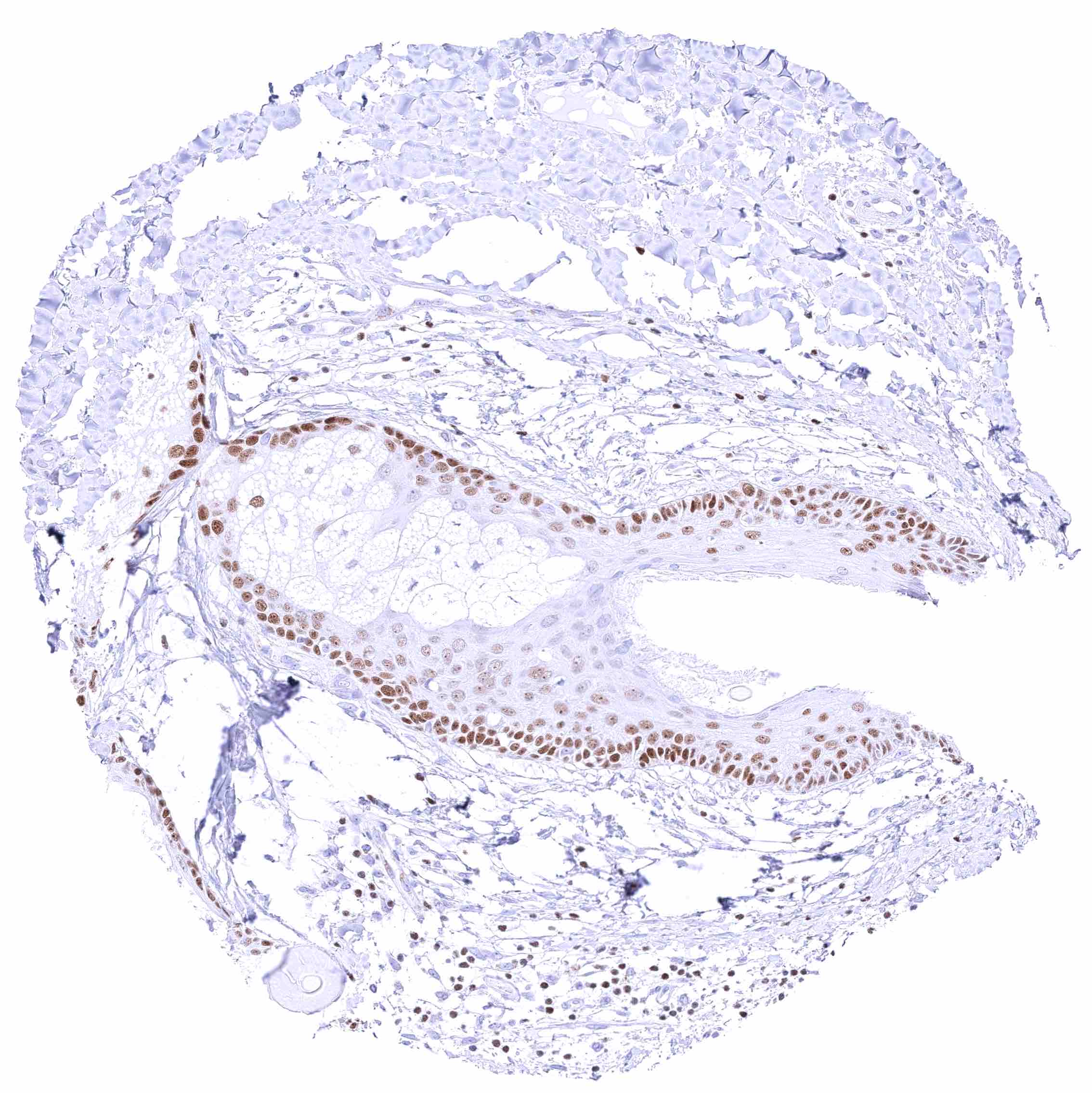
Skin, anal canal – Weak to moderate nuclear GATA3 staining of squamous epithelial cells. The staining predominates in the lower half of the epidermis.
Skin, hair follicel and sebaceous glands – Moderate nuclear GATA3 staining of cells of hair follicles and of peripheral germinative cells of sebaceous glands. GATA3 staining is faint or absent in cells of sebaceous glands.
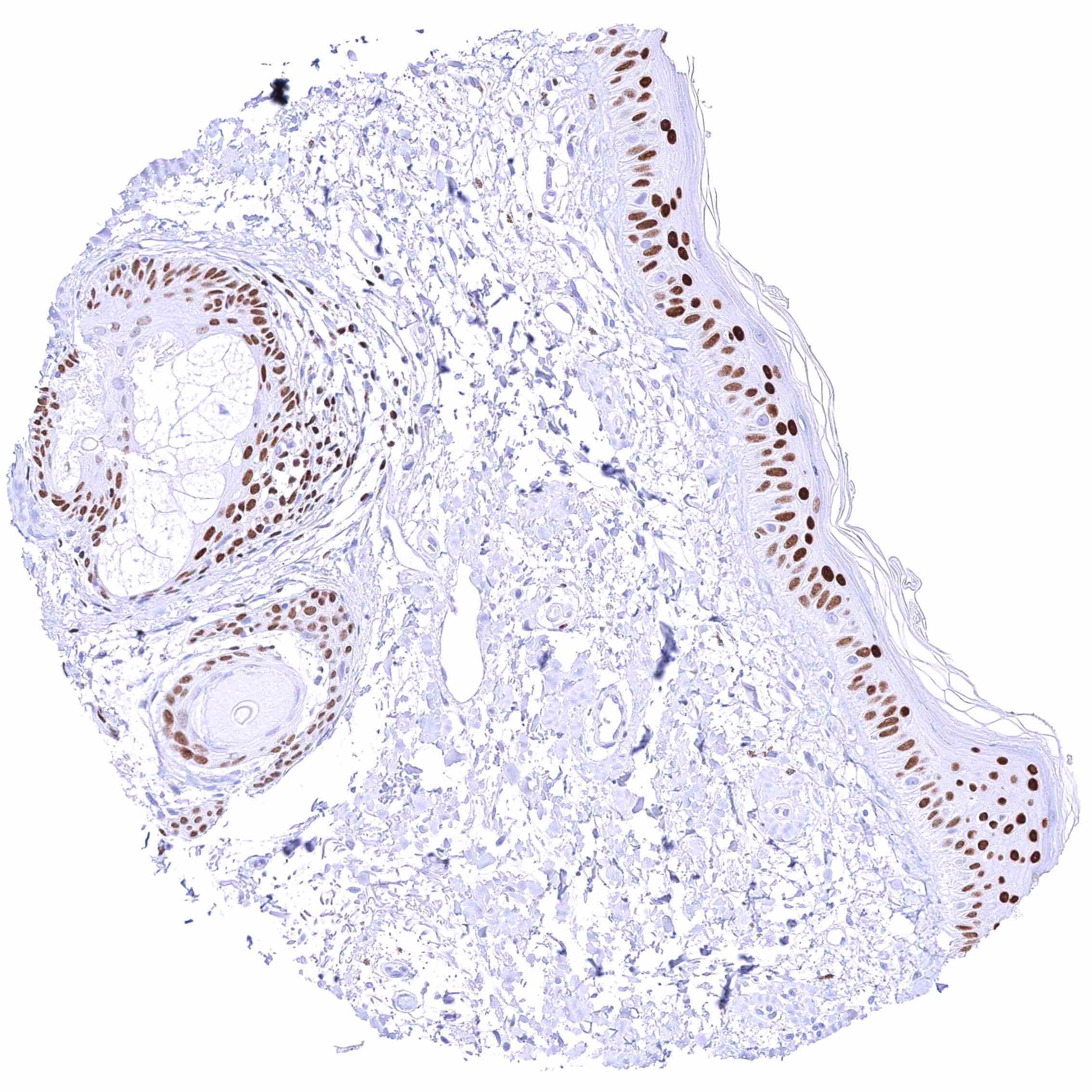
Skin, hair follicel and sebaceous glands – Strong nuclear GATA3 staining of squamous epithelial cells, cells of hair follicles, and peripheral germinative cells of sebaceous glands. Nuclear GATA3 staining is faint or absent in cells of sebaceous glands.
Spleen – Nuclear GATA3 positivity of few lymphocytes.
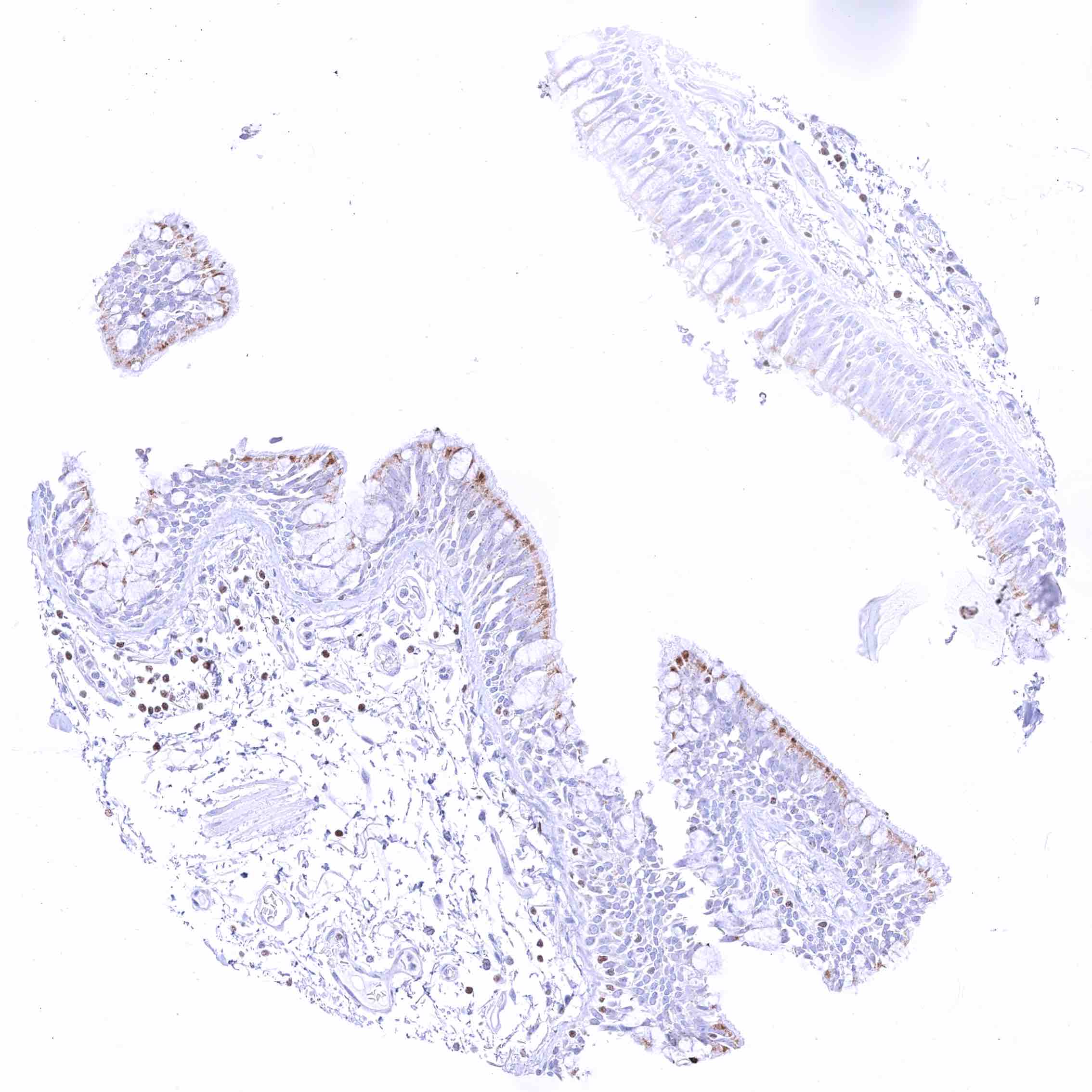
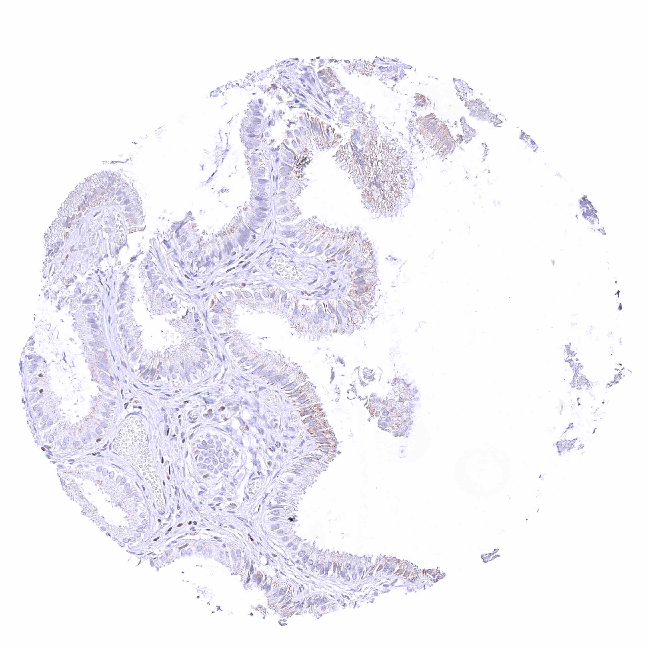
Stomach, antrum.jpeg
Stomach, corpus.jpeg
Stomach, muscular wall – Nuclear GATA3 positivity of few lymphocytes.
Sublingual gland – Moderate to strong, nuclear GATA3 positivity of glandular cells.
Submandibular gland – Moderate to strong, nuclear GATA3 positivity of glandular cells.
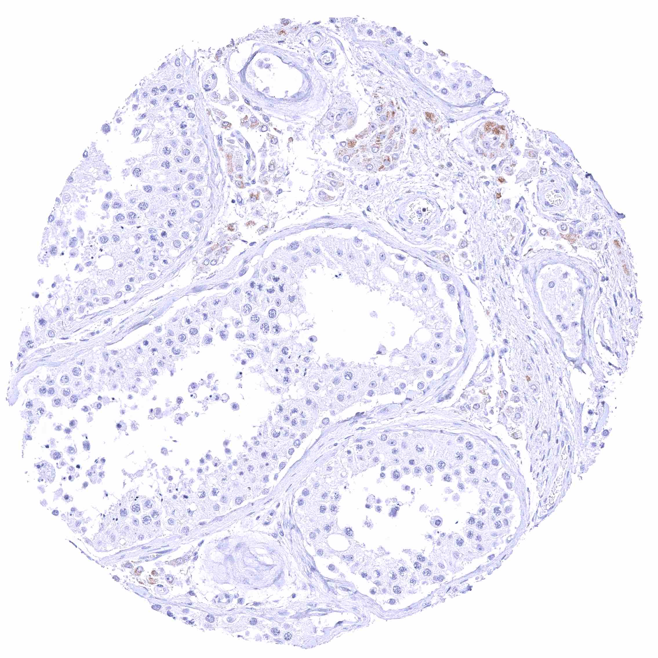
Testis
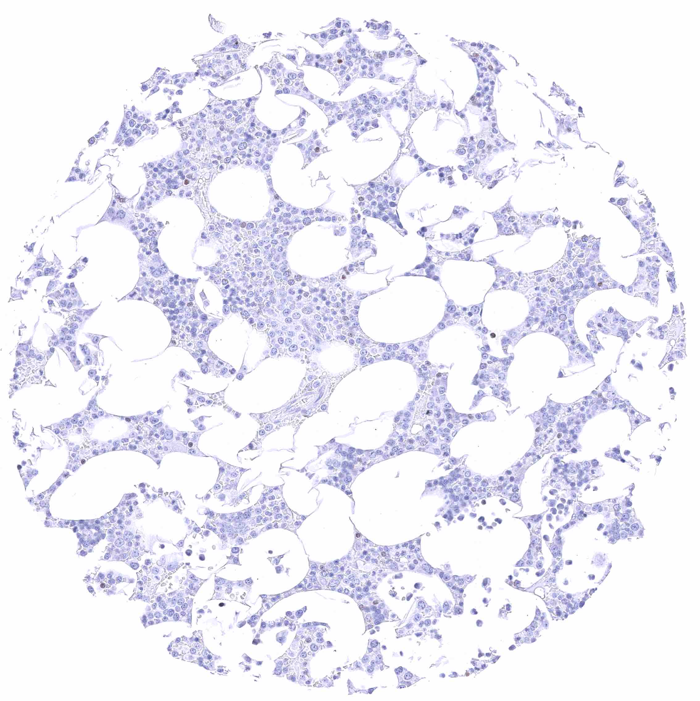
Thymus – Moderate to strong, nuclear GATA3 staining of a large fraction of lymphocytes. .jpeg
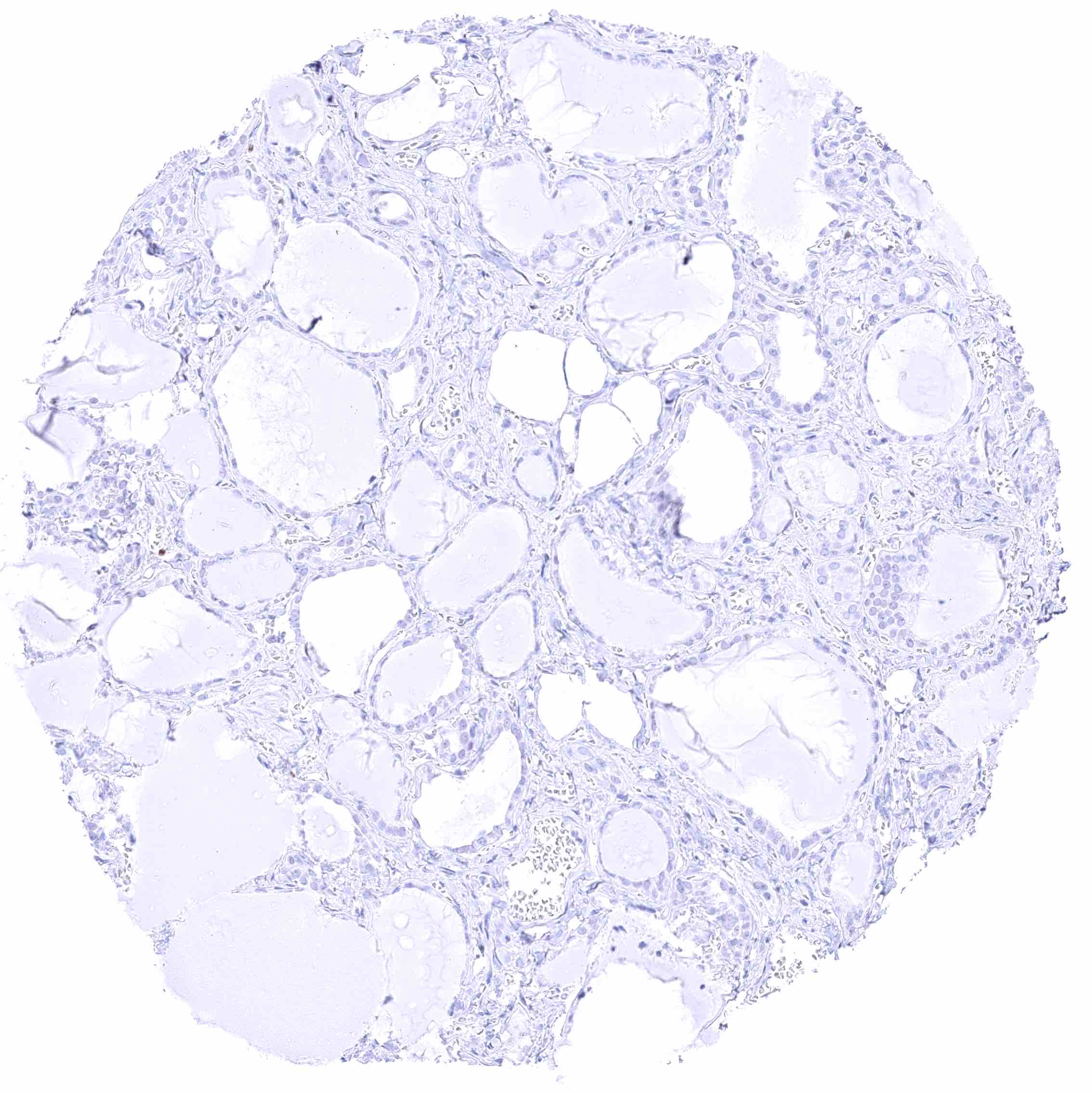
Thyroid gland
Tonsil – Nuclear GATA3 positivity of a significant fraction of lymphocytes.
Tonsil – Nuclear GATA3 positivity of numerous lymphocytes.
Tonsil, surface epithelium – Nuclear GATA3 positivity of few lymphocytes.
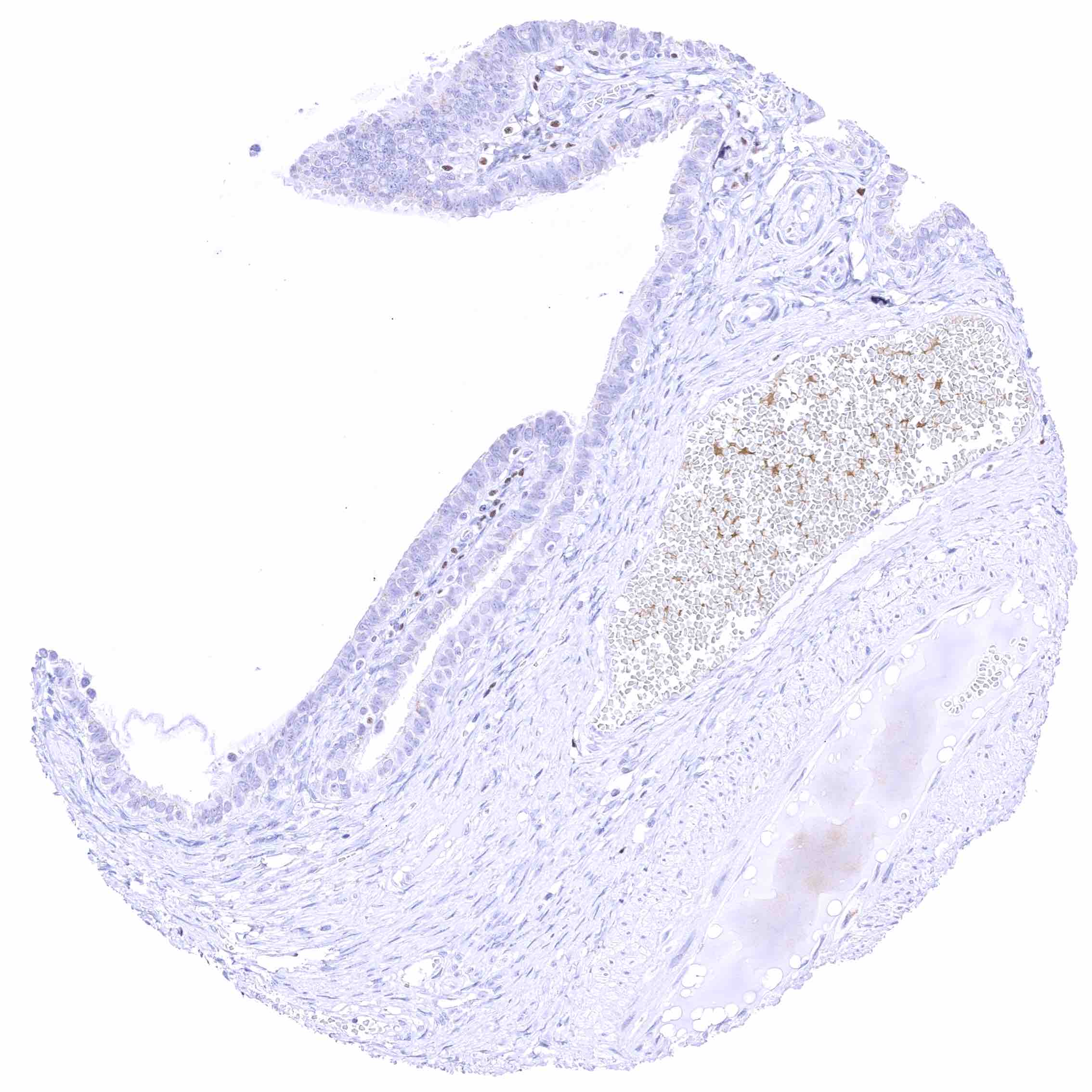
Urinary bladder, muscular wall
Uterus, ectocervix – Weak to moderate nuclear GATA3 staining of squamous epithelial cells.
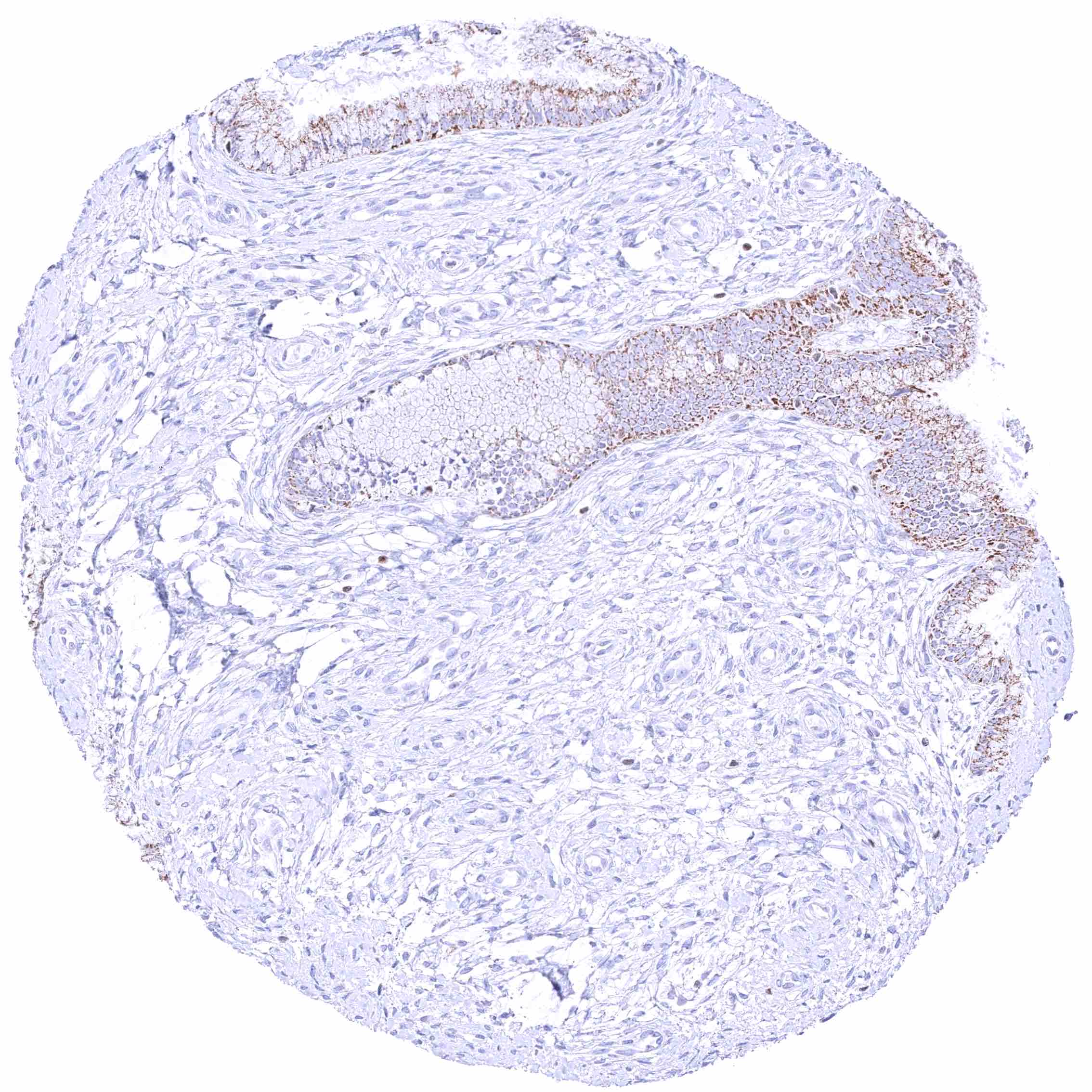
Uterus, endocervix – Granular cytoplasmic GATA3 positivity of epithelial cells.
Uterus, endometrium (pregnancy) – Faint nuclear GATA3 positivity of a subset of decidua cells. .jpeg
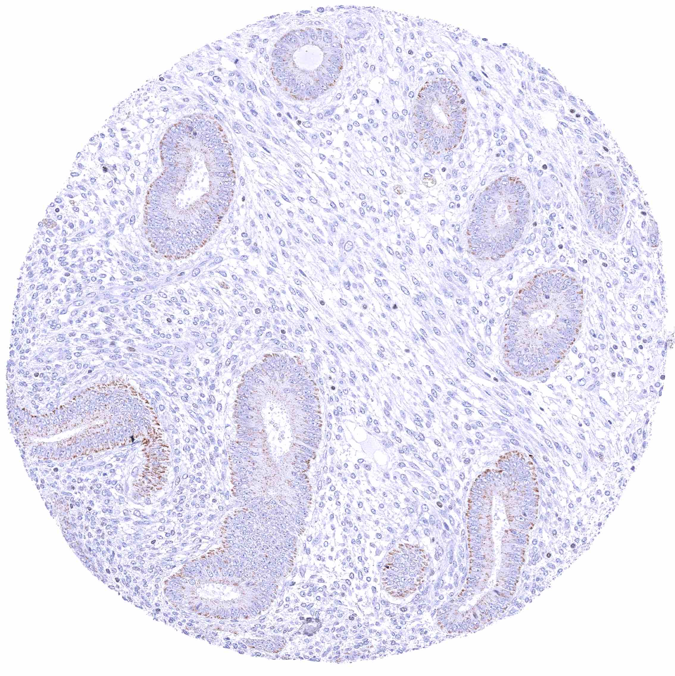
Uterus, endometrium (proliferation) – Granular cytoplasmic GATA3 positivity of some endometrial epithelial cells.
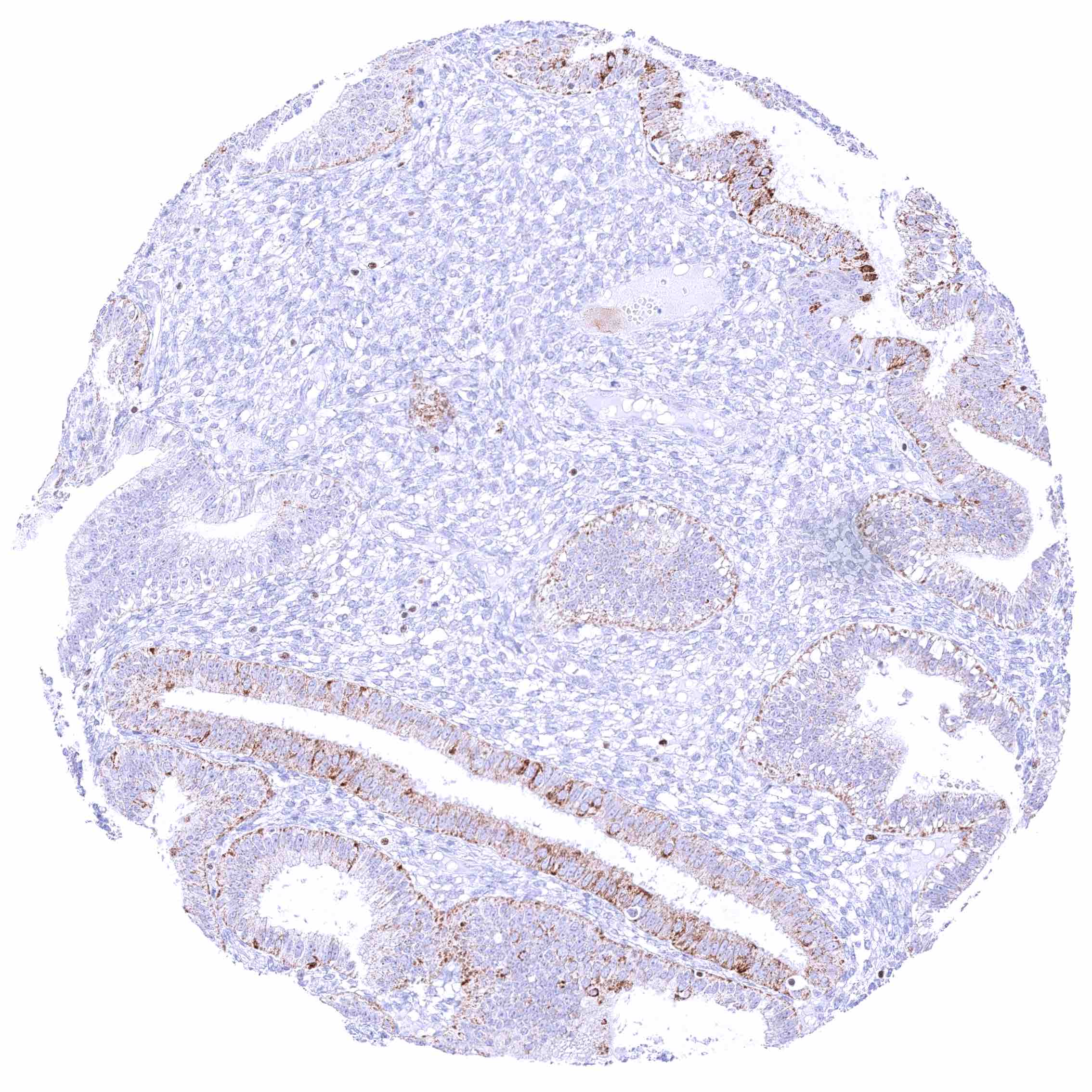
Uterus, endometrium (secretion) – Granular cytoplasmic GATA3 positivity of endometrial epithelial cells. .jpeg
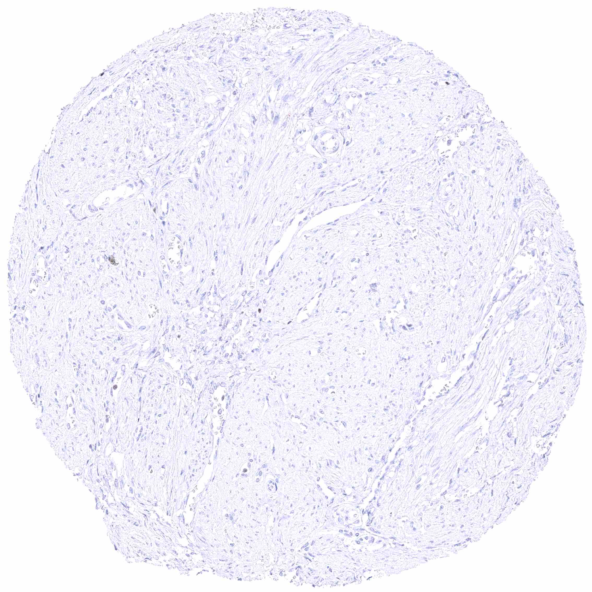
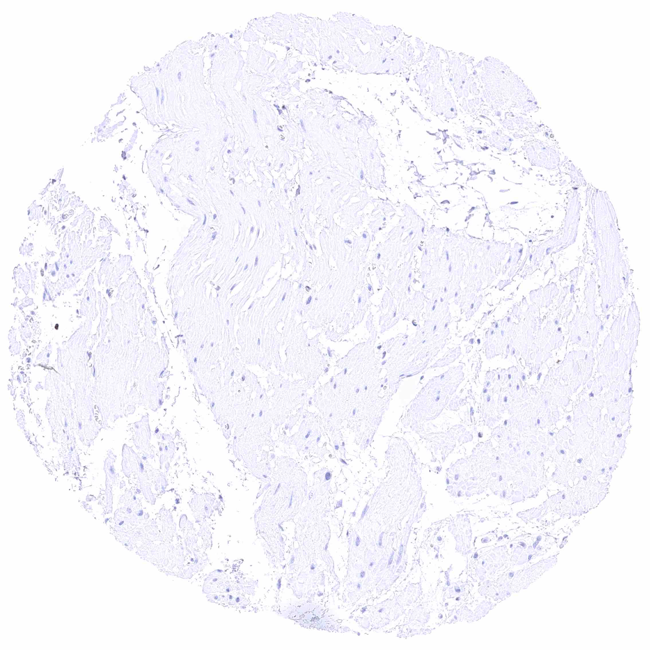
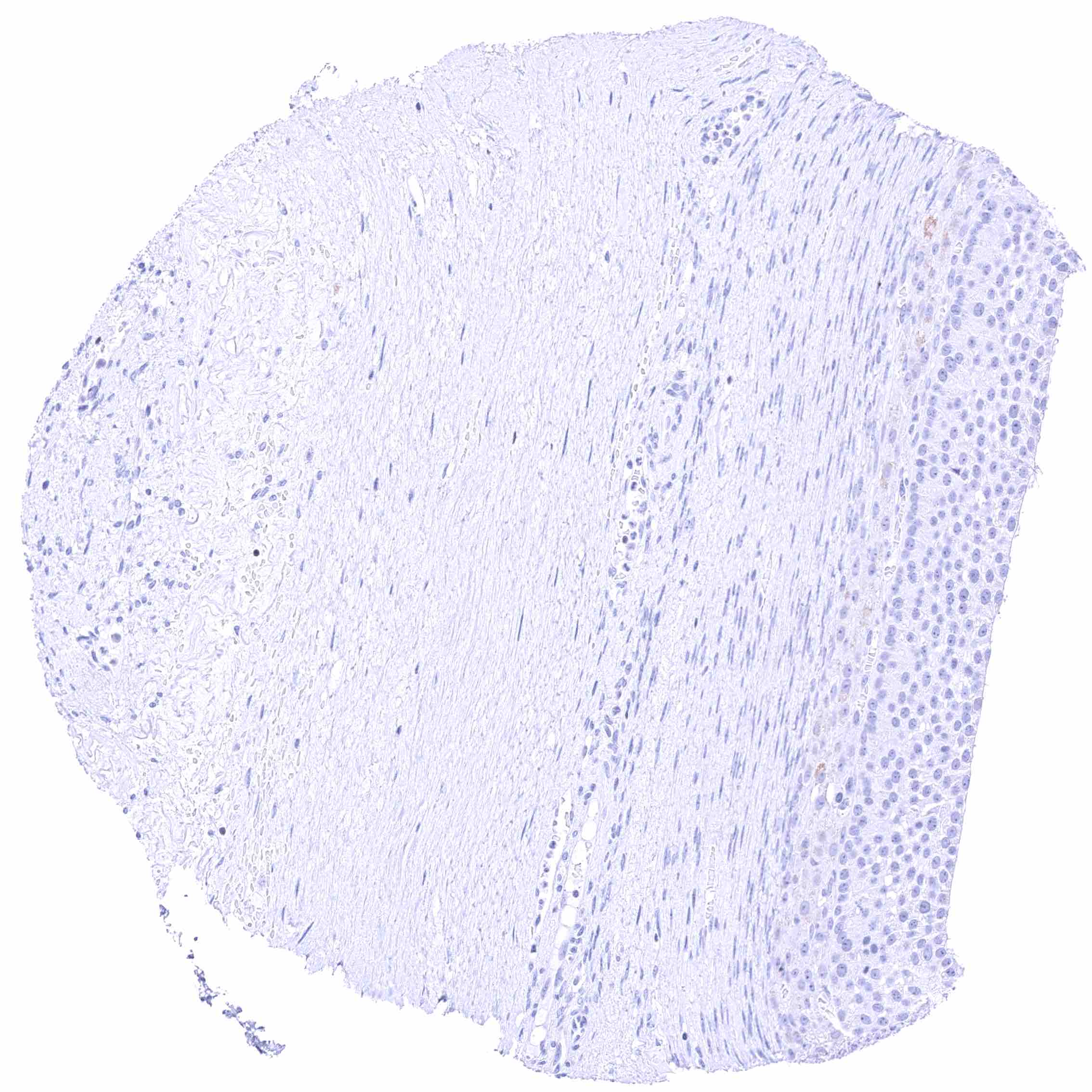
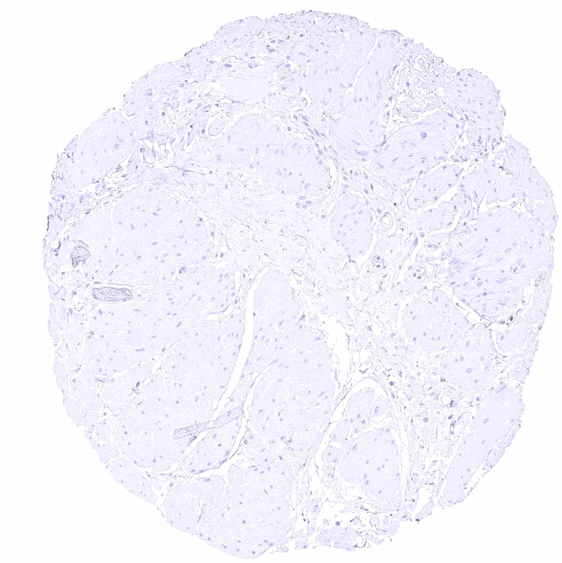
Uterus, myometrium