Adrenal gland - A very faint ER positivity can be observed in a fraction of adrenocortical cells in this sample

Adrenal gland - A very faint ER positivity can be observed in a fraction of adrenocortical cells in this sample.
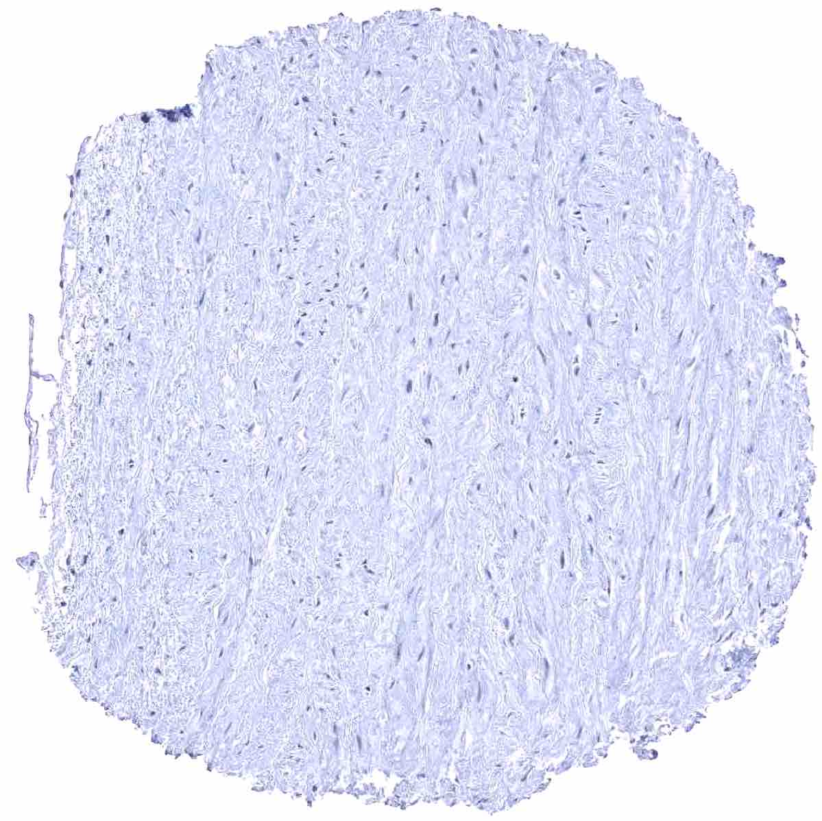
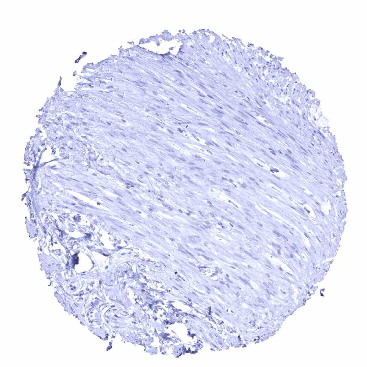
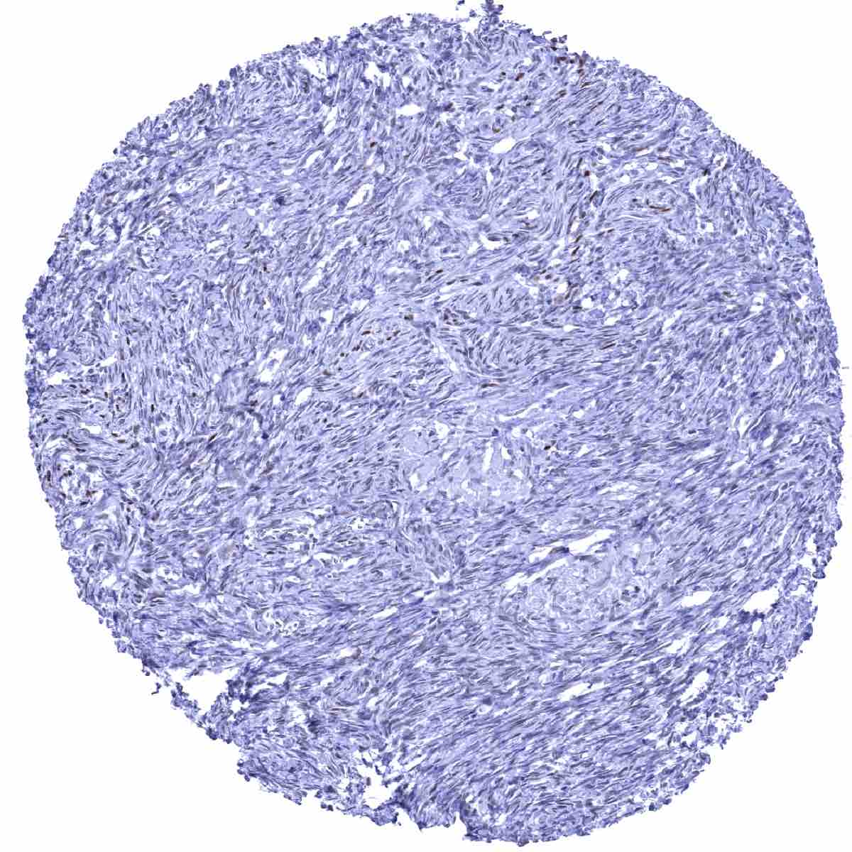
Aorta, media - A faint ER staining of muscular cells of the media can occasionally be seen
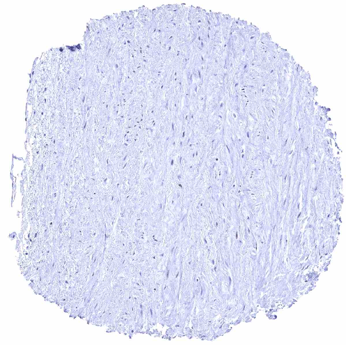
Aorta, media - A faint ER staining of muscular cells of the media can occasionally be seen.
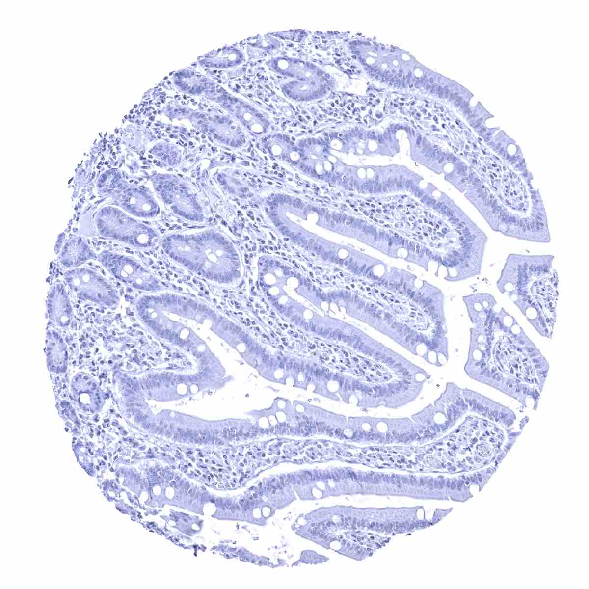
Appendix, mucosa
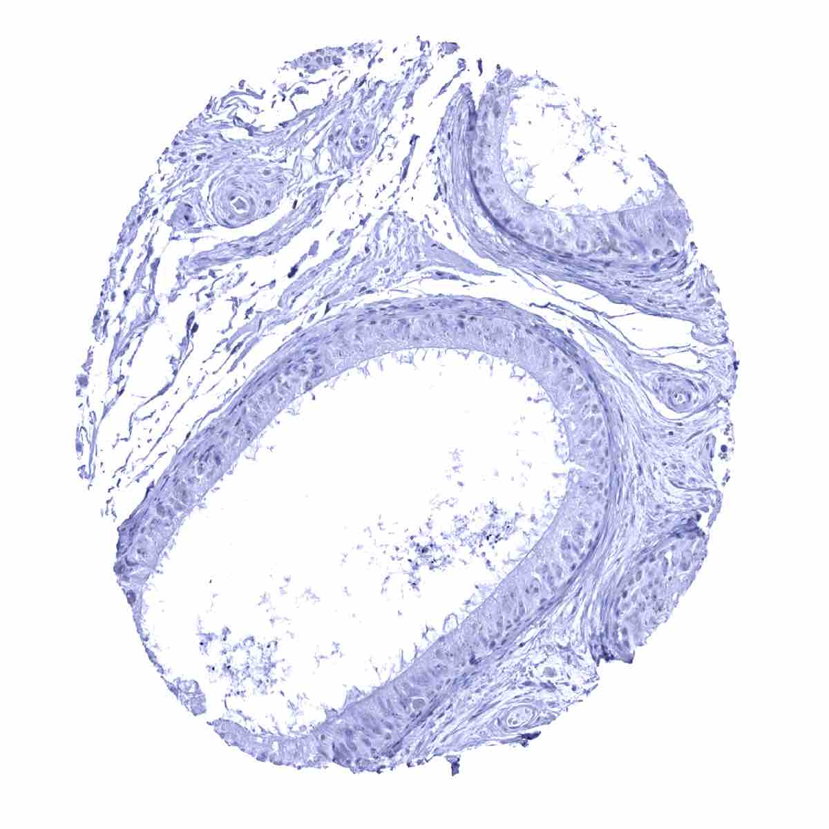
Appendix, muscular wall
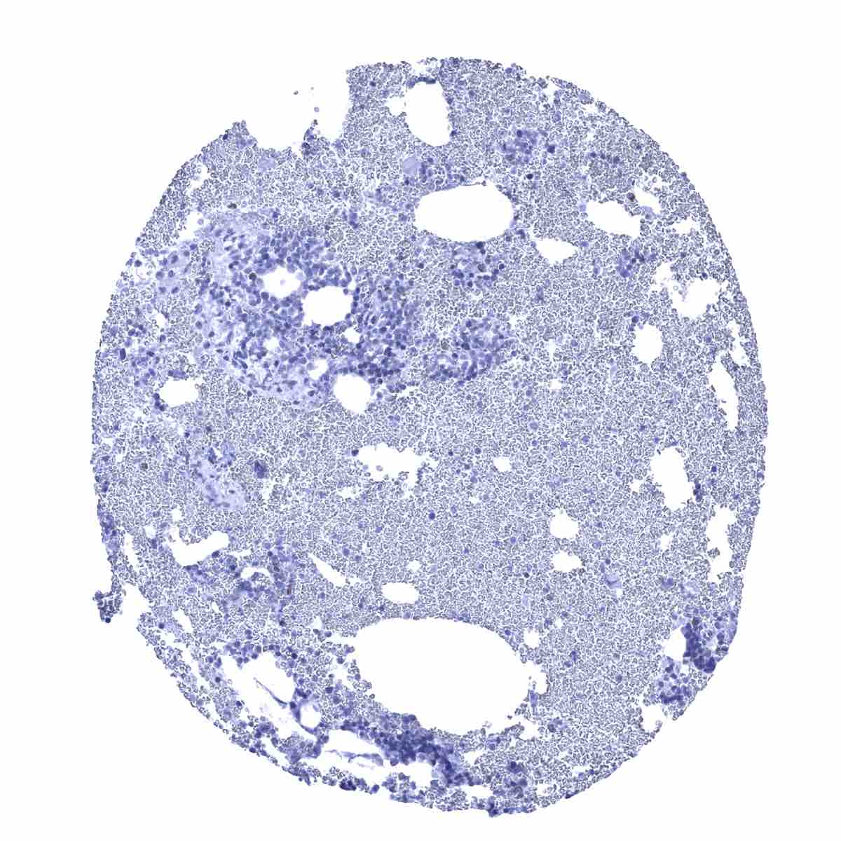
Bone marrow
Breast - A variable intensity ER staining occurs in a fraction of epithelial cells of the breast gland

Bronchus, mucosa

Cerebellum (molecular layer, Purkinje cell layer, granule cell layer, white matter)
Cerebellum (molecular layer, Purkinje cell layer, granule cell layer)
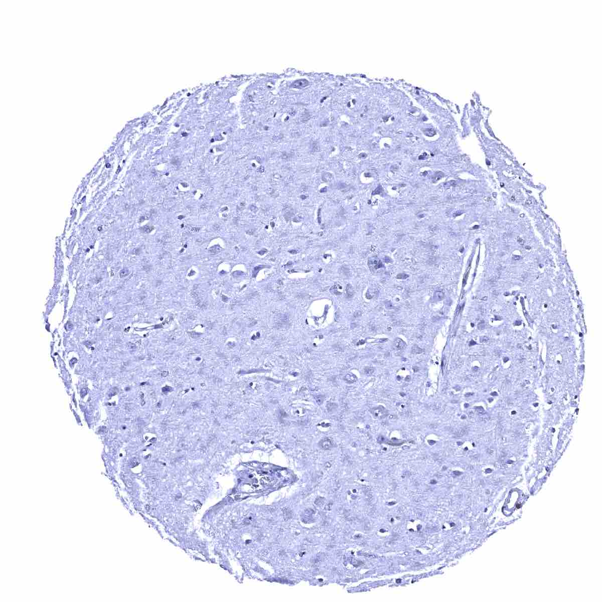
Cerebrum, grey matter

Cerebrum, white matter
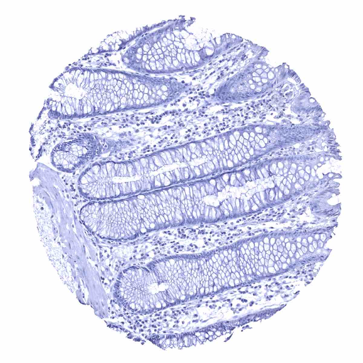
Colon descendens, mucosa
Colon descendens, muscular wall
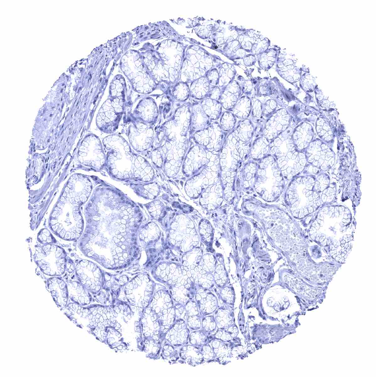
Duodenum, Brunner gland
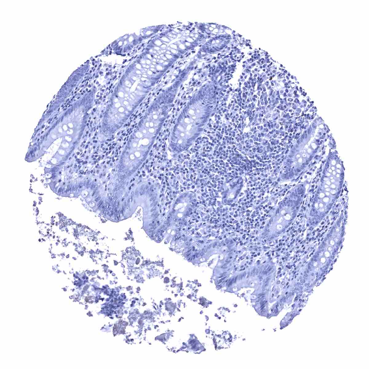
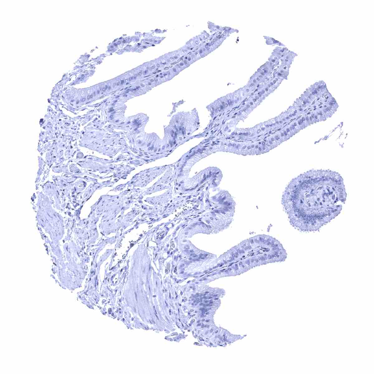
Duodenum, mucosa

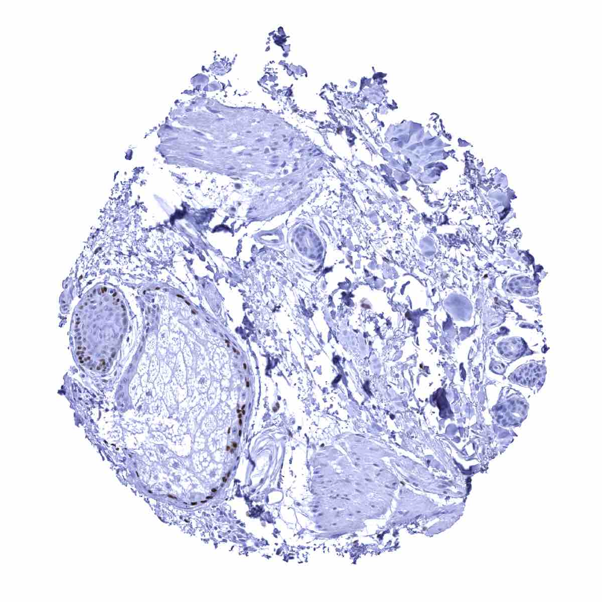
Epididymis, cauda - A moderate to strong ER immunostaining can be seen in epithelial cells of the cauda epididymis
Epididymis, corpus - ER immunostaining is absent epithelial cells of the corpus epididymis
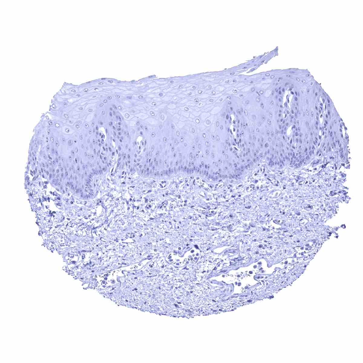
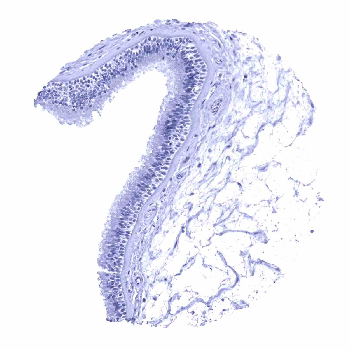
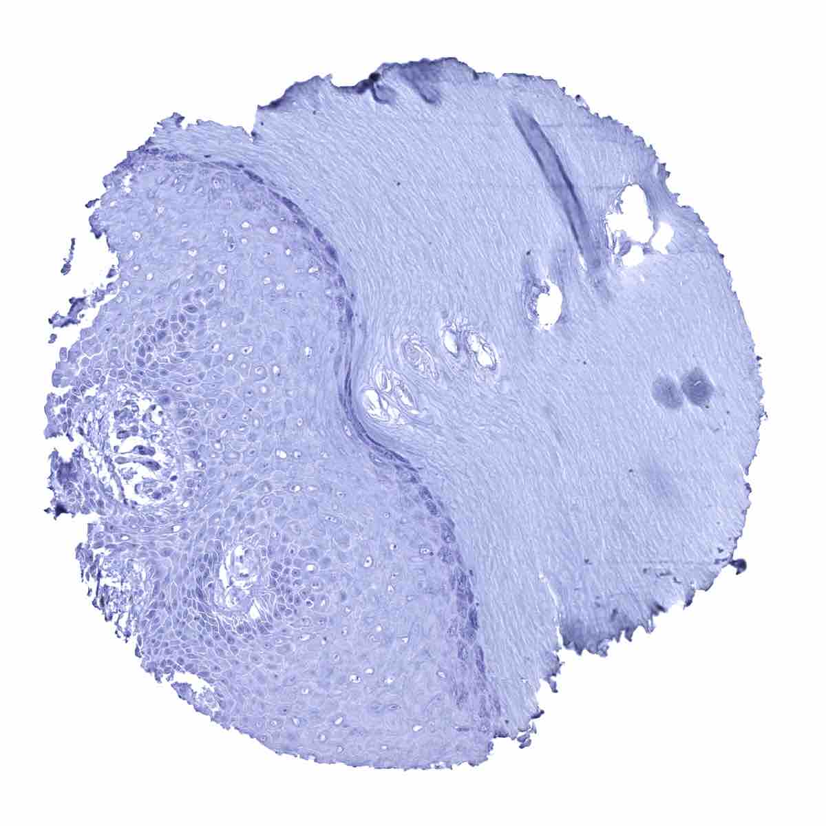
Esophagus, squamous epithelium - Absence of ER staining in the squamous epithelial cells from this esophageal sample
Fallopian tube, mucosa - A strong epithelial and stromal ER positivity is seen in the fallopian tube
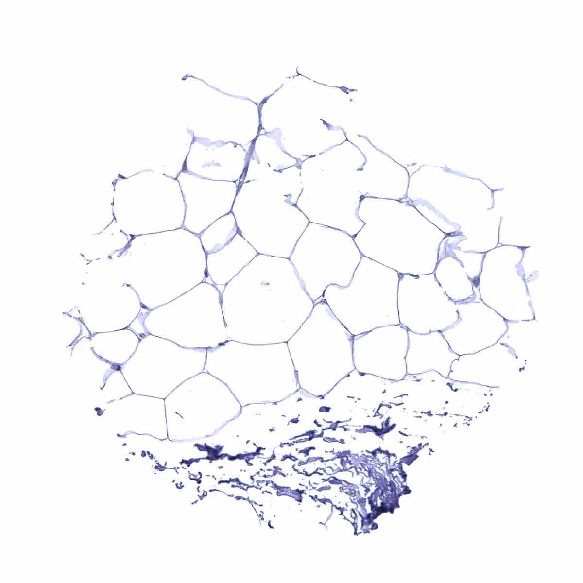
Fat
Gallbladder, epithelium

Heart muscle

Ileum, mucosa

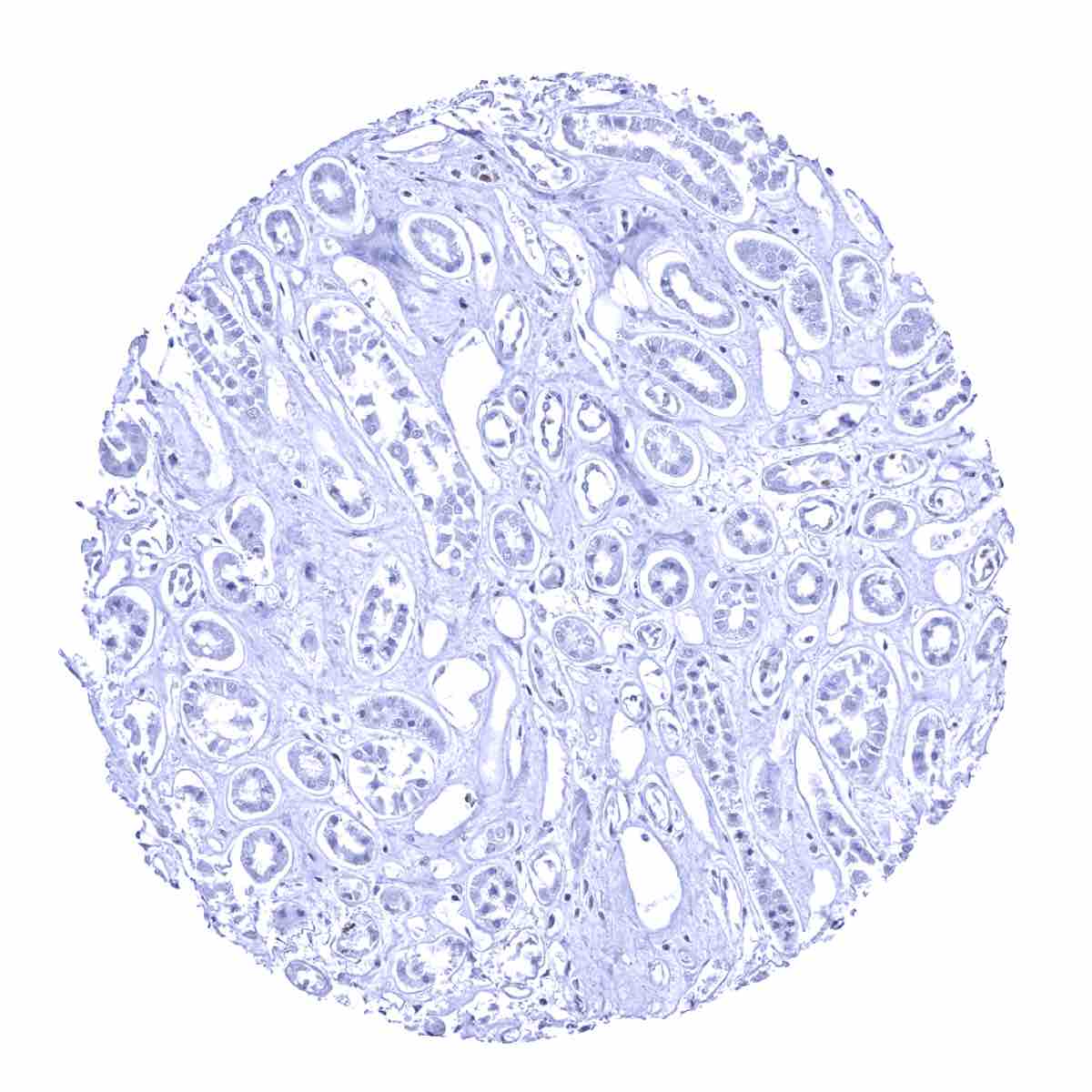
Kidney, cortex
Kidney, medulla

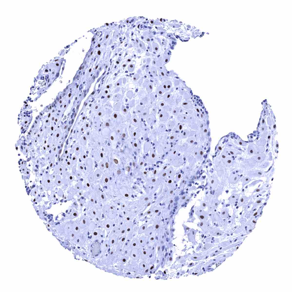
Liver - A variable, weak to moderate ER positivity can be seen in some hepatocytes in a fraction of samples

Liver - ER immunostaining is absent in this liver sample

Lung
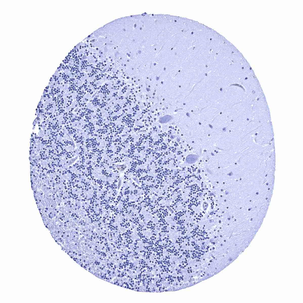
Lymph node - In lymphatic tissues, dispersed follicular dendritic cells in germinal centers show a weak to moderate staining
Ovary, stroma - A faint ER immunostaining can be seen in ovarian stroma cells
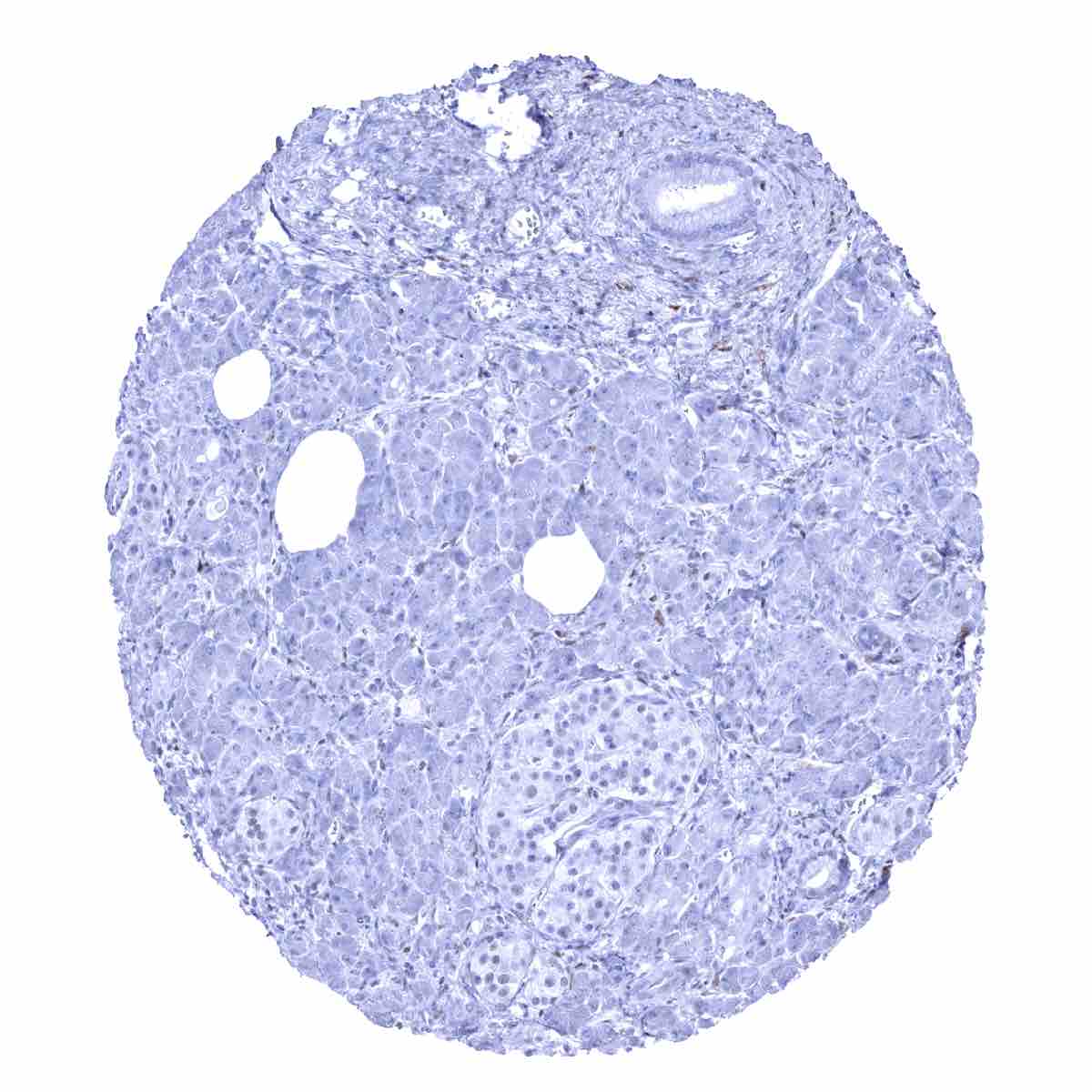
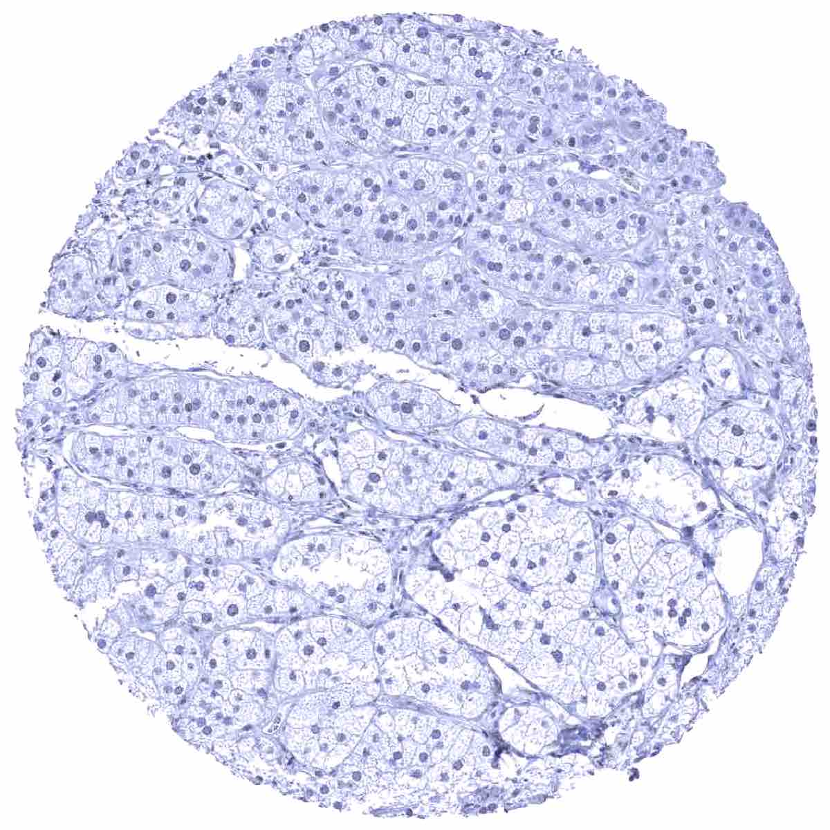
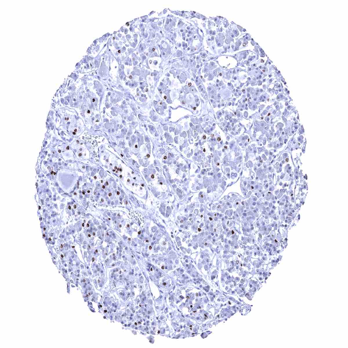
Pancreas - A very faint ER staining of few islet cells and some other cells of the pancreas can be seen
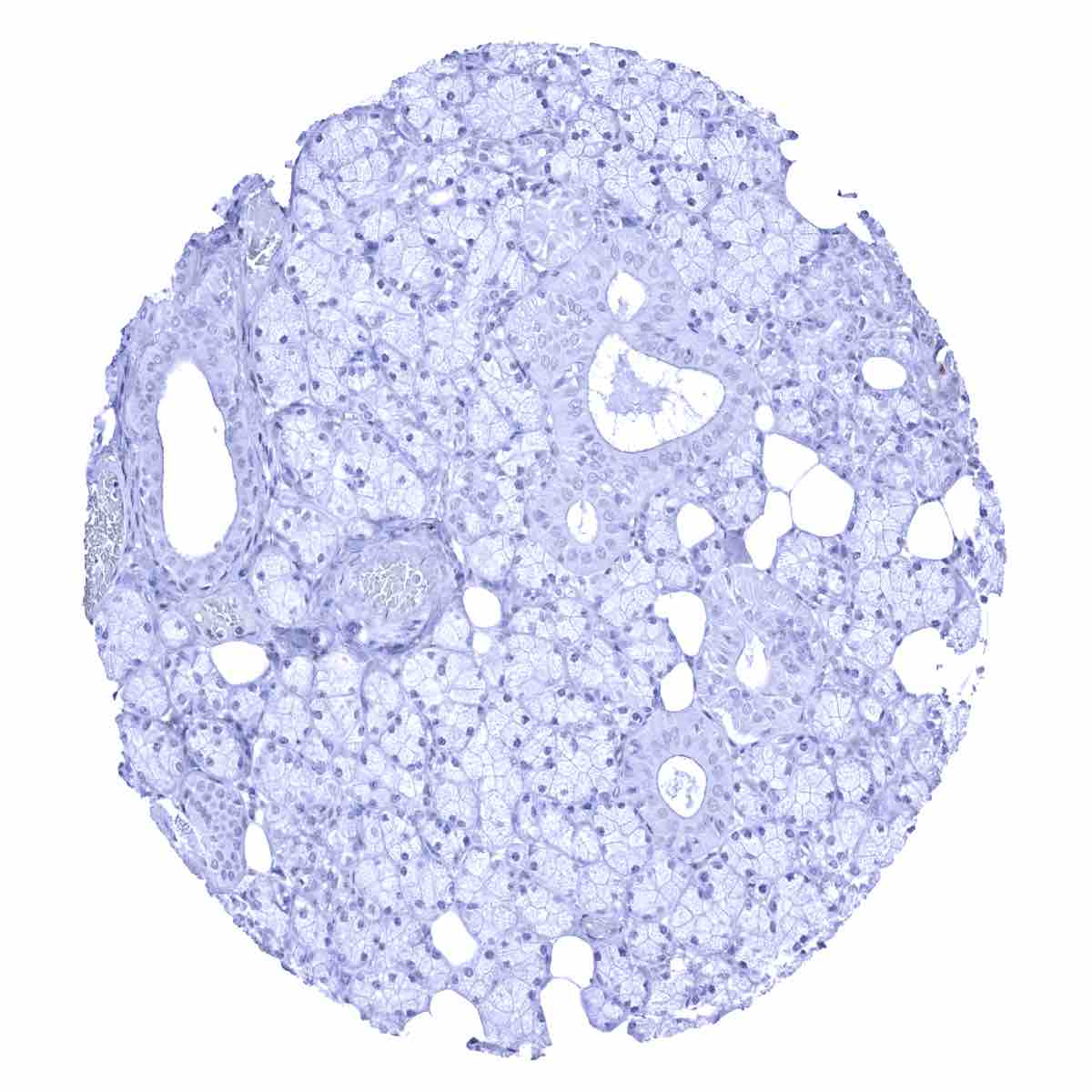
Parathyroid gland

Parotid gland - A variable, weak to moderate ER positivity can occasionally be seen in a fraction of epithelial cells of salivary glands
Parotid gland
Pituitary gland, anterior lobe - A moderate to strong ER immunostaining can be found in a fraction (10-30%) of epithelial cells in the adenohypophysis
Pituitary gland, posterior lobe
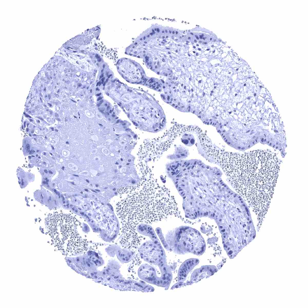
Placenta (amnion)
Placenta (chorion)
Placenta, early
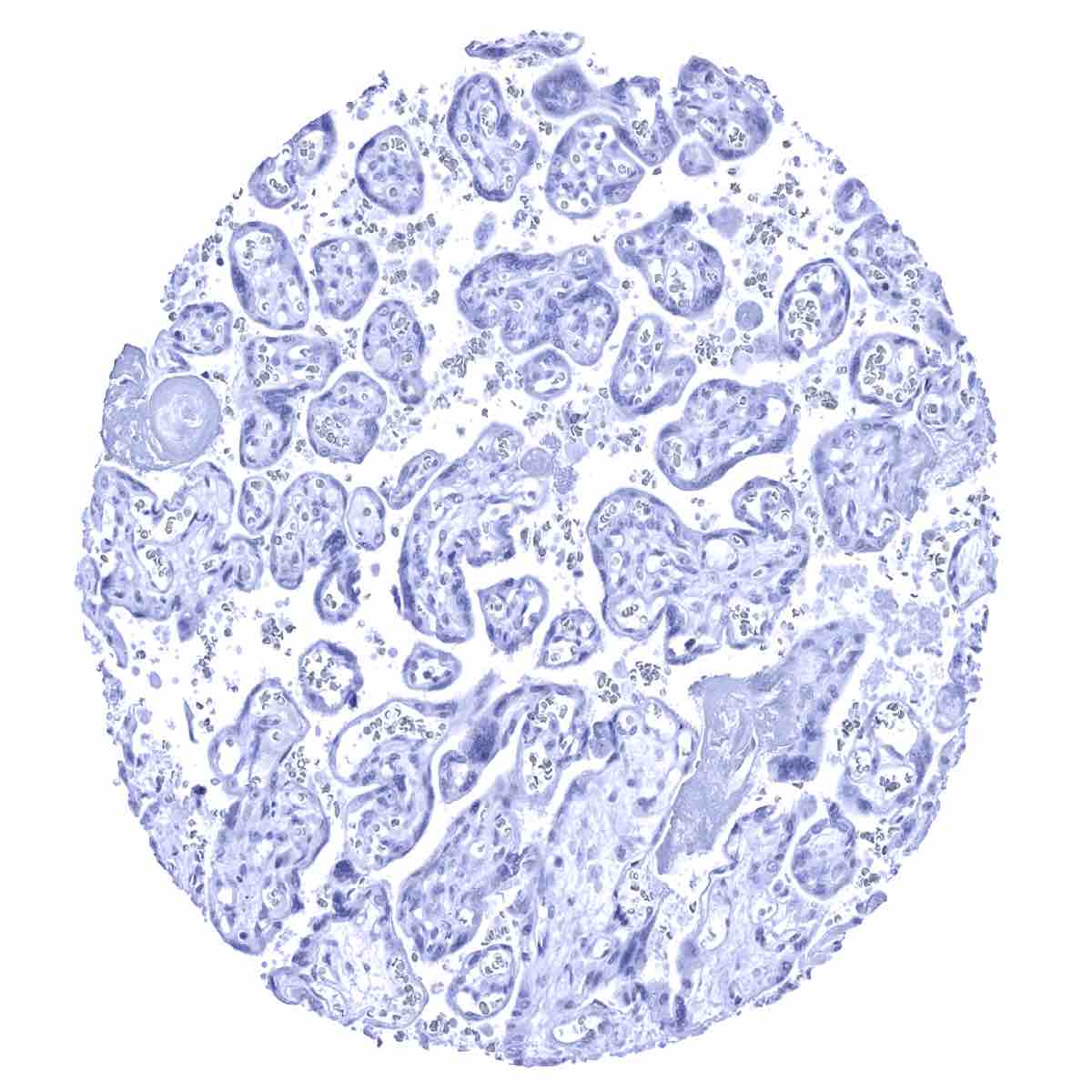
Placenta, mature

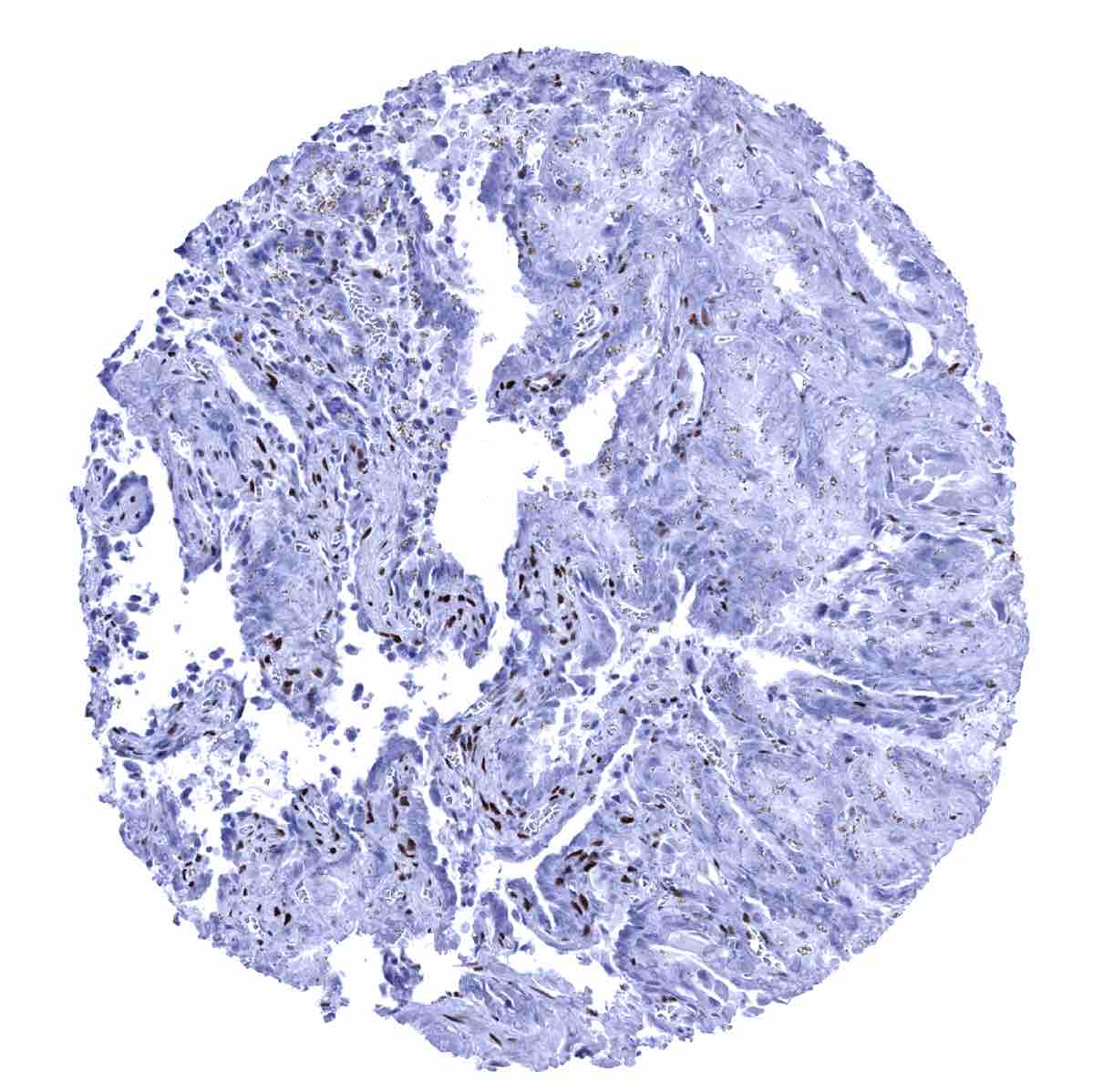
Prostate - A moderate to strong ER immunostaining is seen in stromal cells of the prostate

Rectum, mucosa
Seminal vesicle - A moderate to strong ER immunostaining occurs in stromal cells of the seminal vesicles
Sinus paranasales

Skeletal muscle
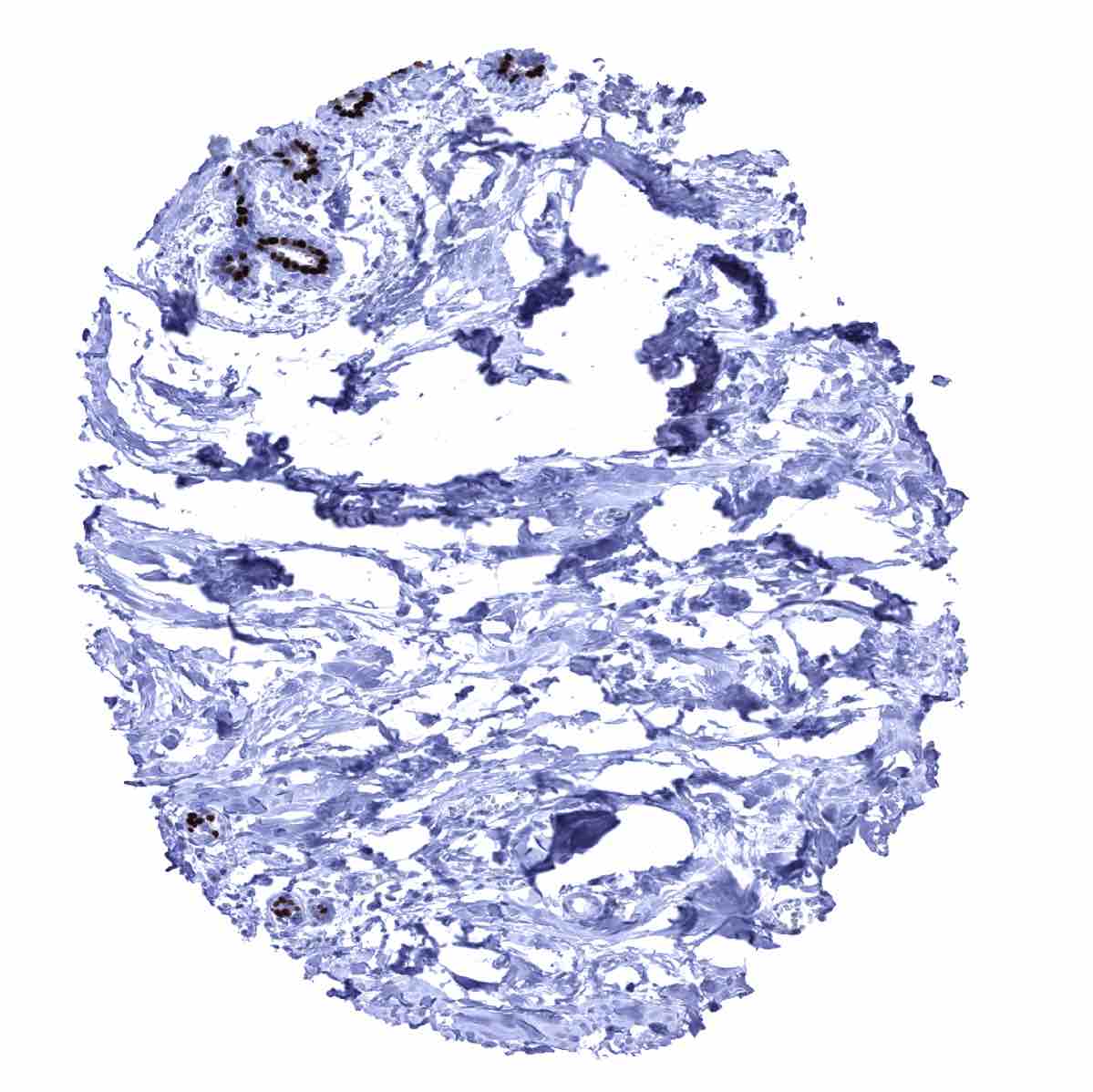
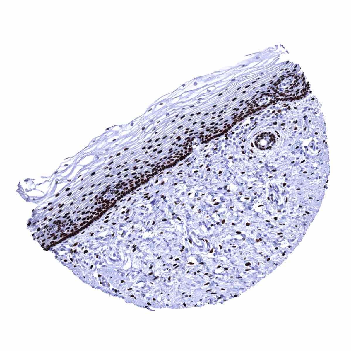
Skin, hairfollicel and sebaceous glands - A moderate ER staining occurs in peripheral germinative cell of sebaceous glands and in peripheral cells of hair follicles
Skin
Spleen
Stomach, antrum

Stomach, corpus

Testis
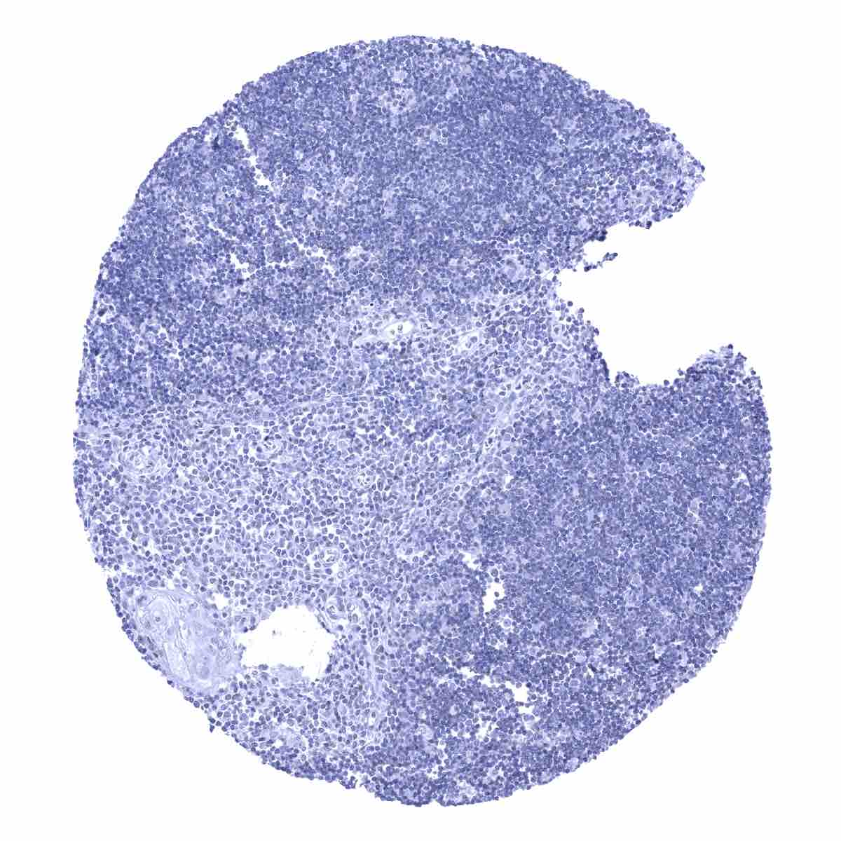
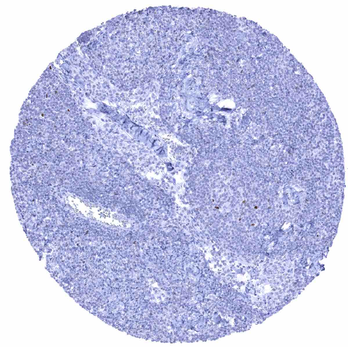
Thymus
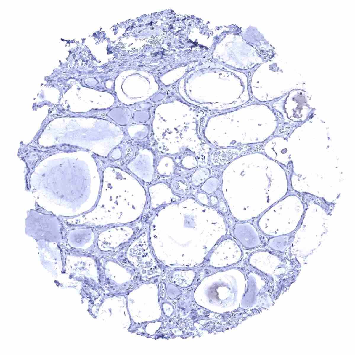
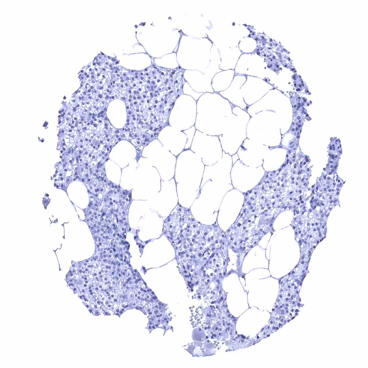
Thyroid gland
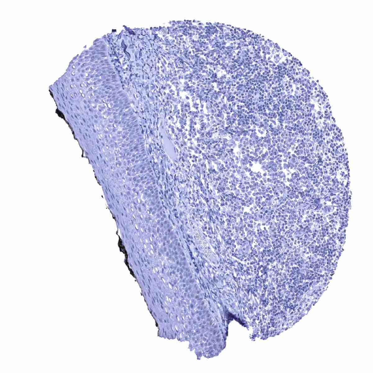
Tonsil, surface epithelium
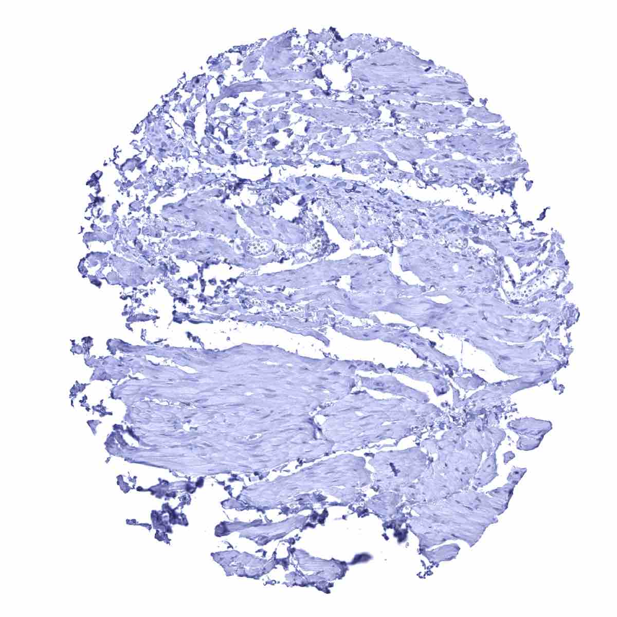
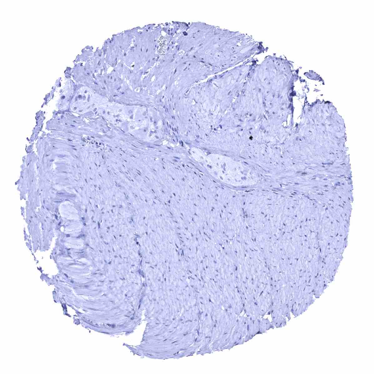
Urinary bladder, muscular wall

Urinary bladder, urothelium - Weak ER staining in a fraction of urothelial cells, moderate ER staining of some stromal cells
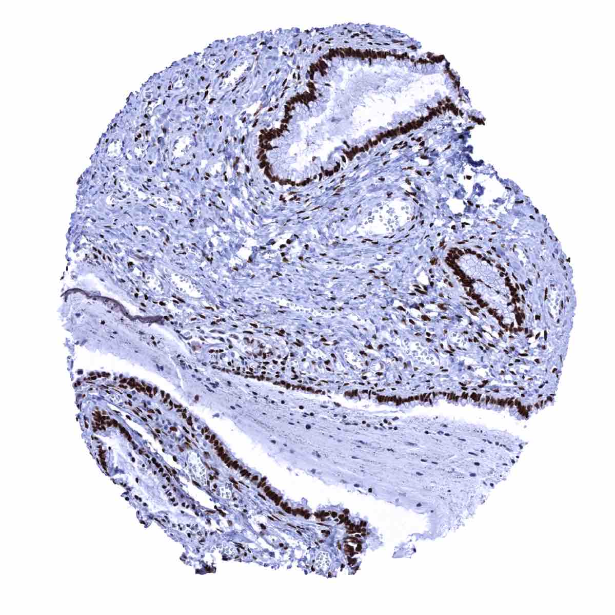
Uterus, cervix - An intense ER staining occurs in stromal cells and in the majority of squamous epithelial cells with a slight decrease of the intensity from the bottom to the top
Uterus, endocervix - ER staining is intense in epithelial and stromal cells of the endocervix
Uterus, endometrium (pregnancy) - A weak to moderate ER immunostaining is seen in decidua cells

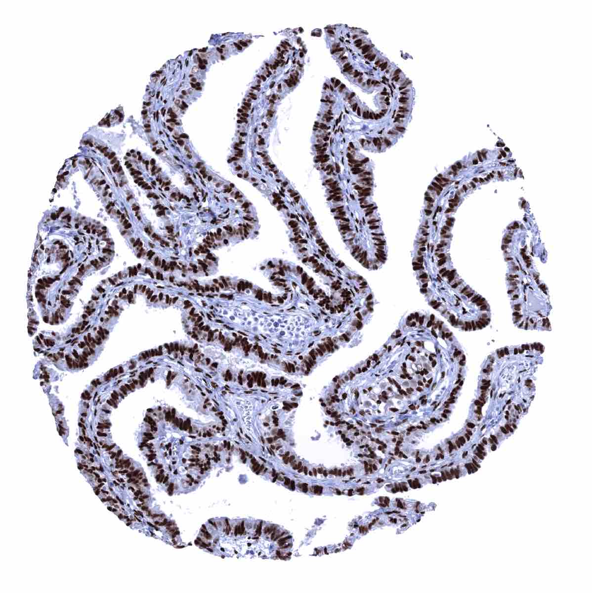
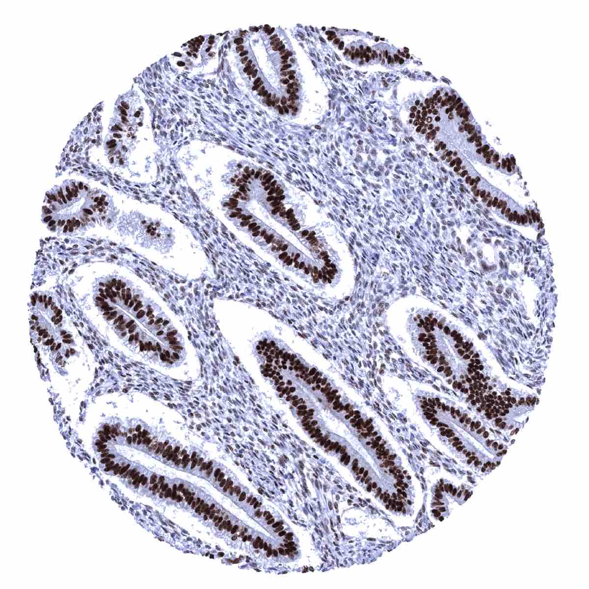
Uterus, endometrium (proliferation) - ER immunostaining is at its highest level in epithelial and stromal cells of the endometrium
Uterus, endometrium (secretion) - ER immunostaining is stronger in epithelial than in stromal cells of this sample of this endometrium in secretion phase

Uterus, myometrium - Strong ER staining of the myometrium muscle cells