Adrenal gland - In the adrenal gland, EpCAM staining can be very faint or even absent.
Adrenal gland - In the adrenal gland, a weak membranous staining of adrenocortical cells can be seen.
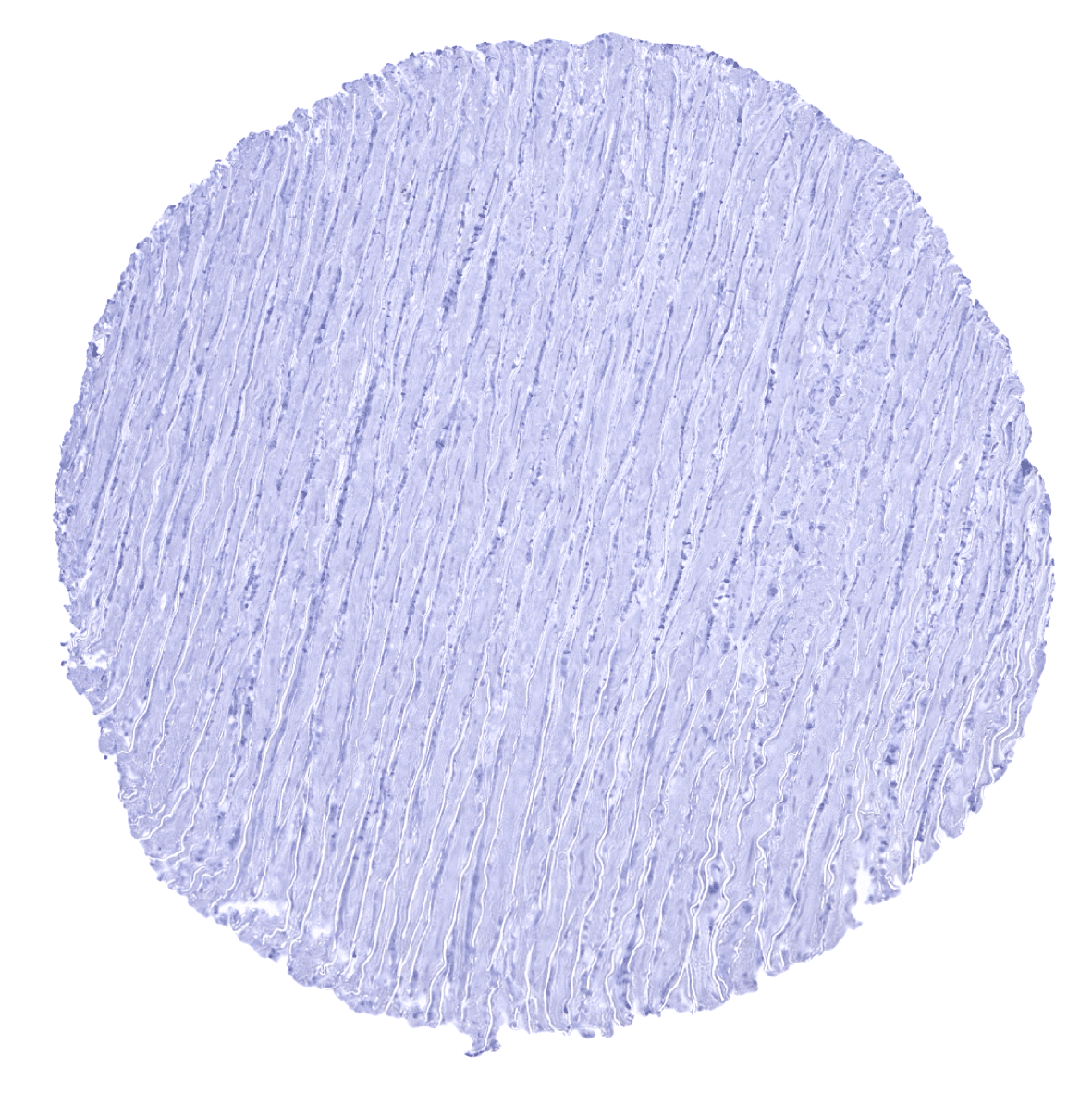
Aorta, media
Appendix, mucosa
Appendix, muscular wall

Bone marrow
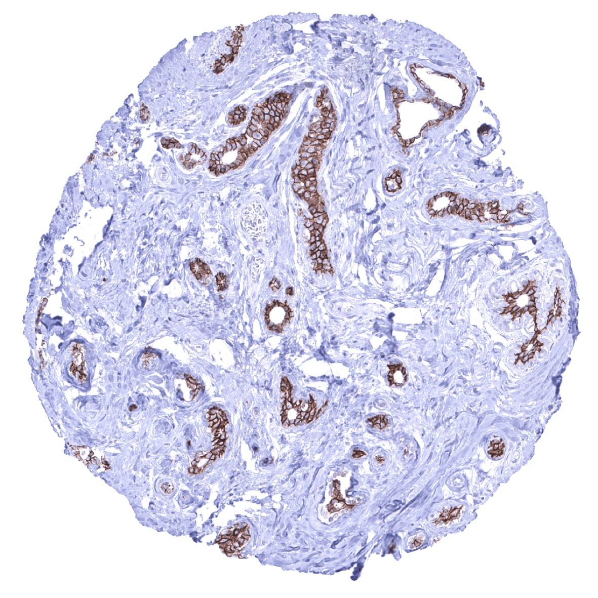
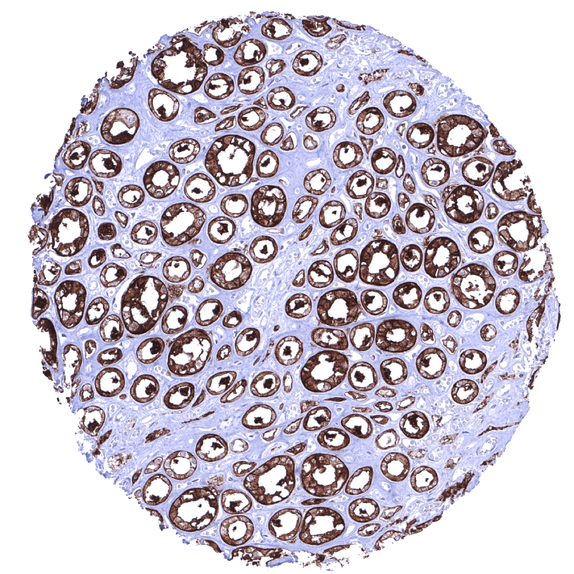
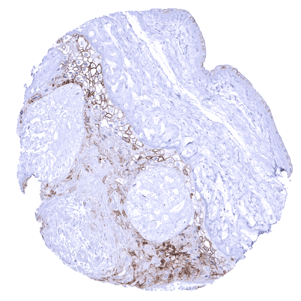
Breast - In the breast gland, luminal cells are strongly positive but basal/myoepithelial cells show much less or absent staining.

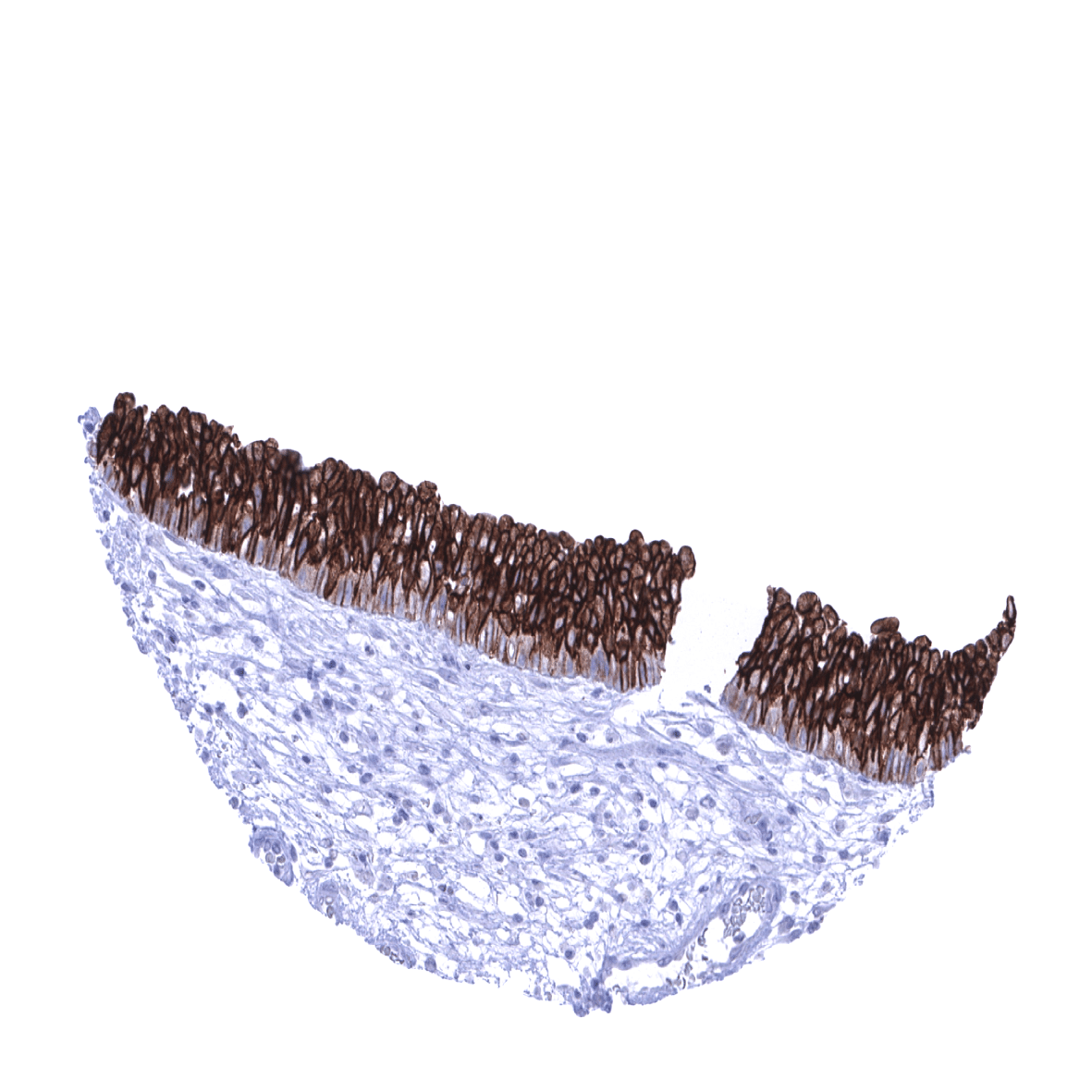
Bronchus, mucosa
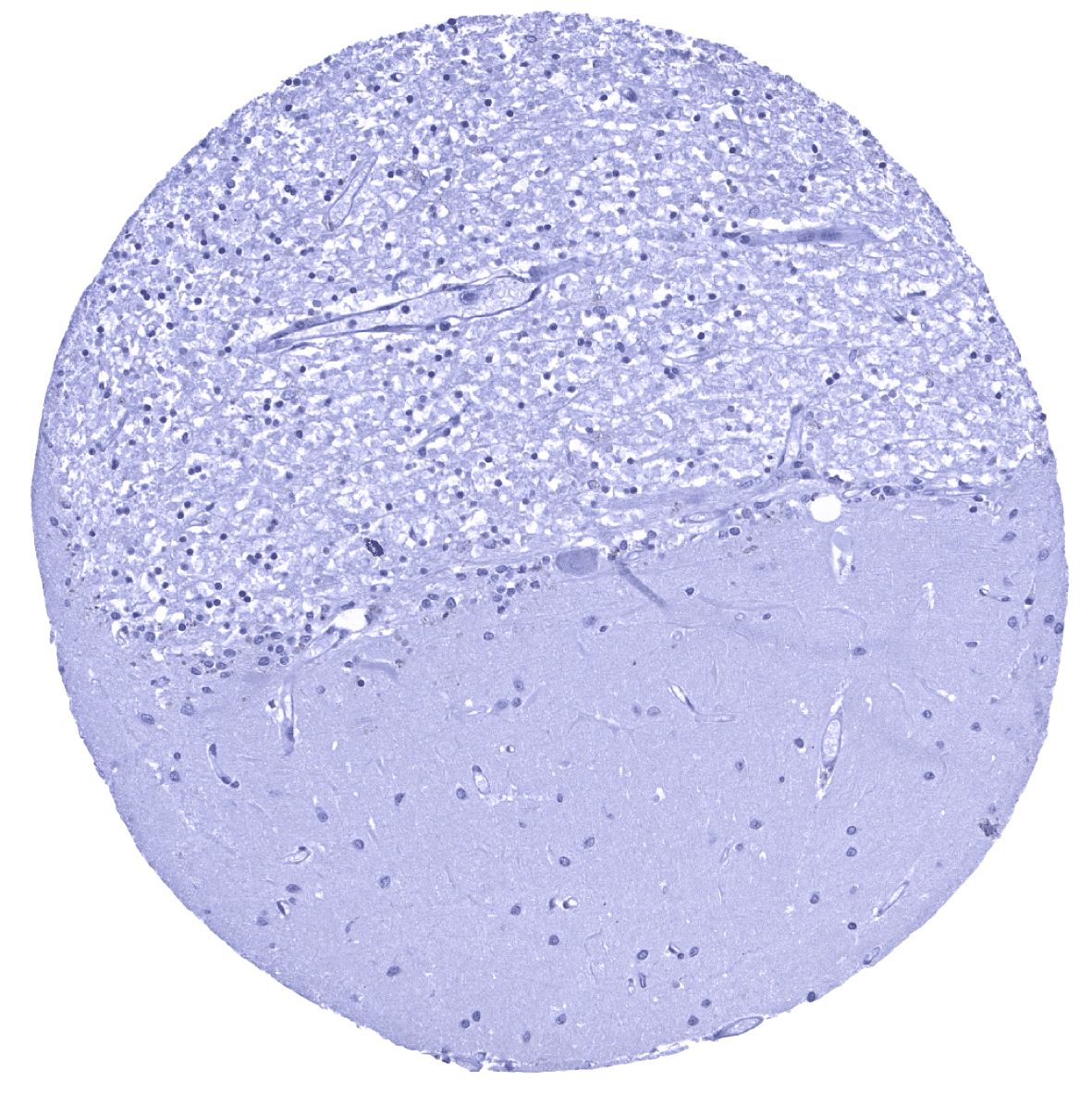
Cerebellum (granule cell layer, white matter)
Cerebellum (molecular layer, Purkinje cell layer, granule cell layer)

Cerebrum, grey matter
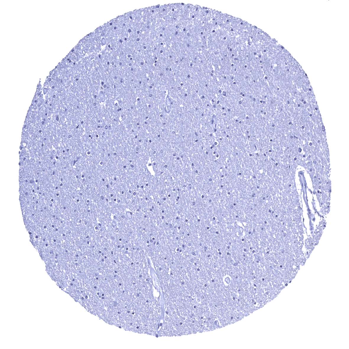
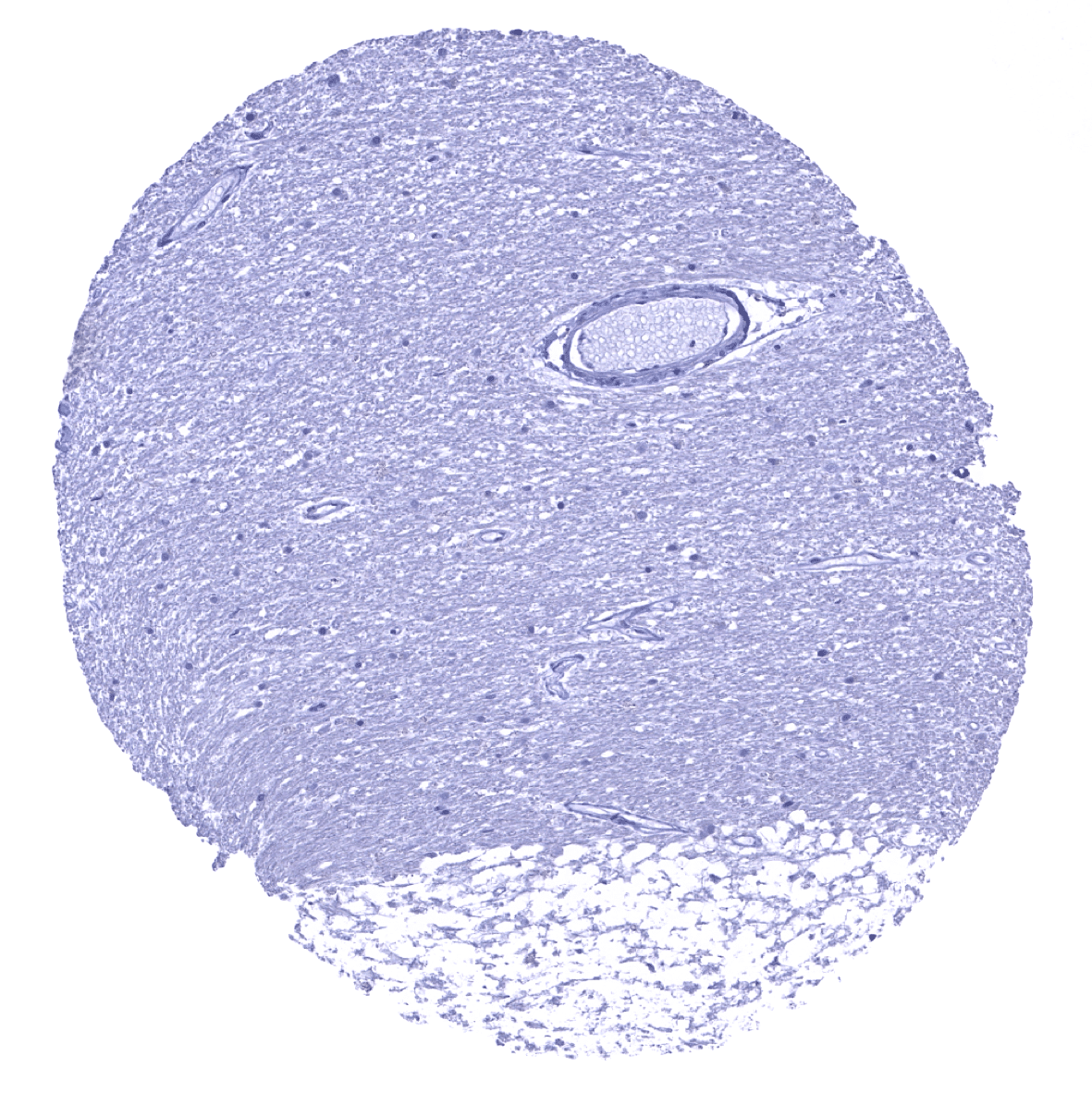
Cerebrum, white matter
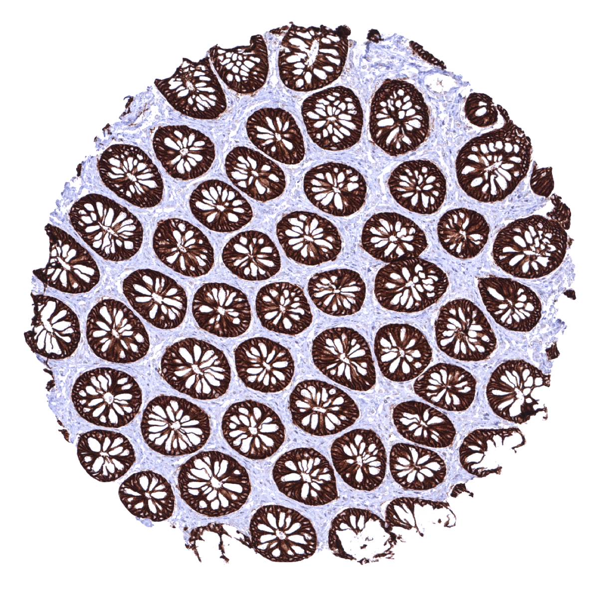
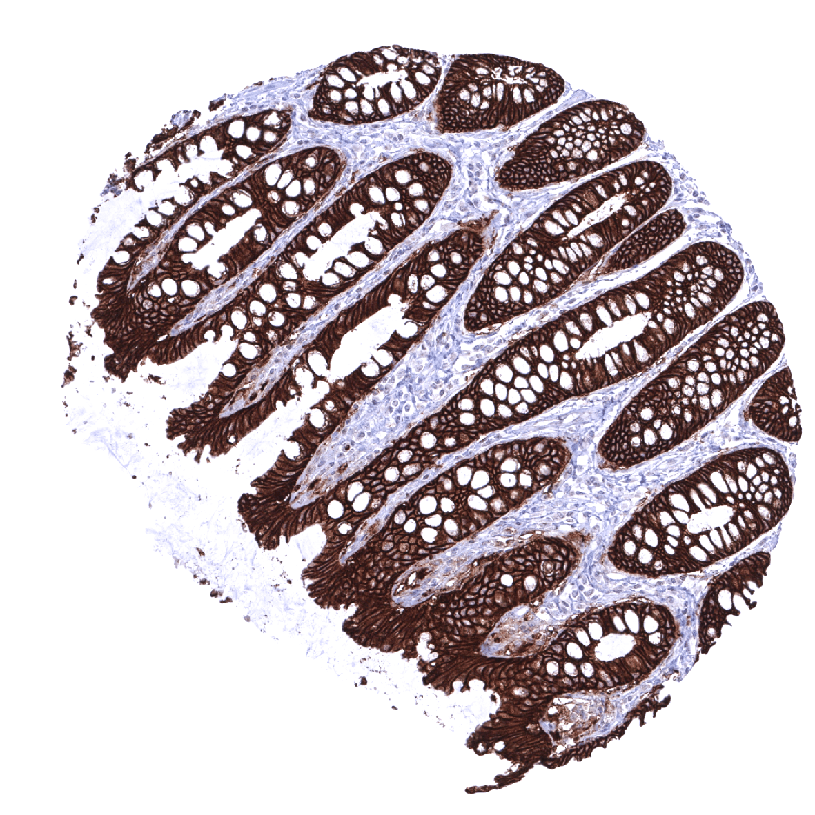
Colon descendens, mucosa
Colon descendens, muscular wall
Duodenum, Brunner gland
Duodenum, mucosa
Epididymis
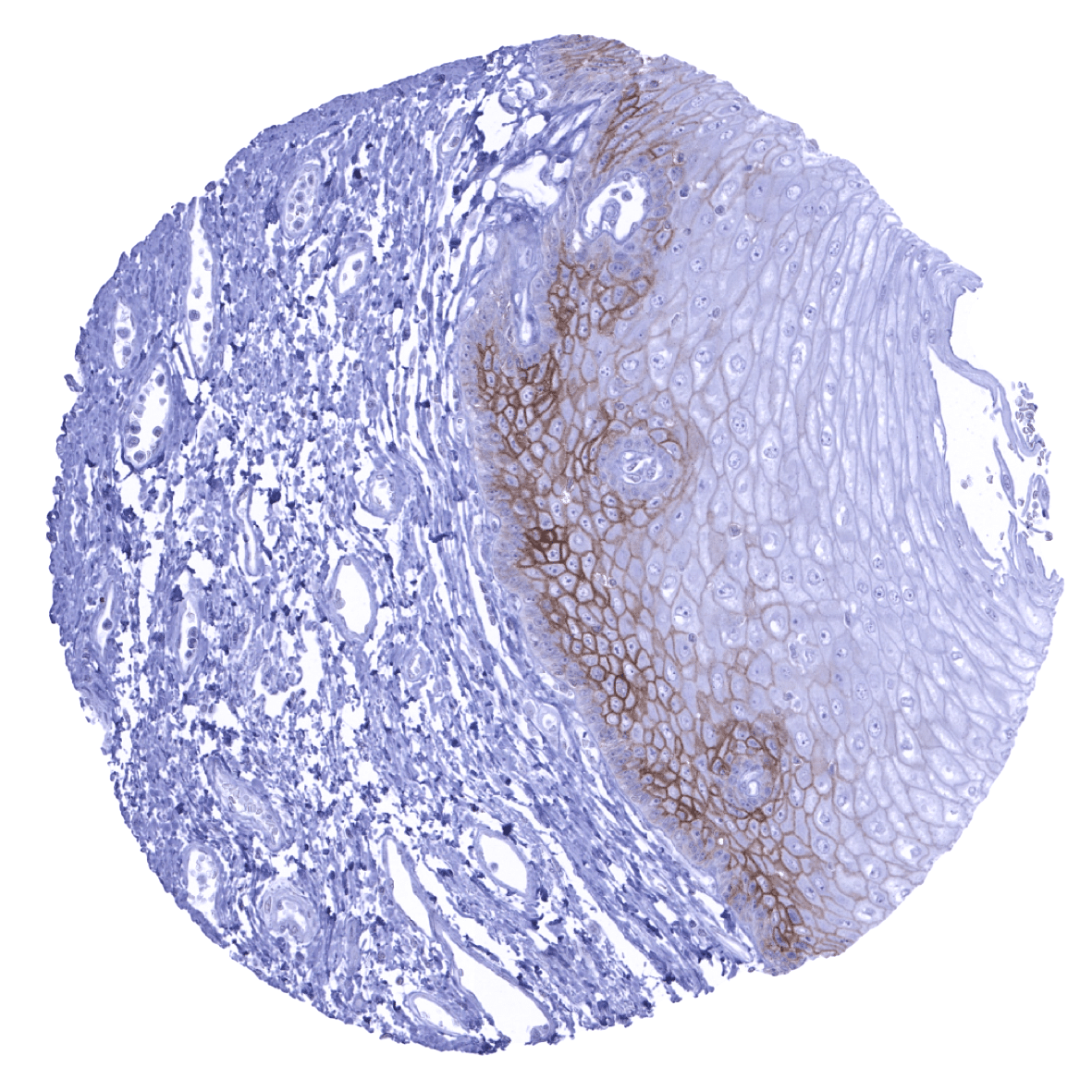
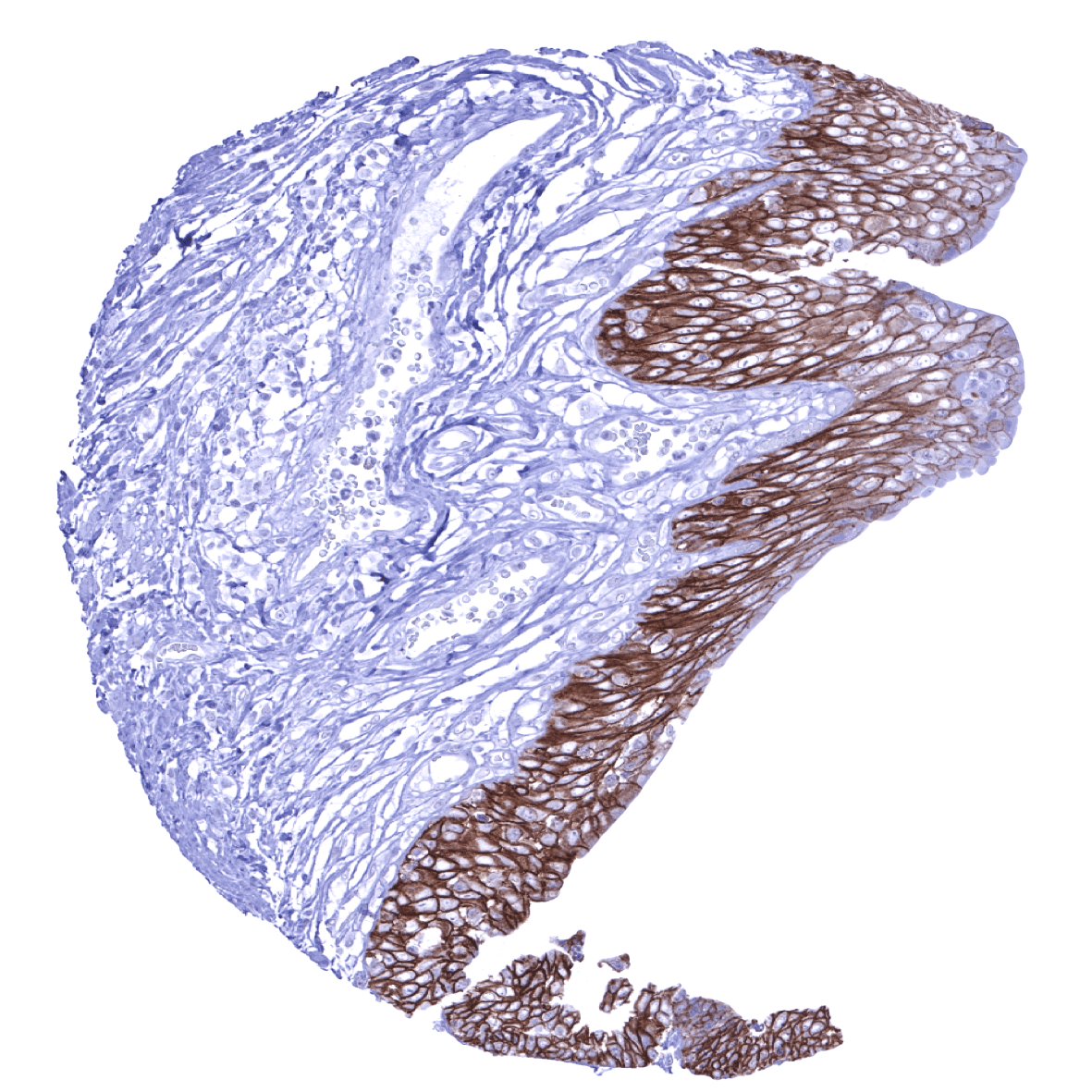
Esophagus, squamous epithelium - A weak to moderate EpCam immunostaining is seen in the suprabasal cell layers of esophageal squamous epithelium.
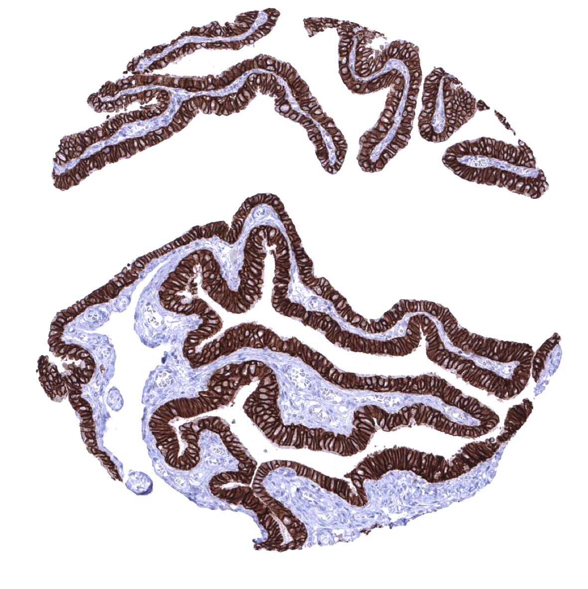
Fallopian tube, mucosa
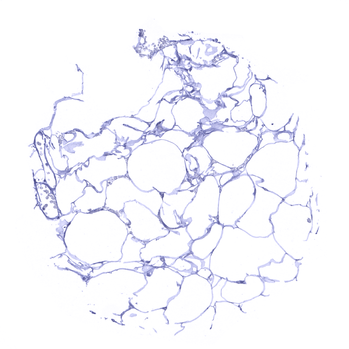
Fat
Gallbladder, epithelium
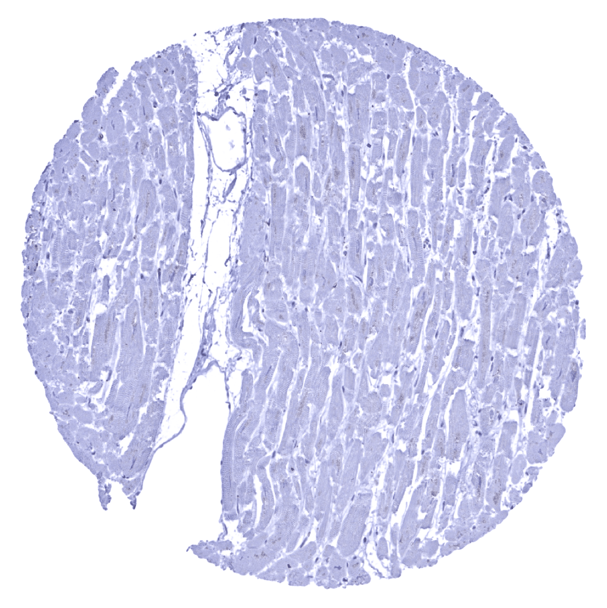
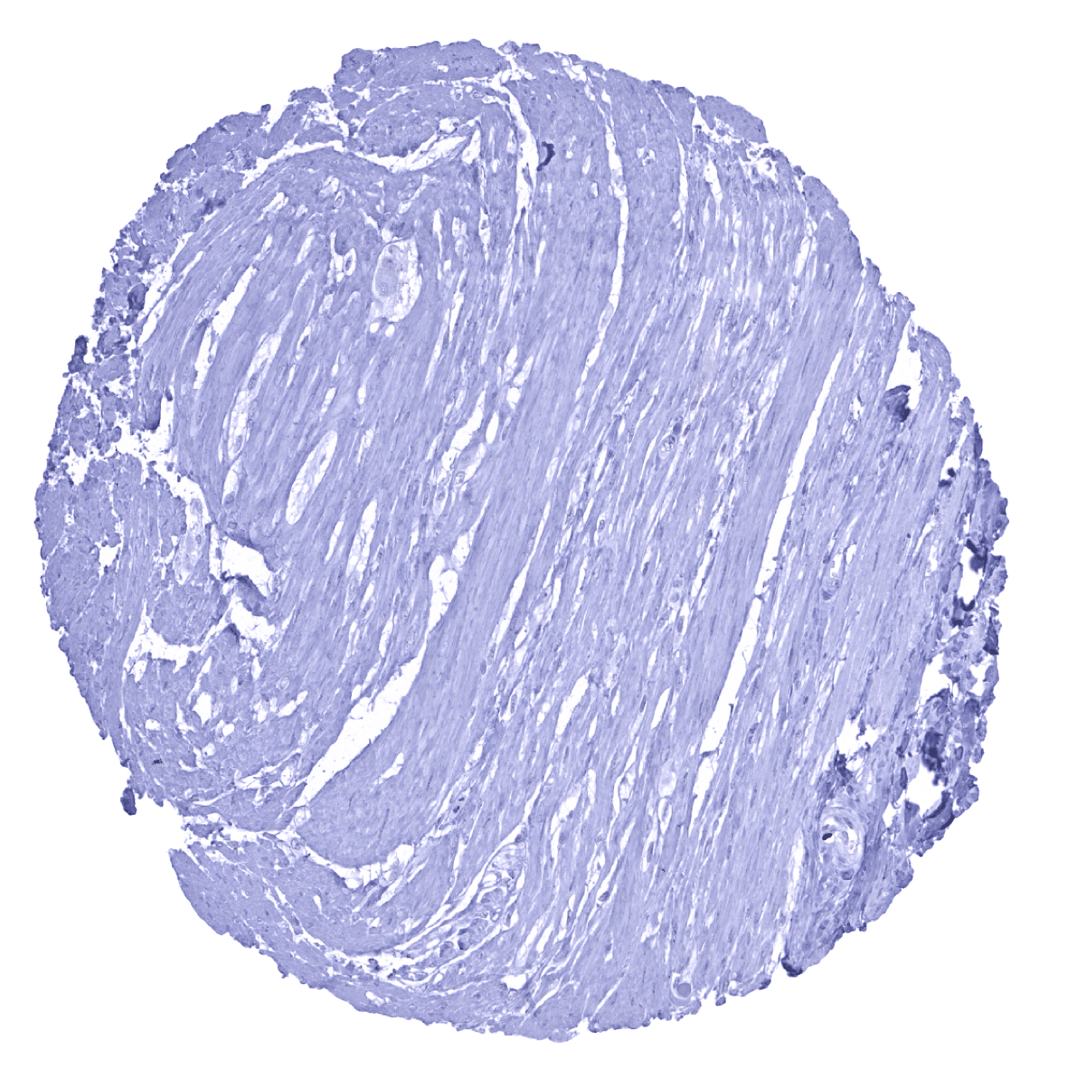
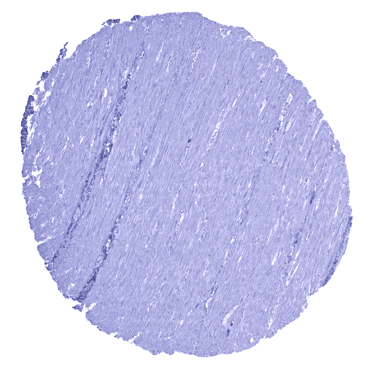
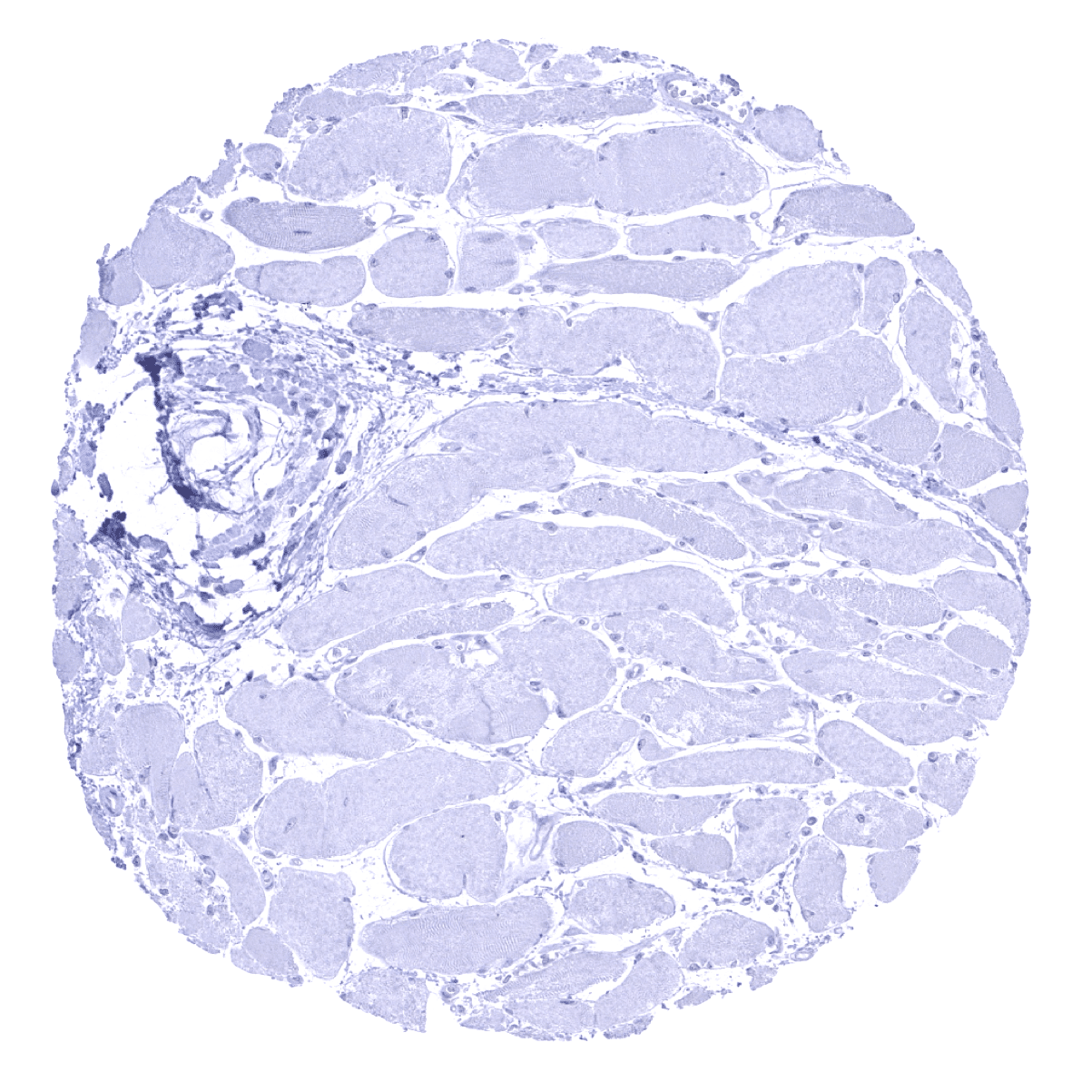
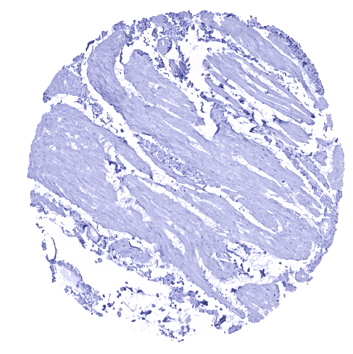
Heart muscle

Ileum, mucosa
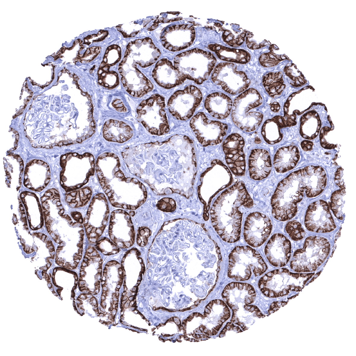
Kidney, cortex - In the kidney, a strong staining is seen in the distal tubuli while staining is less intense and focussed to the basolateral membranes in the majority of proximal tubulus cells. Scattered epithelial cells lining the Bowman capsule are also positive.
Kidney, medulla
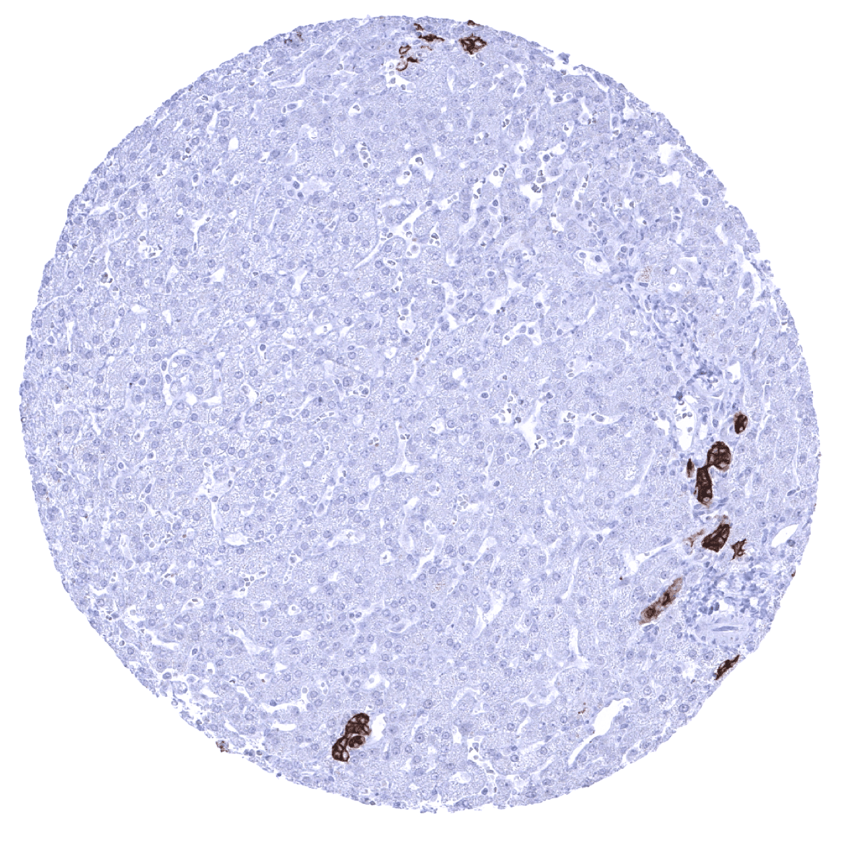
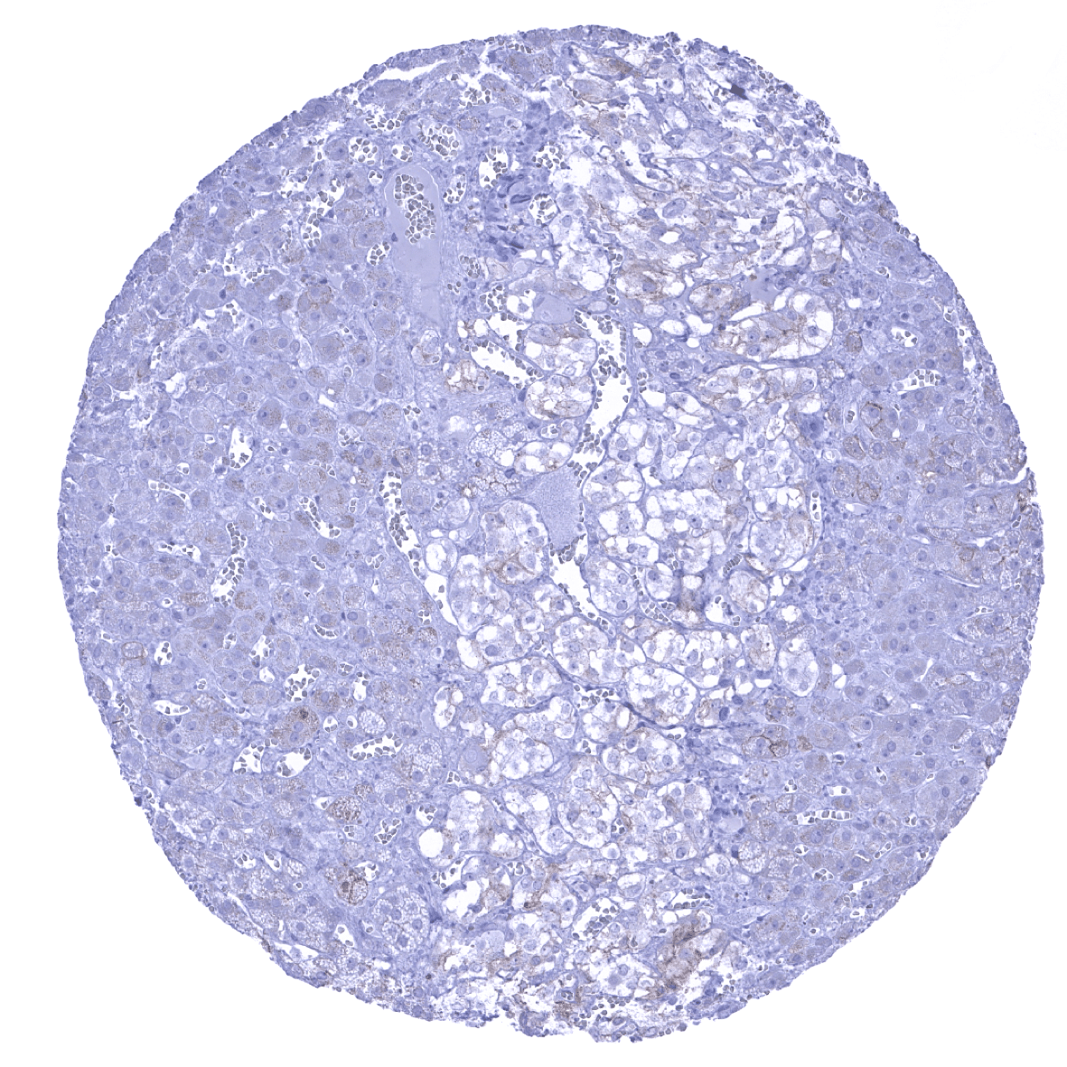
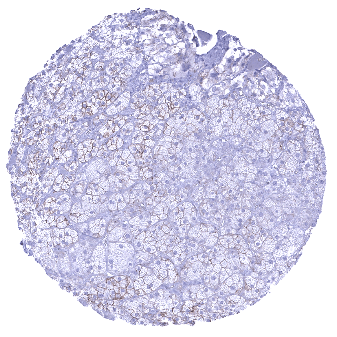
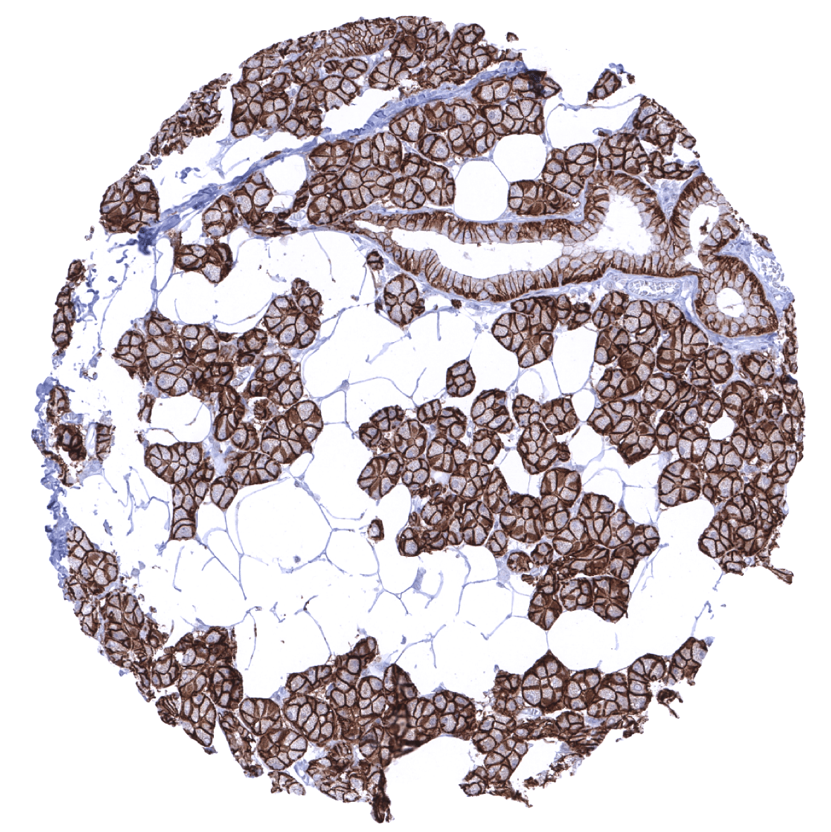
Liver - In the liver, hepatocytes are EpCAM negative but EpCAM is strongly expressed in epithelial cells of the bile ducts.
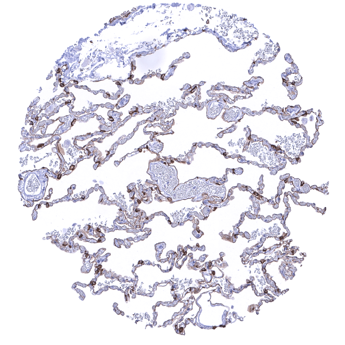
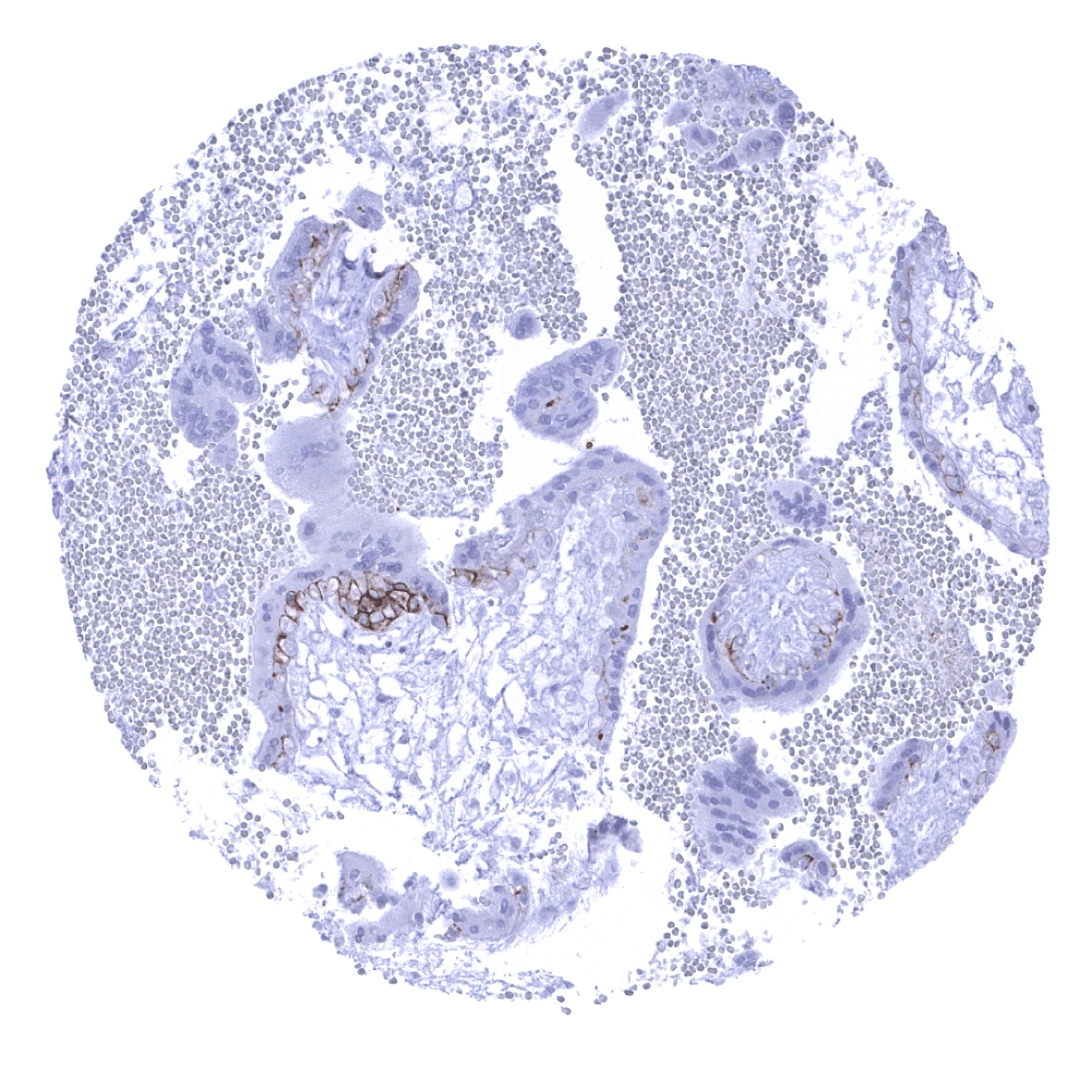
Lung - In the lung, a weak to moderate EpCAM immunostaining is seen in pneumocytes.
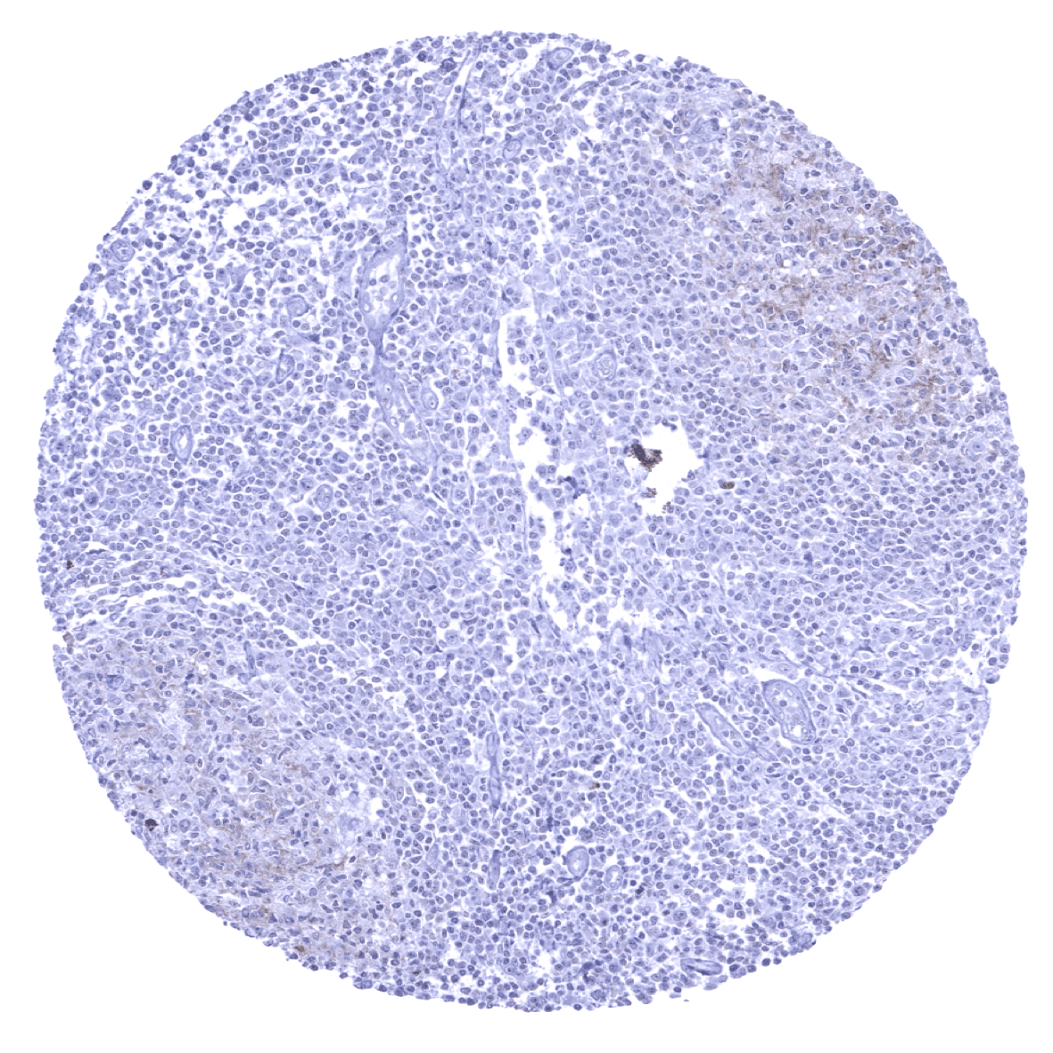
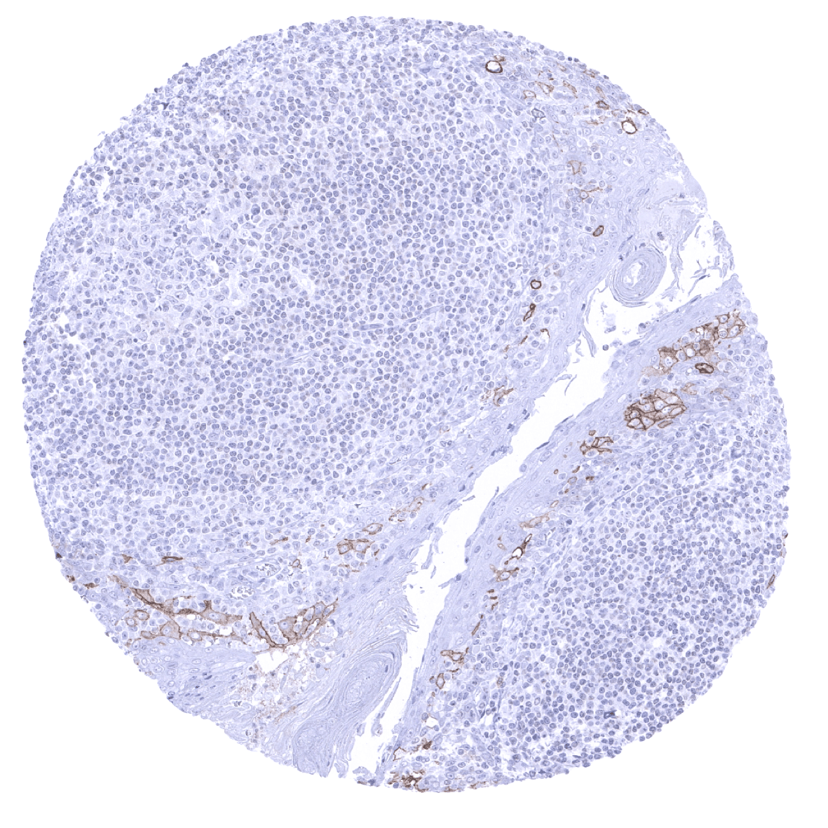
Lymph node
Ovary, stroma
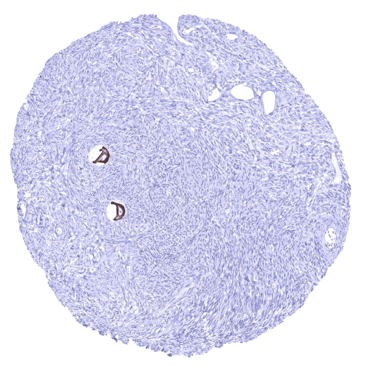
Ovary, stroma - A strong EpCAM immunostaining is seen in oocytes of the ovary.

Pancreas
Parathyroid gland
Parotid gland
Pituitary gland, anterior lobe

Pituitary gland, posterior lobe
Placenta, first trimester - In the first trimester placenta only cytotrophoblast cells shows a weak to moderate EpCAM staining.

Placenta, mature - In the mature placenta, a weak membranous staining of the basal membrane of the trophoblast layer is occasionally seen.
Placenta (amnion and chorion) - EpCam immunostaining is moderate in chorion cells and weak in amnion cells of the placenta.
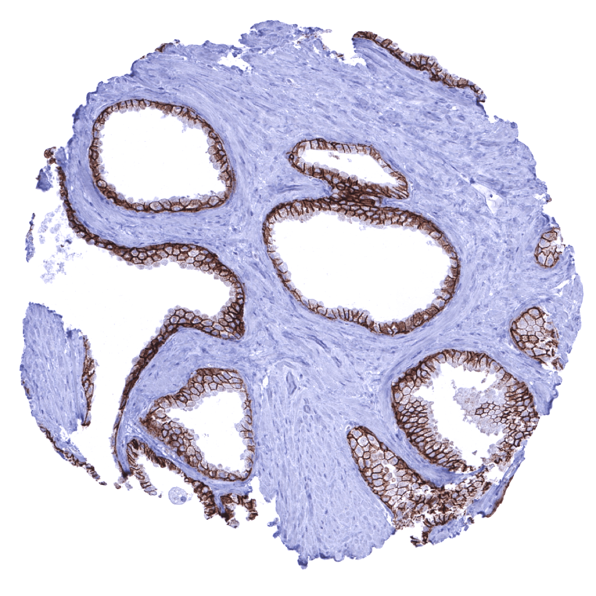
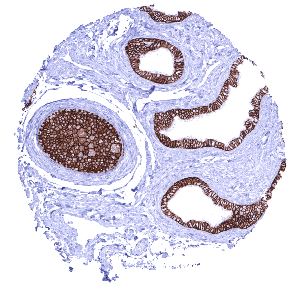
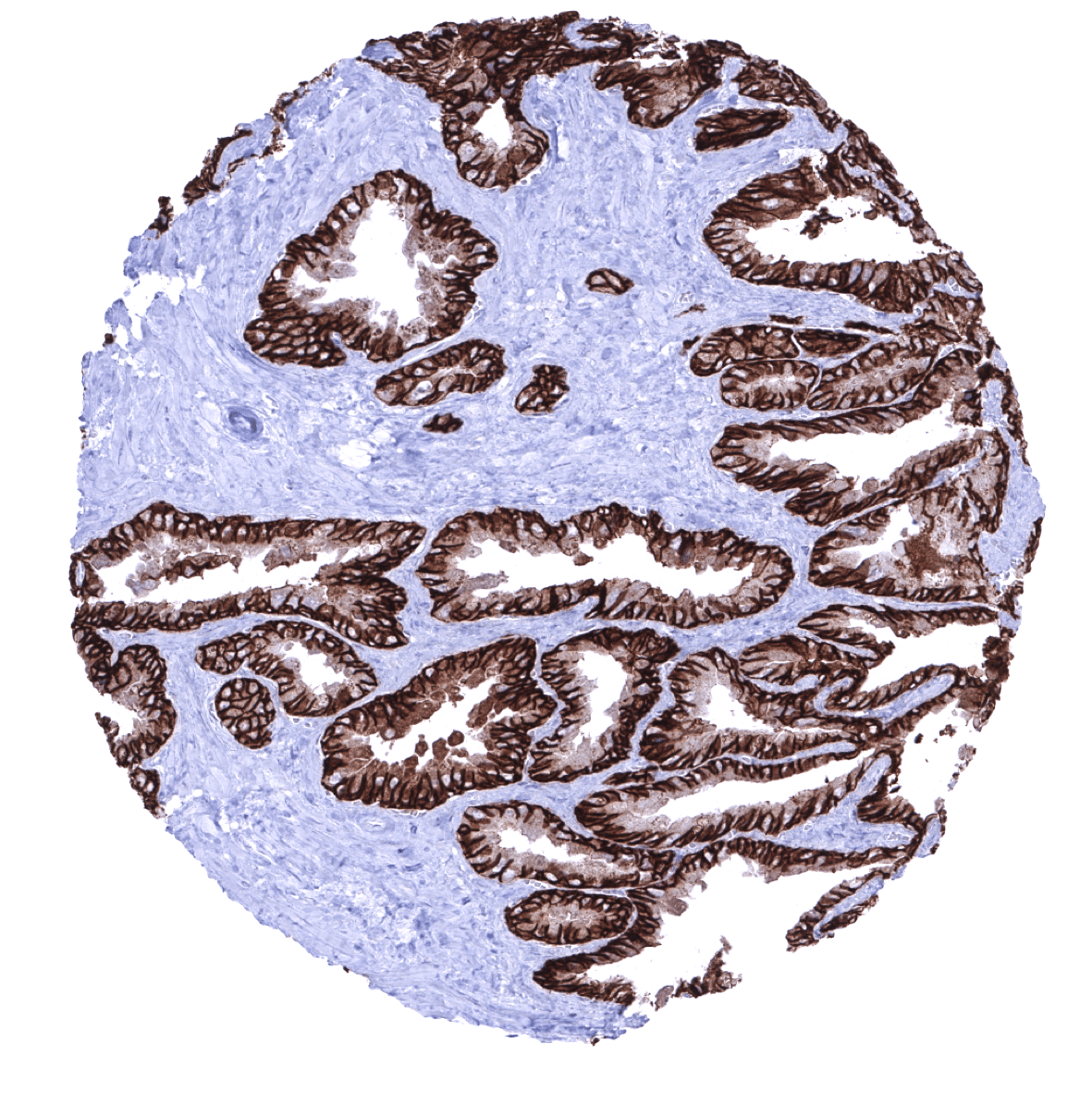
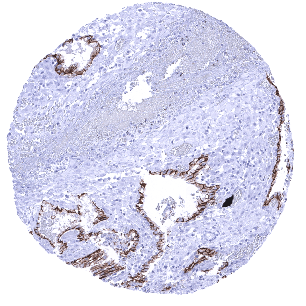
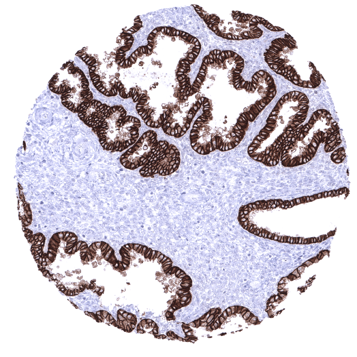
Prostate
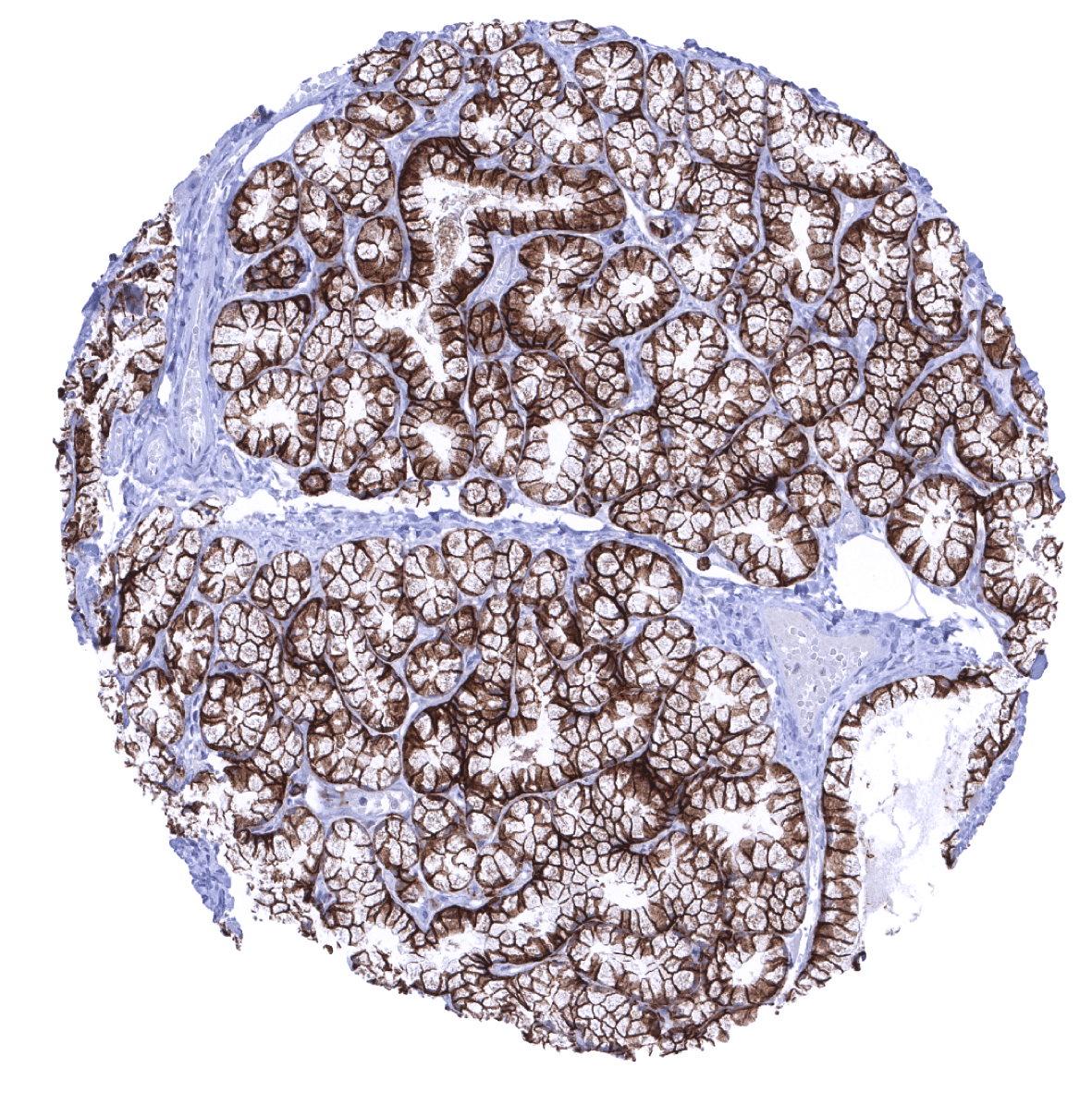
Rectum, mucosa
Seminal vesicle
Sinus paranasales
Skeletal muscle

Skin

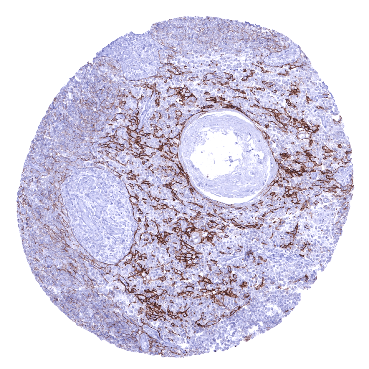
Spleen
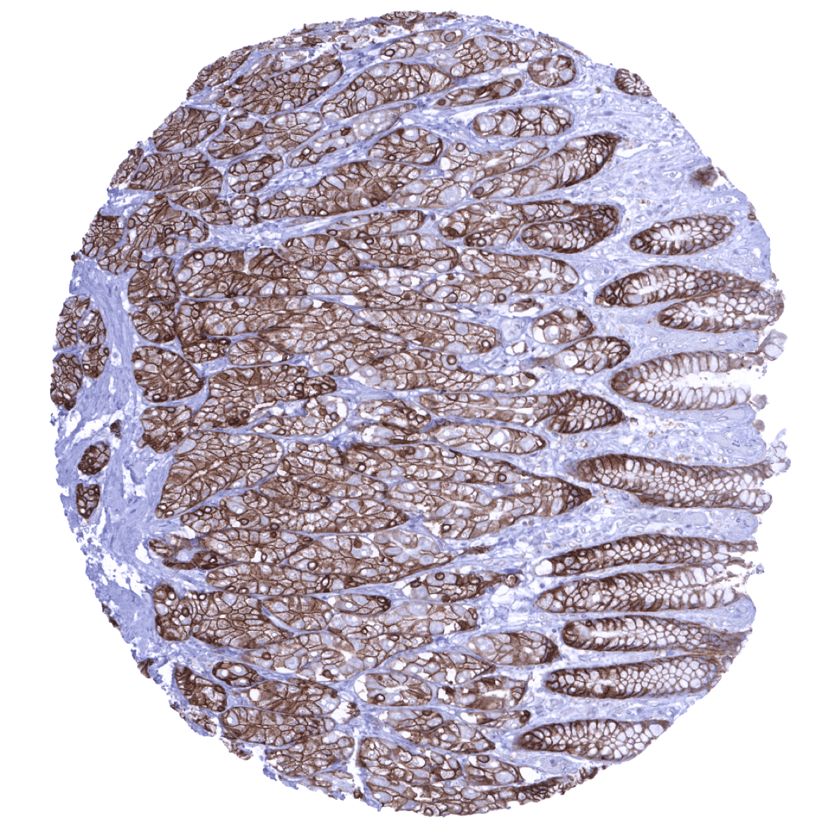
Stomach, antrum
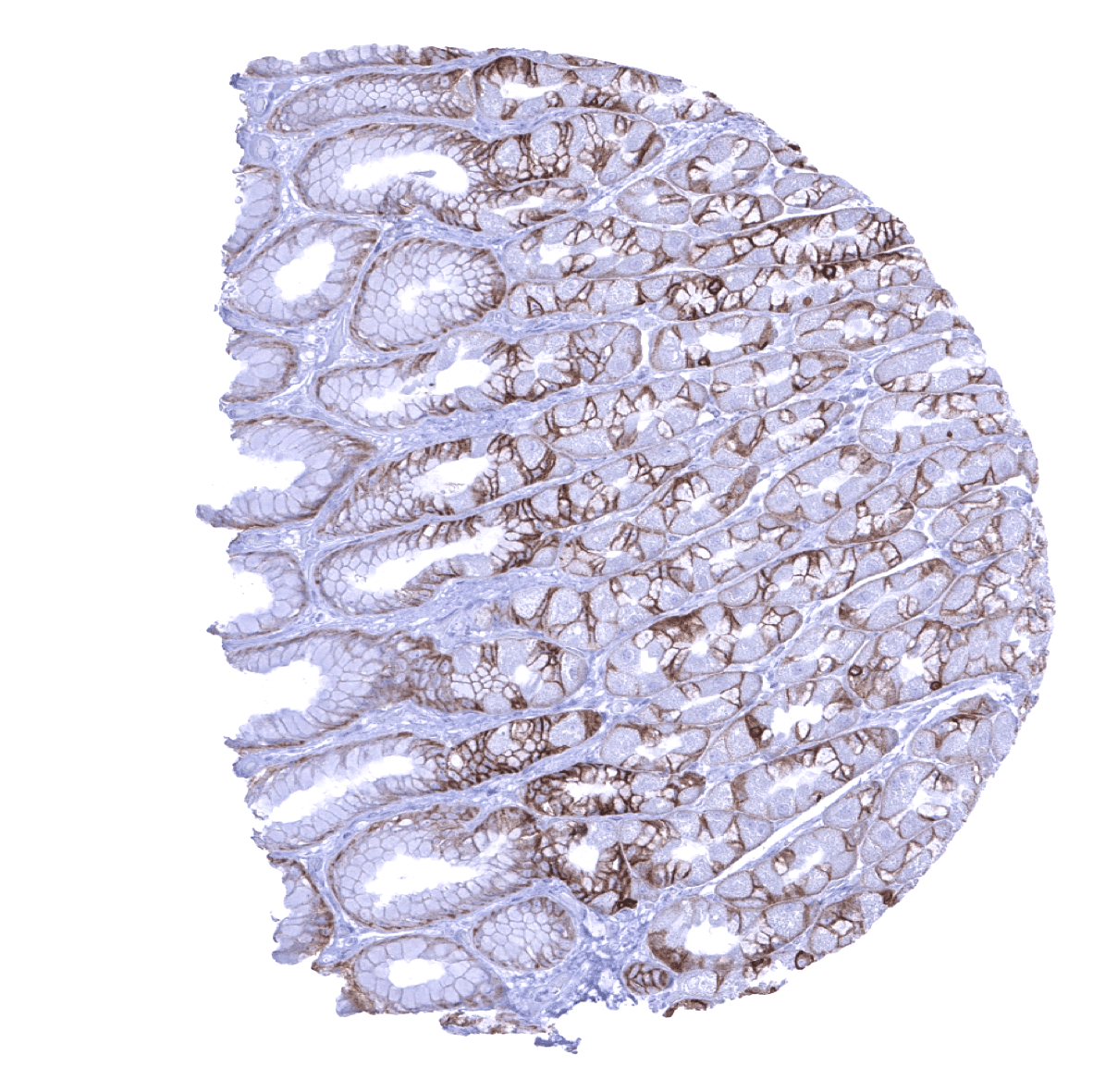
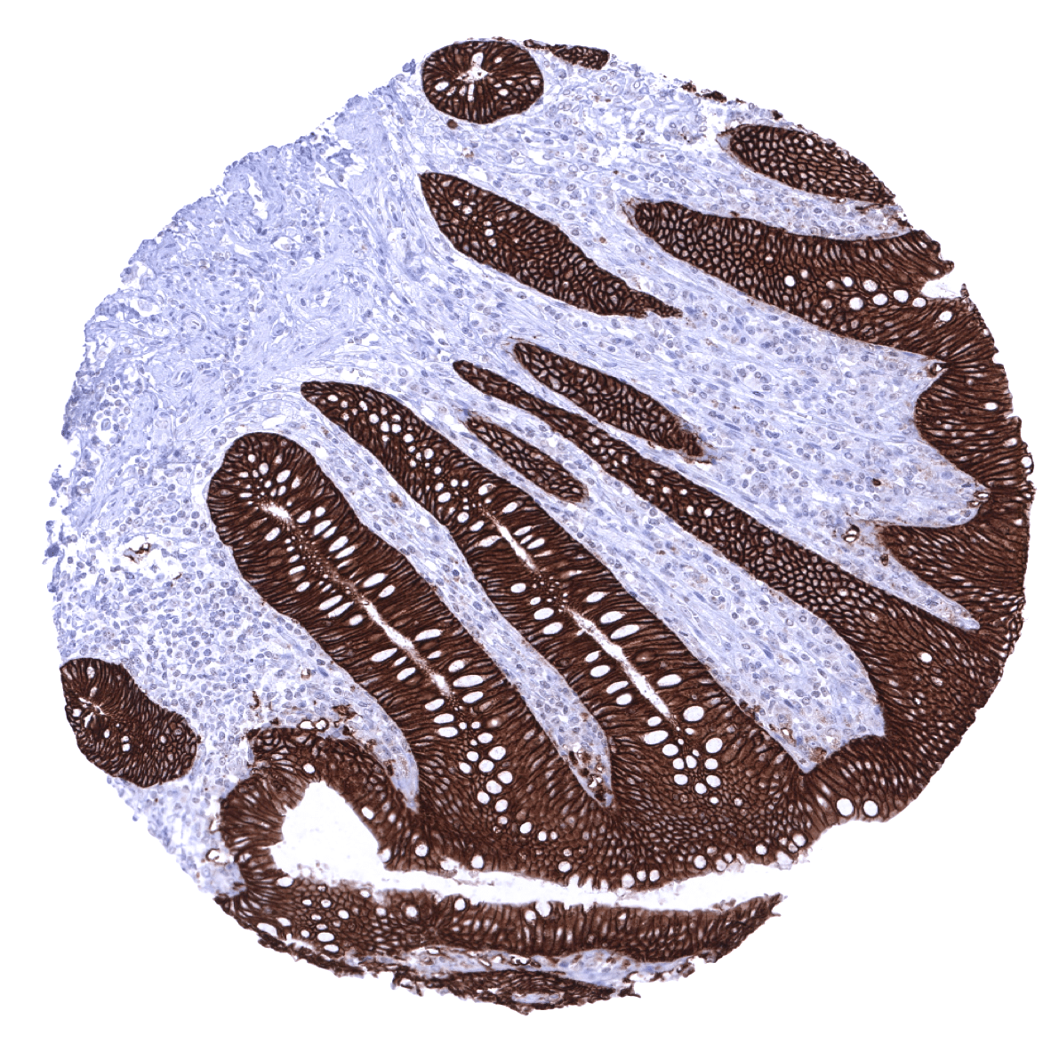
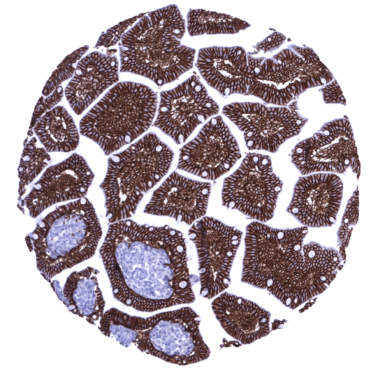
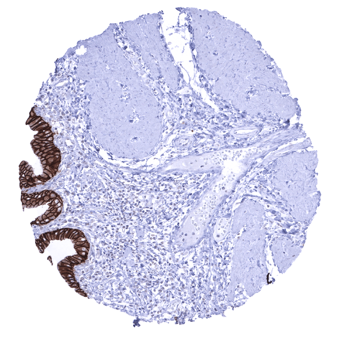
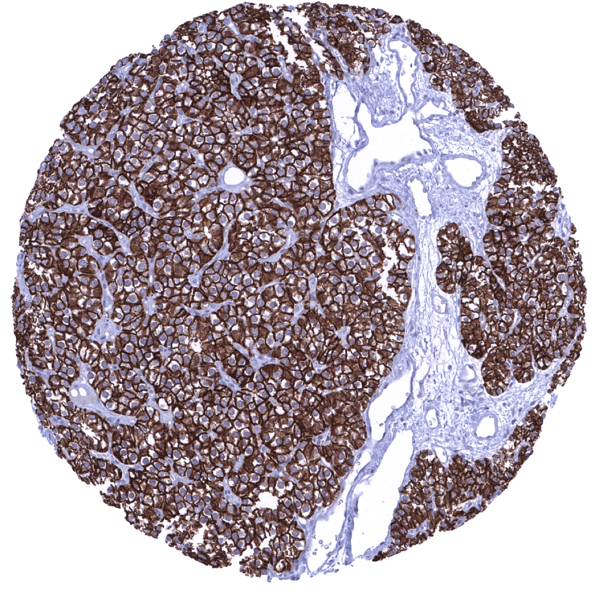
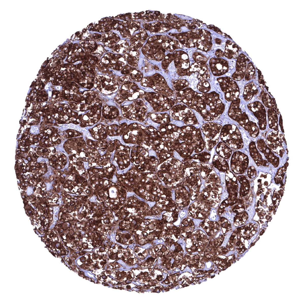
Stomach, corpus - EpCAM is strongly expressed in epithelial cells of the stomach. Only parietal cells show a weaker staining limited to the basolateral membranes.

Testis - In the testis, spermatogonia and spermatocytes show strong EpCAM immunostaining but Sertoli and Leydig cells are negative.
Thymus - Most thymus epithelial cells including corpuscles of Hassall’s show a weak to moderate EpCAM positivity.

Thyroid gland
Tonsil - A moderate EpCAM positivity is seen in a subset of squamous epithelial cells of the tonsil crypts.

Tonsil, surface epithelium - A weak to moderate EpCam immunostaining is seen in the basal cell layers of squamous epithelium of the tonsil surface.
Urinary bladder, muscular wall
Urinary bladder, urothelium

Uterus, ectocervix - A weak EpCam positivity is seen in the basal cell layer of squamous epithelium of the ectocervix.

Uterus, endocervix
Uterus, endometrium (pregnancy)
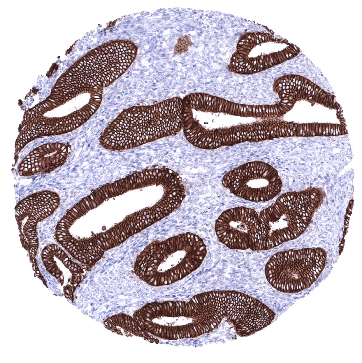
Uterus, endometrium (proliferation)
Uterus, endometrium (secretion)
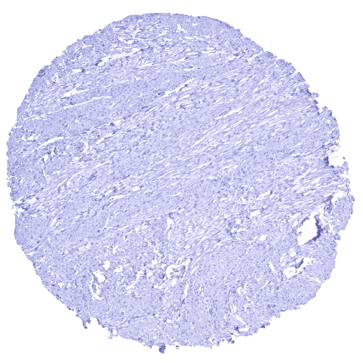
Uterus, myometrium