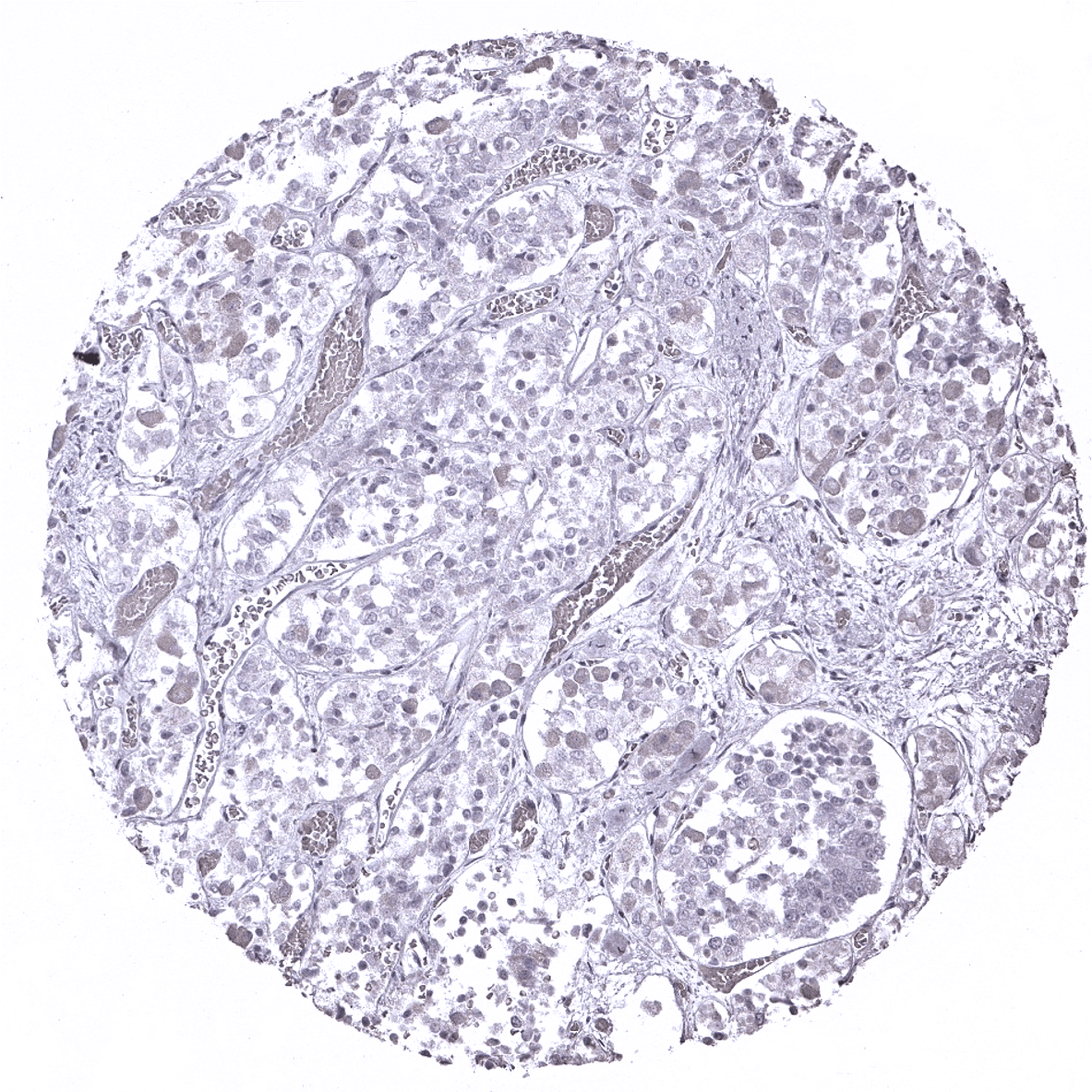
Adrenal gland
Aorta, media
Appendix, mucosa
Appendix, muscular wall

Breast: Prominent DOG-1 staining of myoepithelial cells.
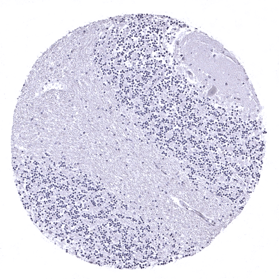
Cerebellum (molecular layer, Purkinje cell layer, granule cell layer, white matter)
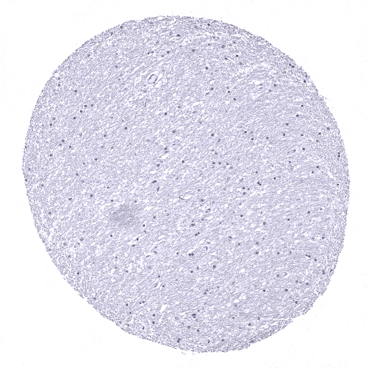
Cerebellum (white matter)
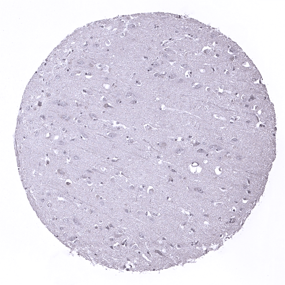
Cerebrum, grey matter
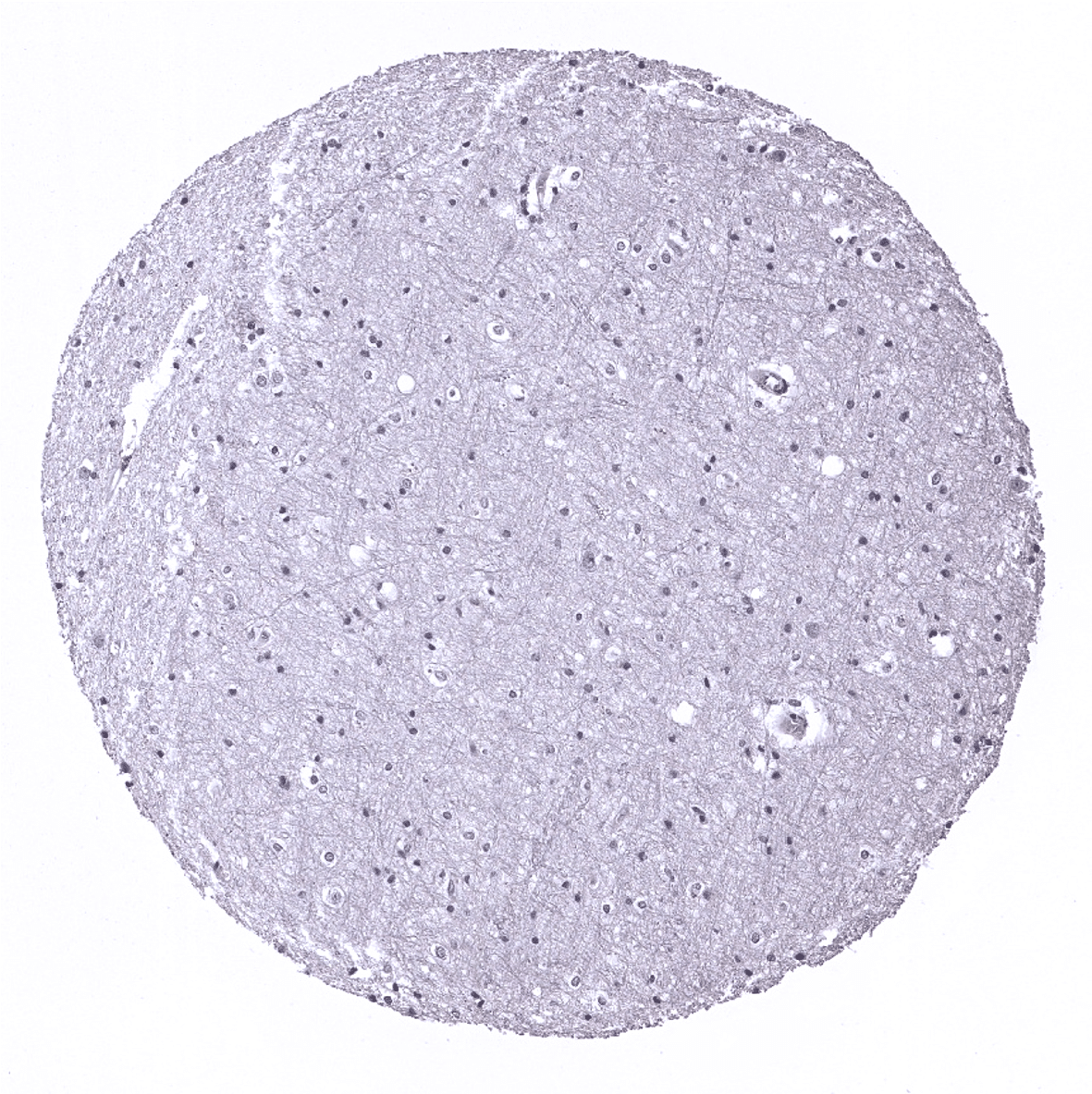
Cerebrum, white matter
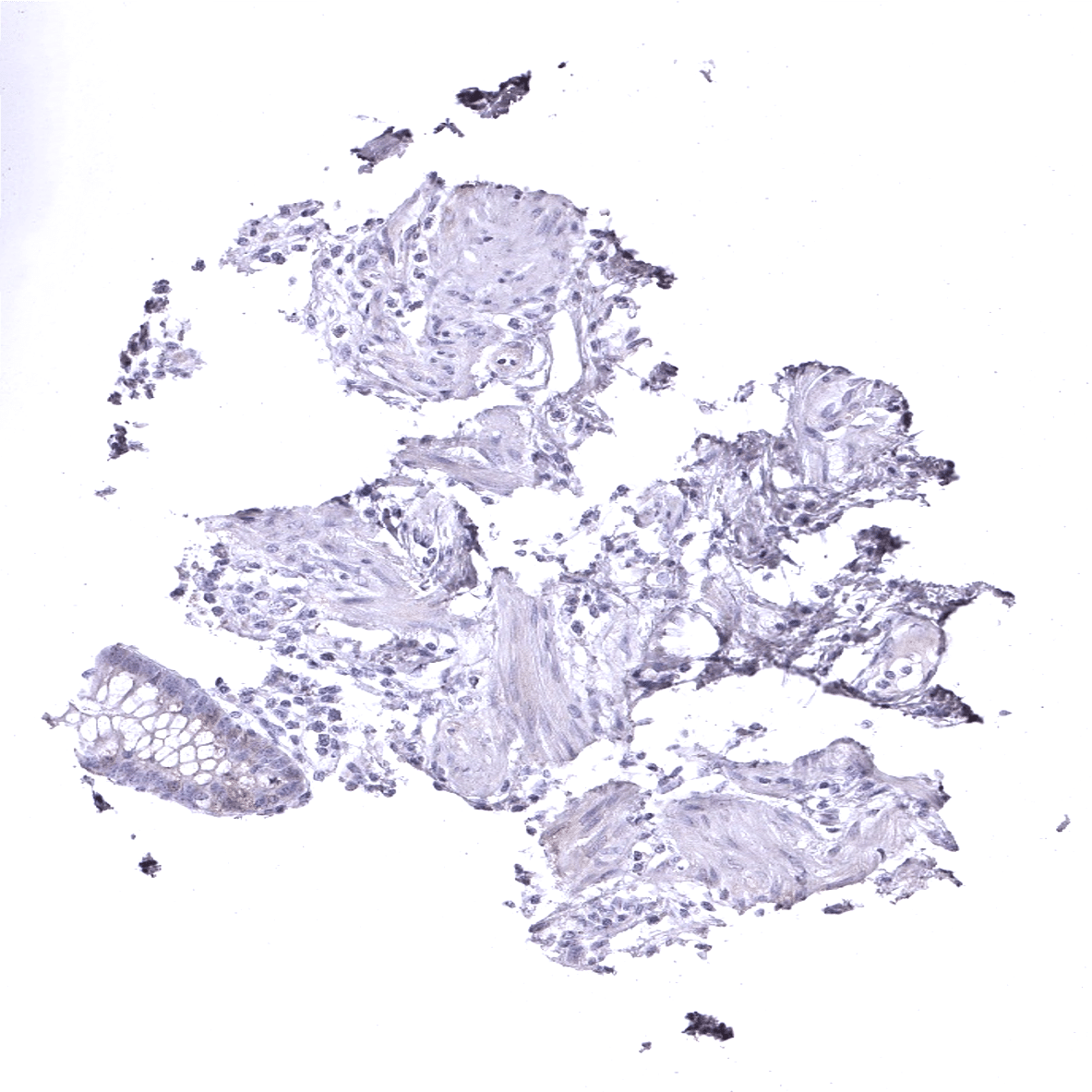
Colon descendens, muscular wall: Few interstitial cells of Cajal show a DOG-1 staining.
Duodenum, Brunner gland
Duodenum, mucosa
Ectocervix
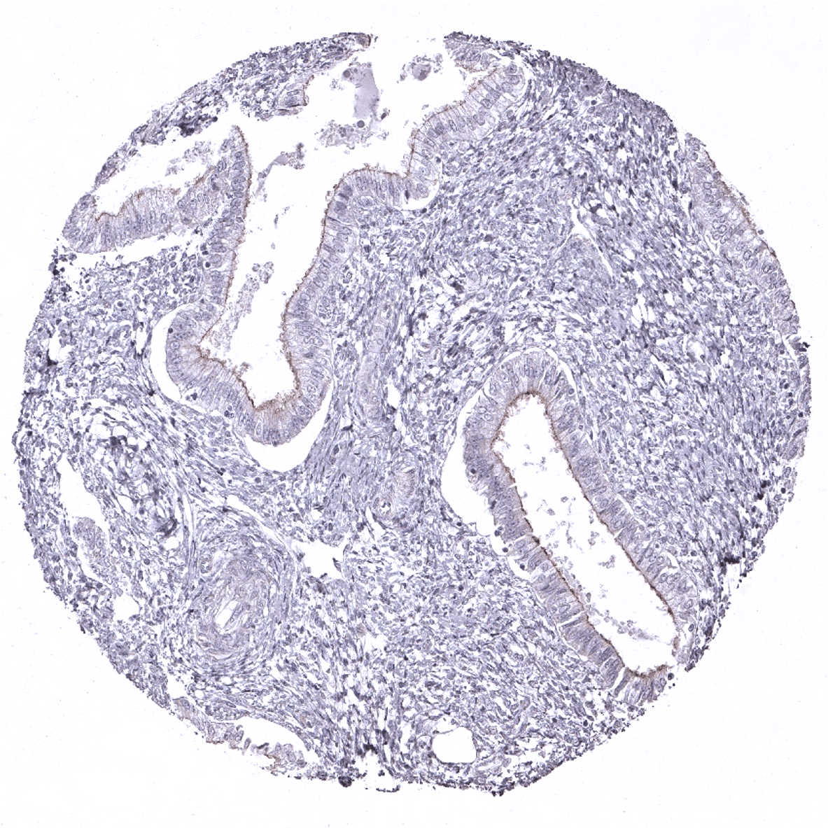
Endocervix
Endometrium, proliferation
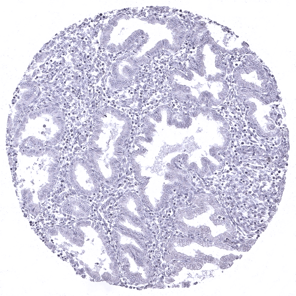
Endometrium, secretion

Epididymis: Strong DOG1 staining of the apical membrane of epithelial cells in the cauda epididymis.
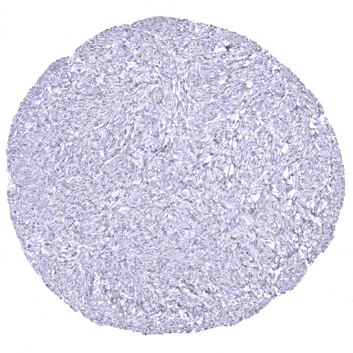
Esophagus, muscular wall: DOG1 immunostaining of interstitial cells of Cajal.
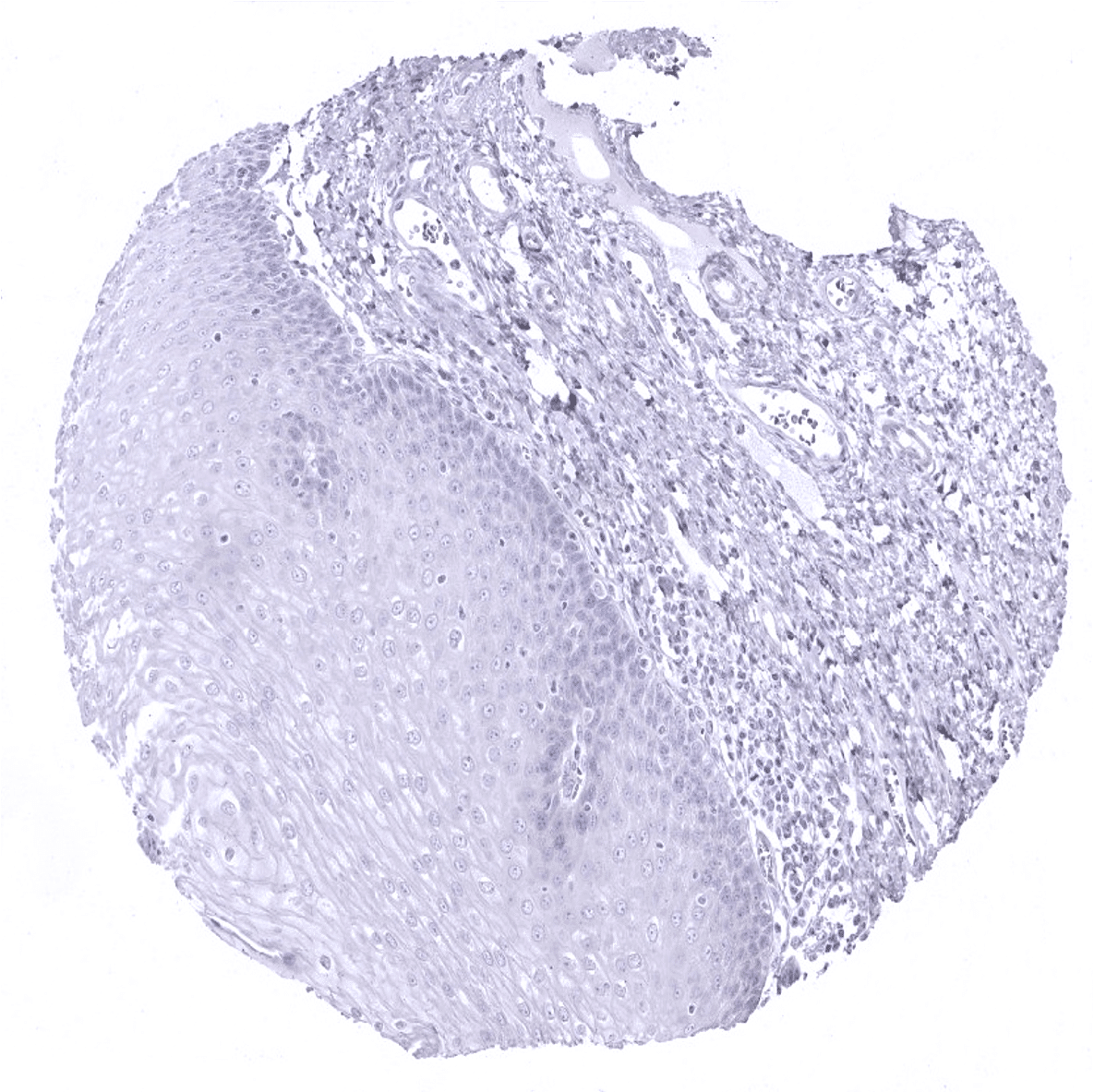
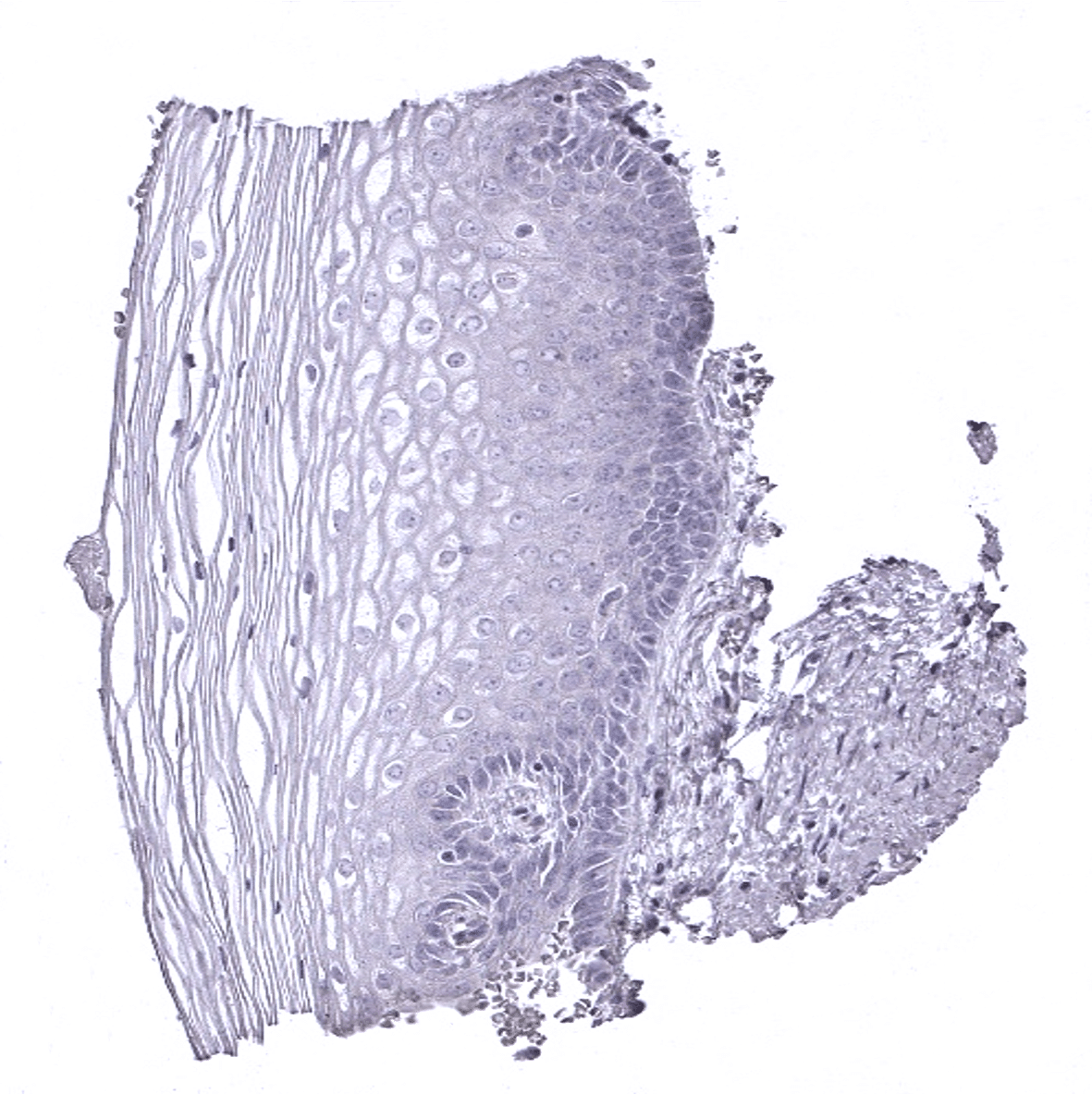
Esophagus, squamous epithelium
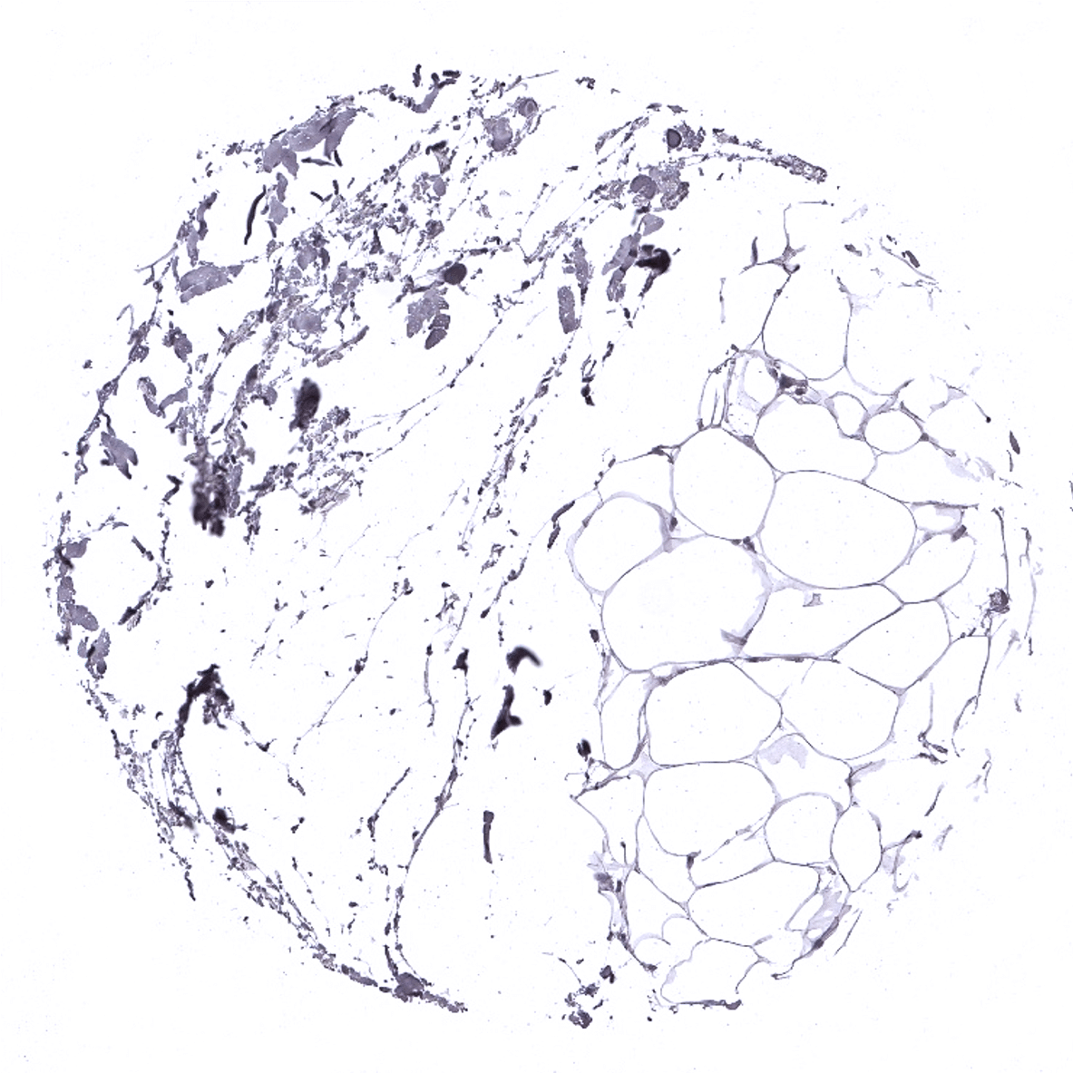
Fat
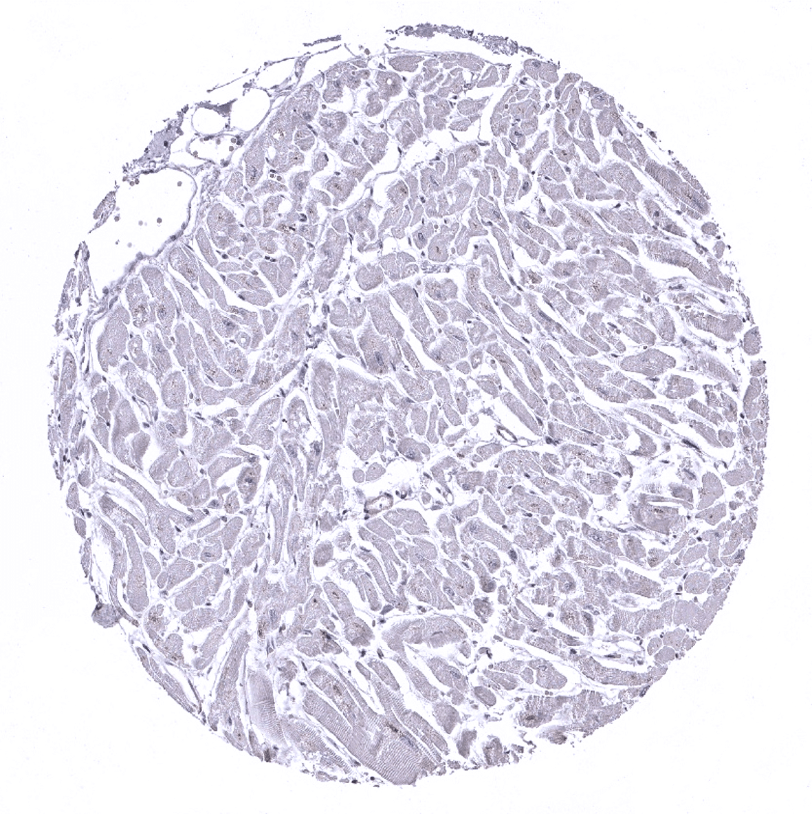
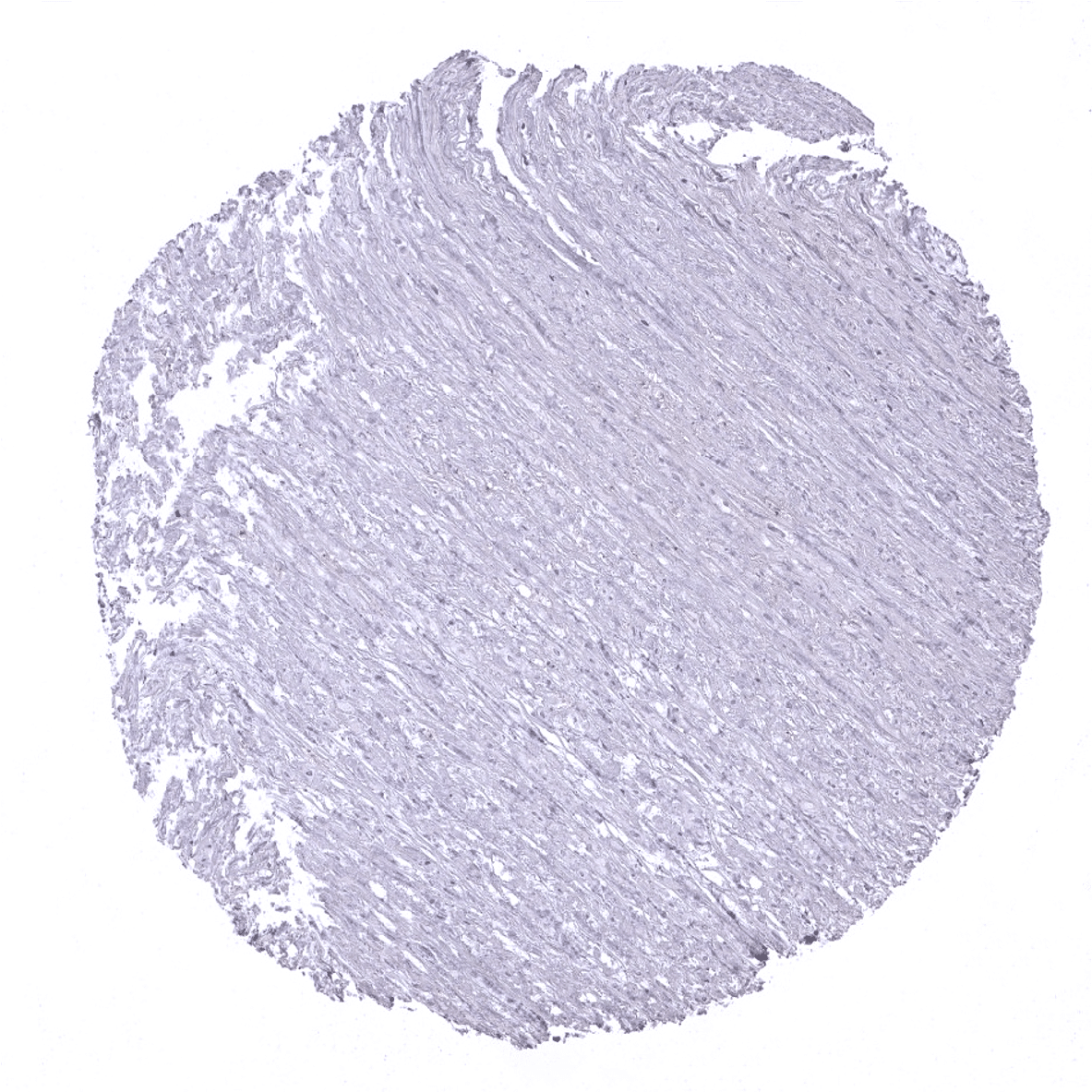
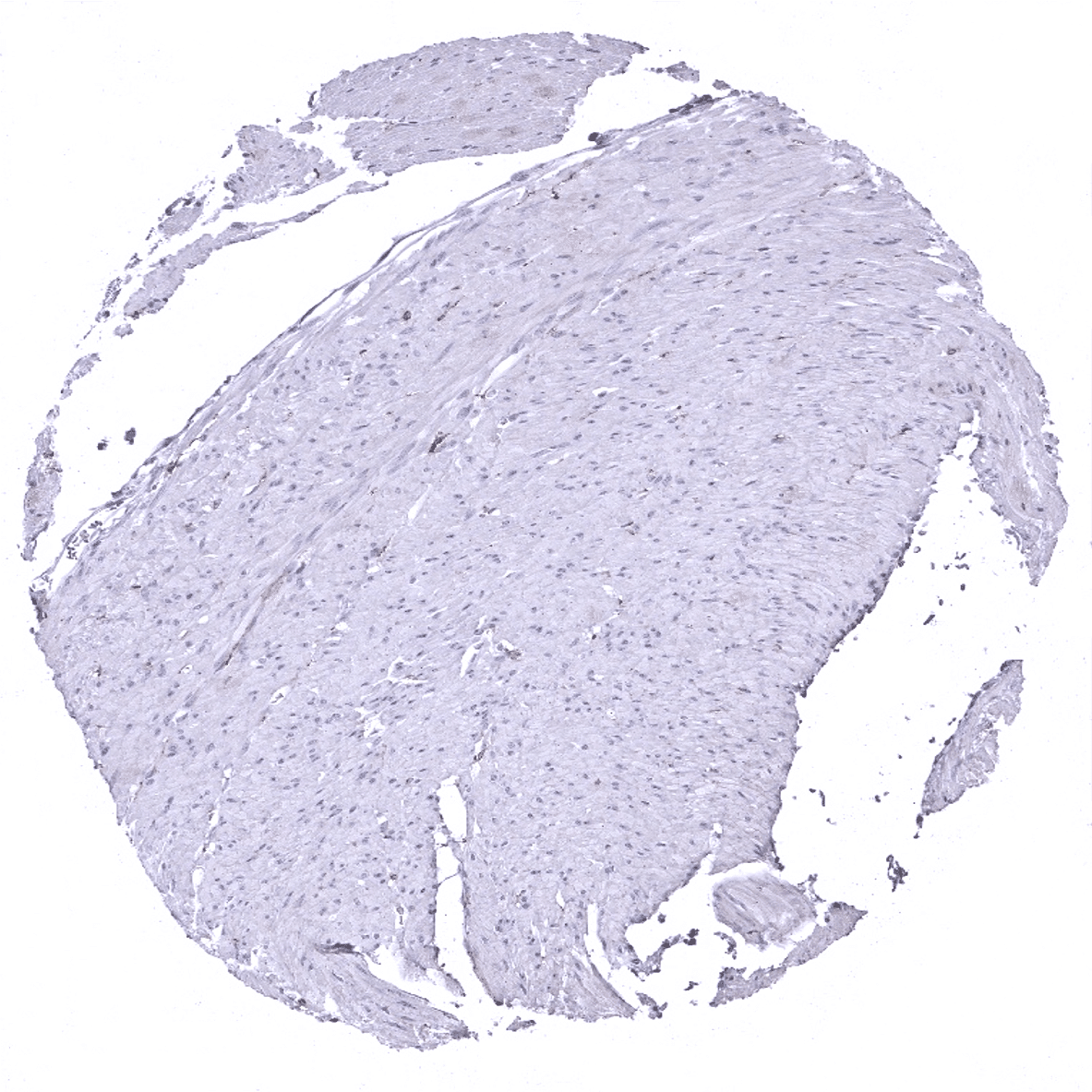
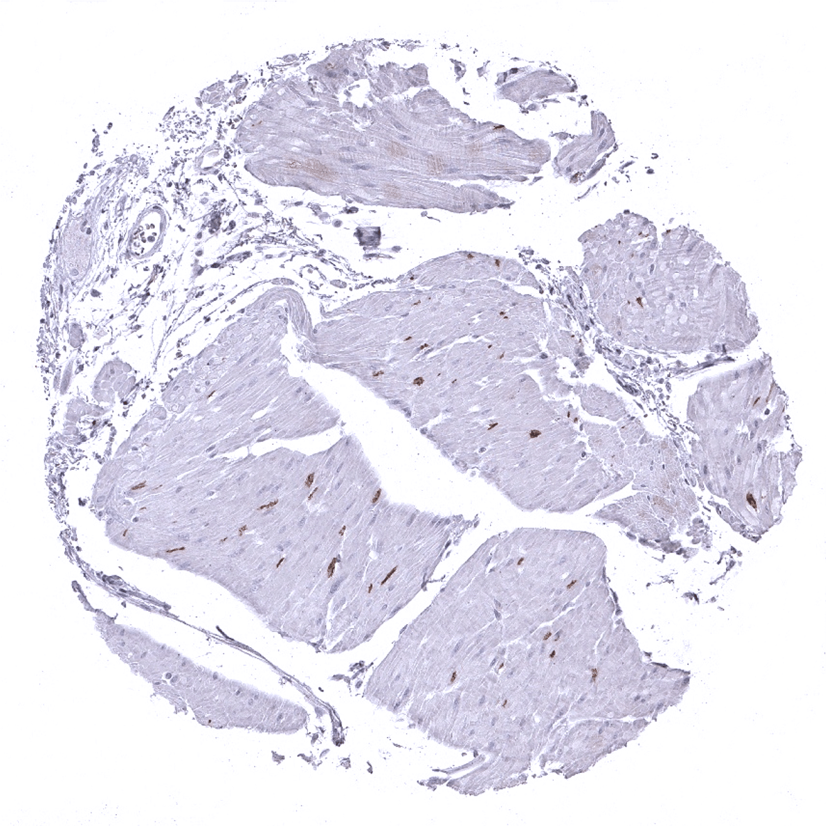
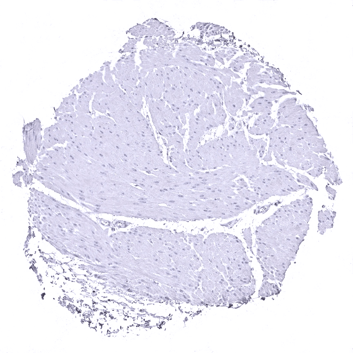
Heart

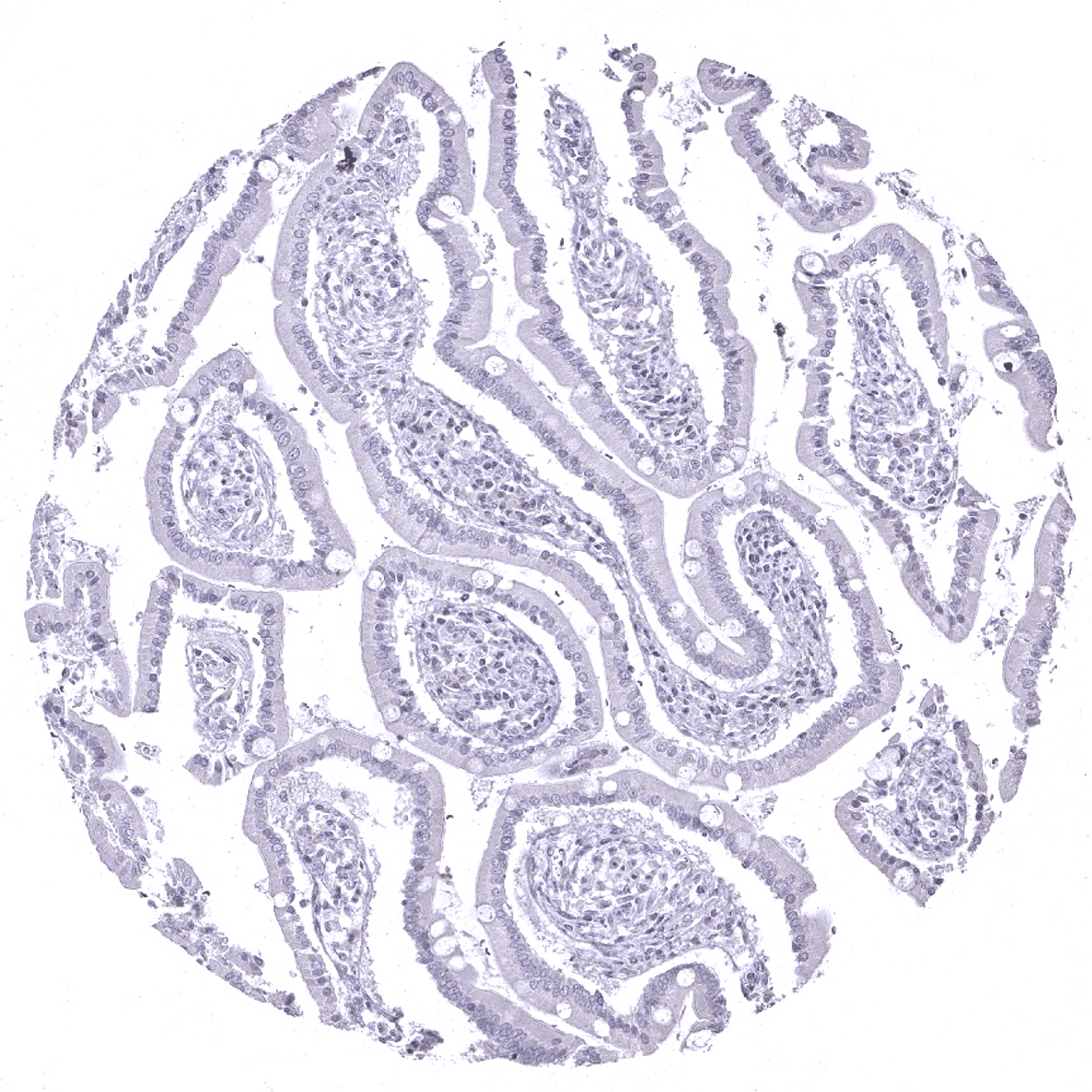
Ileum, mucosa
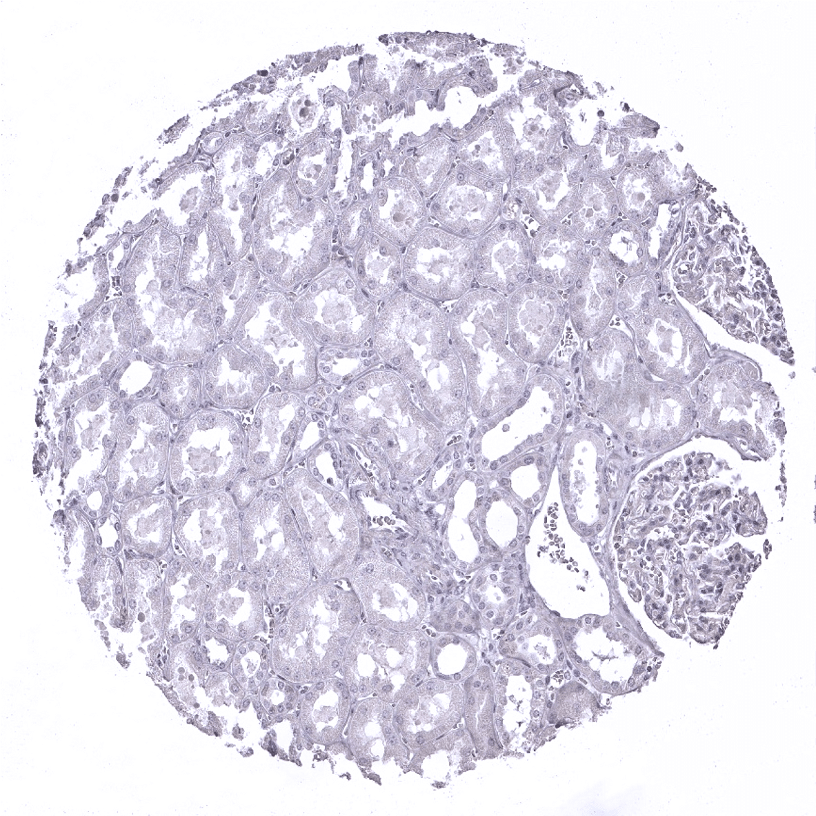
Kidney, cortex: DOG1 immunostaining is absent in the kidney.

Kidney, medulla
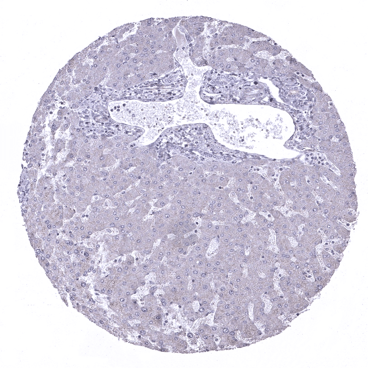
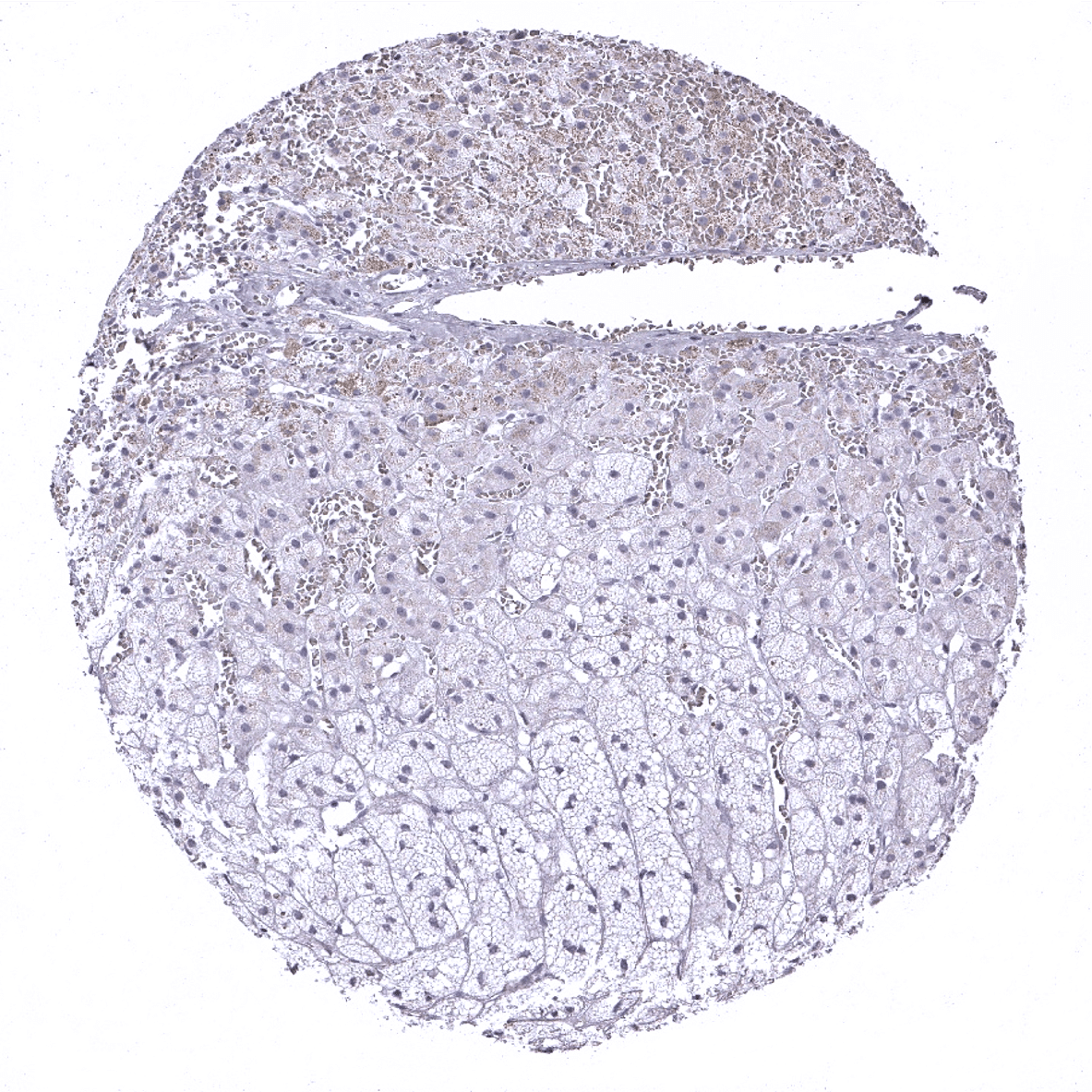
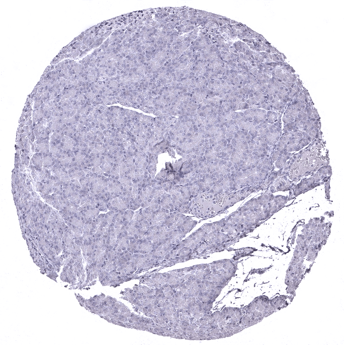
Liver
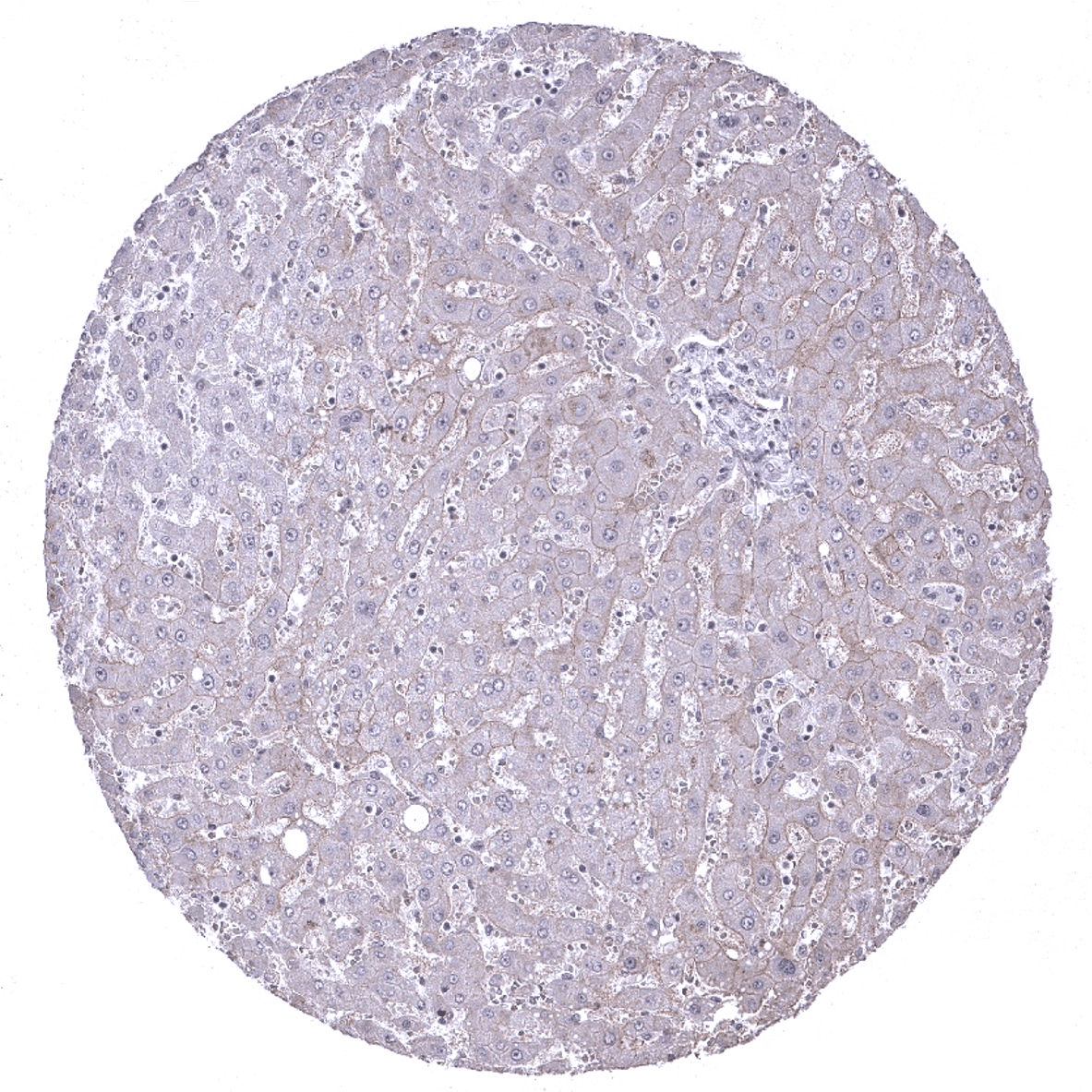
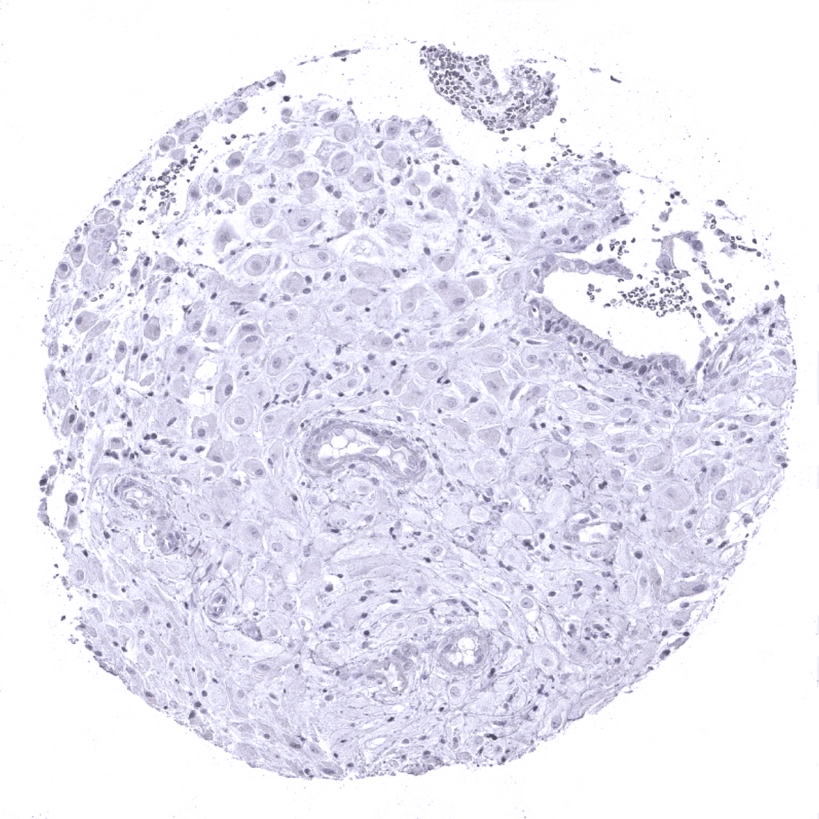
Liver: A weak to moderate membranous DOG1 staining should be seen in hepatocytes.
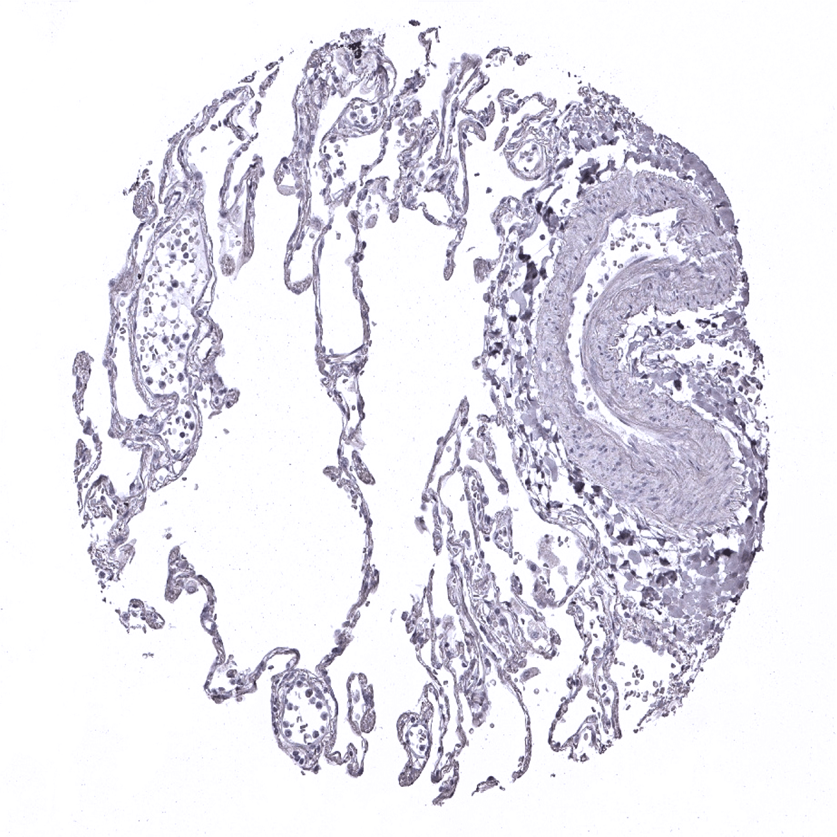
Lung
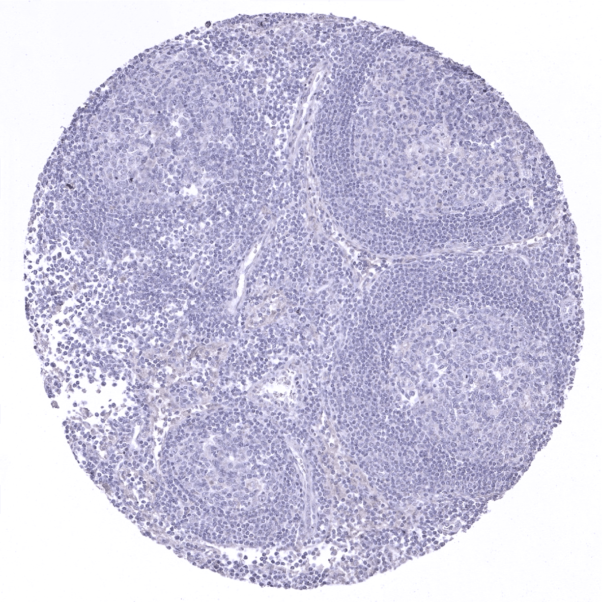
Lymph node
Ovary, stroma
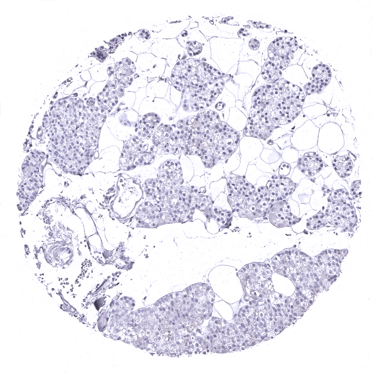
Pancreas
Parathyroid
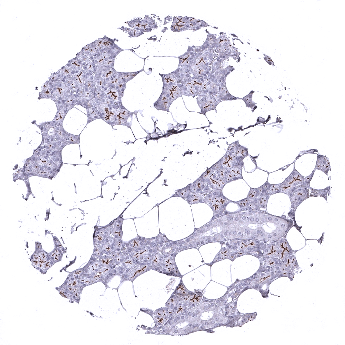
Parotid gland: Strong DOG1 staining of the apical membranes of secreting cells.
Pituitary gland, anterior lobe

Pituitary gland, posterior lobe
Pregnant uterus (decidua)
Placenta, early
Placenta, early
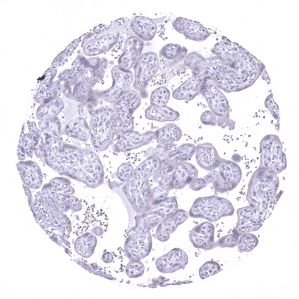
Placenta, mature
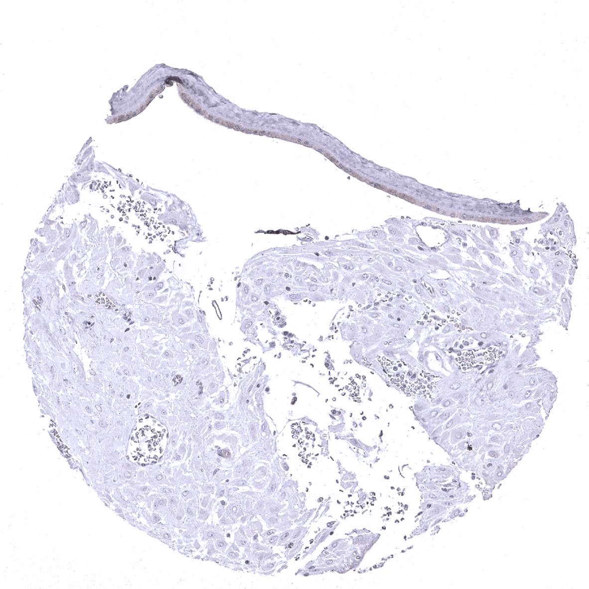
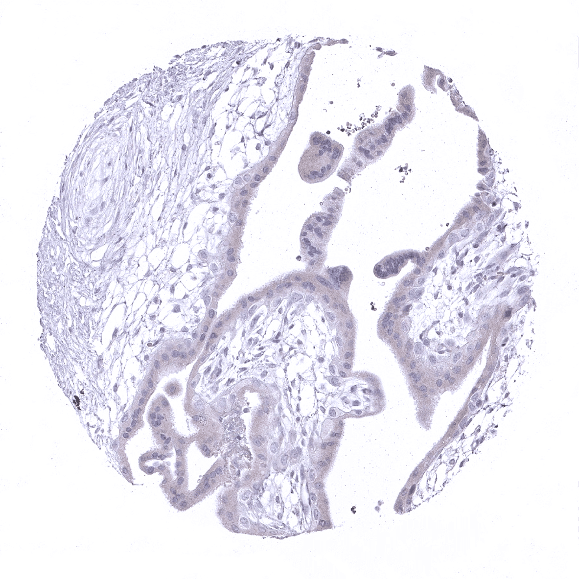
Placenta (amnion and chorion): Weak DOG1 staining of amnion cells.
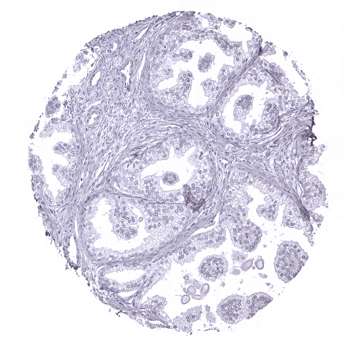
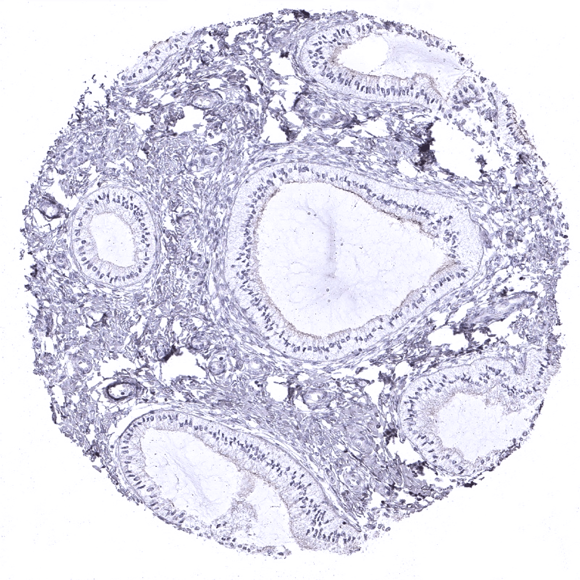
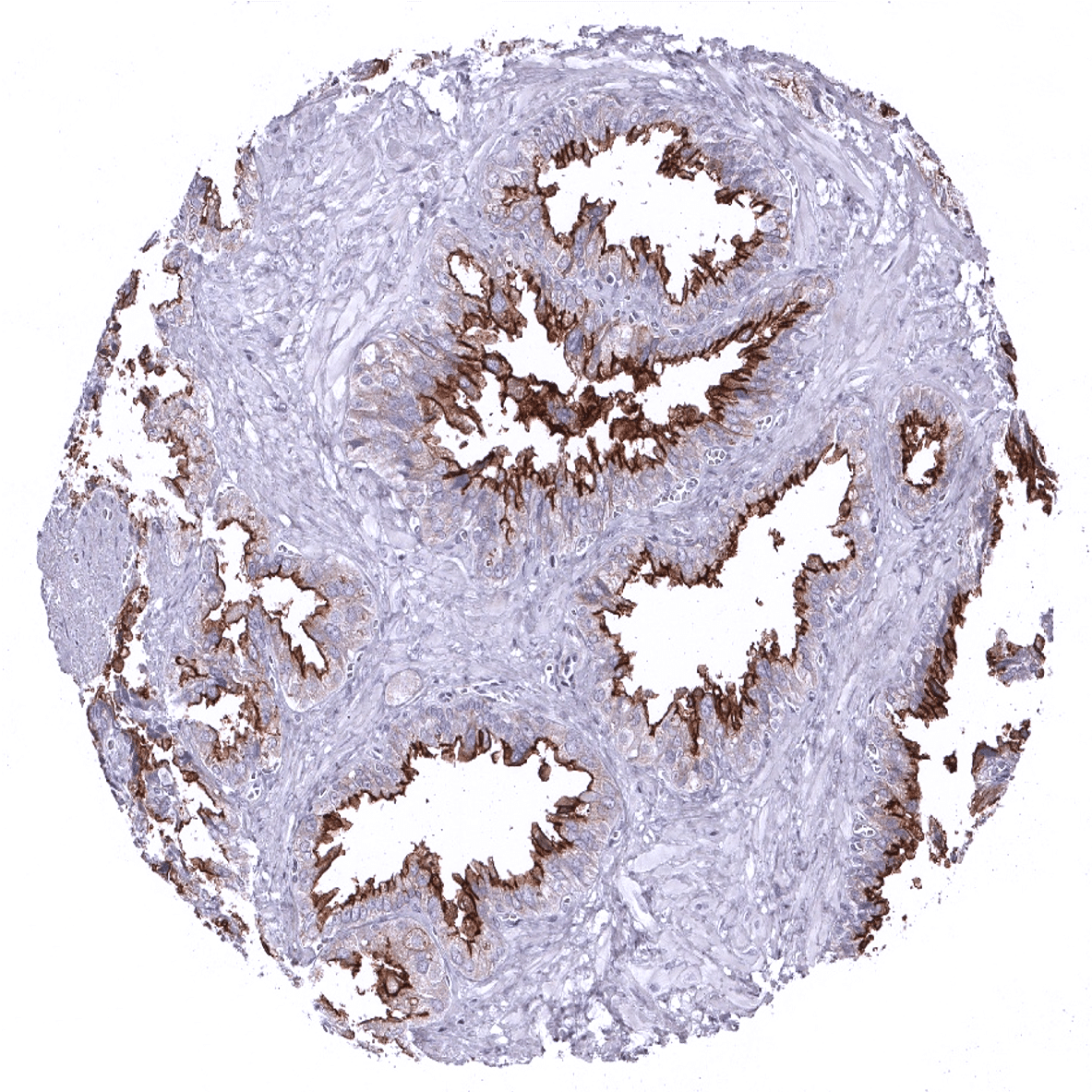
Prostate

Rectum, mucosa
Seminal vesicle: Strong DOG1 staining of apical membranes of glandular cells.
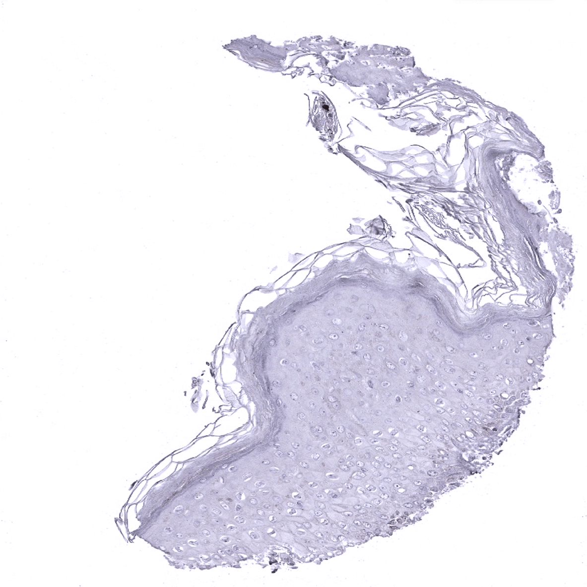
Skin

Spleen
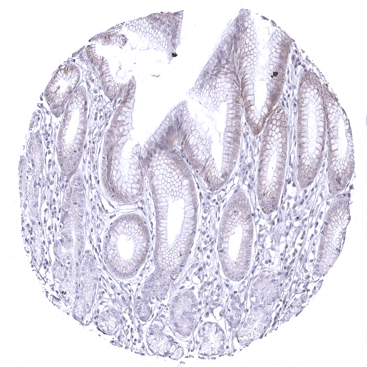
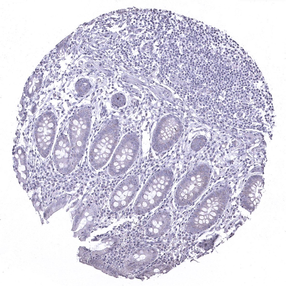
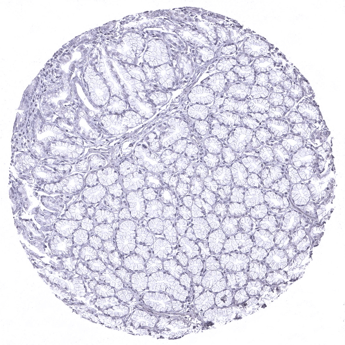
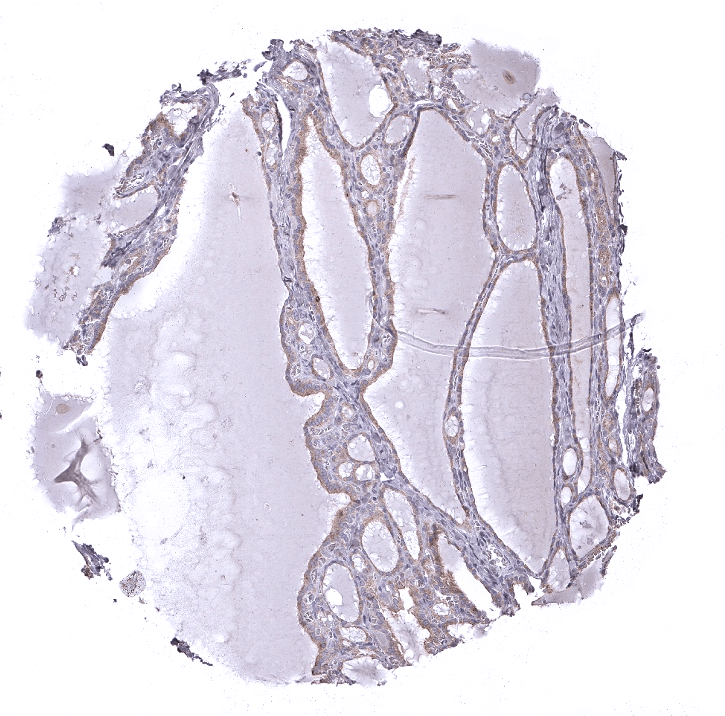
Stomach, antrum: Weak DOG1 staining of epithelial cells, mainly in the isthmus/neck region but also of the surface cell layer.

Stomach, corpus
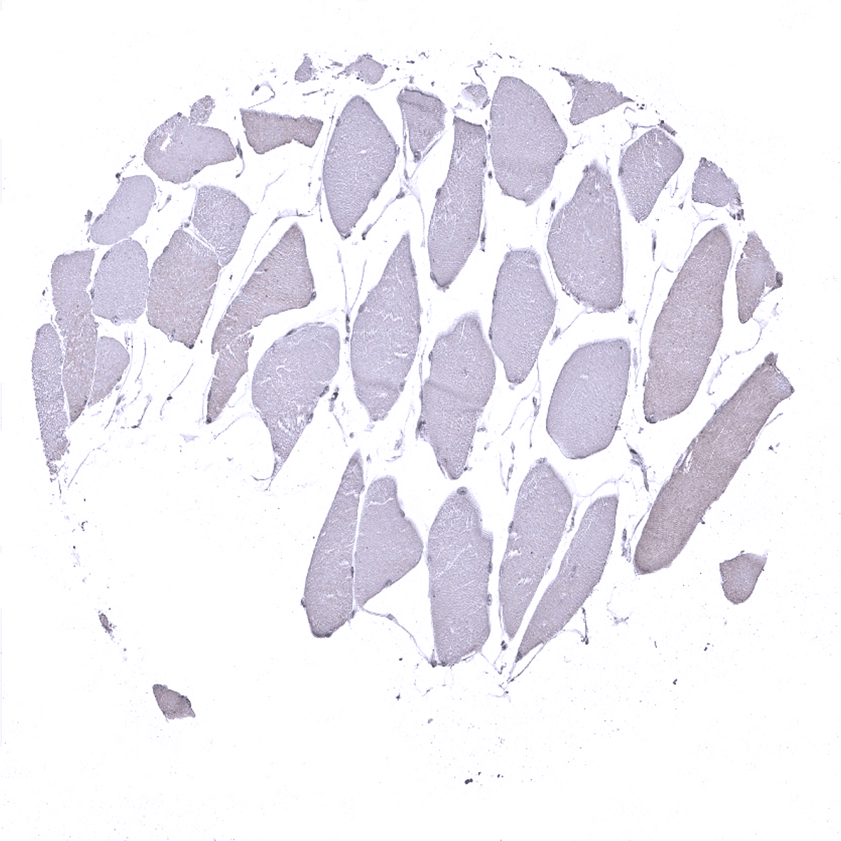
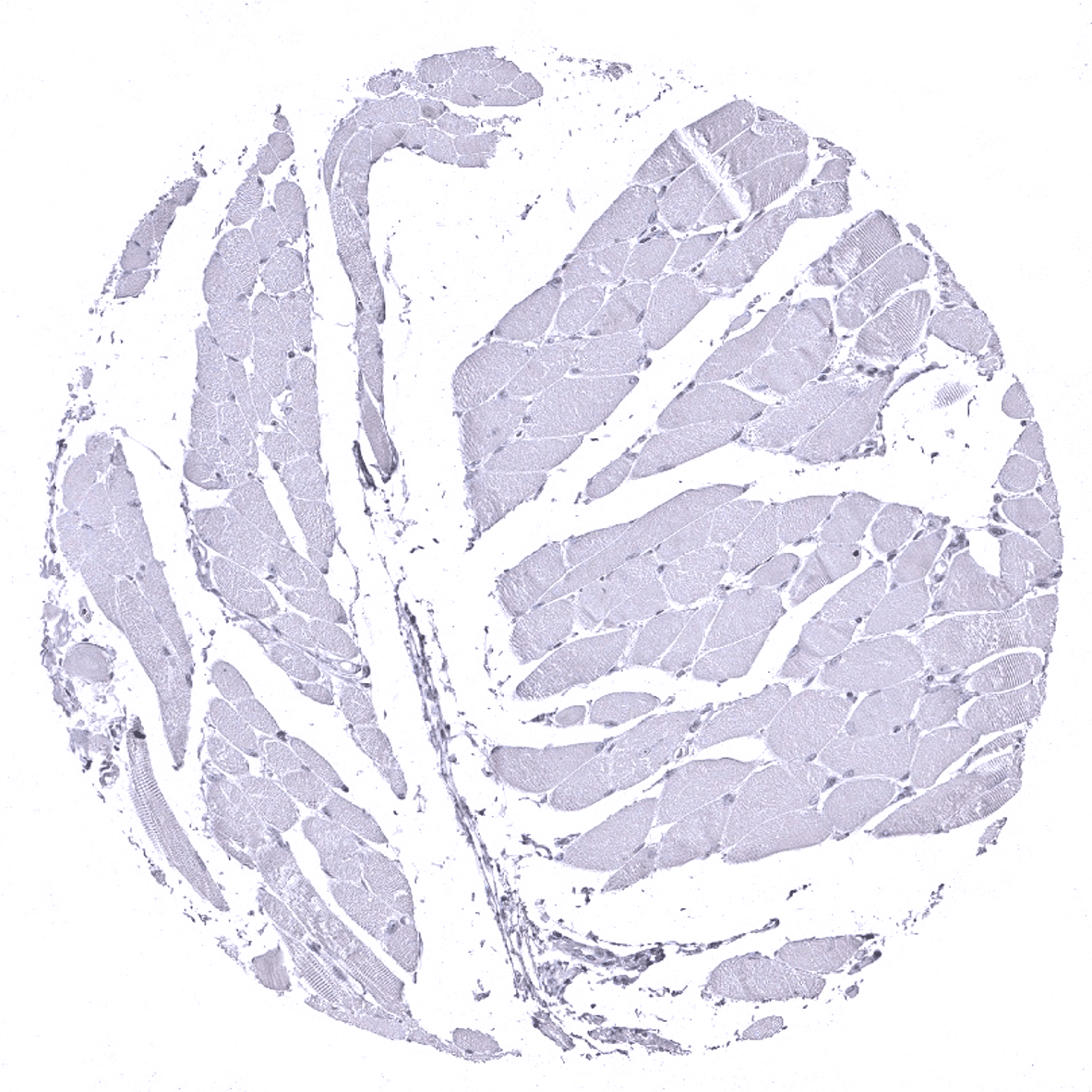
Striated muscle
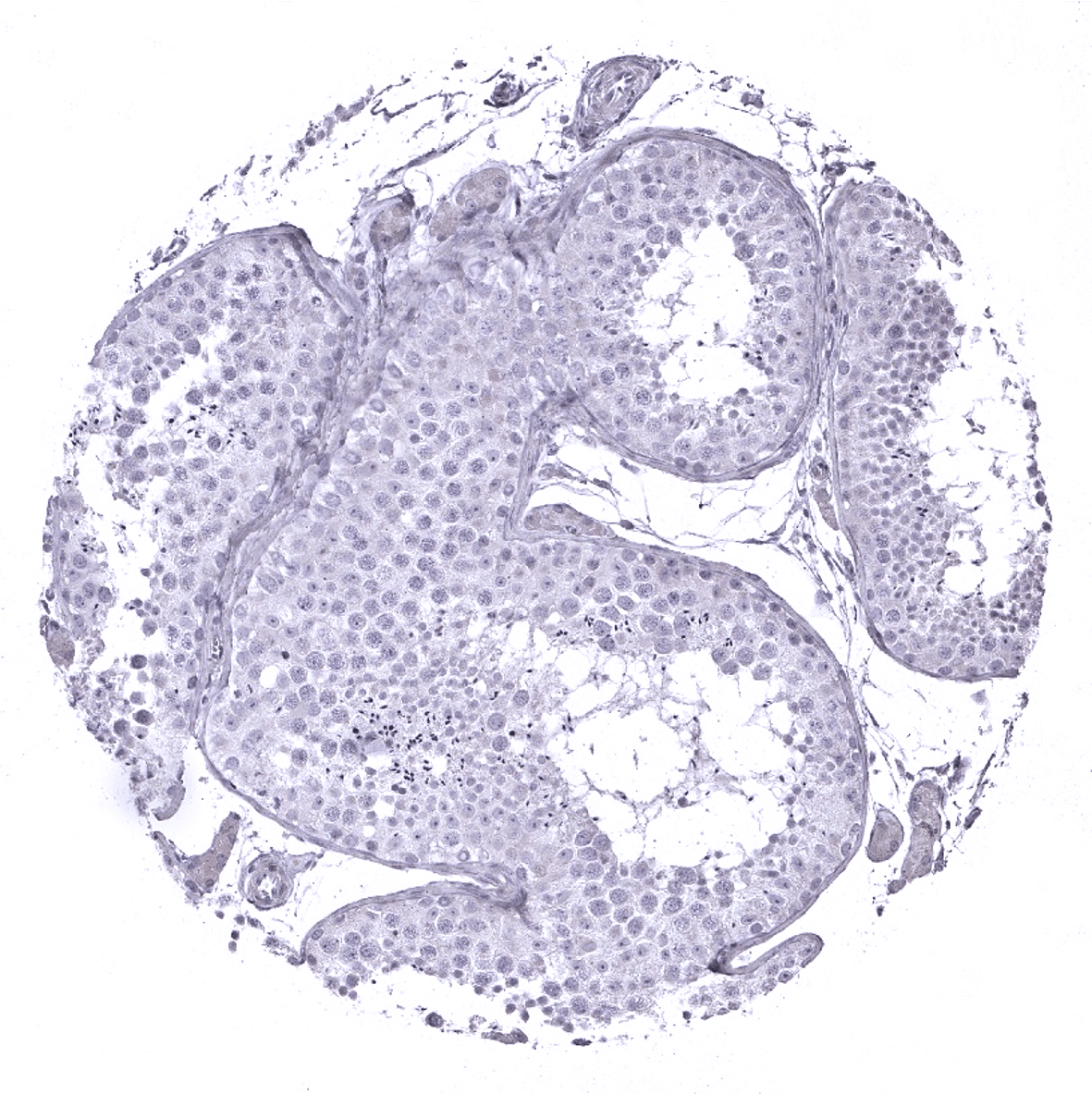
Testis
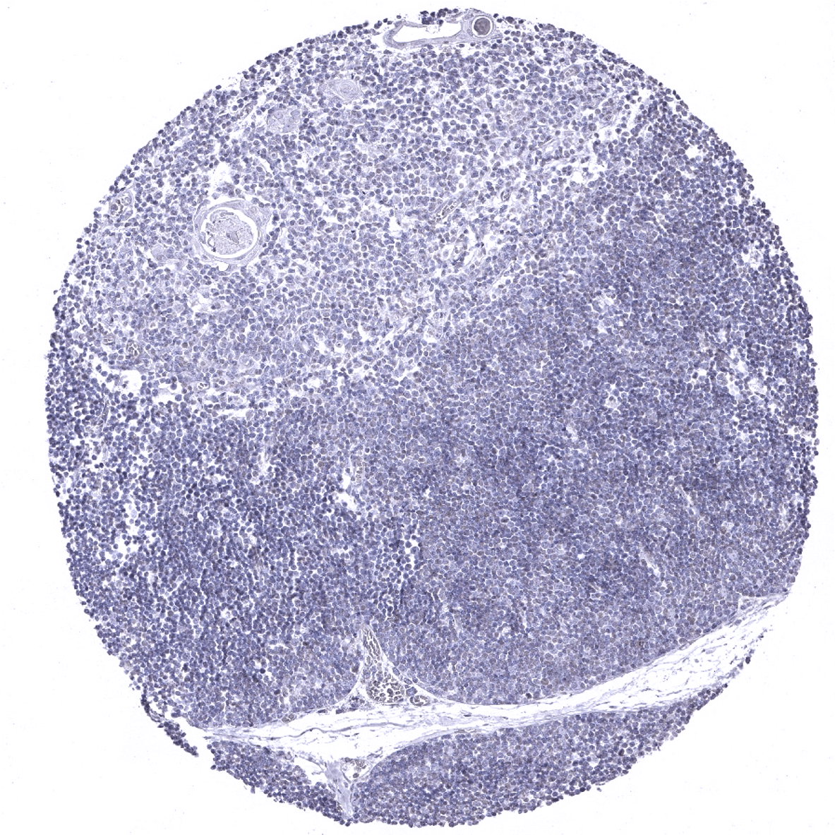
Thymus
Thyroid gland
Tongue, muscle
Tonsil, surface epithelium

Tonsil
Urinary bladder, muscular wall

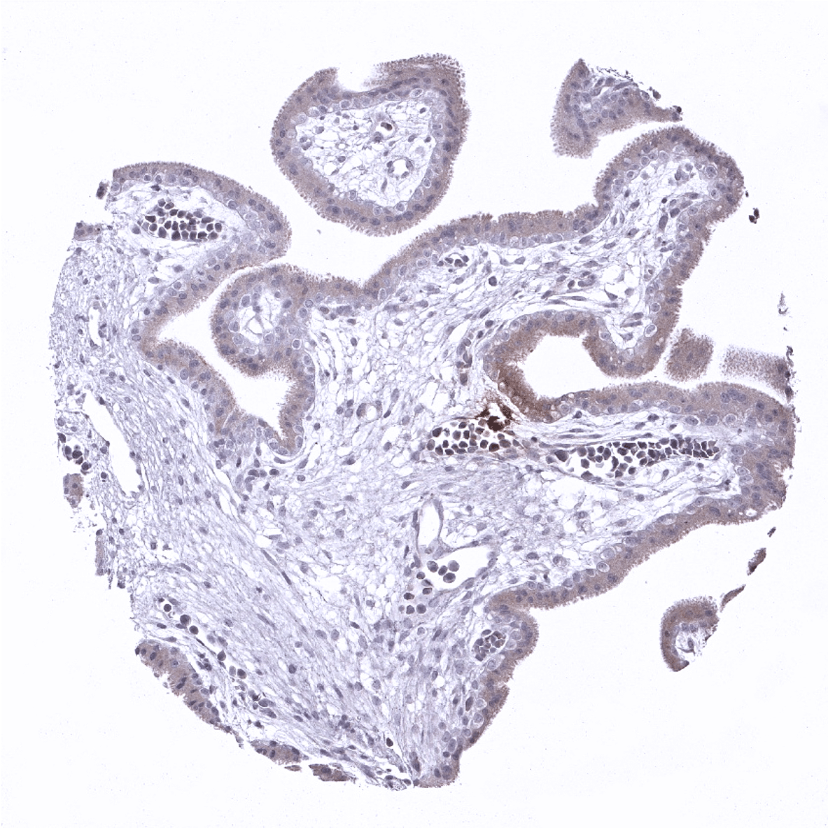
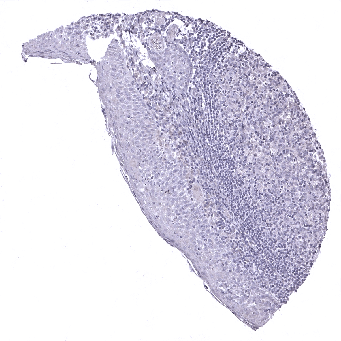
Urinary bladder, urothelium
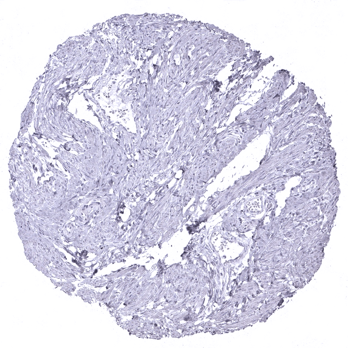
Uterus, myometrium