
Adrenal gland - Chromogranin A staining is absent in the cortex.
Adrenal gland - Intense chromogranin A staining in all cells of the medulla.
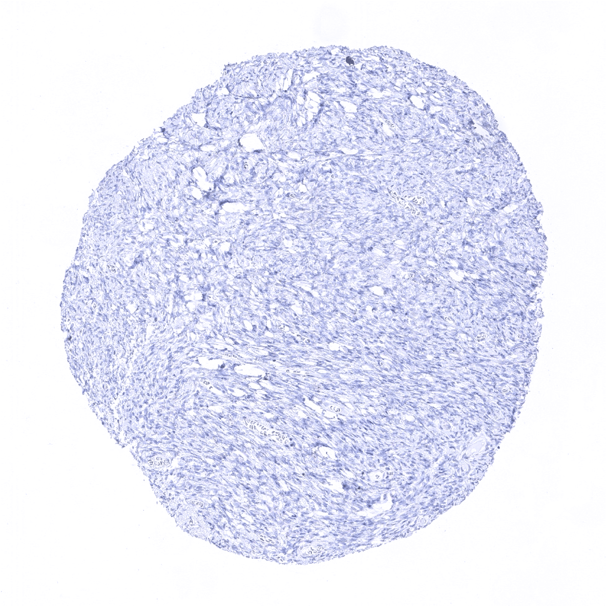
Aorta, media
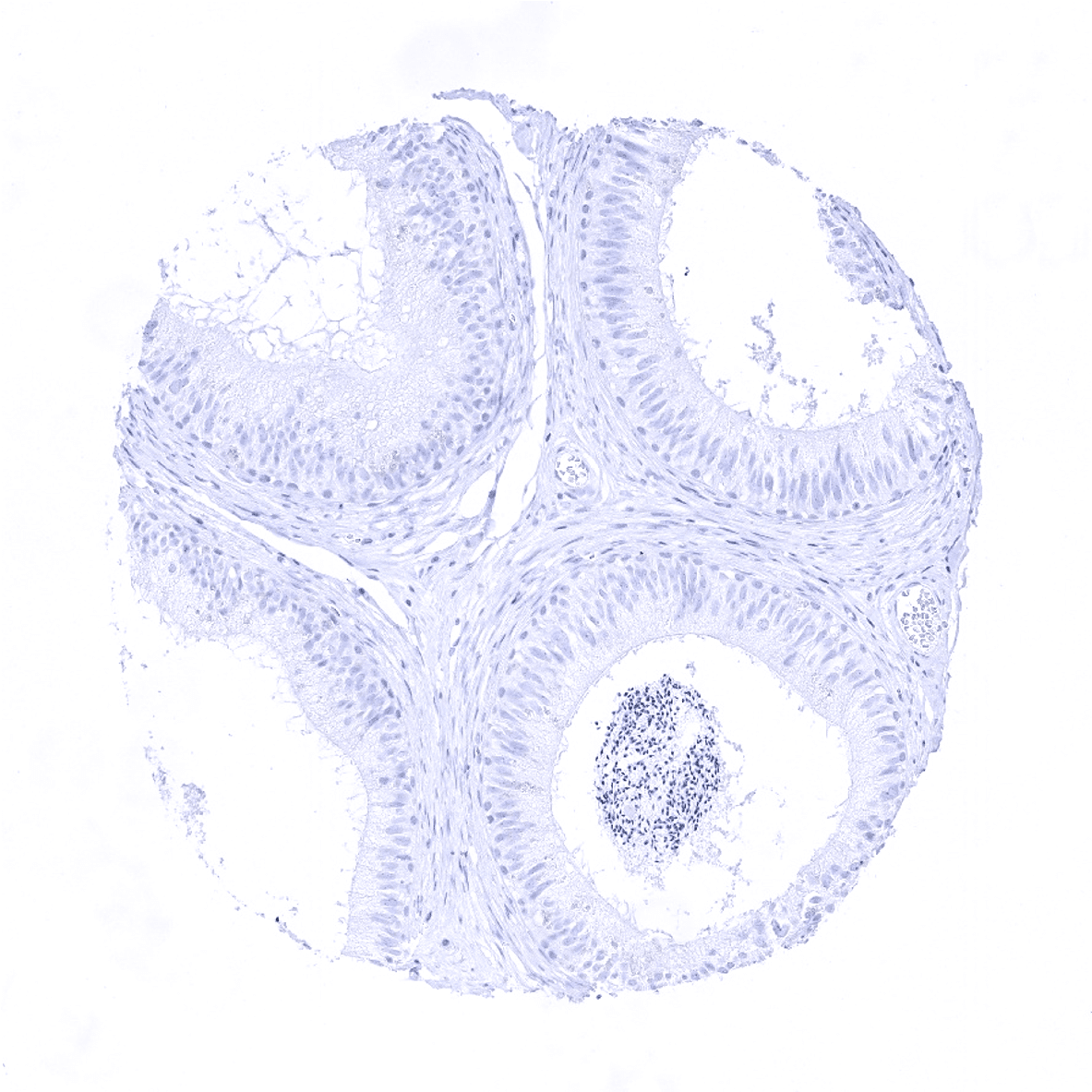
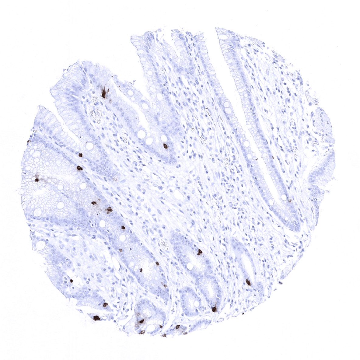
Appendix, mucosa

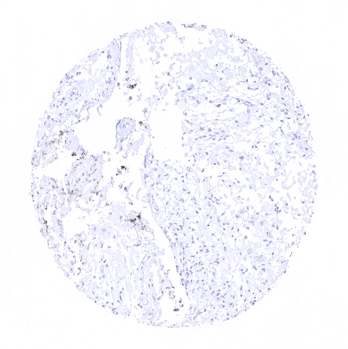
Appendix, muscular wall - Weak chromogranin A immunostaining is seen in axons and ganglion cells of the peripheral nerves.
Bone marrow
Breast
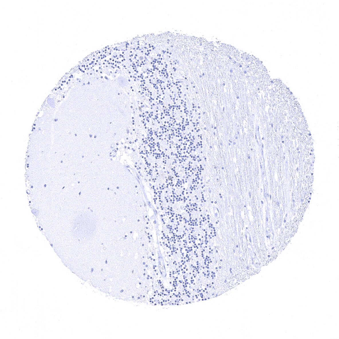
Cerebellum (molecular layer, Purkinje cell layer, granule cell layer, white matter)
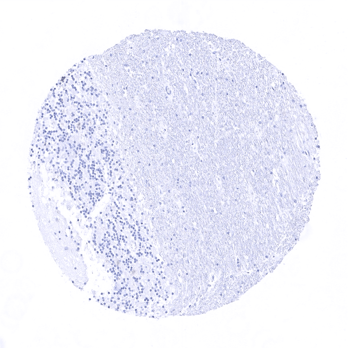
Cerebellum (granule cell layer, white matter)
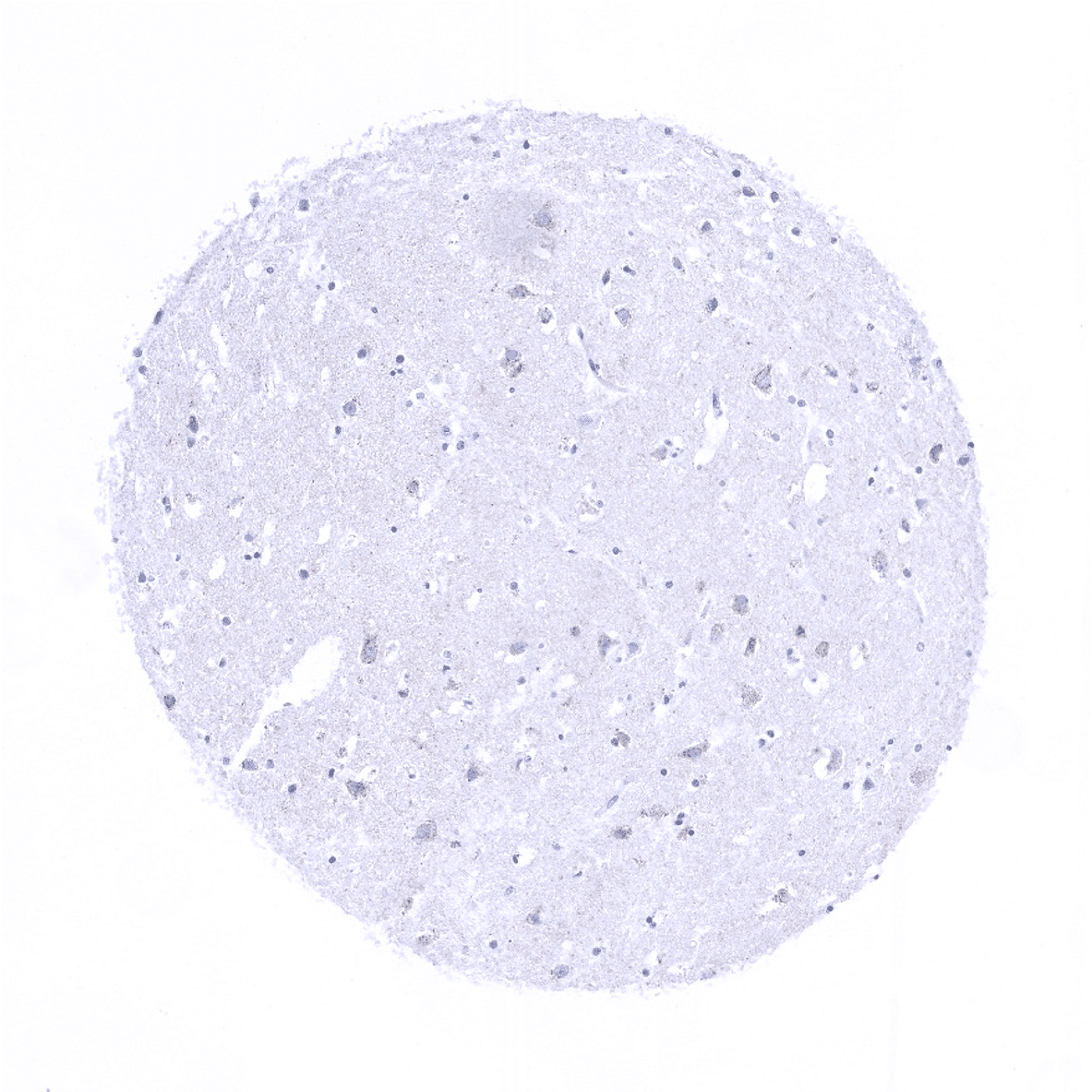
Cerebrum, grey matter

Cerebrum, white matter
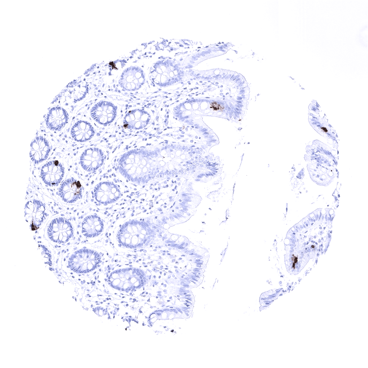
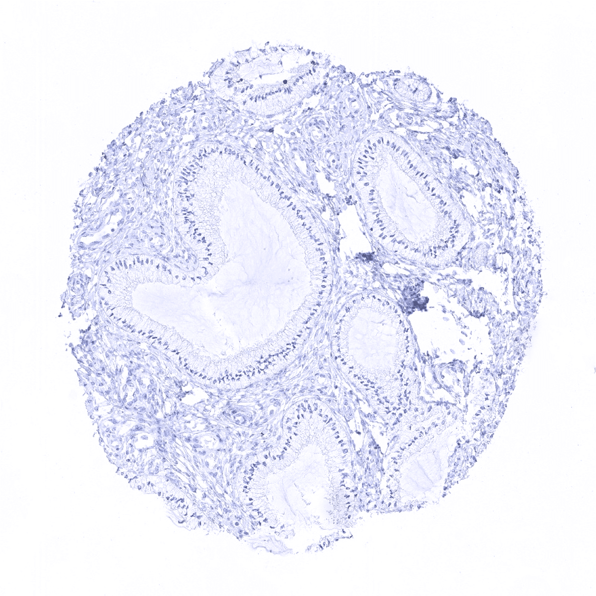
Colon descendens, mucosa
Colon descendens, muscular wall
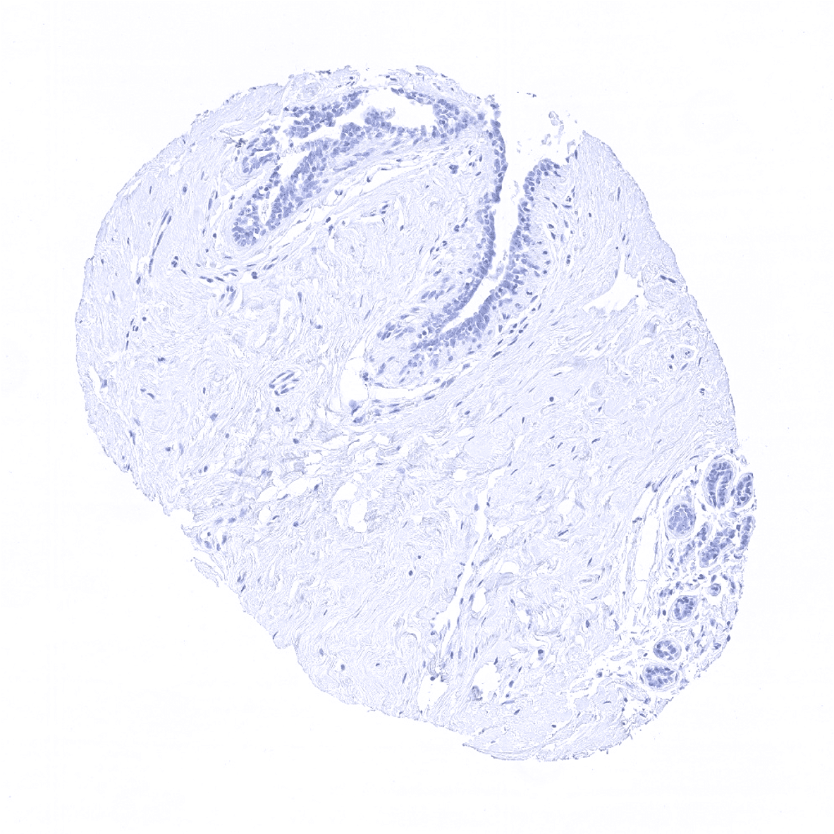
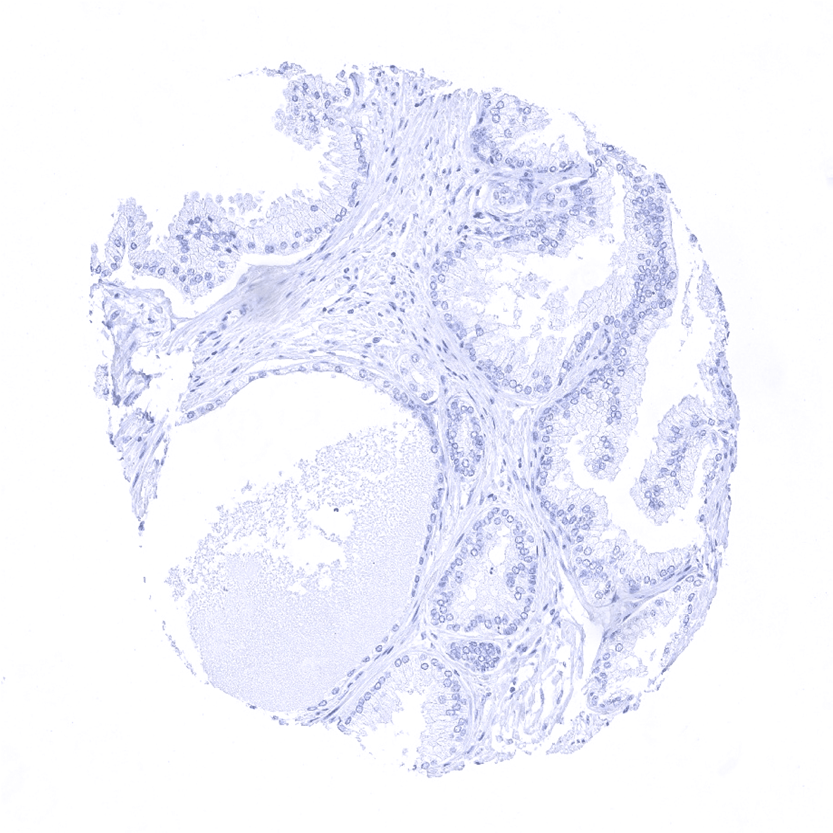
Duodenum, Brunner gland

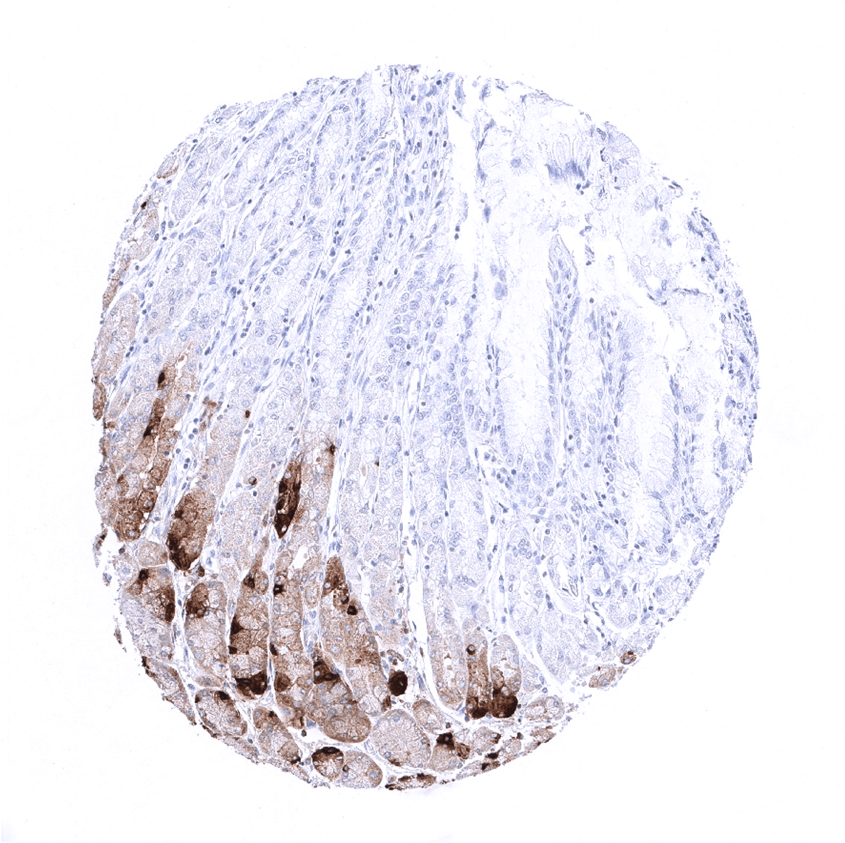
Duodenum, mucosa - Strong chromogranin A immunostaining of neuroendocrine cells. Due to the high level of chromogranin A protein in these cells, adjacent non-neuroendocrine cells show "contamination artifacts".
Ectocervix
Endocervix
Endocervix - Few neuroendocrine cells are chromogranin A positive.
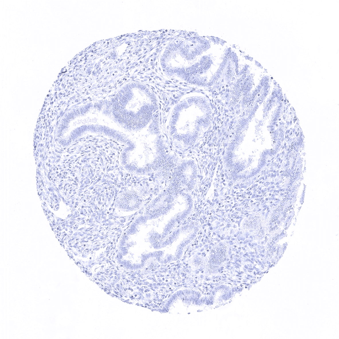
Endometrium, proliferation
Endometrium, secretion
Epididymis

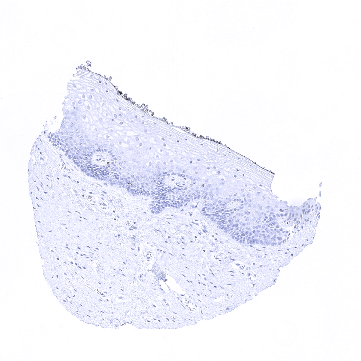
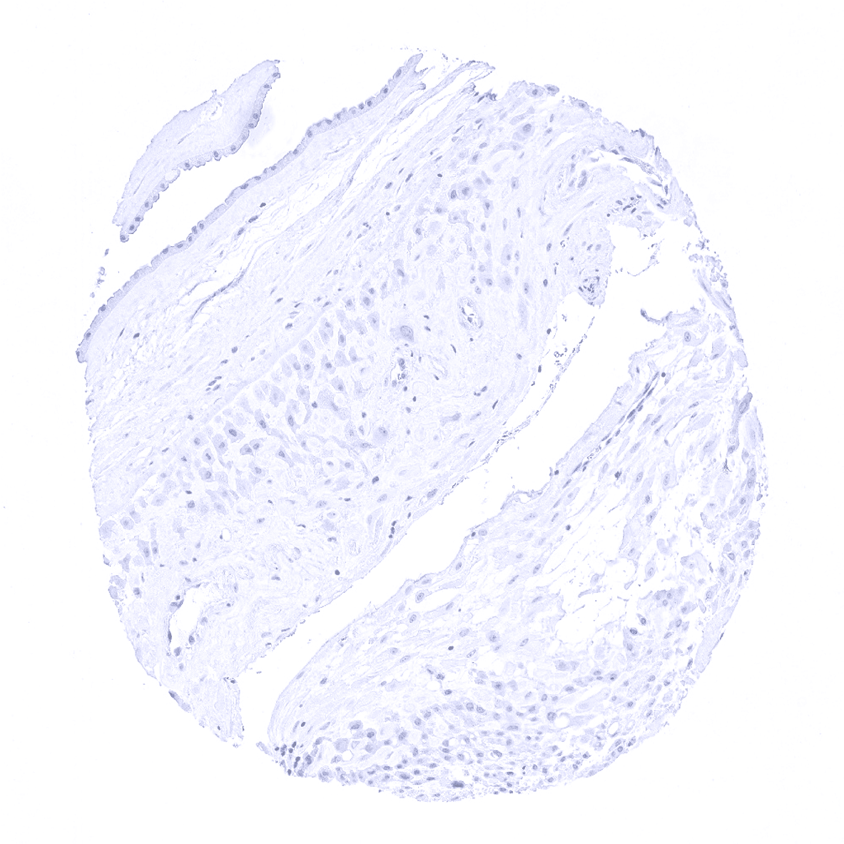
Esophagus, squamous epithelium
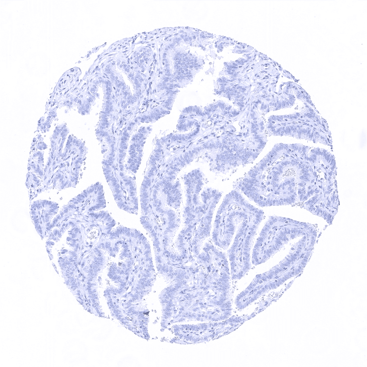
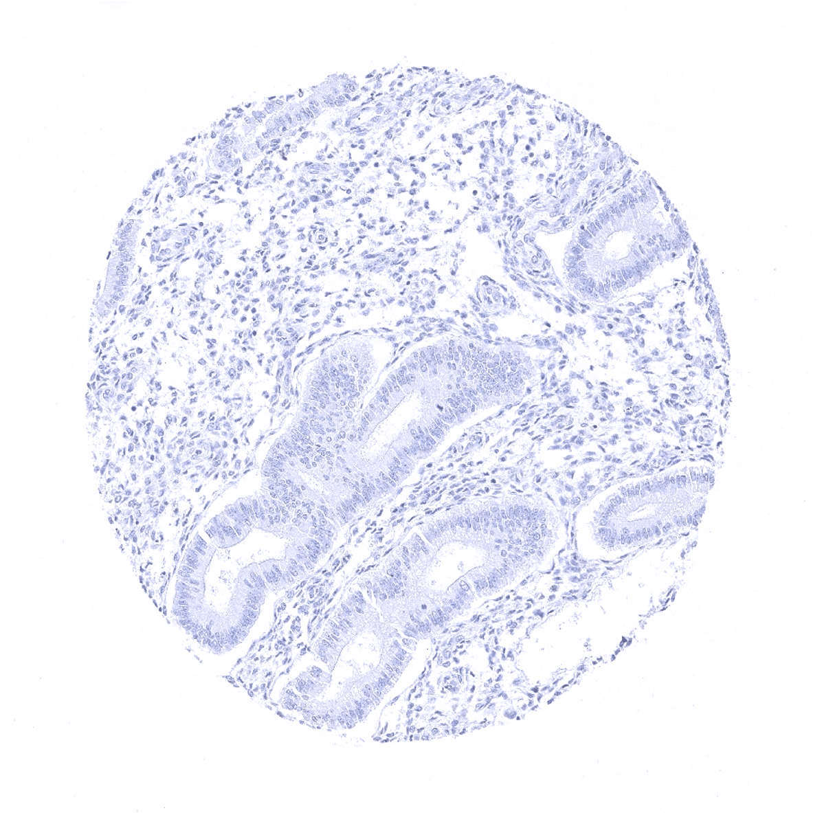
Fallopian tube, mucosa
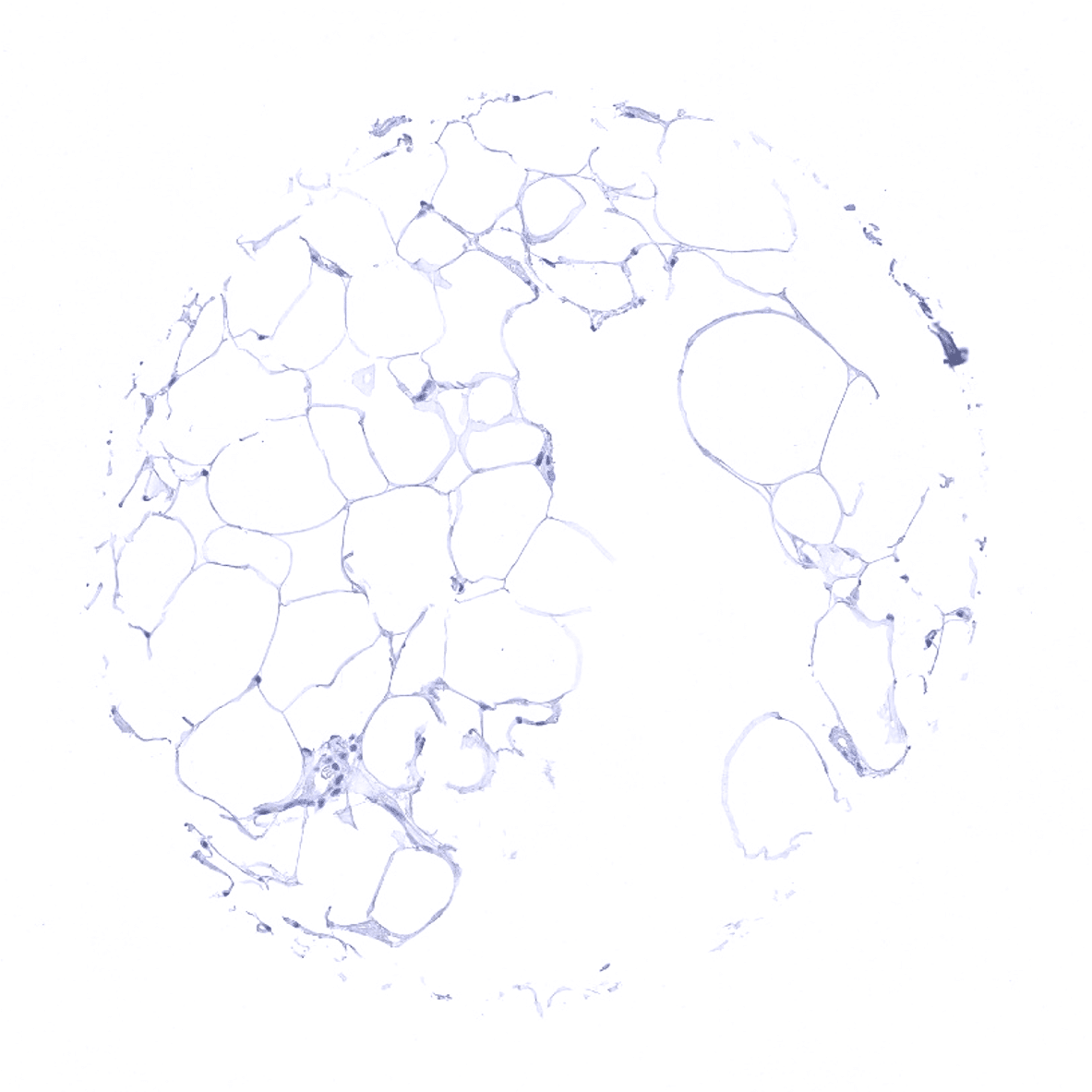
Fat
Gallbladder, epithelium
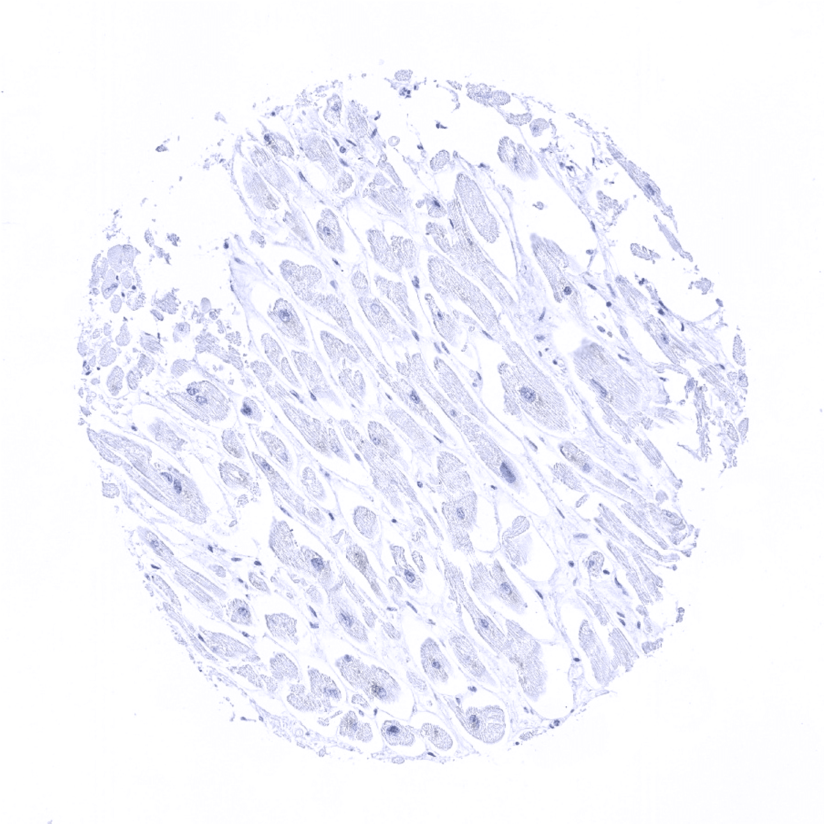
Heart
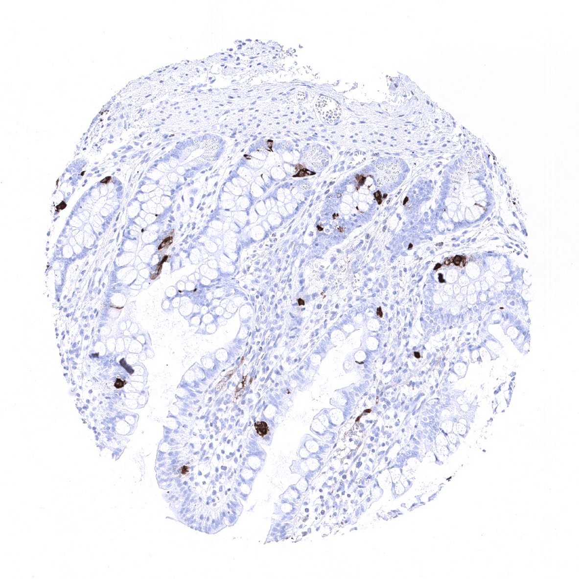
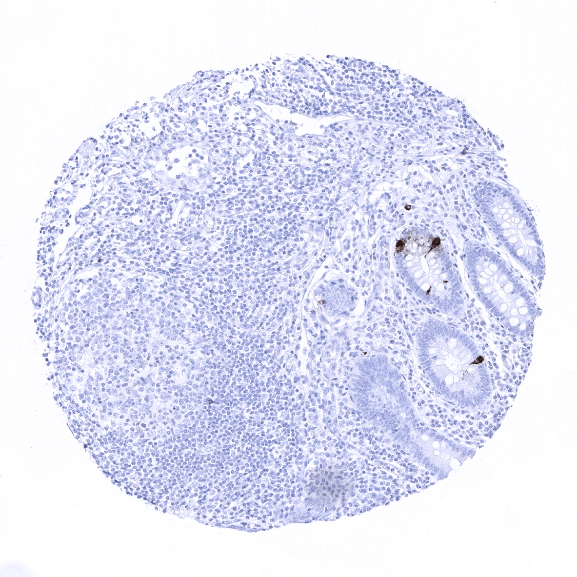
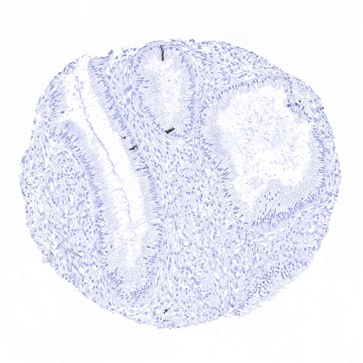
Ileum, mucosa

Kidney, cortex

Kidney, medulla
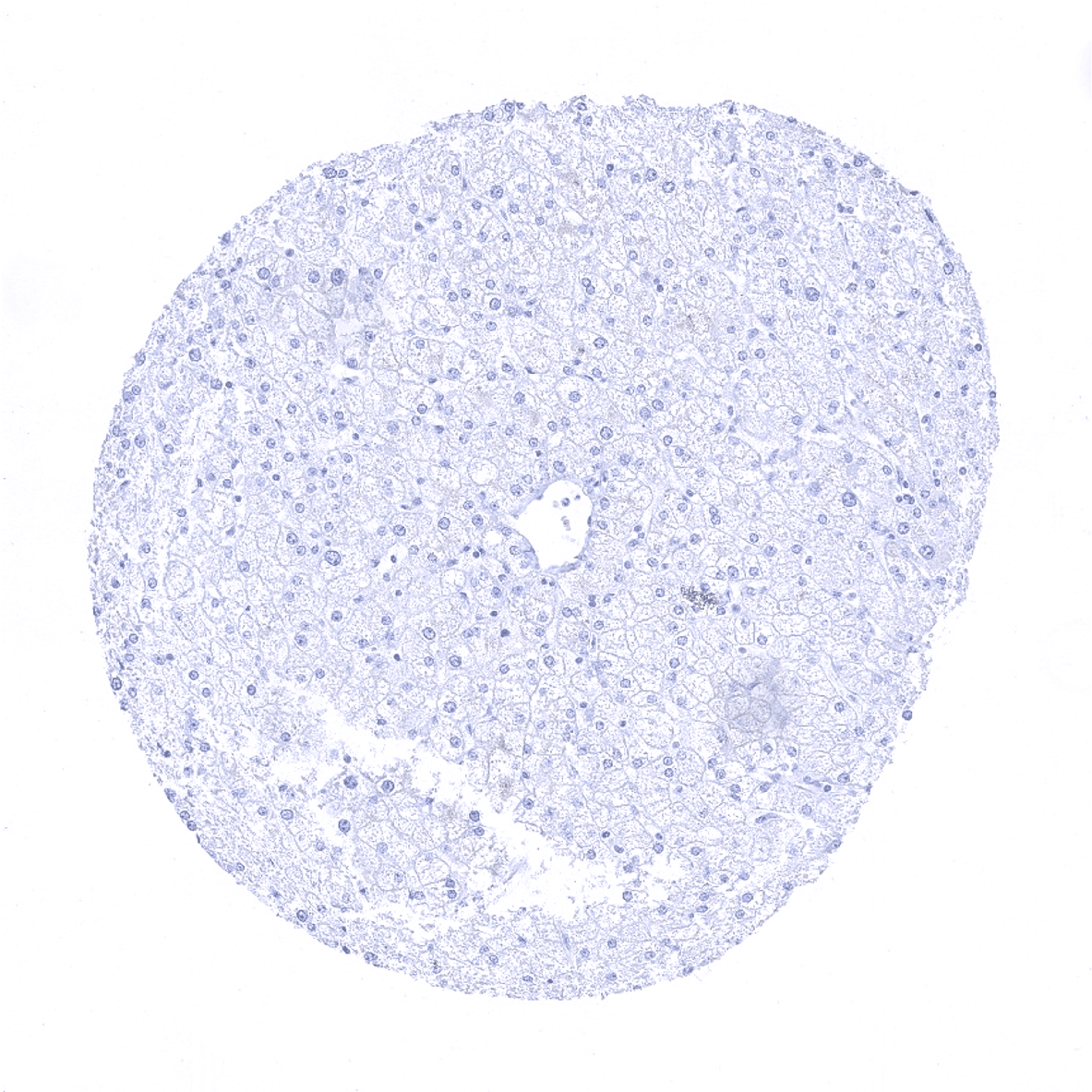
Liver
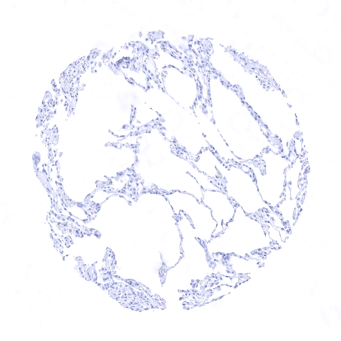
Lung
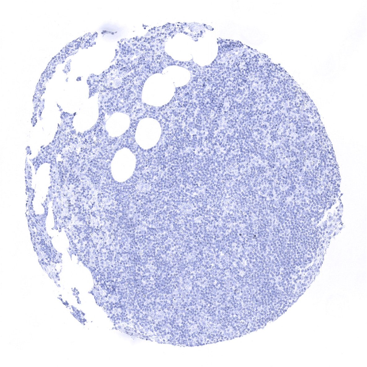
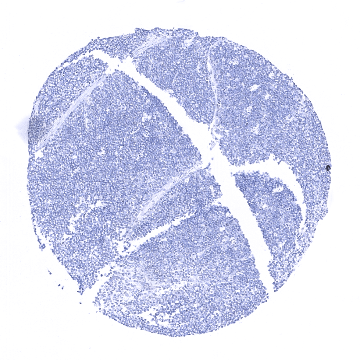
Lymph node
Ovary, stroma
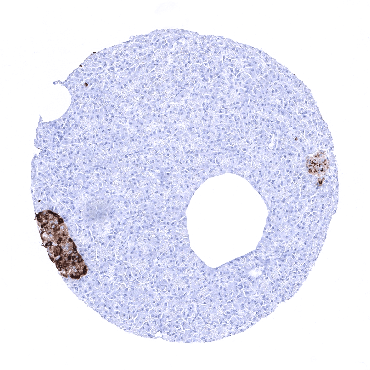
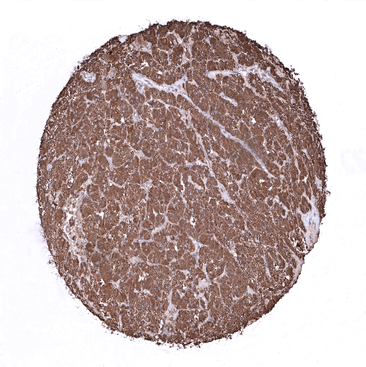
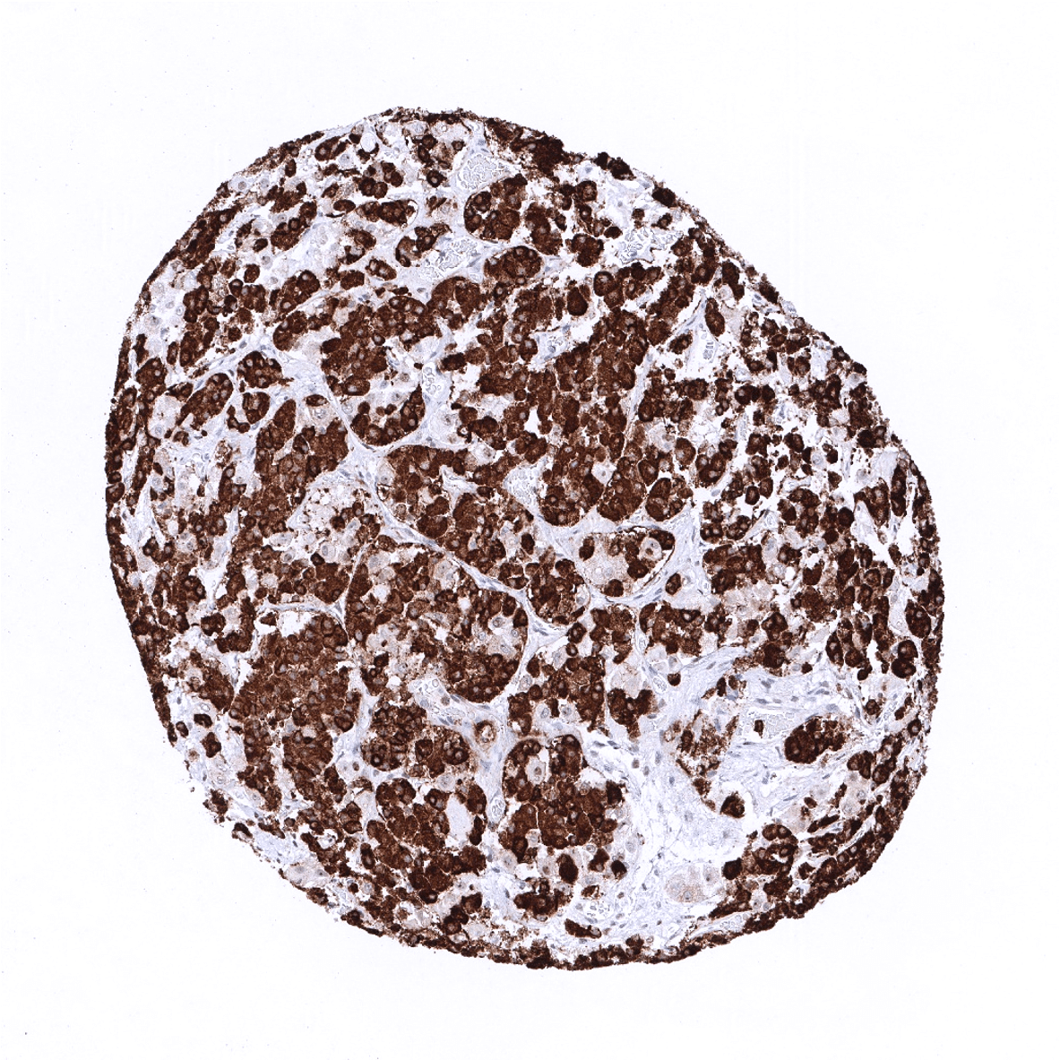
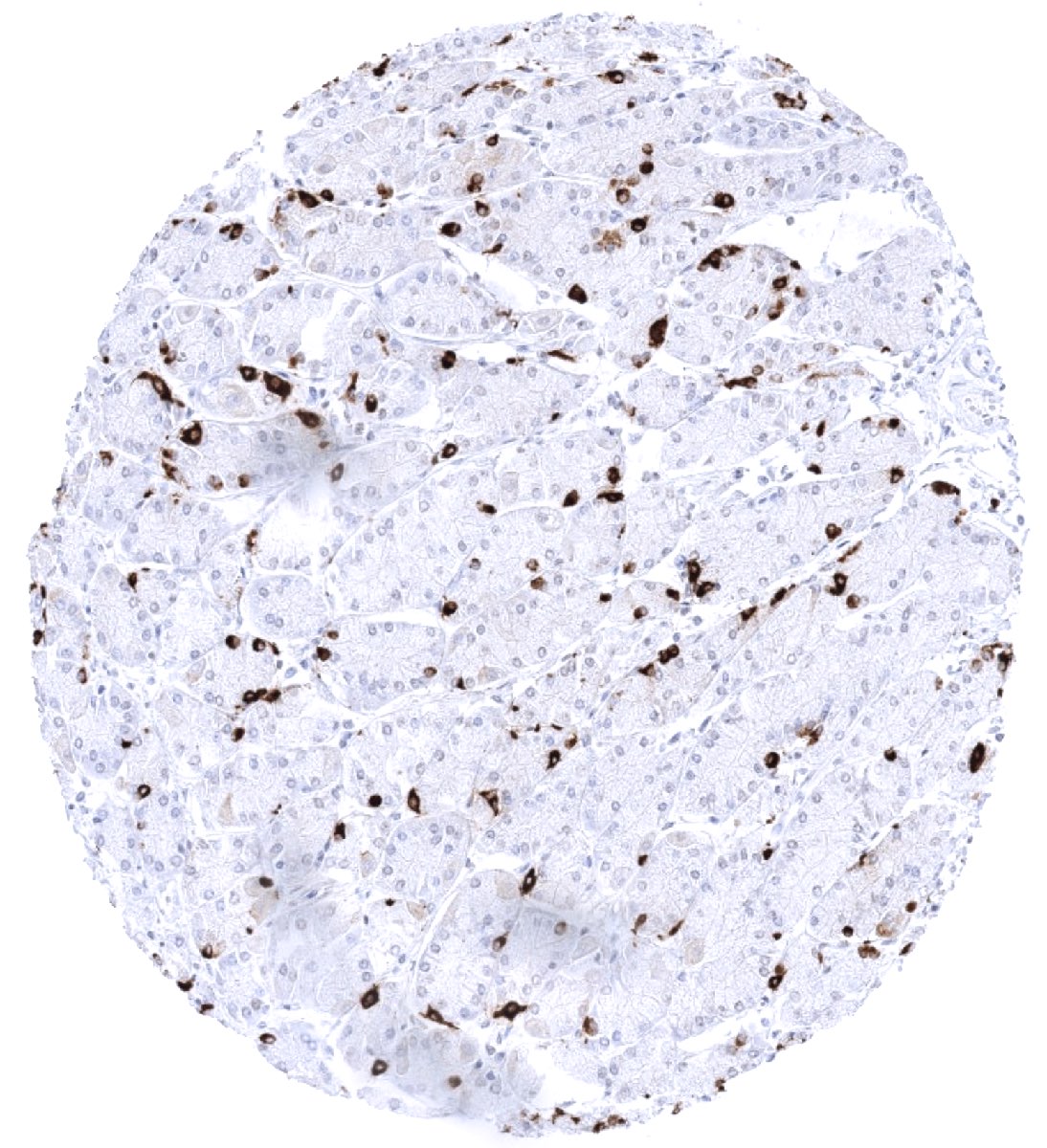
Pancreas - Strong chromogranin A positivity in islets of Langerhans.

Parathyroid gland - Strong chromogranin A staining of epithelial cells.
Parotid gland
Pituitary gland, anterior lobe - Strong chromogranin A staining of epithelial cells.
Pituitary gland, posterior lobe
Pregnant uterus (decidua)
Placenta, early

Placenta, mature
Placenta (amnion and chorion)
Prostate

Seminal vesicle
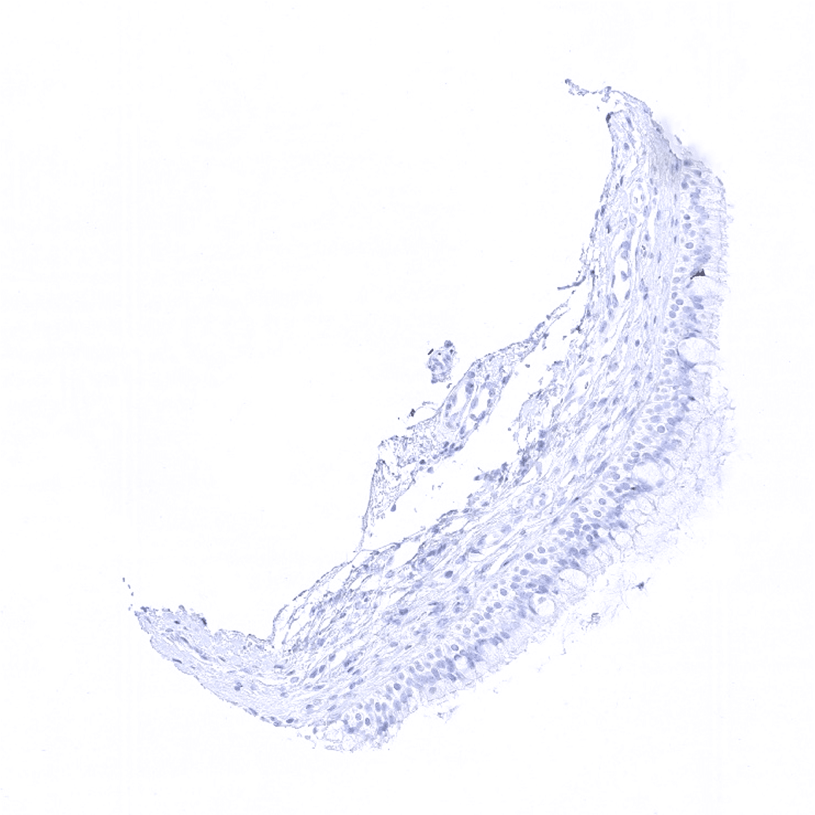
Sinus paranasales
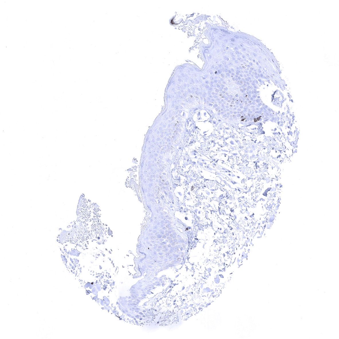
Skin - Few neuroendokrine (Merkel-) cells show chromogranin A immunostaining.
Spleen
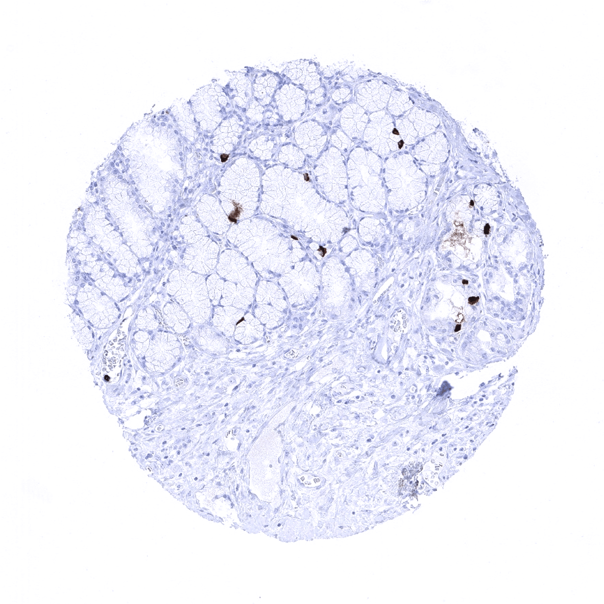
Stomach, antrum
Stomach, corpus
Stomach, corpus - Expression of chromogranin A is so strong in neuroendocrine cells of the stomach, that diffusion of the staining product towards adjacent cells can occur.

Testis
Thymus
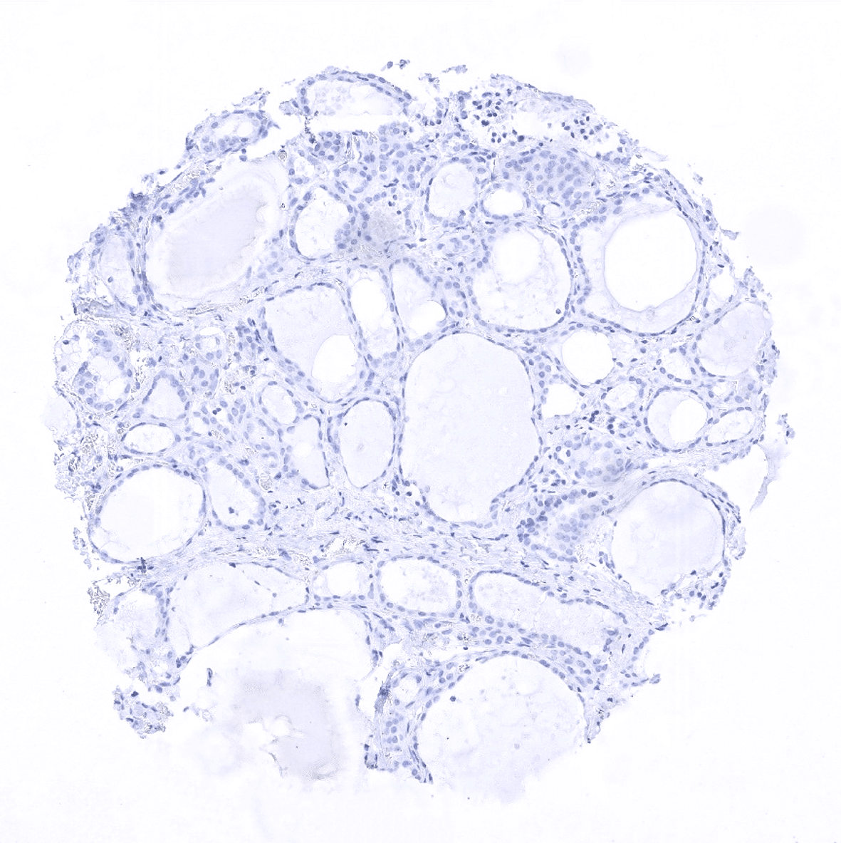
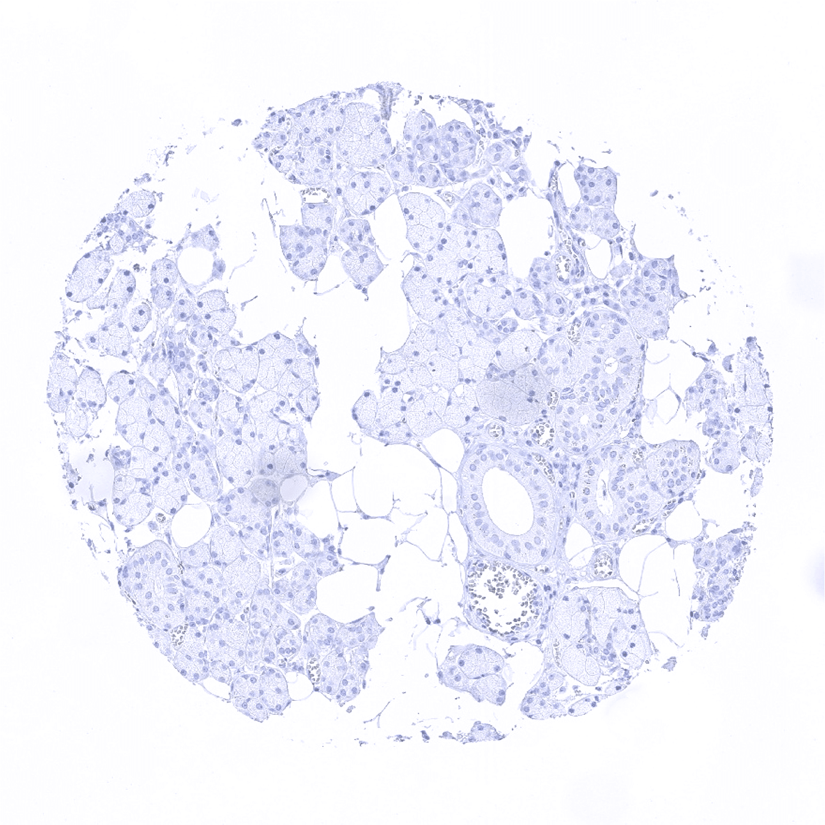
Thyroid gland
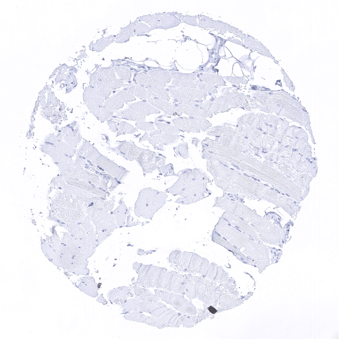
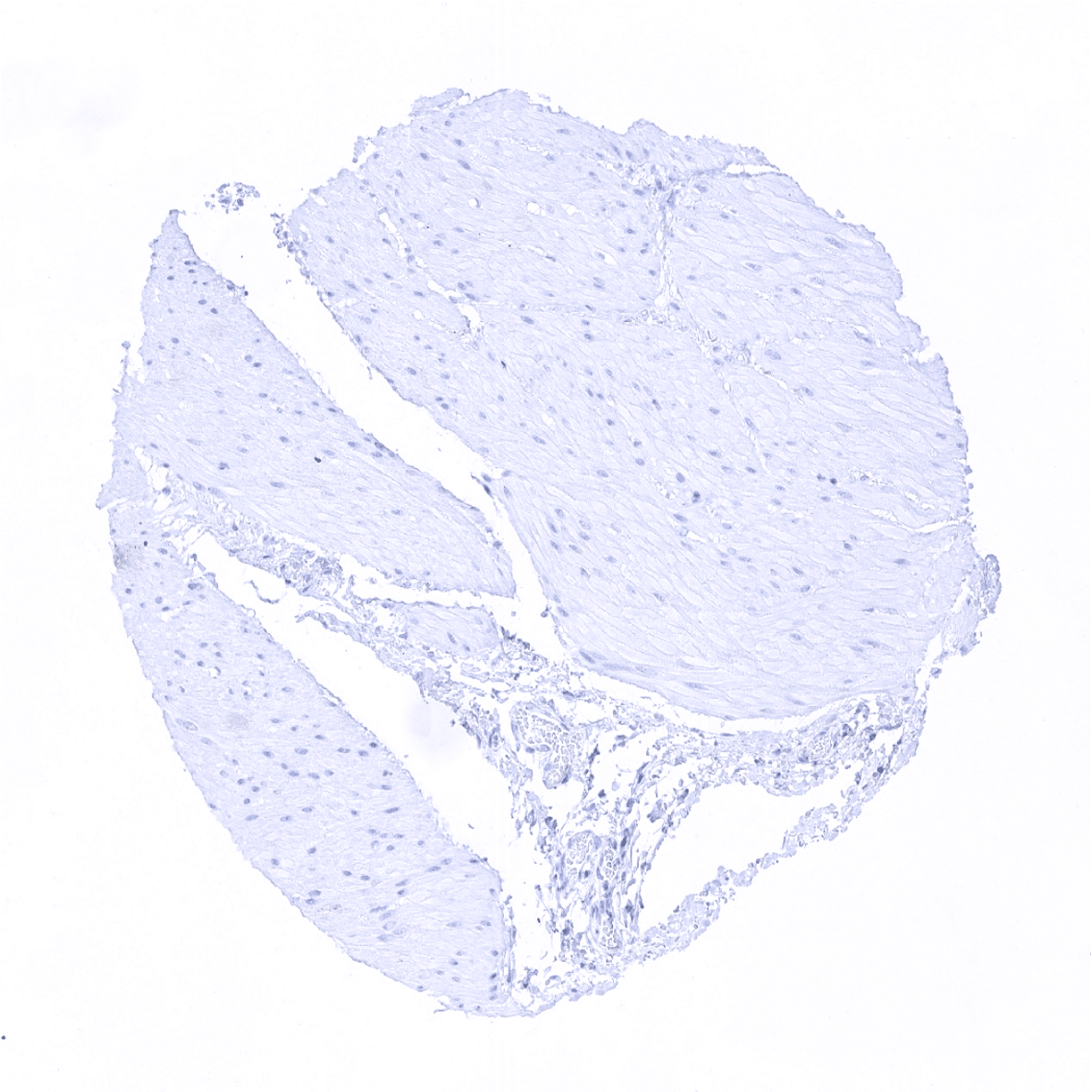
Tongue, muscle
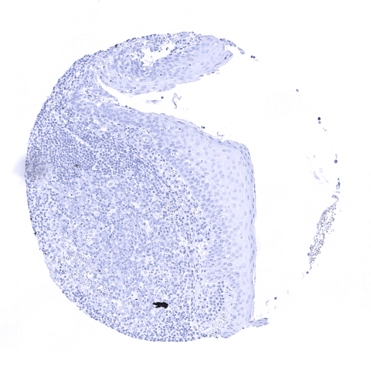
Tonsil, surface epithelium
Tonsil
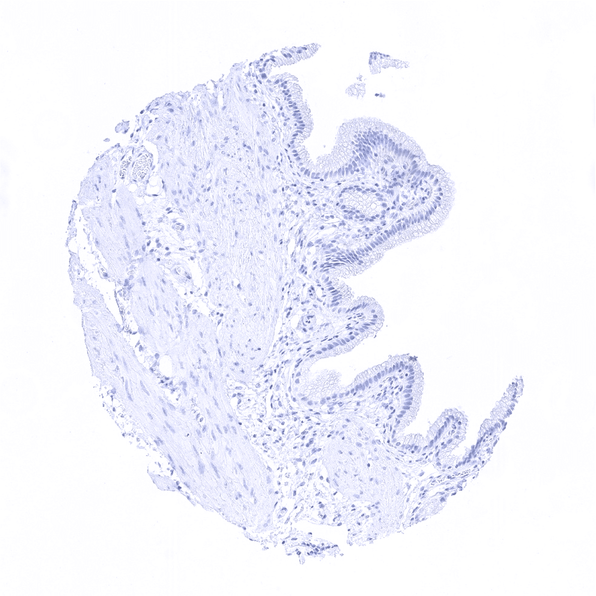
Urinary bladder, muscular wall
Urinary bladder, urothelium
Uterus, myometrium