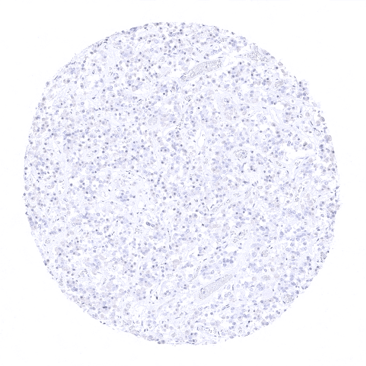
Adrenal gland
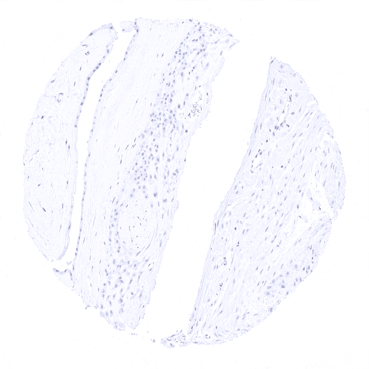
Aorta, media
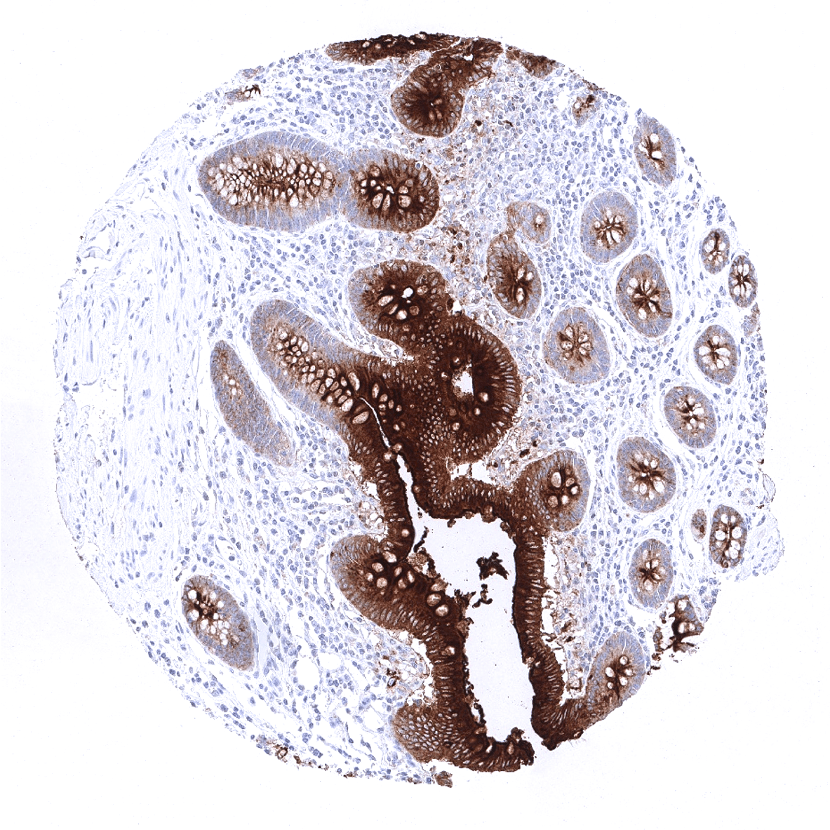
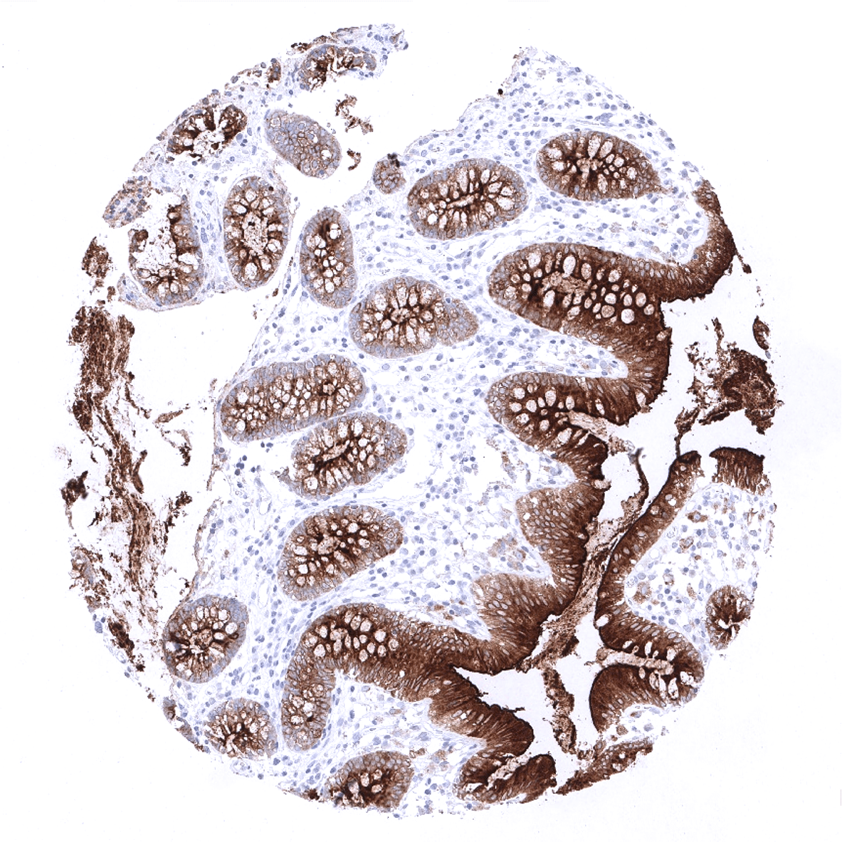
Appendix, mucosa - A strong CEA staining is seen in epithelial cells of the colon mucosa. The intensity is highest in the surface epithelium.
Appendix, muscular wall
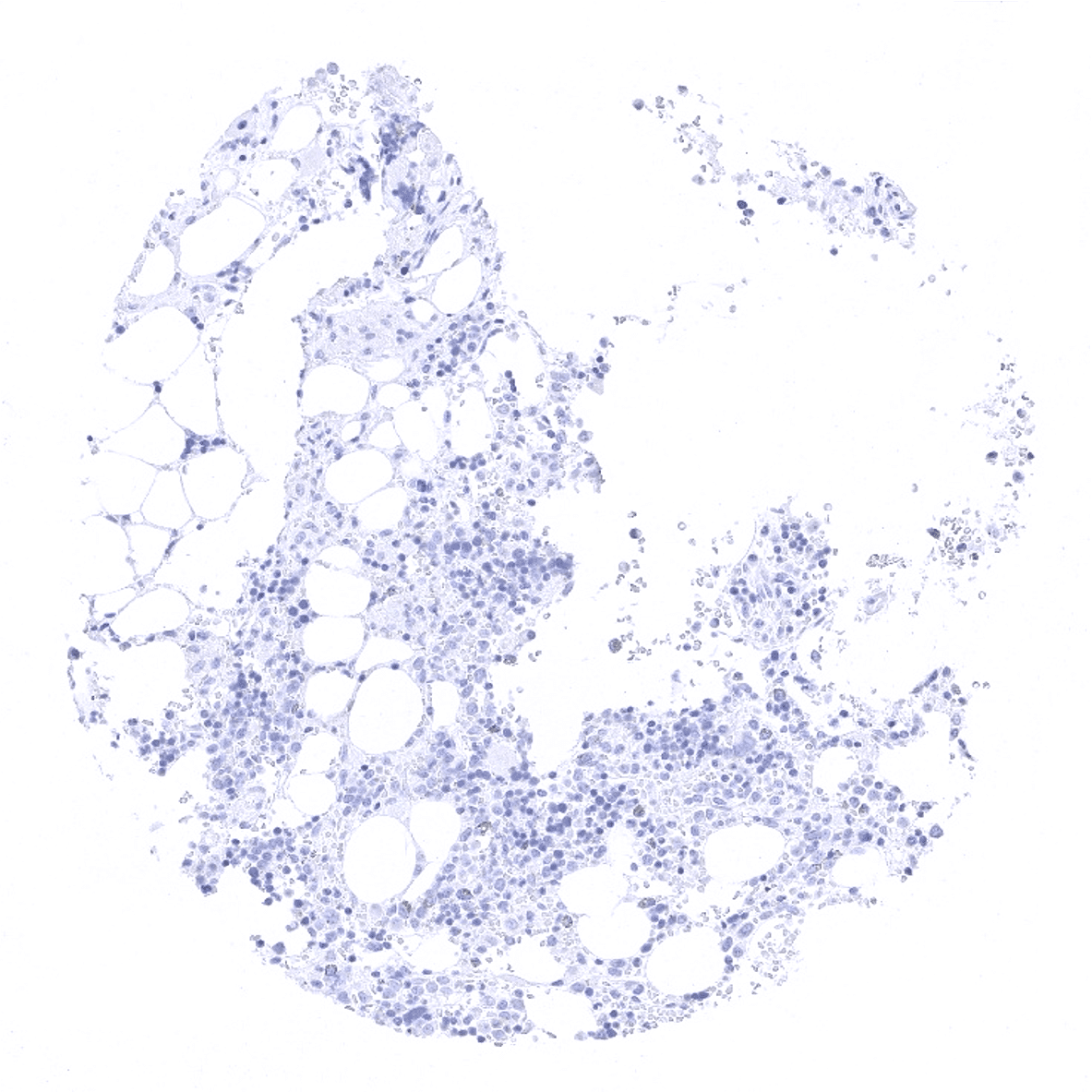
Bone marrow
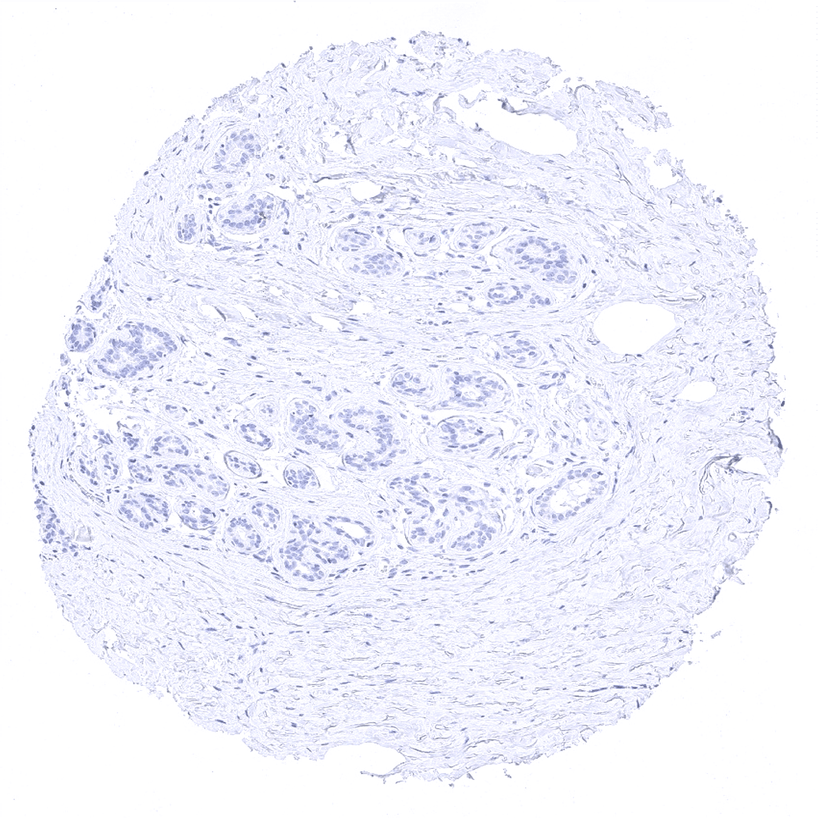
Breast
Bronchus, mucosa - Few CEA positive cells occur in respiratory epithelium.

Cerebellum (molecular layer, Purkinje cell layer, granule cell layer)
Cerebellum (molecular layer, Purkinje cell layer, granule cell layer, white matter)
Cerebellum, grey matter
Cerebellum, white matter

Colon descendens, mucosa - A strong CEA staining is seen in epithelial cells of the colon mucosa. The intensity is highest in the surface epithelium.
Colon descendens, muscular wall
Duodenum, Brunner gland

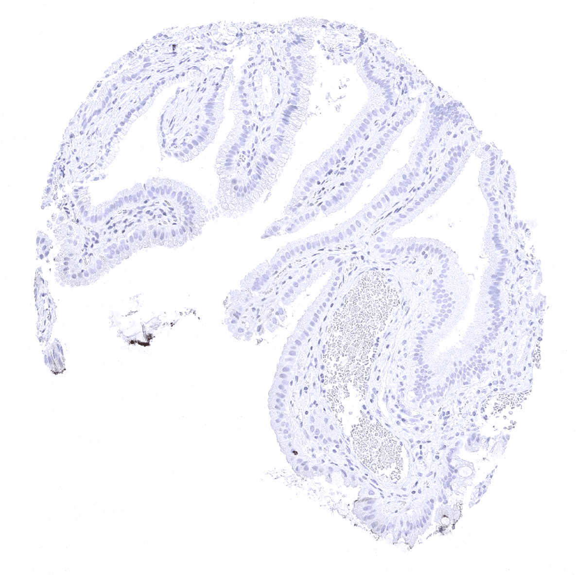
Duodenum, mucosa
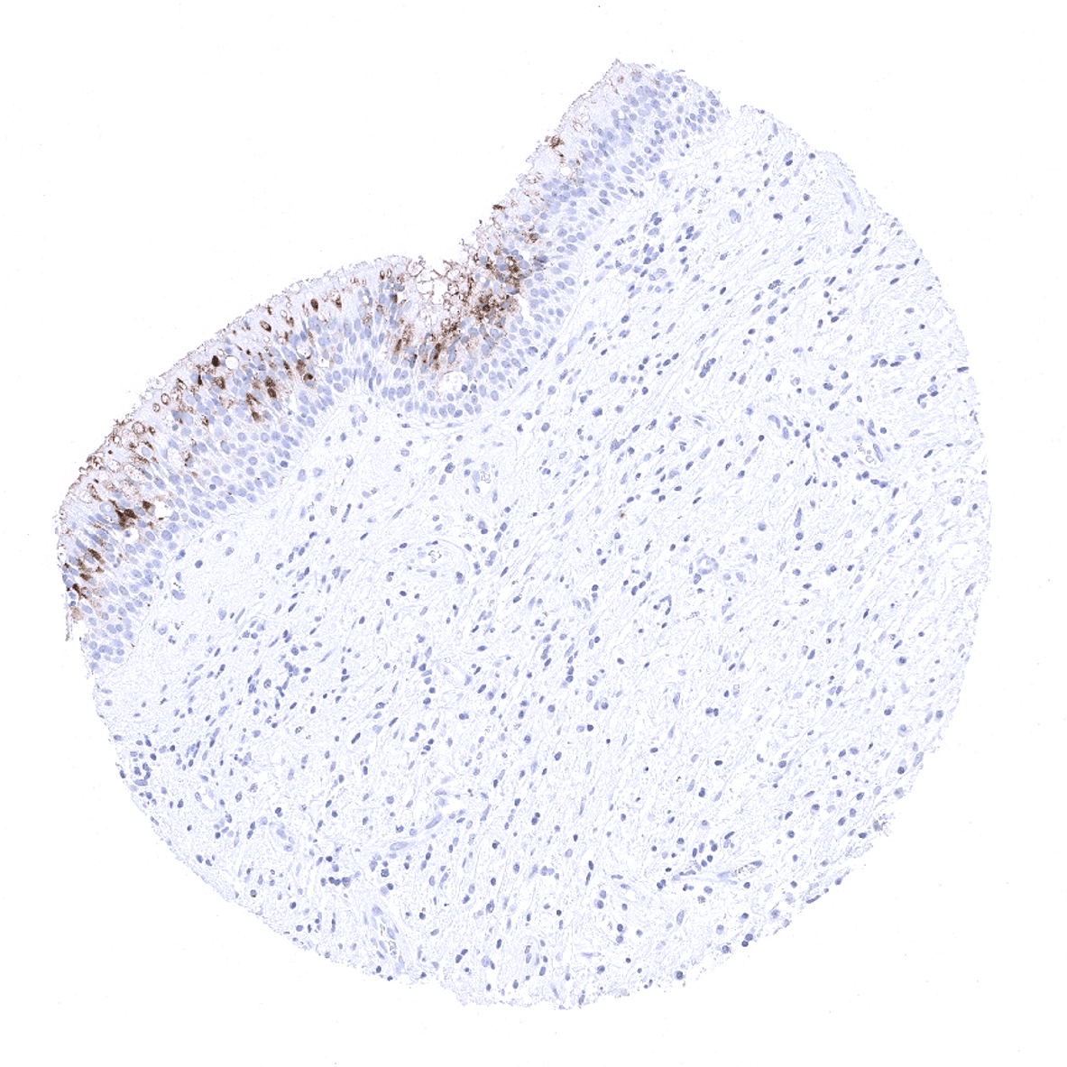
Ectocervix - A moderate to strong CEA staining occurs in the superficial cell layers of the squamous epithelium of the ectocervix.
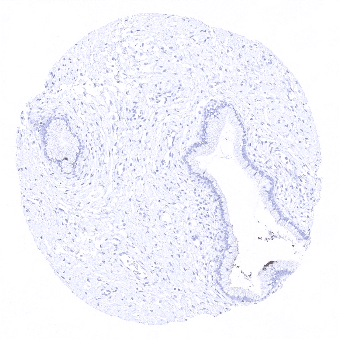
Endocervix - A focal CEA staining is seen in this sample of the endocervix.
Endocervix - Complete lack of CEA immunostaining.
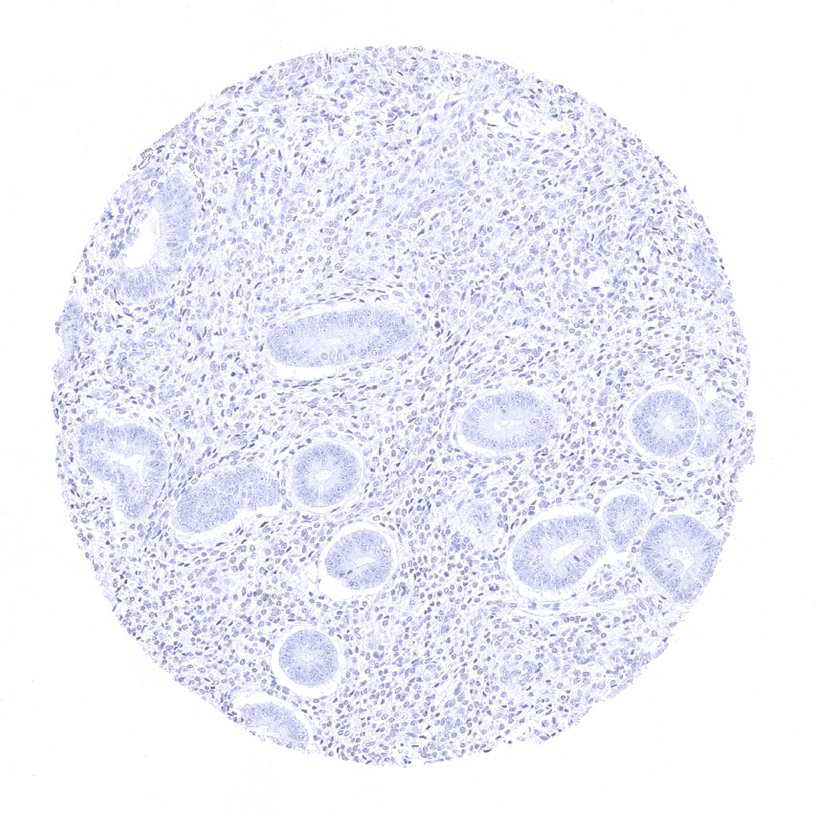
Endometrium, proliferation
Endometrium, secretion
Epididymis
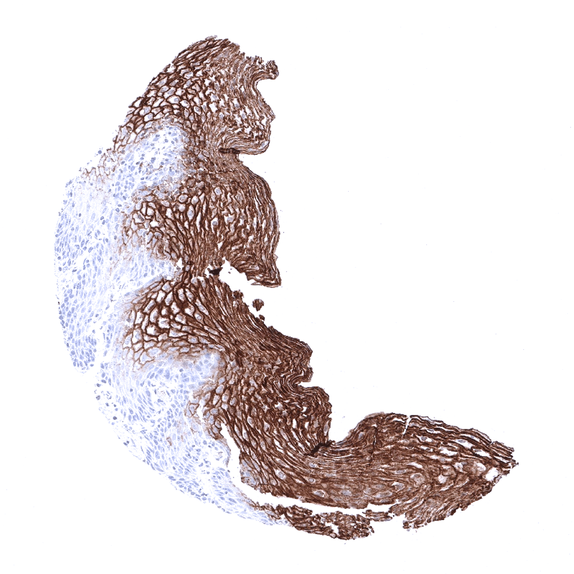
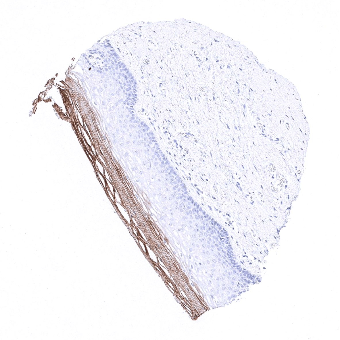
Esophagus, squamous epithelium - A moderate to strong CEA staining occurs in the superficial cell layers of the squamous epithelium of the esophagus.
Fallopian tube, mucosa
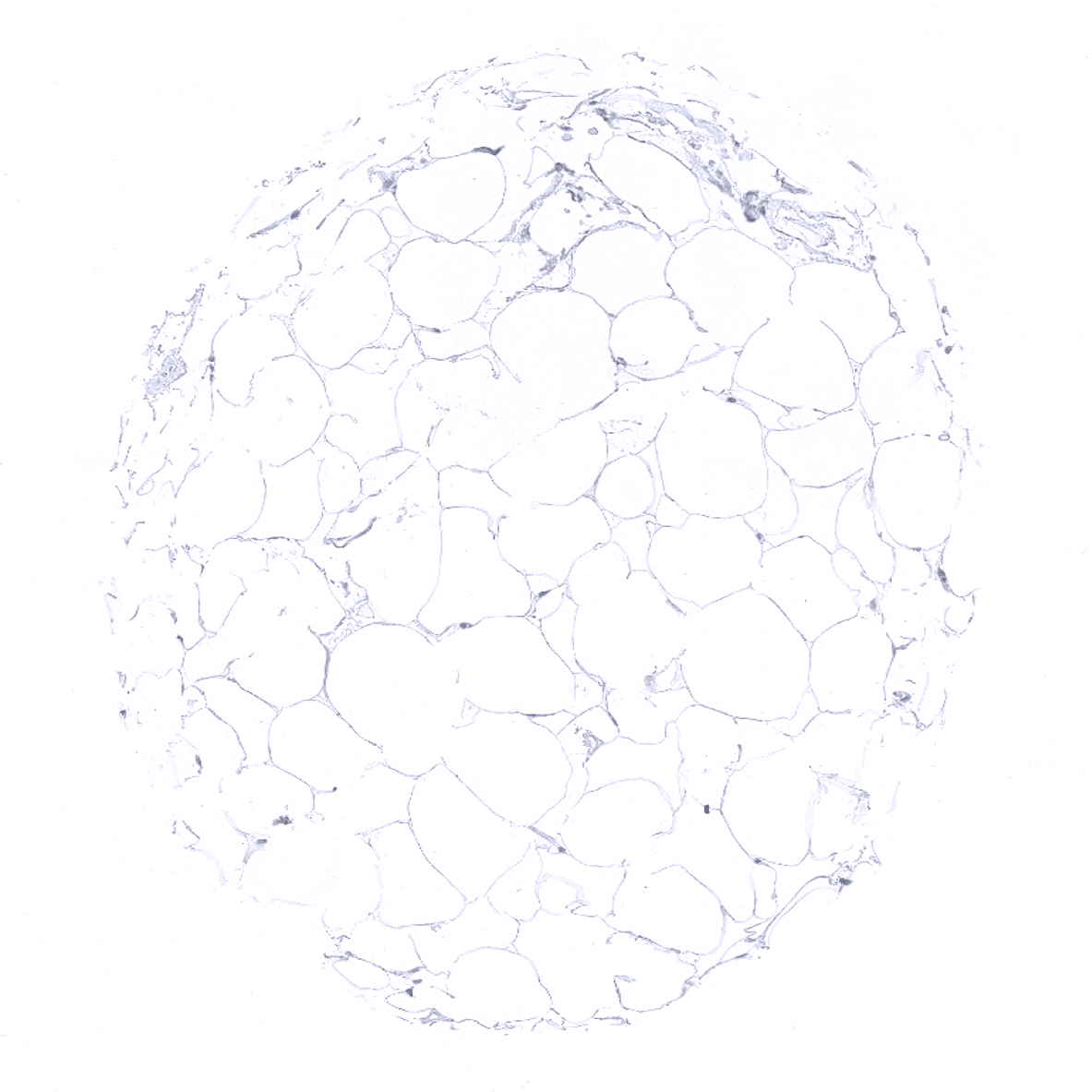
Fat
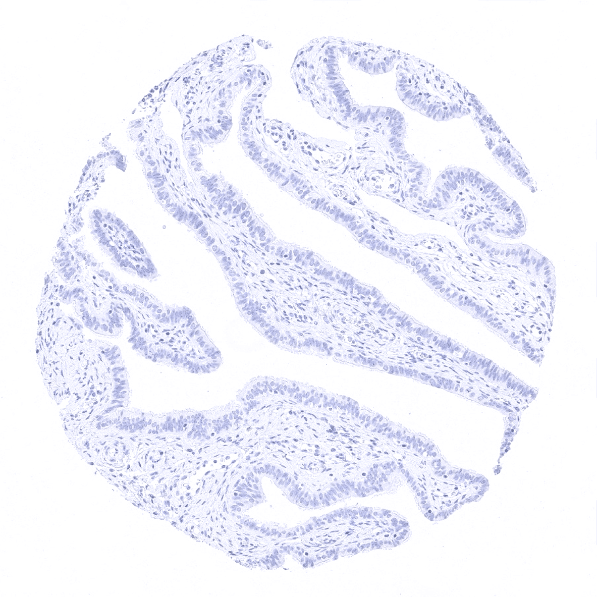
Gallbladder, epithelium
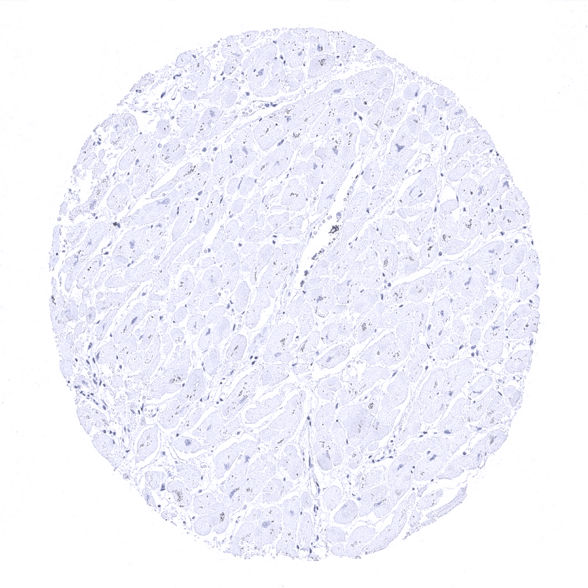
Heart
Ileum, mucosa - A strong CEA staining is seen in epithelial cells of the ileum mucosa. The intensity is highest in the surface epithelium.
Ileum, mucosa - CEA staining is moderate in epithelial cells of this sample of the ileum mucosa.
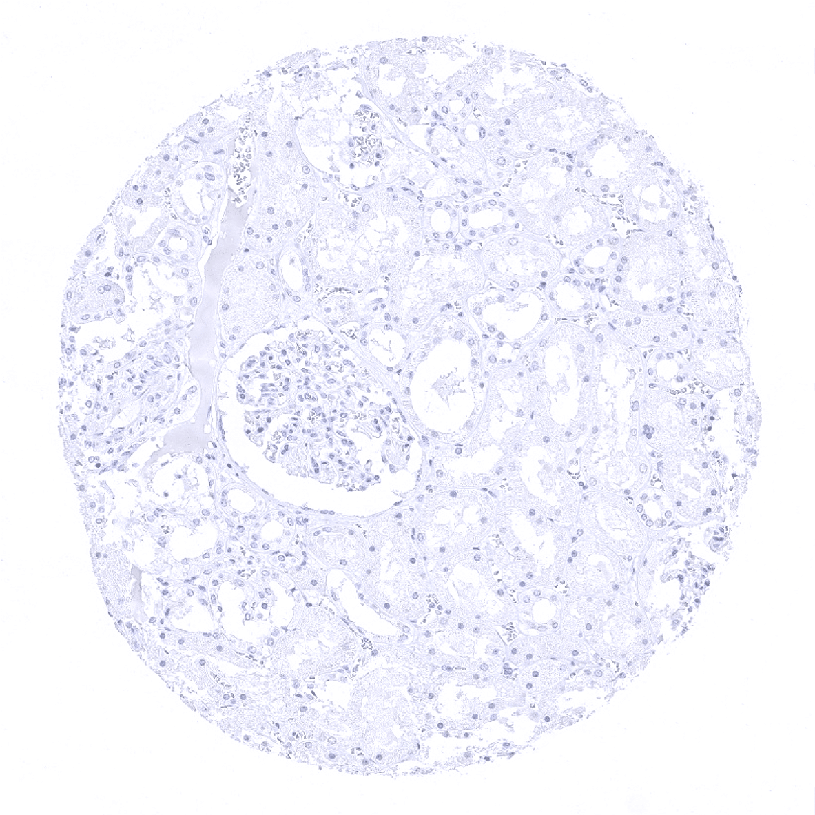
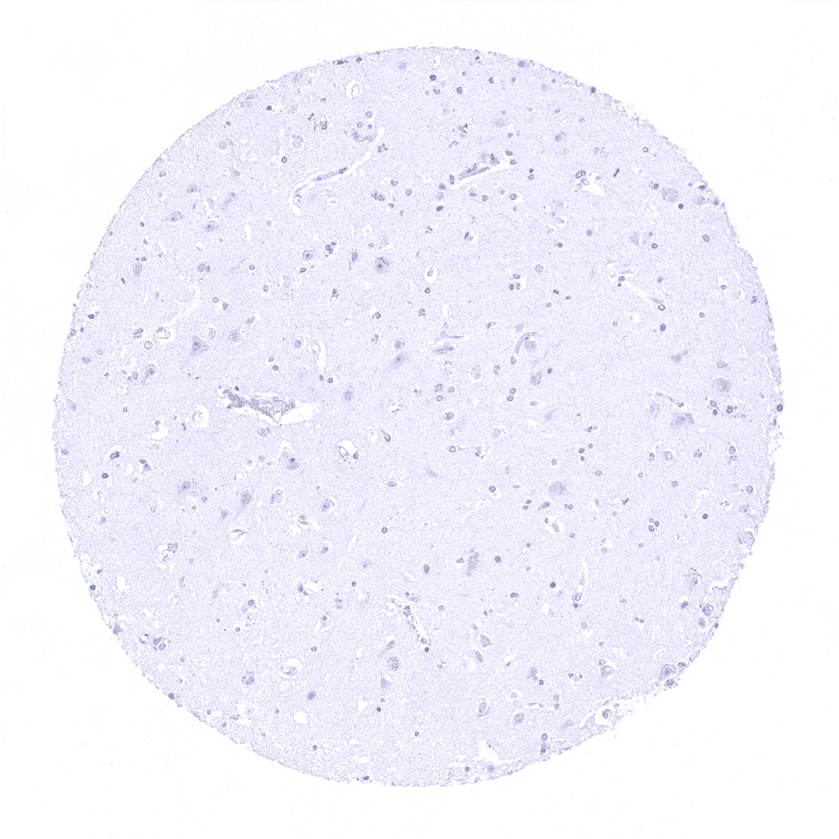
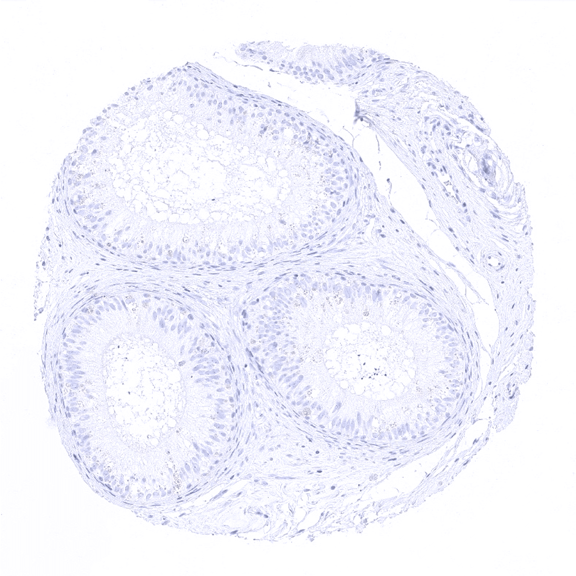
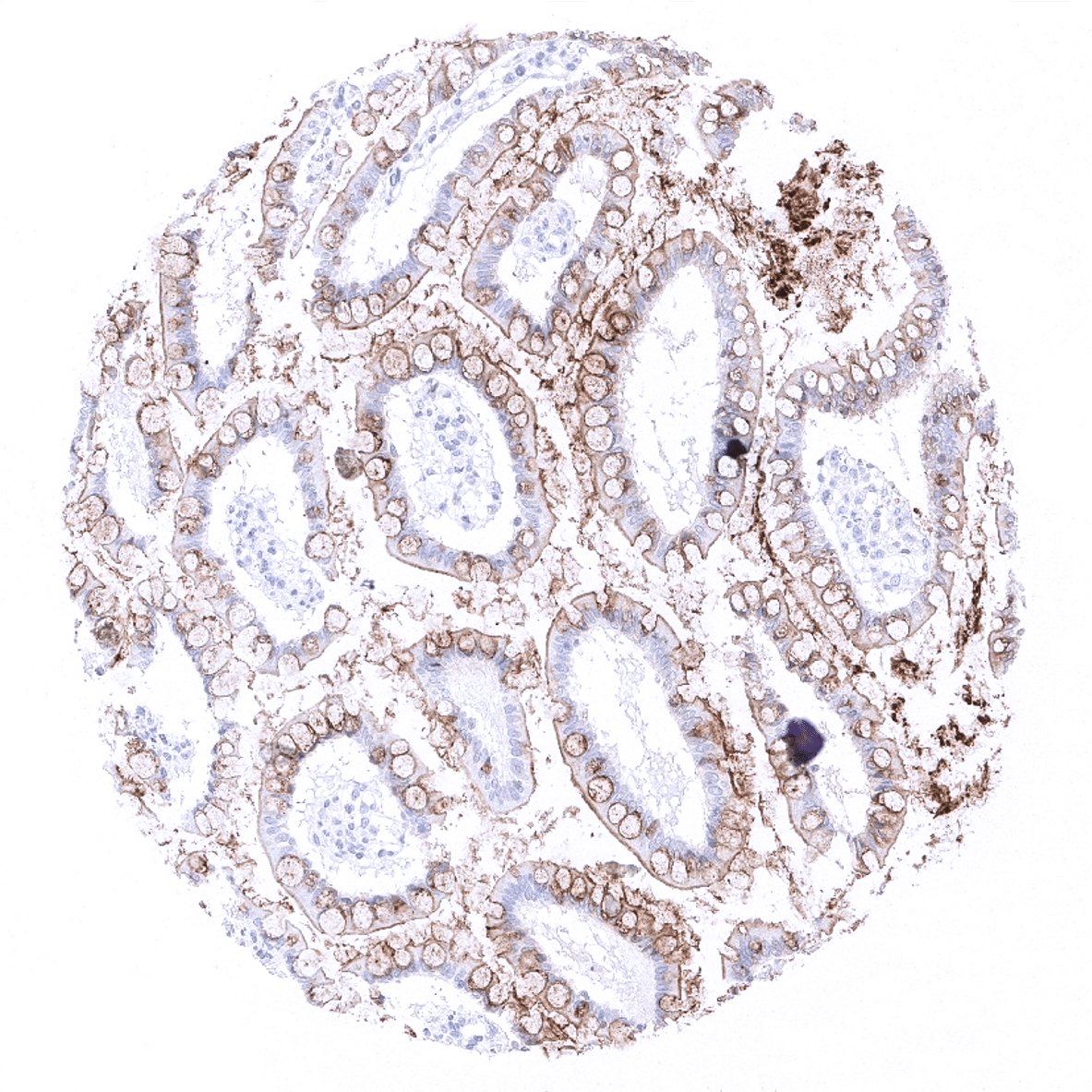
Kidney, cortex - CEA immunostaining is lacking in the kidney.
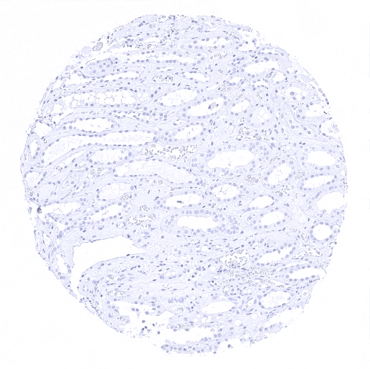
Kidney, medulla
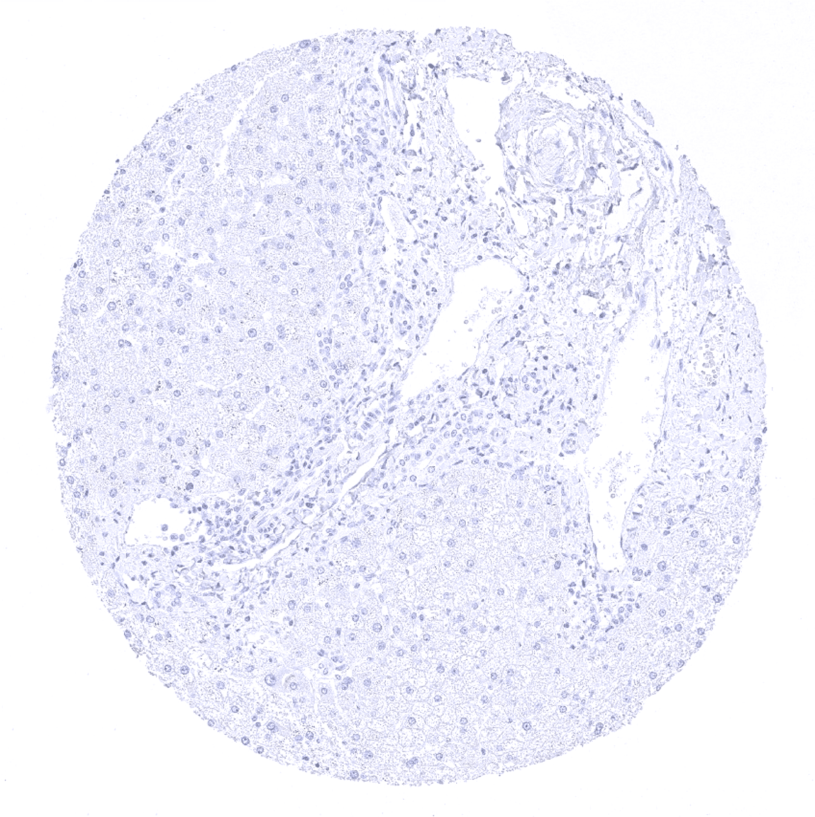
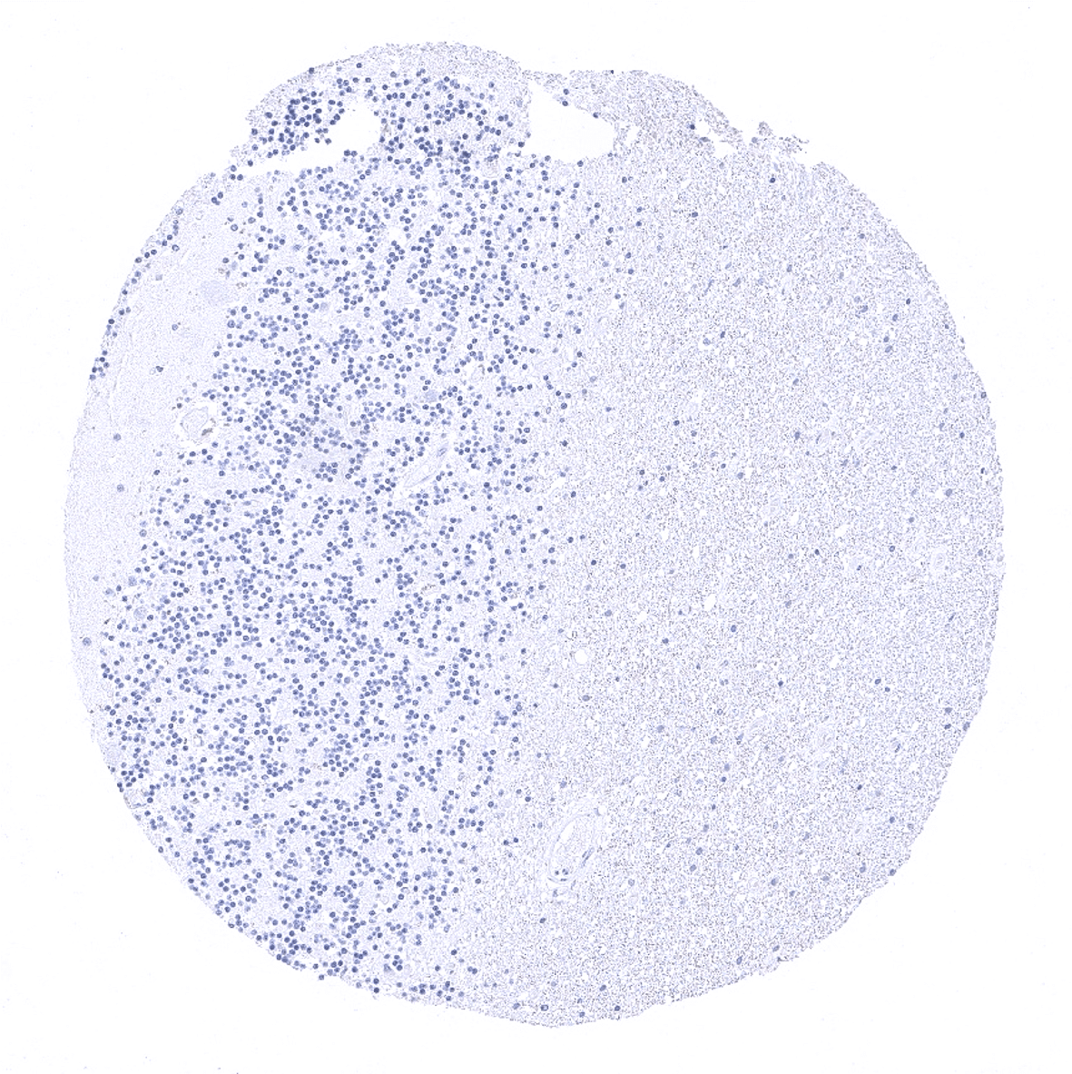
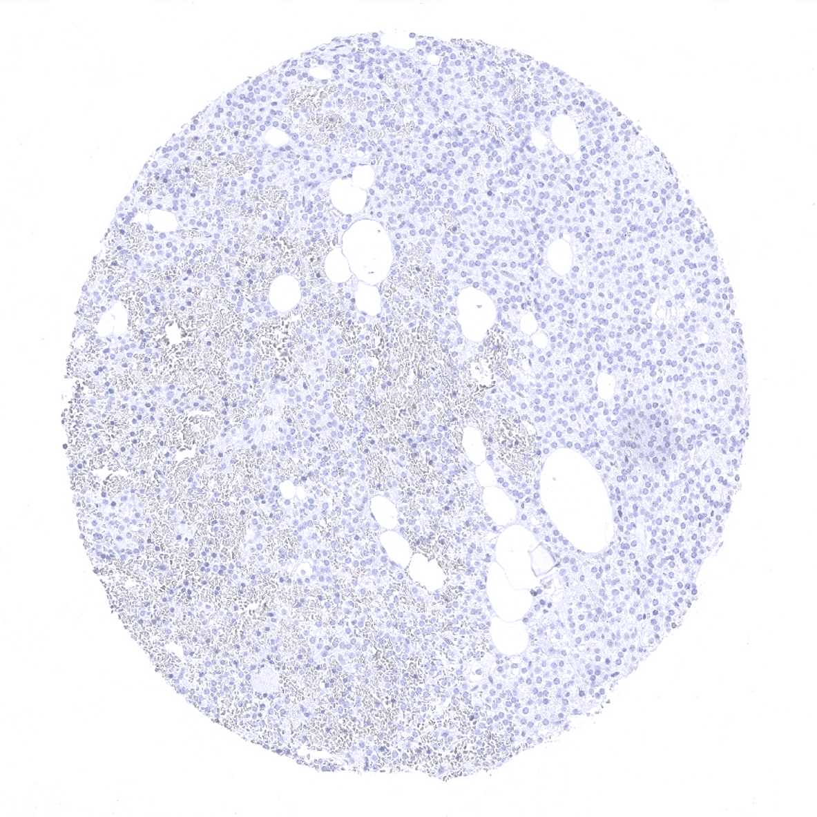
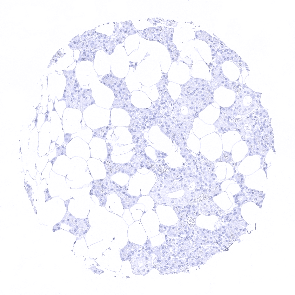
Liver
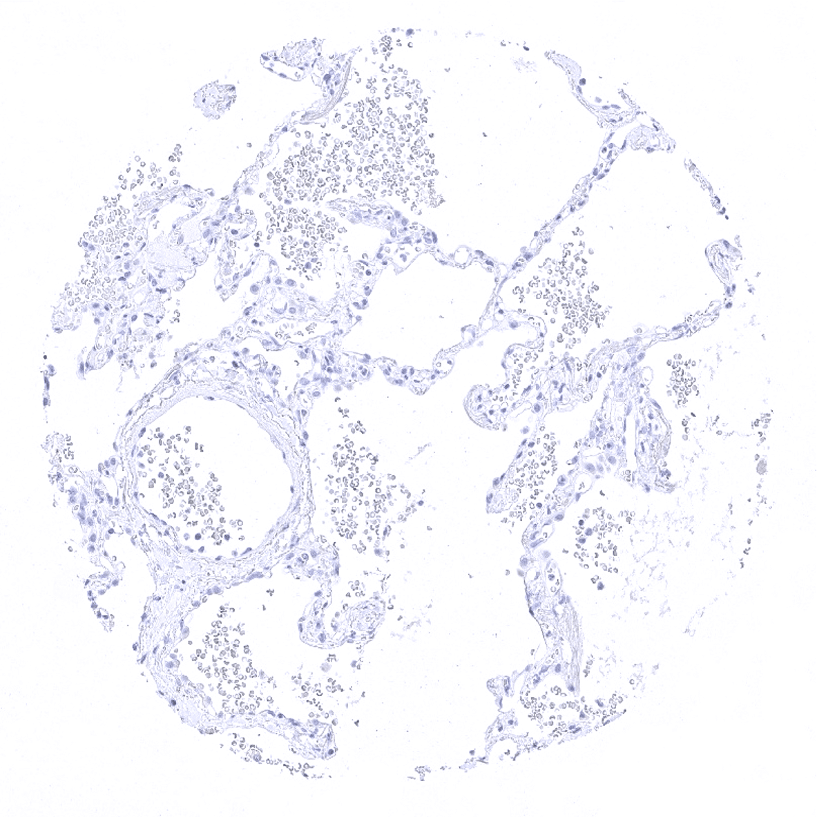
Lung

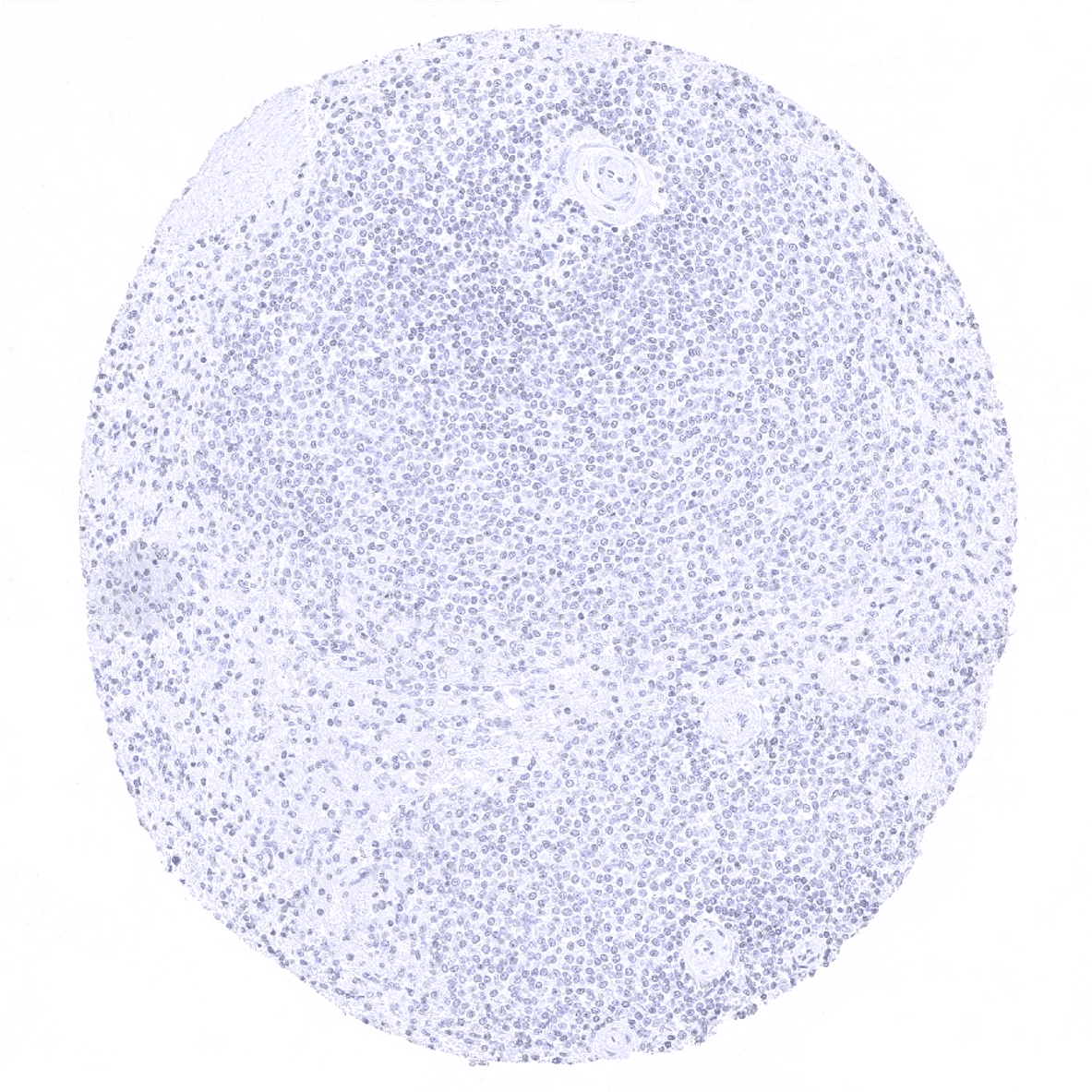
Lymph node
Ovary, stroma
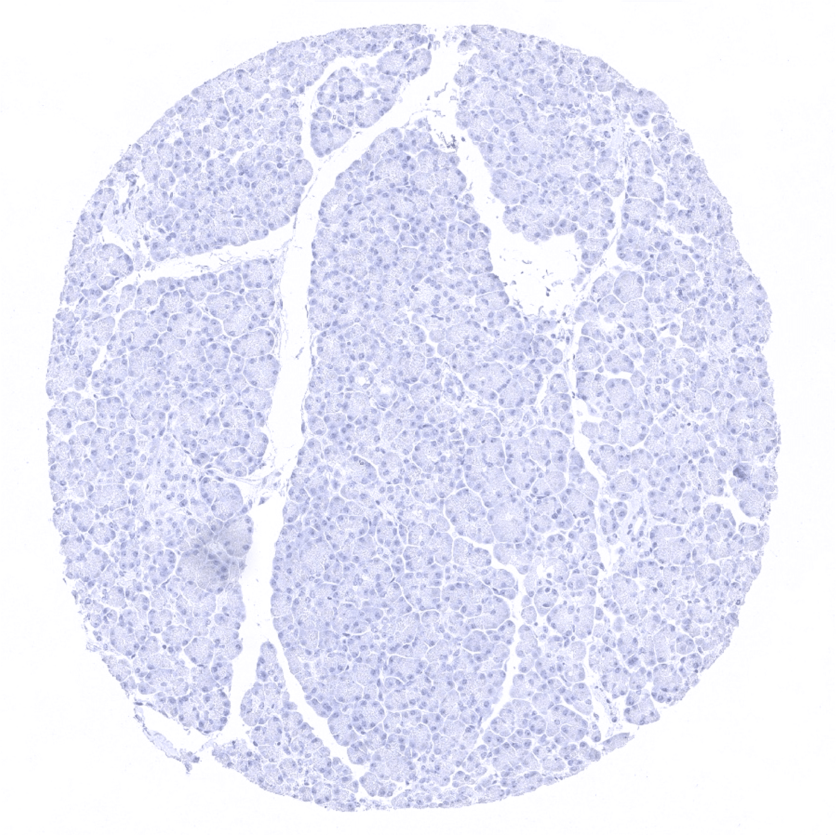
Pancreas
Parathyroid
Parotid gland
Pituitary gland, anterior lobe
Pituitary gland, posterior lobe

Pregnant uterus (decidua)
Placenta early
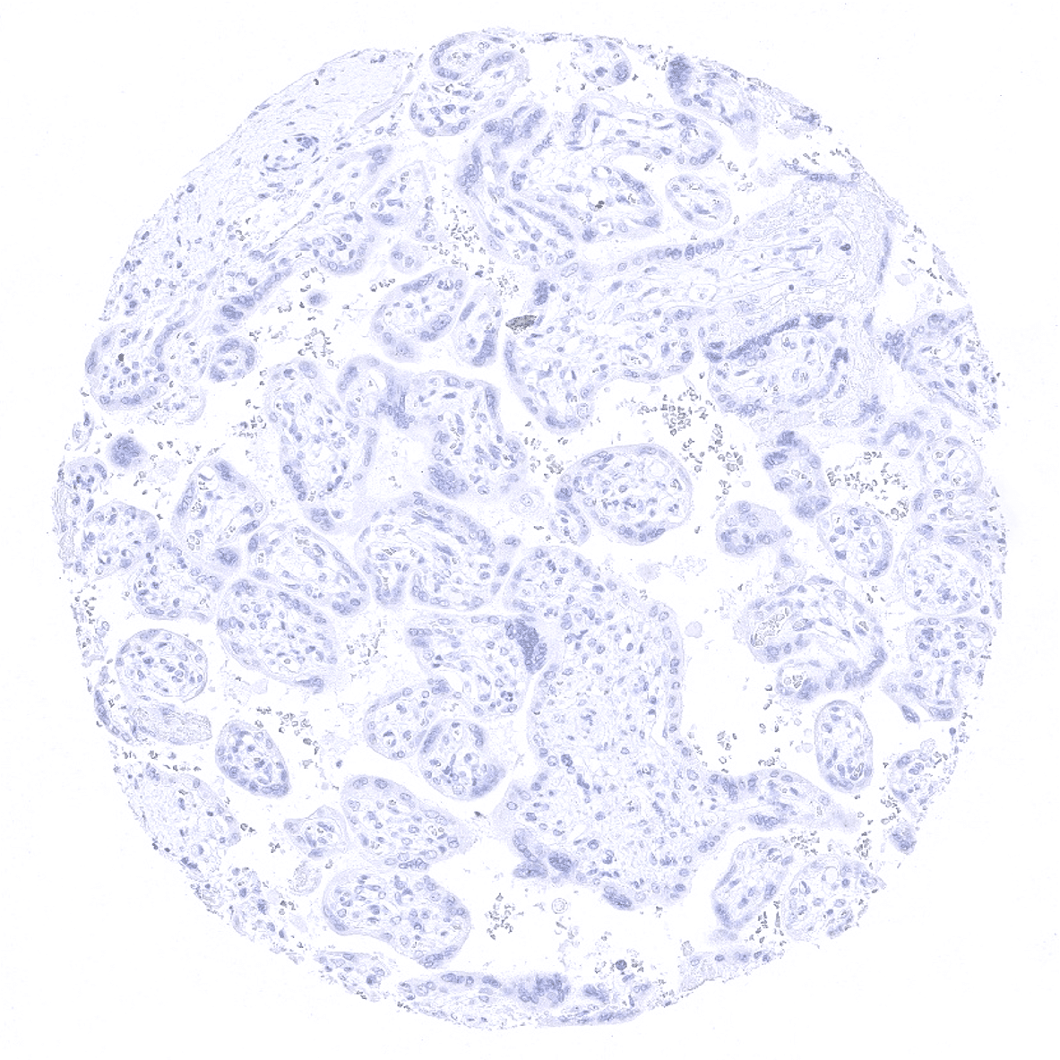
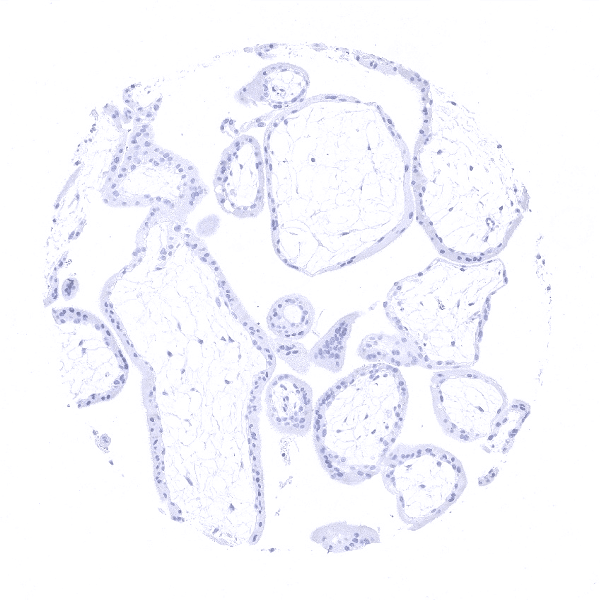
Placenta mature
Placenta (amnion and chorion)

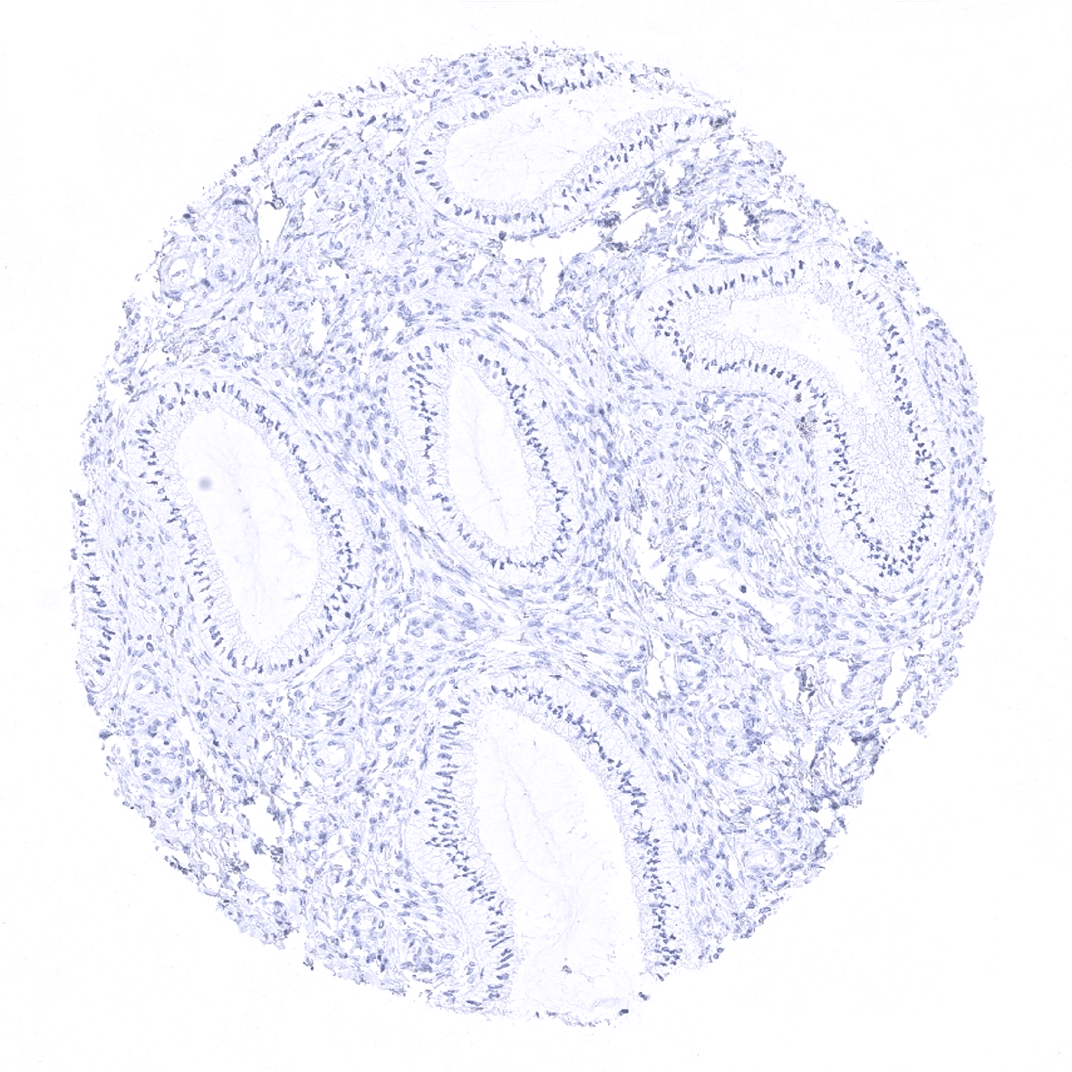
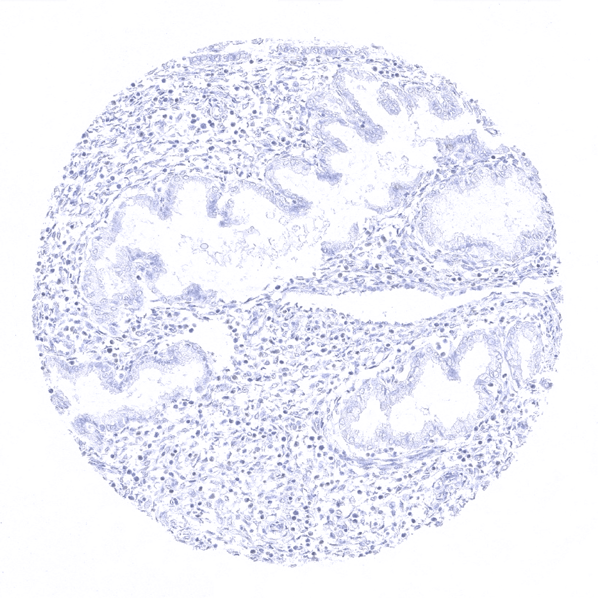
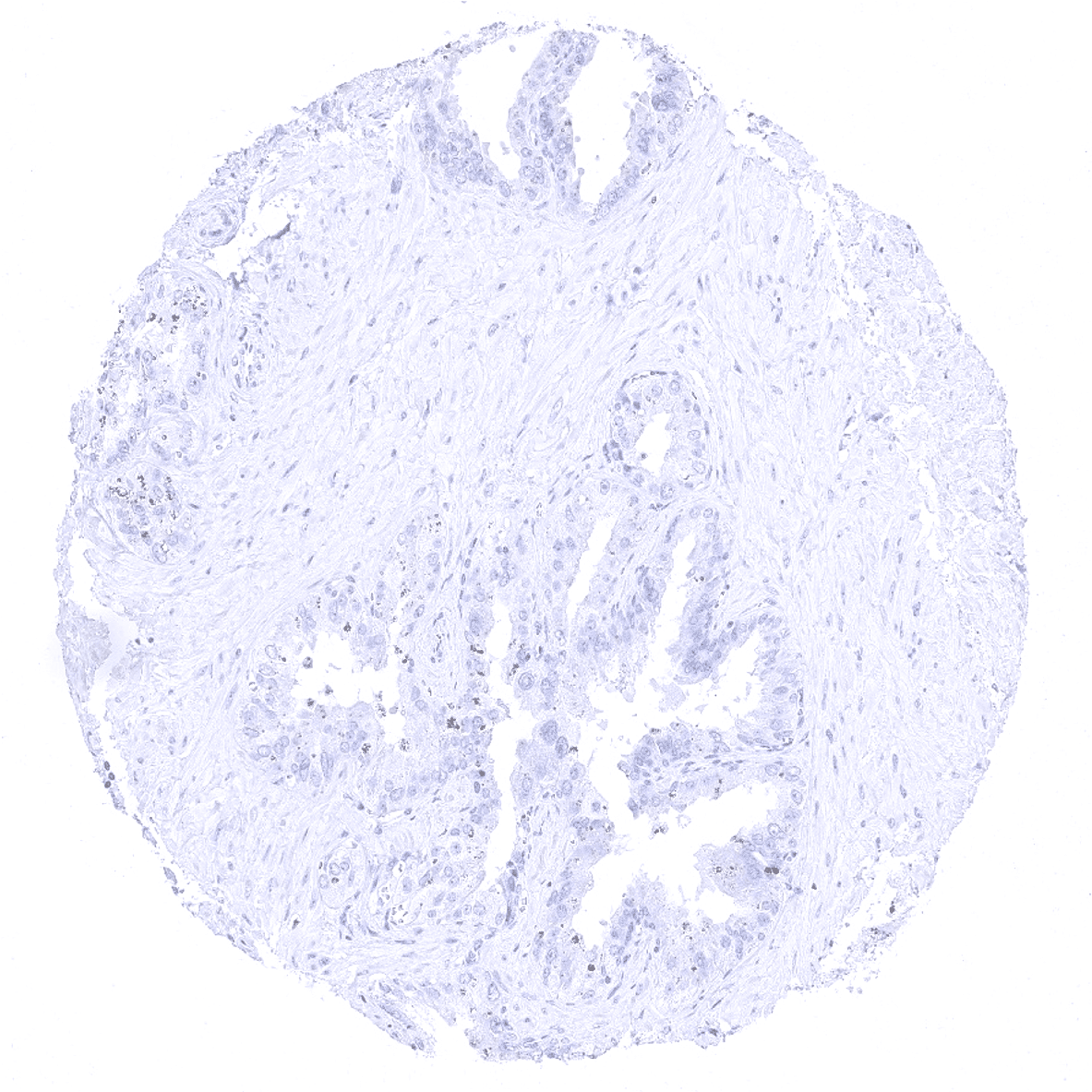
Prostate
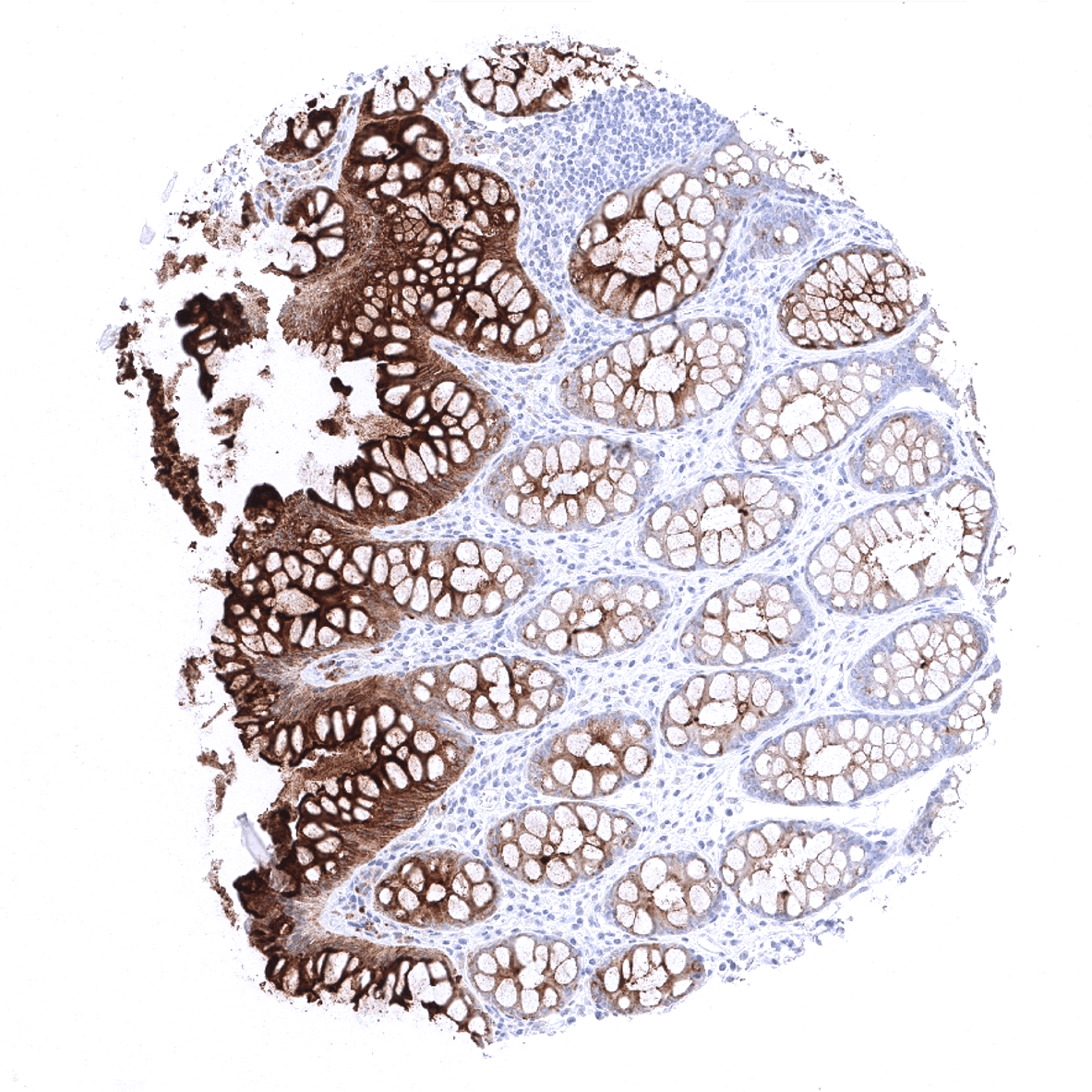
Rectum, mucosa - A strong CEA staining is seen in epithelial cells of the rectum mucosa. The intensity is highest in the surface epithelium.
Seminal vesicle
Sinus paranasales
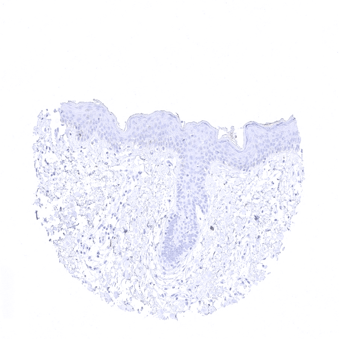
Skin
Spleen
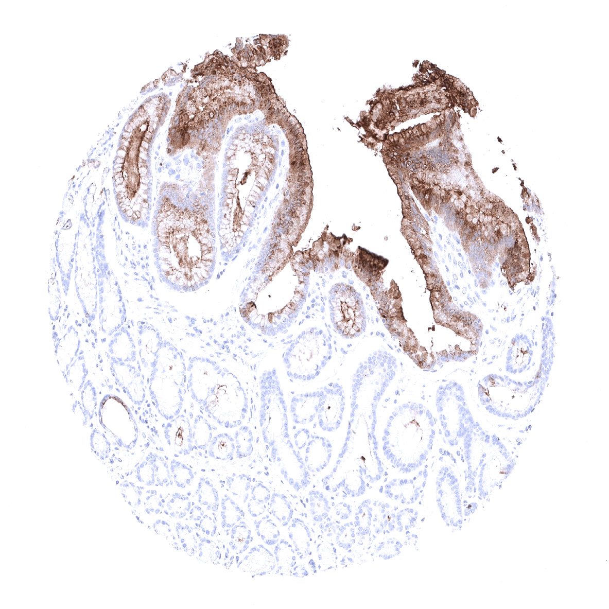
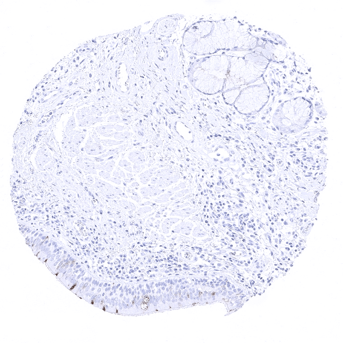
Stomach, antrum - A strong CEA staining is seen in the surface epithelial cells of the stomach.

Stomach, corpus - A strong CEA staining is seen in the surface epithelial cells of the stomach.
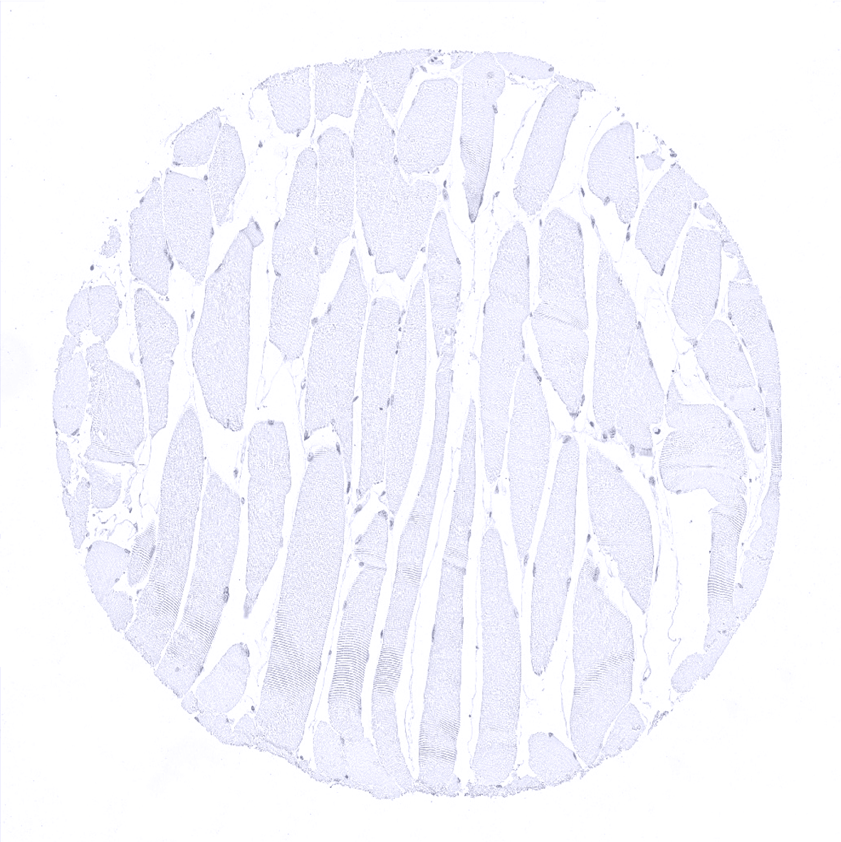
Striated muscle

Testis
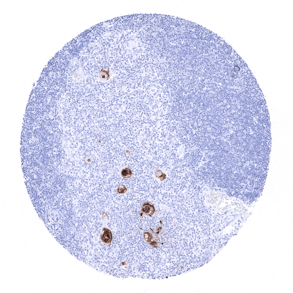
Thymus - A strong CEA staining is seen in corpuscles of Hassall's of the thymus.
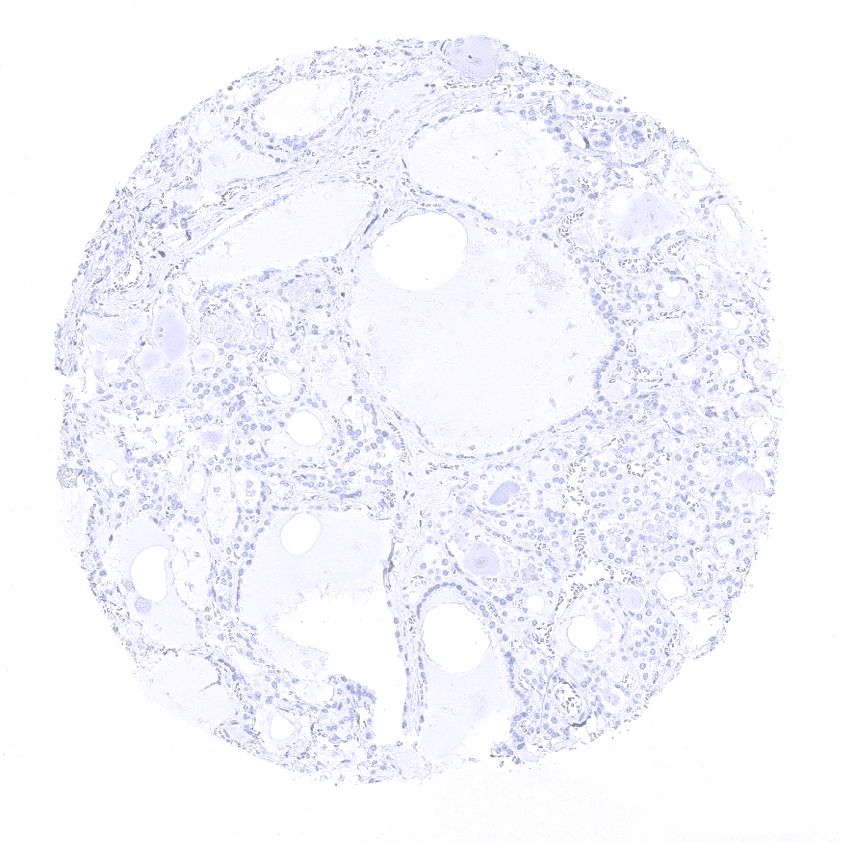
Thyroid
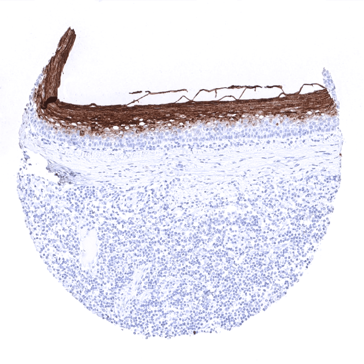
Tonsil, surface epithelium
Urinary bladder, muscular wall
Urinary bladder, urothelium
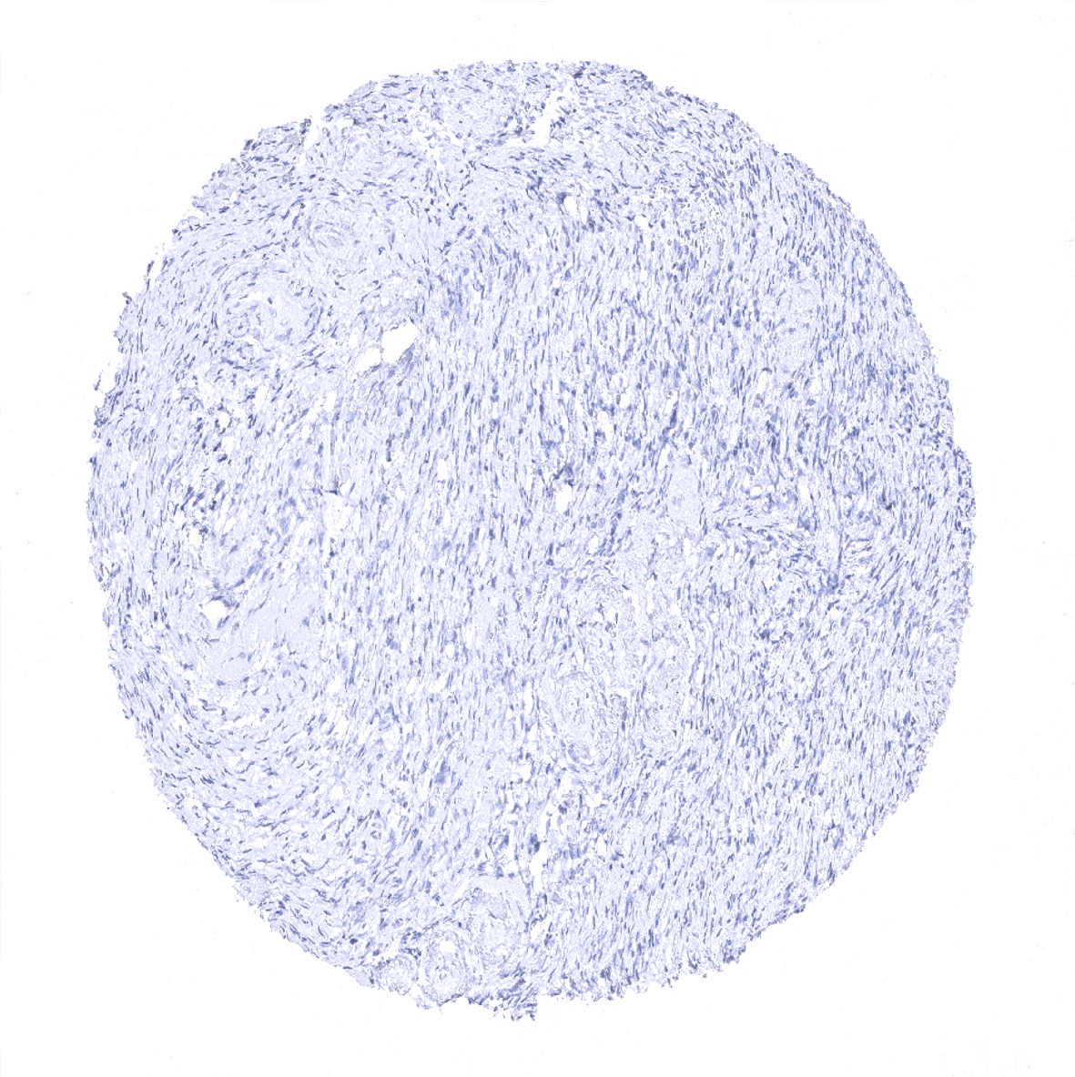
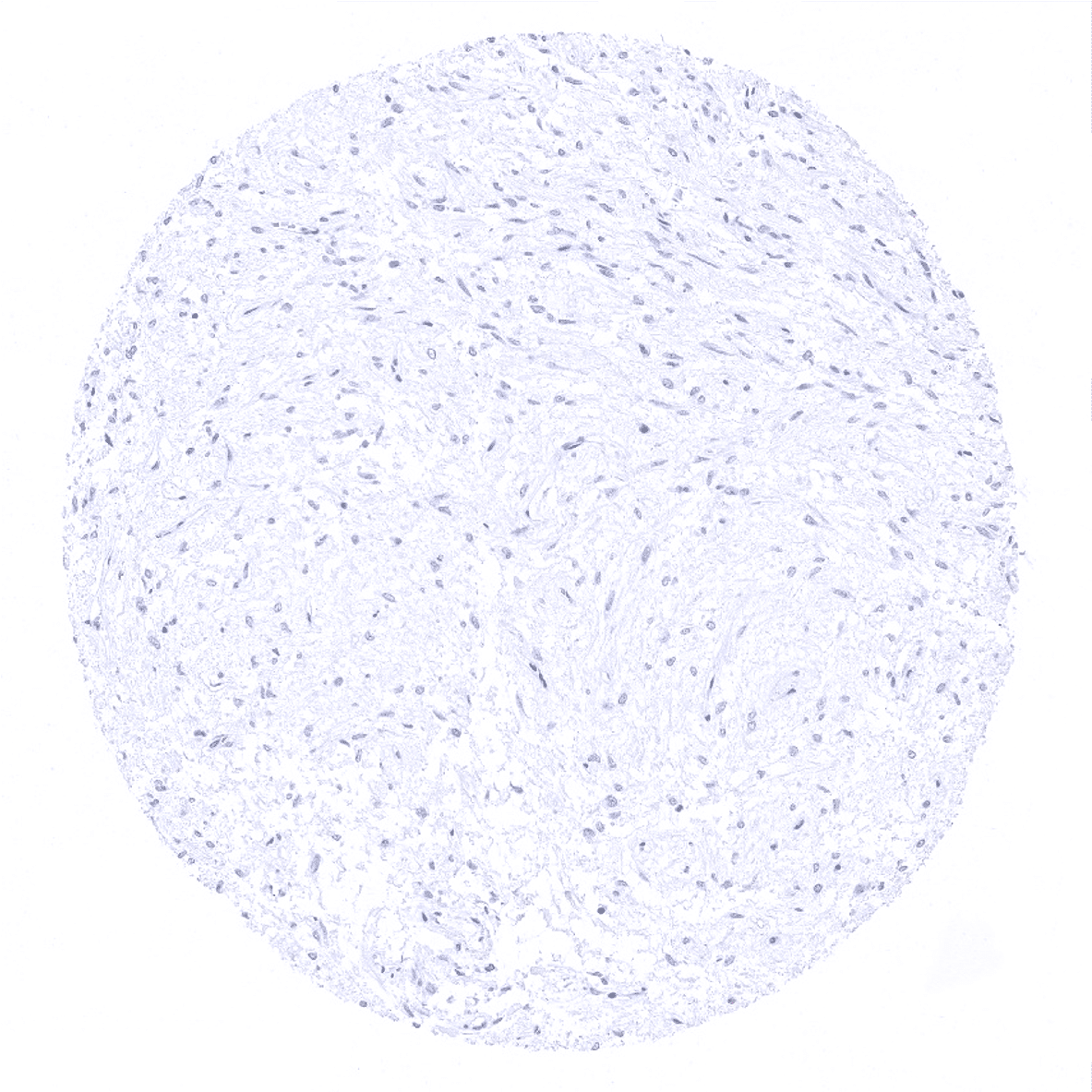
Uterus, myometrium