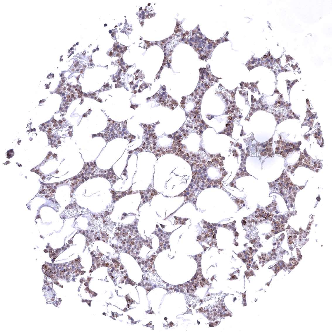
Adrenal gland – Cytoplasmic ATP5J staining occurs in all cell types. It is particularly strong in adrenocortical cells
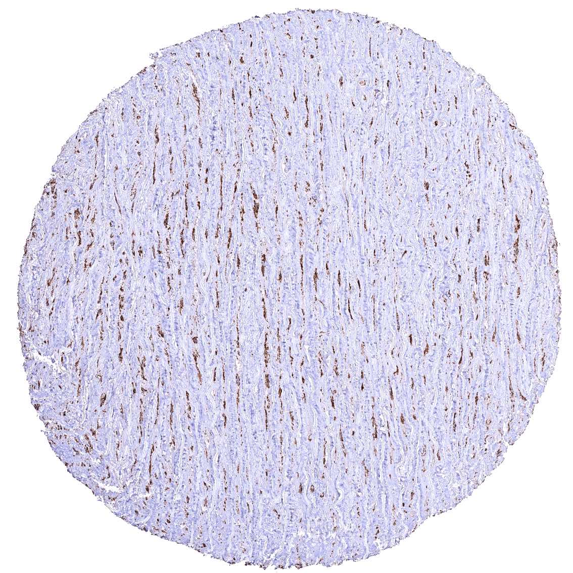
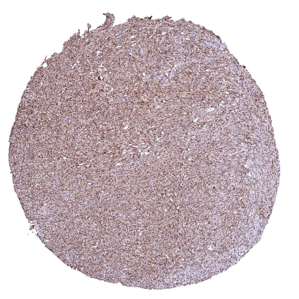
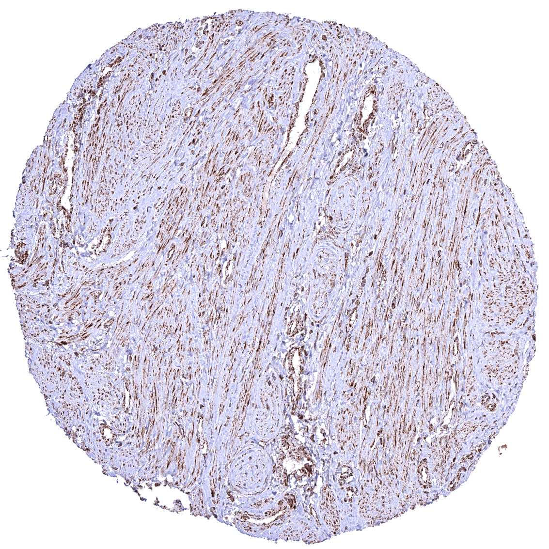
Aorta, media
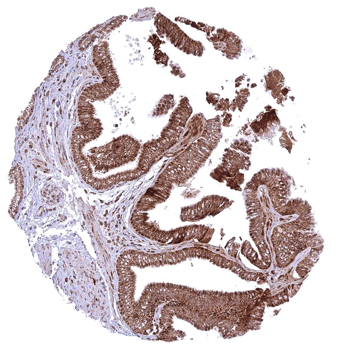
Appendix, mucosa
Appendix, muscular wall
Bone marrow – Cytoplasmic ATP5J staining of variable intensity in all cell types.
Breast
Bronchus, mucosa – Cytoplasmic ATP5J staining occurs in all cell types. A characteristic granular cytoplasmic staining is seen below the apical membranes.
Bronchus, mucosa
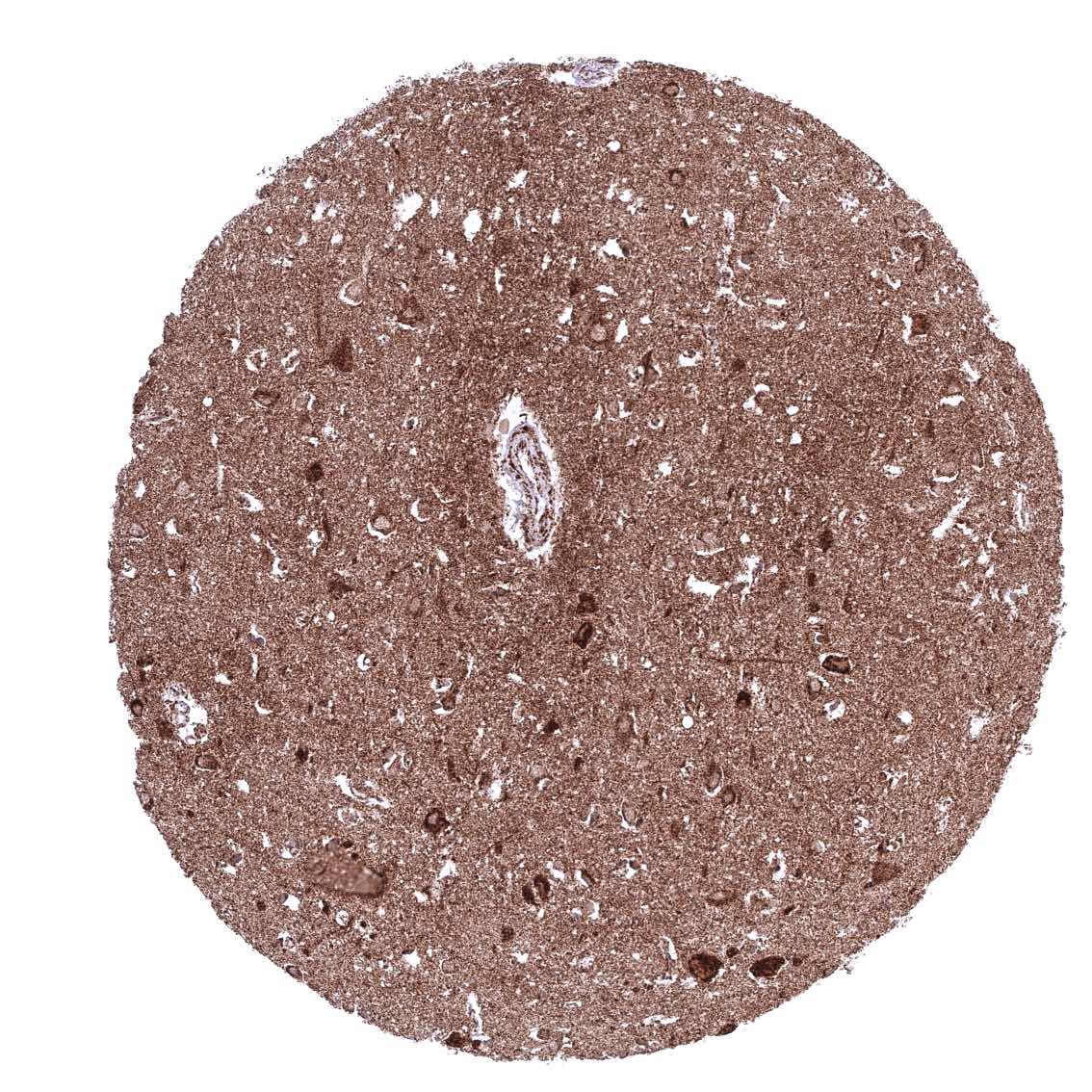
Cerebellum (molecular layer, Purkinje cell layer, granule cell layer, white matter)
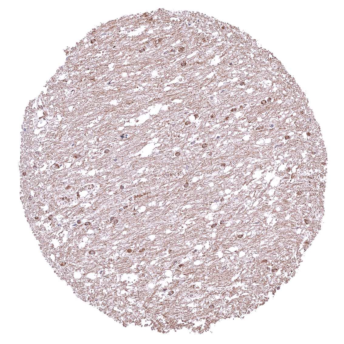
Cerebellum (white matter)
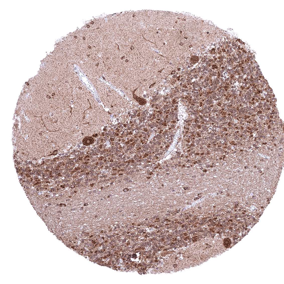
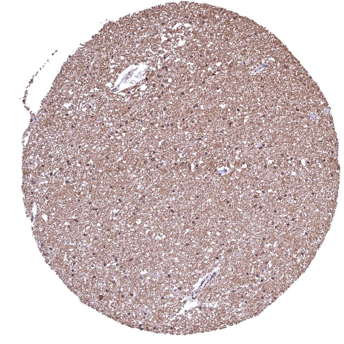
Cerebrum, grey matter
Cerebrum, white matter
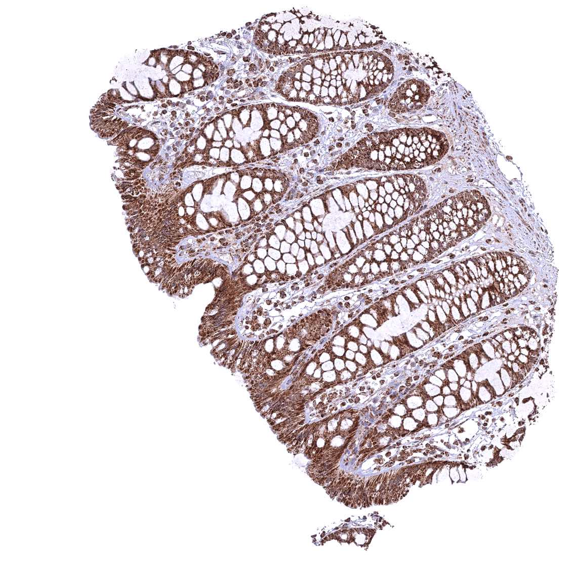
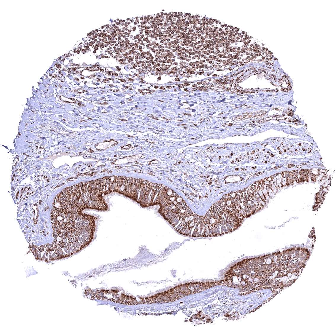
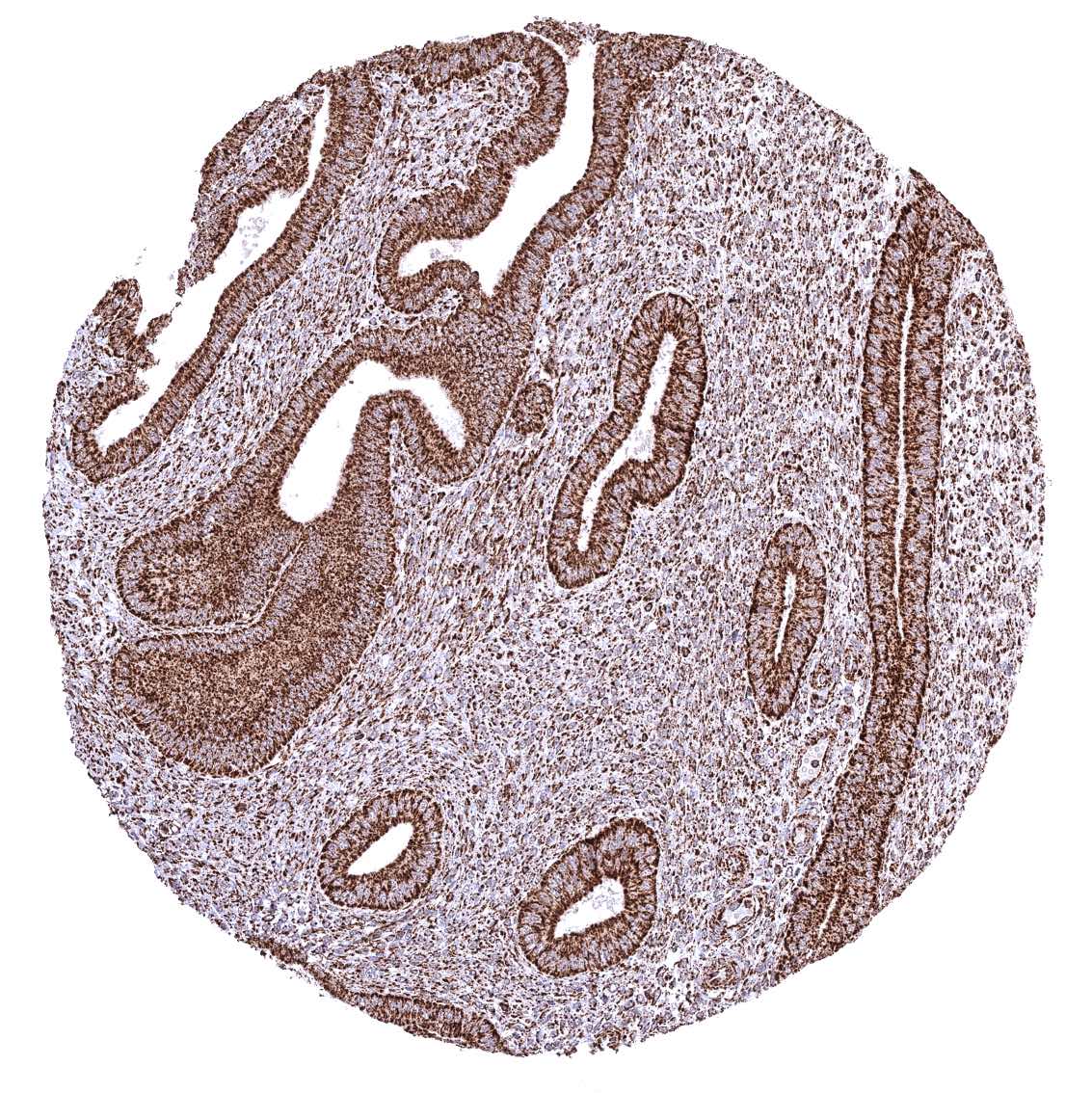
Colon descendens, mucosa
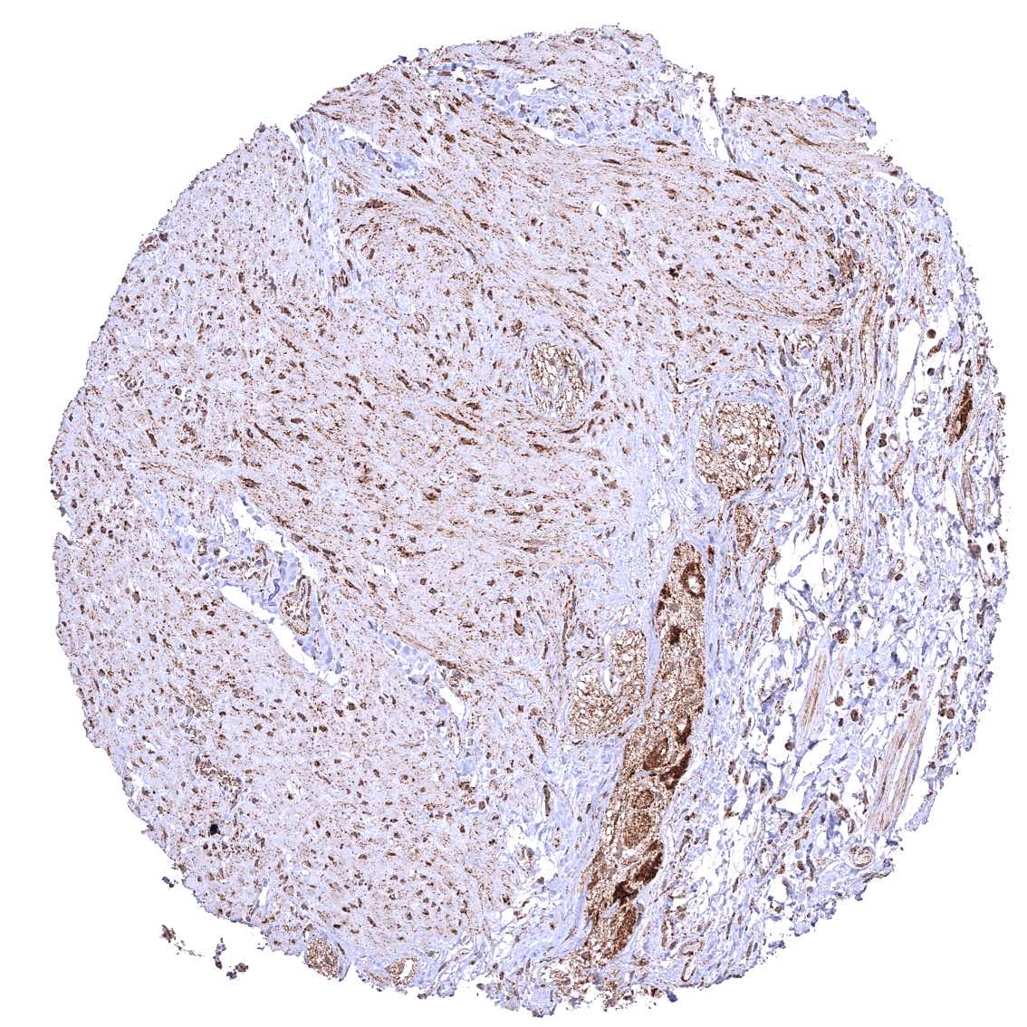
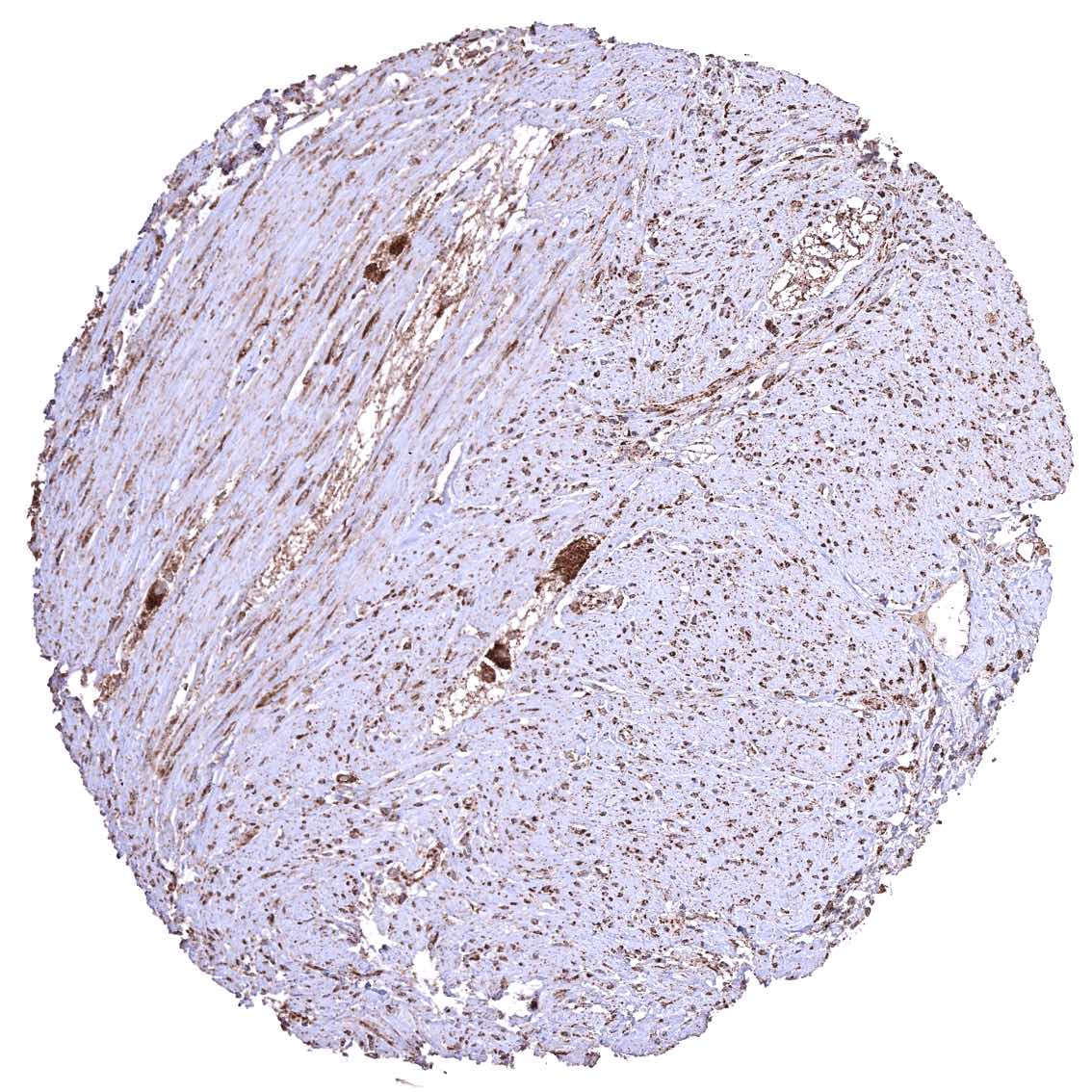
Colon descendens, muscular wall
Duodenum, Brunner gland
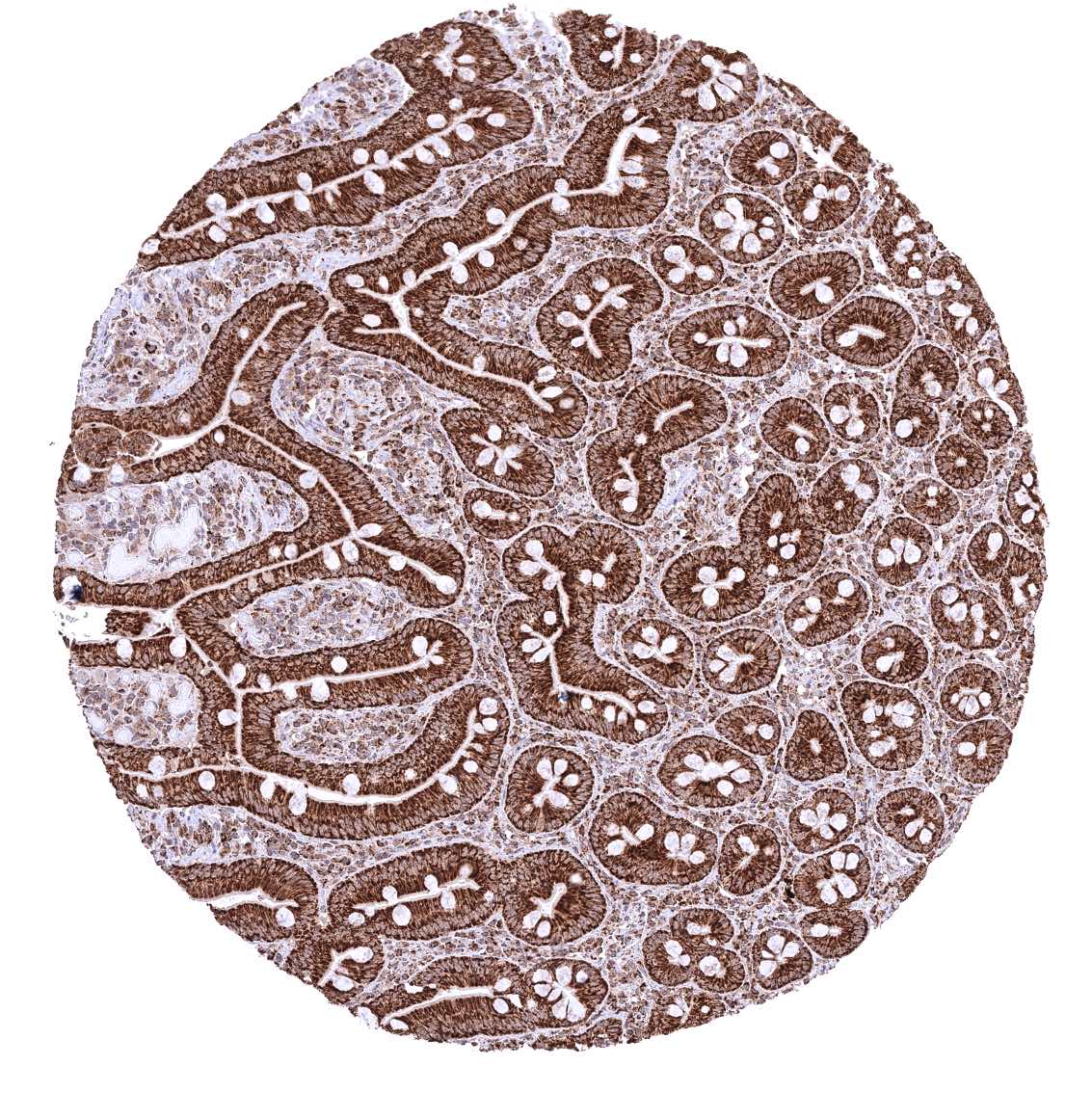
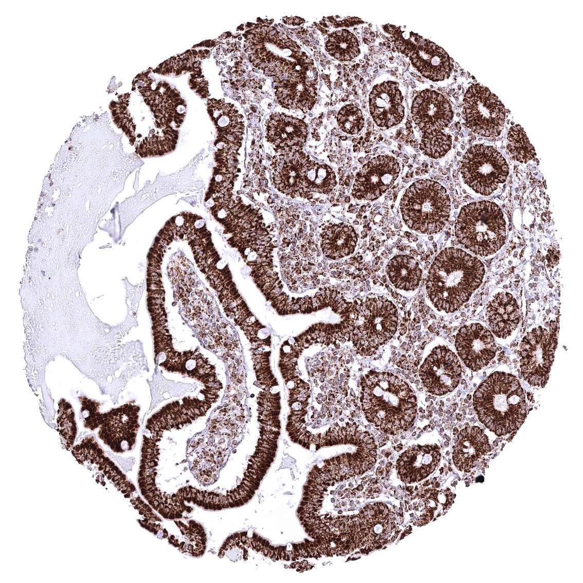
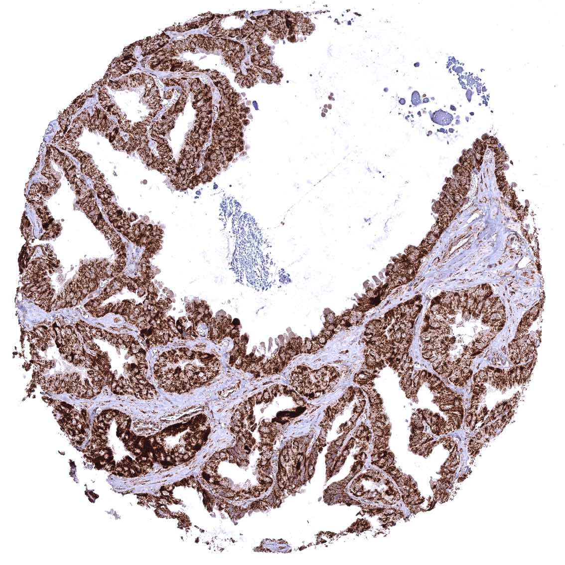
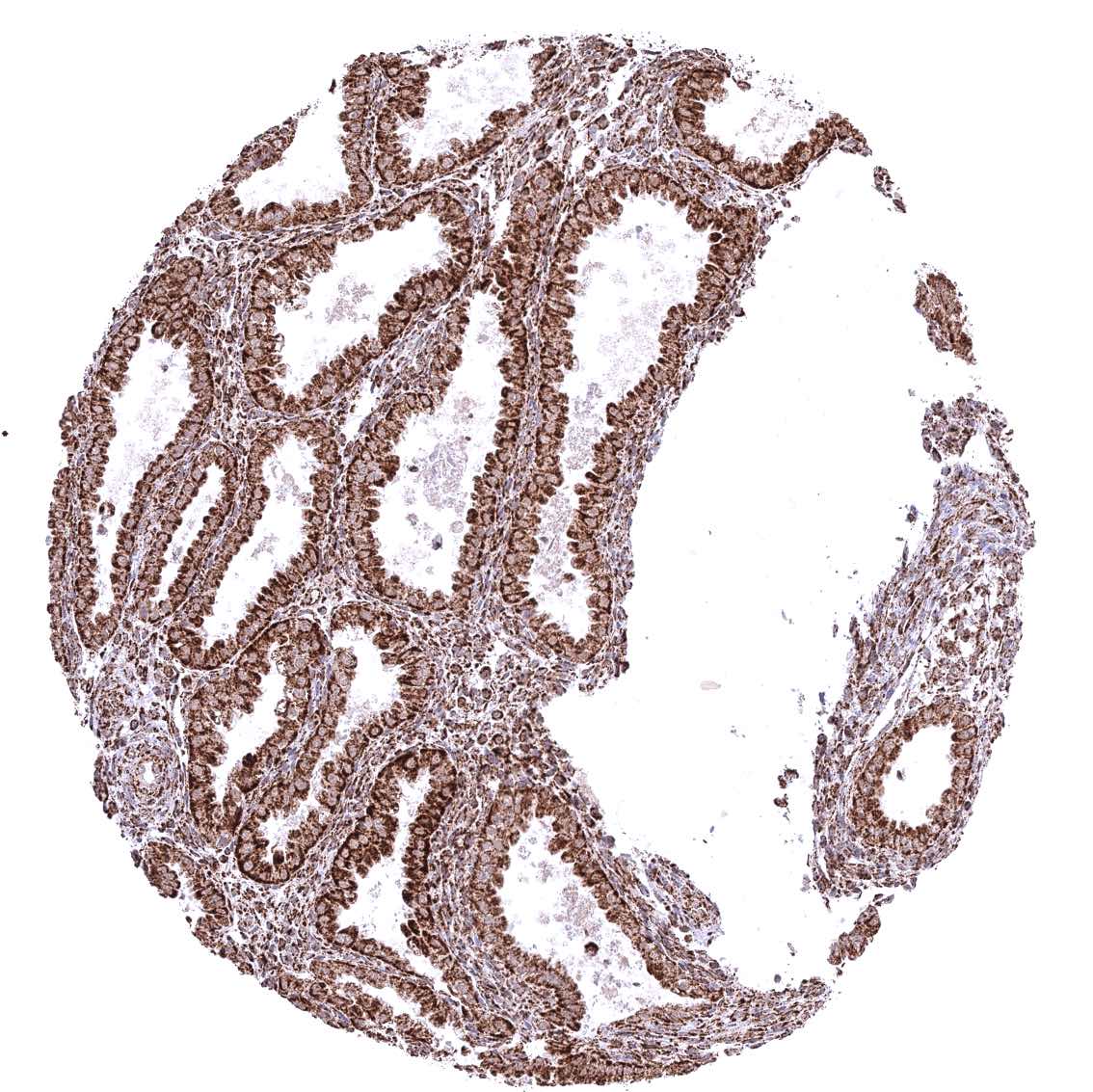
Duodenum, mucosa
Epididymis (Cauda).jpeg
Epididymis (Corpus)
Esophagus, squamous epithelium – Strong granular cytoplasmic ATP5J staining of all cell types. The staining is least intense in superficial cell layers of squamous epithelium.
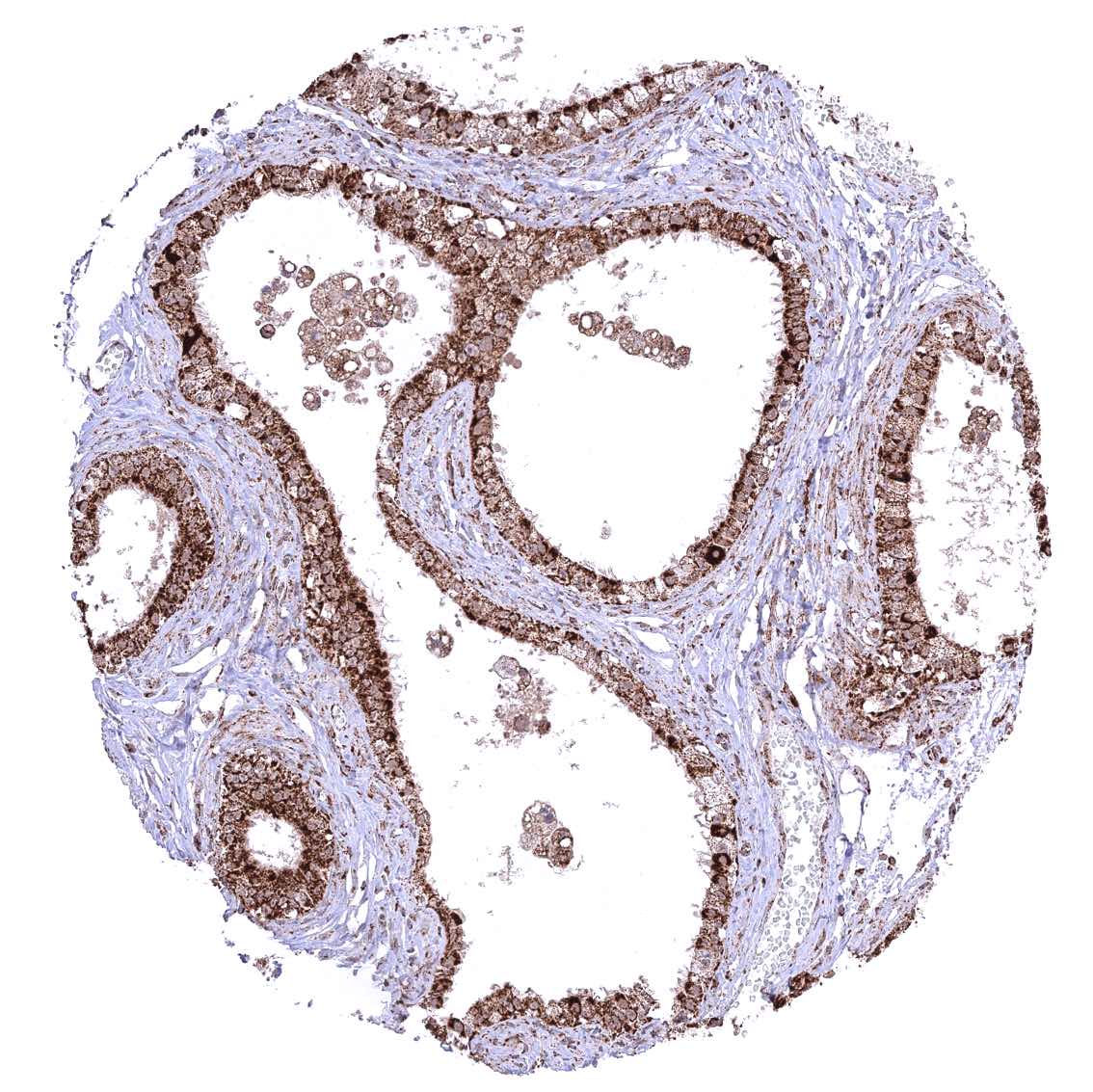
Fallopian tube, mucosa
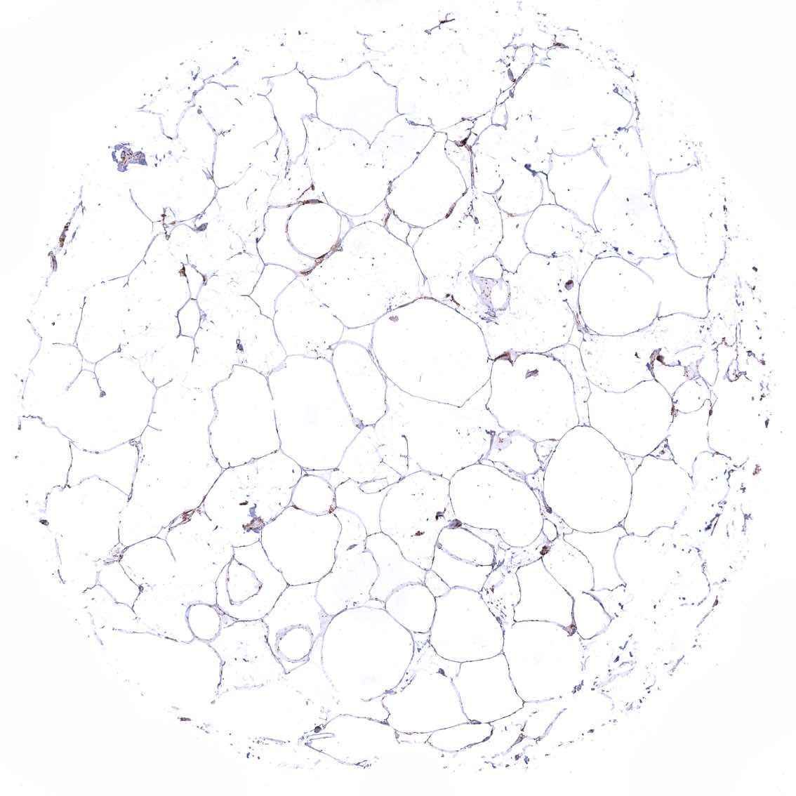
Fat
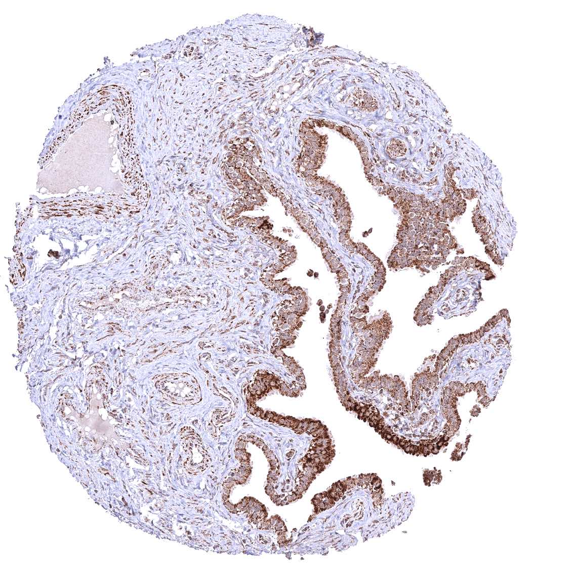
Gallbladder, epithelium
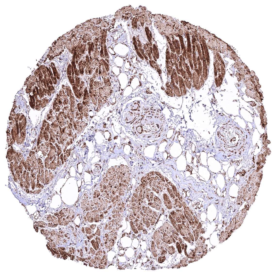
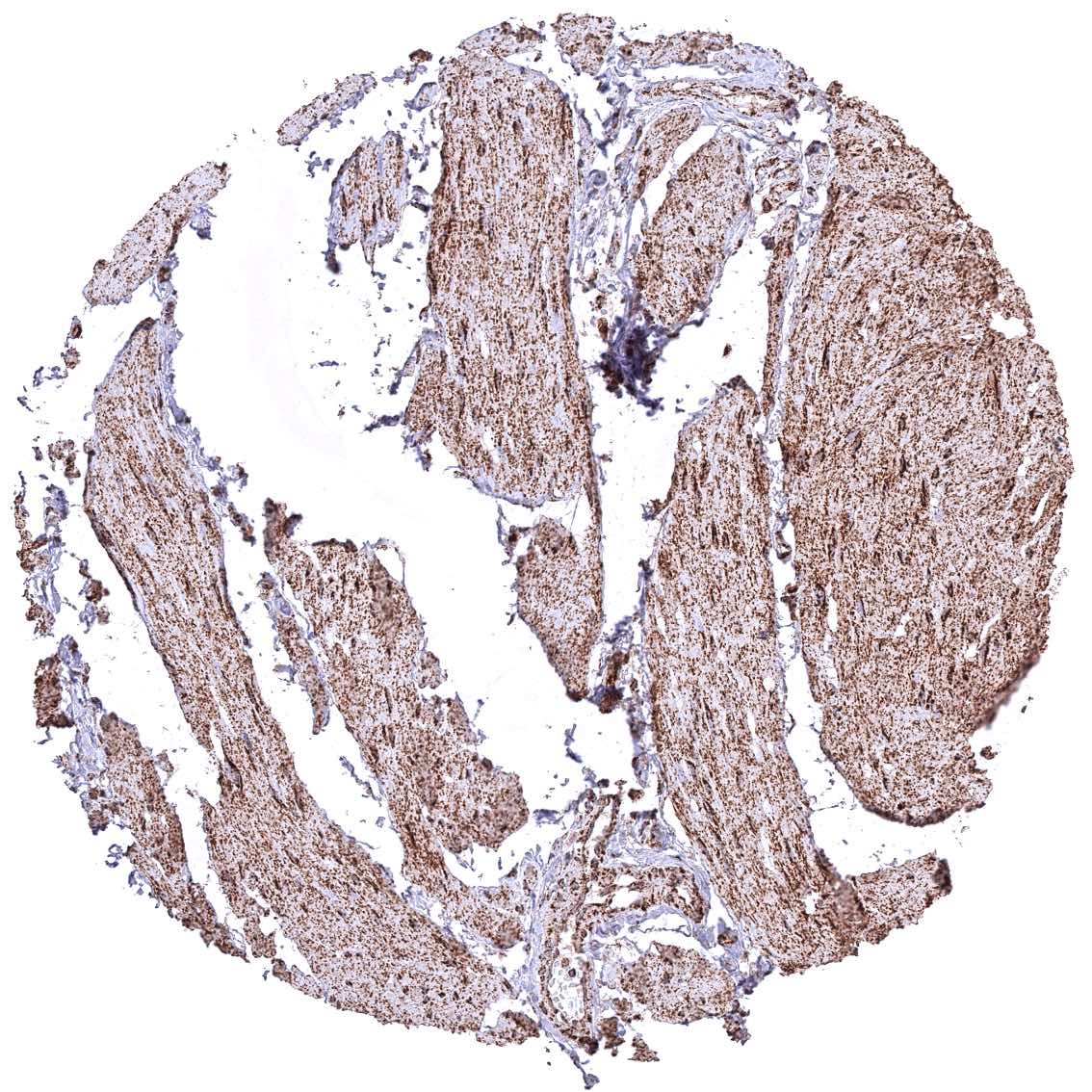
Heart muscle
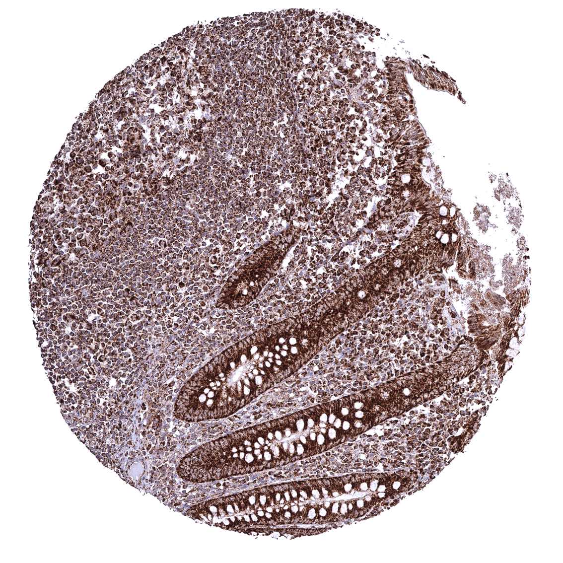
Ileum, mucosa
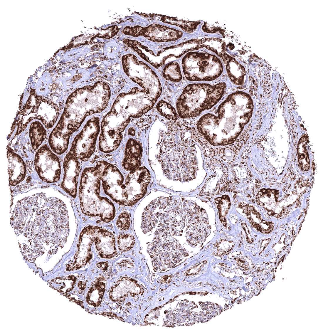
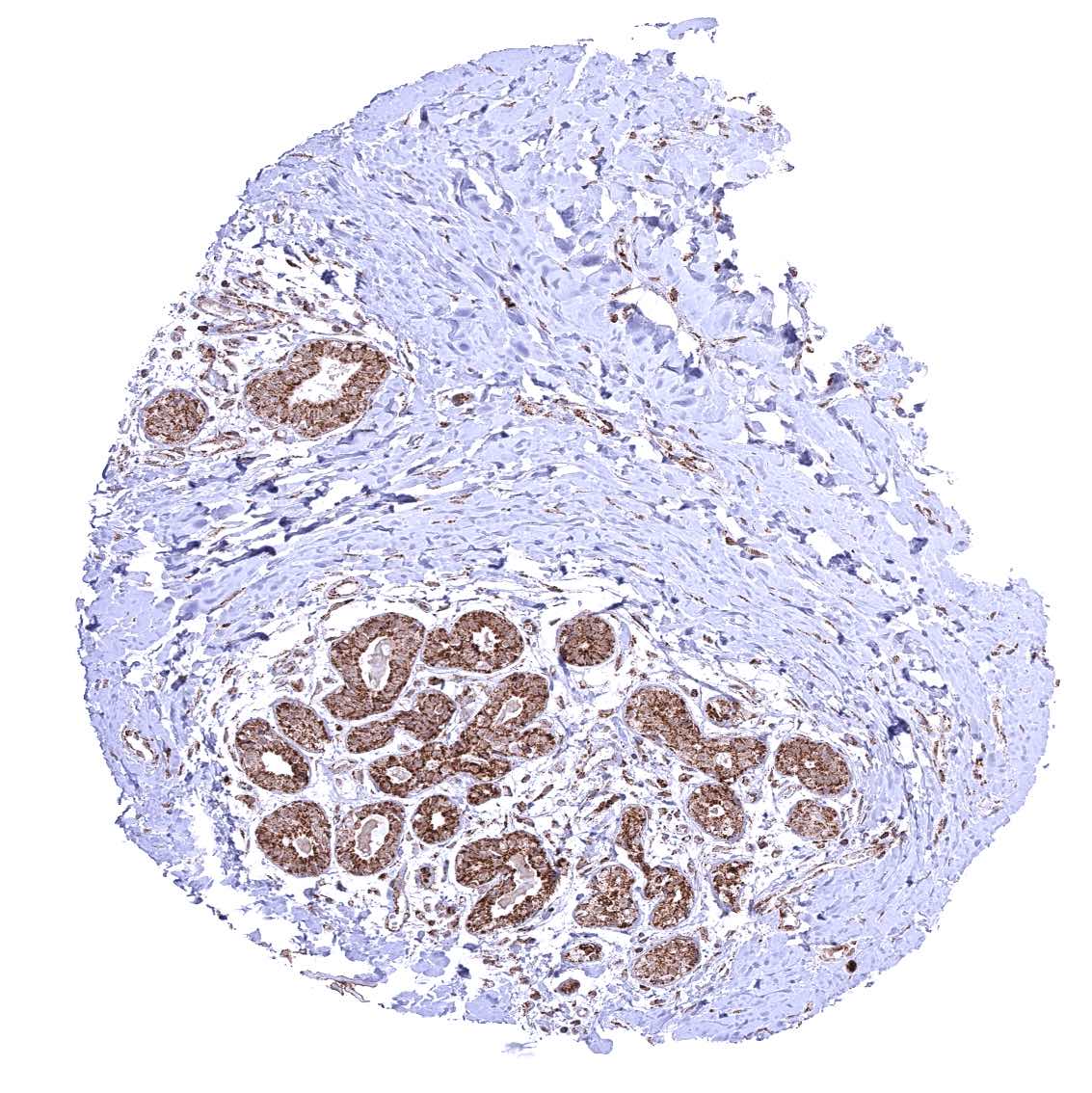
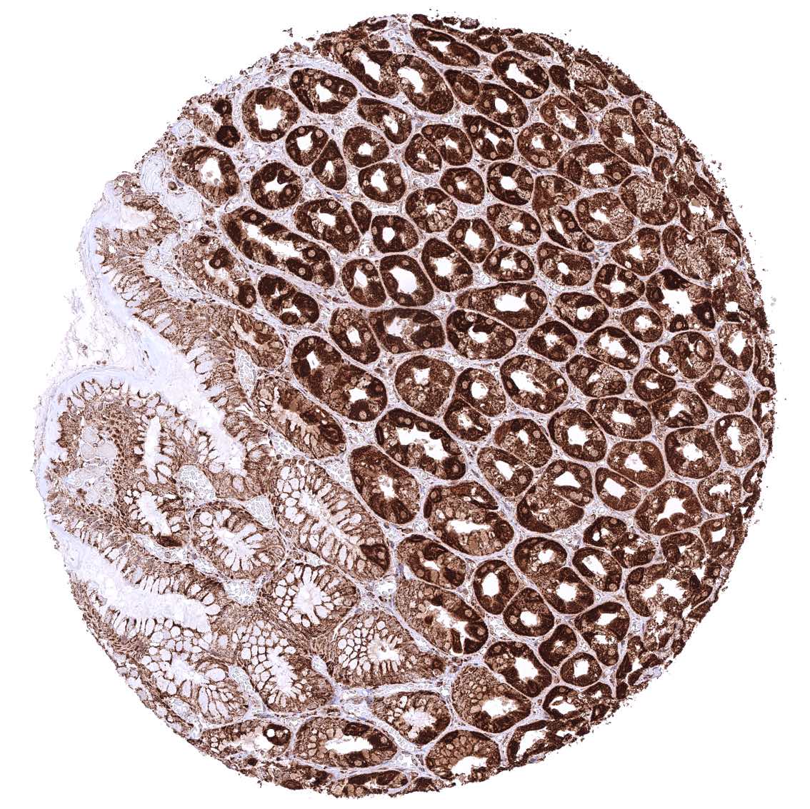
Kidney, cortex – Cytoplasmic ATP5J staining occurs in all cell types. It is intense in all tubuli, moderate in collecting ducts, and least intense in glomeruli.
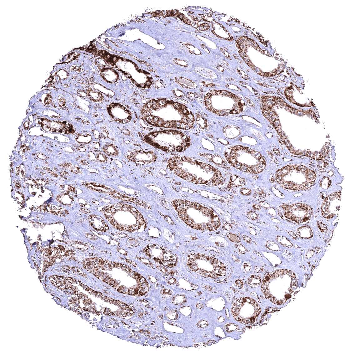
Kidney, medulla
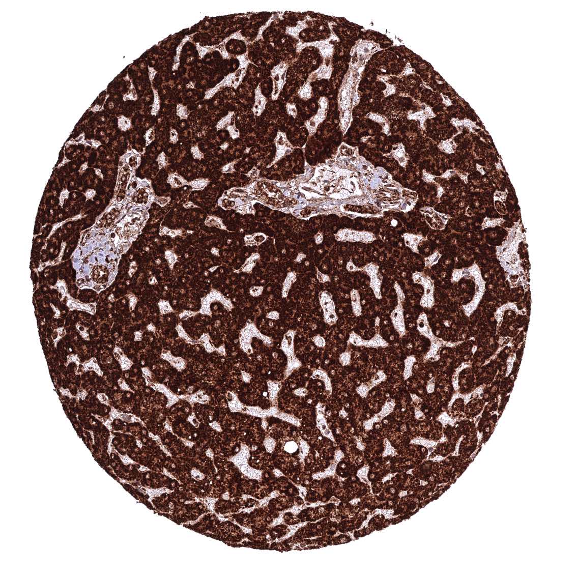
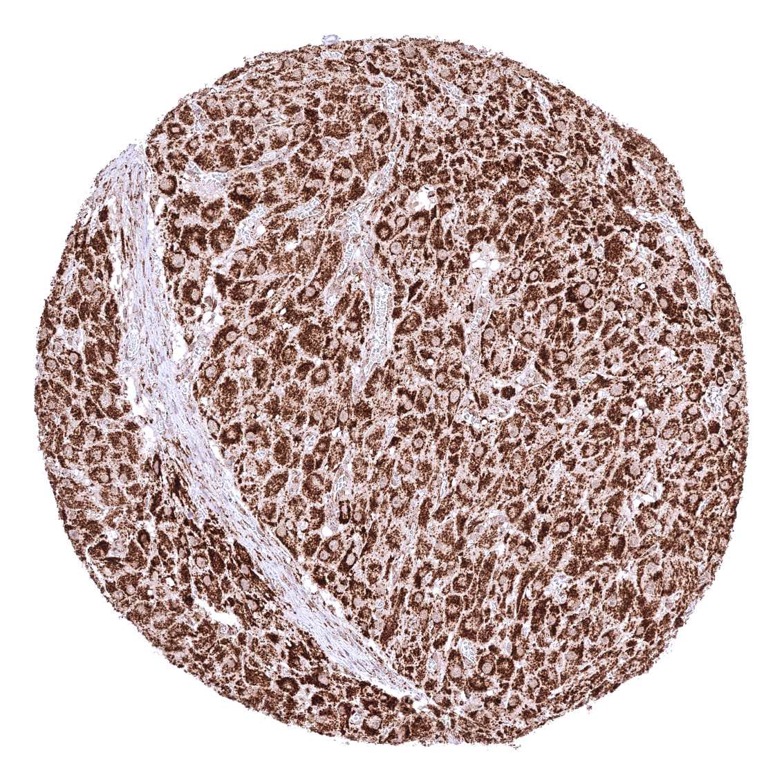
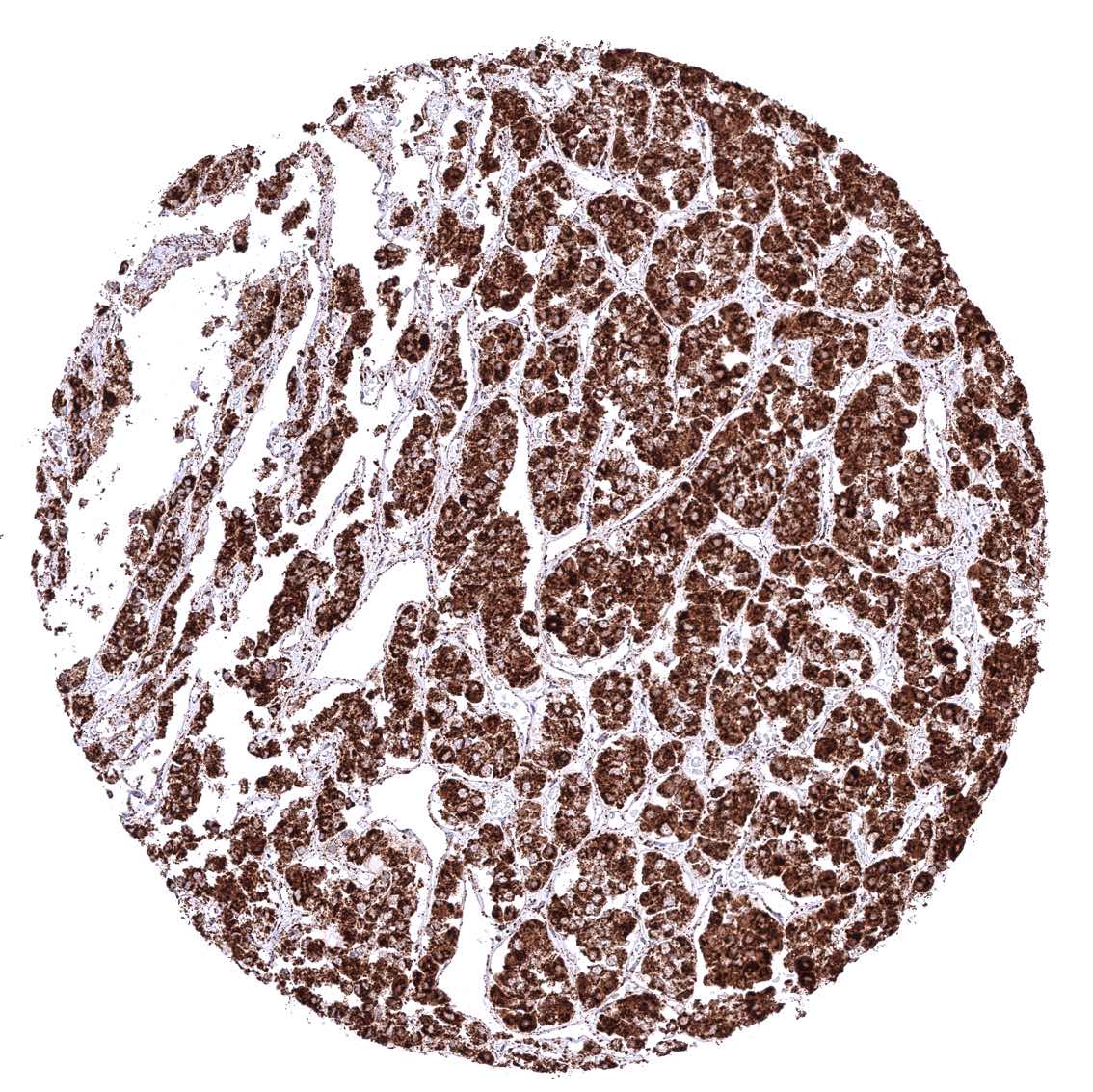
Liver – Cytoplasmic ATP5J staining is particularly intense in hepatocytes.
Lung
Lymph node
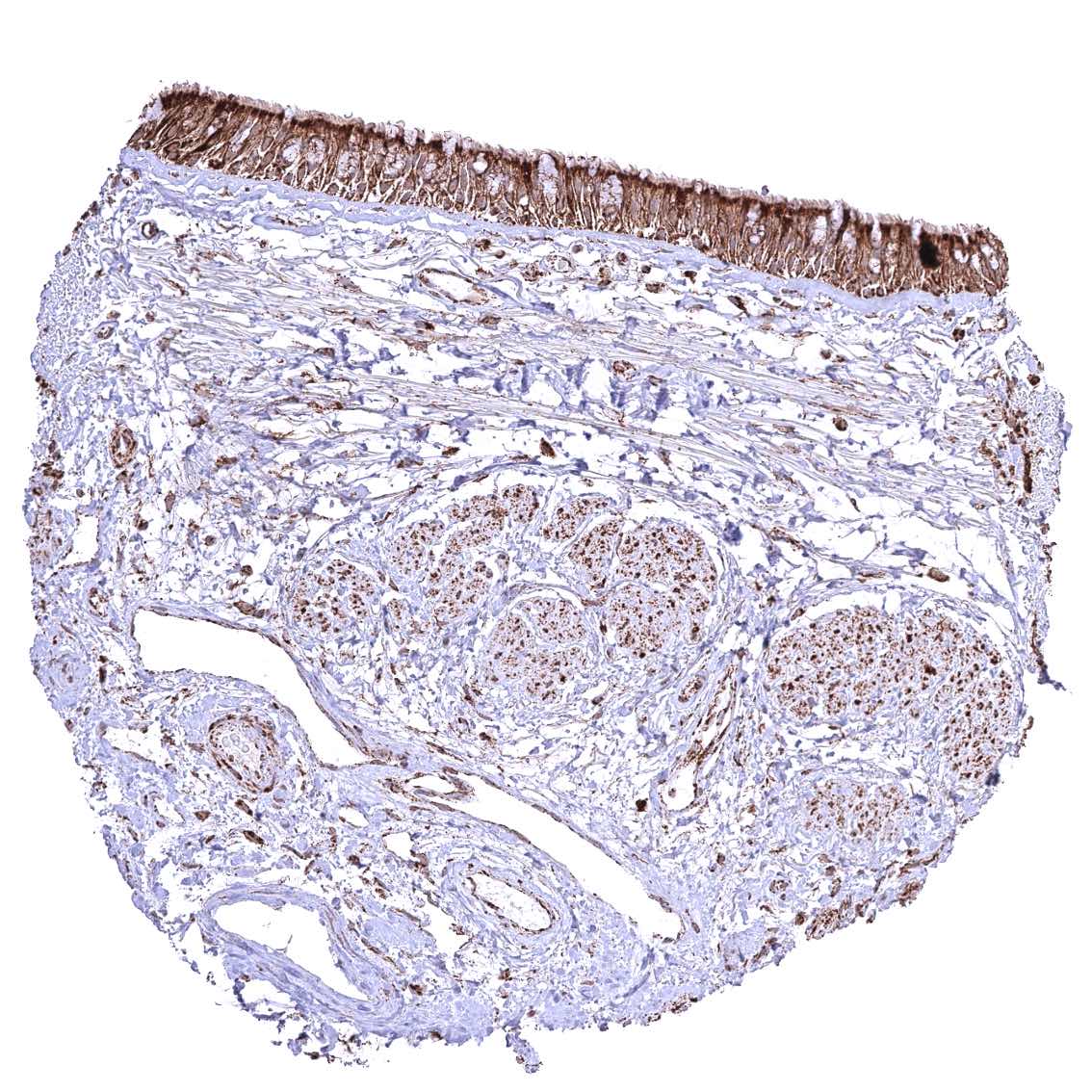
Ovary, corpus luteum
Ovary, stroma
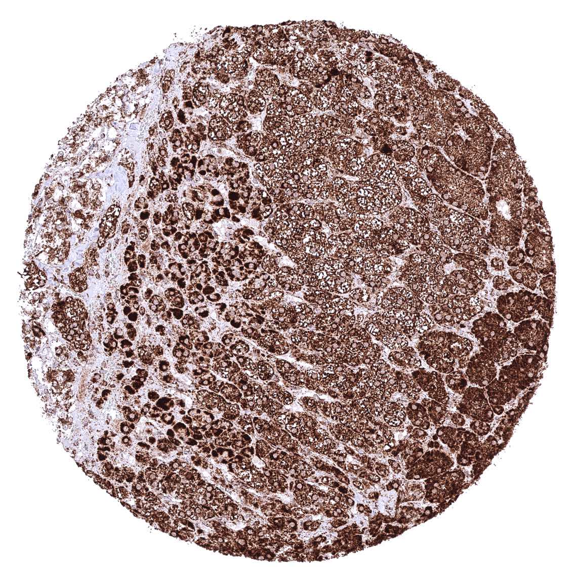
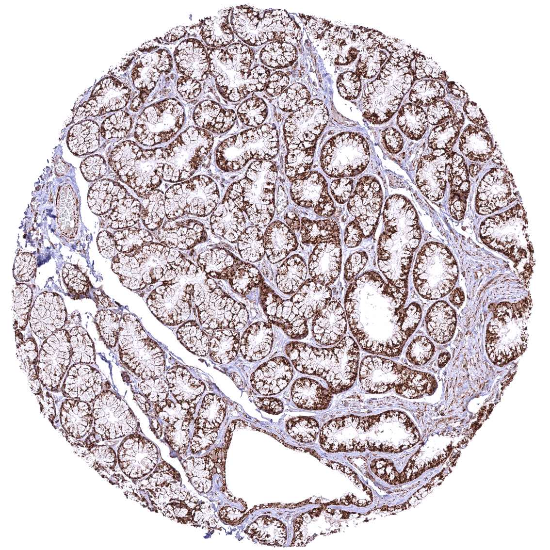
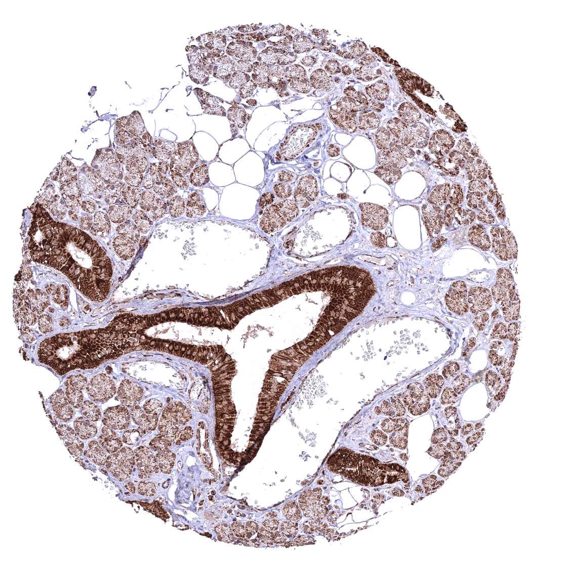
Pancreas – Distinct granular, perinuclear, cytoplasmic ATP5J staining in all cell types. Some cells with higher staining intensity may be related to the excretory system.
Parathyroid gland
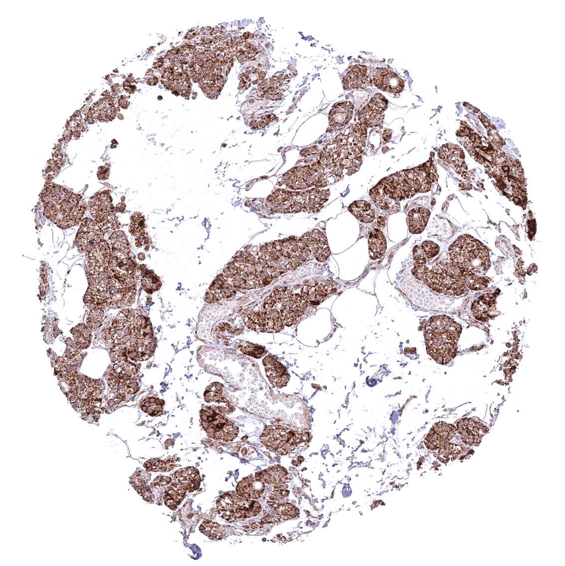
Parotid gland – Cytoplasmic ATP5J staining occurs in all cell types. It is very intense in excretory ducts but rather weak in glandular cells.
Pituitary gland, anterior lobe
Pituitary gland, posterior lobe
Placenta (amnion and chorion) – Distinct cytoplasmic ATP5J staining of amnion and chorion cells.
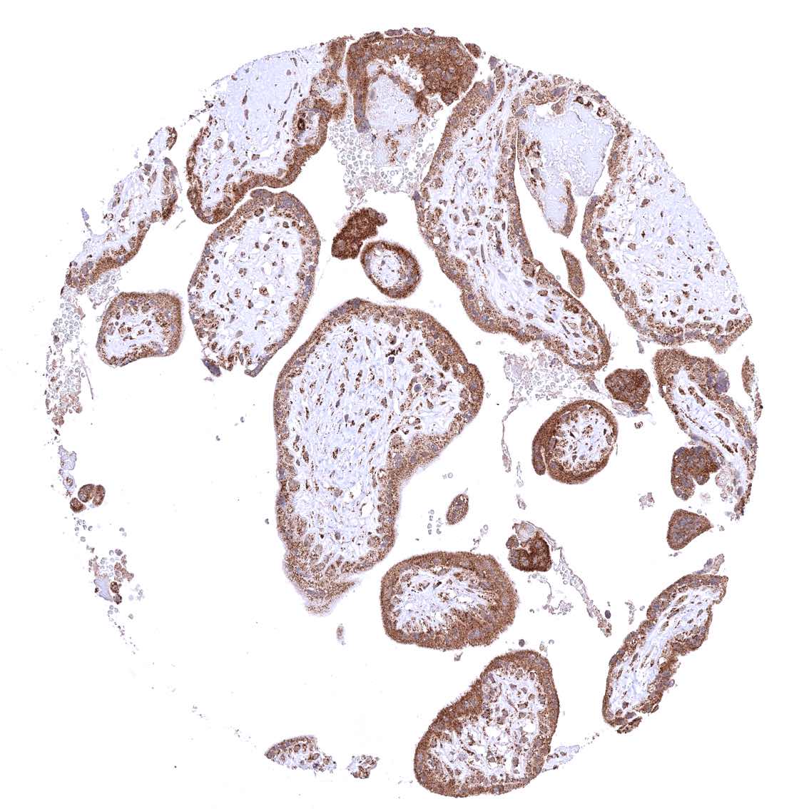
Placenta, early
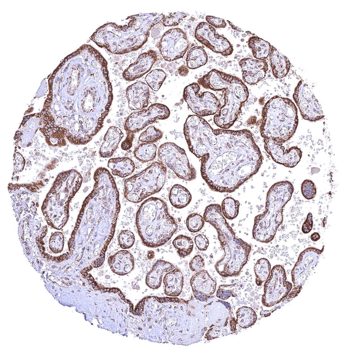
Placenta, mature
Prostate
Rectum, mucosa
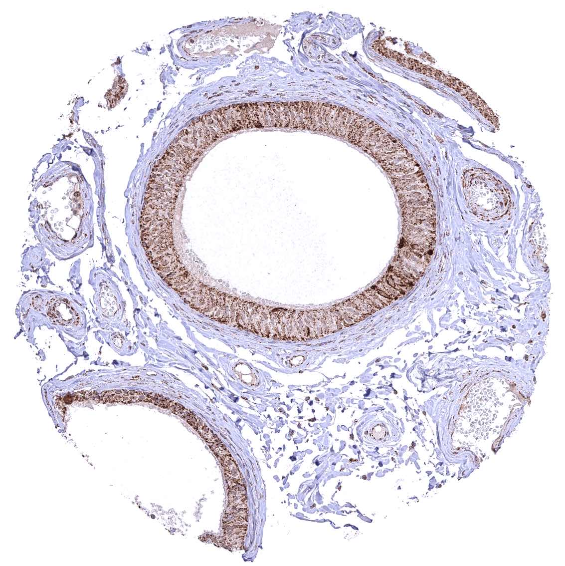
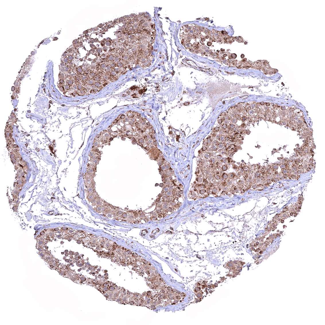
Seminal vesicle
Sinus paranasales – Cytoplasmic ATP5J staining occurs in all cell types. A characteristic granular cytoplasmic staining is seen below the apical membranes.
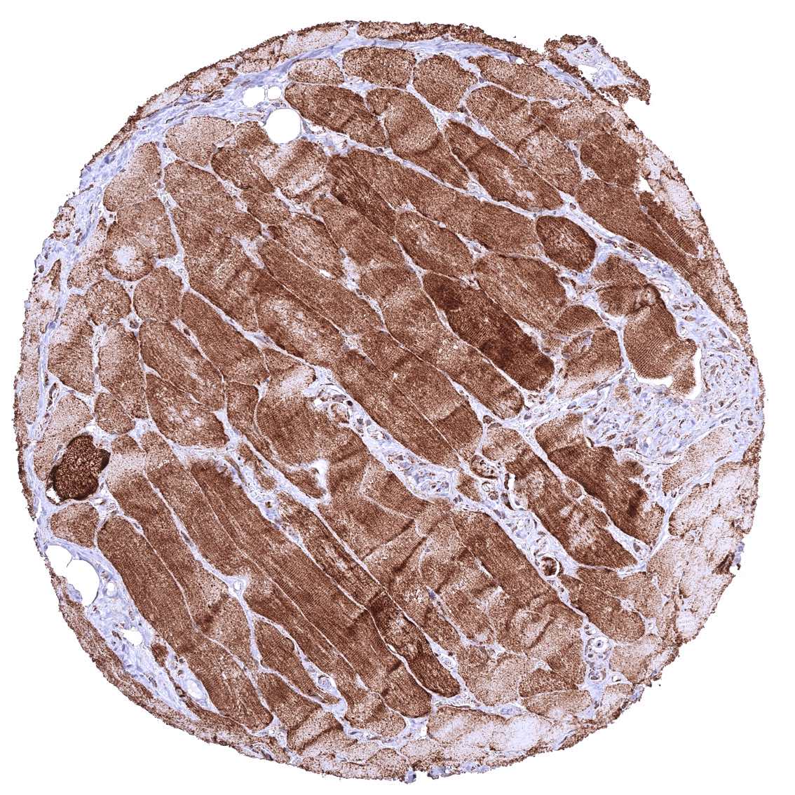
Skeletal muscle.jpeg
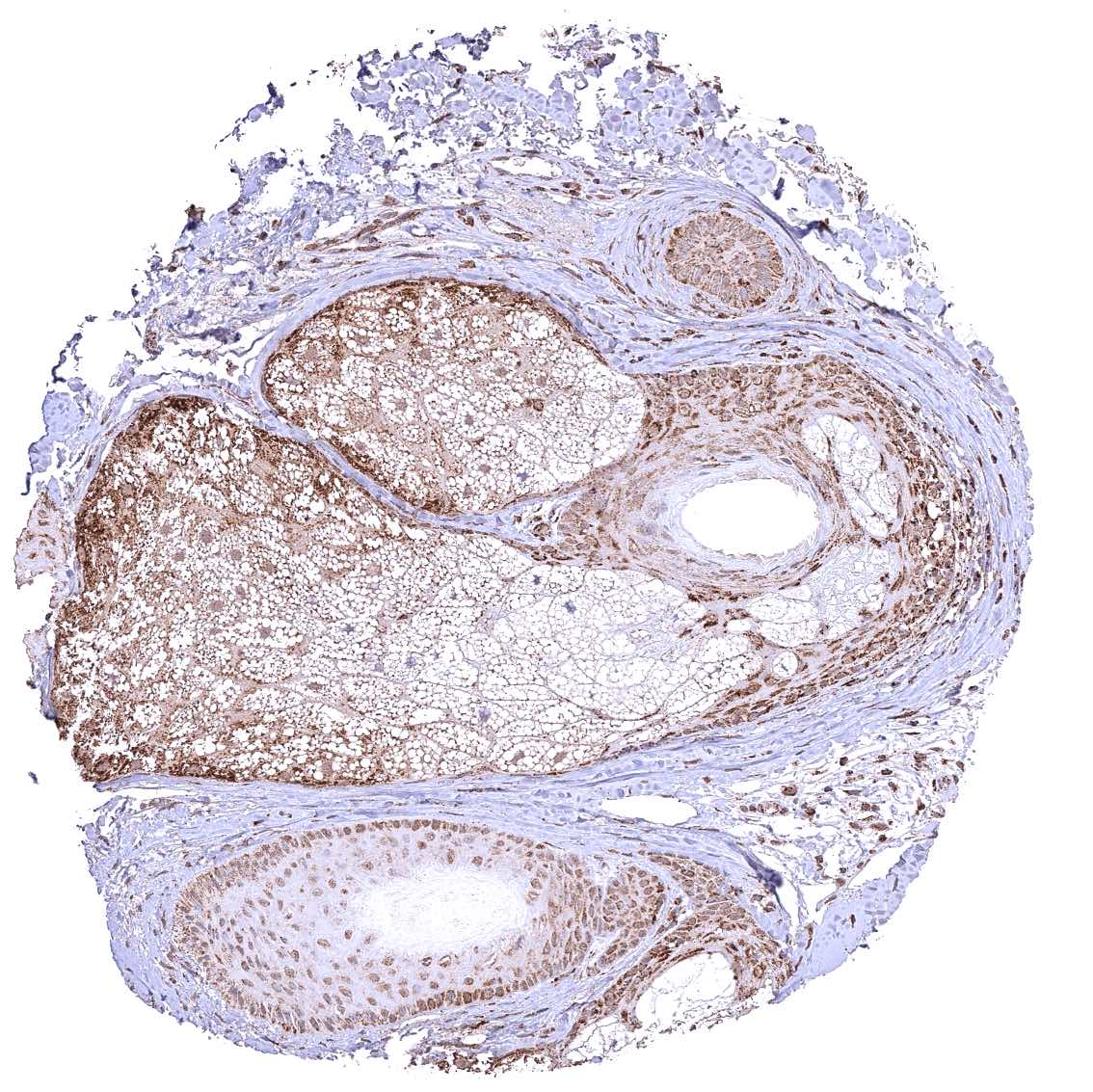
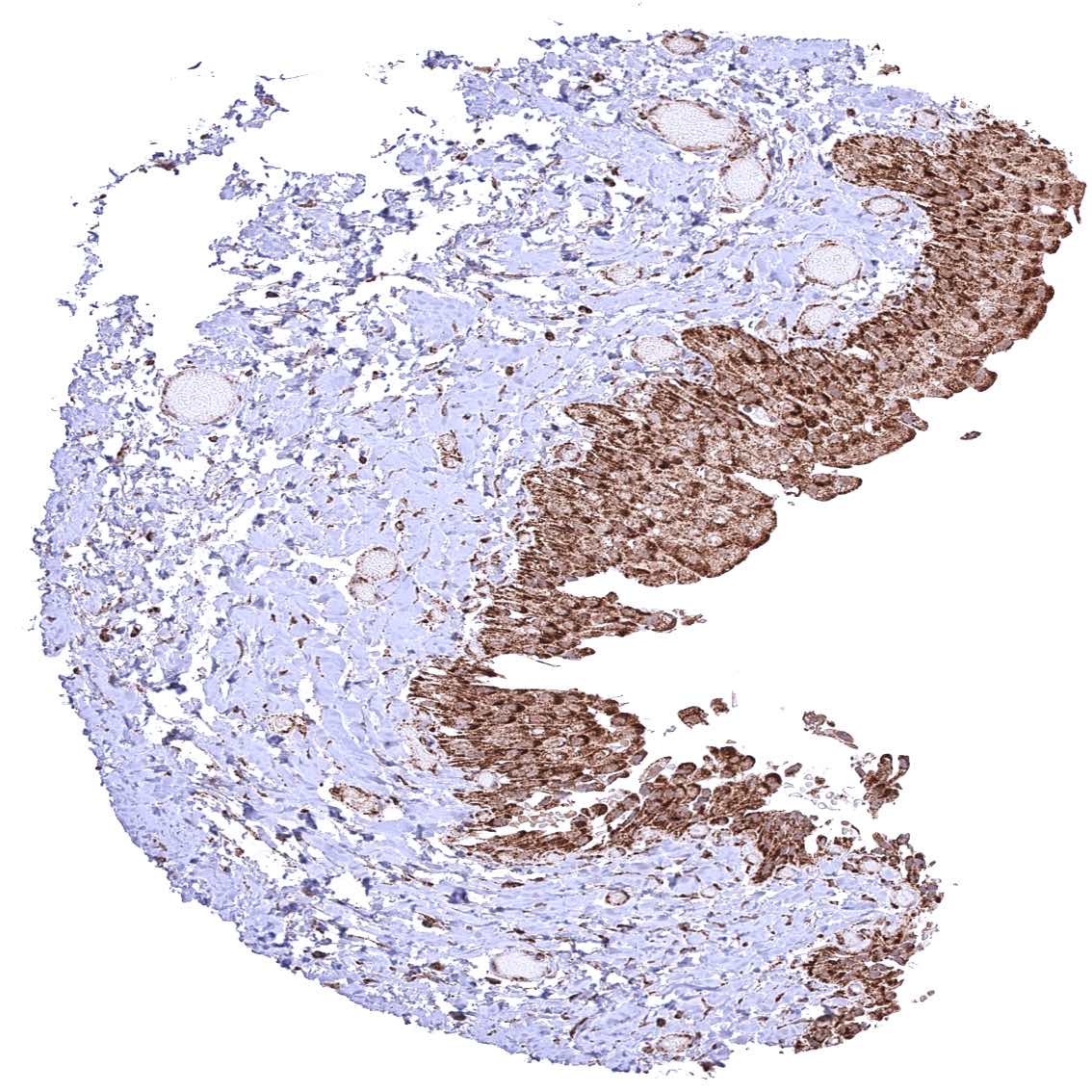
Skin, hairfollicel and sebaceous glands – Distinct cytoplasmic ATP5J staining of all cell types of hair follicles but staining is particularly weak in sebaceous glandular cells.
Skin
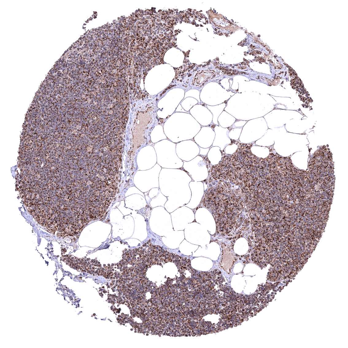
Spleen
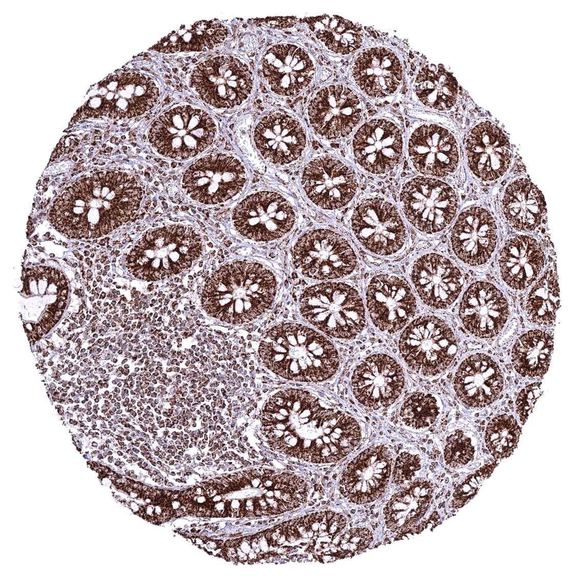
Stomach, corpus – Cytoplasmic ATP5J staining occurs in all cell types. It is most intense in parietal cells and least intense in superficial epithelial cells.
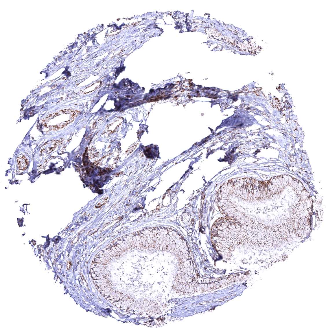
Testis
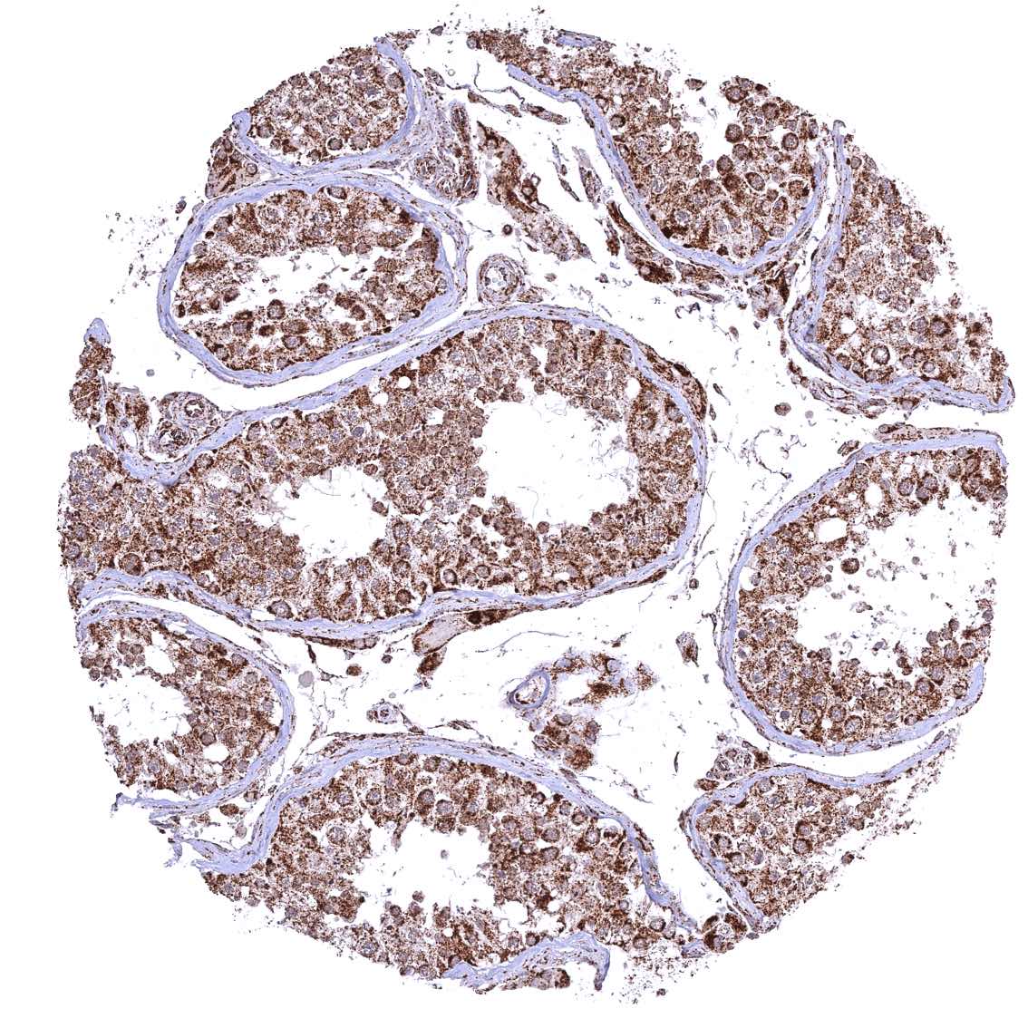
Testis
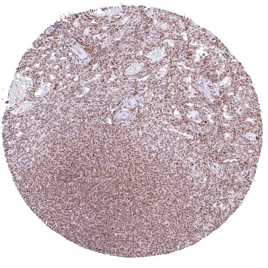
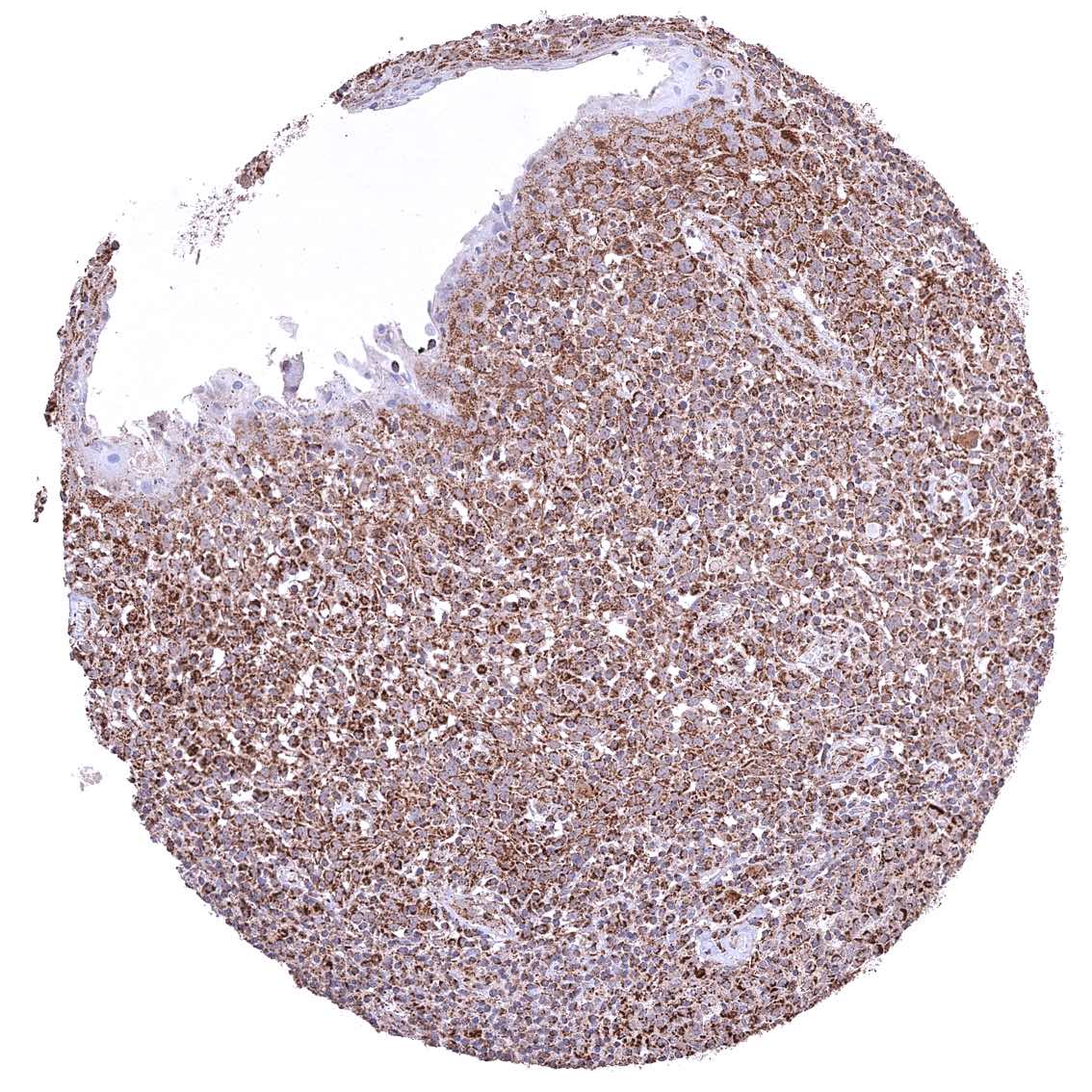
Thymus
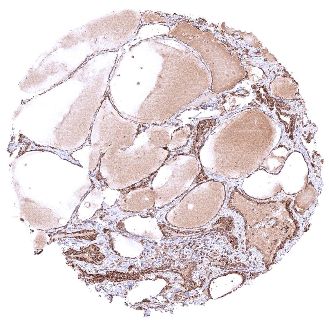
Thyroid gland
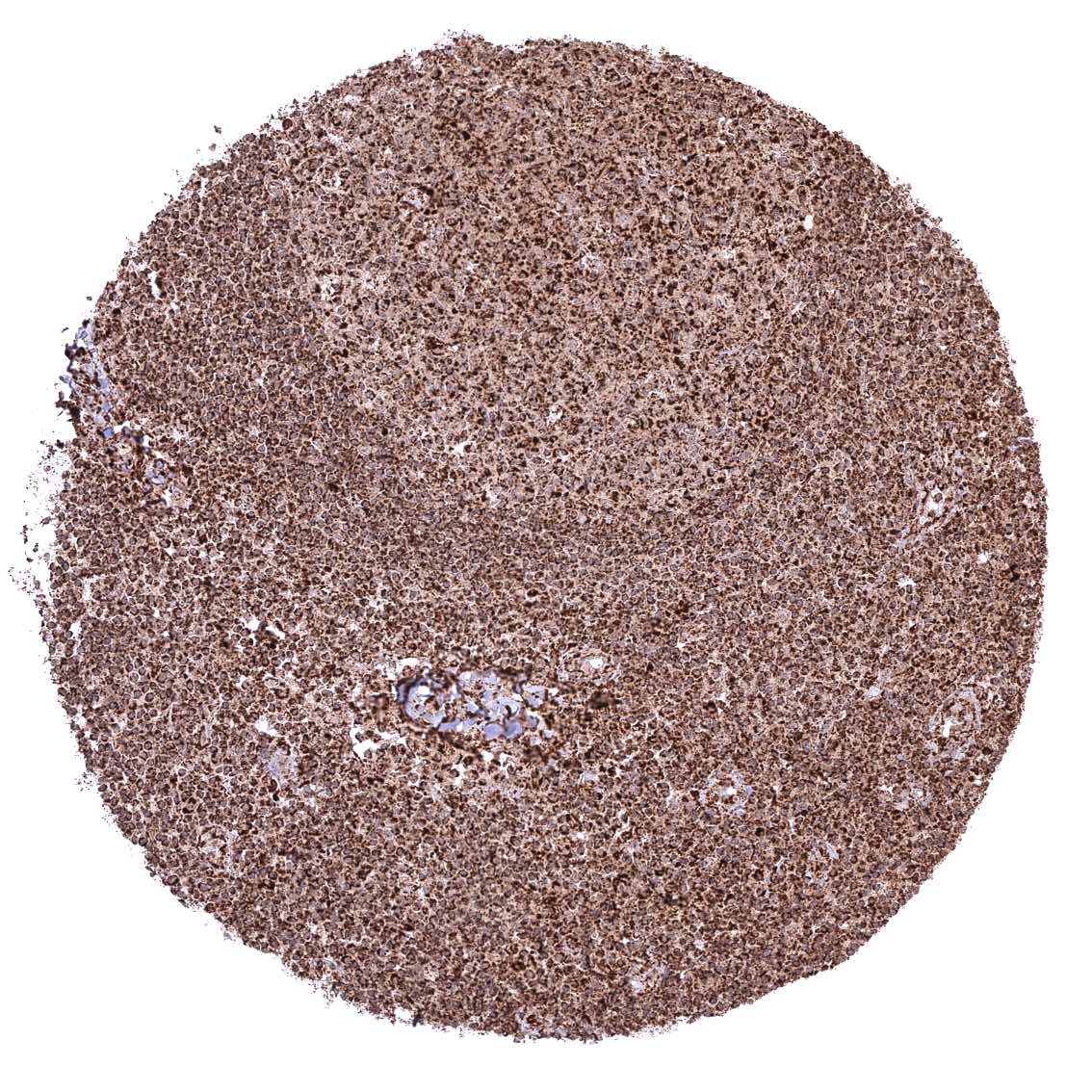
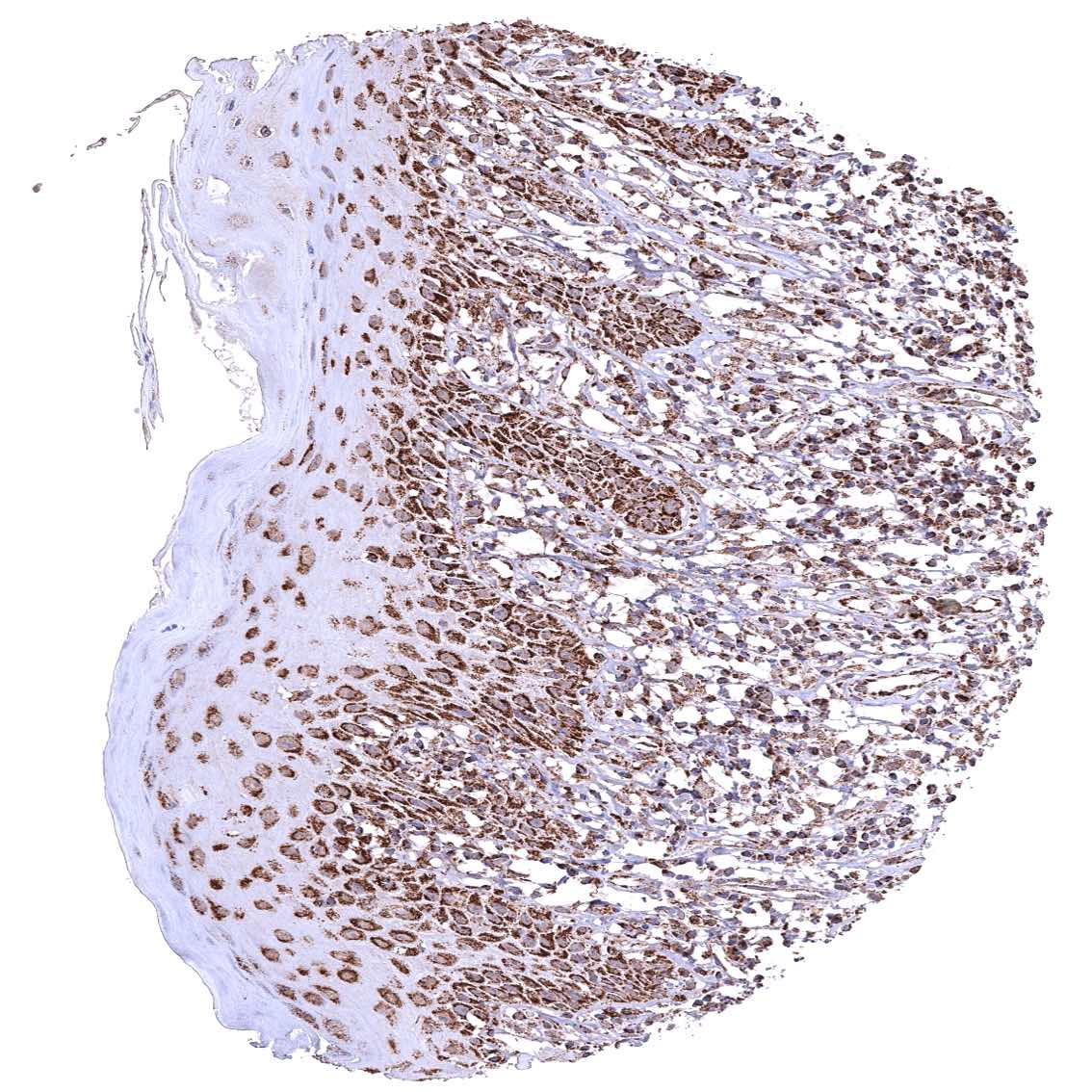
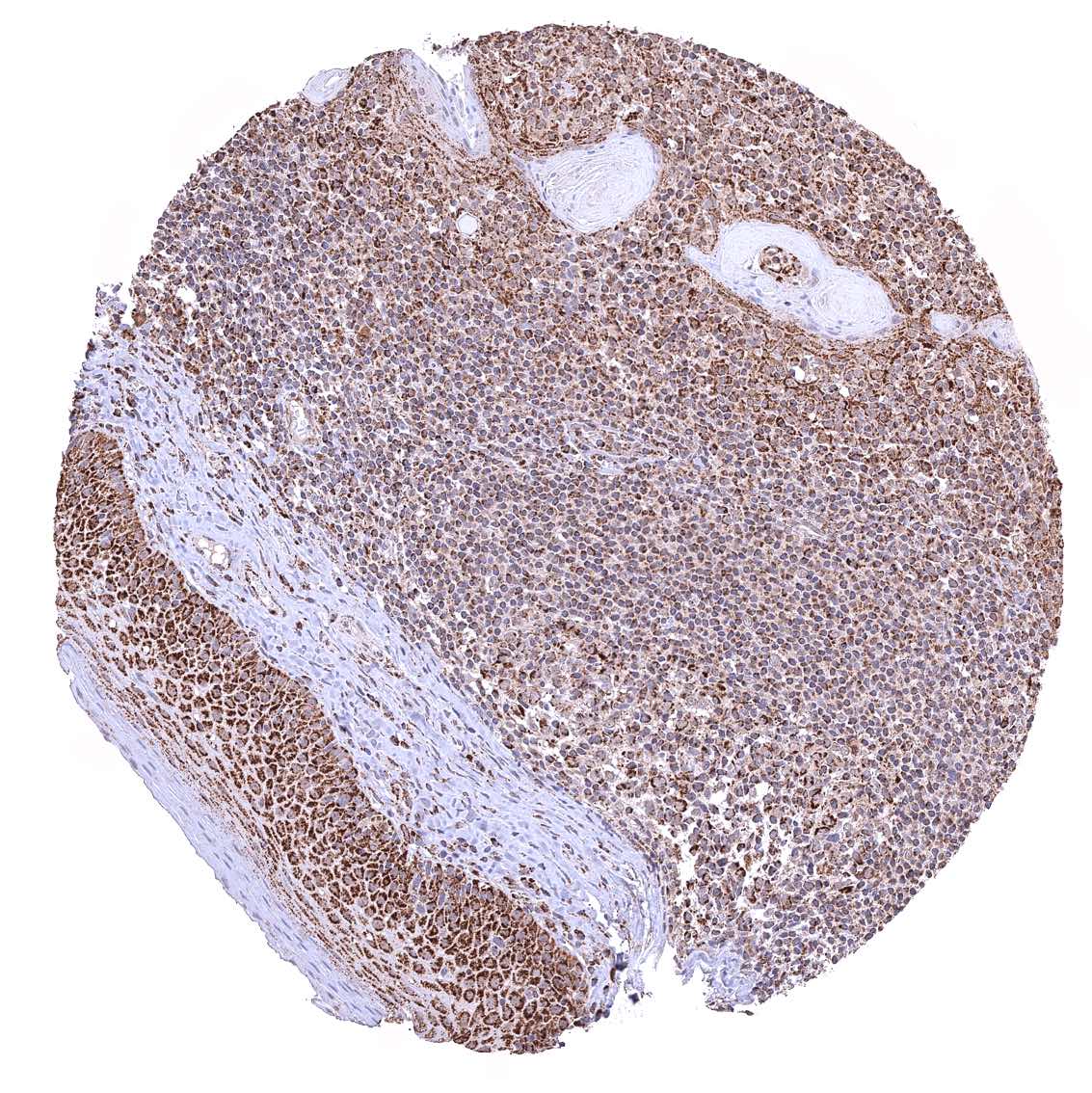
Tonsil – Strong granular cytoplasmic ATP5J staining of all cell types. The staining is least intense in superficial cell layers of squamous epithelium.
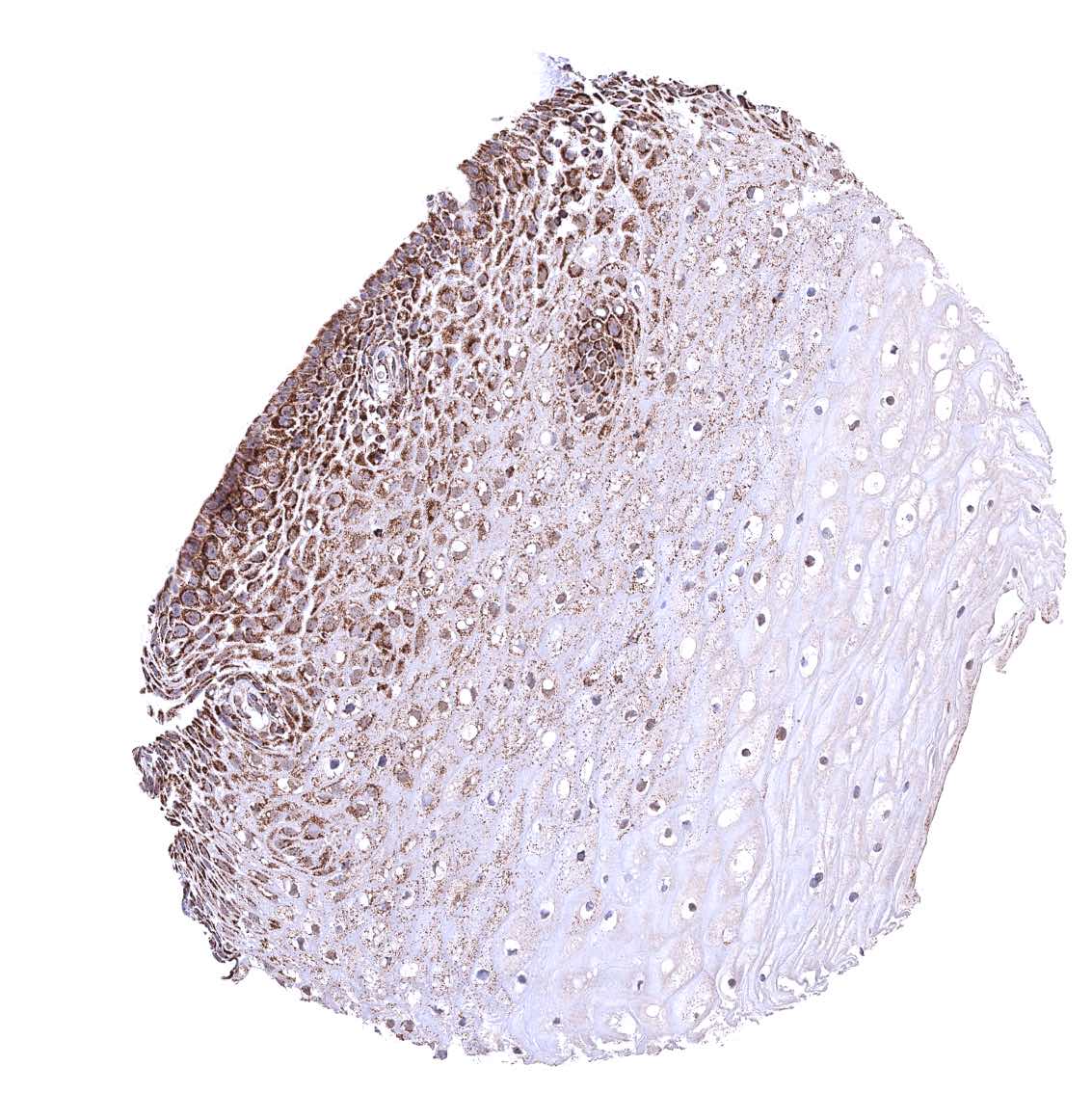
Tonsil, surface epithelium – Strong granular cytoplasmic ATP5J staining of all cell types. The staining is least intense in superficial cell layers of squamous epithelium
Urinary bladder, muscular wall
Urinary bladder, urothelium
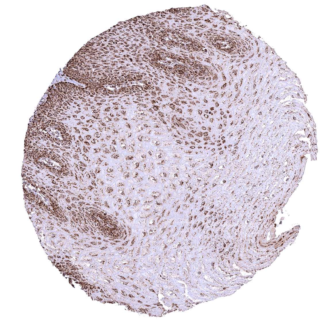
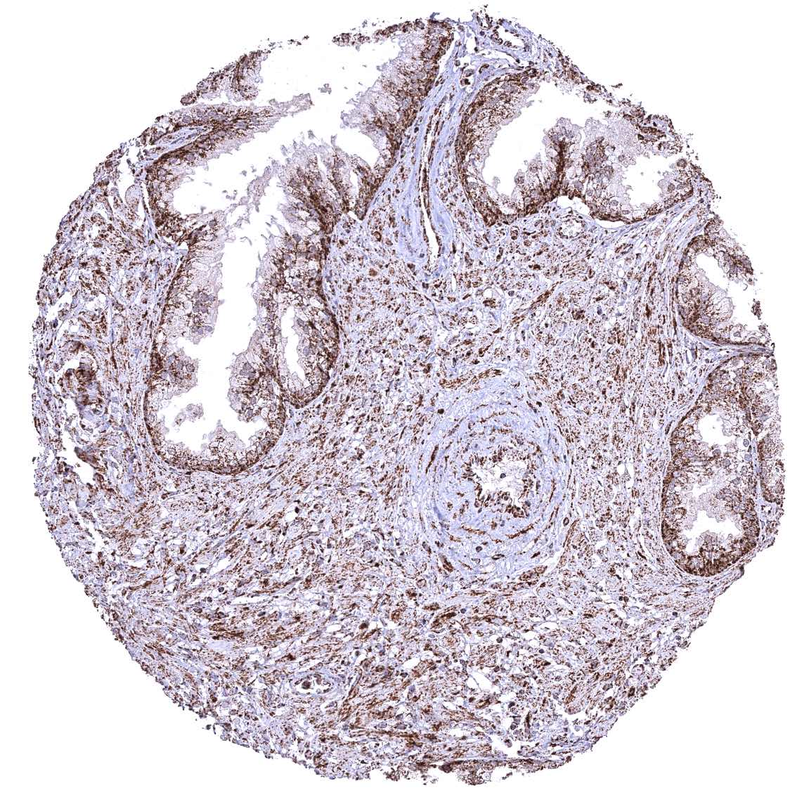
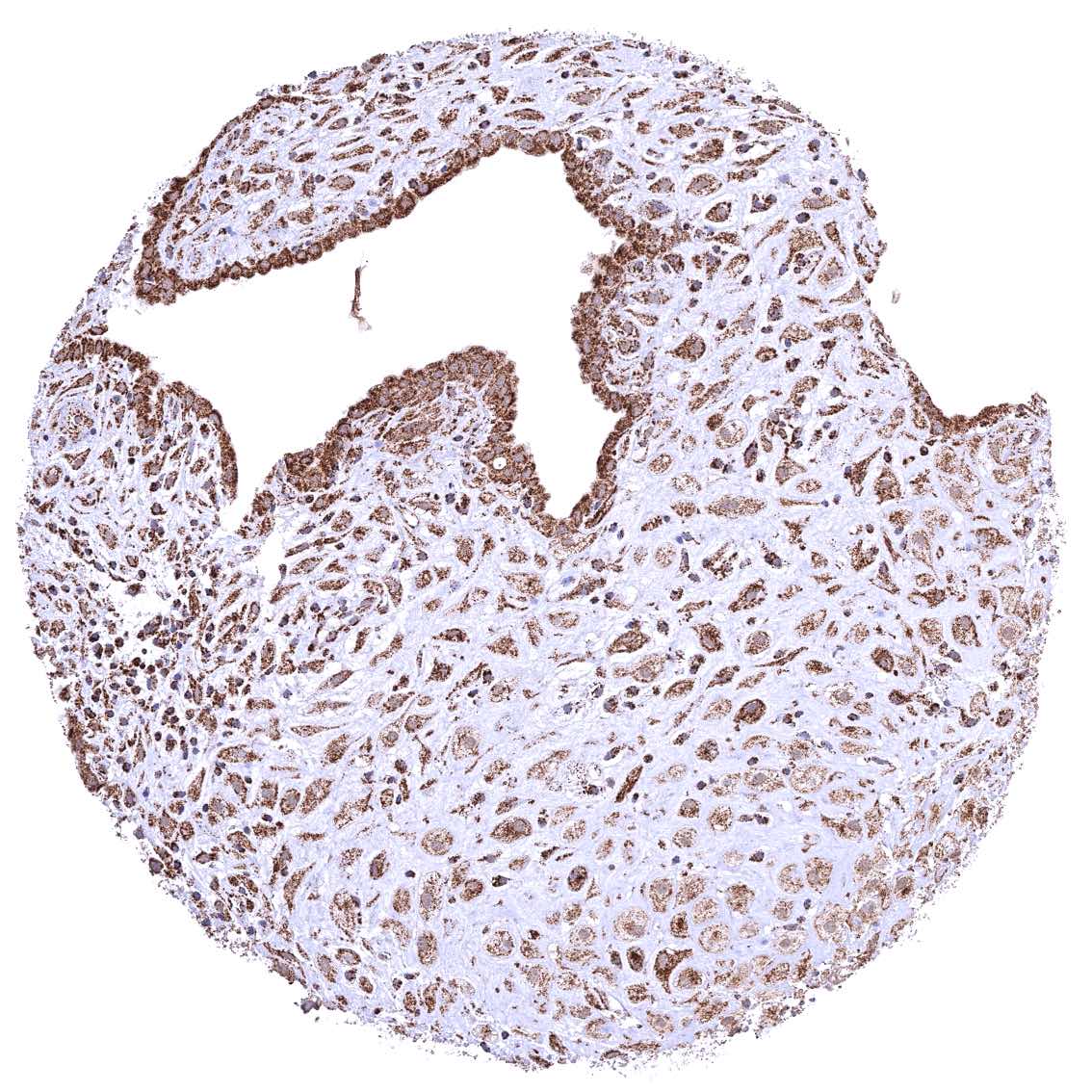
Uterus, ectocervix – Distinct granular, perinuclear, cytoplasmic ATP5J staining in all cell types. The staining is least intense in superficial cell layers of non-keratinizing squamous epithelium.
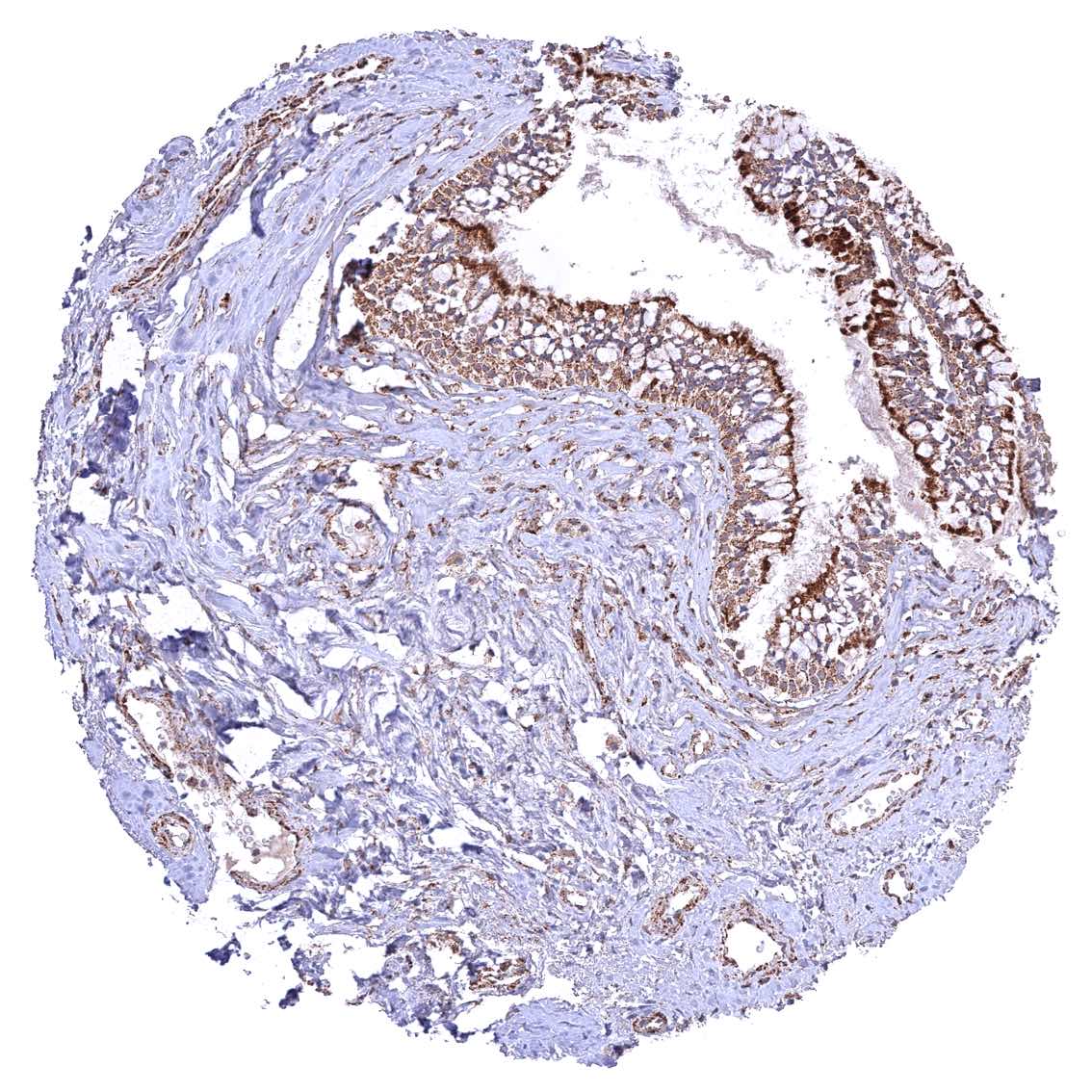
Uterus, endocervix
Uterus, endometrium (pregnancy)
Uterus, endometrium (proliferation)
Uterus, endometrium (secretion)
Uterus, myometrium