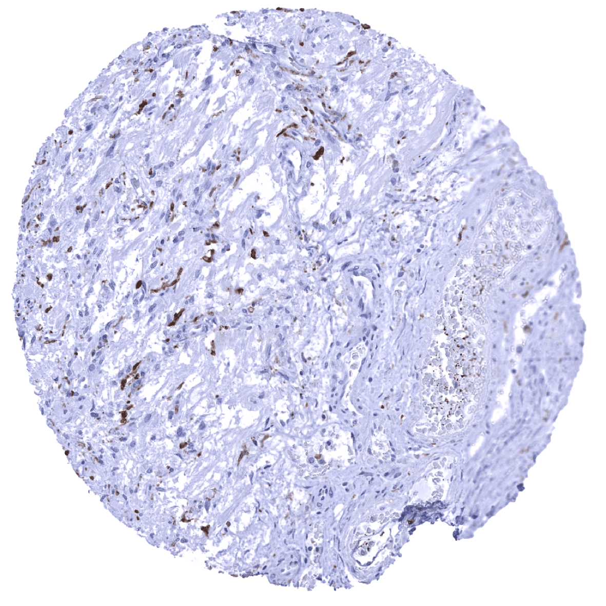
Adrenal gland - A strong AIF1 immunostaining is seen in macrophges in the adrenal gland.
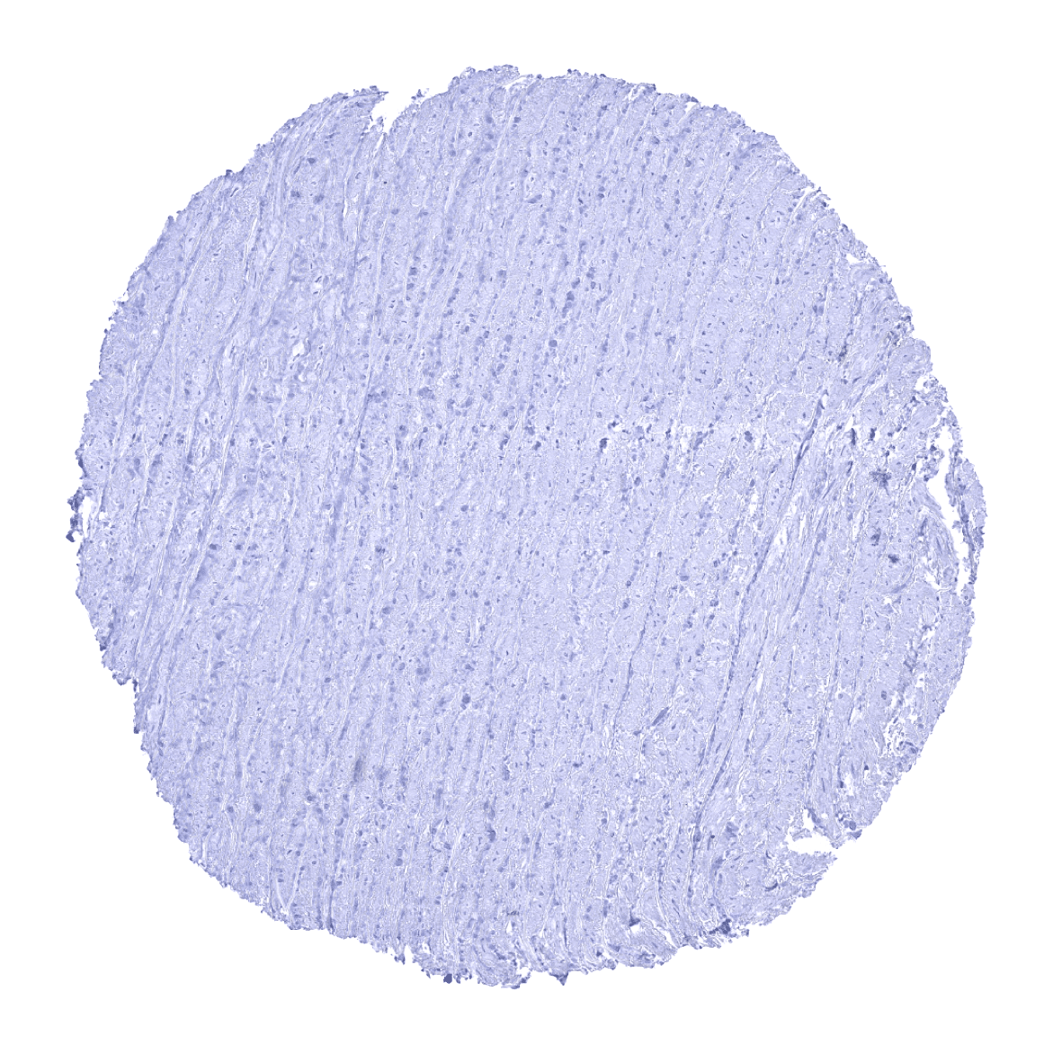
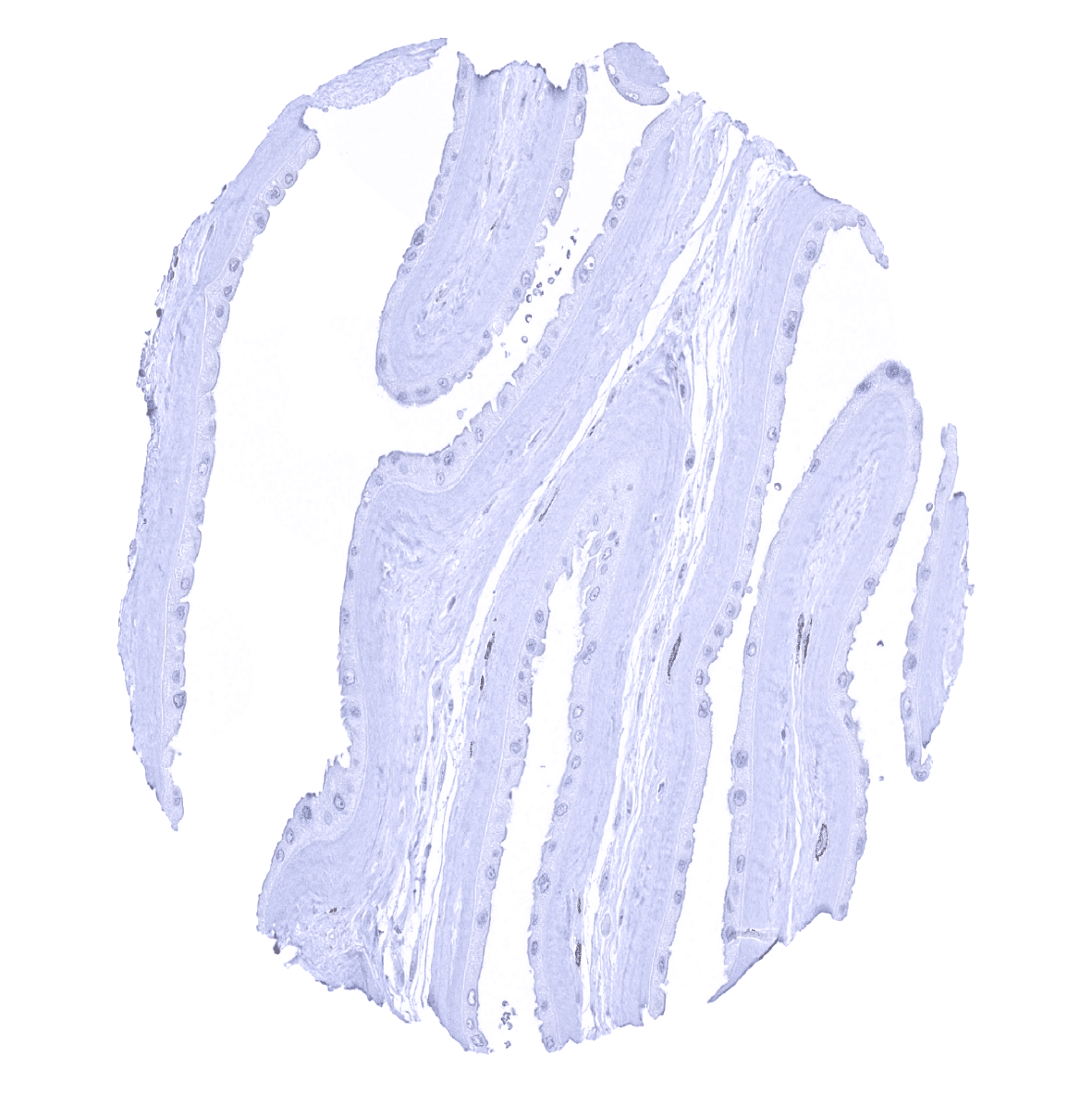
Aorta, media
Appendix, mucosa
Appendix, muscular wall

Bone marrow
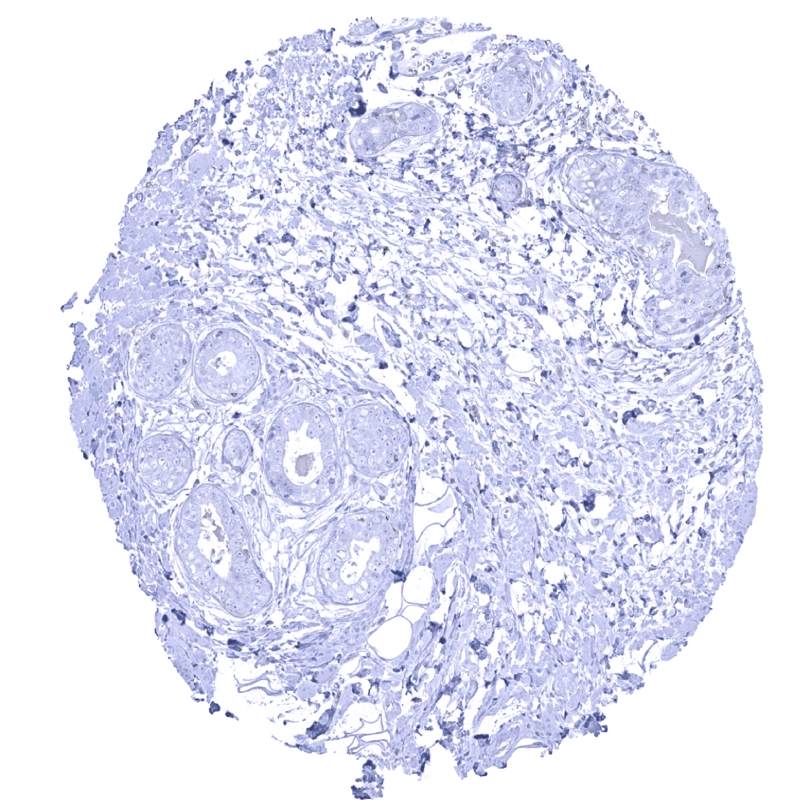
Breast
Bronchus, mucosa
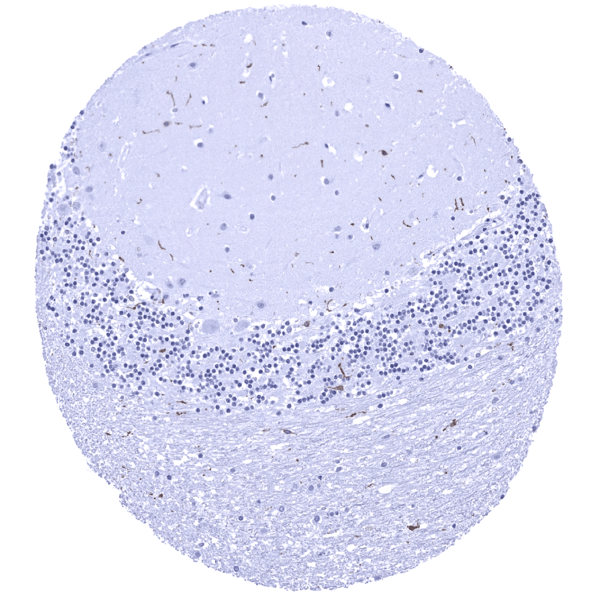
Cerebellum (molecular, Purkinje cell, granule cell layers; white matter) - A distinct AIF1 immunostaining is seen in microglia cells.
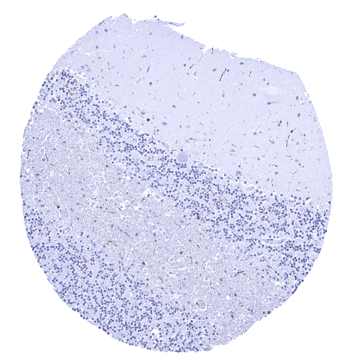
Cerebellum (molecular layer, Purkinje cell layer, granule cell layer, white matter) - A distinct AIF1 immunostaining is seen in microglia cells.
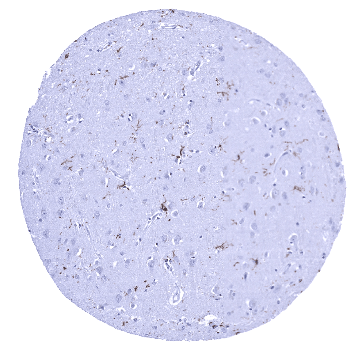
Cerebrum, grey matter - A distinct AIF1 immunostaining is seen in microglia cells.
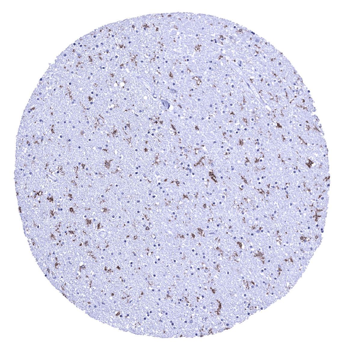
Cerebrum, white matter - A distinct AIF1 immunostaining is seen in microglia cells.
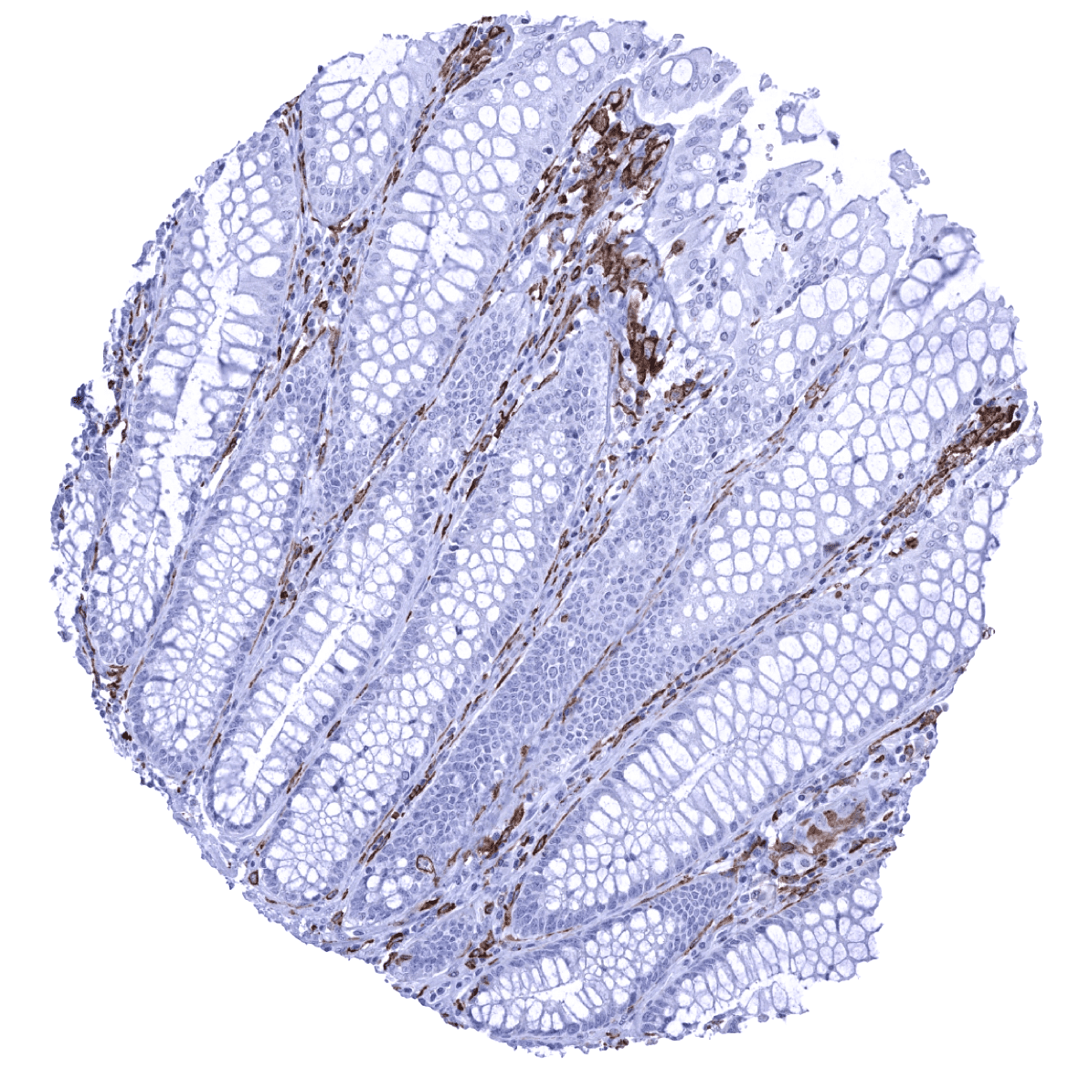
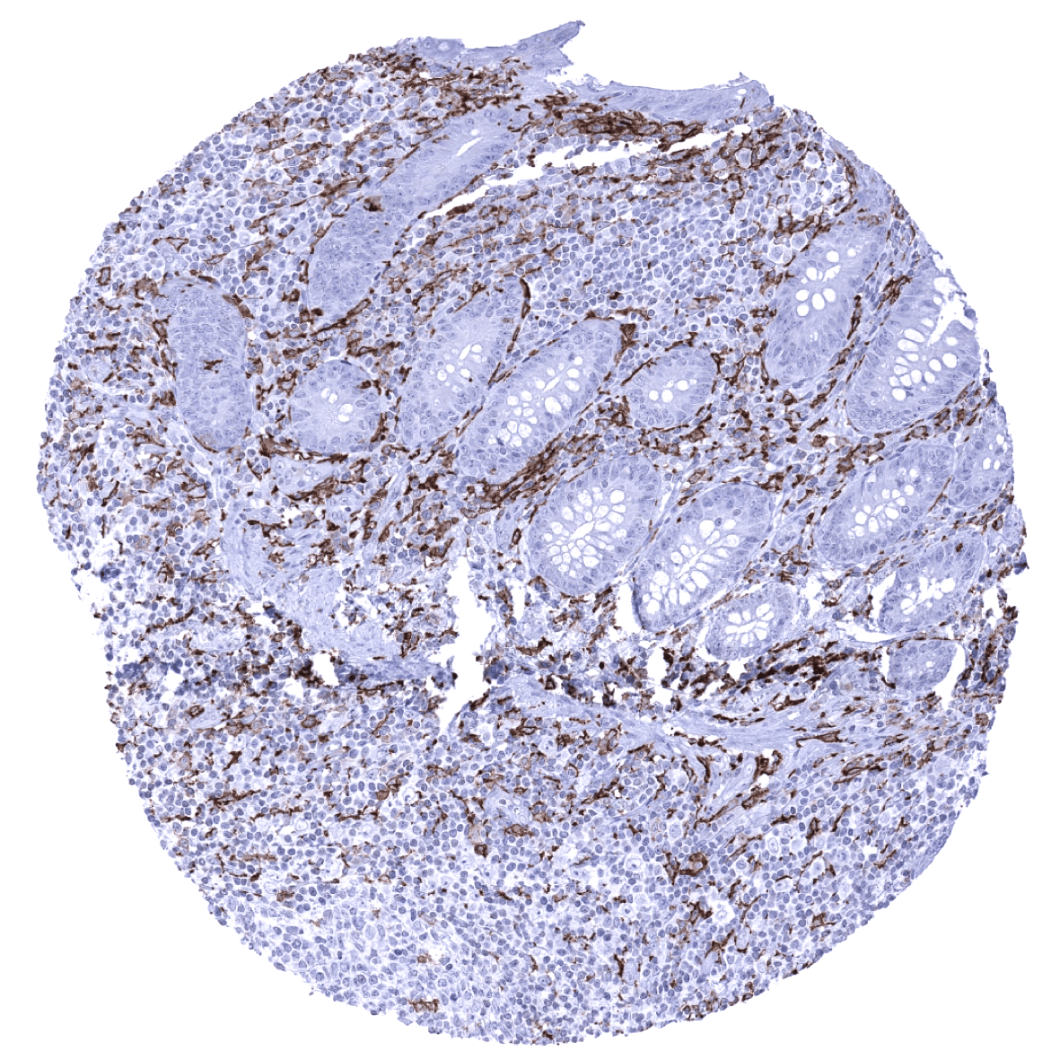
Colon descendens, mucosa - Strong AIF1 positivity is seen in mucosal macrophages.
Colon descendens, muscular wall

Duodenum, Brunner gland

Duodenum, mucosa - A strong AIF1 positivity is seen in mucosal macrophages.

Endocervix
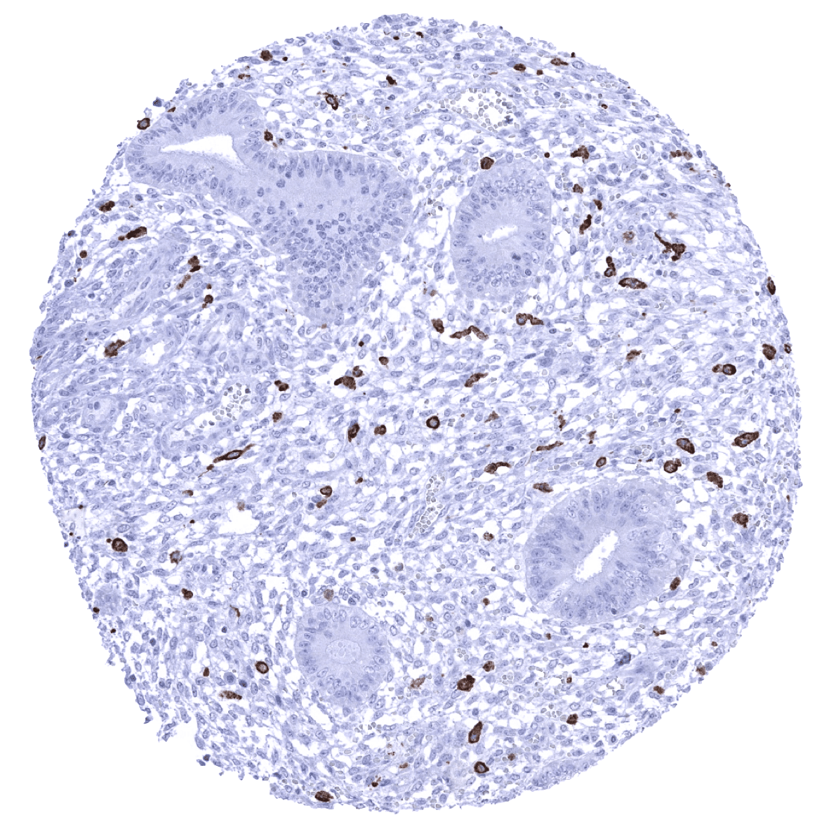
Endometrium, proliferation - Strong AIF1 positivity in scattered macrophages.
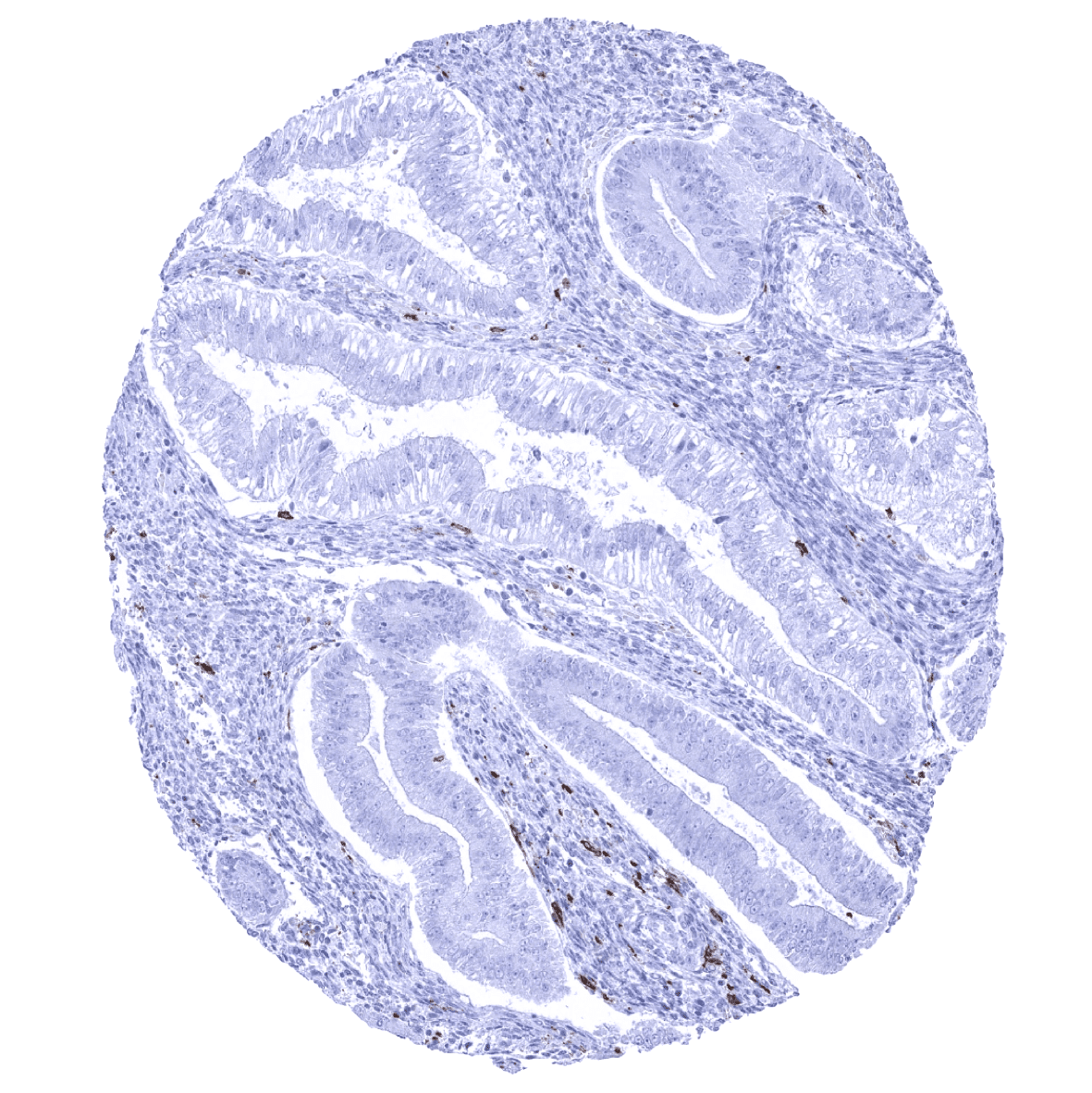
Endometrium
Endometrium, secretion
Epididymis
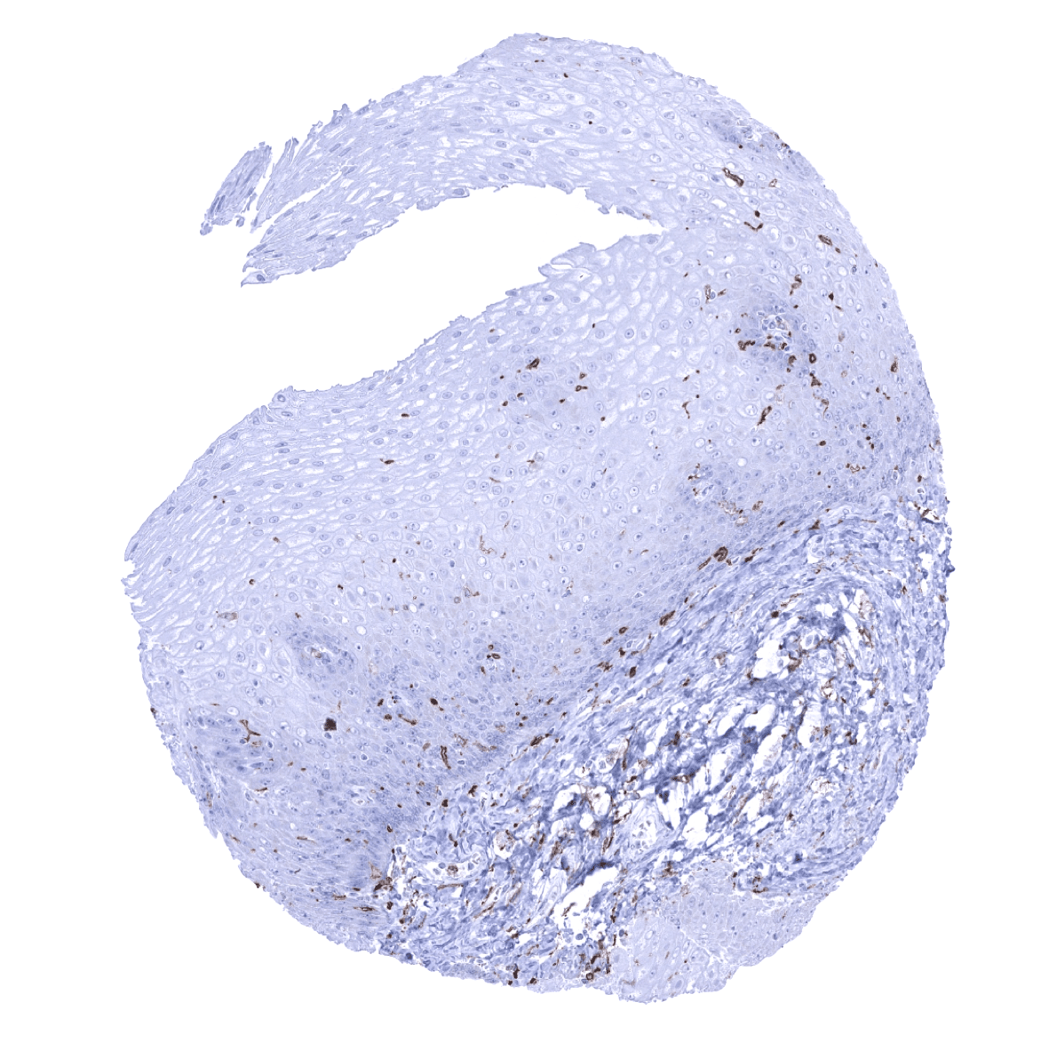
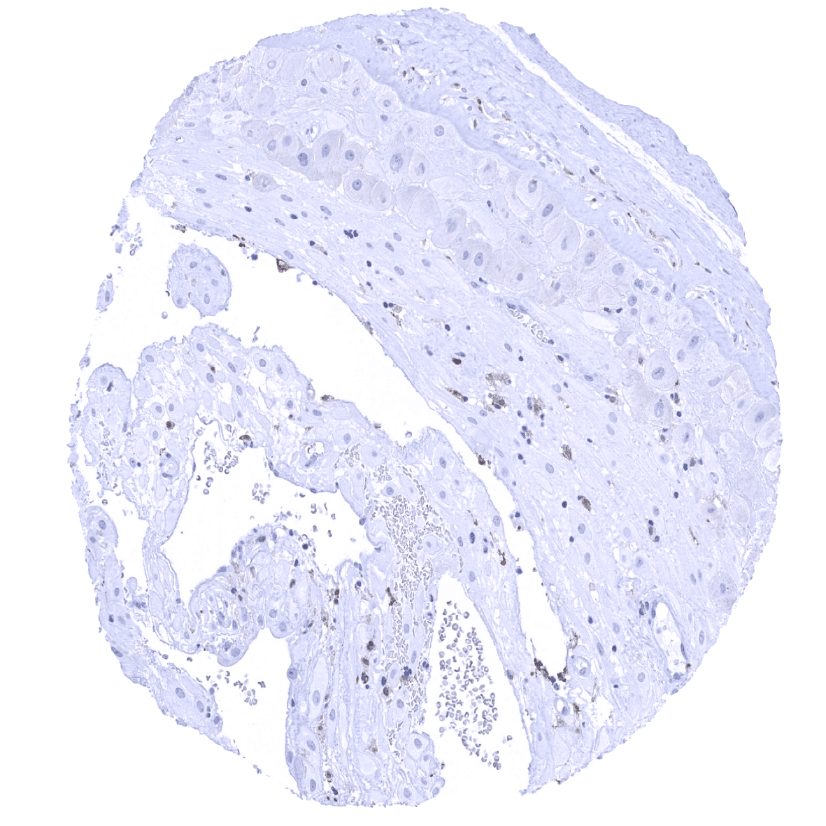
Esophagus, squamous epithelium
Fallopian tube, mucosa
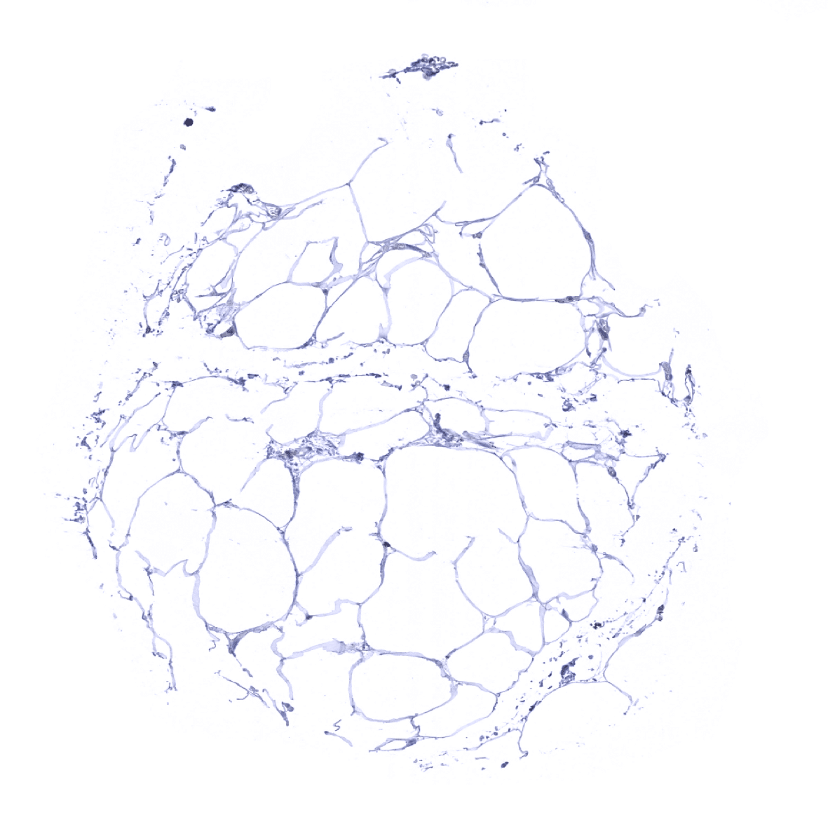
Fat
Gallbladder, epithelium

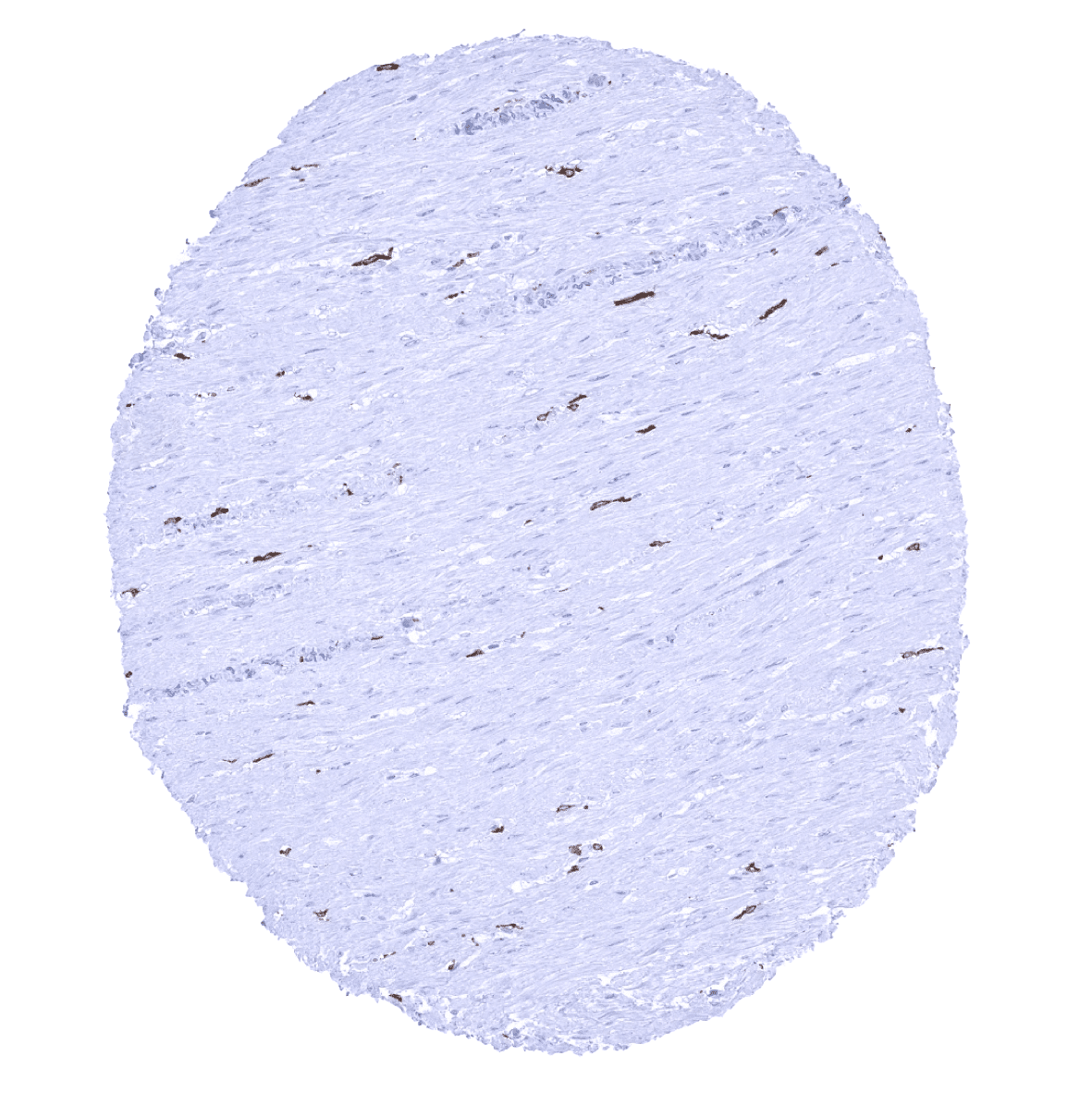
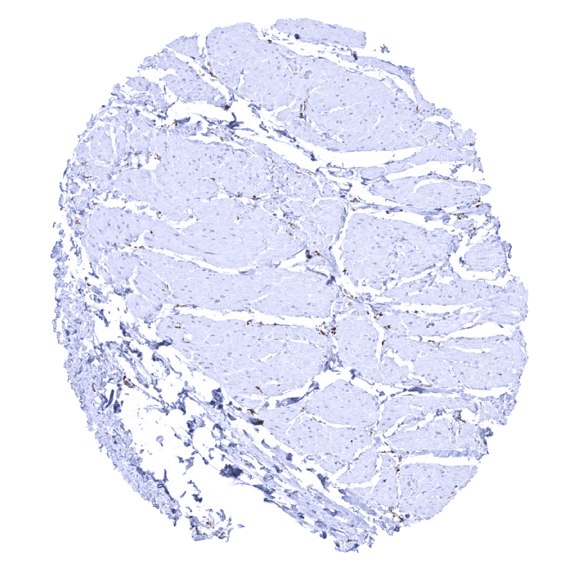
Heart muscle

Ileum, mucosa - A strong AIF1 positivity is seen in mucosal macrophages.
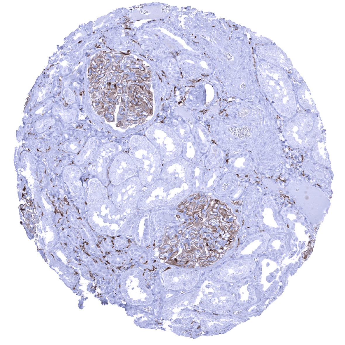
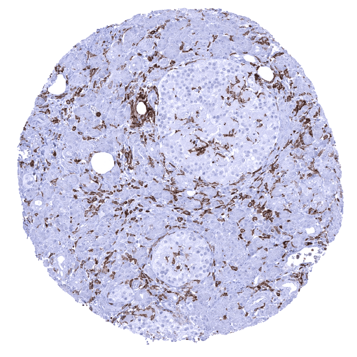
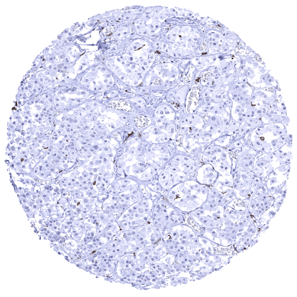
Kidney, cortex - A moderate to strong membranous AIF1 positivity is seen in glomeruli of the kidney. AIF1 staining also occurs in macrophages.
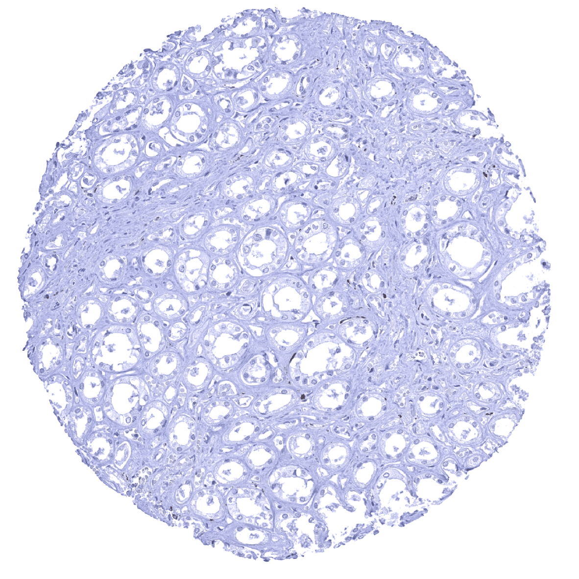
Kidney, medulla
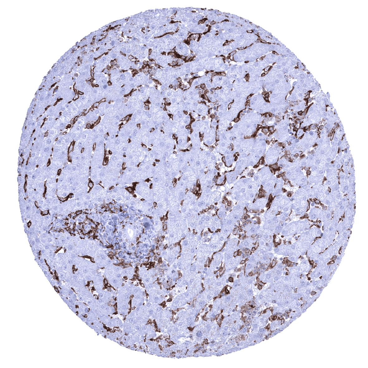
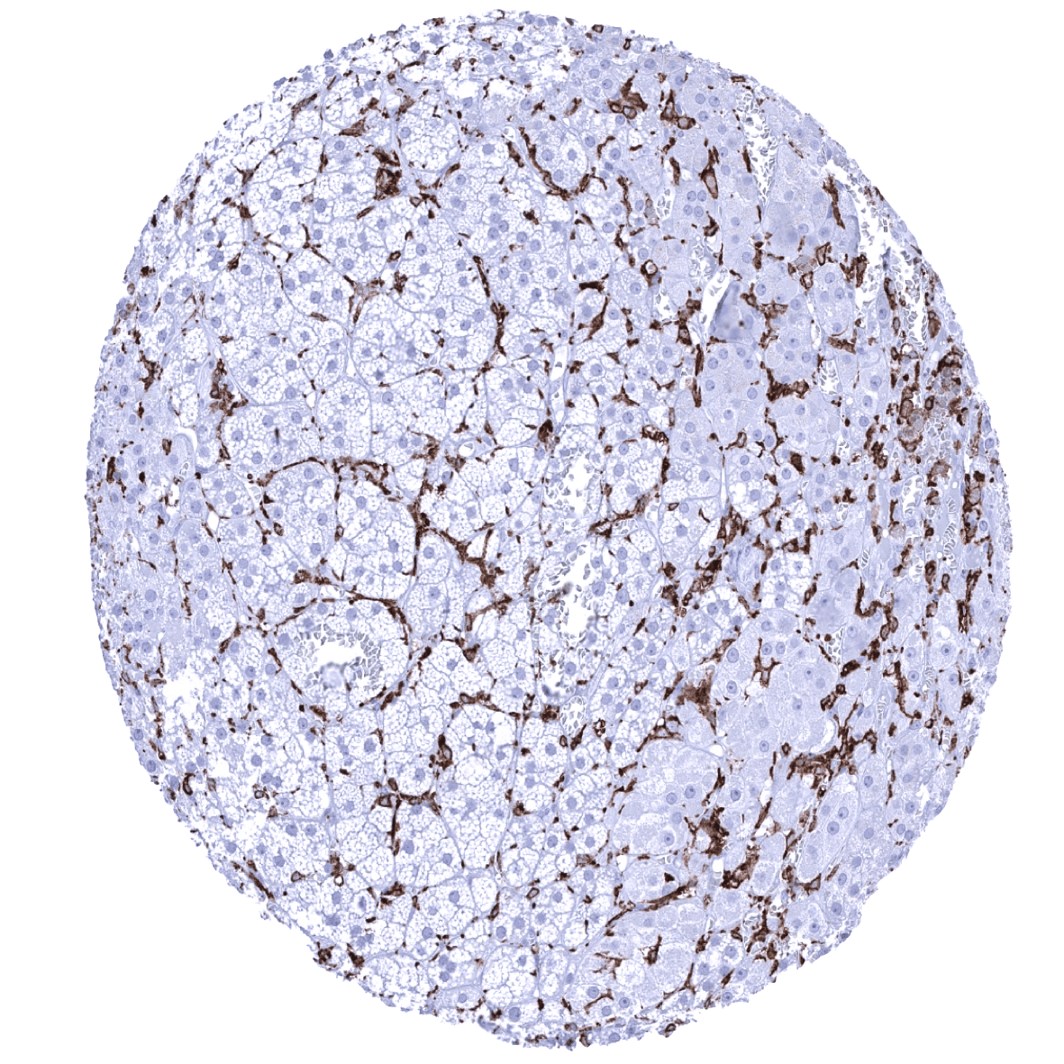
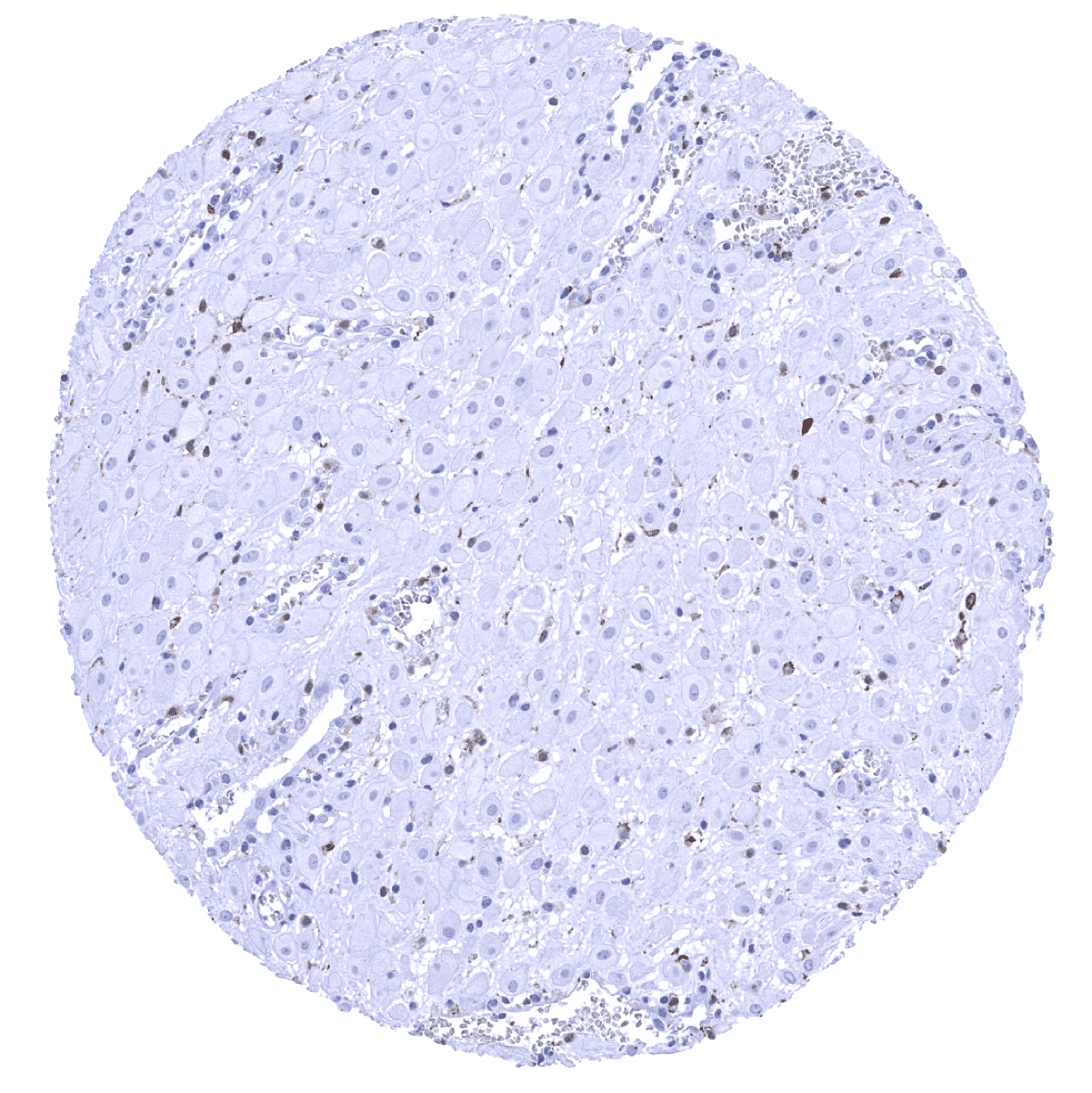
Liver - A strong AIF1 immunostaining occurs in Kupffer cells and intrasinusoidal monocytes.
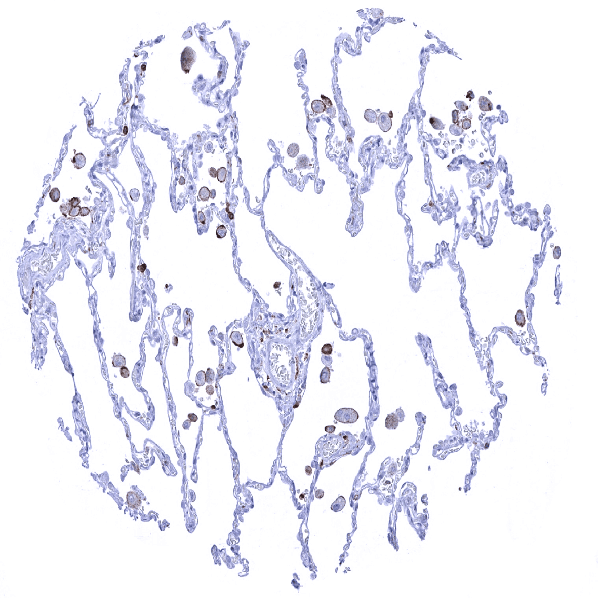
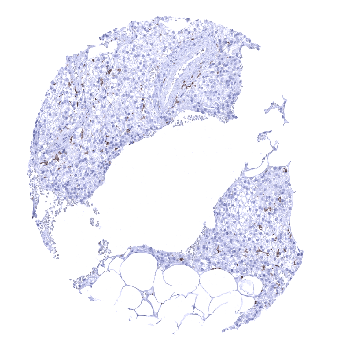
Lung - Alveolar macrophages show a significant AIF1 immunostaining.
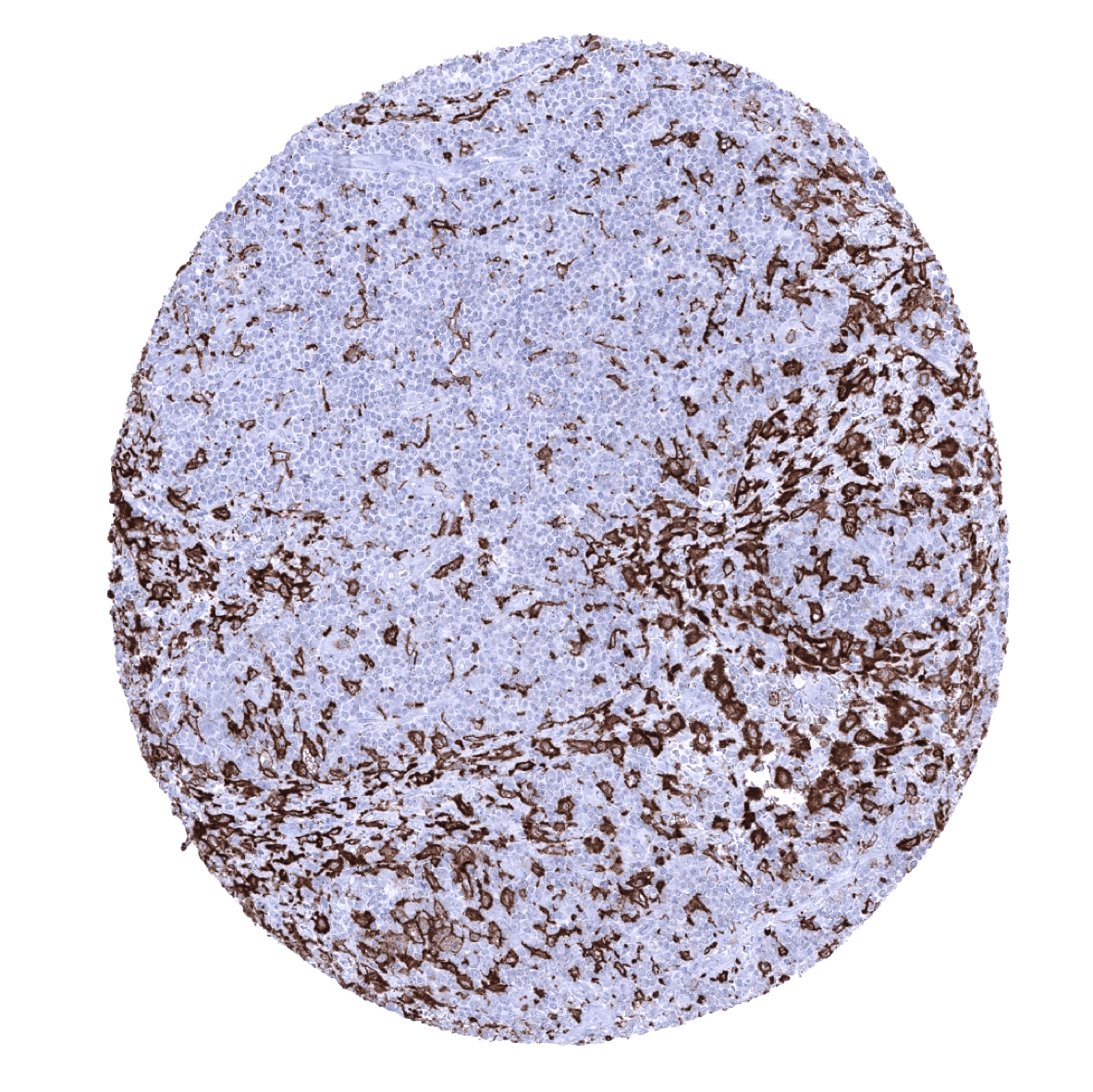
Lymph node - AIF1 positive macrophages are abundant in lymph nodes.
Ovary, stroma
Pancreas - A moderate to strong AIF1 positivity is seen in macrophages of the pancreas.
Parathyroid gland

Parotid gland
Pituitary gland, anterior lobe
Pituitary gland, posterior lobe
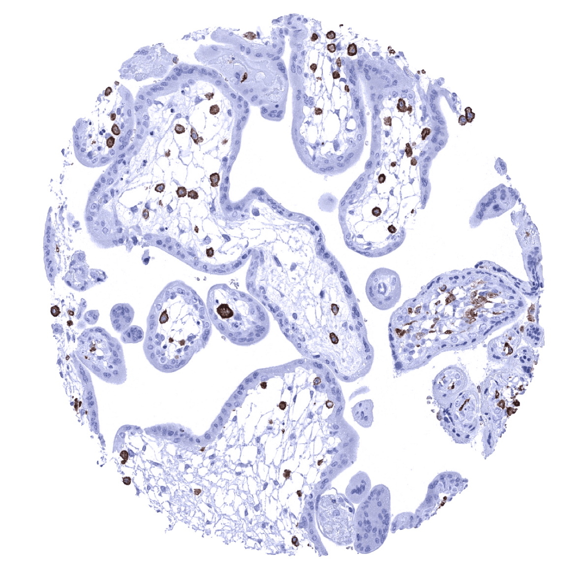
Placenta, early - AIF1 expression is particuarly strong in macrophages of the placenta.
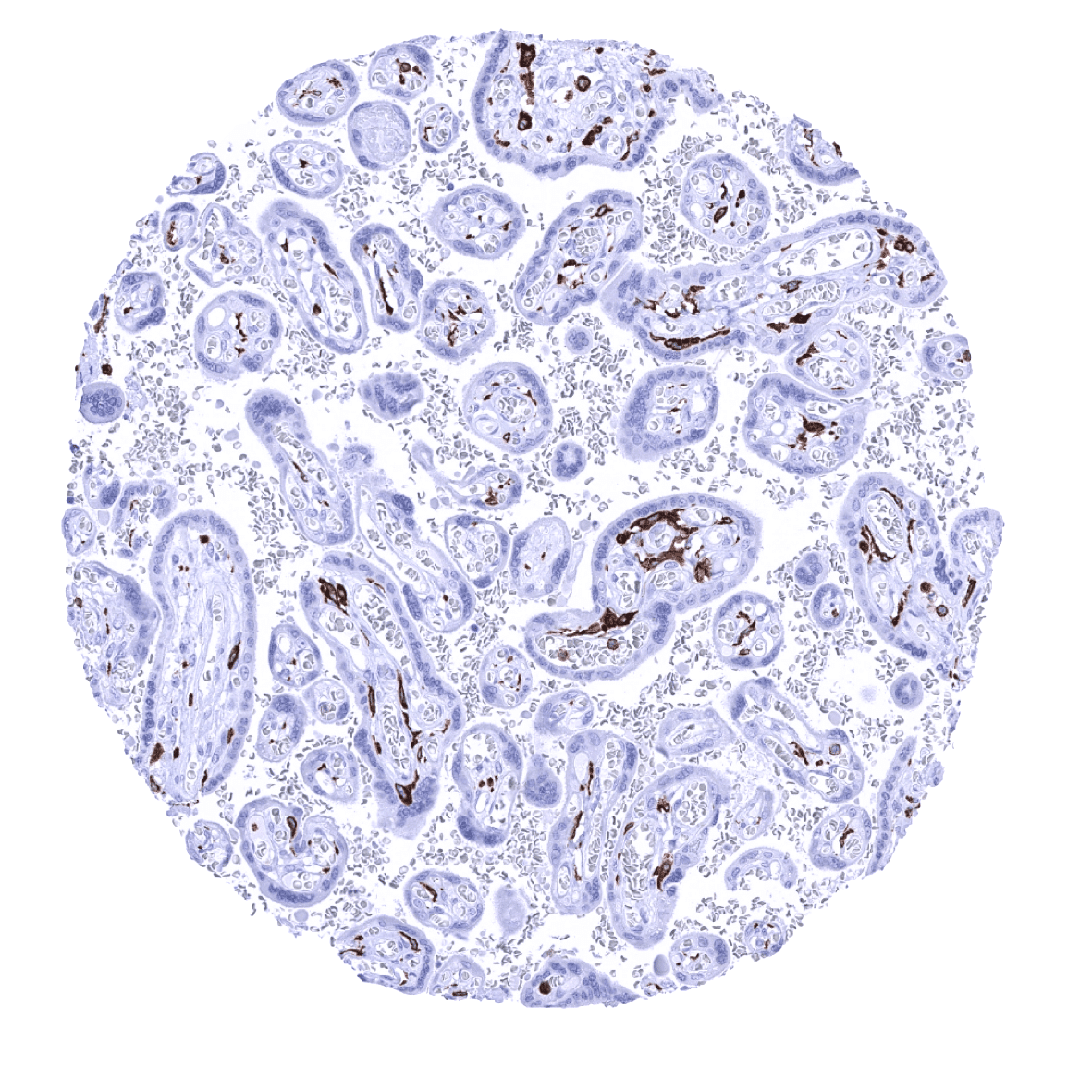
Placenta, mature – A IF1 expression is particuarly strong in macrophages of the placenta.
Placenta (amnion)
Placenta (chorion)
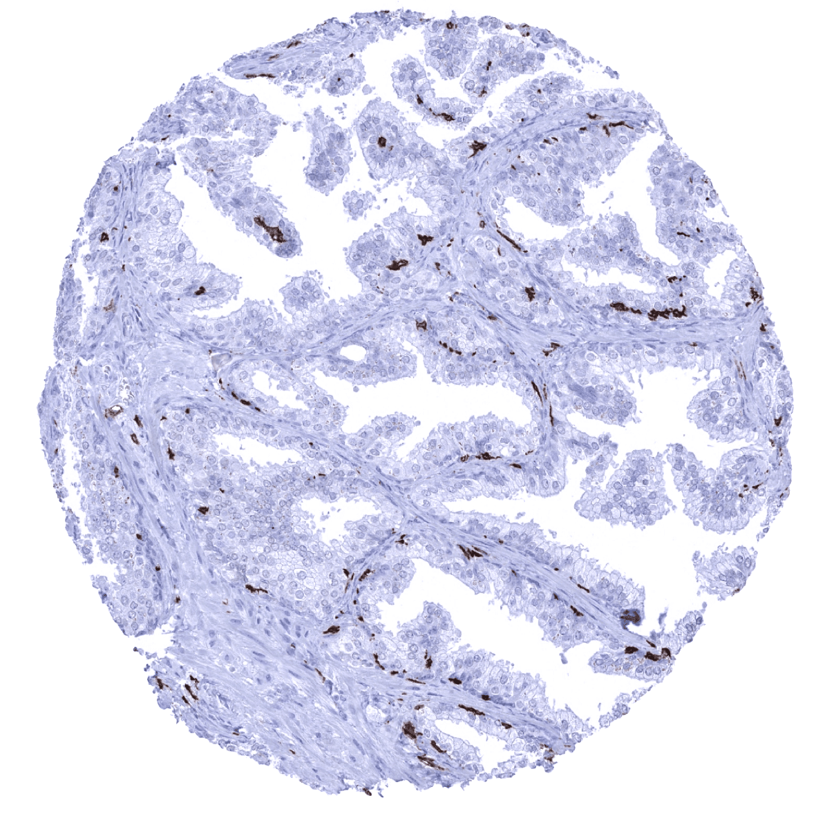
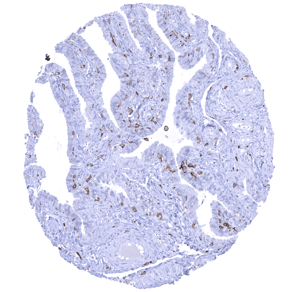
Prostate

Rectum, mucosa - A strong AIF1 positivity is seen in mucosal macrophages.

Seminal vesicle
Sinus paranasales
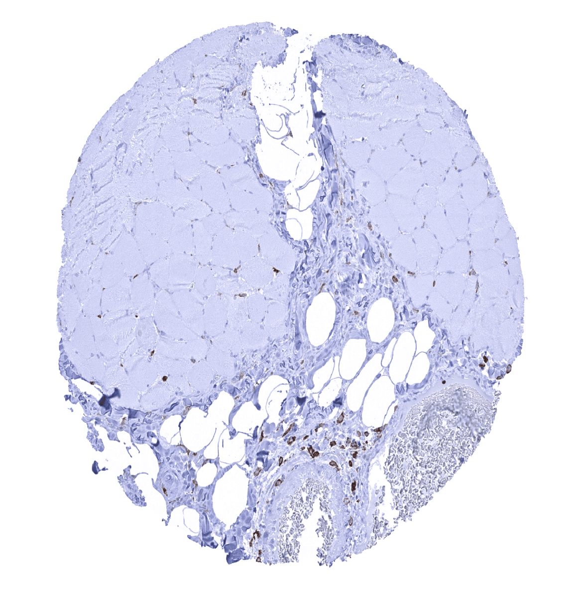
Skeletal muscle

Skin
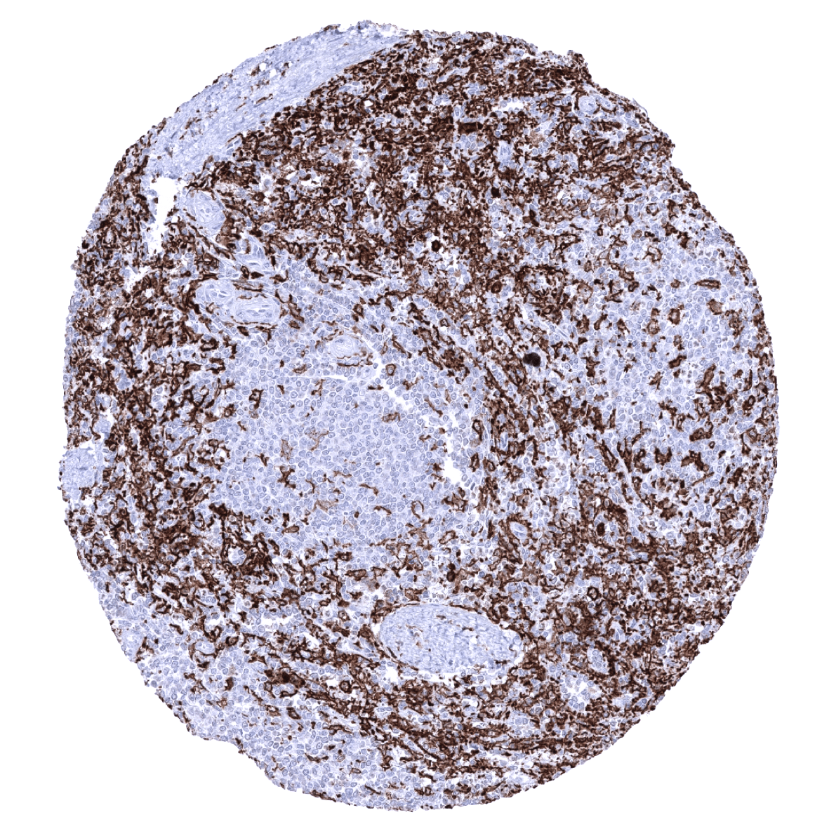
Spleen - Numerous AIF1 positive monocytes/macrophages are seen in the spleen.
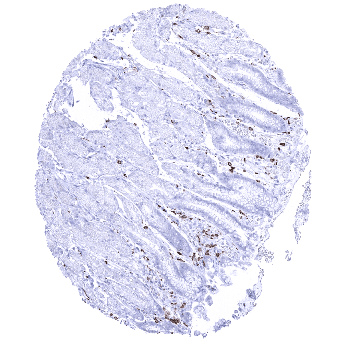
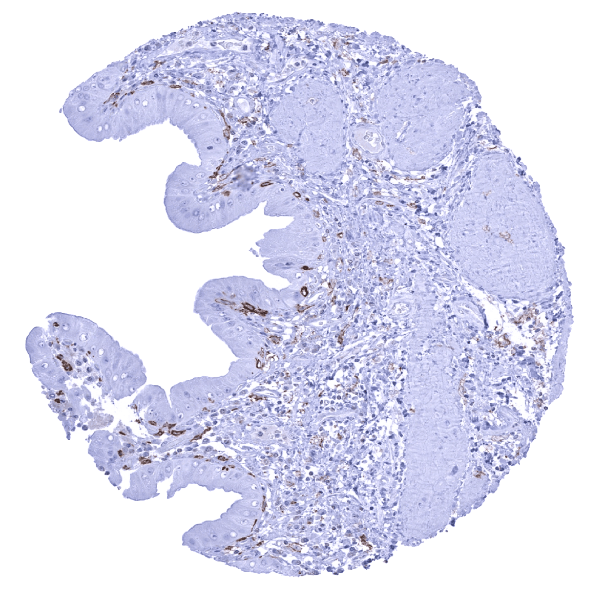
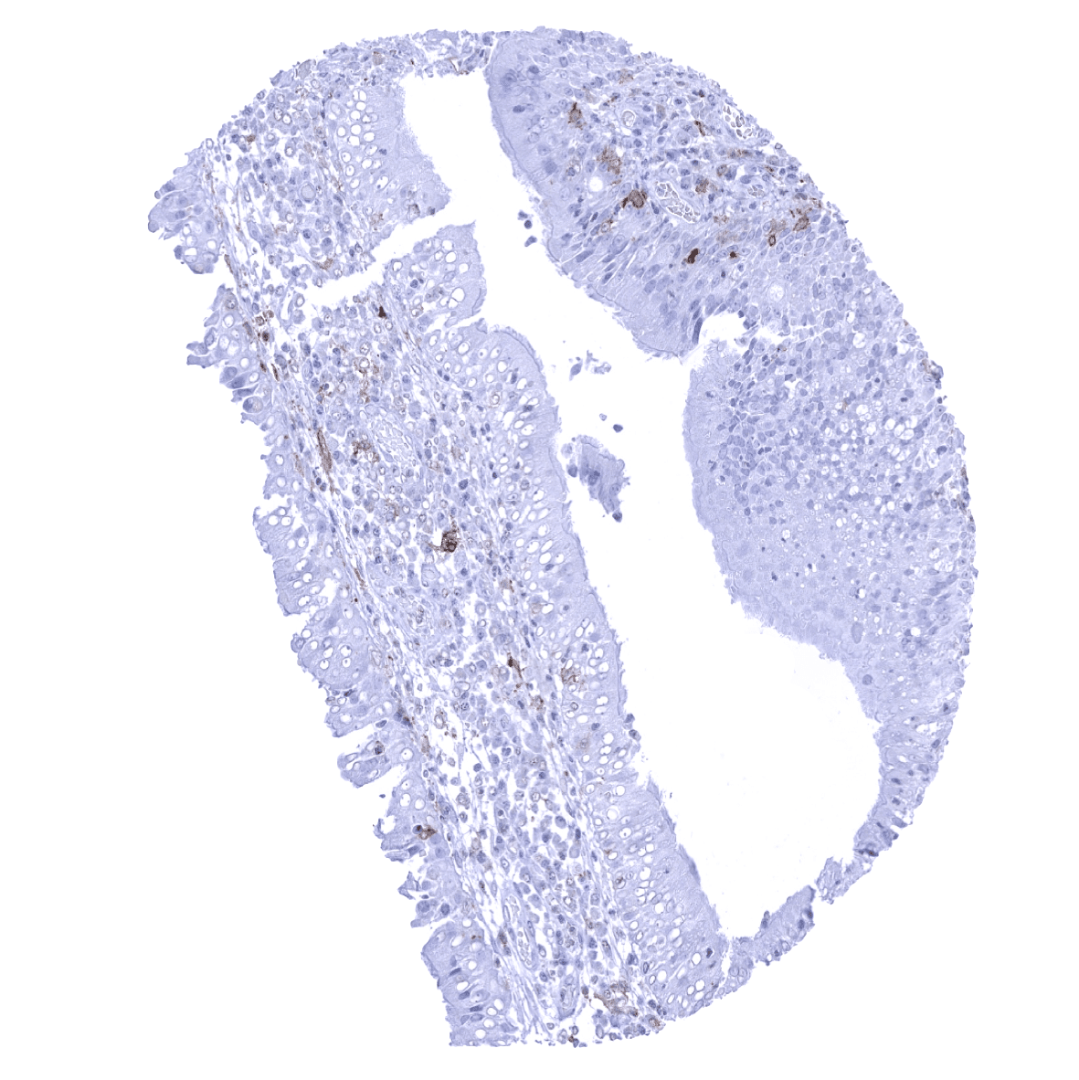
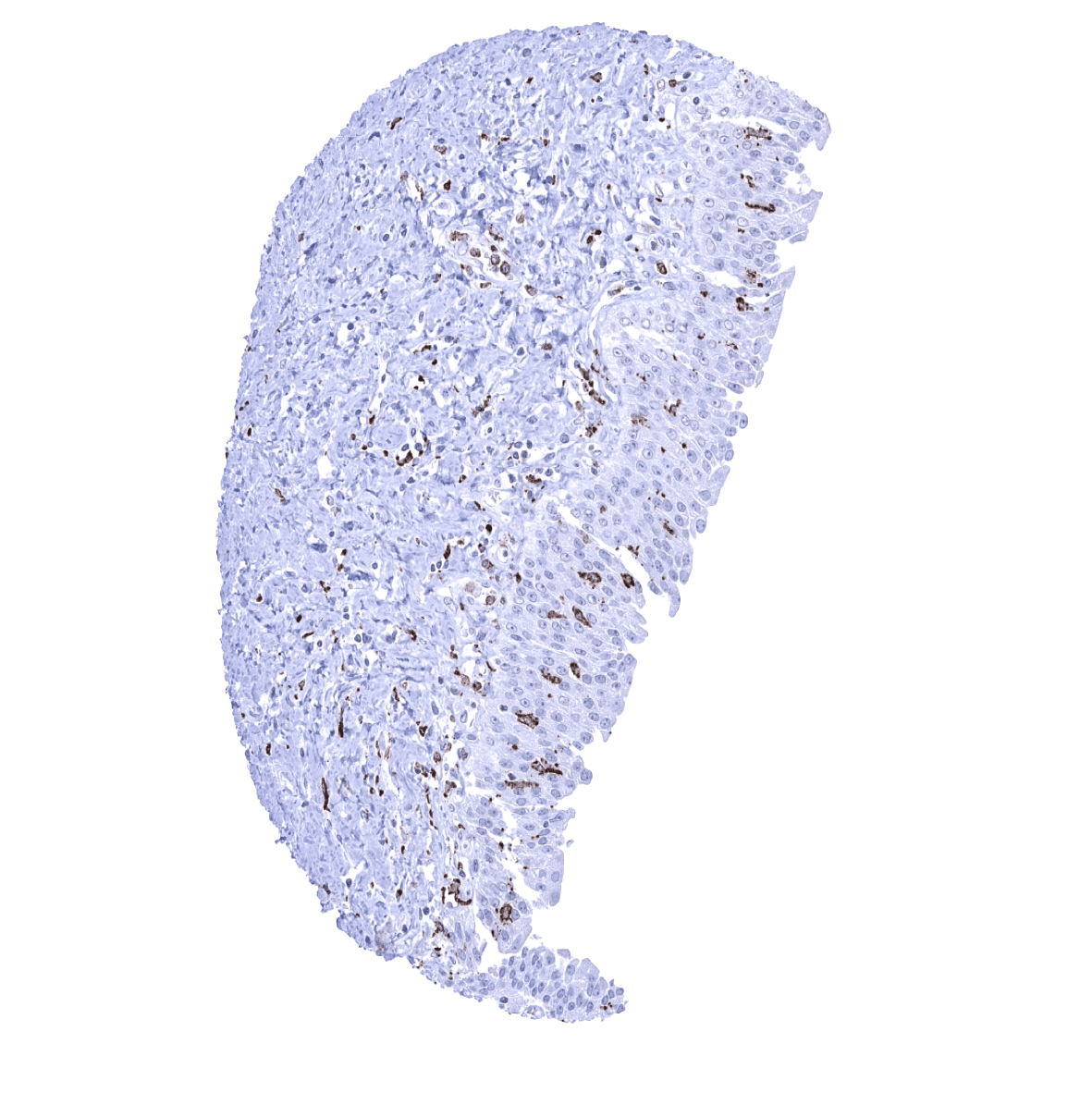
Stomach, antrum

Stomach, corpus

Testis
Thymus
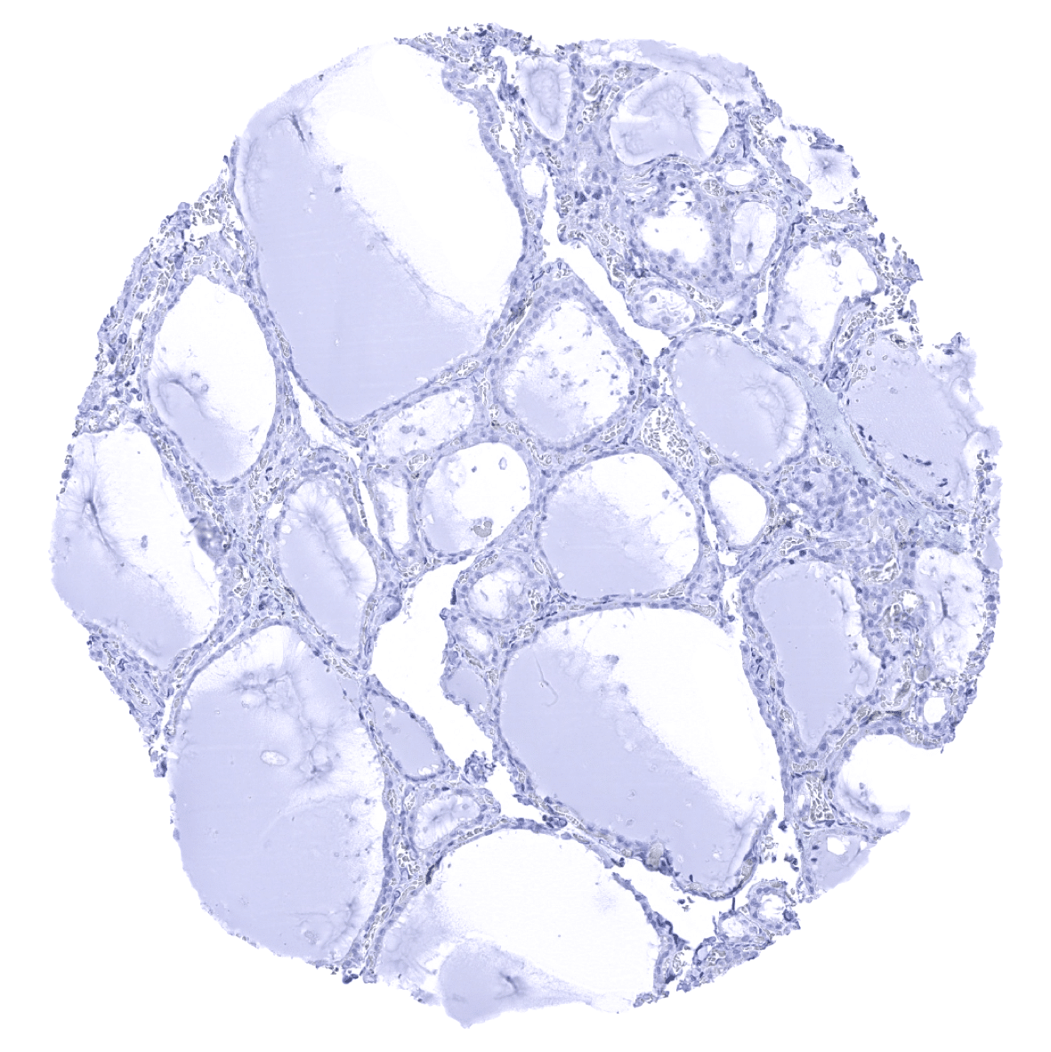
Thyroid gland
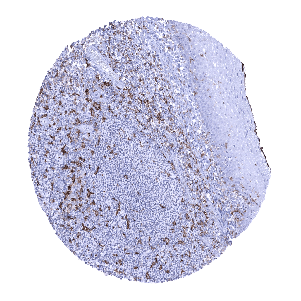
Tonsil, surface epithelium
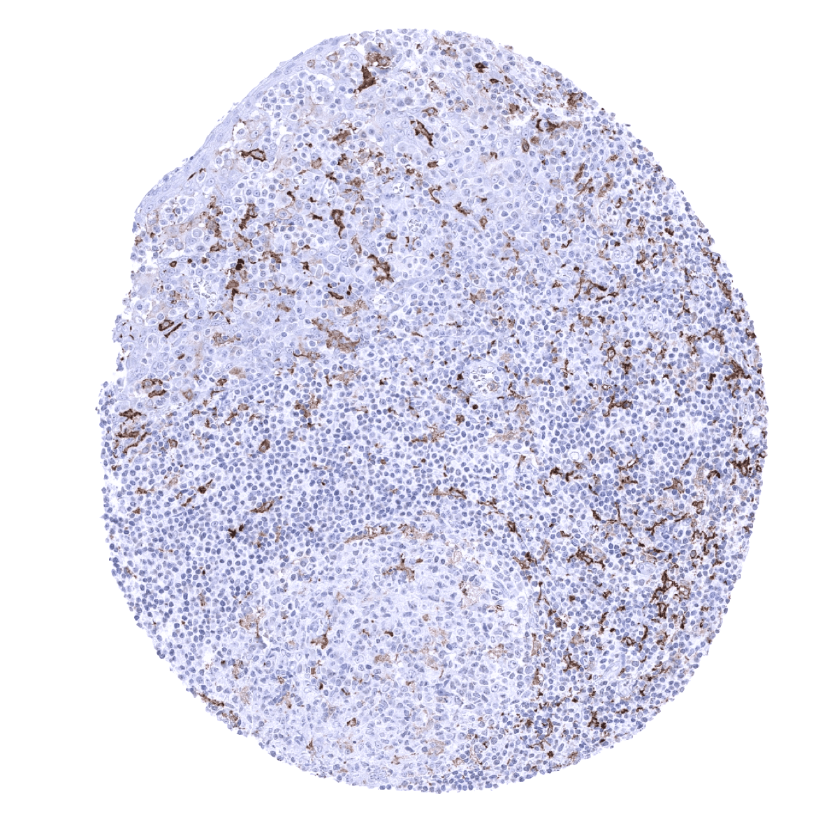
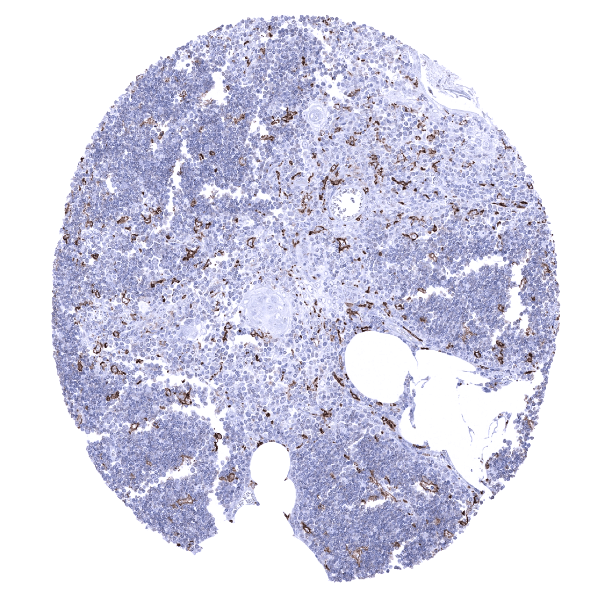
Tonsil
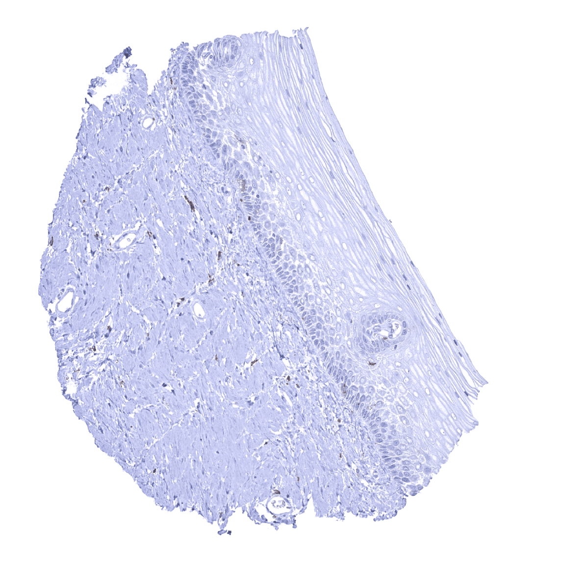
Urinary bladder, muscular wall
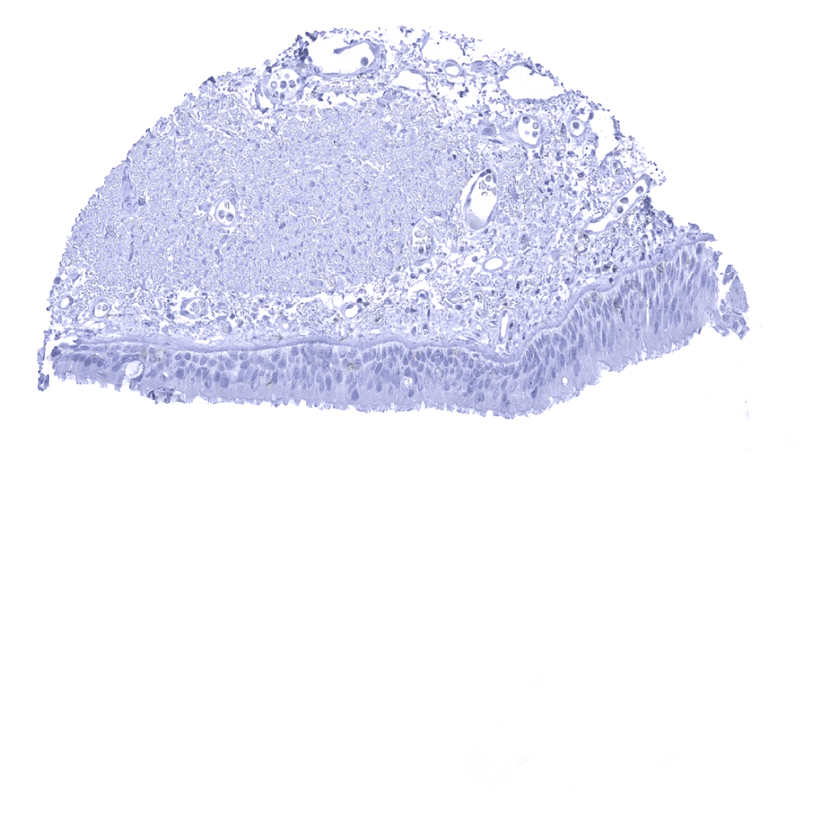
Urinary bladder, urothelium
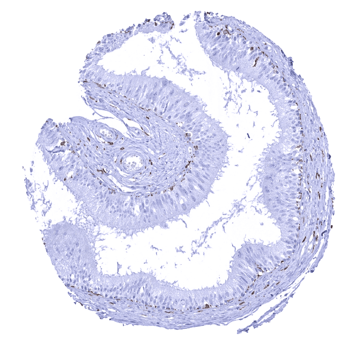
Uterus, ectocervix
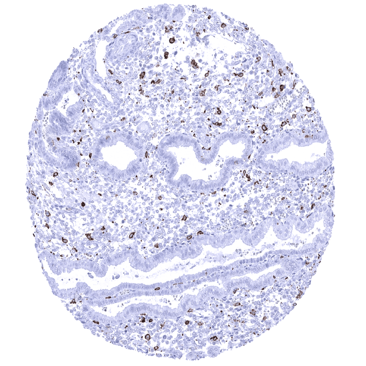
Uterus, endometrium (pregnancy)
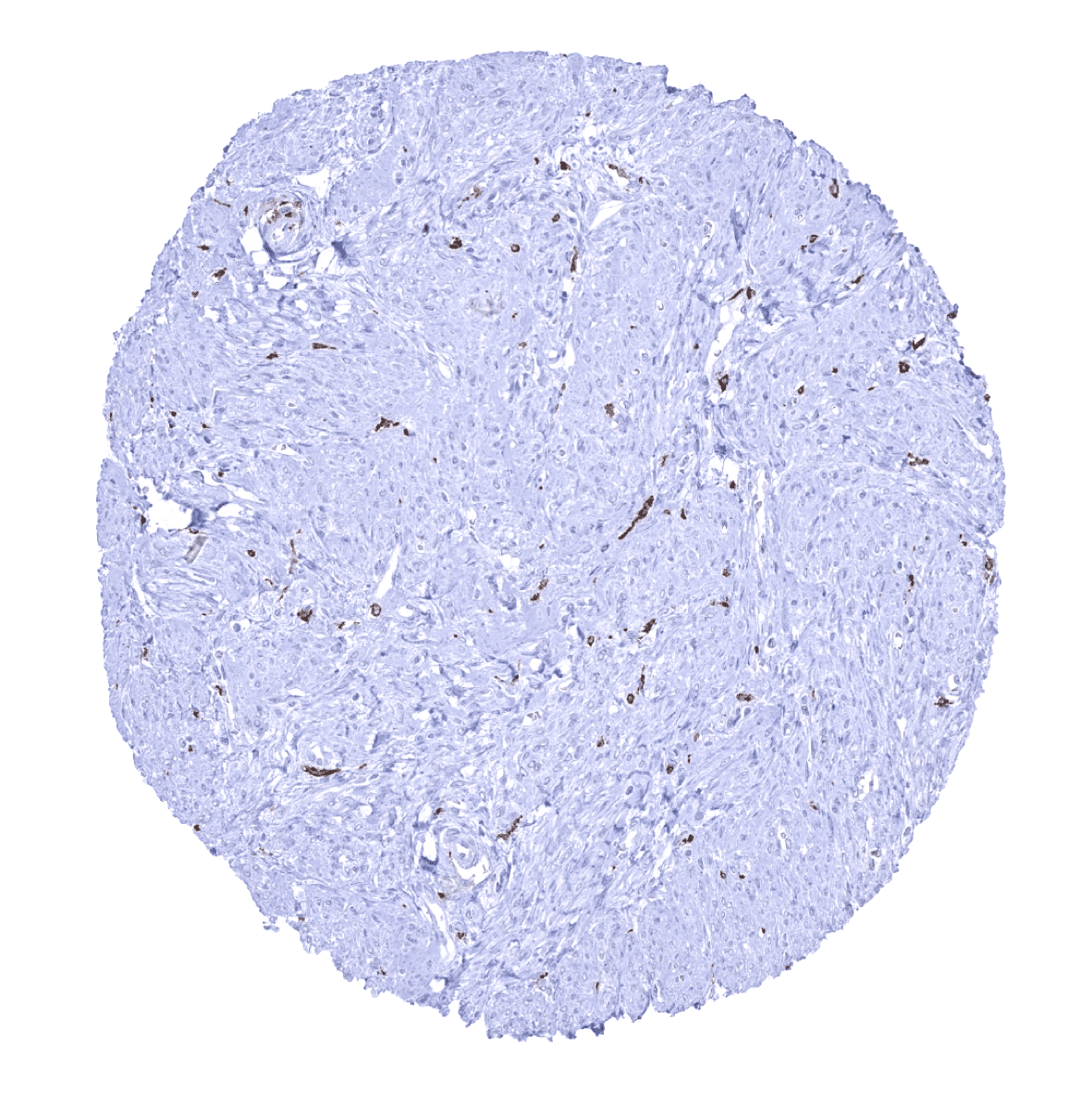
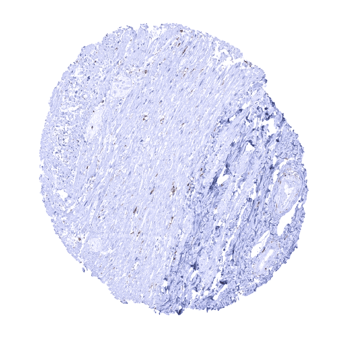
Uterus, myometrium