Product details
Synonyms = high mobility group AT-hook 2 , BABL , HMGI-C , HMGIC , LIPO , STQTL9
Antibody type = Recombinant Rabbit monoclonal / IgG
Clone = HMV314
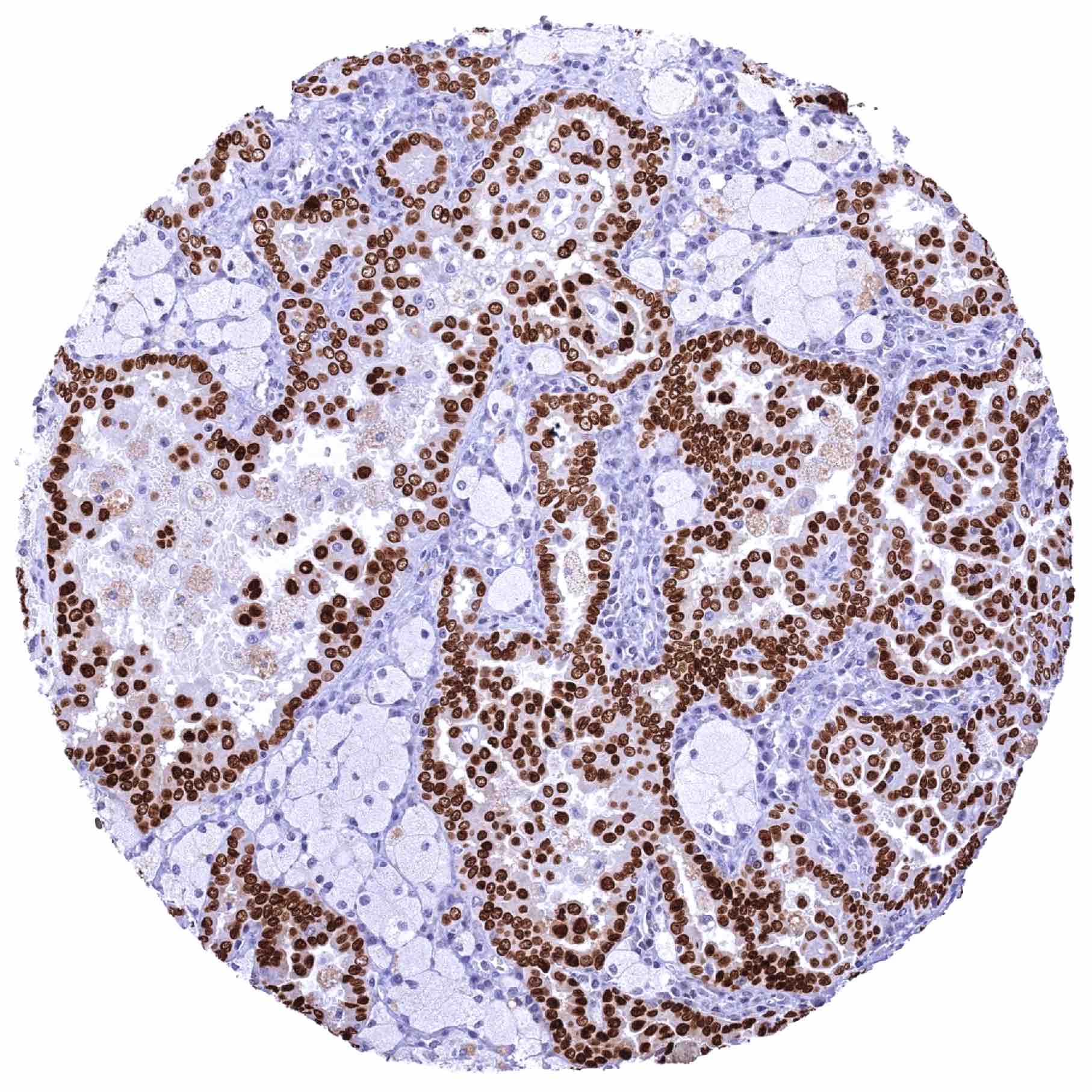
Positive control = Uterus, endocervix: A moderate to strong HMGA2 staining should be seen in epithelial cells of the endocervix.
Negative control = Skin: HMGA2 staining should be absent in all squamous epithelial cells.
Cellular localization = Intracellular
Reactivity = Human
Application = Immunohistochemistry
Dilution = 1:100 – 1:200
Intended Use = Research Use Only
Relevance of Antibody
HMGA2 is a key component of the enhanceosome.
Biology Behind
The high mobility group protein 2 (HMGA2) is a protein of less than 12 kDa, that consists of 108 amino acids and which is coded by the HMGA2 gene located at 12q13-15. The first three (of five) exons encode the AT-binding domain site, which can bind to AT-rich binding sites in the DNA minor groove. These bindings affect the conformation of the DNA and modify transcription by enhancing or suppressing the activities of numerous genes. As such, HMGA2 is an essential component of the enhanceosome. Instead of direct regulation, HMGA2 alters the architecture of DNA and supports the assembly of protein complexes that do regulate the transcription of genes. HMGA2 is preferentially expressed during organogenesis and – with few exceptions – it is hardly expressed in adult tissues. HMGA2 appears to be essential for cell growth regulation. A knock-out of the HMGA2 counterpart in mice results in diet-induced obesity. Specific HMGA2 mutations can lead to unusually small size in mice. Genome-wide association studies have found a relationship between height of humans and HMGA2-linked SNPs. Data suggesting a role of HMGA2 in cancer are rapidly accumulating. Aberrant expression of HMGA2 in adult tissues is commonly associated with both benign and malignant tumor formation. HMGA2 is often re-expressed in human tumors, where it promotes tumorigenesis by multiple mechanisms. HMGA2 was found to increase cancer cell proliferation, inhibit apoptosis, impact several DNA repair mechanisms, endorse epithelial-mesenchymal transition, support a cancer stem cell phenotype, and to foster cancer cell resistance to chemotherapeutic agents. Causes for HMGA2 re-expression in neoplastic tissues include gene fusion, amplification, regulation by specific miRNAs, and other mechanisms.
Staining Pattern in Normal Tissues
Images describing the HMGA2 staining pattern in normal tissues obtained by the antibody HMV314 are shown in our “Normal Tissue Gallery”.
| Brain | Cerebrum | Negative. |
| Cerebellum | Negative. | |
| Endocrine Tissues | Thyroid | Most follicular cells show a faint to weak nuclear HMGA2 staining. |
| Parathyroid | Some epithelial cell groups show a moderate to strong nuclear HMGA2 staining | |
| Adrenal gland | Negative. | |
| Pituitary gland | Weak to moderate nuclear HMGA2 staining of pituicytes. Adenohypophysis is HMGA2 negative. | |
| Respiratory system | Respiratory epithelium | Strong nuclear HMGA2 staining of most respiratory epithelial cells. |
| Lung | Negative. | |
| Gastrointestinal Tract | Salivary glands | Negative. |
| Esophagus | Negative. | |
| Stomach | Weak to moderate nuclear HMGA2 positivity of a fraction of glandular cells. | |
| Duodenum | Weak nuclear HMGA2 staining of duodenal crypt cells. Moderate HMGA2 positivity of Brunner glands. | |
| Small intestine | Very faint nuclear HMGA2 positivity of some epithelial cells. | |
| Appendix | Weak nuclear HMGA2 staining of a fraction of epithelial cells. | |
| Colon | Weak nuclear HMGA2 staining of a fraction of epithelial cells. | |
| Rectum | Weak nuclear HMGA2 staining of a fraction of epithelial cells. | |
| Liver | Weak nuclear HMGA2 staining of a fraction of bile duct epithelial cells. Hepatocytes are negative. | |
| Gallbladder | Negative. | |
| Pancreas | Weak nuclear HMGA2 staining of a fraction of acinar and ductal (small ducts) cells. | |
| Genitourinary | Kidney | Moderate nuclear HMGA2 staining of tubular cells in areas of tissue damage. Faint nuclear HMGA2 staining of a fraction of collecting duct cells. |
| Urothelium | Few urothelial cells show a weak nuclear HMGA2 positivity. | |
| Male genital | Prostate | Negative. |
| Seminal vesicles | Strong nuclear HMGA2 staining of a large fraction of acinar glandular cells. | |
| Testis | Weak to moderate nuclear HMGA2 staining of a spermatocytes and of spermatozoa. | |
| Epididymis | Weak nuclear HMGA2 staining of a fraction of epithelial cells in the cauda while cells in the corpus are HMGA2 negative. | |
| Female genital | Breast | Negative. |
| Uterus, myometrium | Negative. | |
| Uterus, ectocervix | Negative. | |
| Uterus endocervix | Strong nuclear HMGA2 staining of epithelial cells. | |
| Uterus, endometrium | Weak to moderate nuclear HMGA2 staining of a subset of epithelial cells. | |
| Fallopian Tube | Strong nuclear HMGA2 staining of most epithelial cells. | |
| Ovary | Negative. | |
| Placenta early | Strong nuclear HMGA2 staining of stroma cells. Trophoblast cells remain HMGA2 negative. | |
| Placenta mature | Some nuclear HMGA2 staining of trophoblast cells in some samples. Stroma cells are HMGA2 negative. | |
| Amnion | Strong nuclear HMGA2 positivity of amnion cells. | |
| Chorion | Negative. | |
| Skin | Epidermis | Negative. |
| Sebaceous glands | Negative. | |
| Muscle/connective tissue | Heart muscle | Negative. |
| Skeletal muscle | Weak HMGA2 staining of some myocytes | |
| Smooth muscle | Negative. | |
| Vessel walls | Negative. | |
| Fat | Negative. | |
| Stroma | Negative. | |
| Endothelium | Negative. | |
| Bone marrow/ lymphoid tissue | Bone marrow | Moderate nuclear HMGA2 staining of a fraction of cells. |
| Lymph node | Negative. | |
| Spleen | Negative. | |
| Thymus | Negative. | |
| Tonsil | Negative. | |
| Remarks | HMGA2 staining appears to occur in areas of tissue damage. |
The findings described above are thus consistent with the RNA data described in the Human Protein Atlas (Tissue expression HMGA2).
Positive control = Uterus, endocervix: A moderate to strong HMGA2 staining should be seen in epithelial cells of the endocervix.
Negative control = Skin: HMGA2 staining should be absent in all squamous epithelial cells.
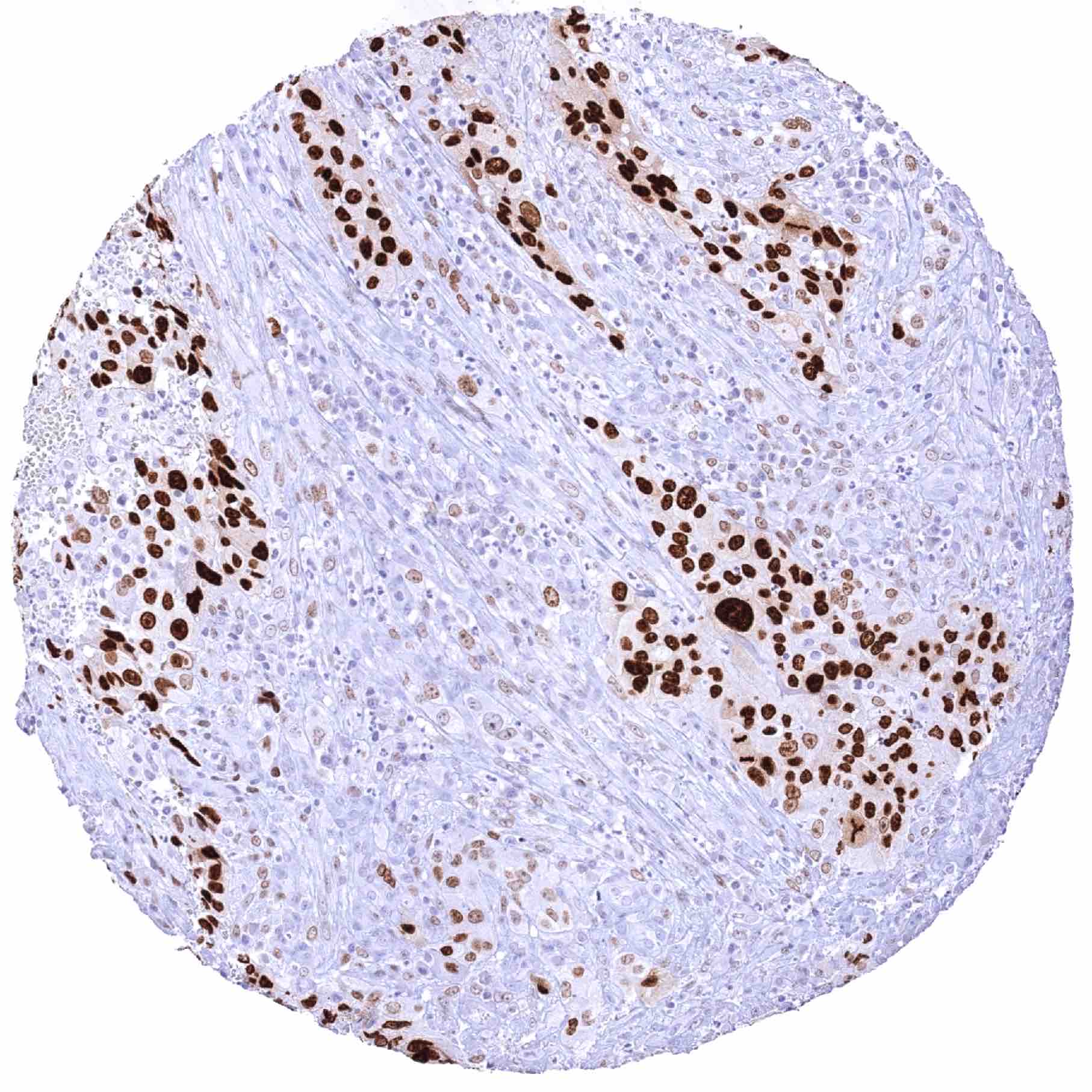
Staining Pattern in Relevant Tumor Types
HMGA2 is expressed in various different tumor entities.
The TCGA findings on HGMA2 RNA expression in different tumor categories have been summarized in the Human Protein Atlas.
Compatibility of Antibodies
No data available at the moment
Protocol Recommendations
IHC users have different preferences on how the stains should look like. Some prefer high staining intensity of the target stain and even accept some background. Others favor absolute specificity and lighter target stains. Factors that invariably lead to more intense staining include higher concentration of the antibody and visualization tools, longer incubation time, higher temperature during incubation, higher temperature and longer duration of the heat induced epitope retrieval (slide pretreatment). The impact of the pH during slide pretreatment has variable effects and depends on the antibody and the target protein.
All images and data shown here and in our image galleries are obtained by the manual protocol described below. Other protocols resulting in equivalent staining are described as well.
Manual protocol
Freshly cut sections should be used (less than 10 days between cutting and staining). Heat-induced antigen retrieval for 5 minutes in an autoclave at 121°C in pH 7,8 Target Retrieval Solution buffer. Apply HMV314 at a dilution of 1:150 at 37°C for 60 minutes. Visualization of bound antibody by the EnVision Kit (Dako, Agilent) according to the manufacturer’s directions.
Potential Research Applications
- The diagnostic, prognostic, and predictive relevance of HMGA2 expression measurement in tumors and in preneoplastic disease needs to be investigated.
- HMGA2 is considered a potential therapeutic target.
Evidence for Antibody Specificity in IHC
There are two ways how the specificity of antibodies can be documented for immunohistochemistry on formalin fixed tissues. These are: 1. Comparison with a second independent method for target expression measurement across a large number of different tissue types (orthogonal strategy), and 2. Comparison with one or several independent antibodies for the same target and showing that all positive staining results are also seen with other antibodies for the same target (independent antibody strategy).
Orthogonal validation: For the antibody HMV314 specificity is suggested by the strong concordance of the immunostaining data with data from three independent RNA screening studies, including the Human Protein Atlas (HPA) RNA-seq tissue dataset, the FANTOM5 project, and the Genotype-Tissue Expression (GTEx) project, which are all summarized in the Human Protein Atlas (Tissue expression HMGA2). HMGA2 positivity by HMV314 is detectable in almost all tissues with documented HMGA2 RNA expression (thyroid, pituitary gland, stomach, colorectum, appendix, small intestine, duodenum, pancreas, testis, cervix uteri, endometrium, fallopian tube, and bone marrow). Ovary and the skin were the only organs with previously described RNA expression but absence of HMGA2 staining by HMV314. Tissues without previously documented HMGA2 RNA expression but HMGA2 positivity by the antibody HMV314 had either very few positive cells that were probably not detected in RNA studies (bile ducts in the liver, some tubuli and collecting ducts in the kidney, distinct groups of epithelial cells in the parathyroid, amnion and stroma cells of the placenta, acinar glandular cells of the seminal vesicle, and a fraction of epithelial cells in the cauda epididymis) or were previously not analyzed on the RNA level (respiratory epithelial cells).
Comparison of antibodies: True expression of HMGA2 in all cell types found to be HMGA2 positive by HMV314 is corroborated by identical stainings of all positive cell types by another commercially available antibody (termed “validation antibody”). With this “validation antibody” an HMGA2 staining in the ovary or the skin could also not be achieved for the tissues analyzed. Independence of the validation antibody is documented by cytoplasmic staining of the granular cell layer in keratinizing squamous epithelium and a granular cytoplasmic apical staining in GIT epithelium which was only seen by the validation antibody and not by HMV314. These stainings are considered antibody specific cross-reactivities of the validation antibody.