295,00 € – 895,00 €
Product details
Synonyms = Calponin 1 basic smooth muscle; Calponin H1 smooth muscle; Calponin-1; CNN1; Cnn1; Sm Calp; SMCC
Antibody type = Recombinant Rabbit monoclonal / IgG
Clone = MSVA-455R
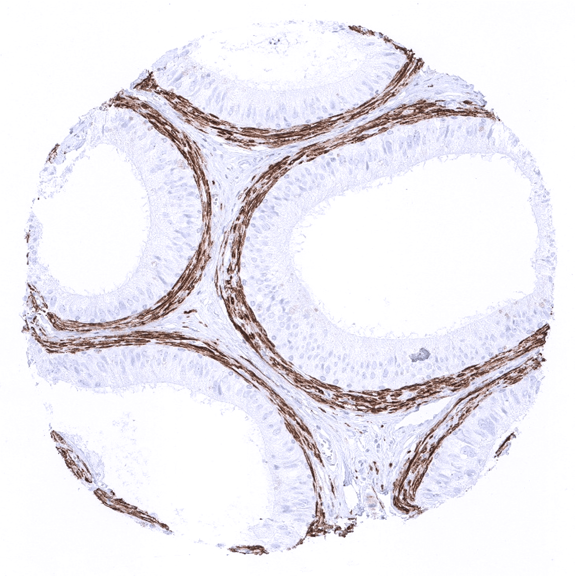
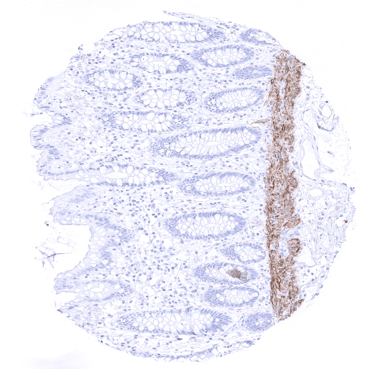
Positive control = Colon: A moderate to strong Calponin 1 staining should be seen in the muscularis mucosae, muscularis propria and in the walls of larger blood vessels.
Negative control = Colon: Calponin 1 staining should be absent in all epithelial cells.
Cellular localization = Cytoplasmic
Reactivity = Human
Application = Immunohistochemistry
Dilution = 1:100 – 1:200
Intended Use = Research Use Only
Relevance of Antibody
Biology Behind
Calponin-1 is an actin filament-associated regulatory protein expressed in smooth muscle cells where it plays a role in fine-tuning smooth muscle contractility (26970176). Calponin 1 functions as an inhibitory regulator of smooth muscle contractility through inhibiting actin/myosin interactions. In this regulation, binding of Ca2+-calmodulin and protein kinase C phosphorylation dissociates calponin 1 from the actin filament and facilitates smooth muscle contraction (Naka et al., 1990). The expression of calponin 1 is highly specific to smooth muscle cells.
Staining Pattern in Normal Tissues
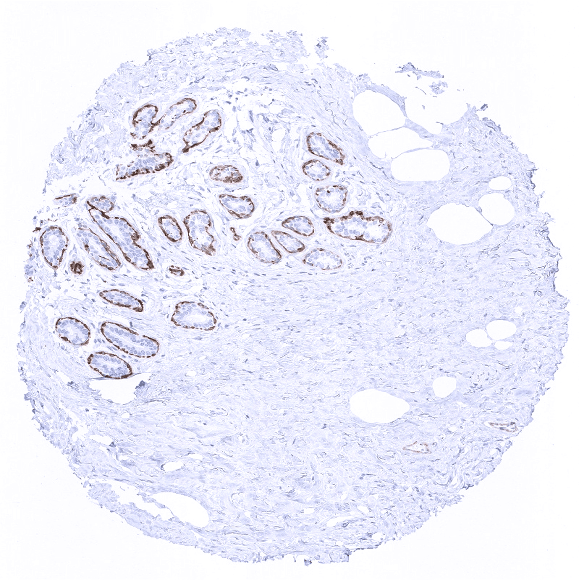
The calponin-1 antibody MSVA-455R strongly stains smooth muscle cells in all organs. This includes a positive staining reaction in all vessels that are large enough to contain smooth muscle cells. In the kidney, arterioles are stained. In addition myoepithelial cells in the breast and in salivary glands bronchial glands. In some testicular samples a weak staining of Sertoli cells is seen.
These findings are largely comparable to the data described in the Human Protein Atlas (Tissue expression Calponin 1).
Suggested positive tissue control: Colon: A moderate to strong Calponin 1 staining should be seen in the muscularis mucosae, muscularis propria and in the walls of larger blood vessels.
Suggested negative tissue control: Colon: Calponin 1 staining should be absent in all epithelial cells.


Staining Pattern in Relevant Tumor Types
Tumors derived from smooth muscle cells mostly stain positive for calponin-1. Calponin-1 immunostaining is also often seen in other tumor samples. Positively stained cells include remnants of smooth muscle in the tumor environment, blood vessels, and myofibroblasts. These cell types are often seen in combination. Epithelial tumor cells are not stained.
The TCGA findings on Calponin 1 RNA expression in different tumor categories have been summarized in the Human Protein Atlas.


Compatibility of Antibodies
No data available at the moment
Protocol Recommendations
IHC users have different preferences on how the stains should look like. Some prefer high staining intensity of the target stain and even accept some background. Others favor absolute specificity and lighter target stains. Factors that invariably lead to more intense staining include higher concentration of the antibody and visualization tools, longer incubation time, higher temperature during incubation, higher temperature and longer duration of the heat induced epitope retrieval (slide pretreatment). The impact of the pH during slide pretreatment has variable effects and depends on the antibody and the target protein.
All images and data shown here and in our image galleries are obtained by the manual protocol described below. Other protocols resulting in equivalent staining are described as well.
-manual protocol
Freshly cut sections should be used (less than 10 days between cutting and staining). Heat-induced antigen retrieval for 5 minutes in an autoclave at 121°C in pH 9 Target Retrieval Solution buffer. Apply MSVA-455R at a dilution of 1:50 at 37°C for 60 minutes. Visualization of bound antibody by the EnVision Kit (Dako, Agilent) according to the manufacturer’s directions.
impact of pH


pH9 is optimal but pH7,8 results in only slightly weaker stainings. At pH6, Calponin-1 immunostaining is markedly reduced.
Potential Research Applications
- The functional role of calponin-1 is not clear in smooth muscle tumors.
- The diagnostic utility of calponin-1 needs to be evaluated.
- The utility of calponin-1 immunohistochemistry for the distinction of myofibroblasts should be investigated.
Evidence for Antibody Specificity in IHC
Specificity of MSVA-455R is documented by strong positive staining in cell types that are well documented to express calponin-1 such as smooth muscle cells and myoepithelial cells as well as absence of staining in all tissues known to not express calponin-1 including tissues notorious for non-specific IHC background such as kidney, colonic mucosa, and epidermis.
