295,00 € – 995,00 €
Product details
Synonyms = pepsinogen 5, group I (pepsinogen A) , pepsinogen 3, group I (pepsinogen A) , pepsinogen 4, group I (pepsinogen A) , Pg5
Antibody type = Recombinant Rabbit monoclonal / IgG
Clone = HMV318
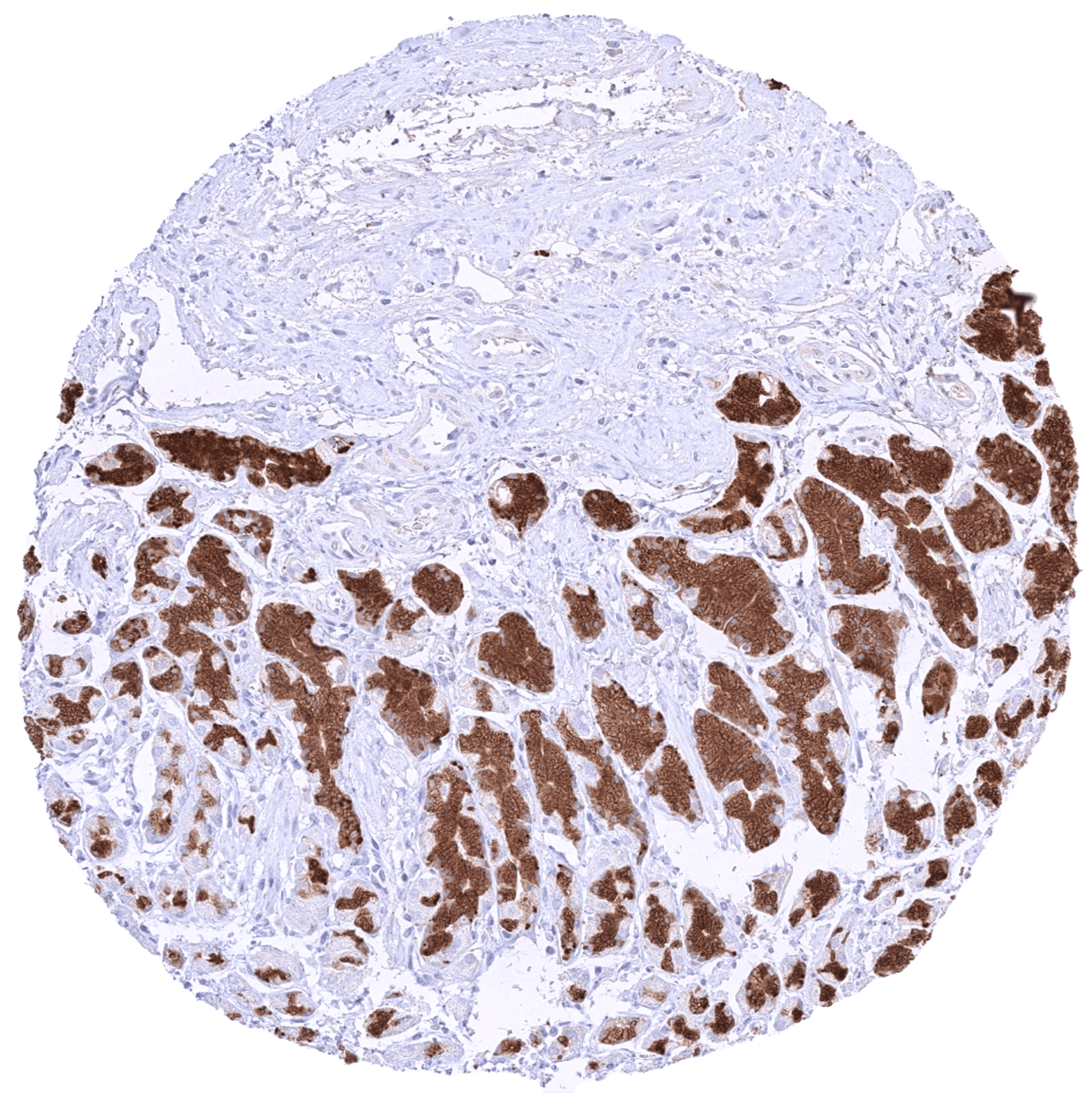
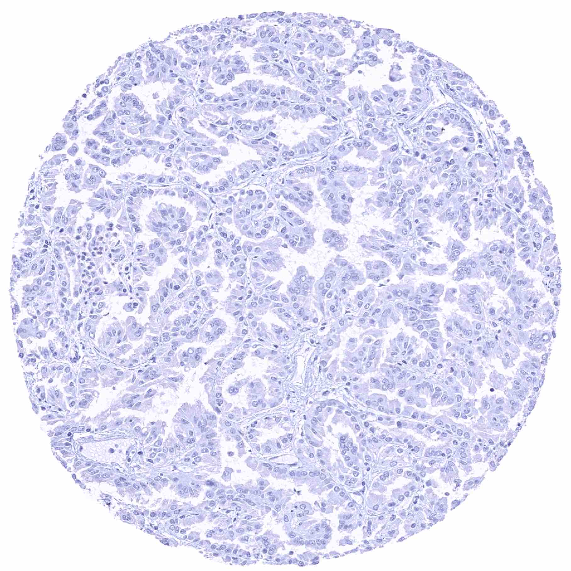
Positive control = Stomach, corpus: A strong pepsinogen-I should be seen in chief cells of the stomach while surface epithelial cells and stroma cells remain negative.
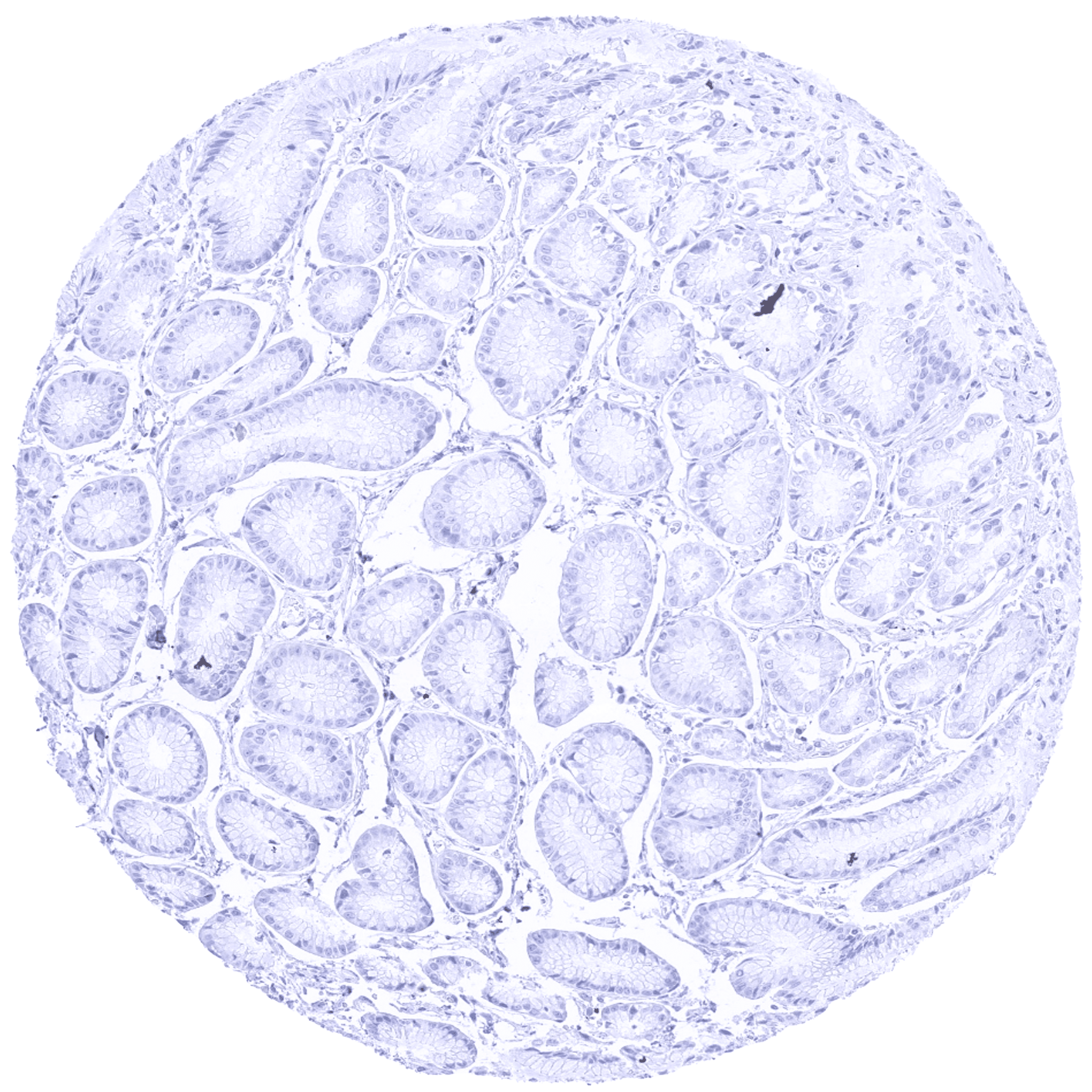
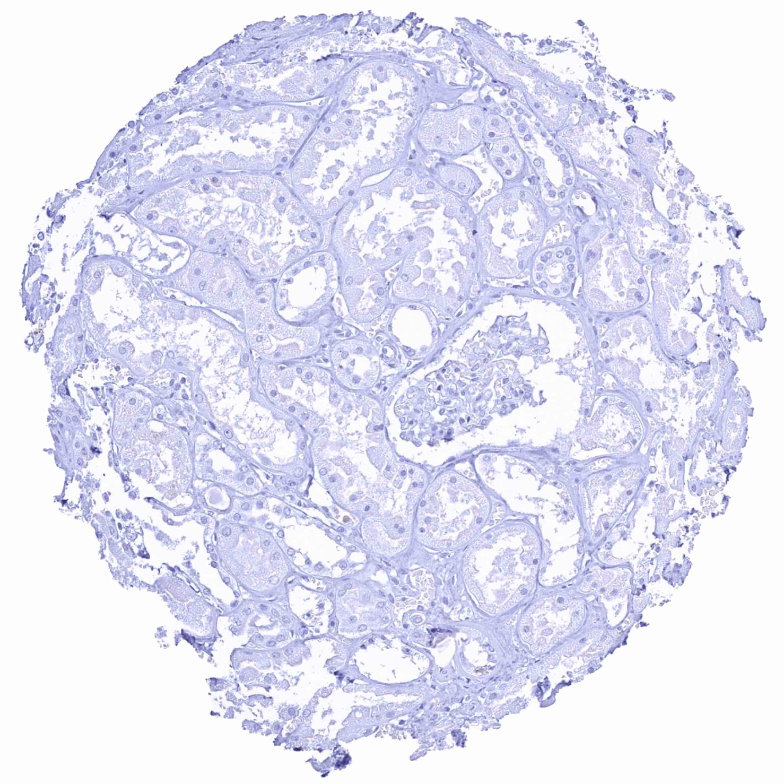
Negative control = Kidney: Pepsinogen-I staining should be completely absent in all cells.
Cellular localization = Secreted
Reactivity = Human
Application = Immunohistochemistry
Dilution = 1:100 – 1:200
Intended Use = Research Use Only
Relevance of Antibody
Pepsinogen I is a Digestive enzyme solely produced by chief cells of gastric glands.
Biology Behind
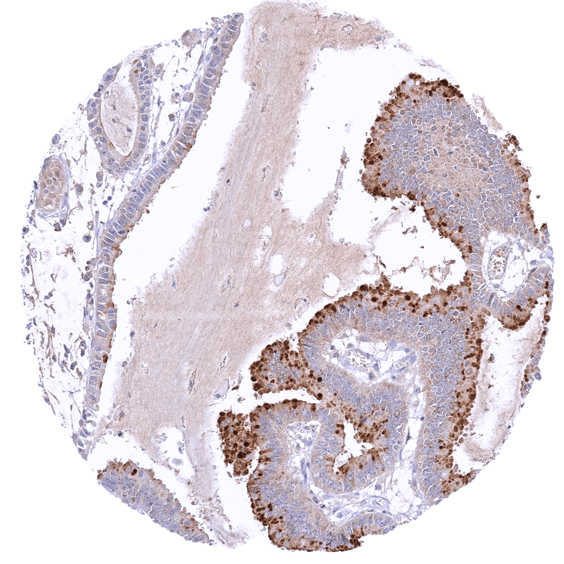
Pepsinogen-I is an endopeptidase coded by the PGA3/PGA4/PGA5 genes on chromosome 11q12.13. Pepsinogen-I is one of three main endopeptidases (enzymes cutting proteins in their -central part) in the digestive system, the other two being trypsin and chymotrypsin. Pepsinogen-I is synthesized and secreted by gastric chief cells in the corpus of the stomach. It is activated to pepsin in the gastric lumen by HCl secreted by fundic parietal cells. Pepsin is most active in acidic environments between pH 1.5 to 2.5. Nutritive proteins are degraded by pepsin and other enzymes to a mixture of oligopeptides that enter the duodenum. The activity of pepsins ends when the HCl driven low pH of the enzyme-(food)protein mixture is terminated when gastric contents are mixed with alkaline bile and pancreatic juice in the duodenum. Expression of pepsinogen-I is normally limited to corpus glands and thus represents a characteristic feature of “gastric adenocarcinoma of the fundic gland type (GA-FG)”, a rare, newly recognized gastric carcinoma variant making up for about 2% of gastric cancers and showing a chief cell differentiation of at least a large fraction of tumor cells. Pepsinogen-I was suggested to represent the predominant IHC marker of GA-FG.
Staining Pattern in Normal Tissues
Images describing the Pepsinogen I staining pattern in normal tissues obtained by the antibody HMV318 are shown in our “Normal Tissue Gallery”.
| Brain | Cerebrum | Negative. |
| Cerebellum | Negative. | |
| Endocrine Tissues | Thyroid | Negative. |
| Parathyroid | Negative. | |
| Adrenal gland | Negative. | |
| Pituitary gland | Negative. | |
| Respiratory system | Respiratory epithelium | Negative. |
| Lung | Negative. | |
| Gastrointestinal Tract | Salivary glands | Negative. |
| Esophagus | Negative. | |
| Stomach | Strong pepsinogen-I positivity in chief cells. Given the high level of pepsinogen I expression in chief cells, adjacent cells/stroma may also show some pepsinogen-I staining due to diffusion of the protein (contamination artifact). | |
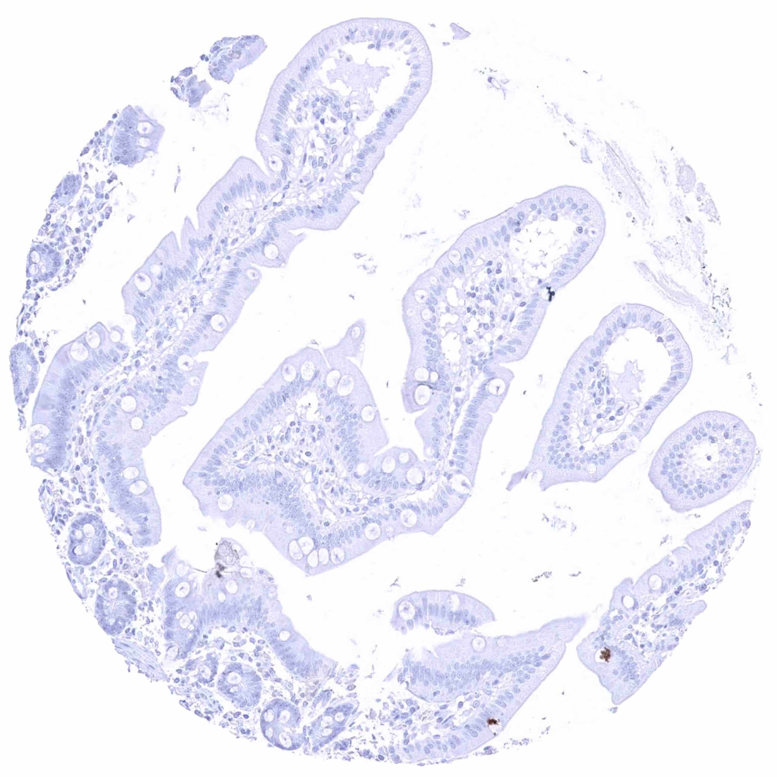
| Duodenum | Negative. | |
| Small intestine | Negative. | |
| Appendix | Negative. | |
| Colon | Negative. | |
| Rectum | Negative. | |
| Liver | Negative. | |
| Gallbladder | Negative. | |
| Pancreas | Negative. | |
| Genitourinary | Kidney | Negative. |
| Urothelium | Negative. | |
| Male genital | Prostate | Negative. |
| Seminal vesicles | Negative. | |
| Testis | Negative. | |
| Epididymis | Negative. | |
| Female genital | Breast | Negative. |
| Uterus, myometrium | Negative. | |
| Uterus, ectocervix | Negative. | |
| Uterus endocervix | Negative. | |
| Uterus, endometrium | Negative. | |
| Fallopian Tube | Negative. | |
| Ovary | Negative. | |
| Placenta early | Negative. | |
| Placenta mature | Negative. | |
| Amnion | Negative. | |
| Chorion | Negative. | |
| Skin | Epidermis | Negative. |
| Sebaceous glands | Negative. | |
| Muscle/connective tissue | Heart muscle | Negative. |
| Skeletal muscle | Negative. | |
| Smooth muscle | Negative. | |
| Vessel walls | Negative. | |
| Fat | Negative. | |
| Stroma | Negative. | |
| Endothelium | Negative. | |
| Bone marrow/ lymphoid tissue | Bone marrow | Negative. |
| Lymph node | Negative. | |
| Spleen | Negative. | |
| Thymus | Negative. | |
| Tonsil | Negative. | |
| Remarks | Pepsinogen-I is very highly expressed in gastric chief cells. Therefore, adjacent cells may also show some staining due to diffusion of the protein in chief-cell adjacent structures (contamination artifact). |
These findings are largely comparable to the RNA and protein data described in the Human Protein Atlas (Tissue expression Pepsinogen I).
Pepsinogen-I staining by HMV318 is only seen in chief cells of the gastric corpus while the stomach is the only organ for which Pepsinogen RNA has been detected.
Positive control = Stomach, corpus: A strong pepsinogen-I should be seen in chief cells of the stomach while surface epithelial cells and stroma cells remain negative.
Negative control = Kidney: Pepsinogen-I staining should be completely absent in all cells.
Staining Pattern in Relevant Tumor Types
Pepsinogen-I positivity is characteristically expressed in gastric adenocarcinoma of the fundic gland type. Expression in other tumor entities has not been described.
The TCGA findings on Pepsinogen I RNA expression in different tumor categories have been summarized in the Human Protein Atlas.
Compatibility of Antibodies
No data available at the moment
Protocol Recommendations
IHC users have different preferences on how the stains should look like. Some prefer high staining intensity of the target stain and even accept some background. Others favor absolute specificity and lighter target stains. Factors that invariably lead to more intense staining include higher concentration of the antibody and visualization tools, longer incubation time, higher temperature during incubation, higher temperature and longer duration of the heat induced epitope retrieval (slide pretreatment). The impact of the pH during slide pretreatment has variable effects and depends on the antibody and the target protein.
All images and data shown here and in our image galleries are obtained by the manual protocol described below. Other protocols resulting in equivalent staining are described as well.
Manual protocol
Freshly cut sections should be used (less than 10 days between cutting and staining). Heat-induced antigen retrieval for 5 minutes in an autoclave at 121°C in pH 7,8 Target Retrieval Solution buffer. Apply HMV318 at a dilution of 1:200 at 37°C for 60 minutes. Visualization of bound antibody by the EnVision Kit (Dako, Agilent) according to the manufacturer’s directions.
Potential Research Applications
- The clinical characteristics of the fundic gland type of gastric cancer are not yet well enough established.
- Whether pepsinogen-I can also occur in non-gastric cancers is unknown.
Evidence for Antibody Specificity in IHC
There are two ways how the specificity of antibodies can be documented for immunohistochemistry on formalin fixed tissues. These are: 1. Comparison with a second independent method for target expression measurement across a large number of different tissue types (orthogonal strategy), and 2. Comparison with one or several independent antibodies for the same target and showing that all positive staining results are also seen with other antibodies for the same target (independent antibody strategy).
Orthogonal validation: For the antibody HMV318 specificity is proven by the perfect concordance of the immunostaining data with data from three independent RNA screening studies, including the Human Protein Atlas (HPA) RNA-seq tissue dataset, the FANTOM5 project, and the Genotype-Tissue Expression (GTEx) project, which are all summarized in the Human Protein Atlas (Tissue expression Pepsinogen I). Pepsinogen-I positivity by HMV318 was solely detectable in chief cells of the gastric mucosa. This fits perfectly with the complete restriction of Pepsinogen-I RNA expression to the stomach.
Comparison of antibodies: Not done due to the unequivocal result of orthogonal validation.
