295,00 € – 995,00 €
Product details
Synonyms = Glycoprotein m6a, M6A, Gpm6
Antibody type = Recombinant Rabbit monoclonal / IgG
Clone = HMV3973
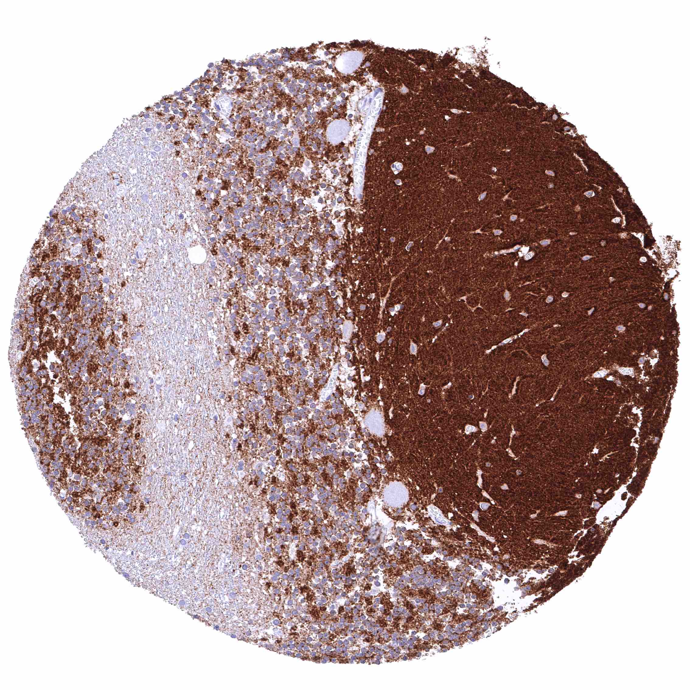
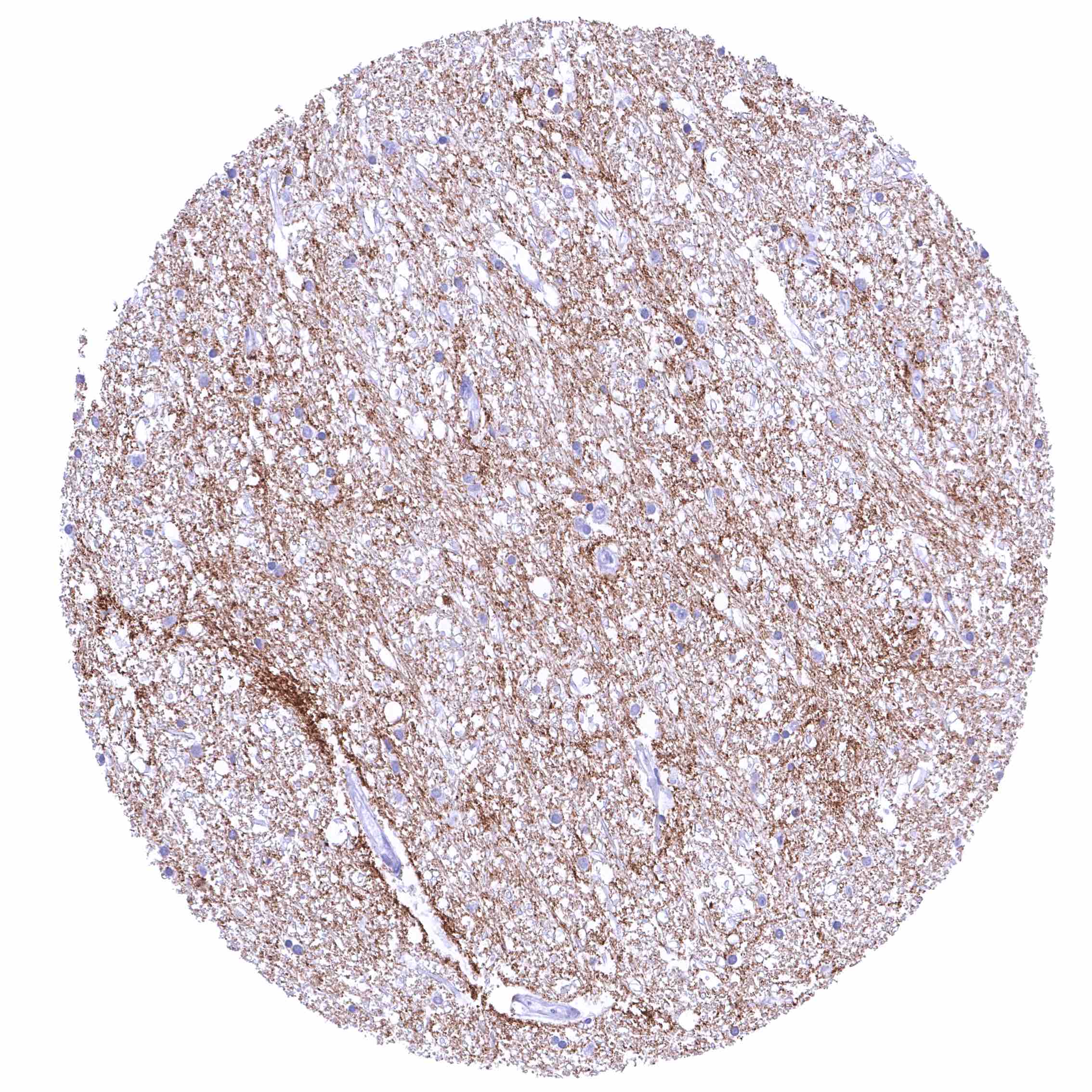
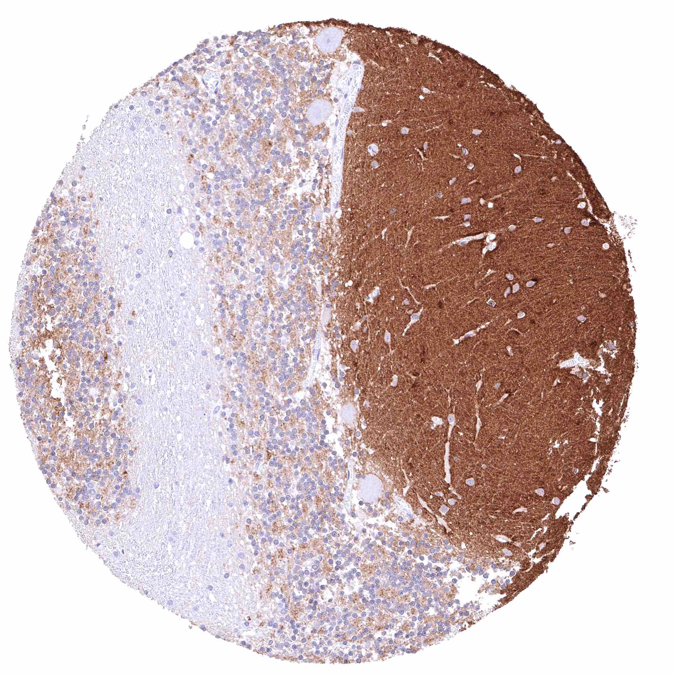
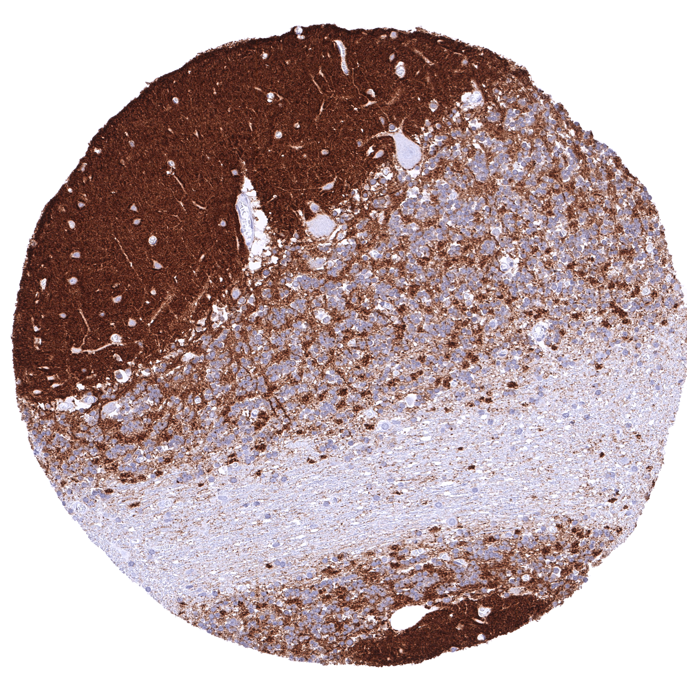
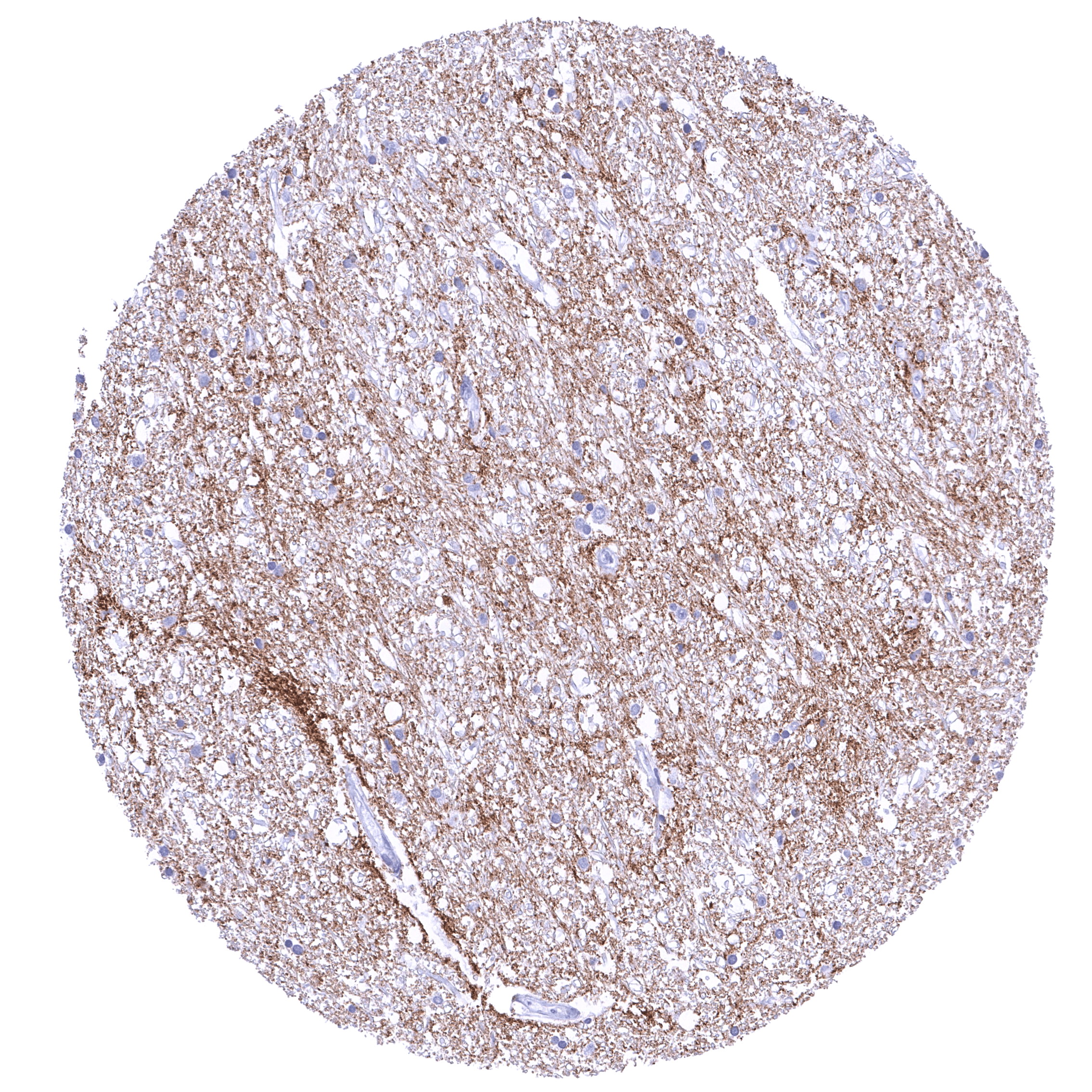
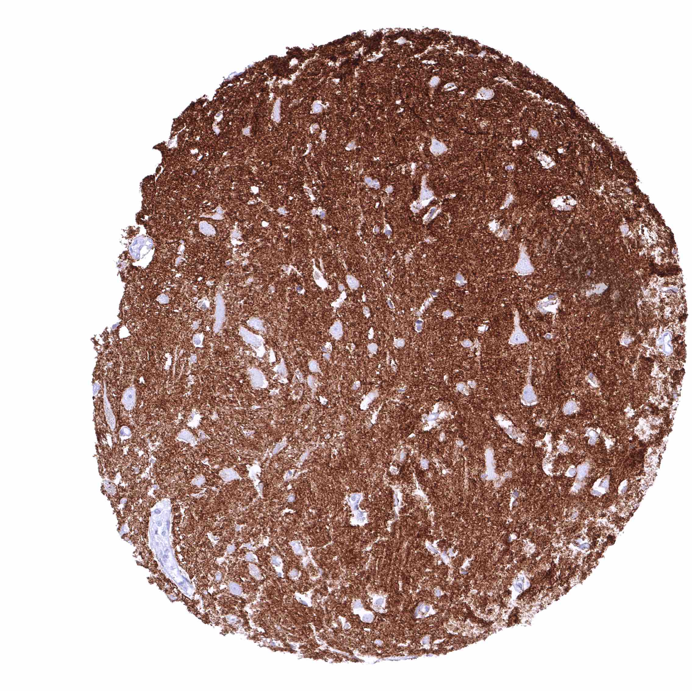
Positive control = Cerebrum (grey matter): A strong membranous GPM6A staining of neurons should be seen in combination with a strong fibrillar staining (axons).
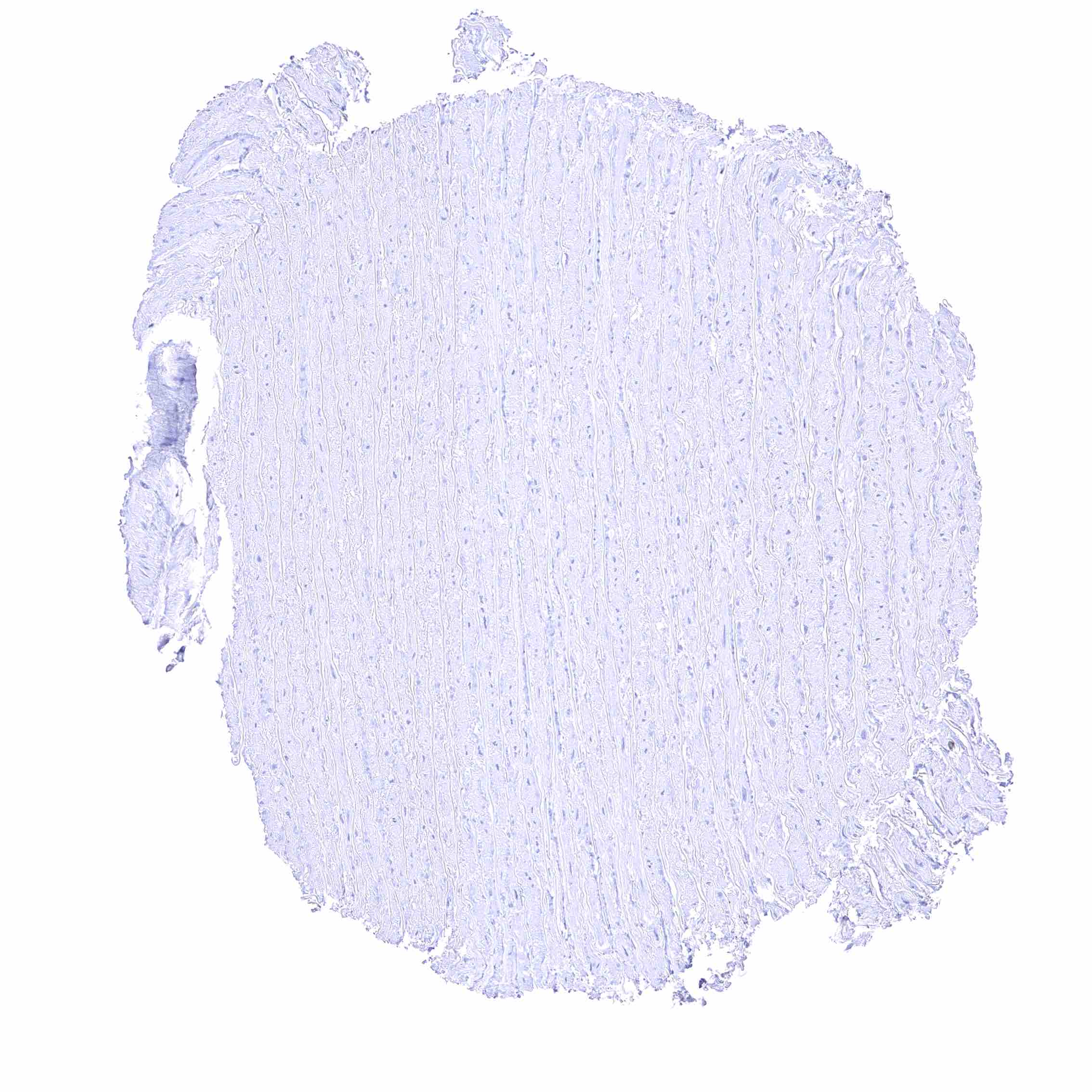
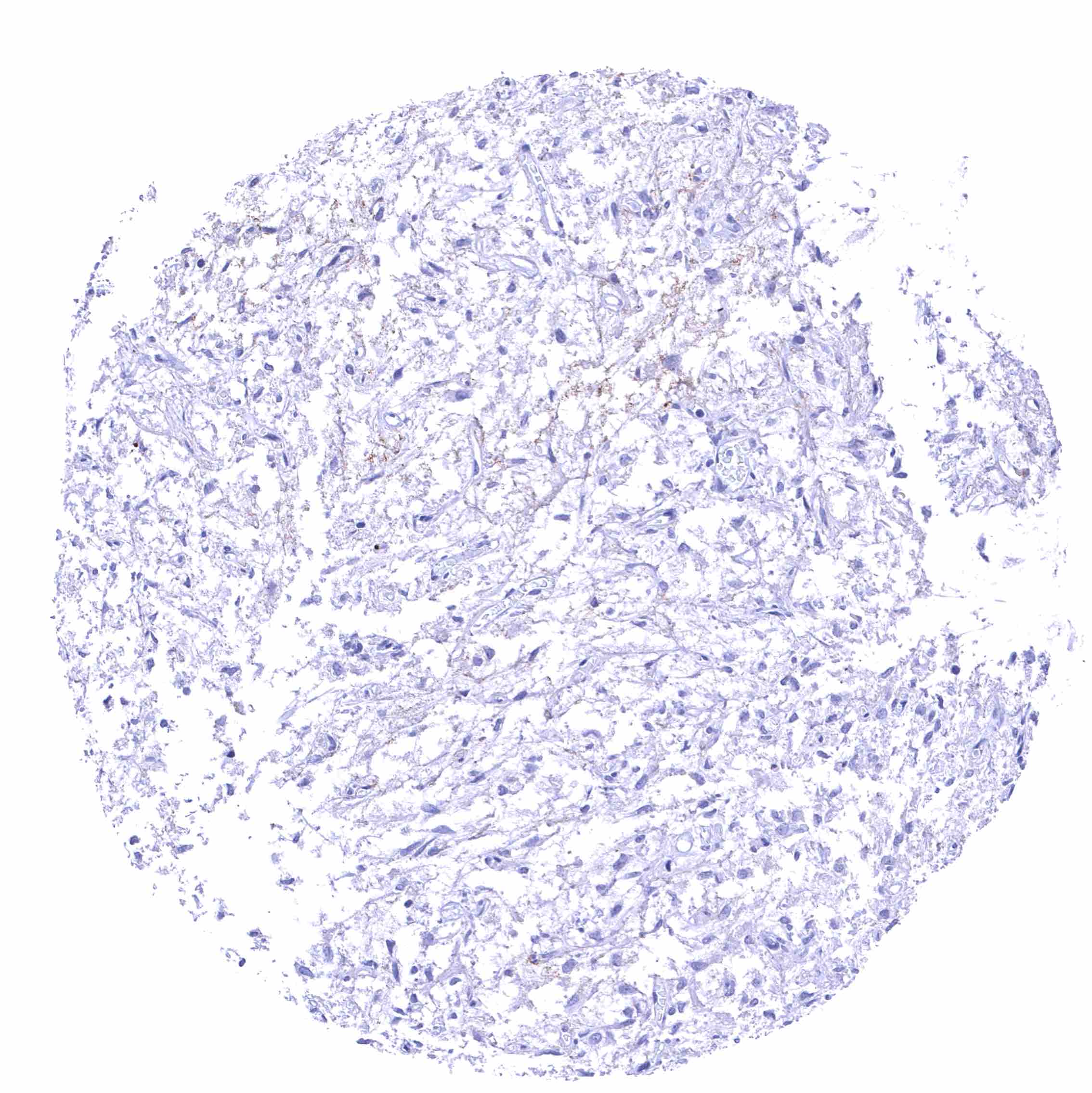
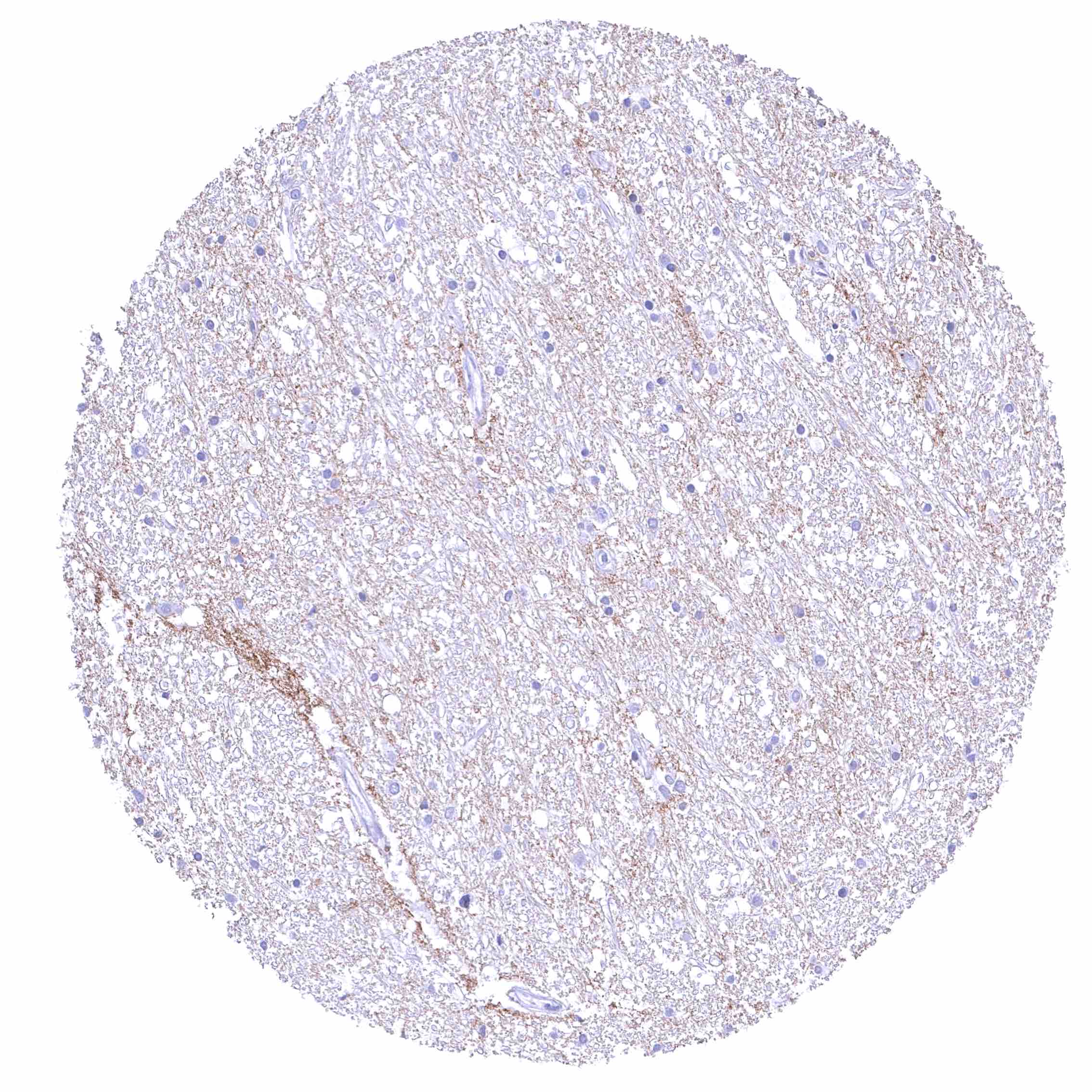
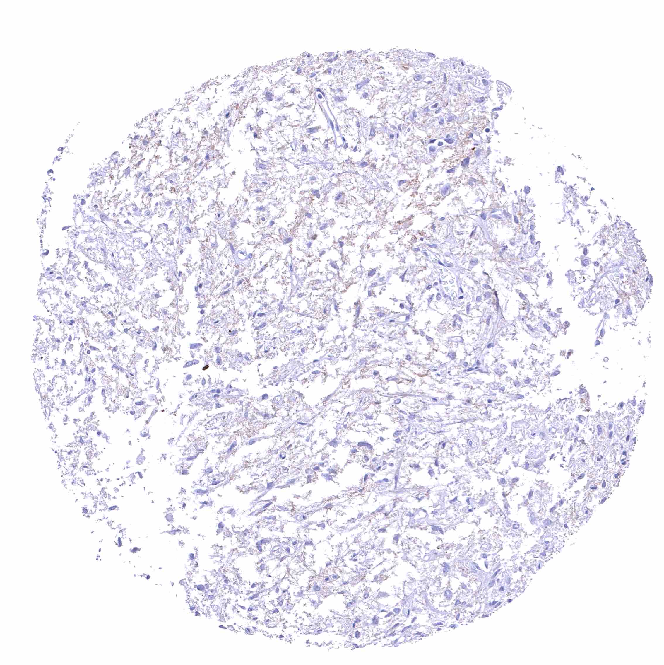
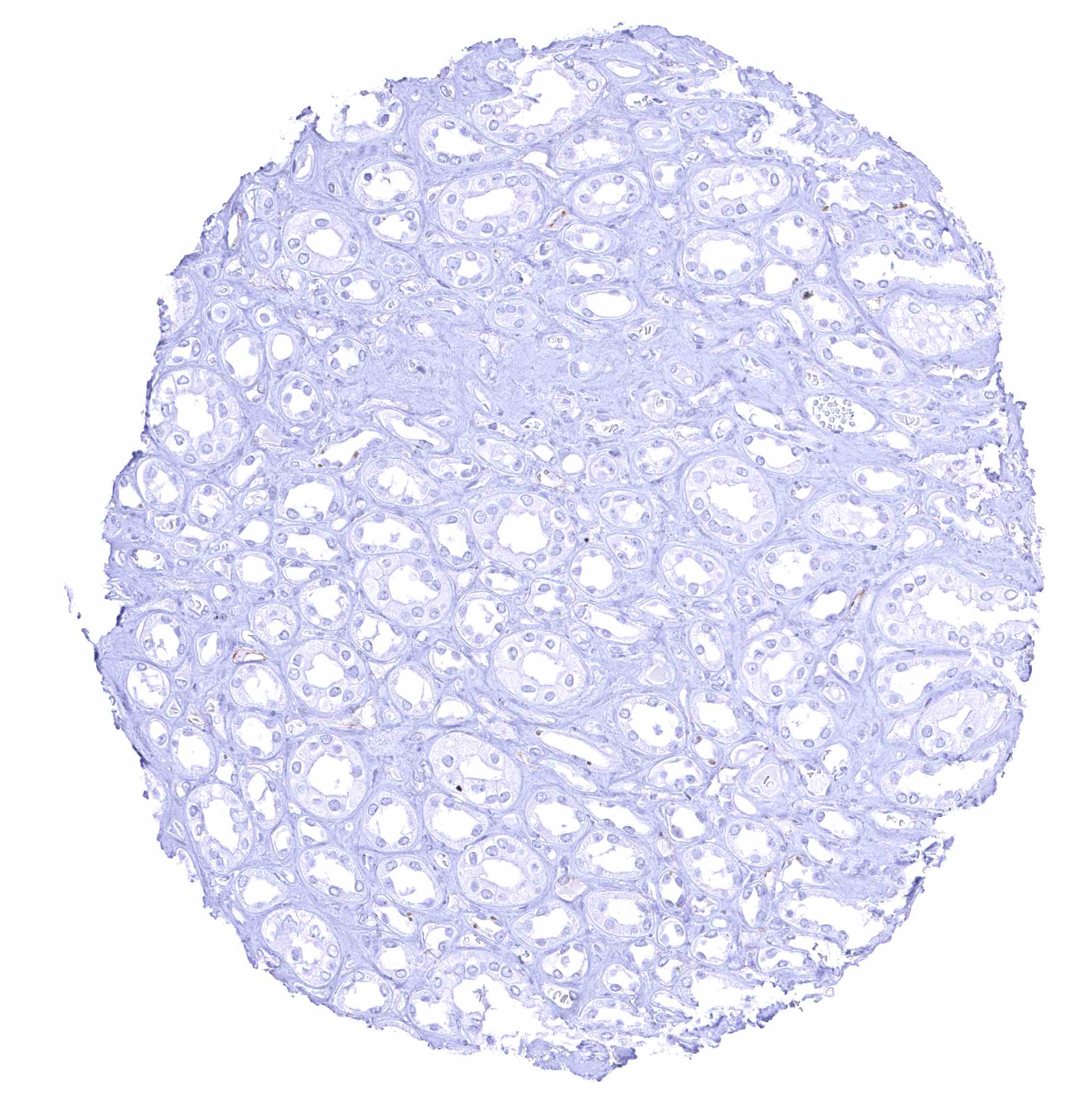
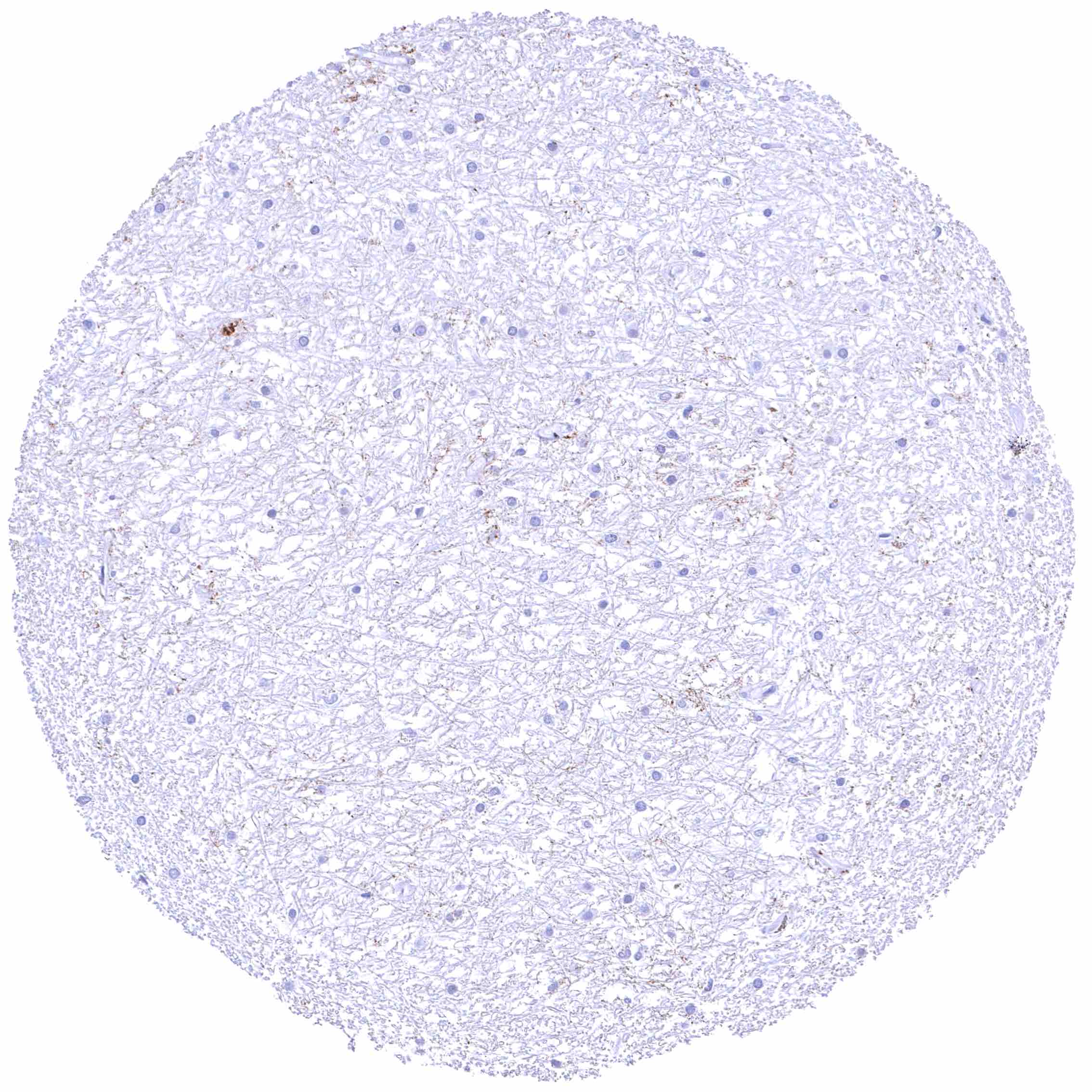
Negative control = Tonsil: GPM6A staining should be completely absent in all cell types.
Cellular localization = Cytoplasmic
Reactivity = Human
Application = Immunohistochemistry
Dilution = 1:100 – 1:200
Intended Use = Research Use Only
Relevance of Antibody
GPM6A is a major component of the myelin sheath in the brain.
Biology Behind
Glycoprotein M6A (GPM6A) is a membranous glycoprotein coded by the GPM6A gene at chromosome 4q34.2. Together with PLP1, DM20, and GPM6B, GPM6A forms the proteolipid protein (PLP) family which are the main group of proteins of the myelin sheath in the brain. GPM6A is highly expressed in neurons of the central nervous system. It is assumed that GPM6A has a role in intracellular transport. It plays a role in important processes including neuronal differentiation, axon growth, synaptogenesis, migration, and the formation of the spine. Altered expression levels or molecular structure of GPM6A has been found in several neuropsychiatric disorders. Including schizophrenia and depression. GPM6A has also been suspected to play a role in extracranial cancer.
Staining Pattern in Normal Tissues
Among normal tissues, GPM6A expression is limited to the brain. Images describing the GPM6A staining pattern in normal tissues obtained by the antibody HMV3973 are shown in our “Normal Tissue Gallery”.
| Brain | Cerebrum | Strong membranous GPM6A staining of neurons especially along axons, at least in specific regions of the cerebrum. |
| Cerebellum | Strong membranous GPM6A staining of neurons including Purkinje cells, and a fraction of cells in the granule cell layer. Positivity also occurs along axons, but it appears to be weak and less common in the white matter. | |
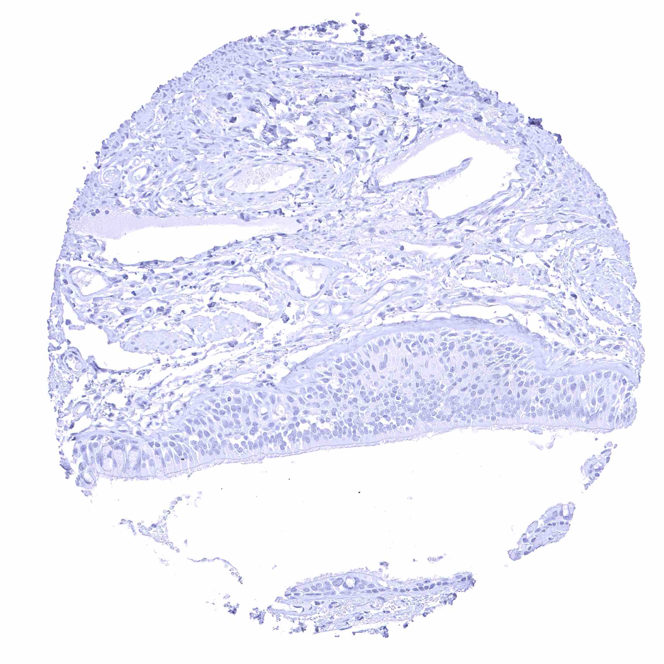
| Endocrine Tissues | Thyroid | Negative. |
| Parathyroid | Negative. | |
| Adrenal gland | Negative. | |
| Pituitary gland | Moderate fibrillar GPM6A staining of the neurohypophysis while cells in the adenohypophysis remain GPM6A negative. | |
| Respiratory system | Respiratory epithelium | Negative. |
| Lung | Negative. | |
| Gastrointestinal Tract | Salivary glands | Negative. |
| Esophagus | Negative. | |
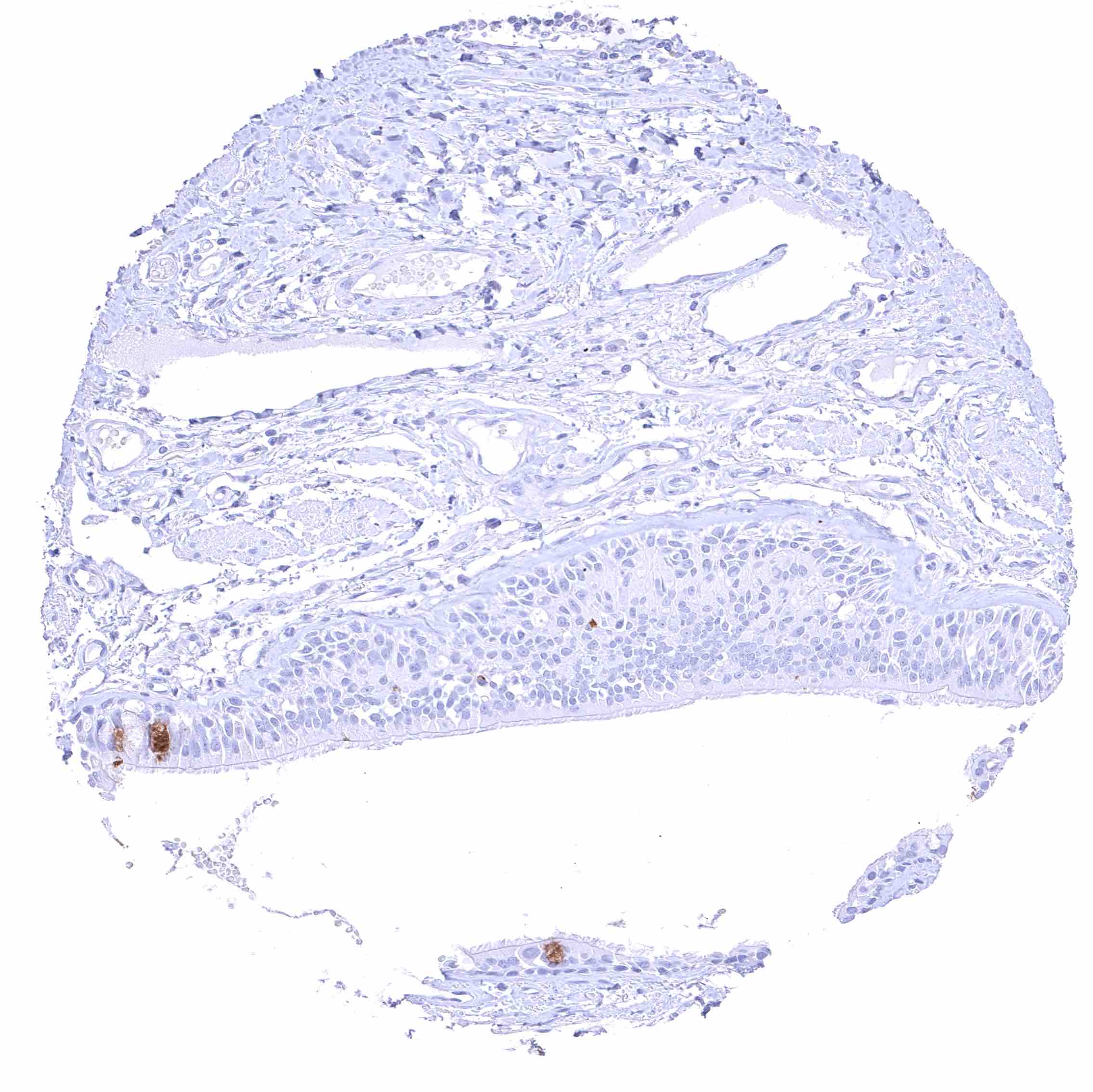
| Stomach | Negative. | |
| Duodenum | Negative. | |
| Small intestine | Negative. | |
| Appendix | Negative. | |
| Colon | Negative. | |
| Rectum | Negative. | |
| Liver | Negative. | |
| Gallbladder | Negative. | |
| Pancreas | Negative. | |
| Genitourinary | Kidney | Negative. |
| Urothelium | Negative. | |
| Male genital | Prostate | Negative. |
| Seminal vesicles | Negative. | |
| Testis | Negative. | |
| Epididymis | Negative. | |
| Female genital | Breast | Negative. |
| Uterus, myometrium | Negative. | |
| Uterus, ectocervix | Negative. | |
| Uterus endocervix | Negative. | |
| Uterus, endometrium | Negative. | |
| Fallopian Tube | Negative. | |
| Ovary | Negative. | |
| Placenta early | Negative. | |
| Placenta mature | Negative. | |
| Amnion | Negative. | |
| Chorion | Negative. | |
| Skin | Epidermis | Negative. |
| Sebaceous glands | Negative. | |
| Muscle/connective tissue | Heart muscle | Negative. |
| Skeletal muscle | Negative. | |
| Smooth muscle | Negative. | |
| Vessel walls | Negative. | |
| Fat | Negative. | |
| Stroma | Negative. | |
| Endothelium | Negative. | |
| Bone marrow/ lymphoid tissue | Bone marrow | Negative. |
| Lymph node | Negative. | |
| Spleen | Negative. | |
| Thymus | Negative. | |
| Tonsil | Negative. | |
| Remarks |
The findings described above are thus consistent with the RNA data described in the Human Protein Atlas (Tissue expression GPM6A).
Positive control = Cerebrum (grey matter): A strong membranous GPM6A staining of neurons should be seen in combination with a strong fibrillar staining (axons).
Negative control = Tonsil: GPM6A staining should be completely absent in all cell types.
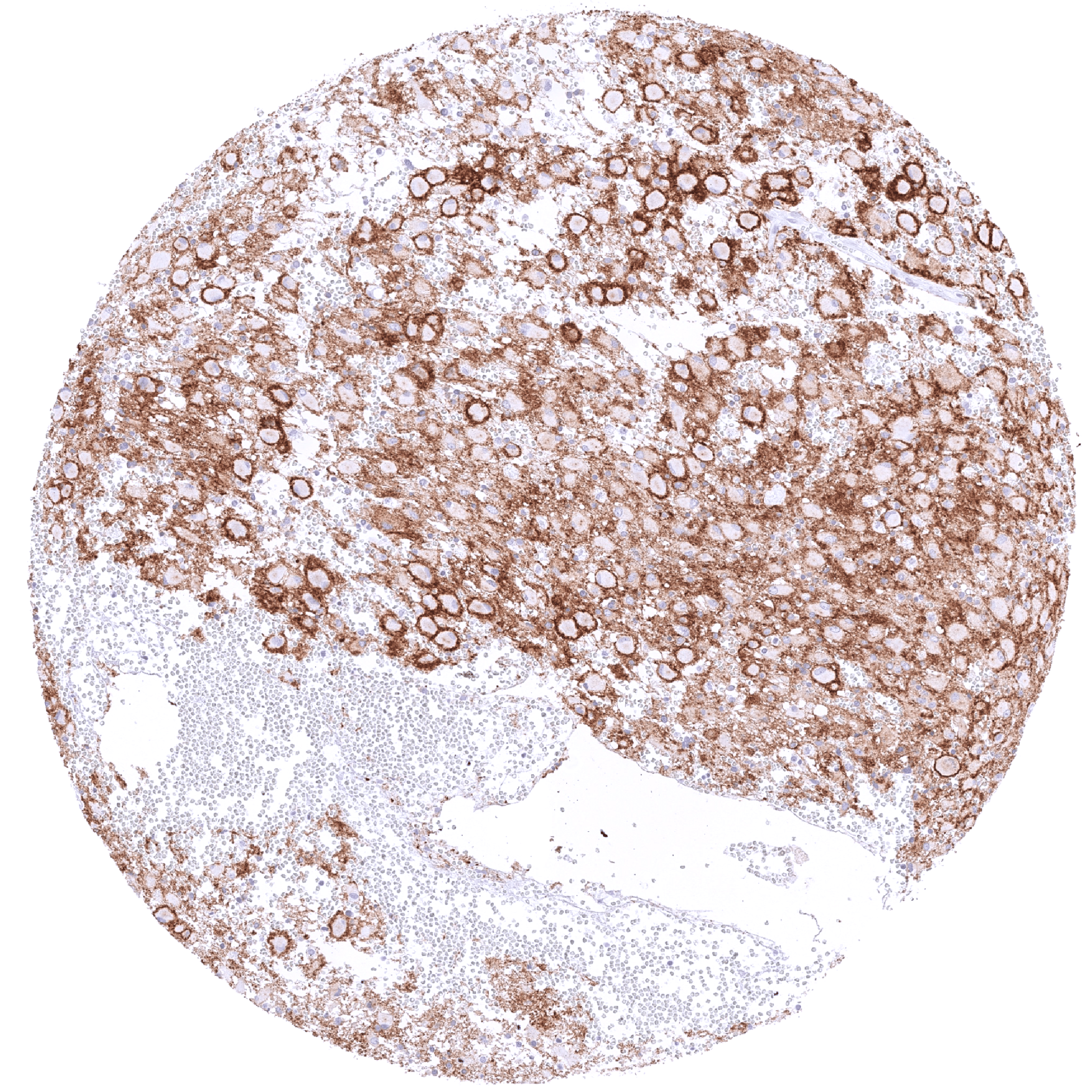
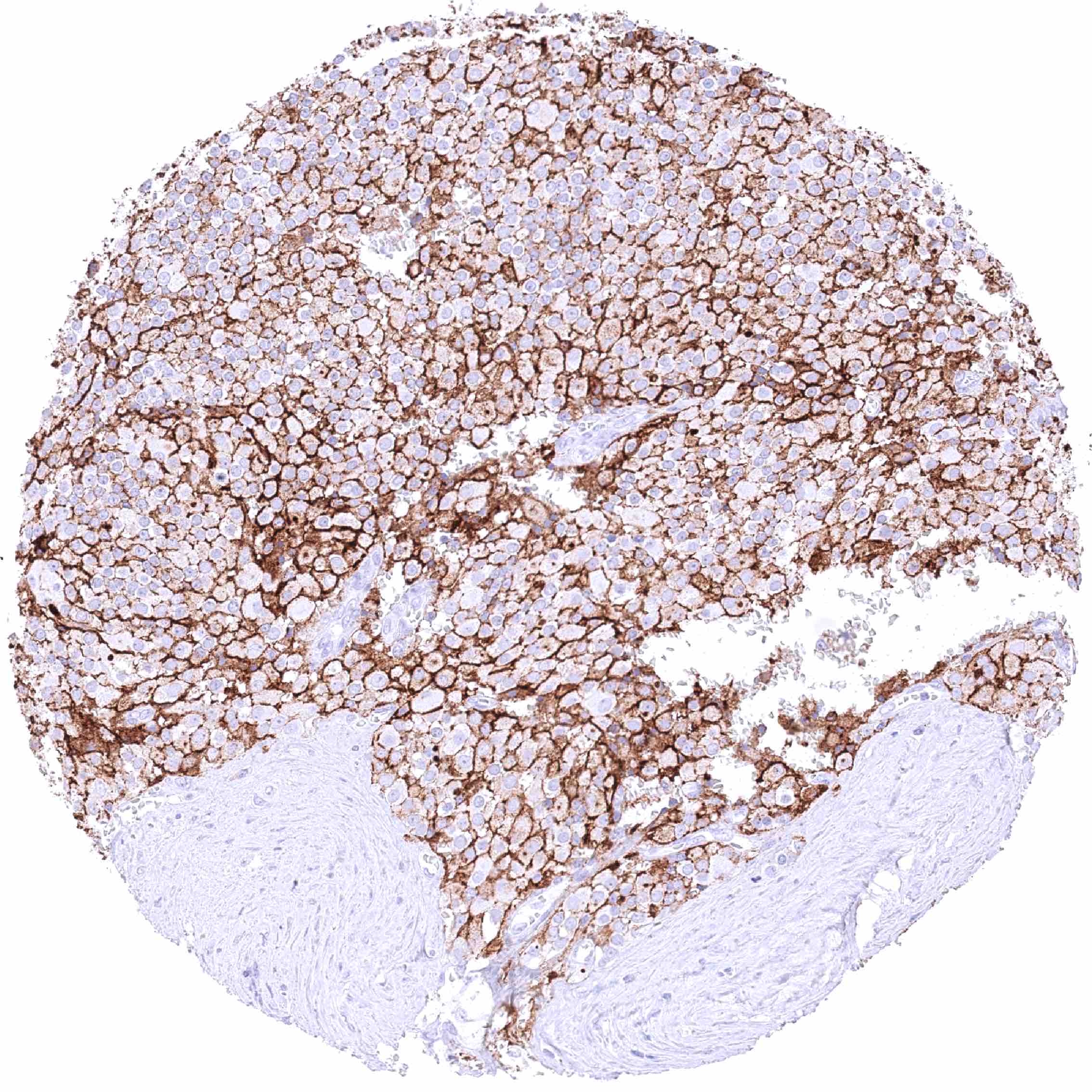
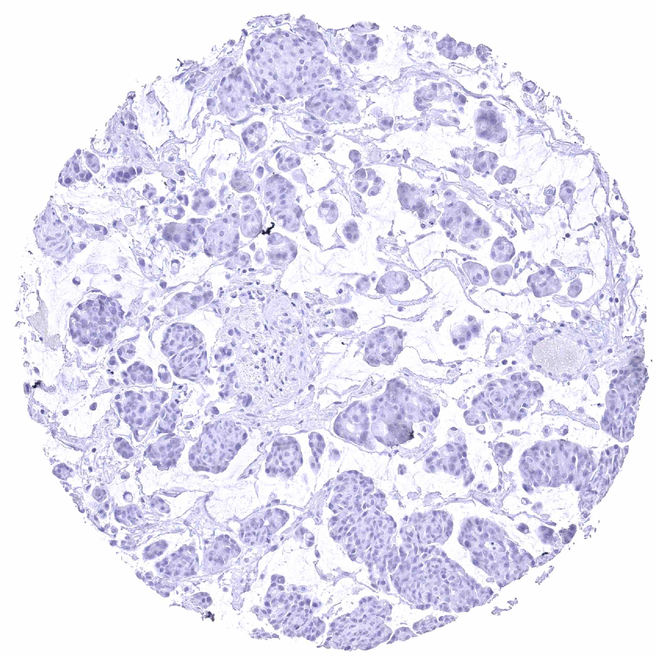
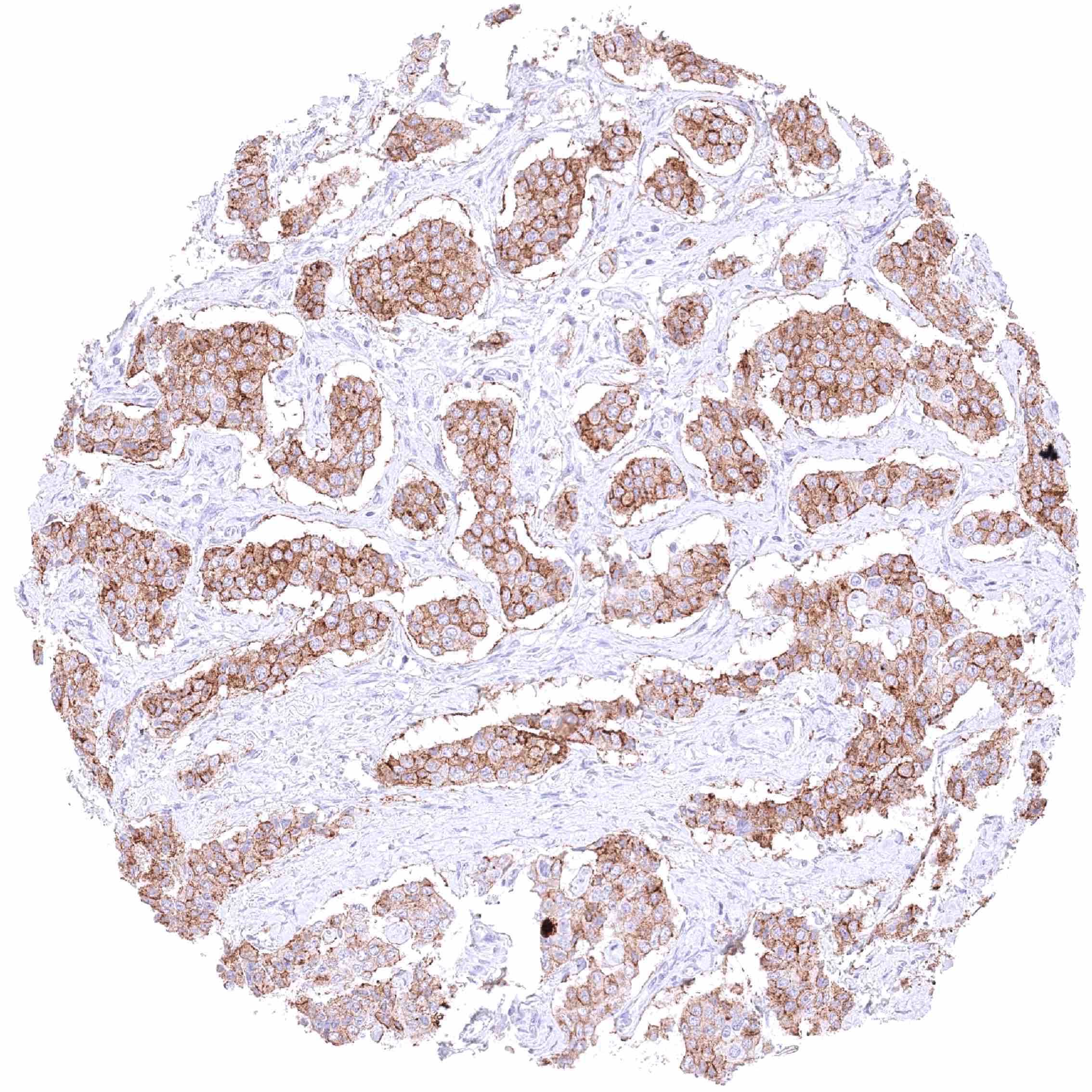
Staining Pattern in Relevant Tumor Types
GPM6A expression has been described to occur in cancer, but systematic data are lacking.
The TCGA findings on GPM6A RNA expression in different tumor categories have been summarized in the Human Protein Atlas.
Compatibility of Antibodies
No data available at the moment
Protocol Recommendations
IHC users have different preferences on how the stains should look like. Some prefer high staining intensity of the target stain and even accept some background. Others favor absolute specificity and lighter target stains. Factors that invariably lead to more intense staining include higher concentration of the antibody and visualization tools, longer incubation time, higher temperature during incubation, higher temperature and longer duration of the heat induced epitope retrieval (slide pretreatment). The impact of the pH during slide pretreatment has variable effects and depends on the antibody and the target protein.
All images and data shown here and in our image galleries are obtained by the manual protocol described below. Other protocols resulting in equivalent staining are described as well.
Manual protocol
Freshly cut sections should be used (less than 10 days between cutting and staining). Heat-induced antigen retrieval for 5 minutes in an autoclave at 121°C in pH 7,8 Target Retrieval Solution buffer. Apply HMV3973 at a dilution of 1:150 at 37°C for 60 minutes. Visualization of bound antibody by the EnVision Kit (Dako, Agilent) according to the manufacturer’s directions.
Potential Research Applications
- The role of GPM6A in the function of the brain needs to be further evaluated.
- The role of GPM6A in cancer is not understood.
- The role of GPM6A in neuropsychiatric disorders needs to be further evaluated.
Evidence for Antibody Specificity in IHC
There are two ways how the specificity of antibodies can be documented for immunohistochemistry on formalin fixed tissues. These are: 1. Comparison with a second independent method for target expression measurement across a large number of different tissue types (orthogonal strategy), and 2. Comparison with one or several independent antibodies for the same target and showing that all positive staining results are also seen with other antibodies for the same target (independent antibody strategy).
Orthogonal validation: For the antibody HMV3973 specificity is supported by the complete concordance of the immunostaining data with data from three independent RNA screening studies, including the Human Protein Atlas (HPA) RNA-seq tissue dataset, the FANTOM5 project, and the Genotype-Tissue Expression (GTEx) project, which are all summarized in the Human Protein Atlas (Tissue expression GPM6A). GPM6A immunostaining by using HMV3973 was only detected in the brain which also represented the only organ with documented RNA expression. GPM6A immunostaining by HMV3973 was always absent in organs lacking GPM6A RNA expression.
Comparison of antibodies: True expression of GPM6A in cell types with documented GPM6A immunostaining by HMV3973 is further validated by identical (although less intense) staining patterns obtained by a second, independent GPM6A antibody, termed “validation antibody”. An additional granular staining of matrix material in the aortic wall and membranes of few epithelial cells in the rectal mucosa which was only seen by the validation antibody was considered an antibody specific cross-reactivity of the validation antibody.