295,00 € – 995,00 €
Product details
Synonyms = Apoptosis regulator Bcl-2, B-cell CLL/lymphoma-2
Antibody type = Recombinant Mouse monoclonal / IgG1, kappa
Clone = MSVA-402M
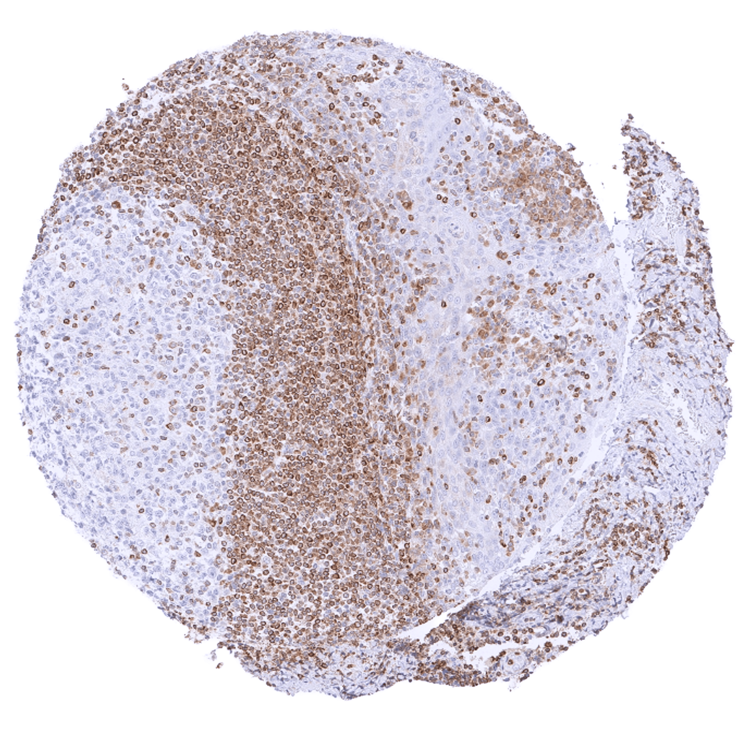
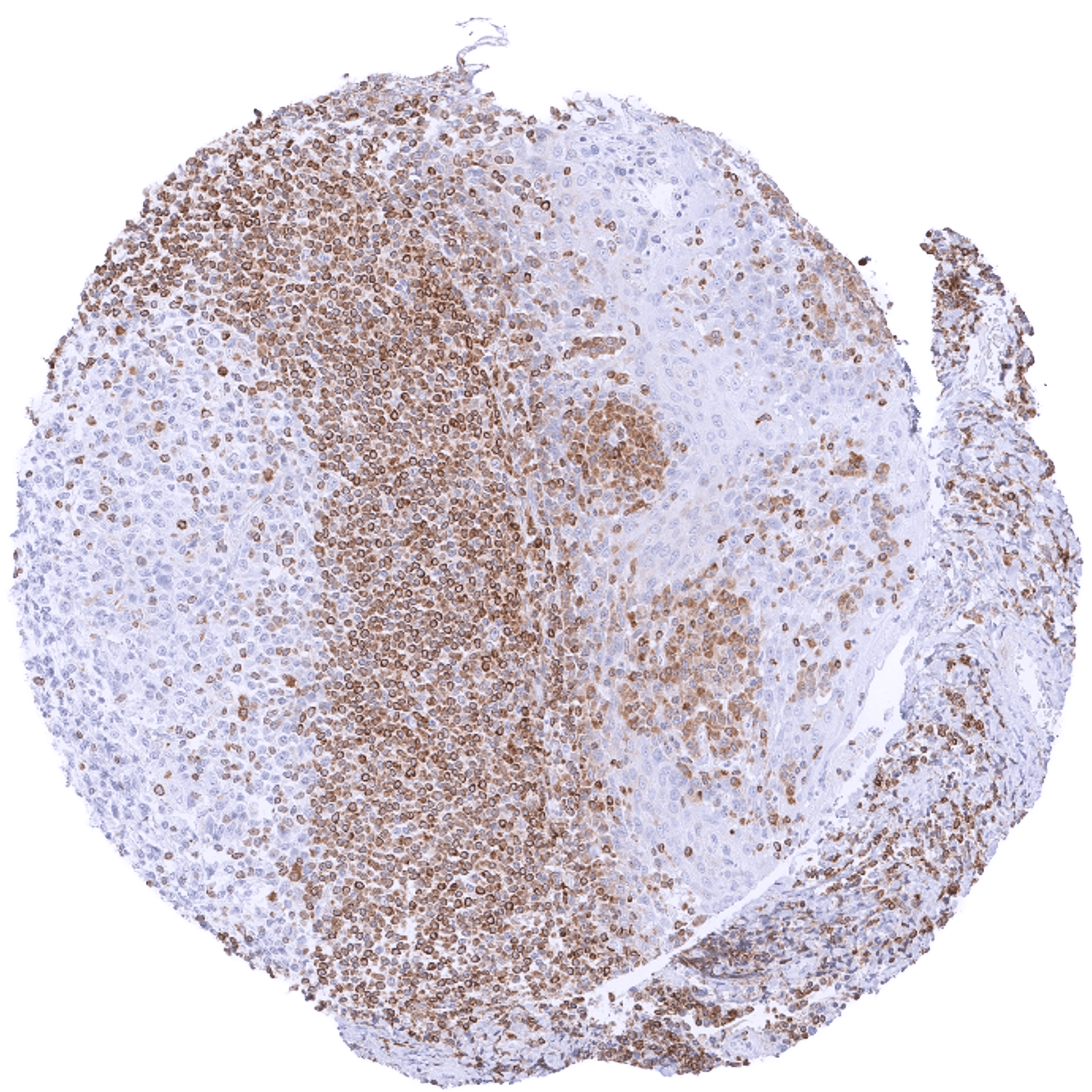
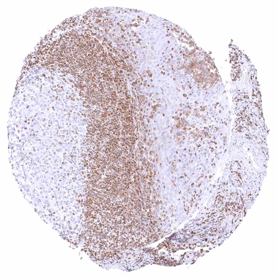
Positive control = Tonsil: A strong cytoplasmic bcl-2 staining should be seen in most interfollicular lymphocytes while most germinal centre cells are bcl-2 negative.
Negative control = Tonsil: The vast majority of lymphocytic cells from germinal centres must be bcl-2 negative while interfollicular lymphocytes are mostly positive.
Cellular localization = Cytoplasmic
Reactivity = Human
Application = Immunohistochemistry
Dilution = 1:100 – 1:200
Intended Use = Research Use Only
Relevance of Antibody
Bcl-2 is a critical apoptosis inhibitor protein.
Biology Behind
Bcl-2 (B-cell lymphoma 2) is the founding member of the bcl-2 family of proteins which all share a role in regulating apoptosis. The protein is coded by the bcl-2 gene on chromosome 18q21.33. Bcl-2 is a potent inhibitor of apoptosis. The protein is located at the outer membrane of mitochondria, where it inhibits the activity of pro-apoptotic proteins which often act at the mitochondrial membrane to promote its permeabilization. Alterations of the bcl-2 gene have been identified as a key driving event in cancer where high bcl-2 expression is a cause for poor prognosis and resistance to cancer treatments. Considering the pivotal importance of apoptosis for the homeostasis of all tissues, bcl-2 also has a role in many other disease types including neurodegenerative, cardiovascular, autoimmune, and infectious diseases. Bcl-2 is an emerging drug target. Venetoclax, a bcl-2 specific small inhibiting molecule, has been approved for the treatment of several neoplasms of hematopoietic and lymphatic tissues.
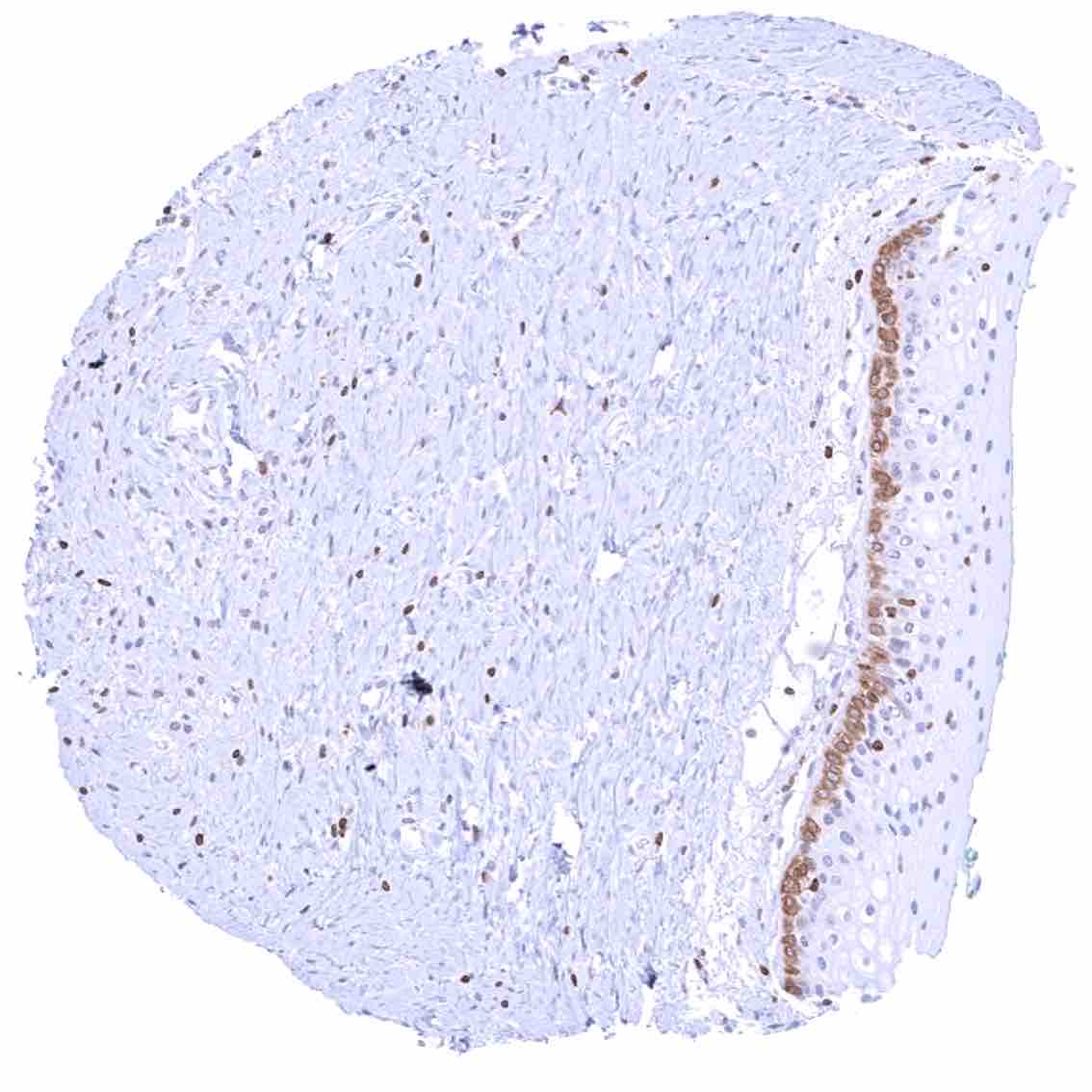
Staining Pattern in Normal Tissues
Images describing the Bcl-2 staining pattern in normal tissues obtained by the antibody MSVA-402M are shown in our “Normal Tissue Gallery”.
| Brain | Cerebrum | Negative. |
| Cerebellum | Negative. | |
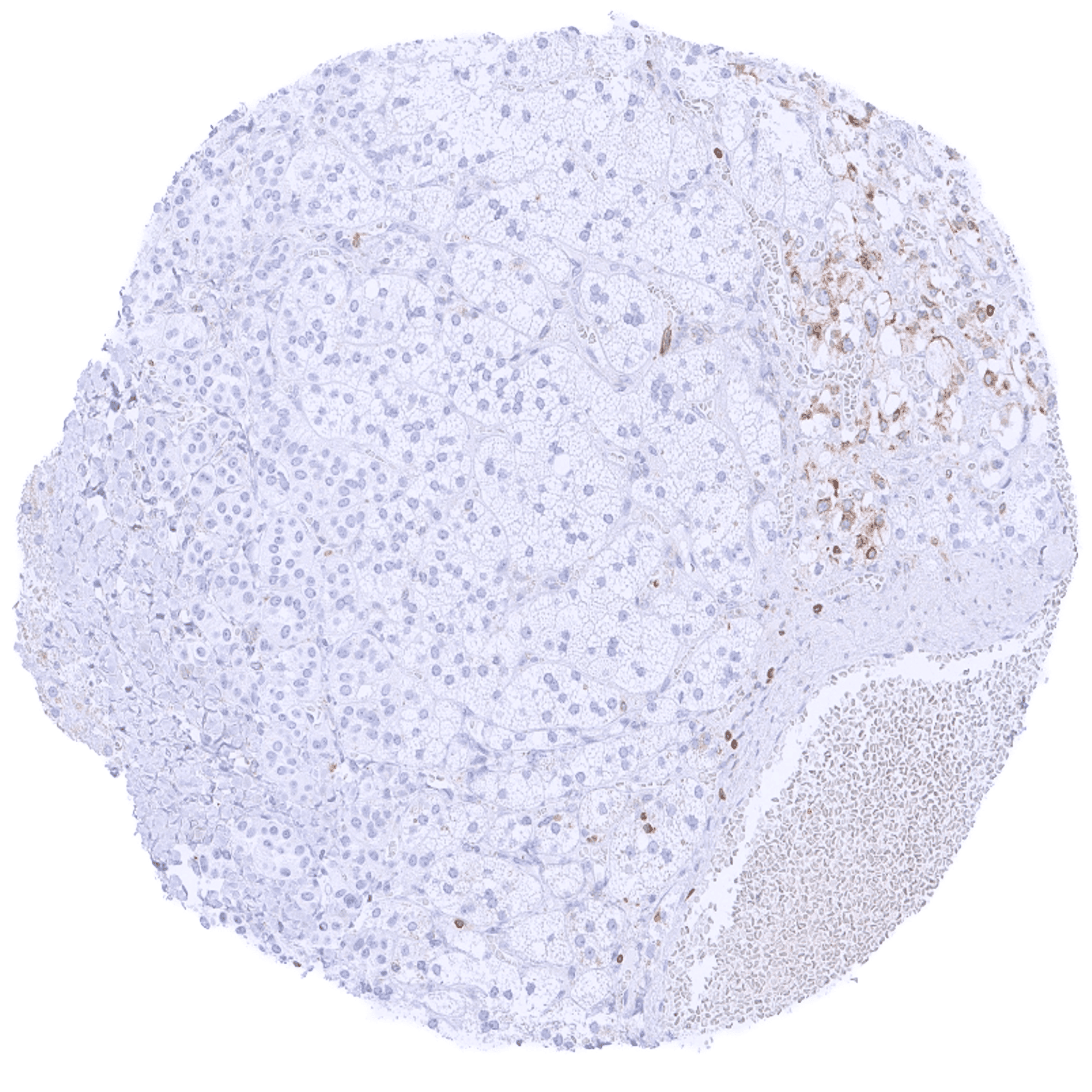
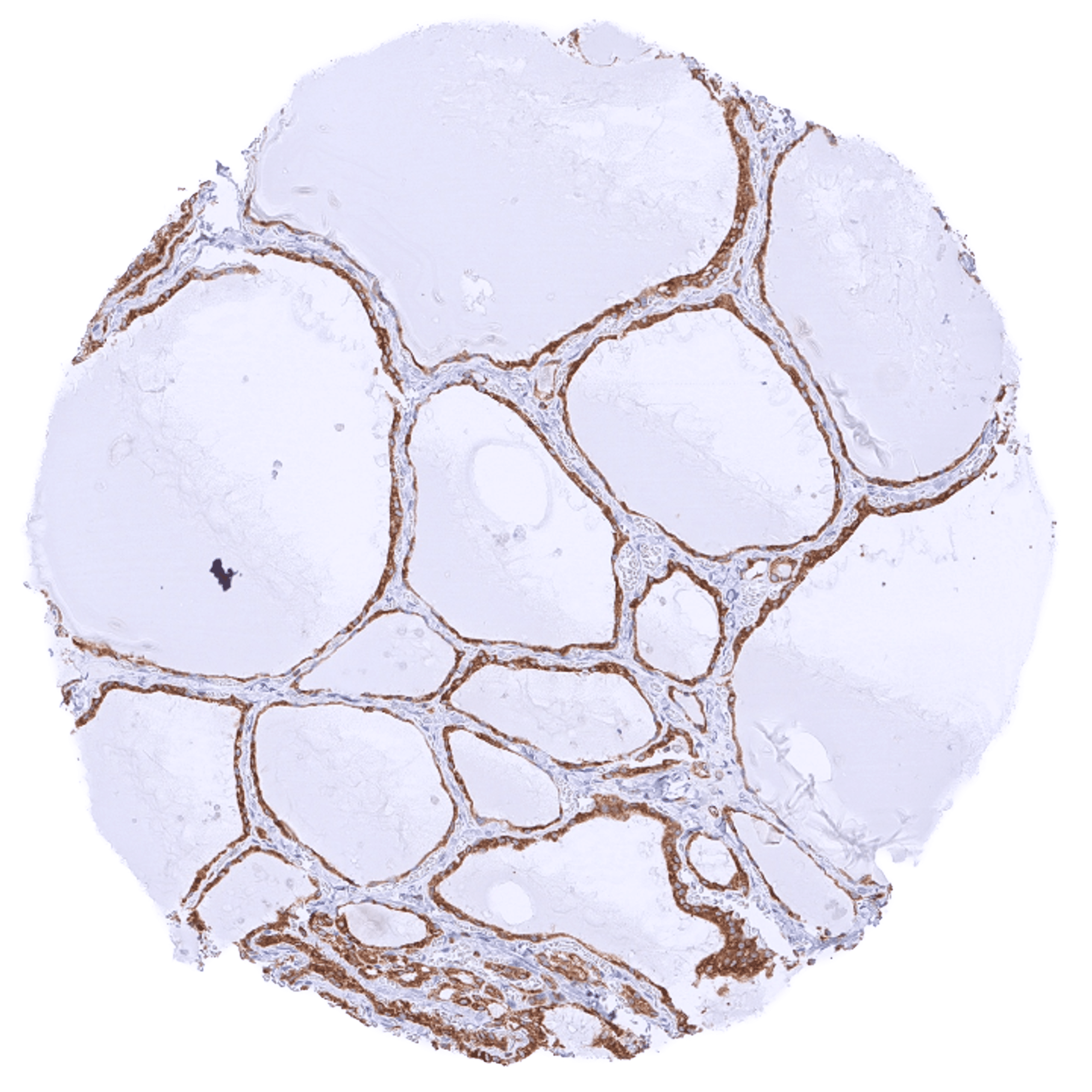
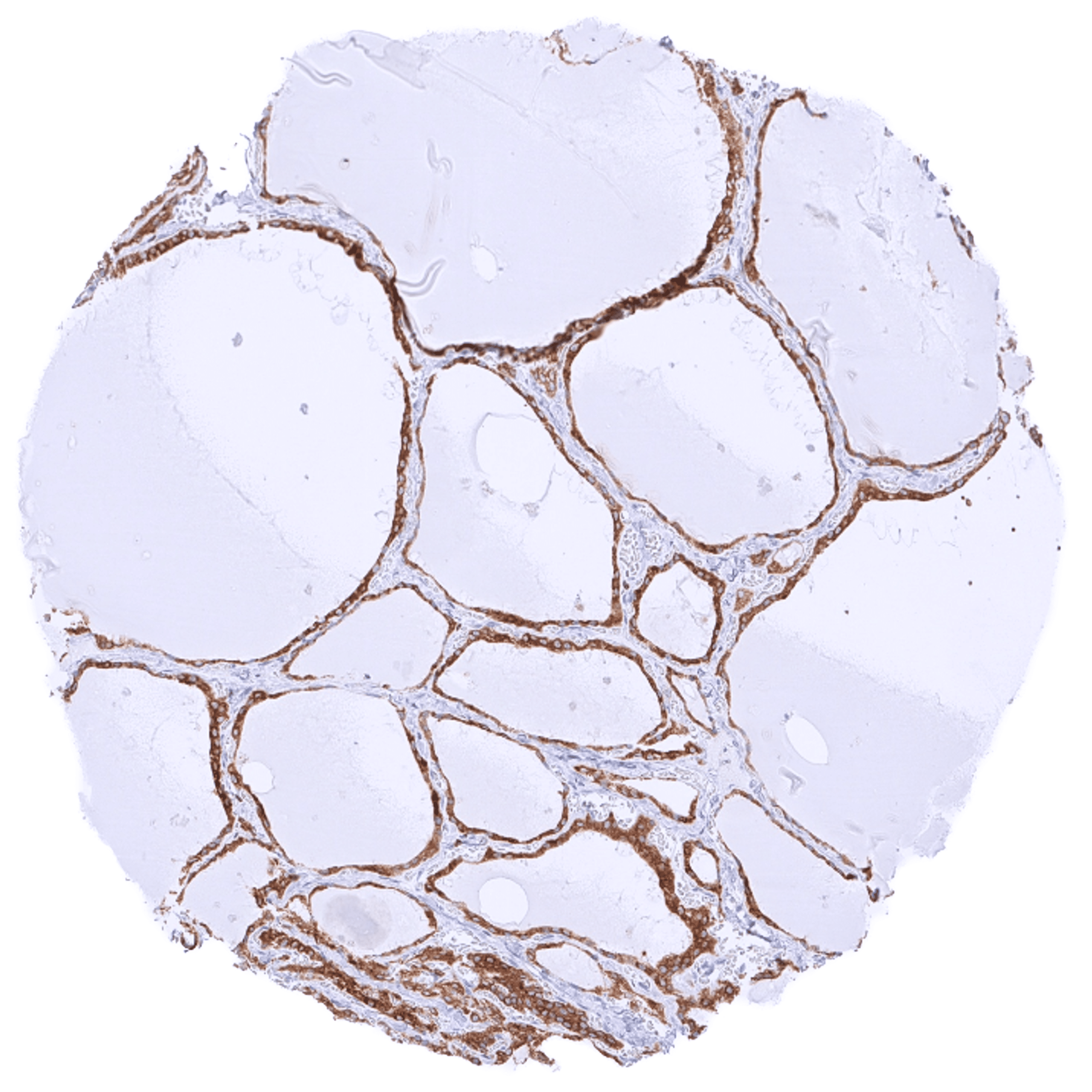
| Endocrine Tissues | Thyroid | Strong cytoplasmic bcl-2 staining of follicular cells. |
| Parathyroid | Moderate cytoplasmic bcl-2 staining of medullary cells while cortical cells are negative. | |
| Adrenal gland | Moderate cytoplasmic bcl-2 staining of medullary cells while cortical cells are negative. | |
| Pituitary gland | Few bcl-2 positive cells in the adenohypophysis. | |
| Respiratory system | Respiratory epithelium | Weak to moderate cytoplasmic bcl-2 staining of basal cells. |
| Lung | Pneumocytes are largely negative while a weak cytoplasmic bcl-2 staining can appear in some bronchiolar cells. | |
| Gastrointestinal Tract | Salivary glands | A moderate cytoplasmic bcl-2 staining can occur in myoepithelial cells of excretory ducts. |
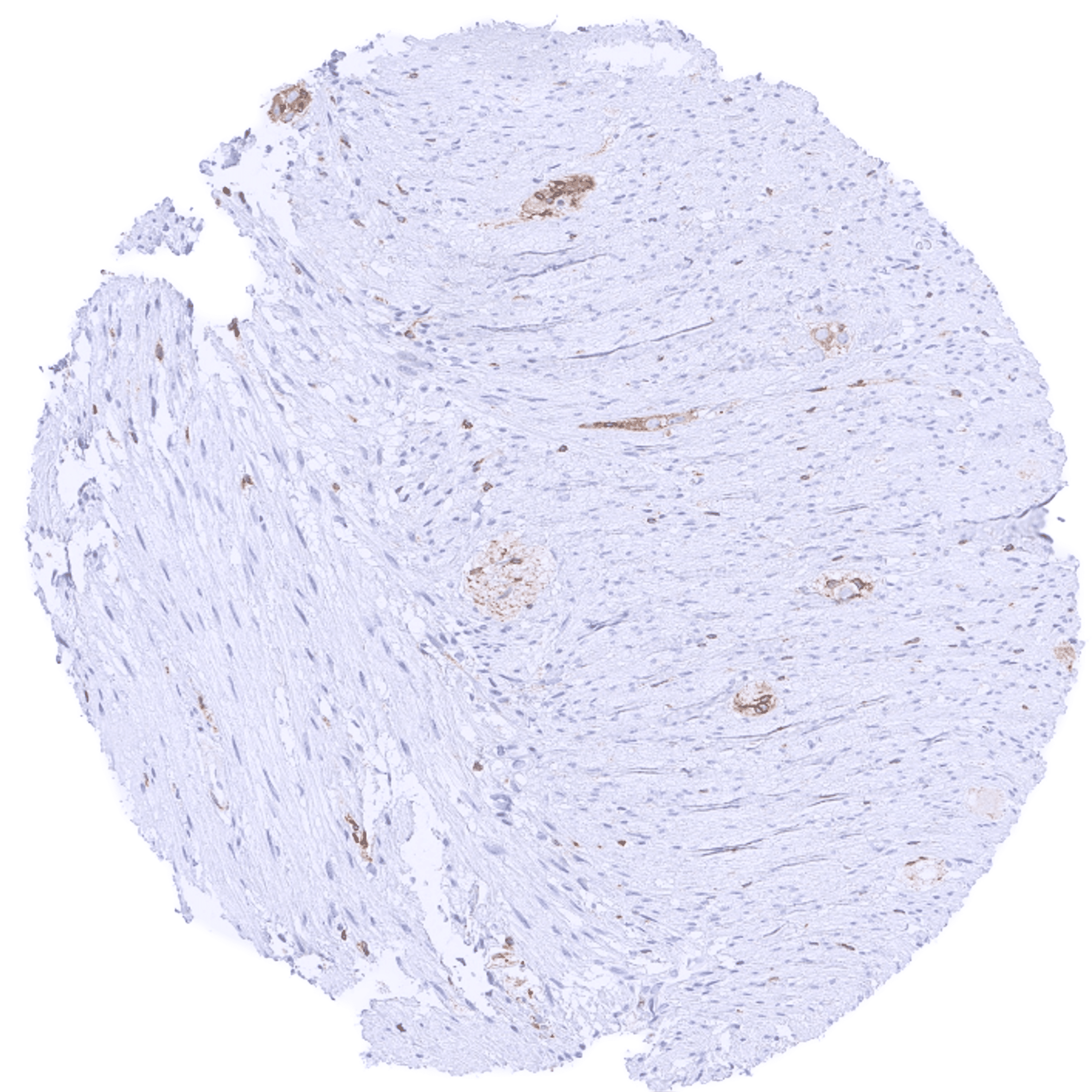
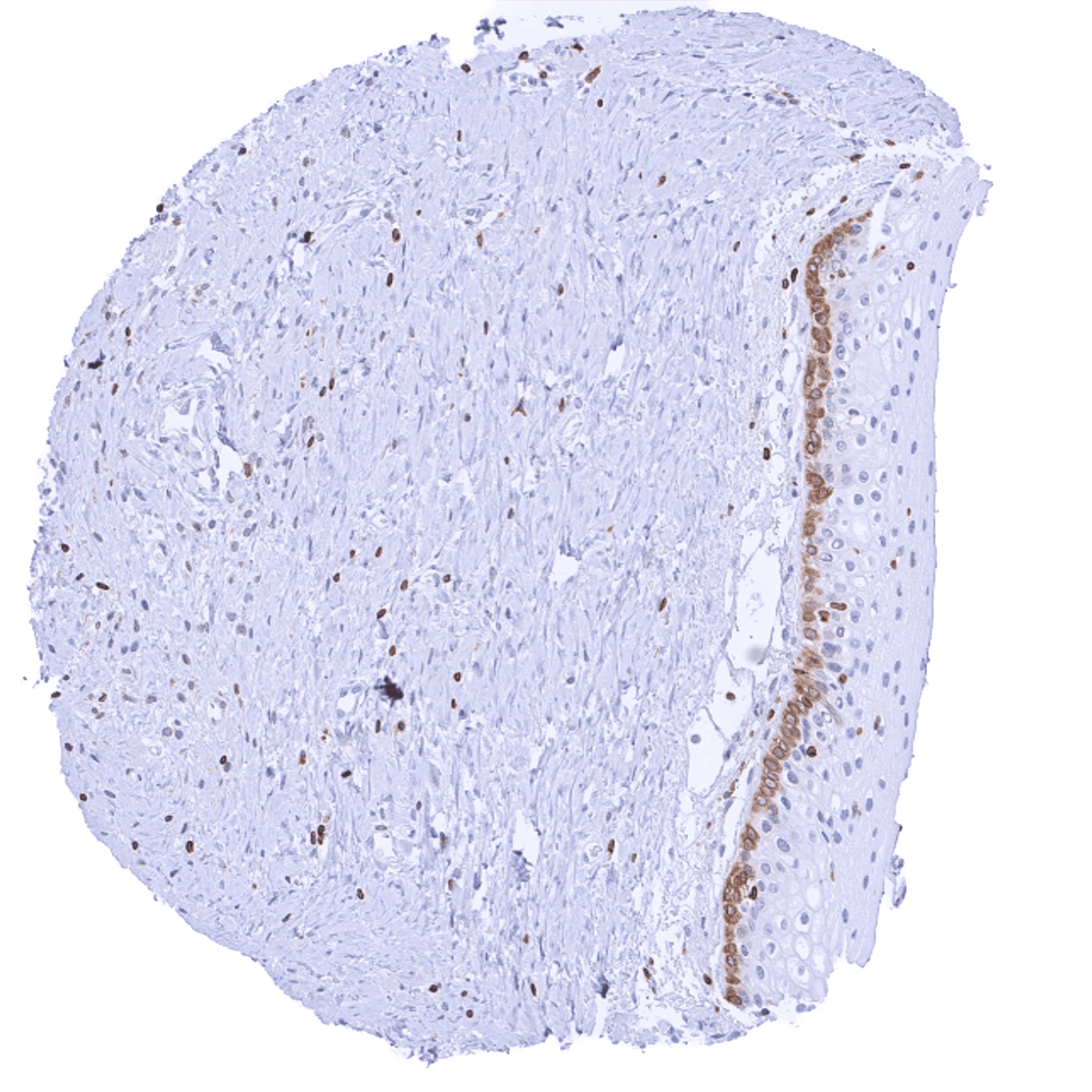
| Esophagus | Squamous epithelium was bcl-2 negative in few analyzed samples (basal cell positivity may occur based on findings in other types of squamous epithelium). Moderate cytoplasmic bcl-2 staining of ganglion cells of the muscular wall. | |
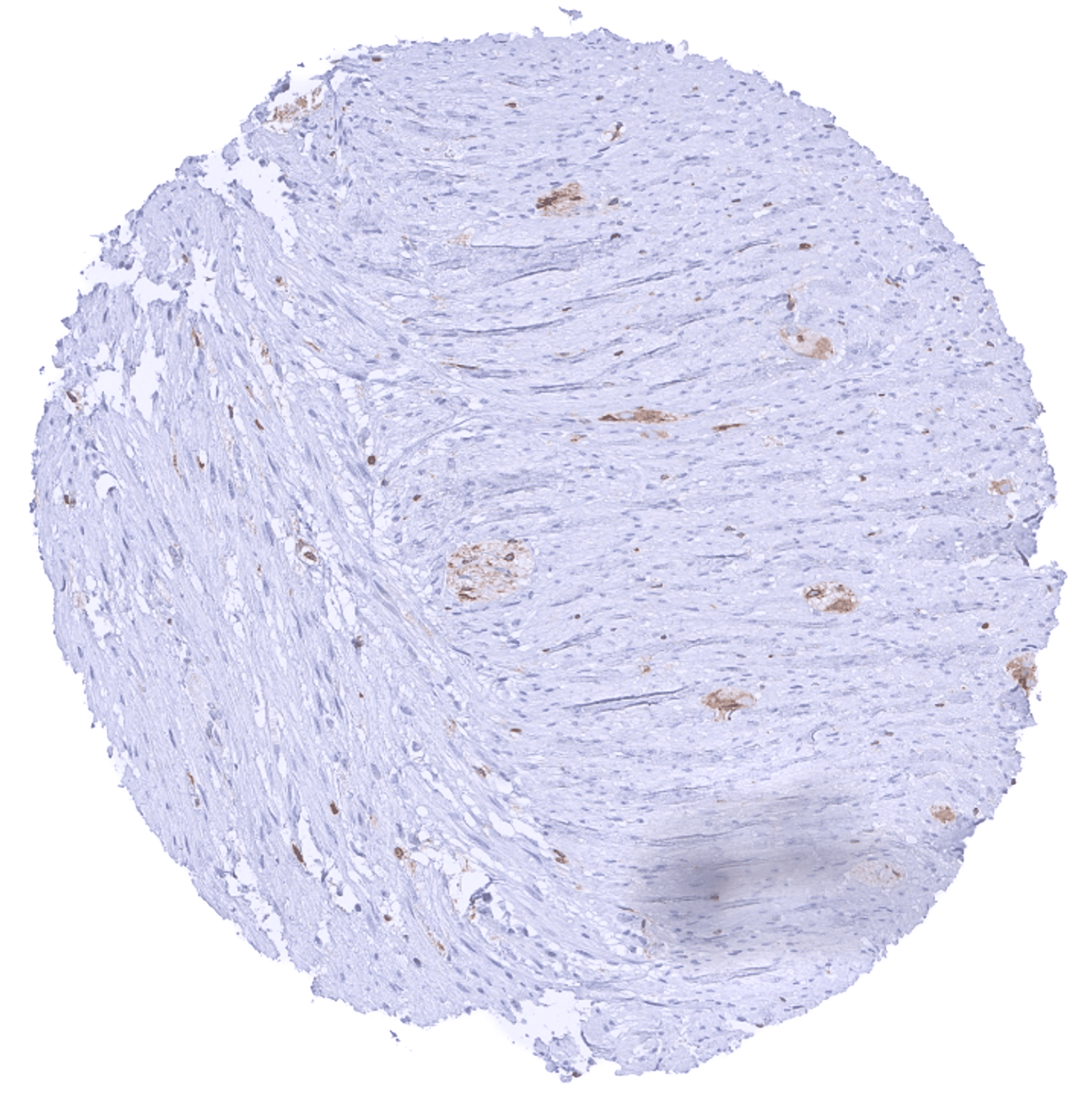
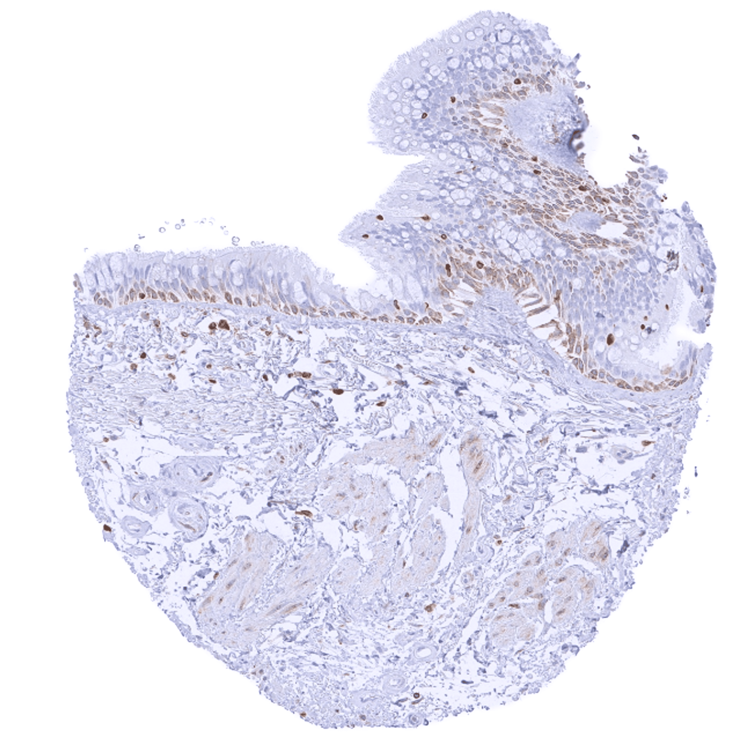
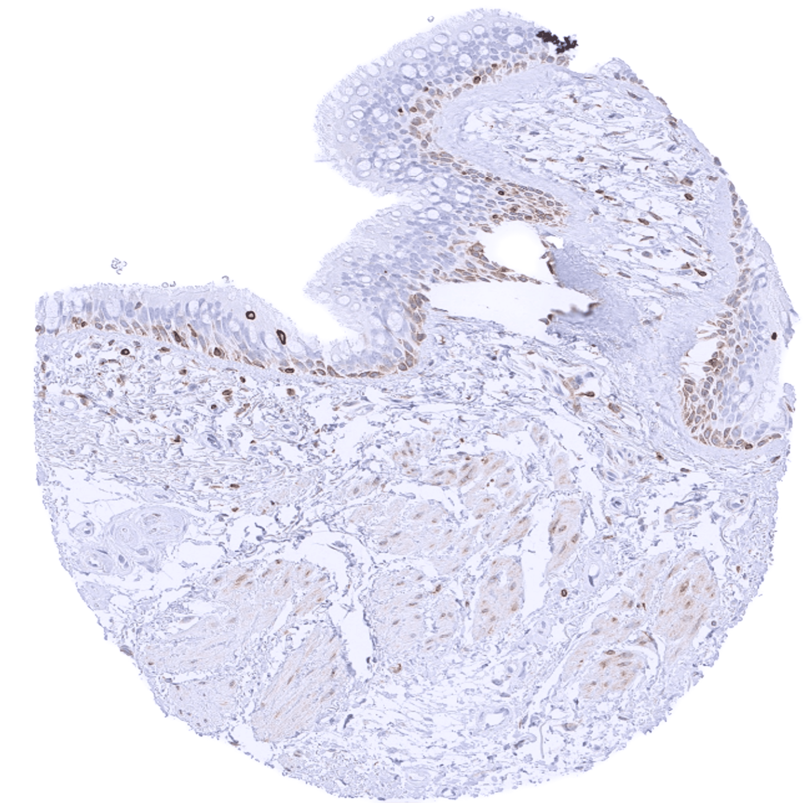
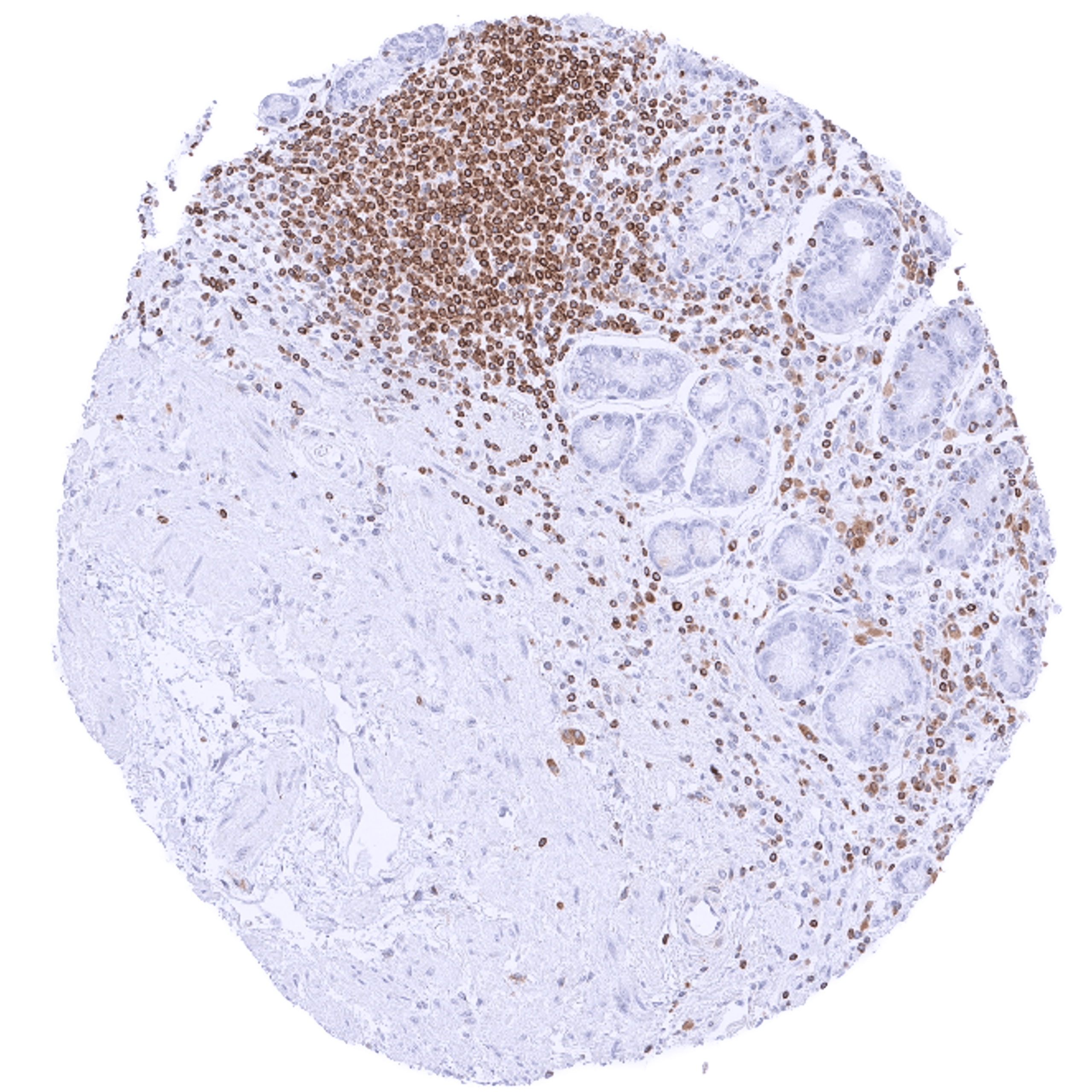
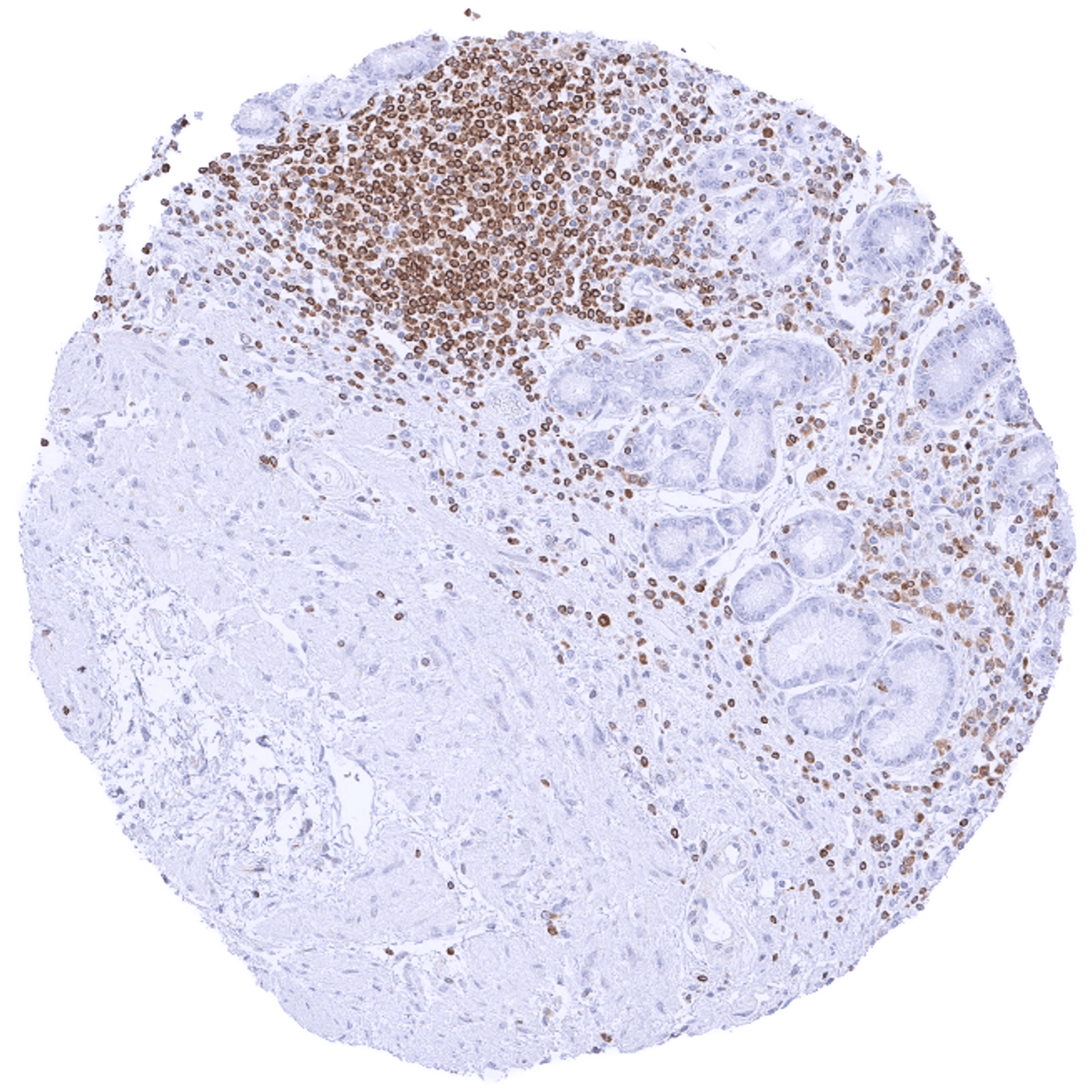
| Stomach | The epithelium is largely negative. Moderate cytoplasmic bcl-2 staining of nerves in the muscular wall. | |
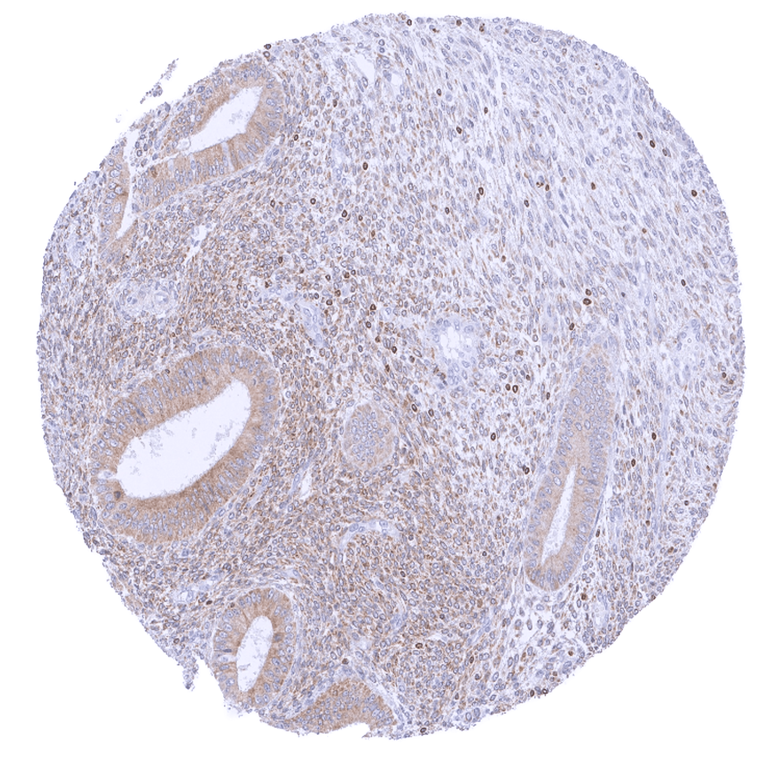
| Duodenum | Cytoplasmic bcl-2 staining is largely limited to lymphocytes. The epithelium is mostly negative although some weak staining can occur in crypt cells. | |
| Small intestine | Cytoplasmic bcl-2 staining is largely limited to lymphocytes. The epithelium is mostly negative although some weak staining can occur in crypt cells. | |
| Appendix | Cytoplasmic bcl-2 staining is largely limited to (non-germinal centre) lymphocytes. A weak bcl-2 staining occurs in crypt cells. | |
| Colon | Weak bcl-2 staining of crypt cells. | |
| Rectum | Weak bcl-2 staining of crypt cells. | |
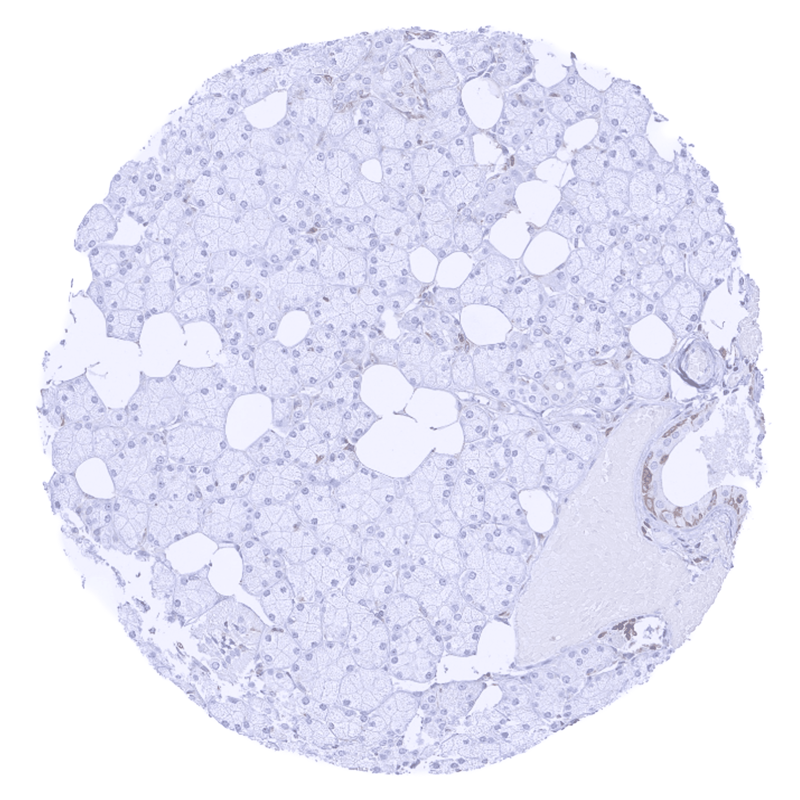
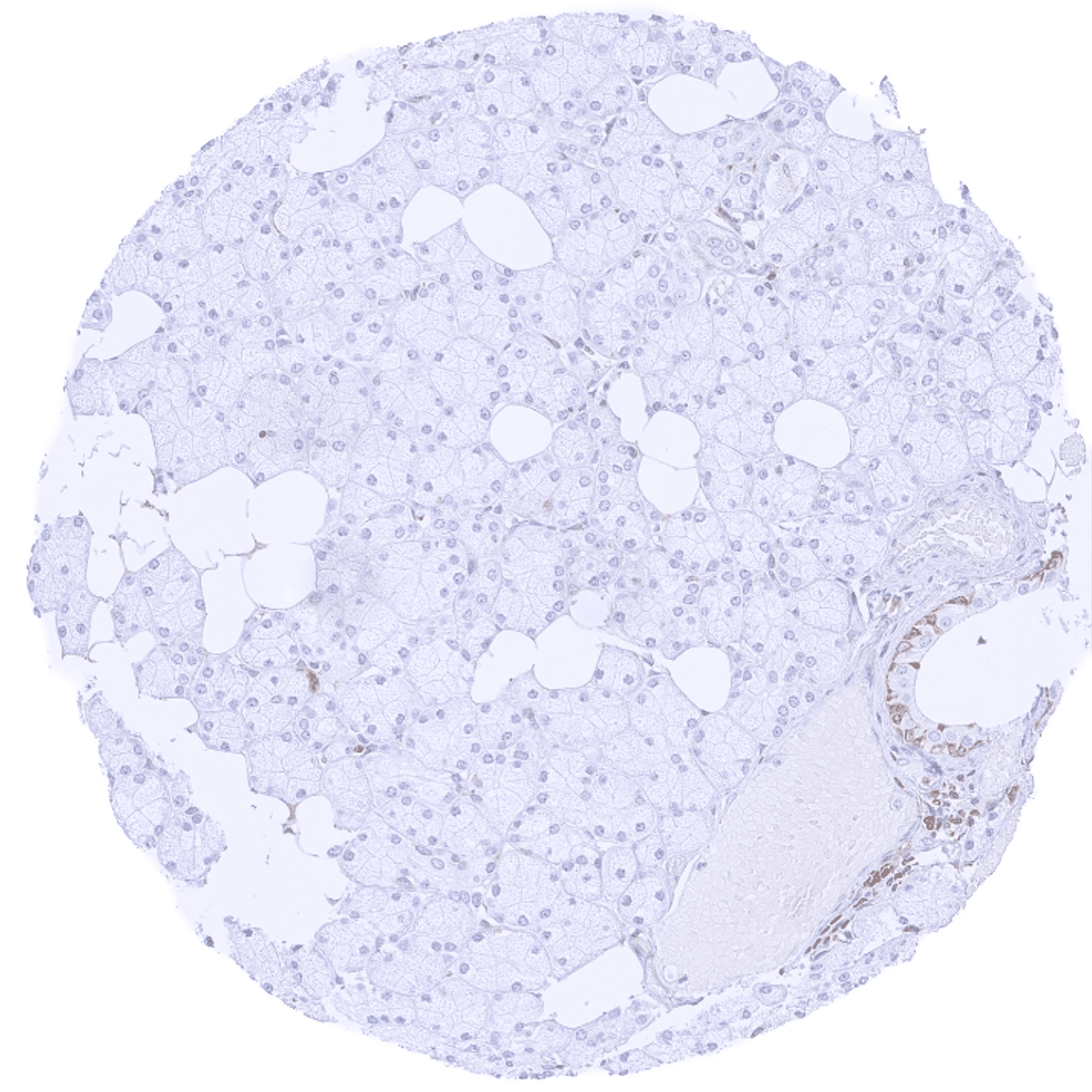
| Liver | Negative. | |
| Gallbladder | The epithelium is largely bcl-2 negative. | |
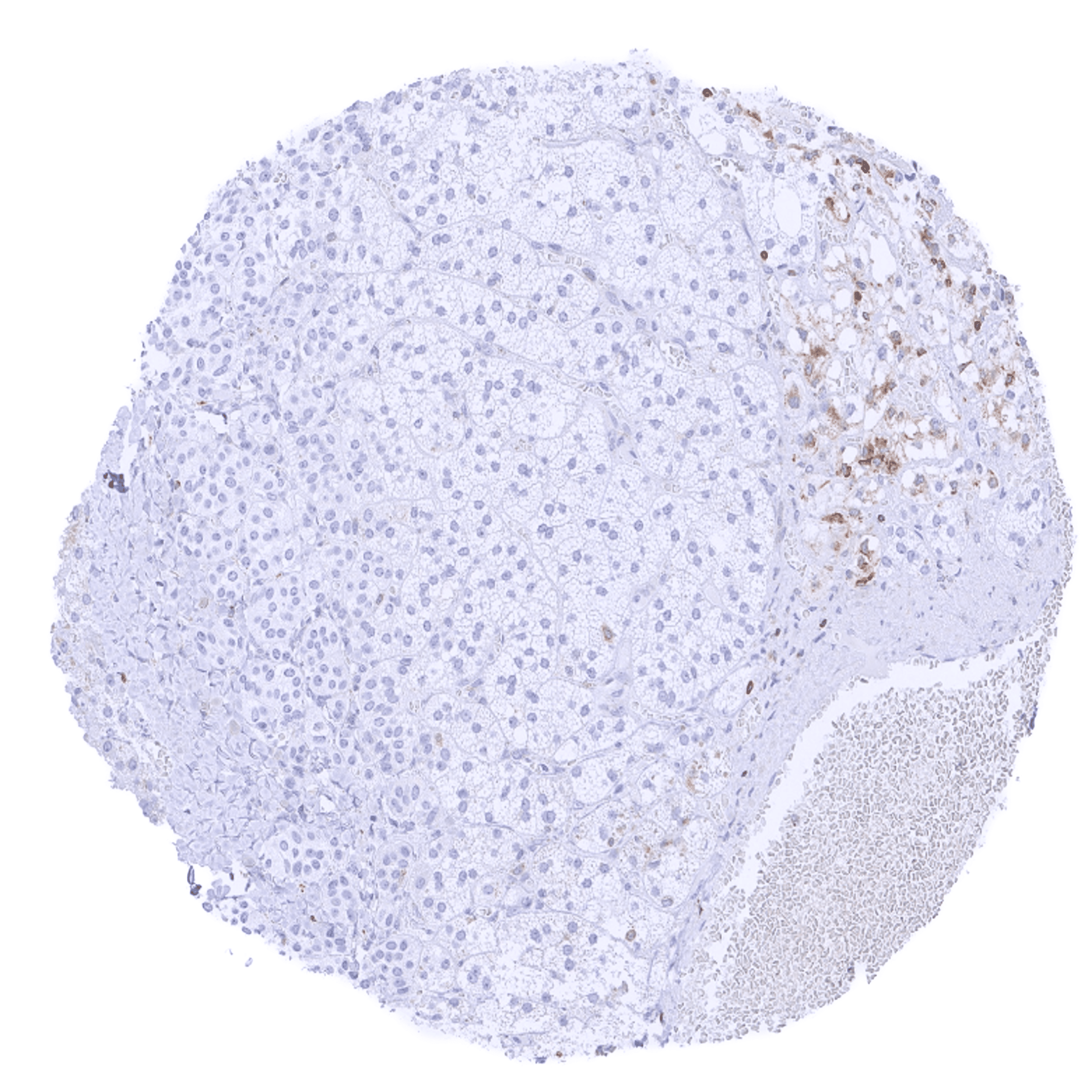
| Pancreas | A weak bcl-2 staining can occur in excretory duct cells (not in all samples). | |
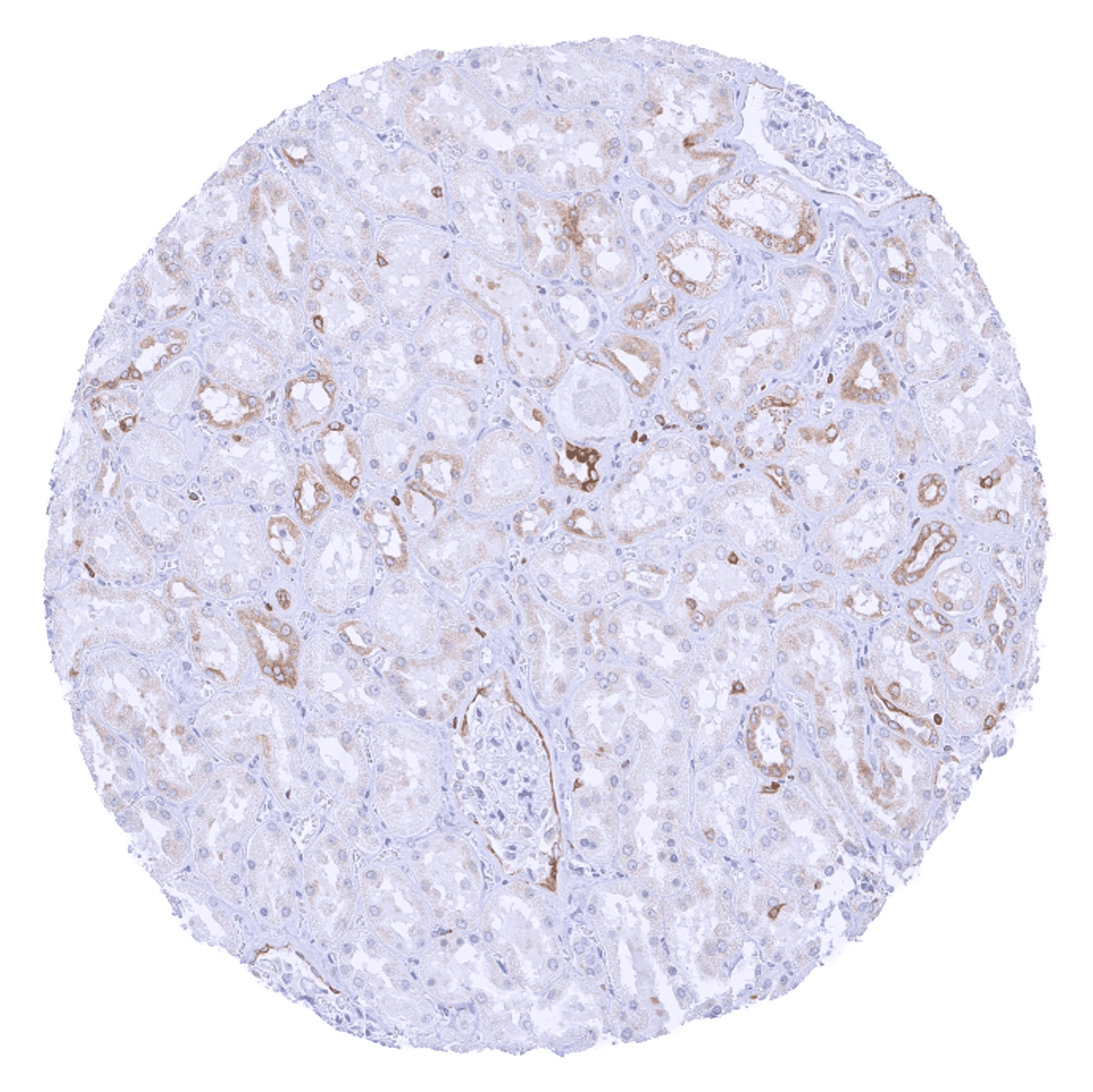
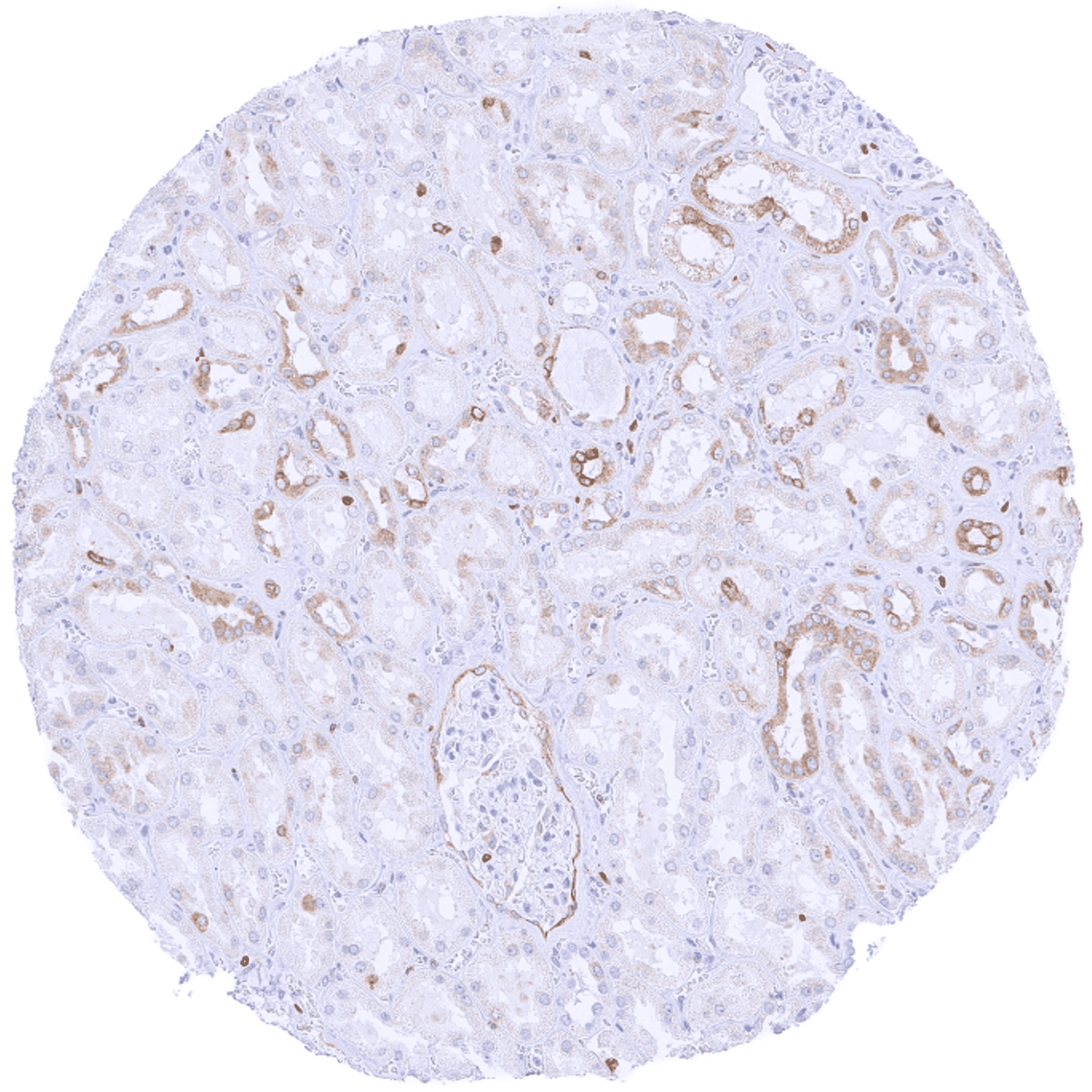
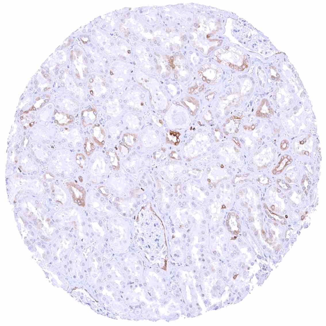
| Genitourinary | Kidney | A variable (usually weak to moderate) bcl-2 staining can occur in a fraction of (mostly distal) tubuli and collecting ducts. Staining may be more prominent in samples suffering from some degree of tissue damage. |
| Urothelium | Negative. | |
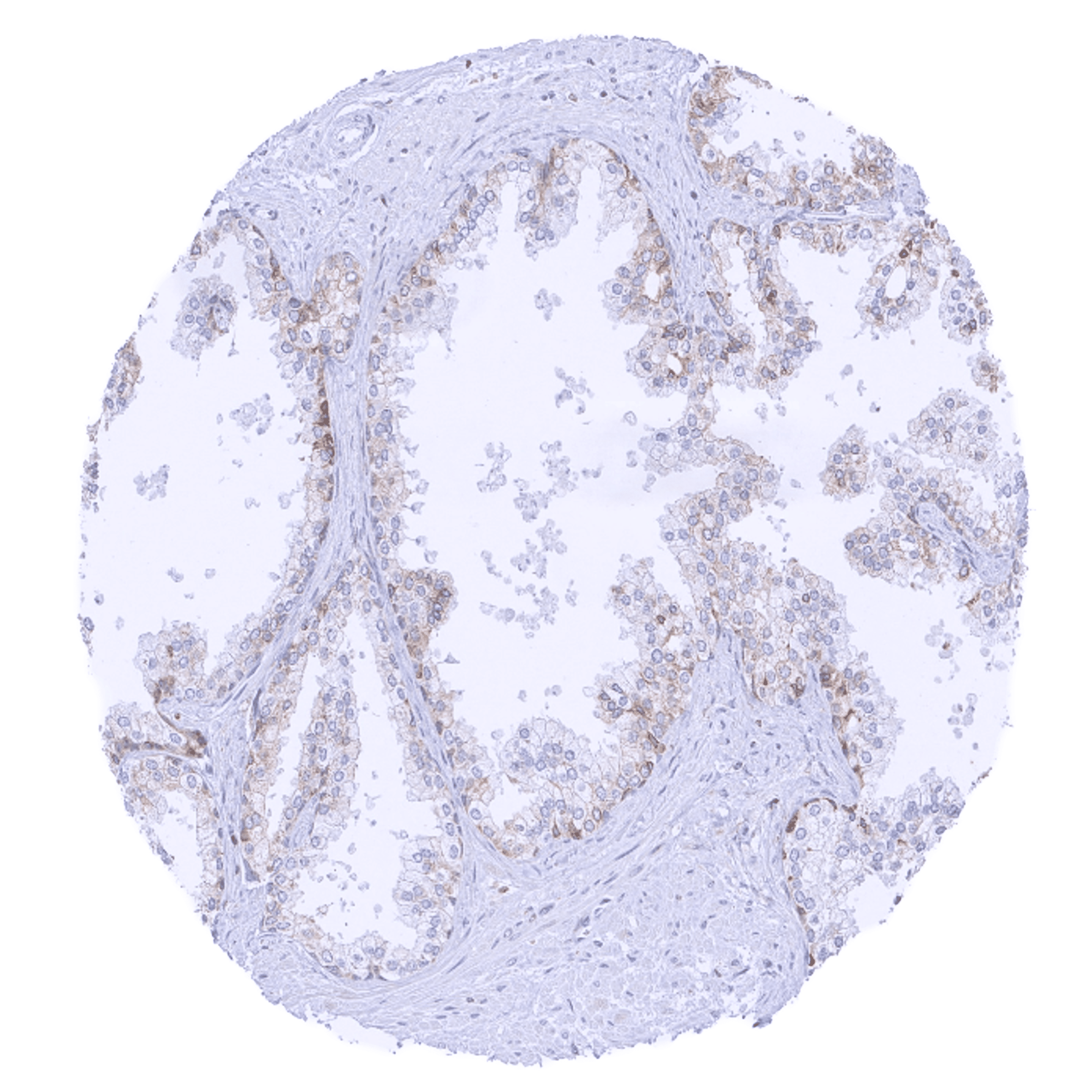
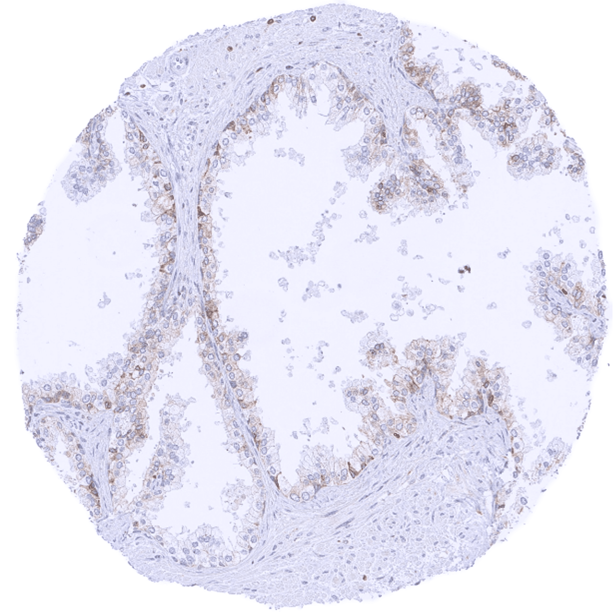
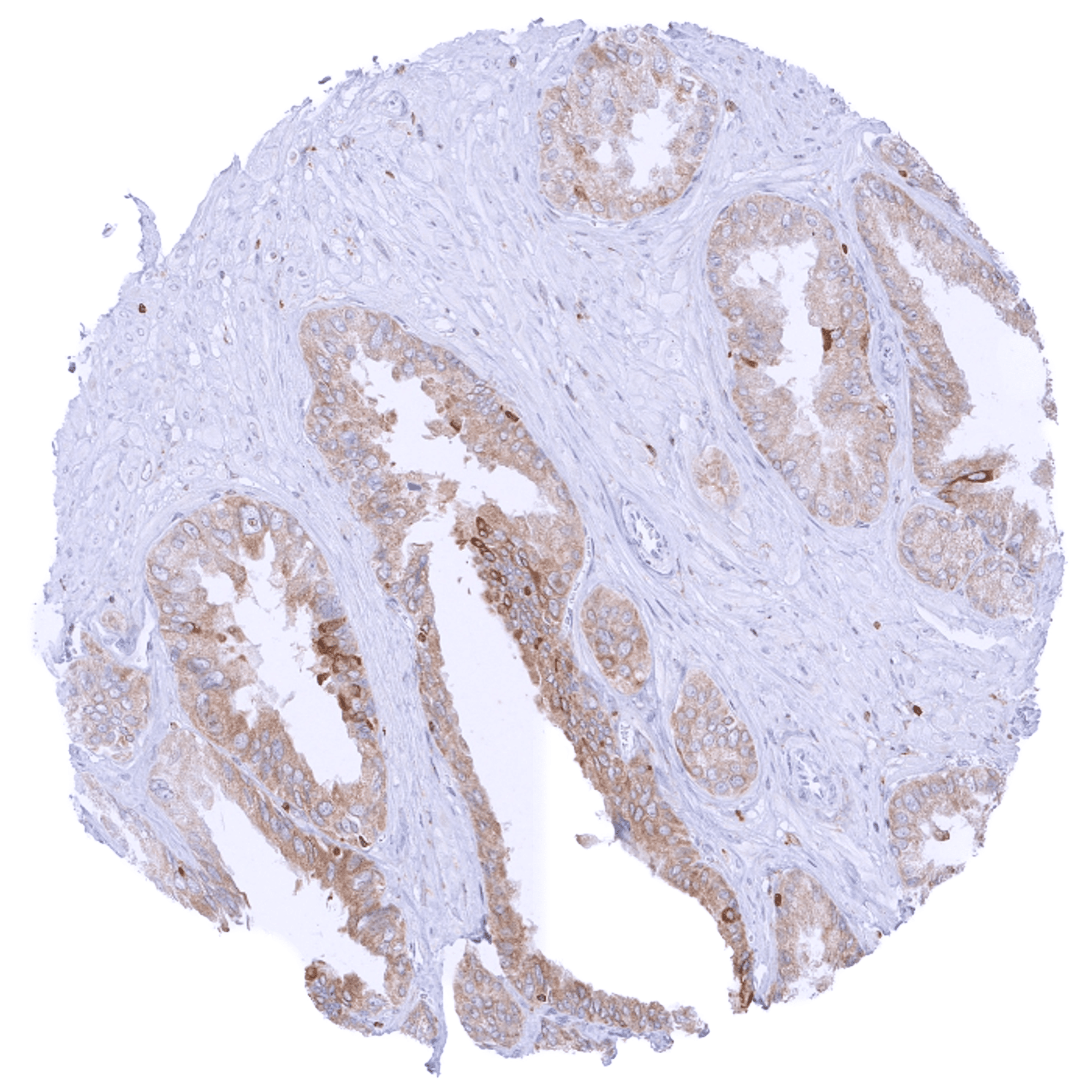
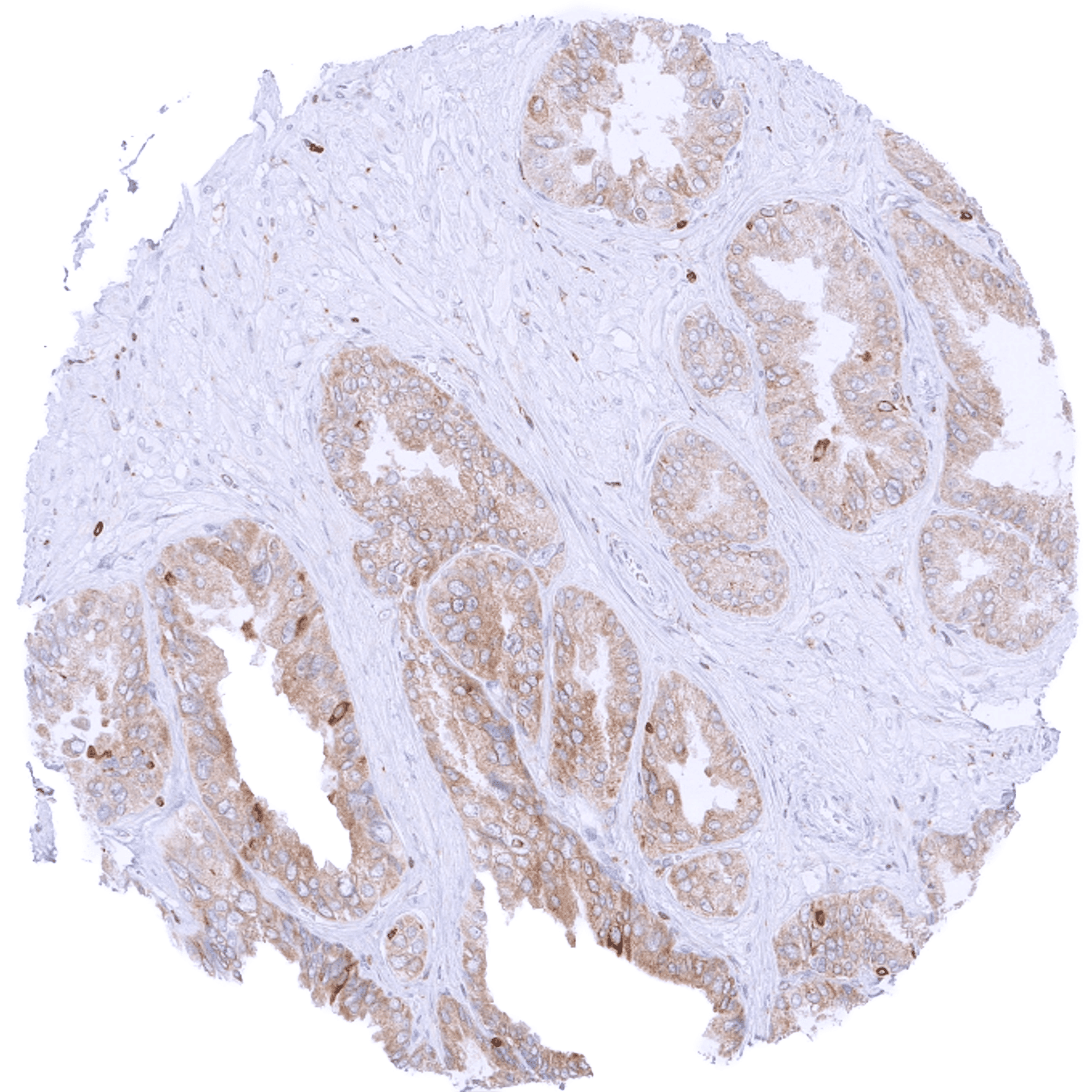
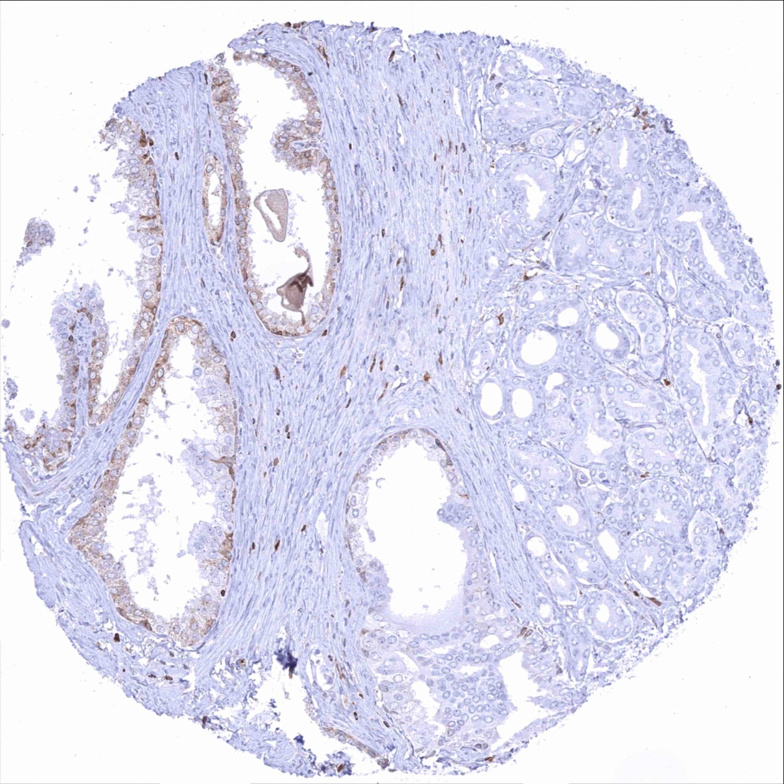
| Male genital | Prostate | Weak to moderate bcl-2 staining of basal cells. |
| Seminal vesicles | Moderate to strong bcl-2 staining of epithelial cells. | |
| Testis | Negative. | |
| Epididymis | Weak to moderate bcl-2 staining of epithelial cells. | |
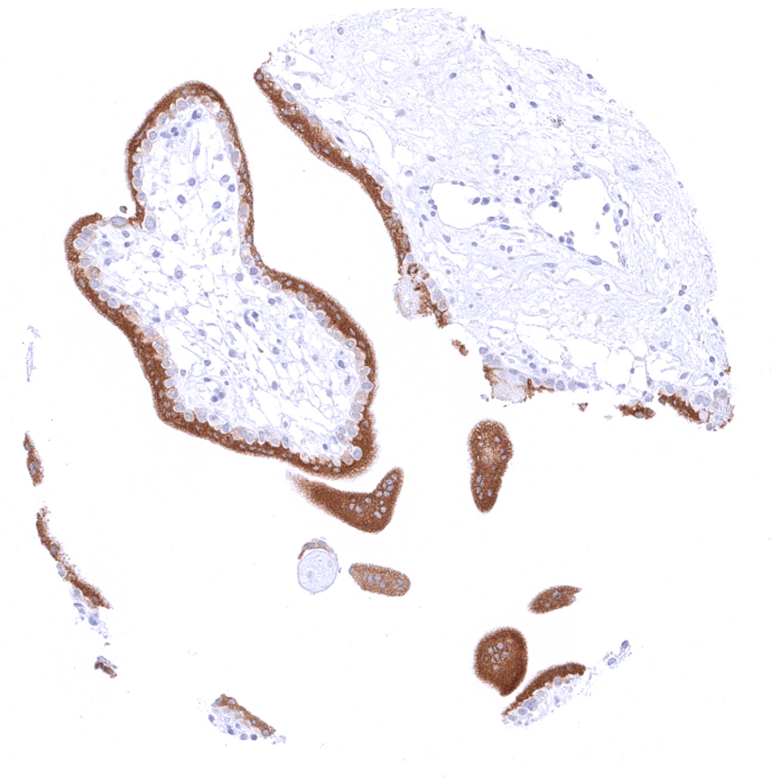
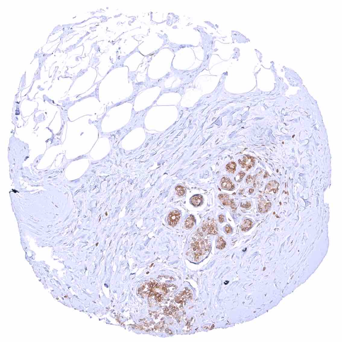
| Female genital | Breast | Distinct cytoplasmic bcl-2 staining of epithelial cells (acinar cells stain stronger than myoepithelial cells). |
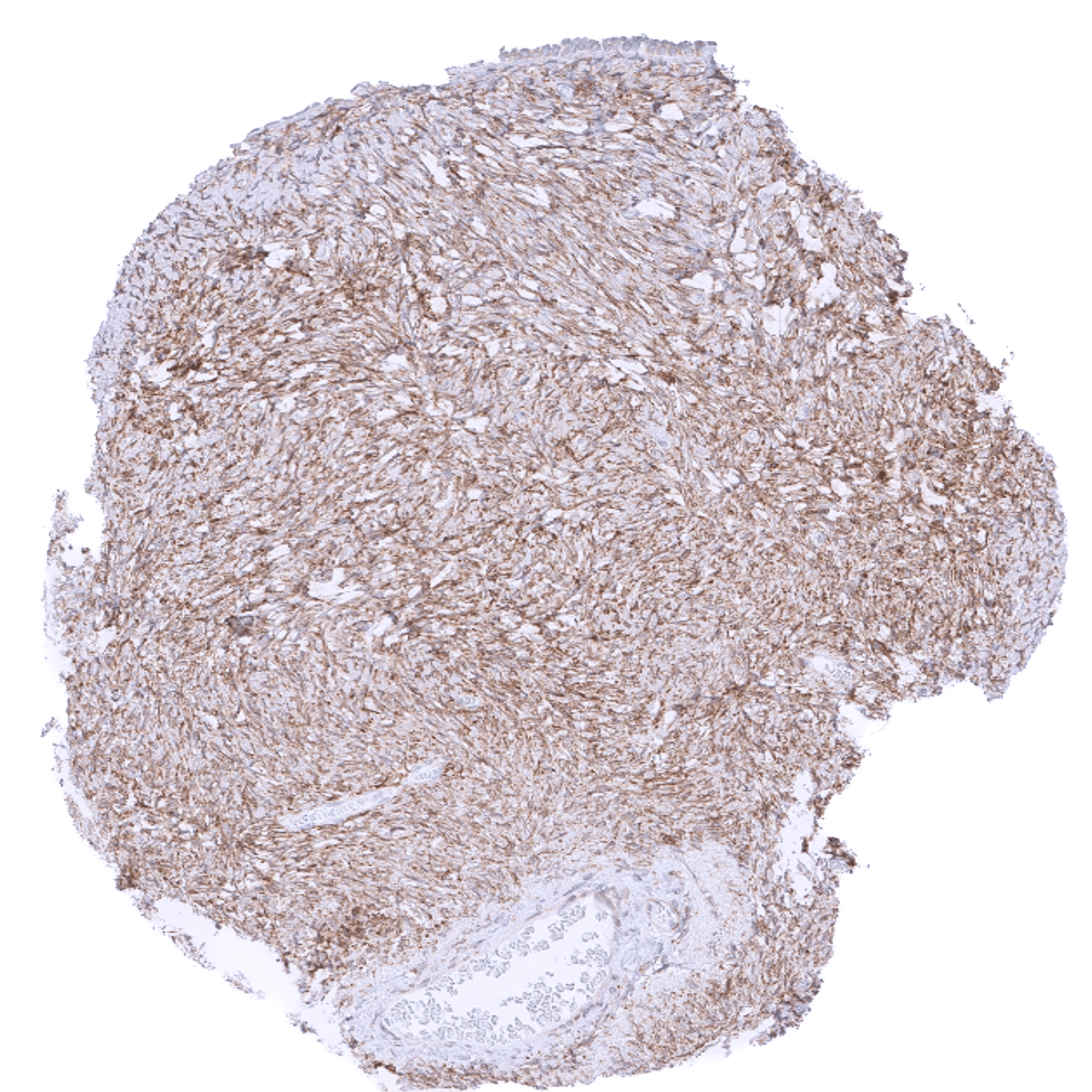
| Uterus, myometrium | Weak to moderate cytoplasmic bcl-2 staining of smooth muscle cells. | |
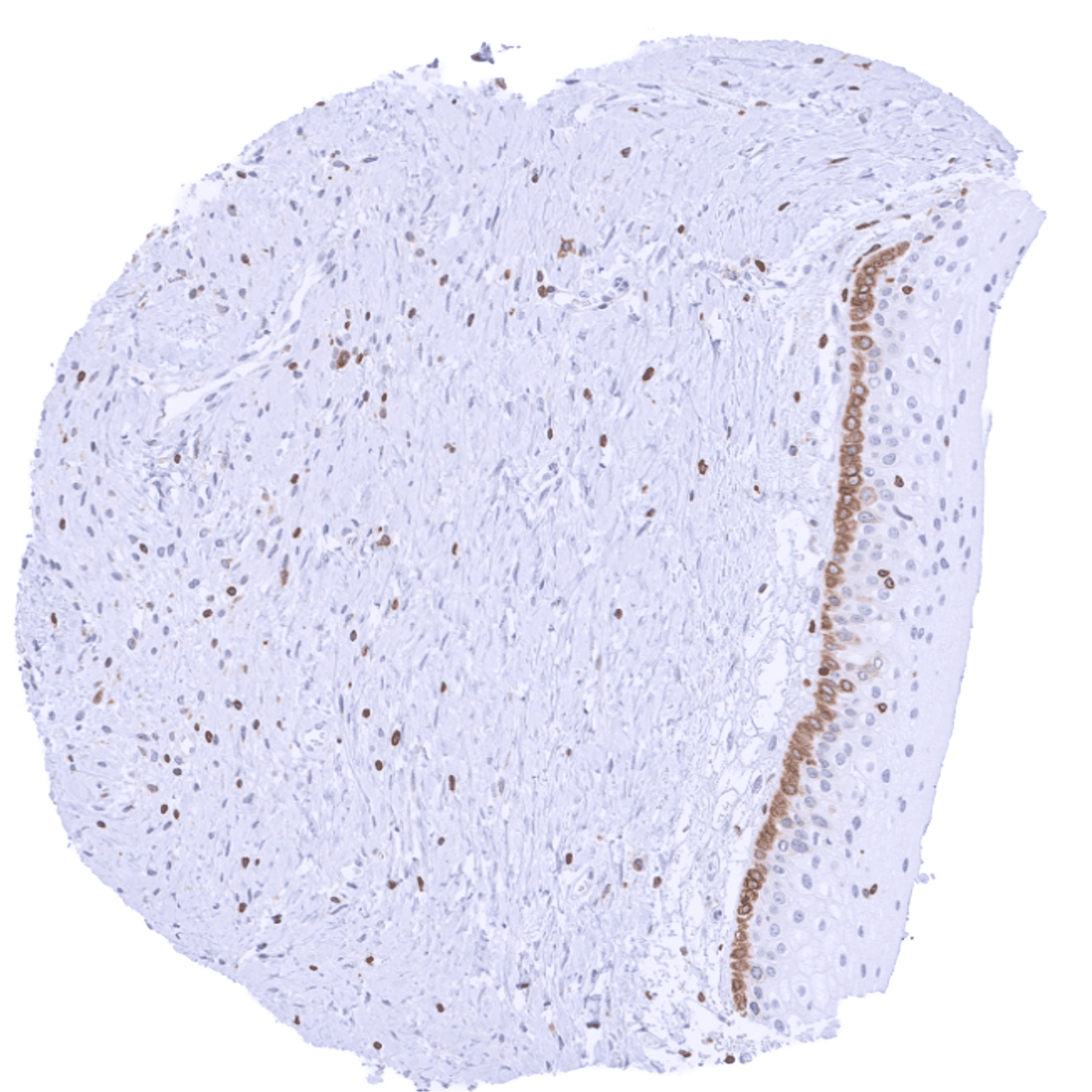
| Uterus, ectocervix | Variable (weak to strong) cytoplasmic bcl-2 staining of basal cells of the squamous epithelium. | |
| Uterus endocervix | Negative. | |
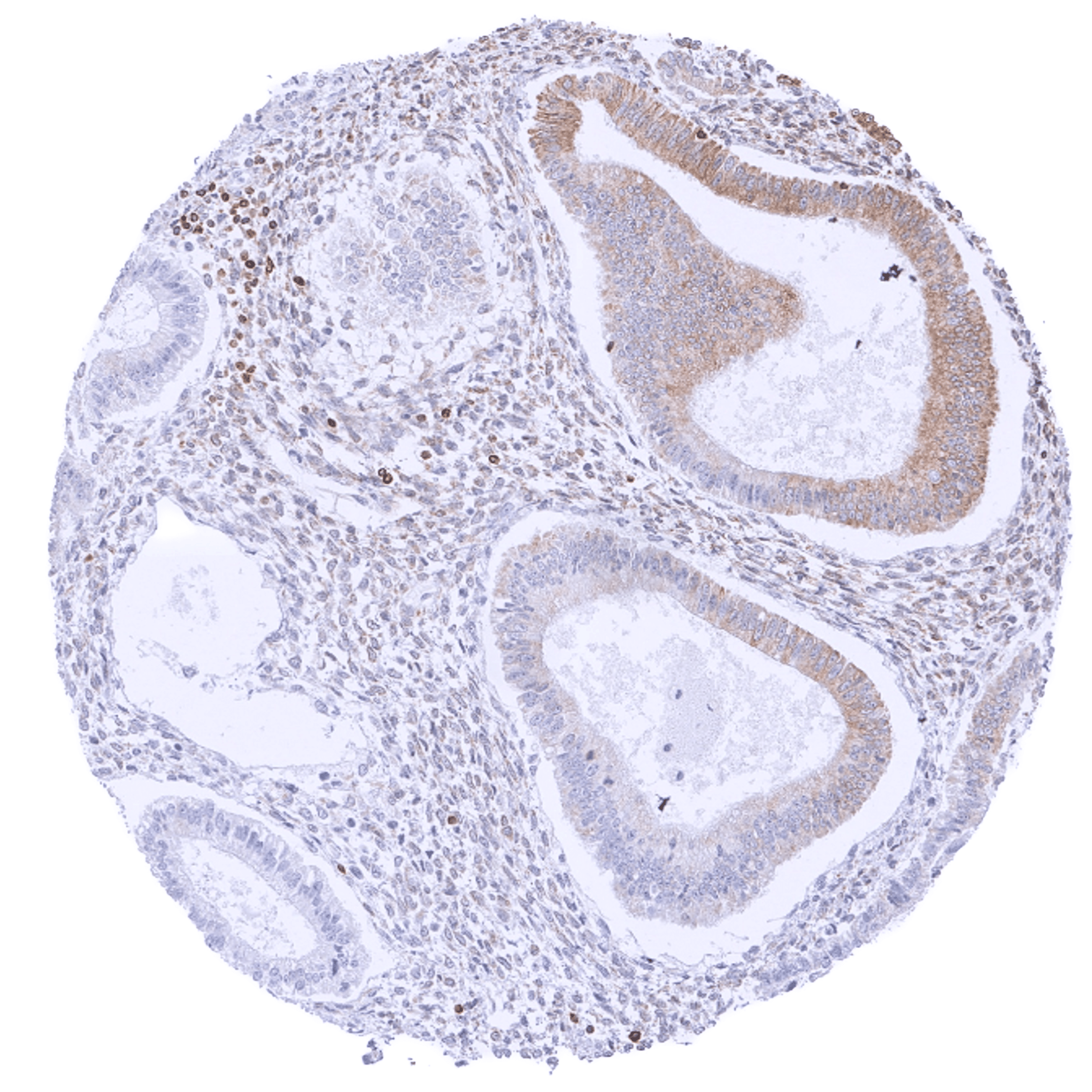
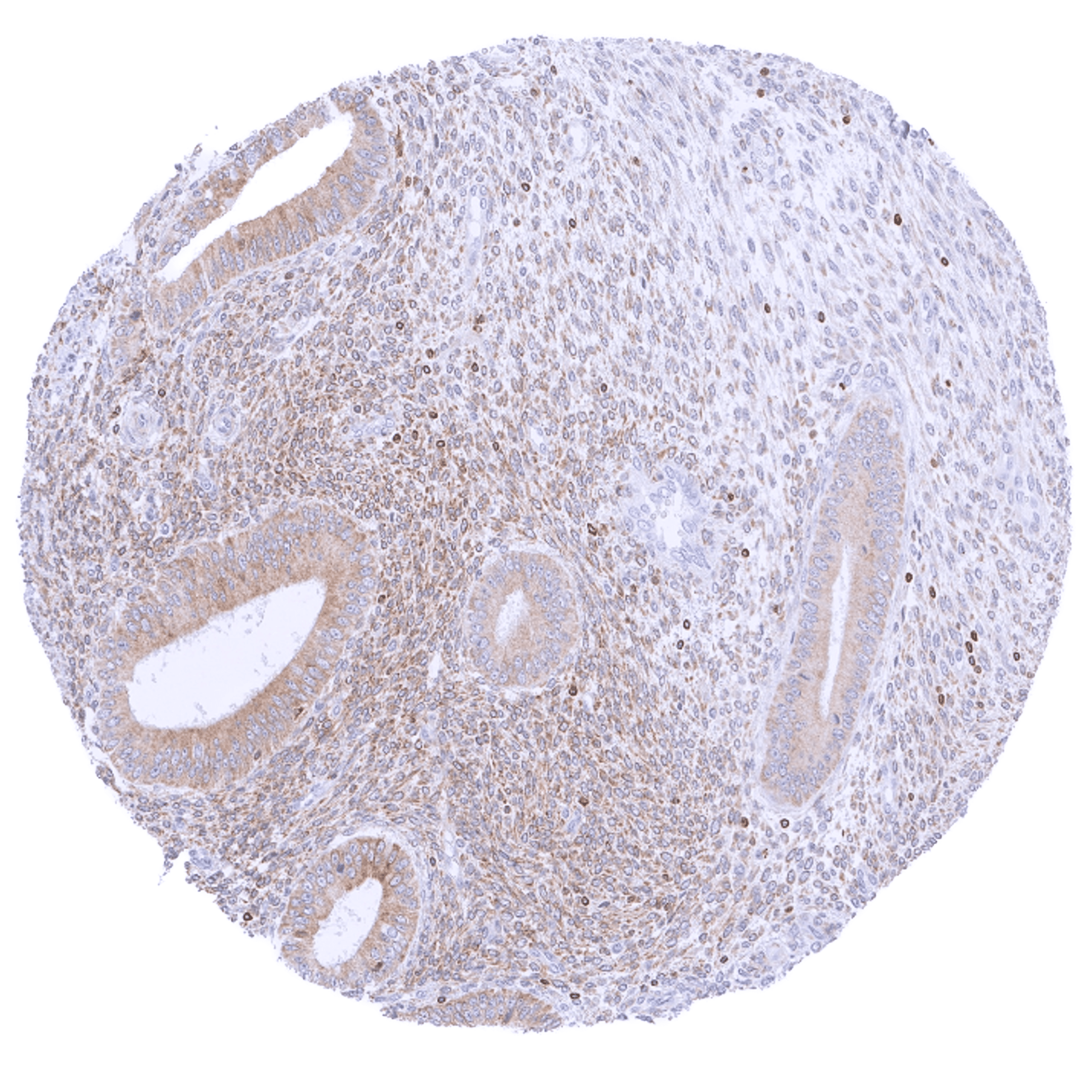
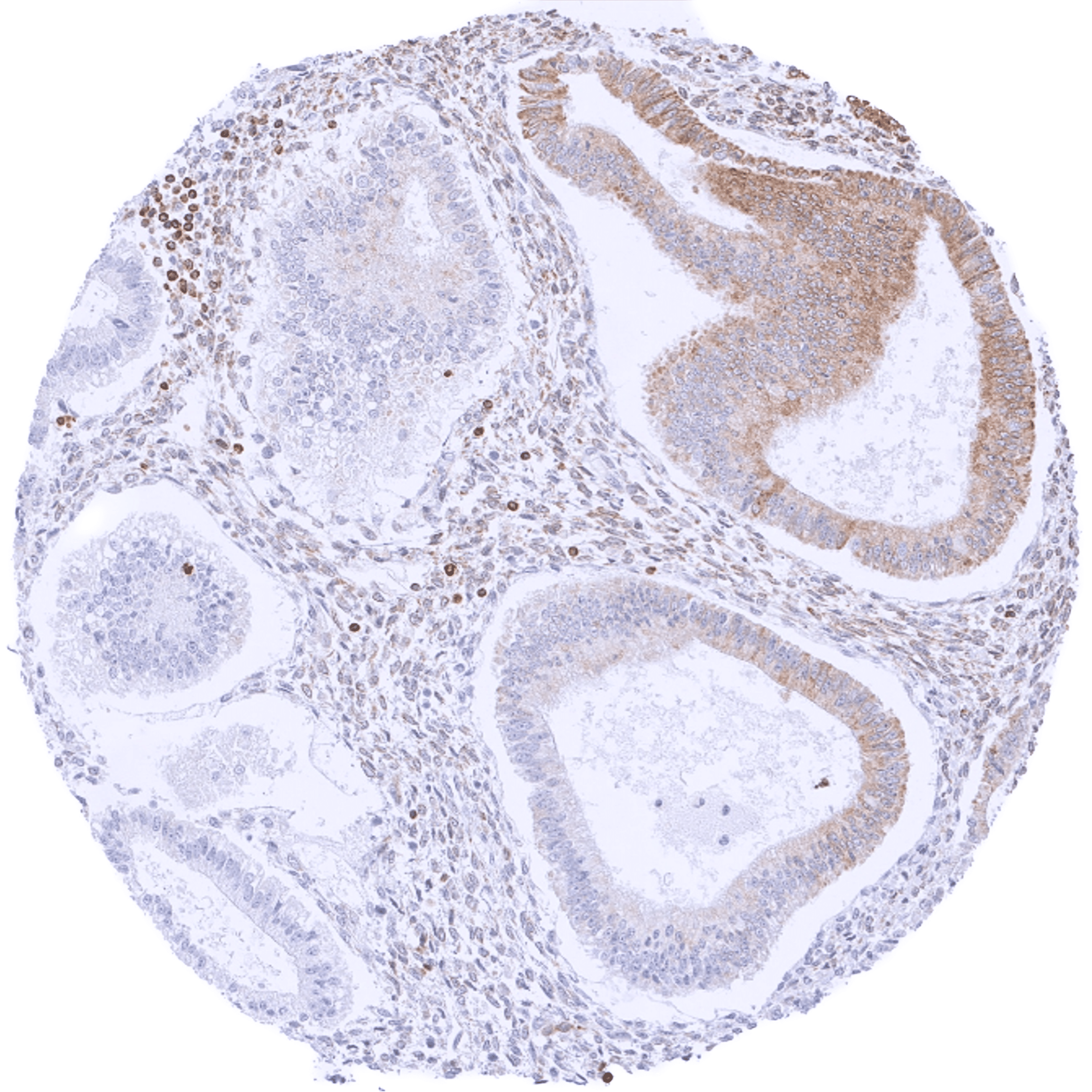
| Uterus, endometrium | Variable (negative to strong) cytoplasmic bcl-2 staining of epithelial cells. Staining of stromal cells is usually markedly weaker. | |
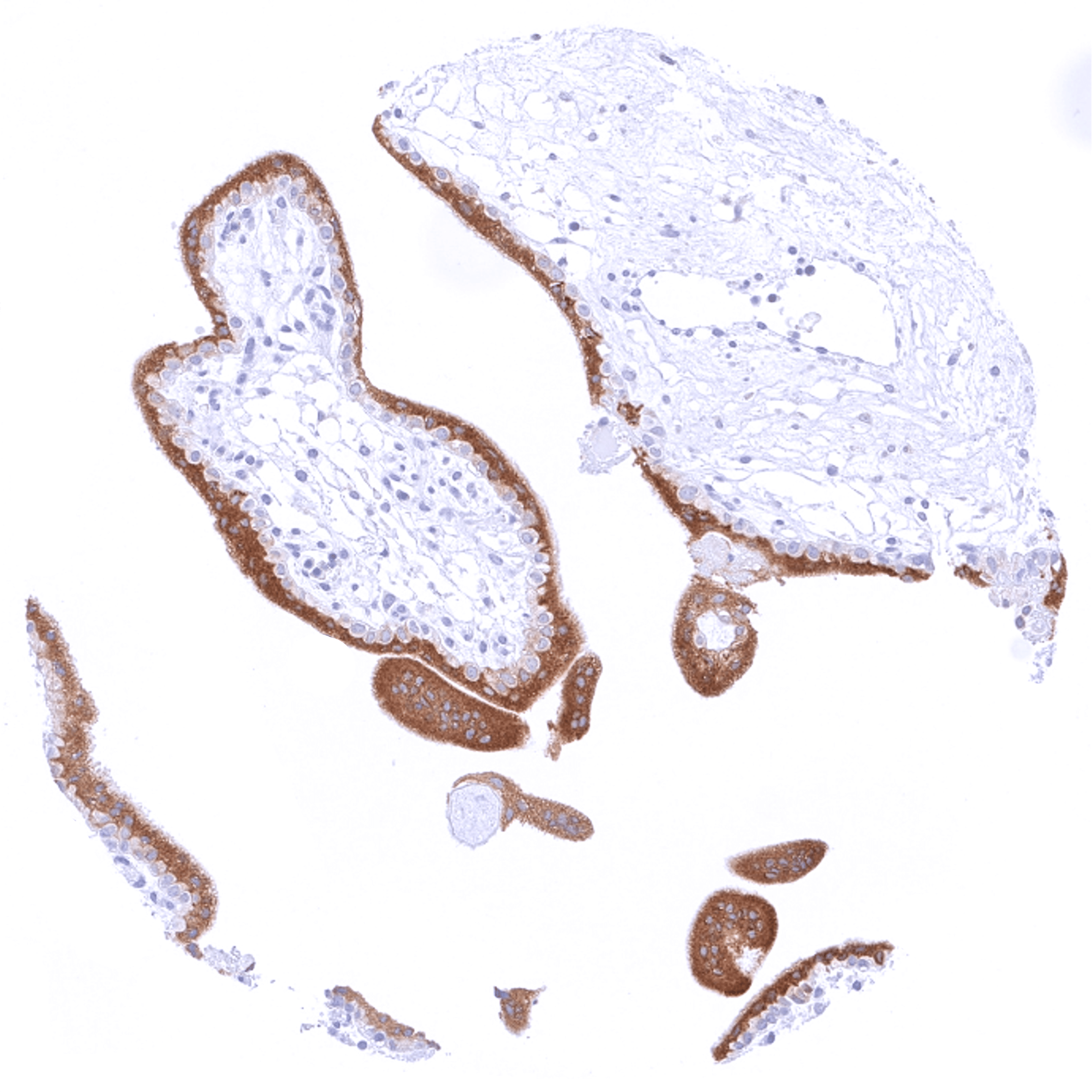
| Fallopian Tube | Strong cytoplasmic bcl-2 staining of a subset of epithelial cells. | |
| Ovary | Moderate to strong cytoplasmic bcl-2 staining of stroma cells. Corpus luteum cells are bcl-2 negative. | |
| Placenta early | Moderate to strong cytoplasmic bcl-2 staining of syncytiotrophoblast cells while the cytotrophoblast and other cells remain negative. | |
| Placenta mature | Moderate to strong cytoplasmic bcl-2 staining of trophoblast cells. | |
| Amnion | Negative. | |
| Chorion | Negative. | |
| Skin | Epidermis | Weak cytoplasmic bcl-2 staining of basal cells of the squamous epithelium. |
| Sebaceous glands | Negative. | |
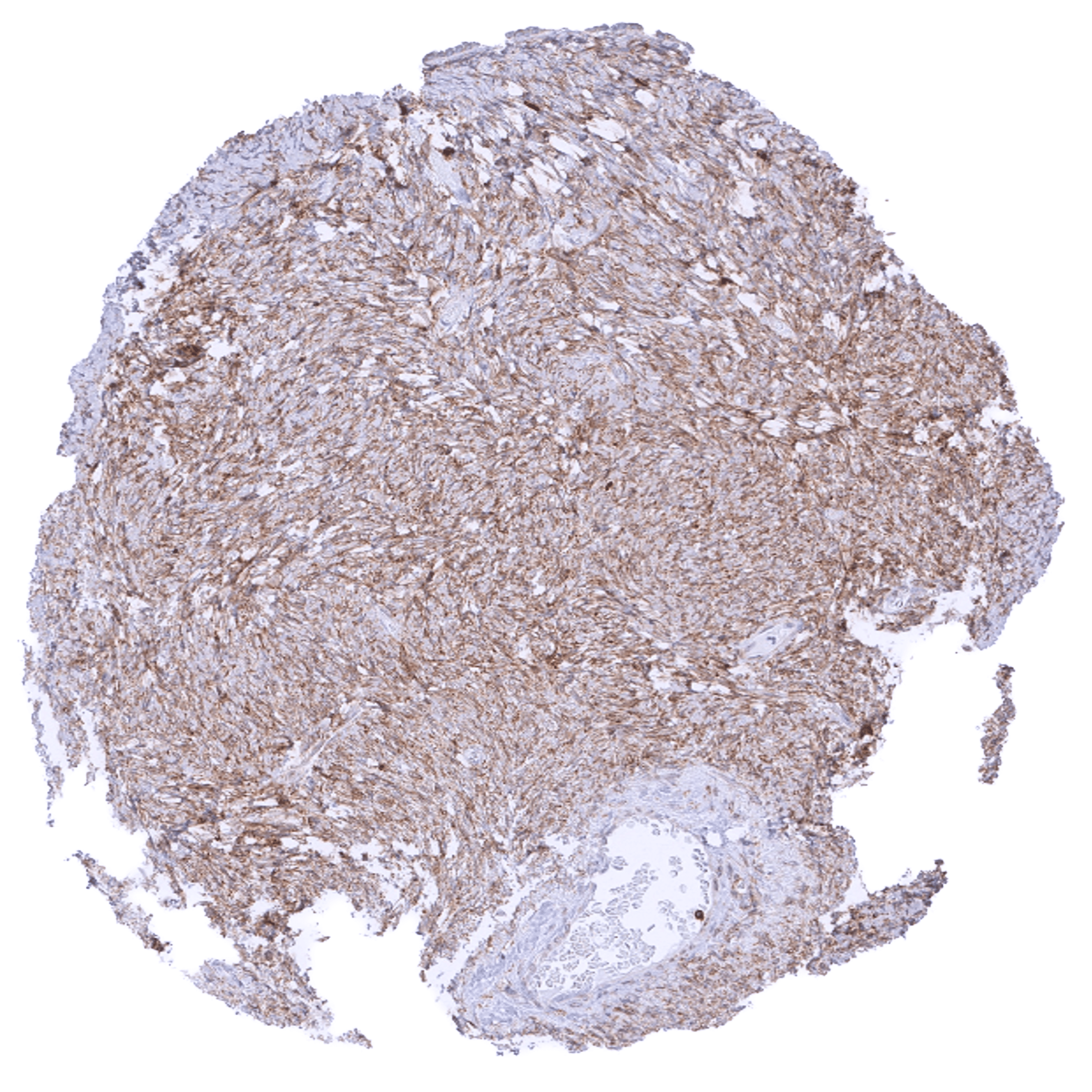
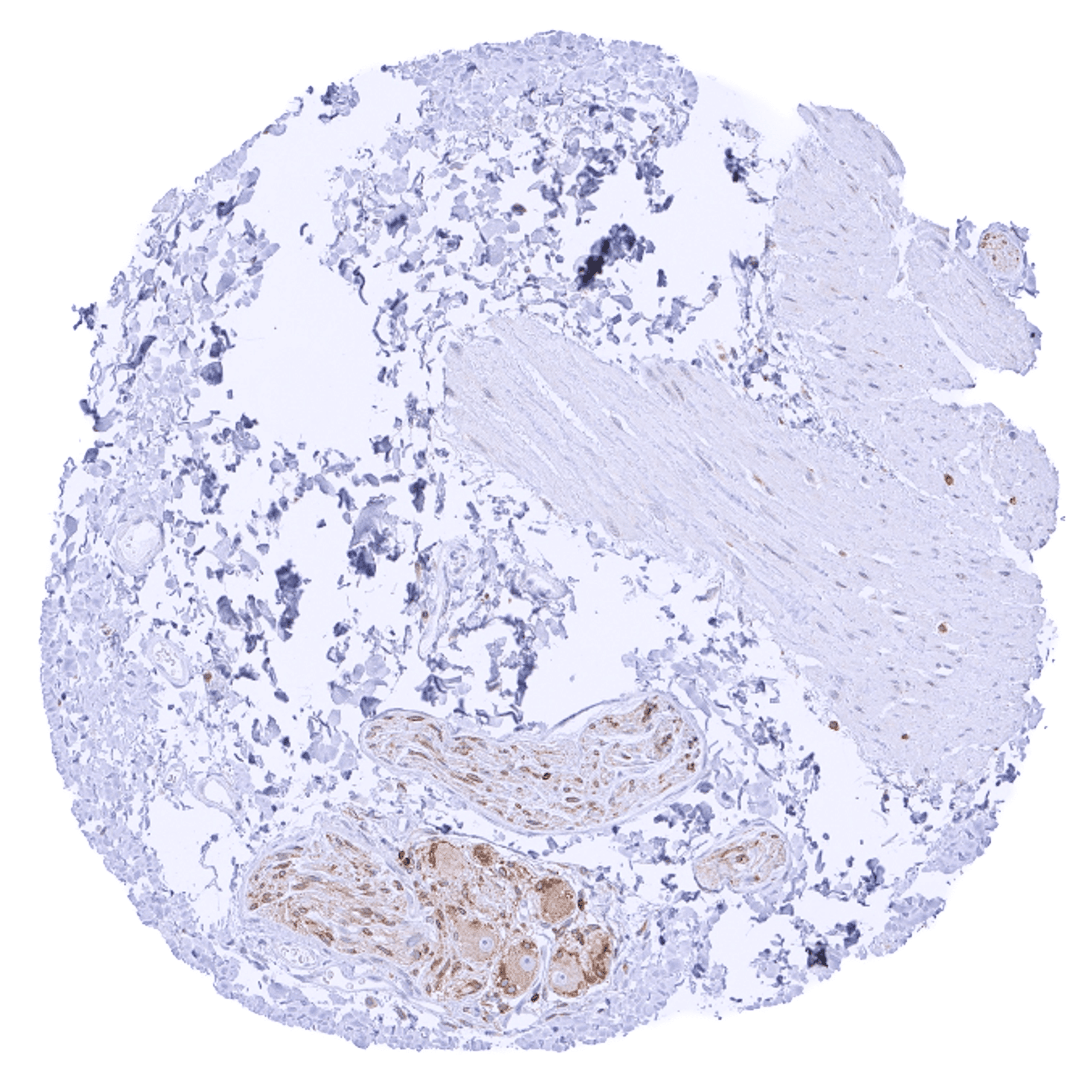
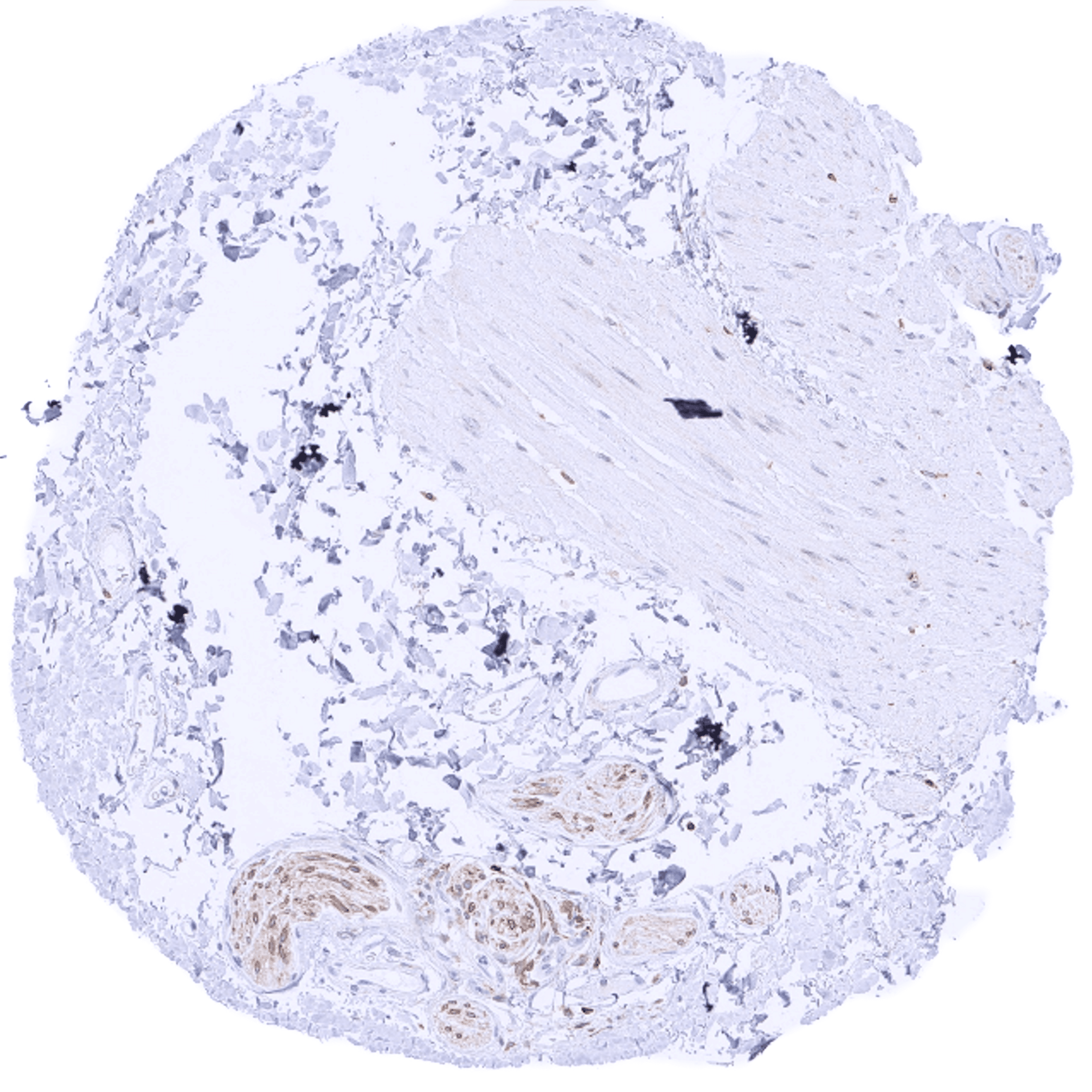
| Muscle/connective tissue | Heart muscle | Negative. |
| Skeletal muscle | Faint cytoplasmic bcl-2 staining of muscle fibers. | |
| Smooth muscle | A faint cytoplasmic bcl-2 staining of muscle fibers can occur in some cases. | |
| Vessel walls | Negative. | |
| Fat | Negative. | |
| Stroma | Negative or faint bcl-2 staining. | |
| Endothelium | Negative. | |
| Bone marrow/ lymphoid tissue | Bone marrow | Only few cells are bcl-2 positive. |
| Lymph node | Strong bcl-2 positivity of a large fraction of lymphocytic cells in the interfollicular area and around germinal centres while almost all cells in germinal centres are bcl-2 negative. | |
| Spleen | Strong bcl-2 positivity of the lymphocytes of the white pulpa and of a small fraction of cells in the red pulpa. | |
| Thymus | Strong bcl-2 positivity of a large fraction of lymphocytic cells in the medulla while corpuscles of Hassall’s are negative. Bcl-2 staining is less intense, variable, and often negative in lymphocytic cells of the cortex. | |
| Tonsil | Strong bcl-2 positivity of a large fraction of lymphocytic cells in the interfollicular area and around germinal centres while almost all cells in germinal centres are bcl-2 negative. Squamous epithelium is bcl-2 negative although the basal cell layer may show weak positivity. | |
| Remarks |
The findings described above are thus consistent with the the Human Protein Atlas (Tissue expression Bcl-2). Bcl-2 staining by MSVA-402M is preferentially seen in lymphatic cells (except germinal centres) but it also occurs in various other cell types.
Positive control = Tonsil: A strong cytoplasmic bcl-2 staining should be seen in most interfollicular lymphocytes while most germinal centre cells are bcl-2 negative.
Negative control = Tonsil: The vast majority of lymphocytic cells from germinal centres must be bcl-2 negative while interfollicular lymphocytes are mostly positive.
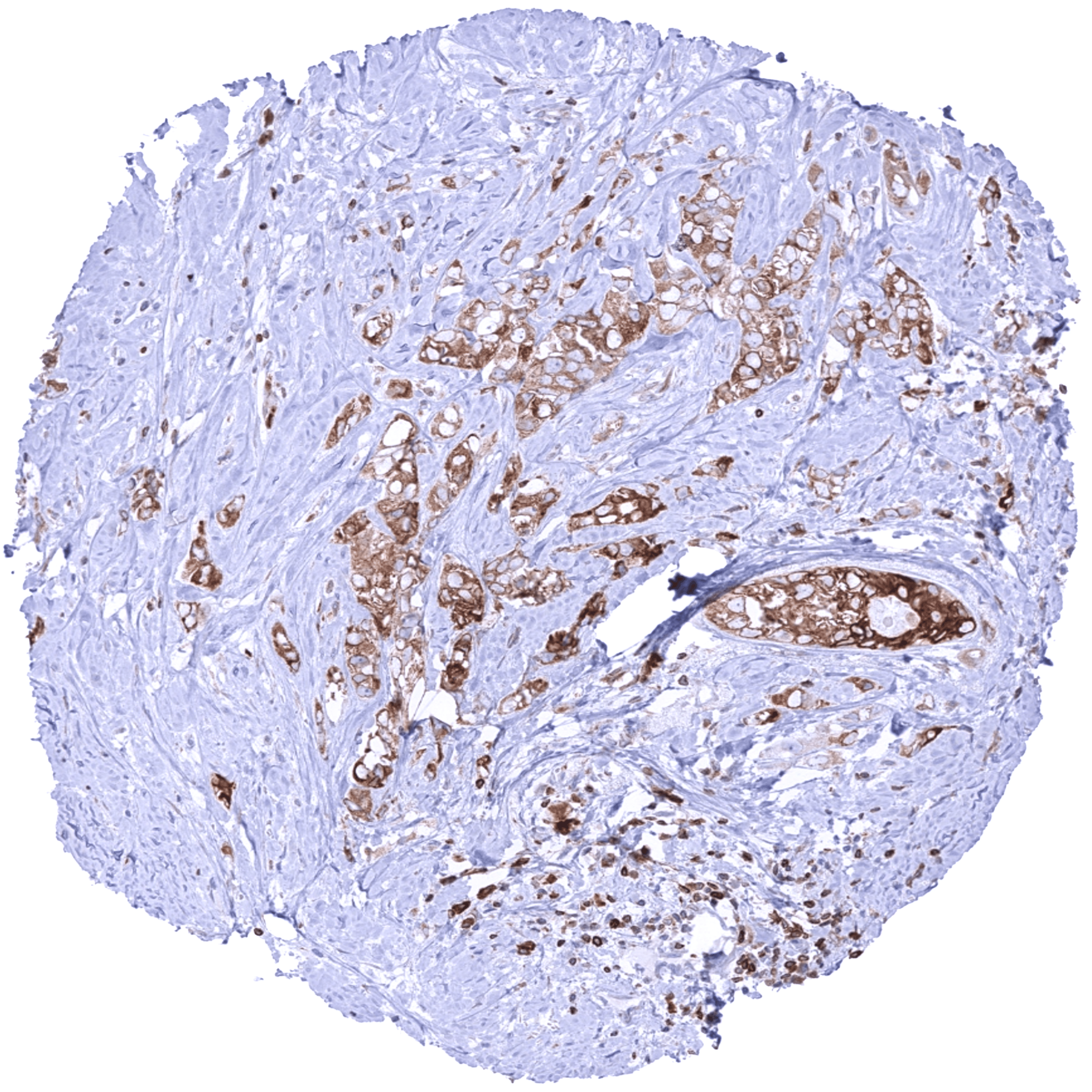
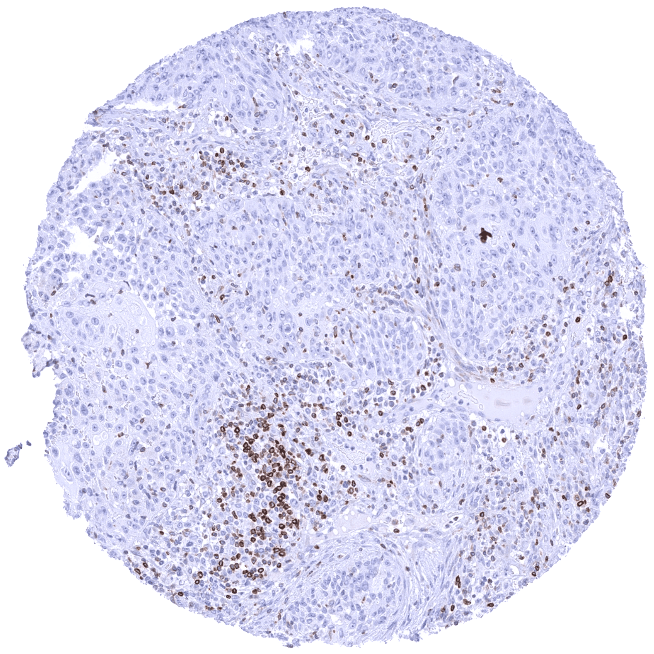
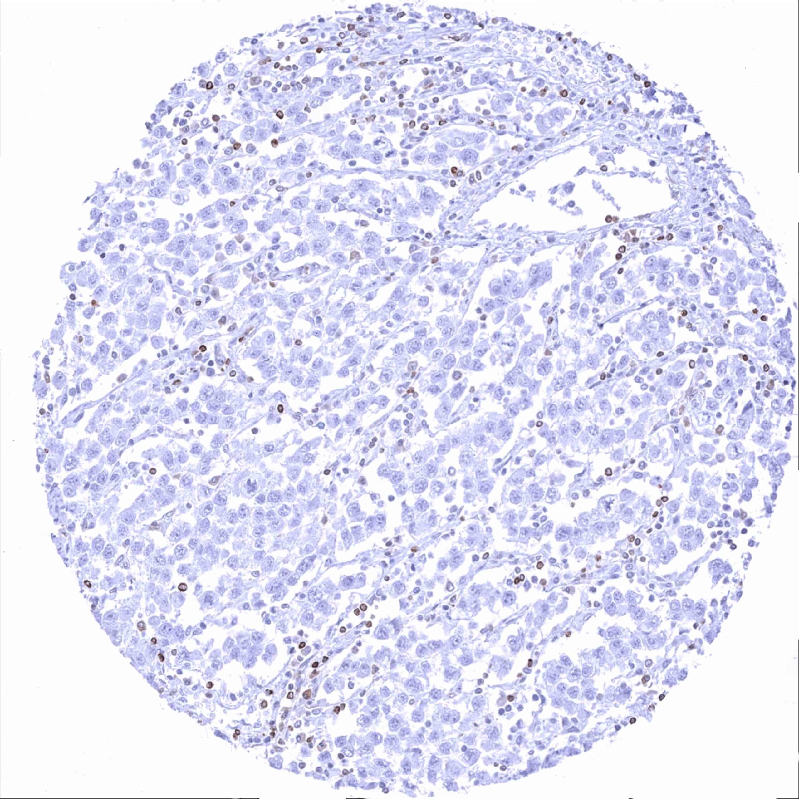
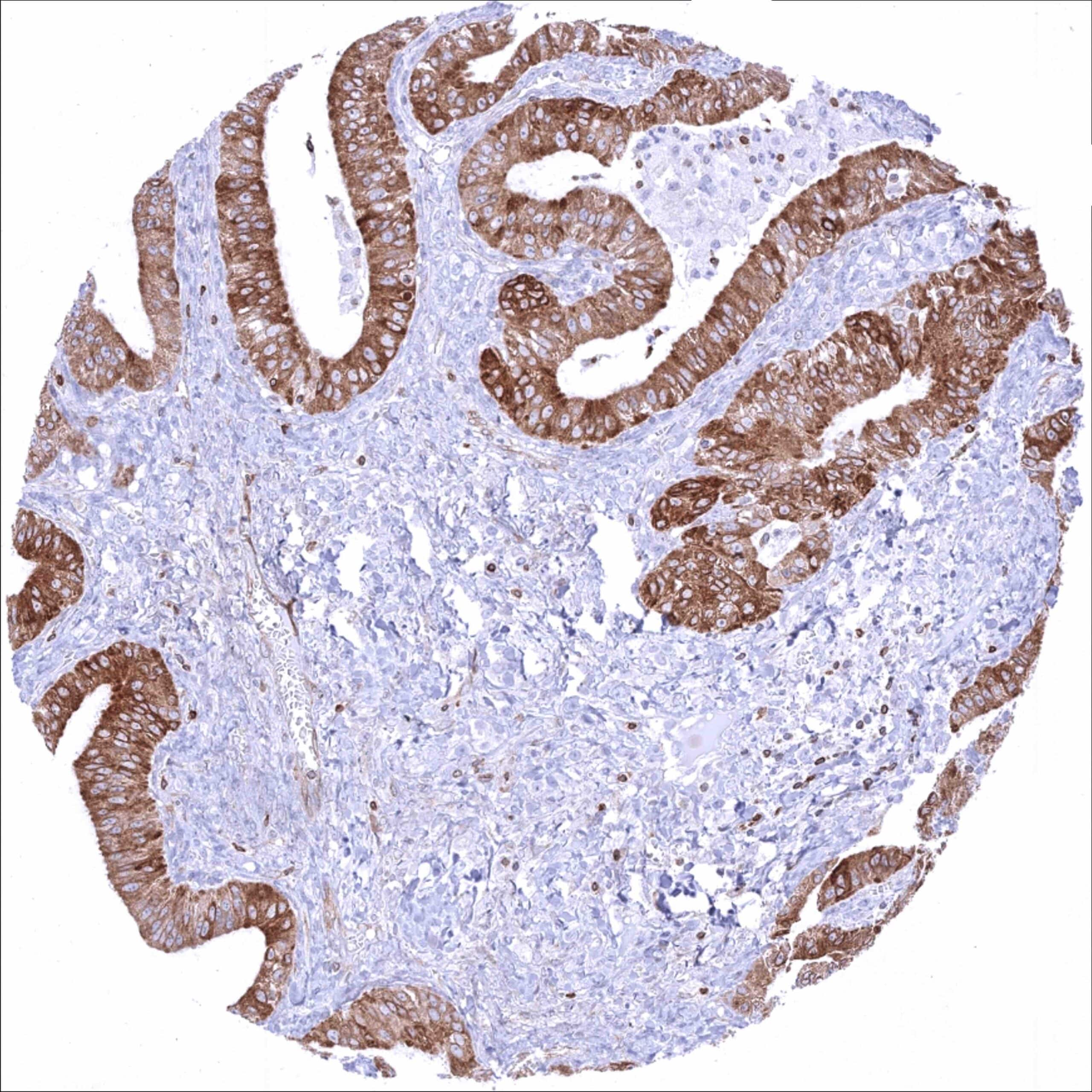
Staining Pattern in Relevant Tumor Types
Bcl-2 expression of variable intensity can occur in all types of cancer. It is particularly common in follicular lymphoma.
The TCGA findings on Bcl-2 RNA expression in different tumor categories have been summarized in the Human Protein Atlas.
Compatibility of Antibodies
No data available at the moment
Protocol Recommendations
IHC users have different preferences on how the stains should look like. Some prefer high staining intensity of the target stain and even accept some background. Others favor absolute specificity and lighter target stains. Factors that invariably lead to more intense staining include higher concentration of the antibody and visualization tools, longer incubation time, higher temperature during incubation, higher temperature and longer duration of the heat induced epitope retrieval (slide pretreatment). The impact of the pH during slide pretreatment has variable effects and depends on the antibody and the target protein.
All images and data shown here and in our image galleries are obtained by the manual protocol described below. Other protocols resulting in equivalent staining are described as well.
Manual protocol
Freshly cut sections should be used (less than 10 days between cutting and staining). Heat-induced antigen retrieval for 5 minutes in an autoclave at 121°C in pH 7,8 Target Retrieval Solution buffer. Apply MSVA-402M at a dilution of 1:150 at 37°C for 60 minutes. Visualization of bound antibody by the EnVision Kit (Dako, Agilent) according to the manufacturer’s directions.
Potential Research Applications
- The prevalence and clinical significance of bcl-2 expression in different cancer types is insufficiently explored.
- The role of bcl-2 in cells of the tumor microenvironment and its potential inhibition needs to be investigated.
- The potential of bcl-2 inhibition in subtypes of solid cancers needs to be investigated.
- The role of bcl-2 and its inhibition in neurodegenerative, cardiovascular, autoimmune, and infectious diseases needs to be explored.
Evidence for Antibody Specificity in IHC
There are two ways how the specificity of antibodies can be documented for immunohistochemistry on formalin fixed tissues. These are: 1. Comparison with a second independent method for target expression measurement across a large number of different tissue types (orthogonal strategy), and 2. Comparison with one or several independent antibodies for the same target and showing that all positive staining results are also seen with other antibodies for the same target (independent antibody strategy).
Orthogonal validation: For the antibody MSVA-402M specificity is supported by the good concordance of the immunostaining data with data from three independent RNA screening studies, including the Human Protein Atlas (HPA) RNA-seq tissue dataset, the FANTOM5 project, and the Genotype-Tissue Expression (GTEx) project, which are all summarized in the Human Protein Atlas (Tissue expression Bcl-2). In particular, immunostaining by using MSVA-402M was highest in lymphatic tissues and in the thyroid but particularly low in the liver and the pancreas. However, because bcl-2 staining occurs in variable portions of inflammatory cells in all tissues, orthogonal validation based on data from disaggregated tissue is not sufficient for bcl-2 antibody validation.
Comparison of antibodies: True expression of bcl-2 in all cell types with documented bcl-2 immunostaining by MSVA-402M is validated by identical staining patterns obtained by a second, independent commercially available bcl-2 antibody, termed “validation antibody” for all analyzed tissues.