Adrenal gland – STING staining of endothelial cells and of macrophages (STING immunohistochemistry)

Aorta, media – STING staining of endothelial cells and of a subset of mesenchymal cells (STING immunohistochemistry)
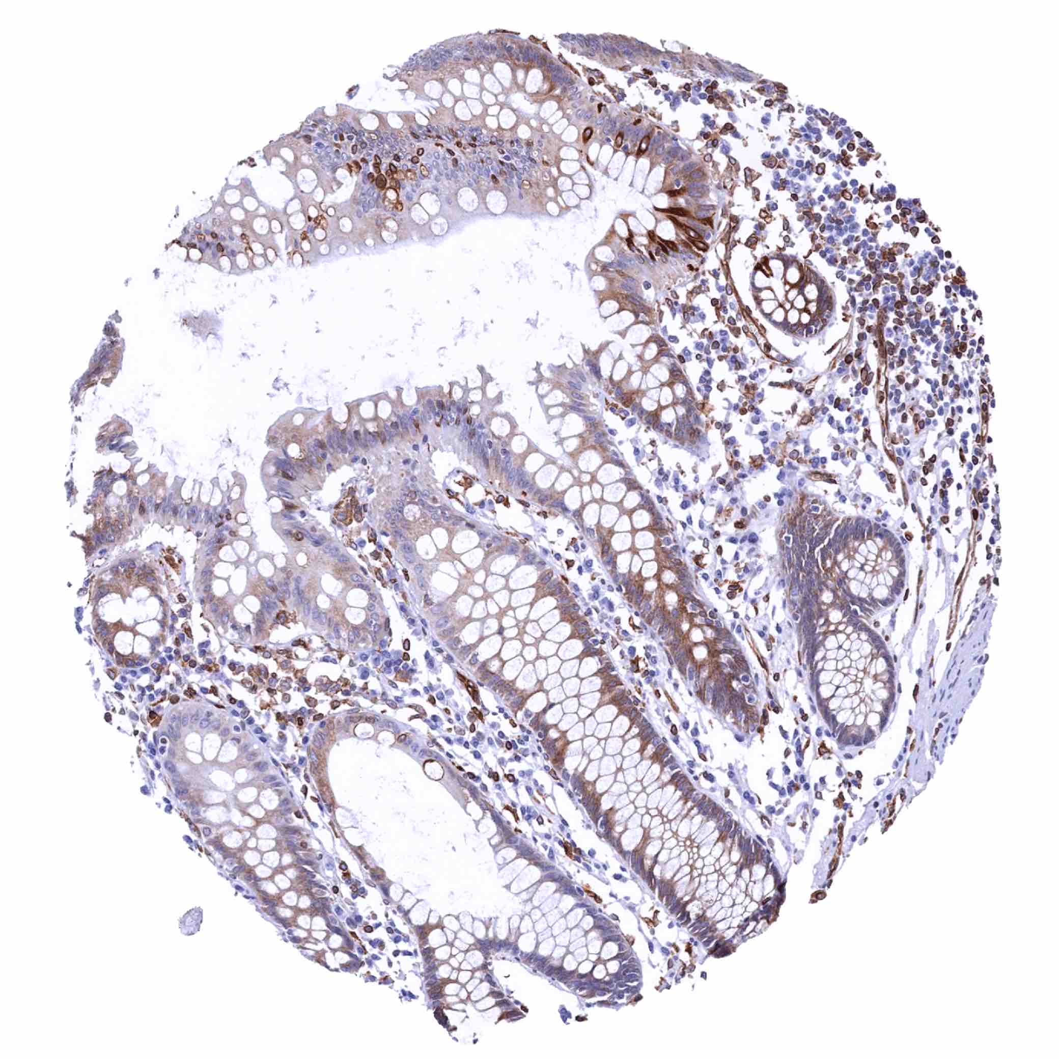
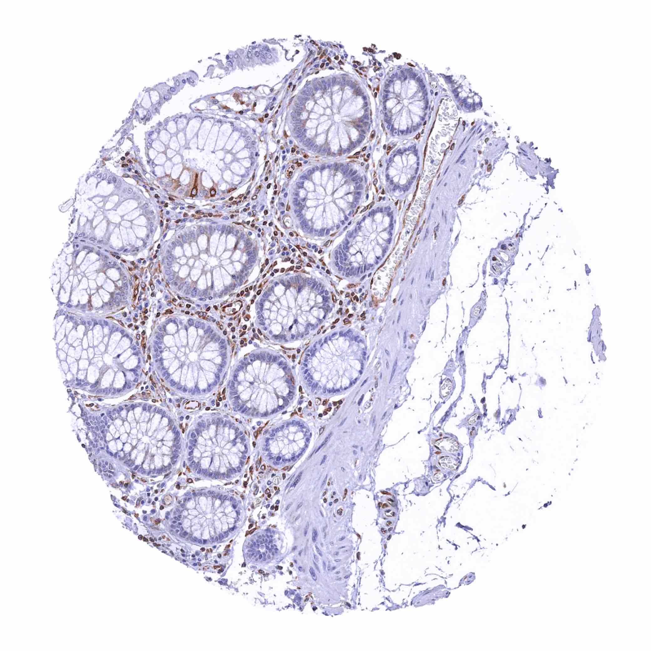
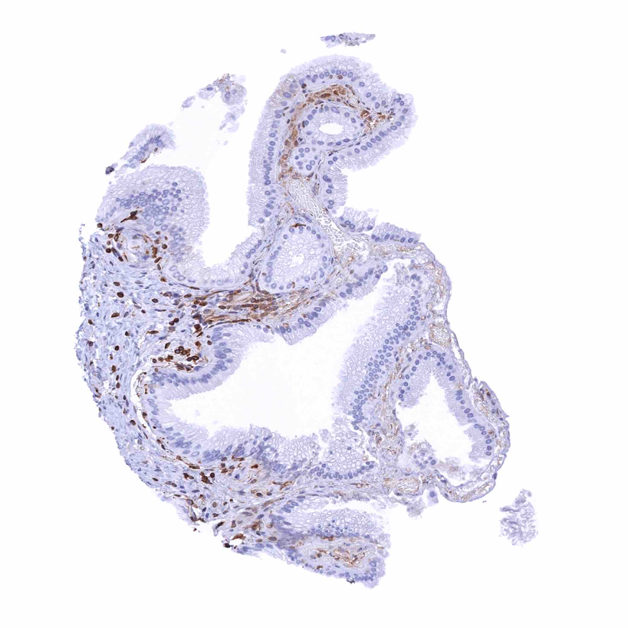
Appendix, mucosa – STING staining is rather weak and involves only a fraction of epithelial cells in this sample (STING immunohistochemistry)
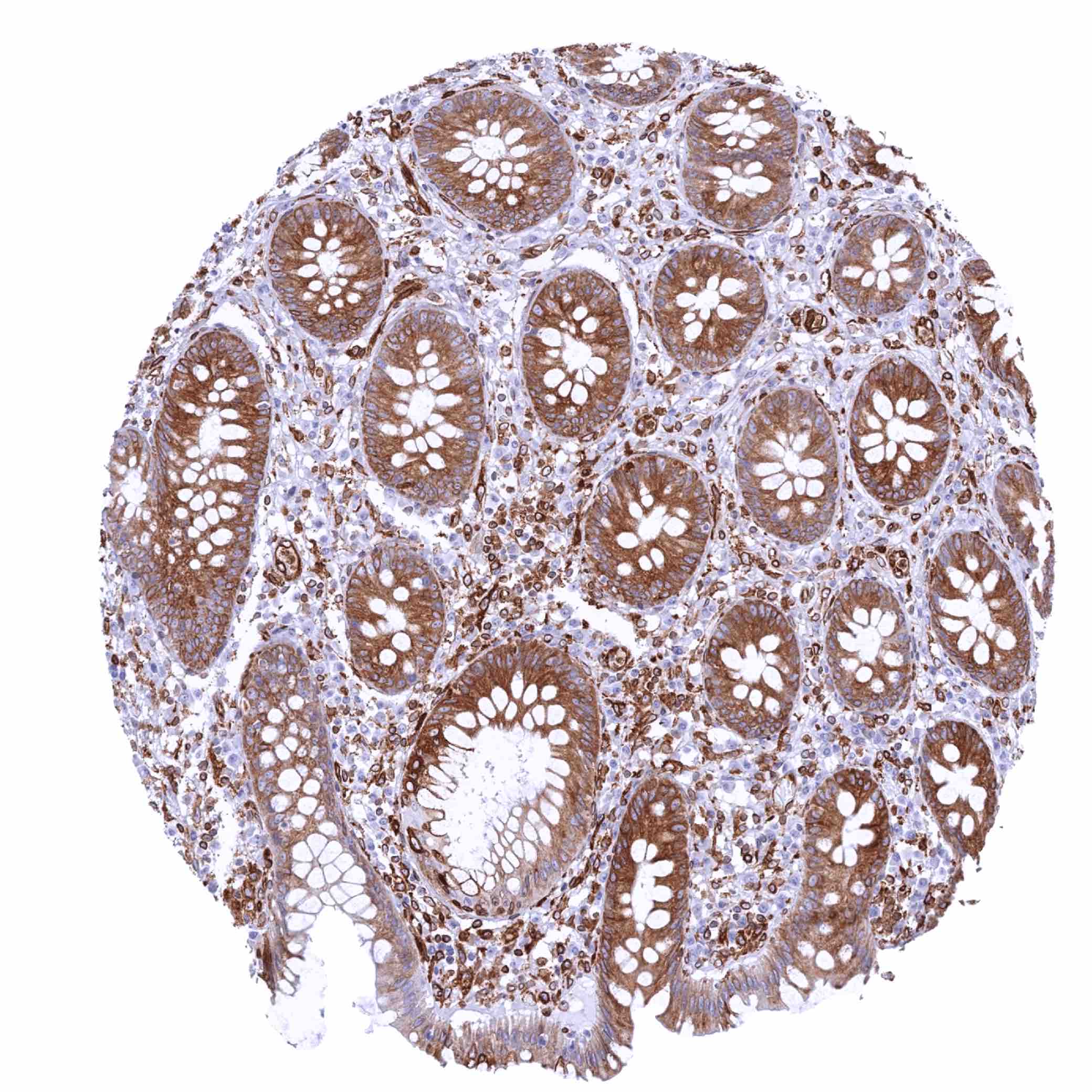
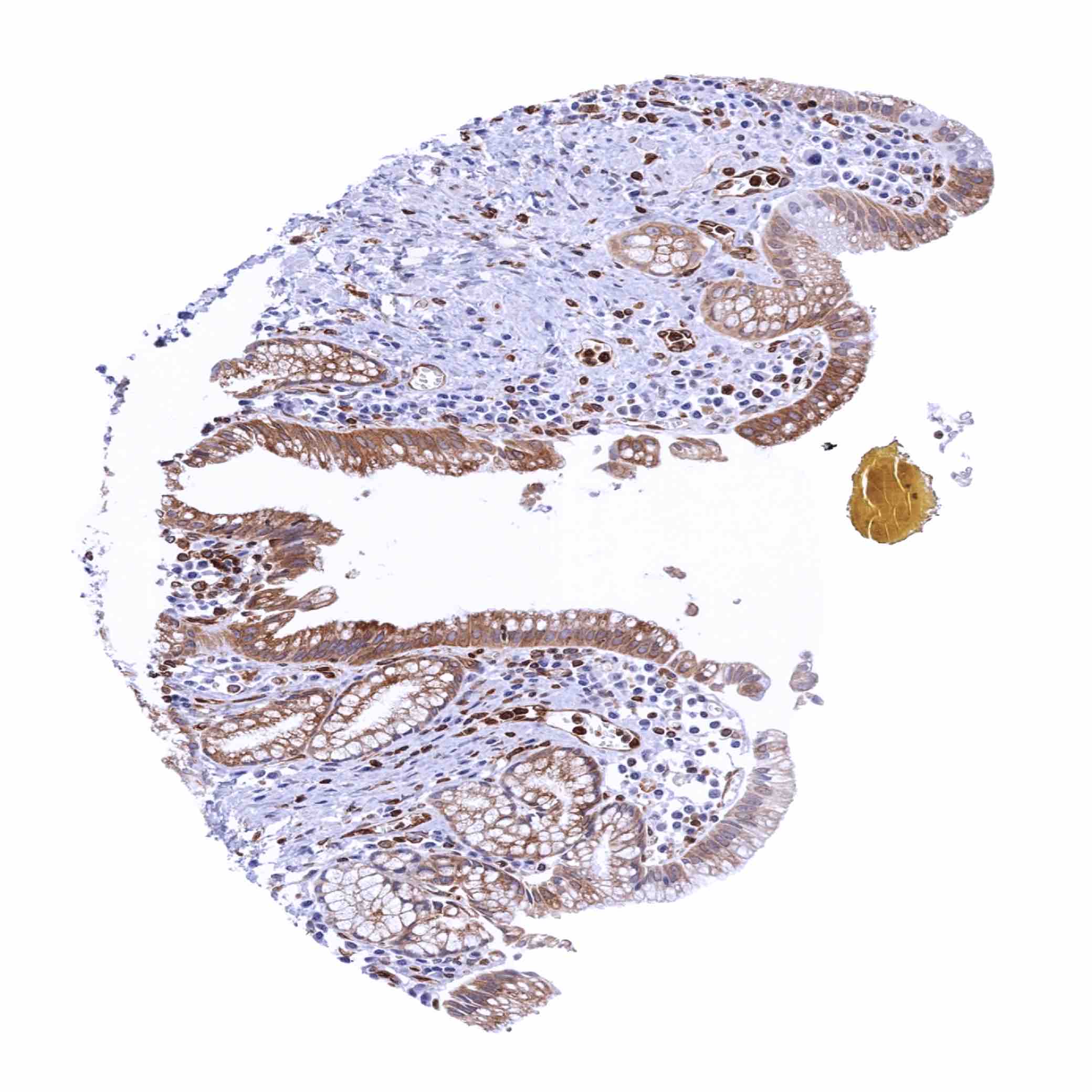
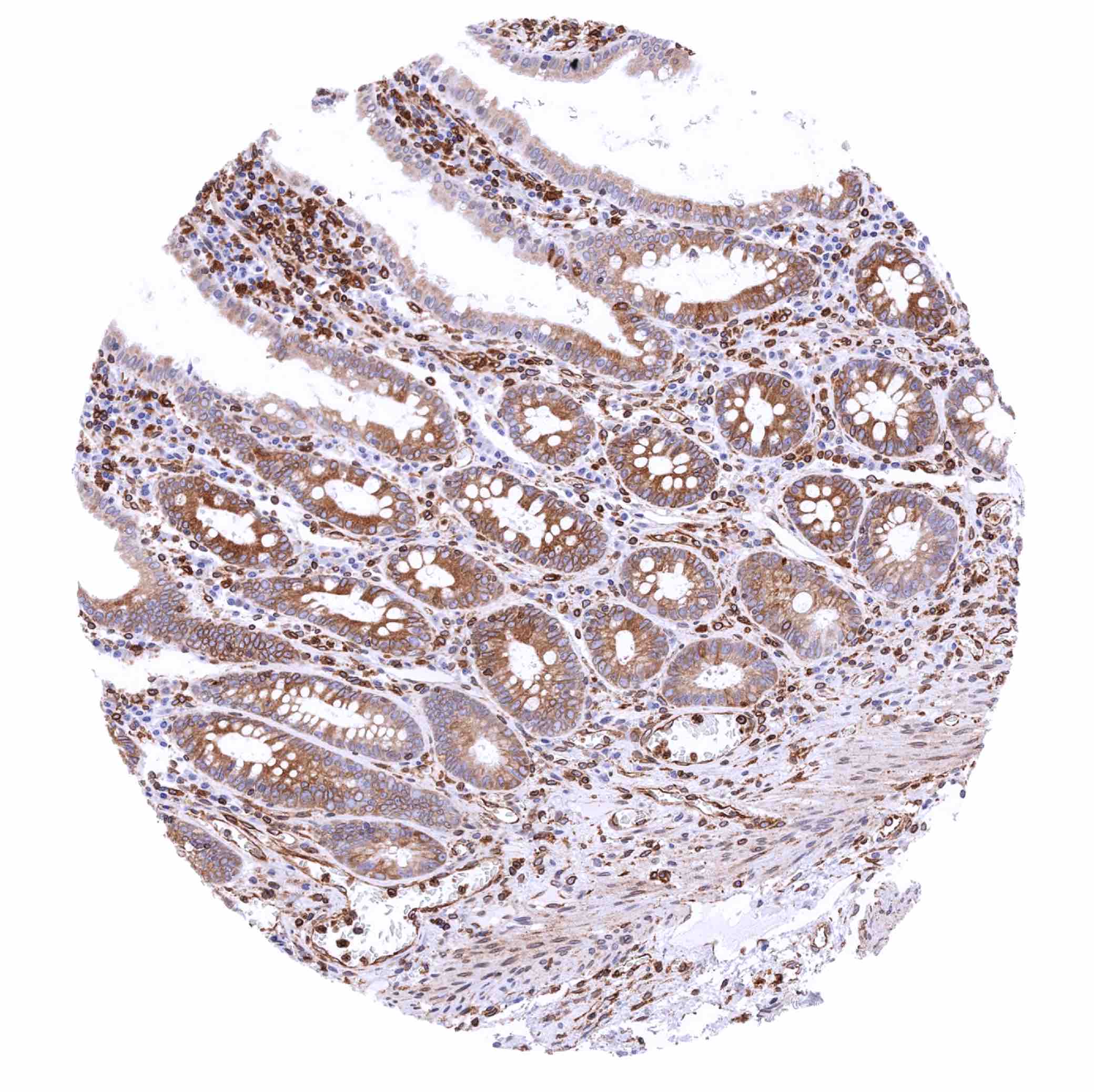
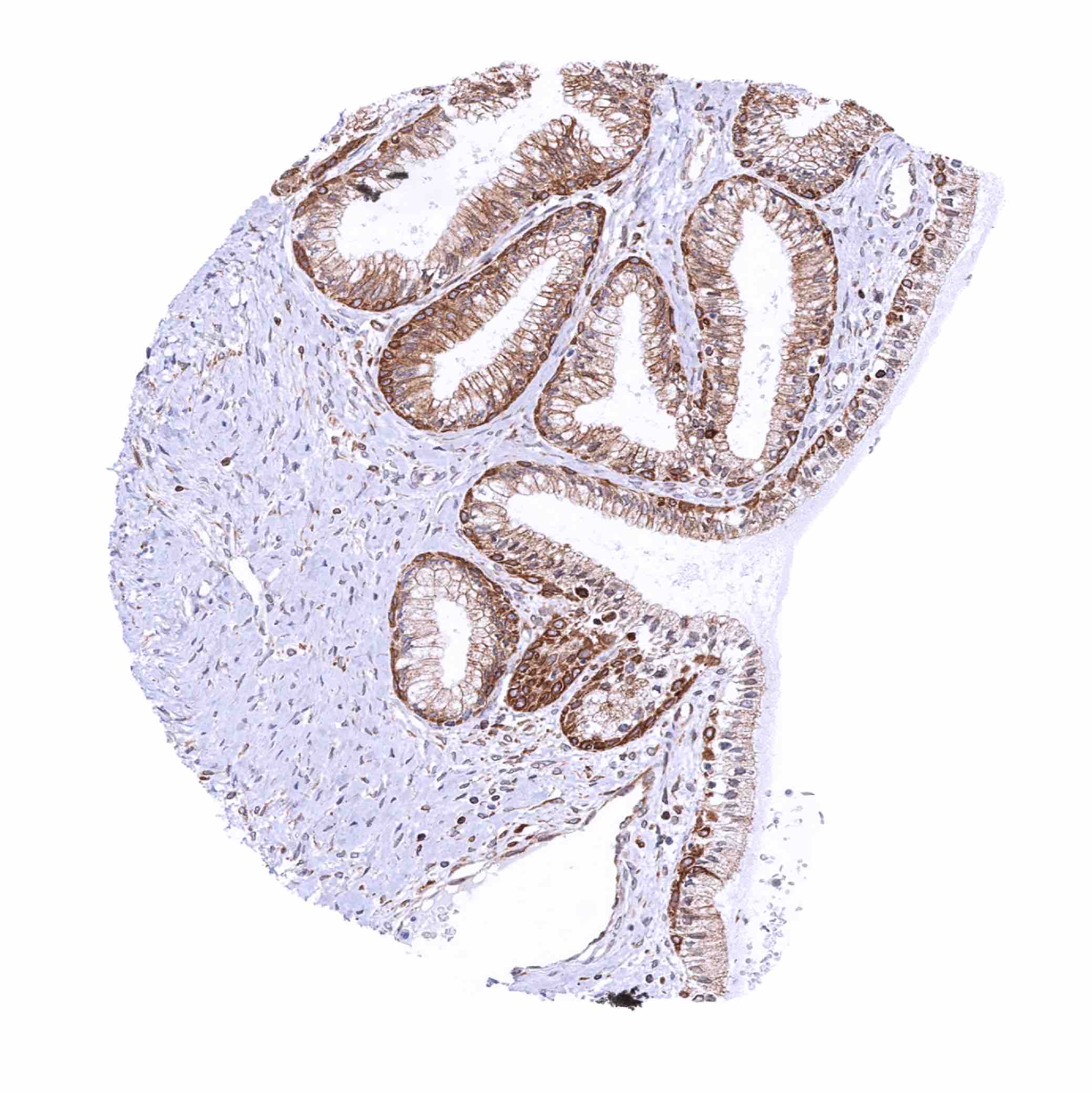
Appendix, mucosa – STING staining is strong in all epithelial cells in this sample (STING immunohistochemistry)
Appendix, mucosa – STING staining of inflammatory cells predominates in the interfollicular zone (STING immunohistochemistry)
Appendix, muscular wall – STING staining of endothelial cells (STING immunohistochemistry)
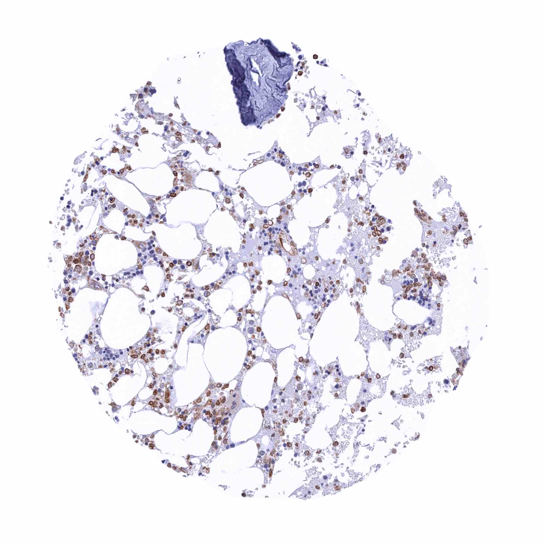
Bone marrow – STING staining of endothelial cells and a fraction of hematopoetic cells (STING immunohistochemistry)
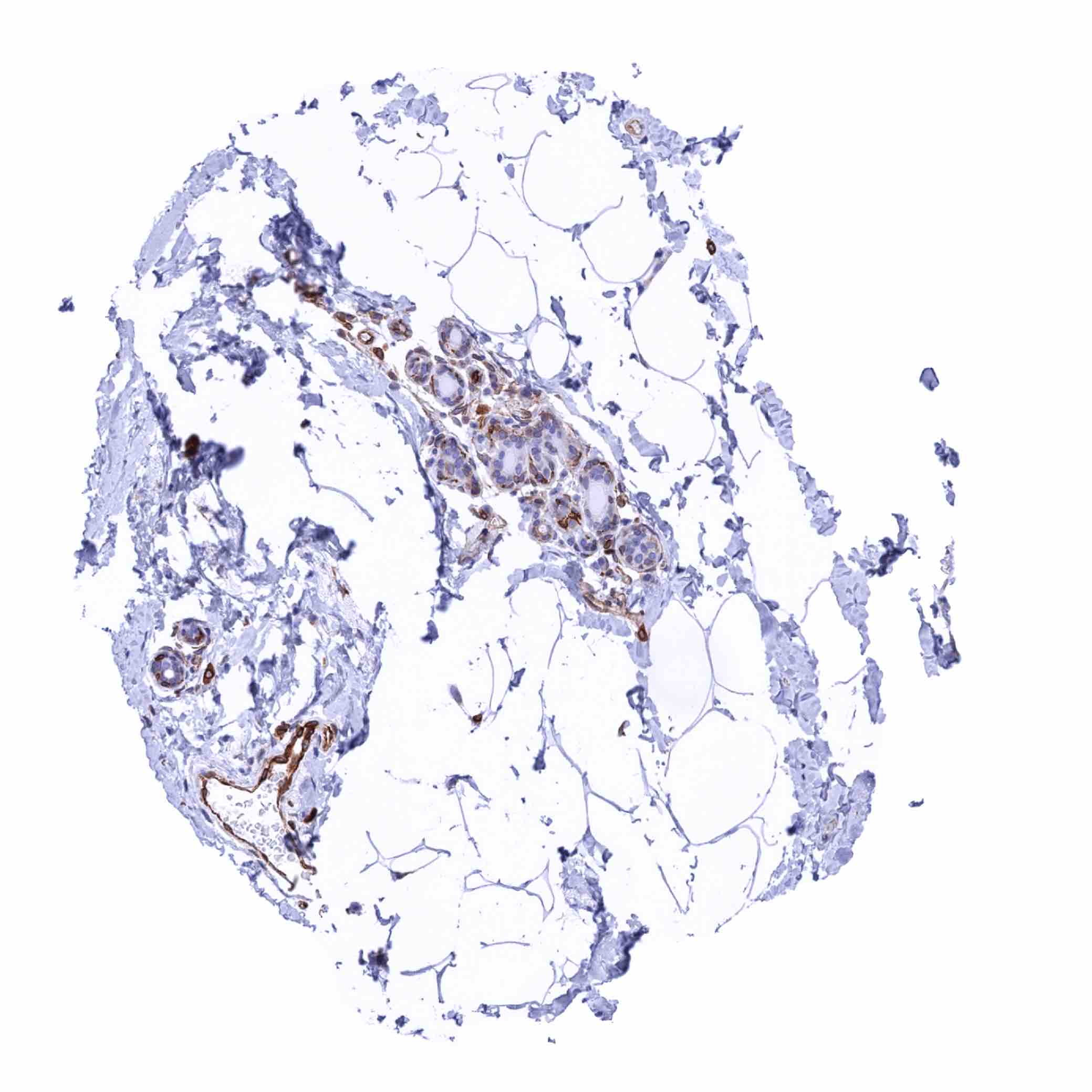
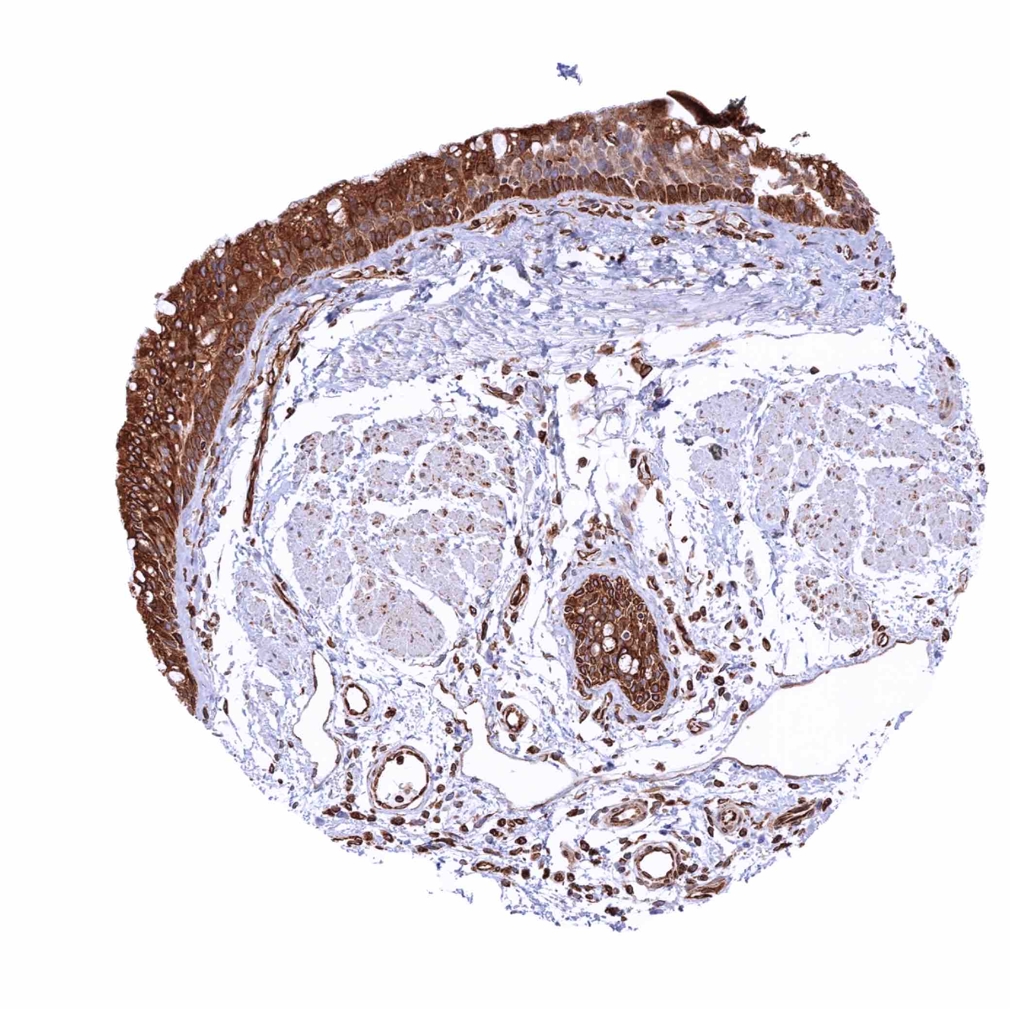
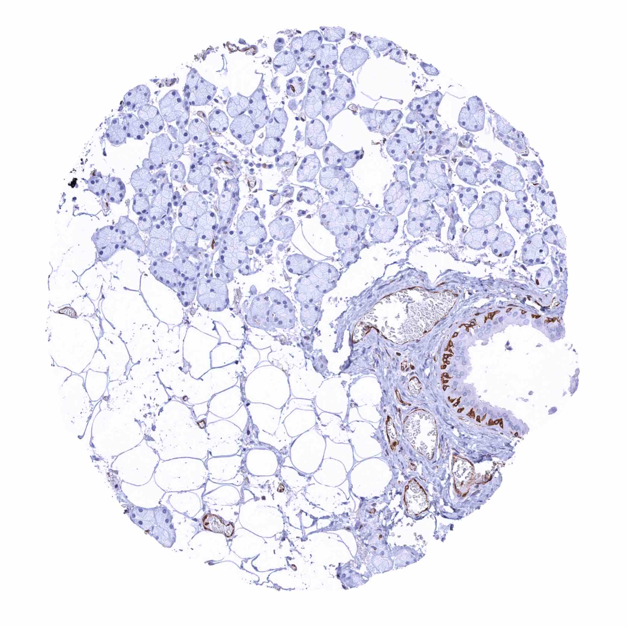
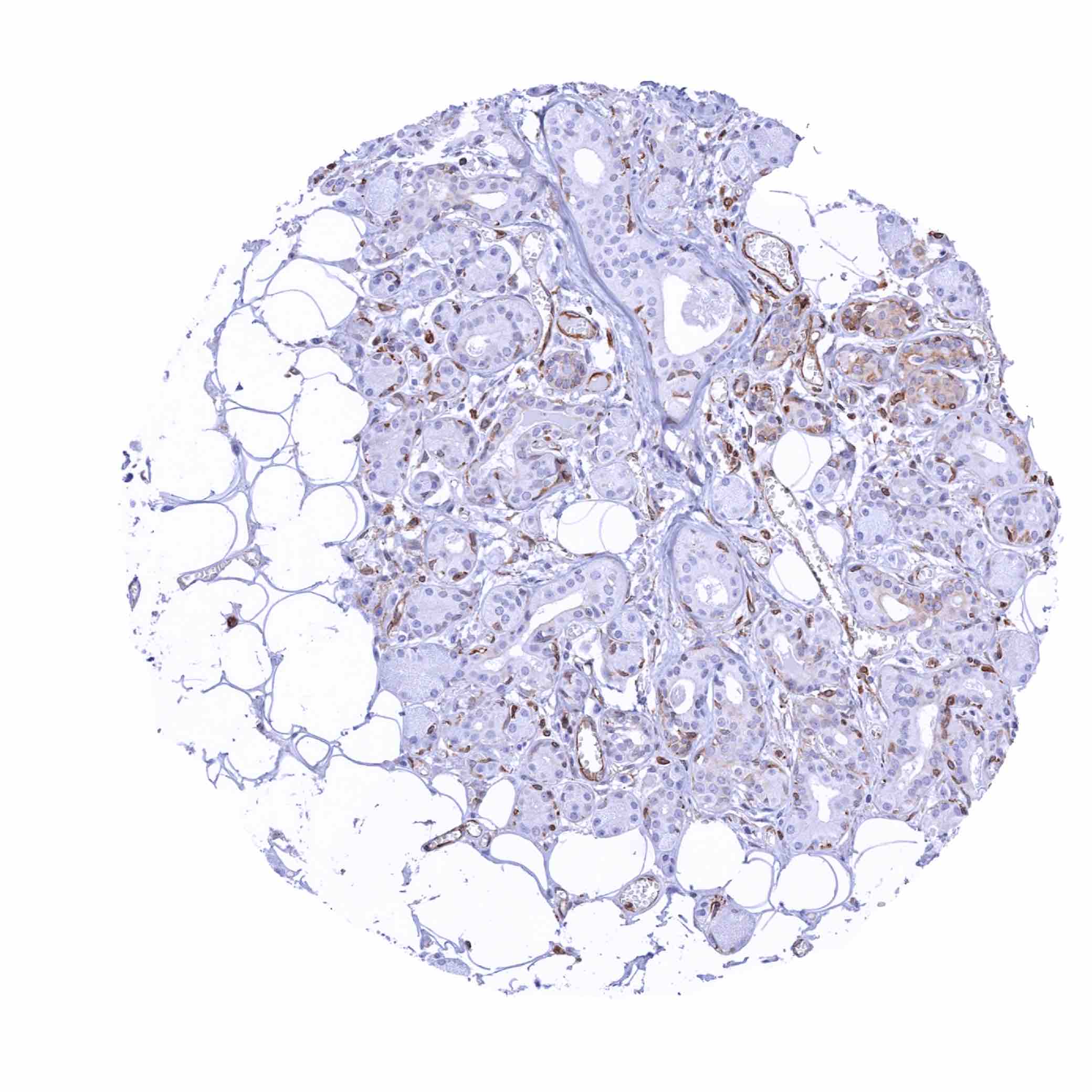
Breast – STING staining of some myoepithelial cells and (weakly) of luminal cells (STING immunohistochemistry)
Breast
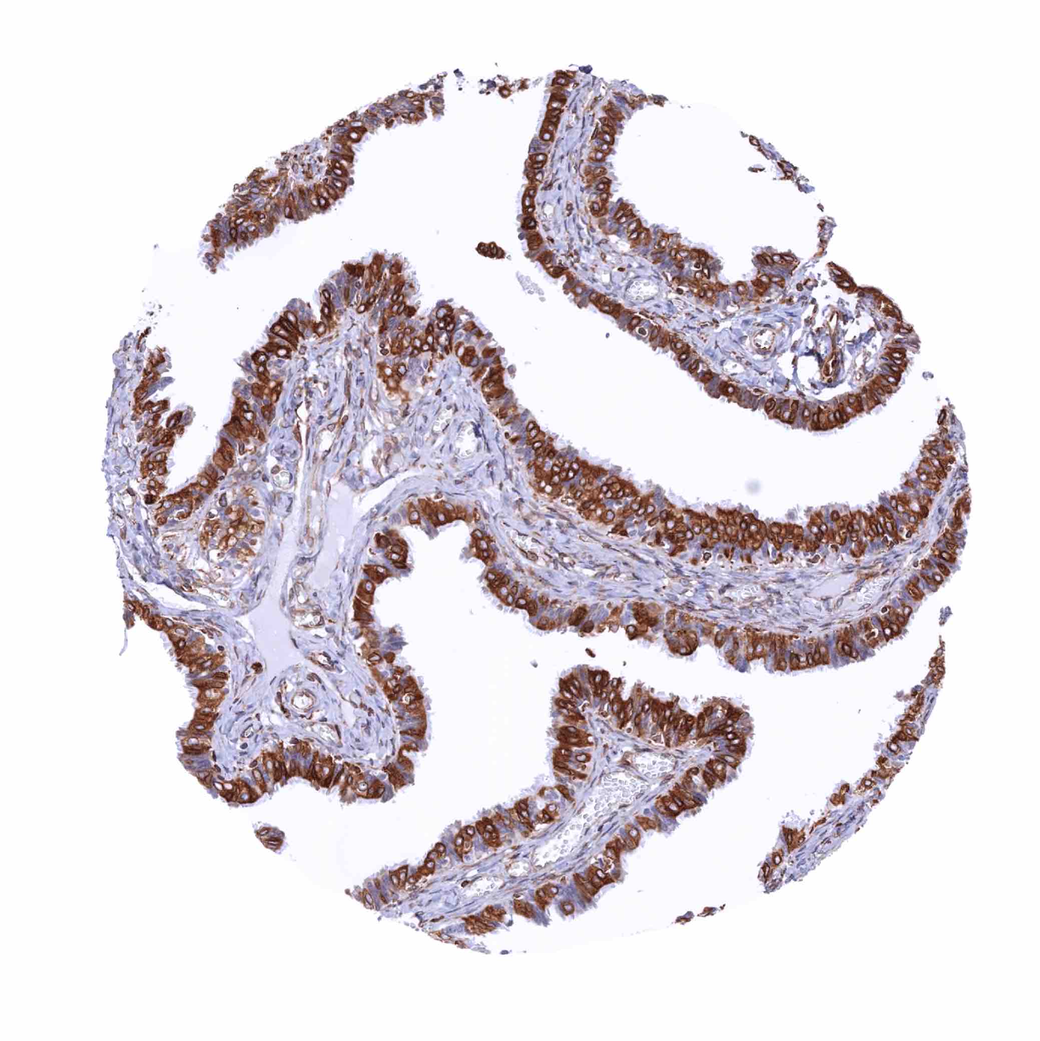
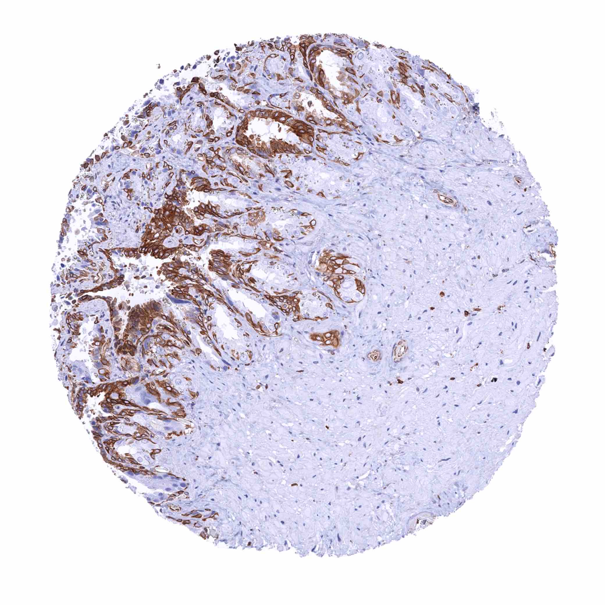
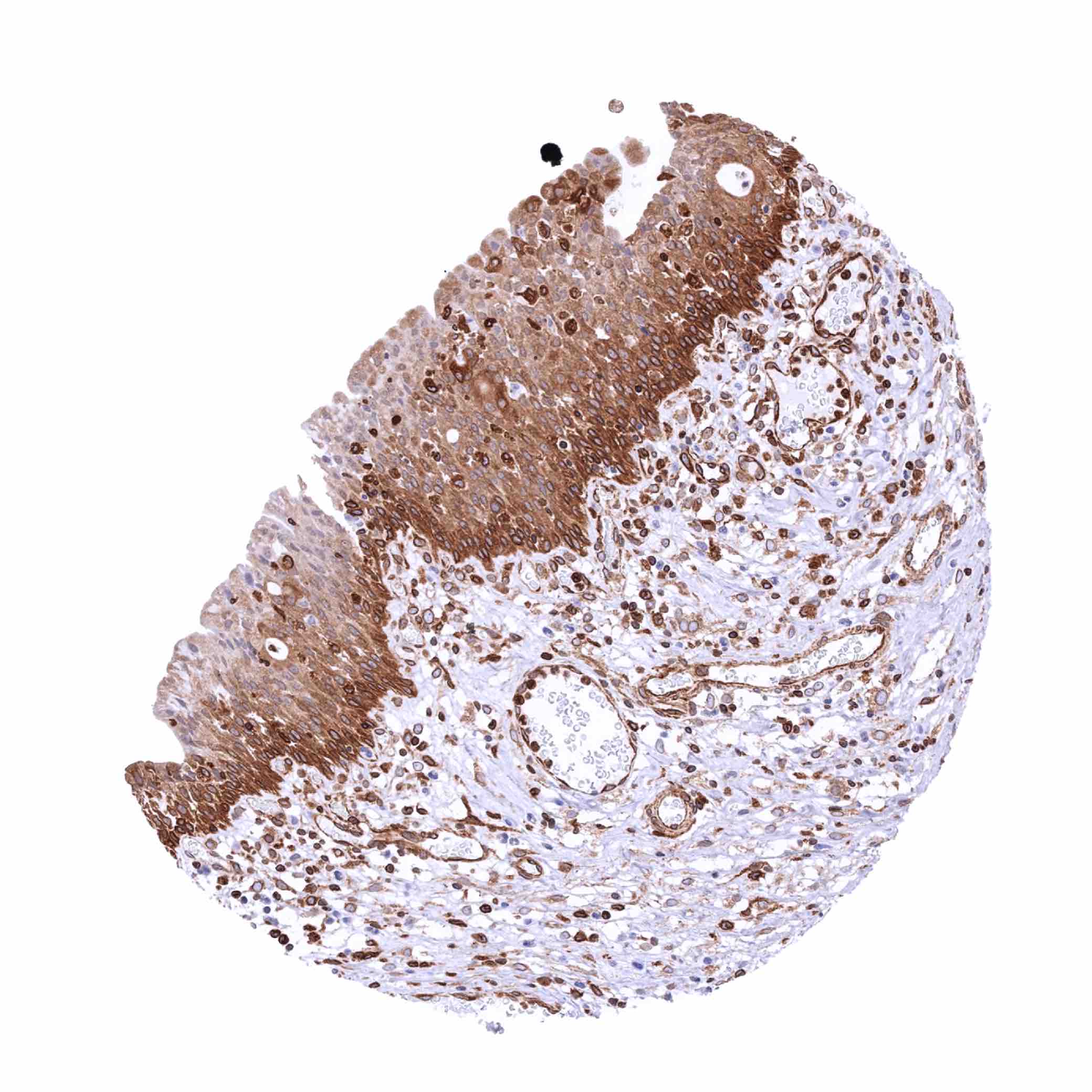
Bronchus, glands – Strong STING staining of the respiratory epithelium (STING immunohistochemistry)
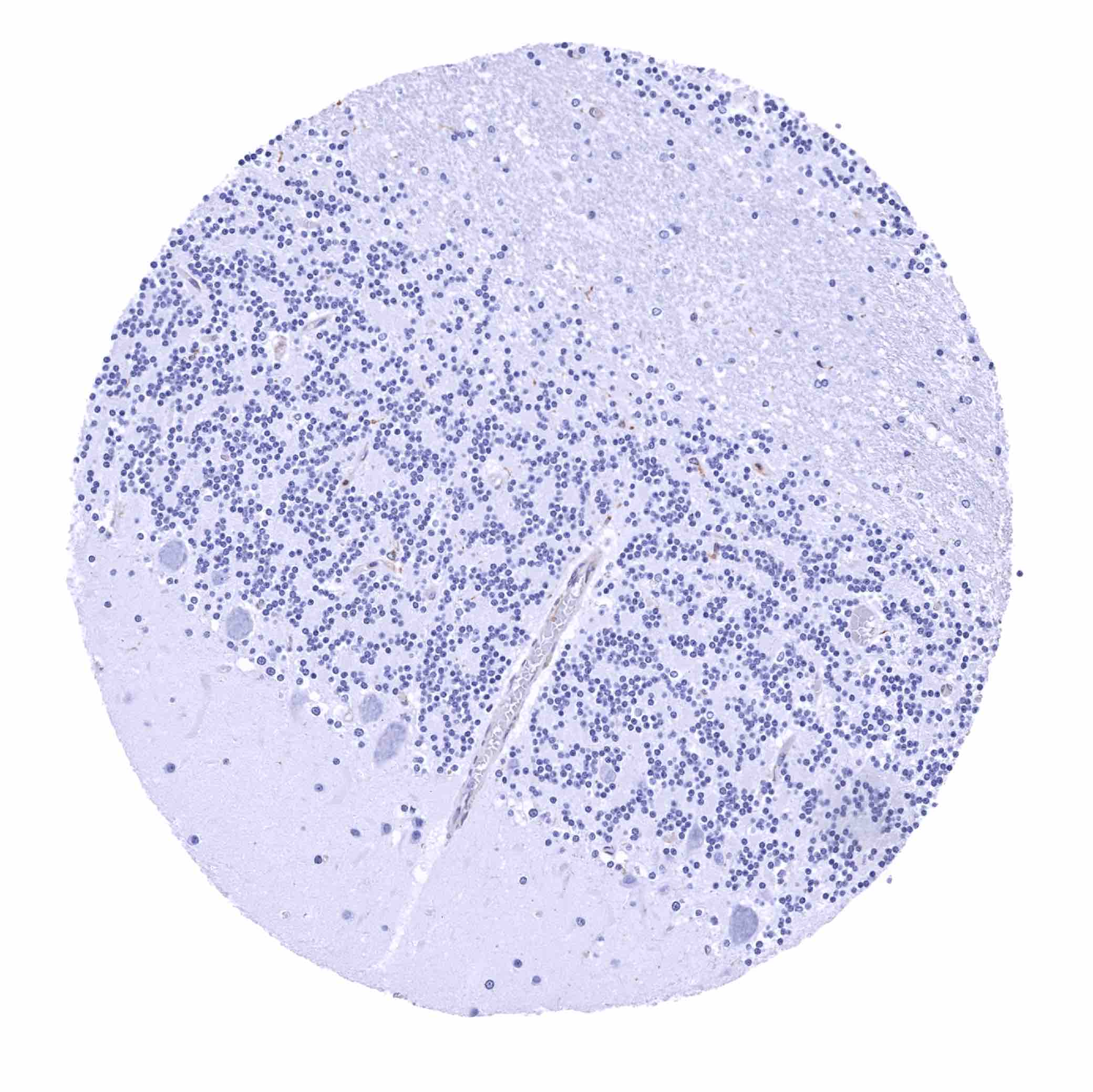
Cerebellum (molecular layer, Purkinje cell layer, granule cell layer, white matter)
Cerebellum (white matter)
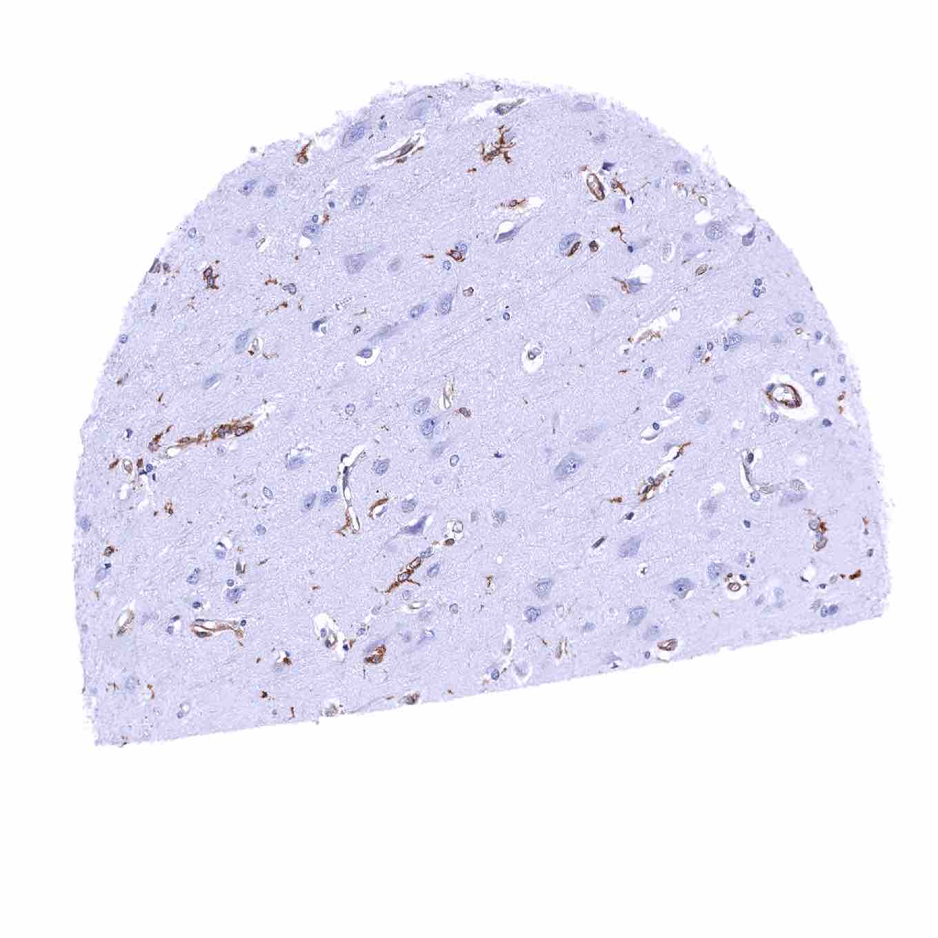
Cerebrum, grey matter – STING staining of endothelial cells (STING immunohistochemistry)
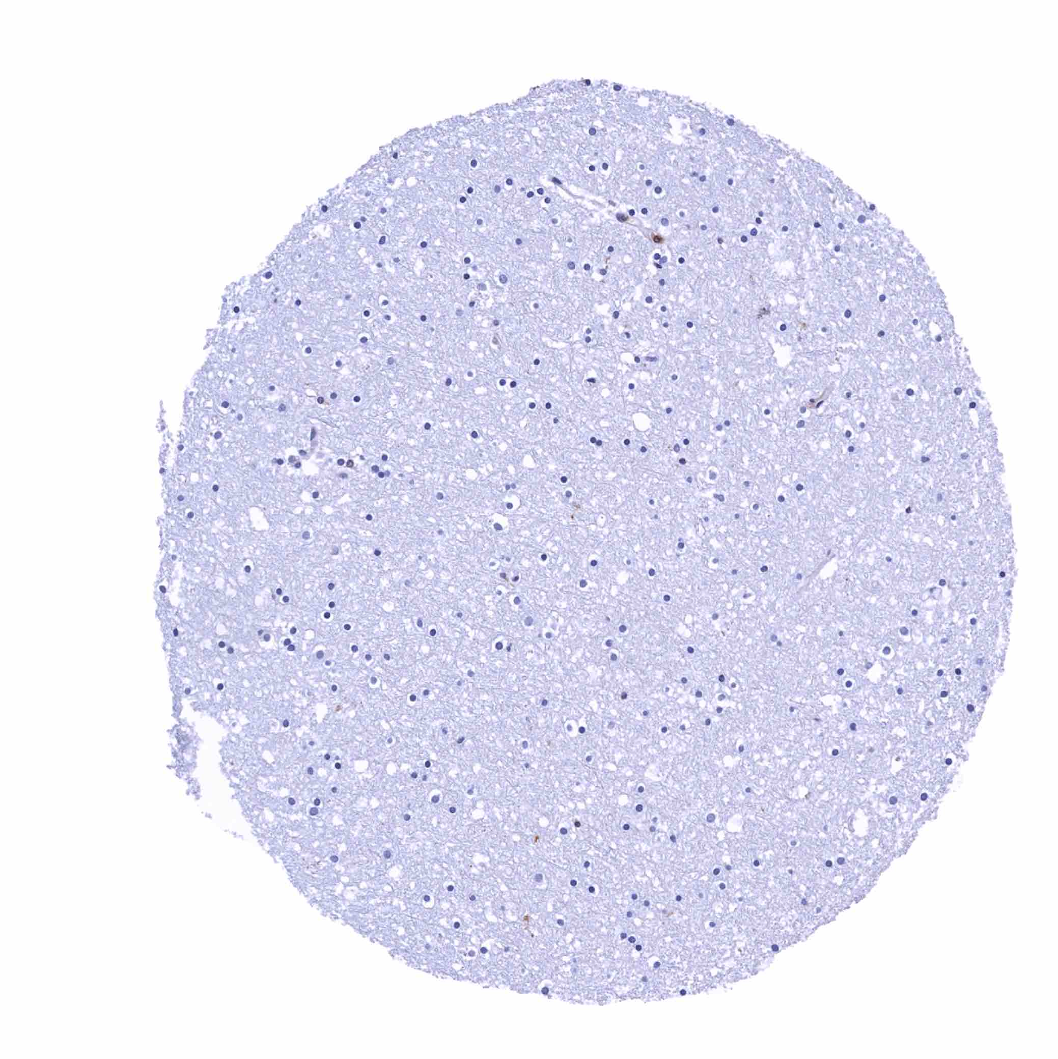
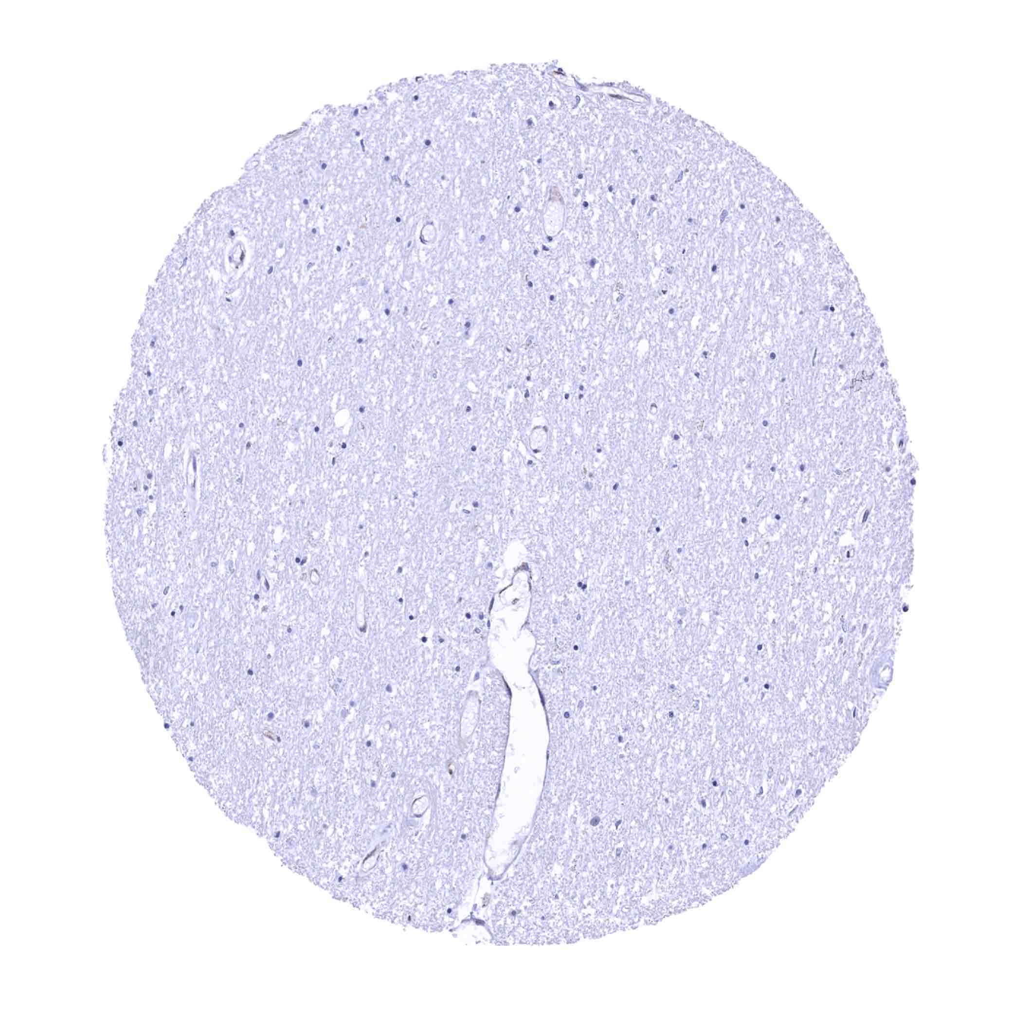
Cerebrum, white matter
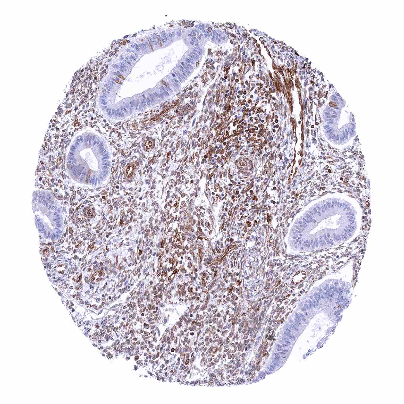
Colon descendens, mucosa – STING staining of only a very small subset of epithelial cells in this sample (STING immunohistochemistry)
Colon descendens, muscular wall – STING staining of endothelial cells (STING immunohistochemistry)
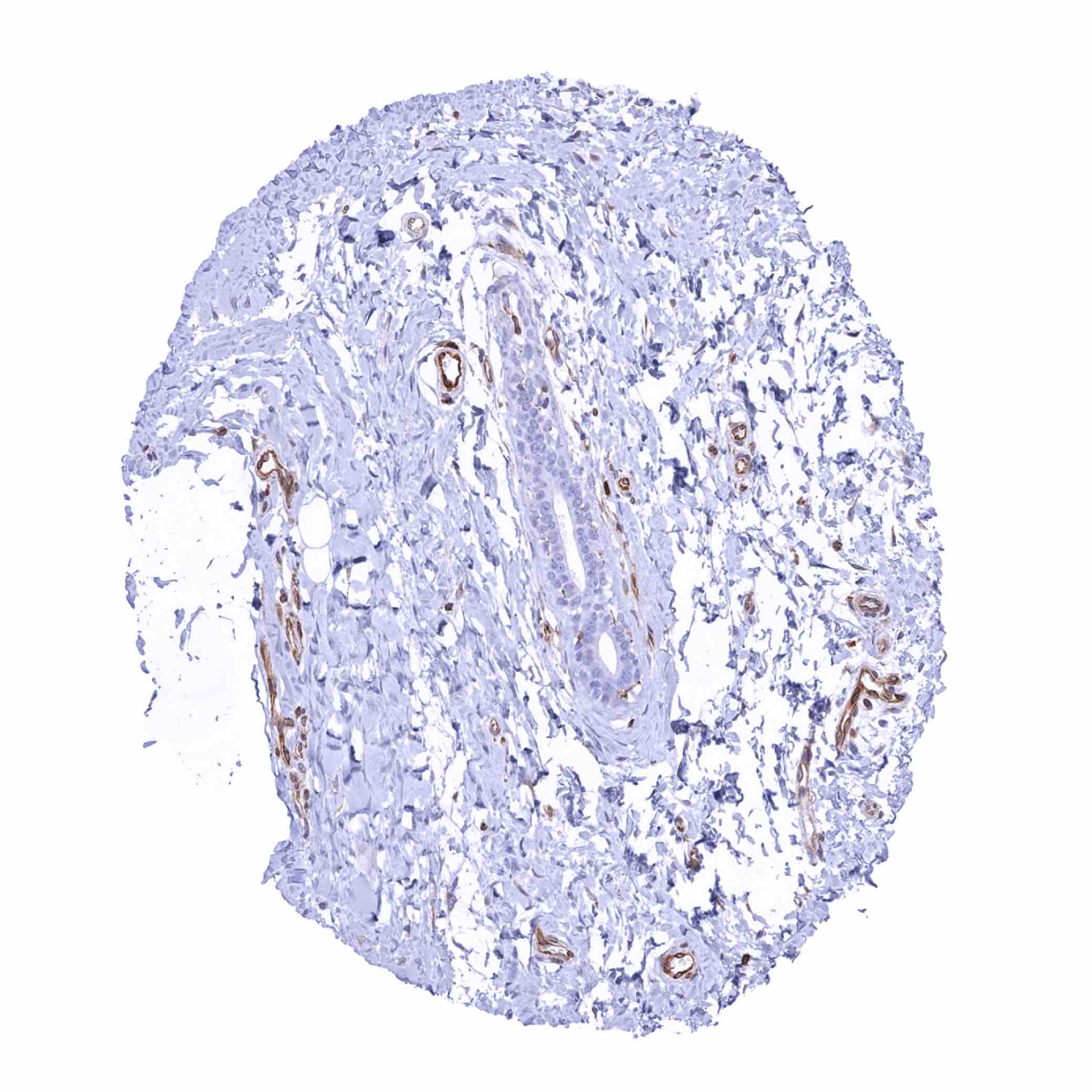
Duodenum, Brunner gland
Duodenum, mucosa – STING staining of endothelial cells and a subset of lymphocytes (STING immunohistochemistry)

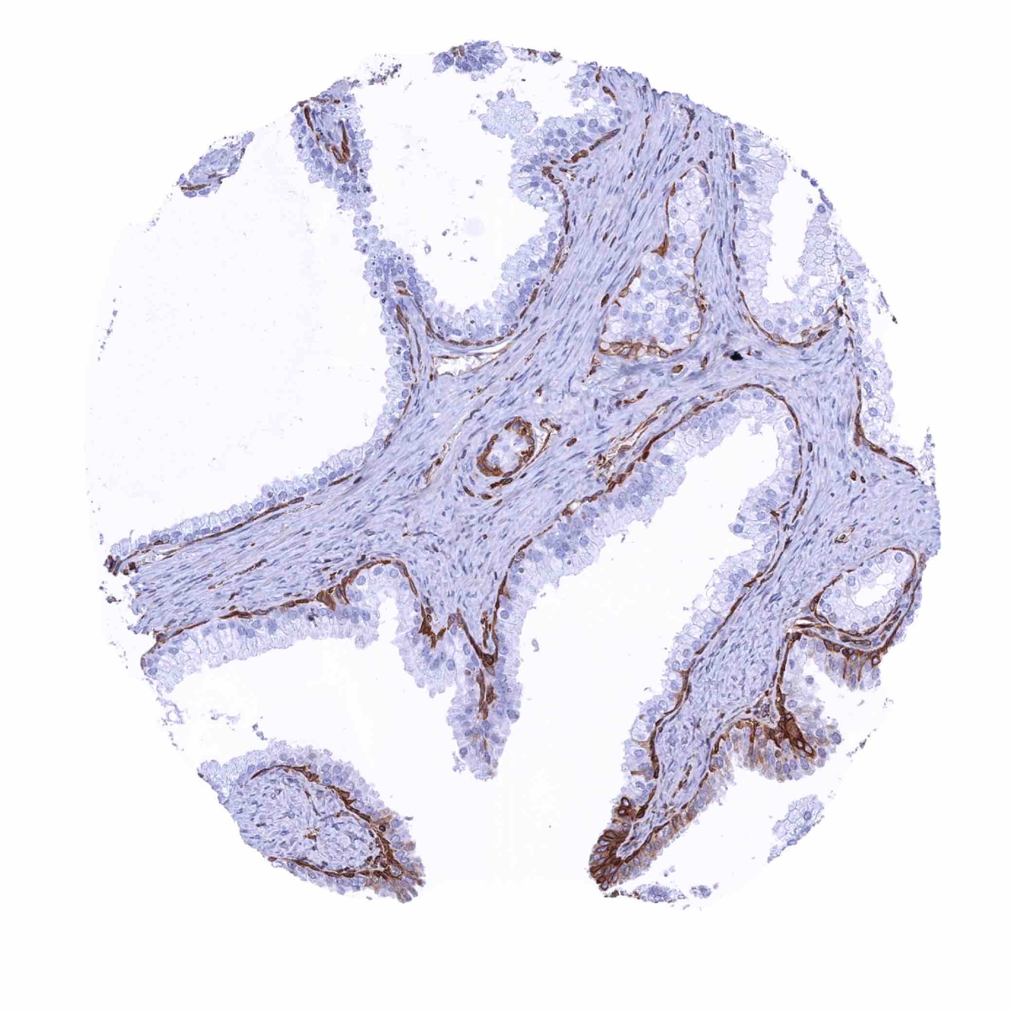
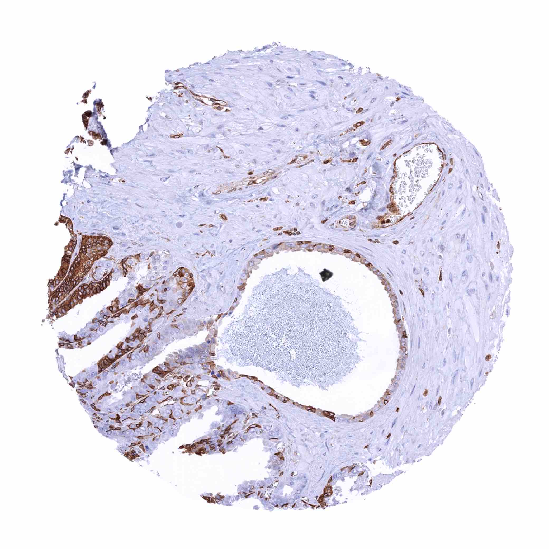
Epididymis – Moderate to strong STING staining of a fraction of epithelial cells in the cauda epididymis (STING immunohistochemistry)

Epididymis – Moderate to strong STING staining of basal cells in the caput epididymis (STING immunohistochemistry)
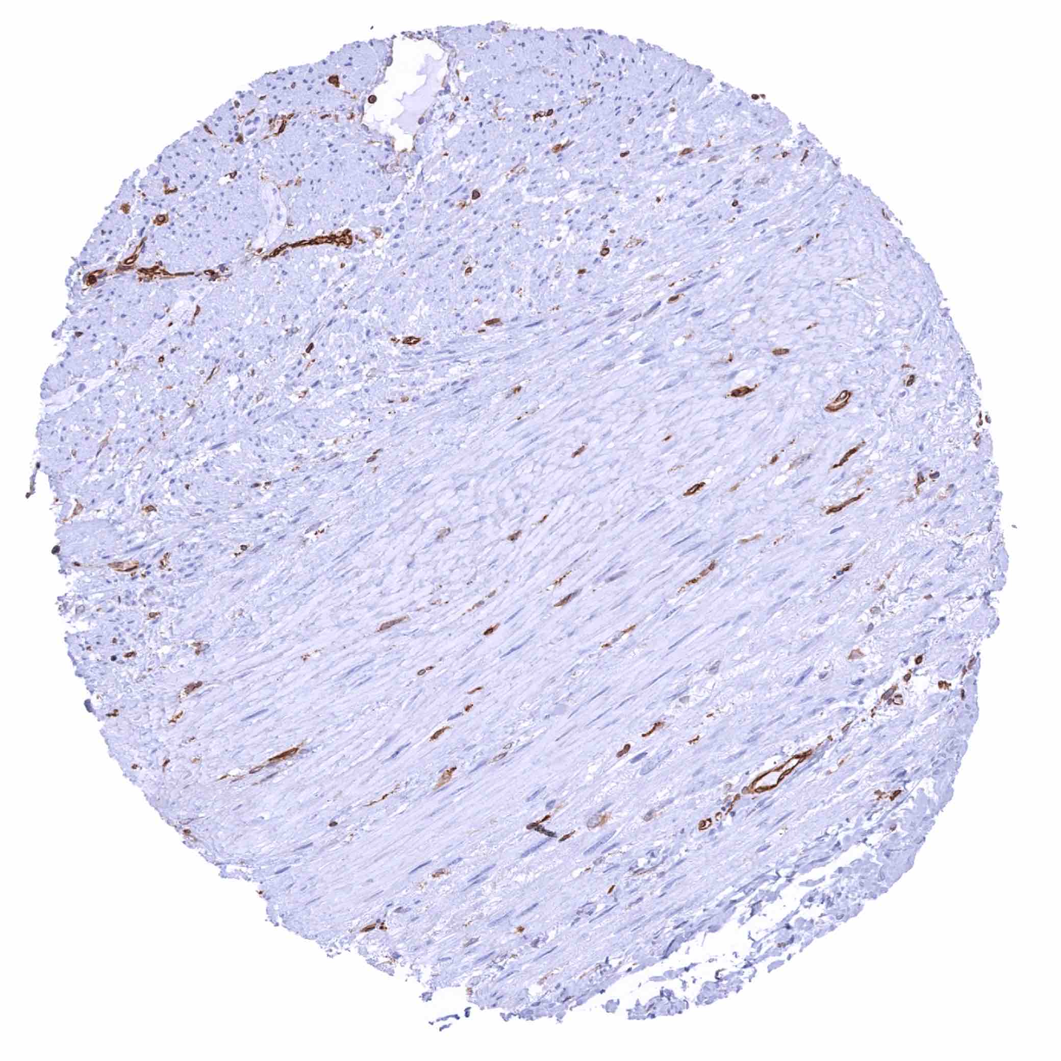
Esophagus, muscular wall – STING staining of endothelials cells and of a fraction of smooth muscle cells (STING immunohistochemistry)
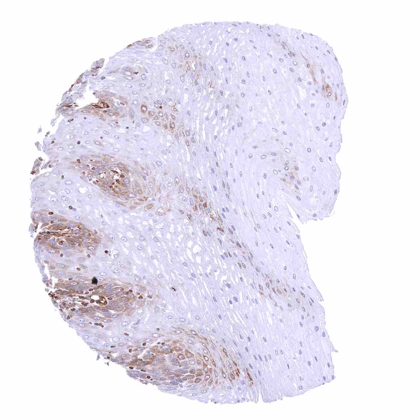
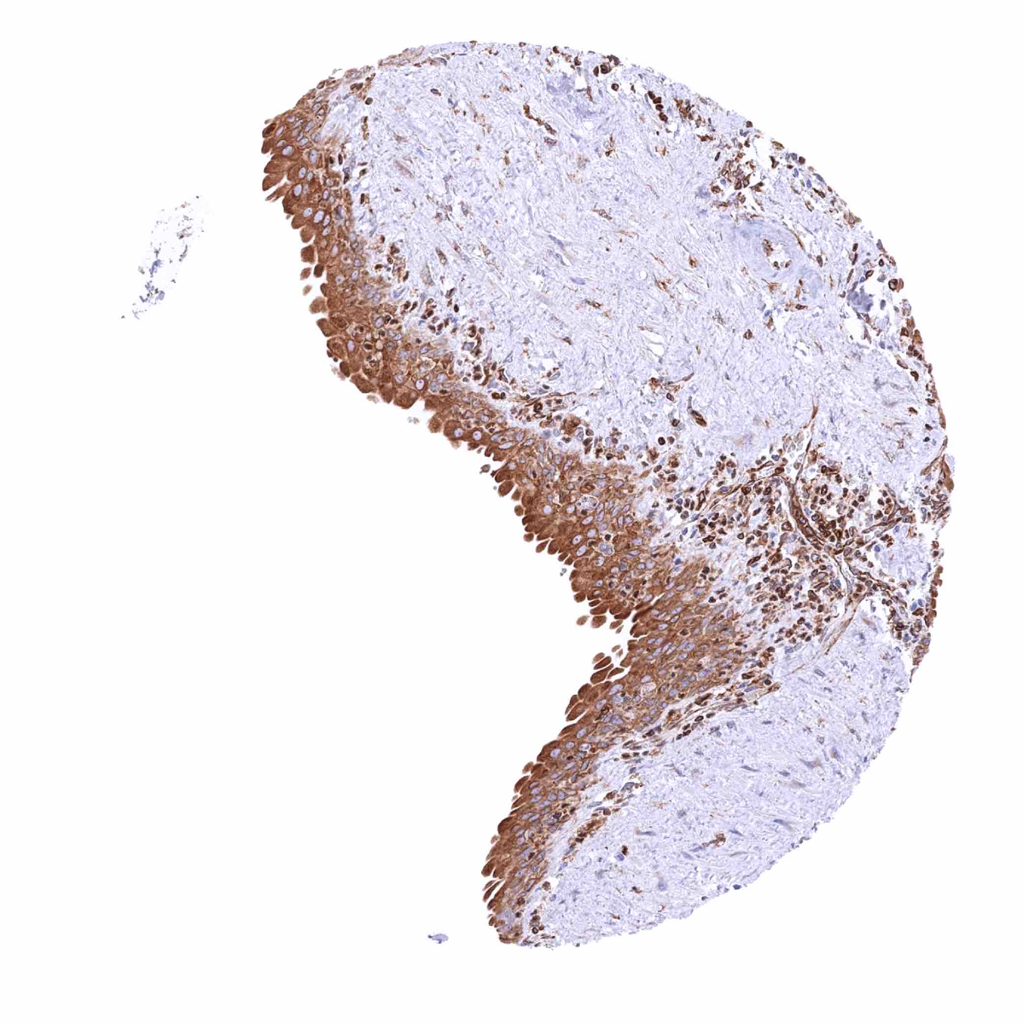
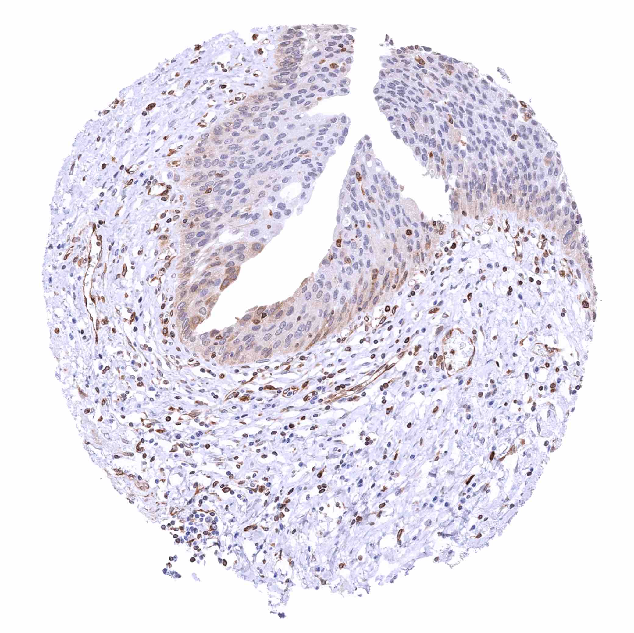
Esophagus, squamous epithelium – Weak to moderate STING staining of the basal cell layers of the squamous epithelium (STING immunohistochemistry)
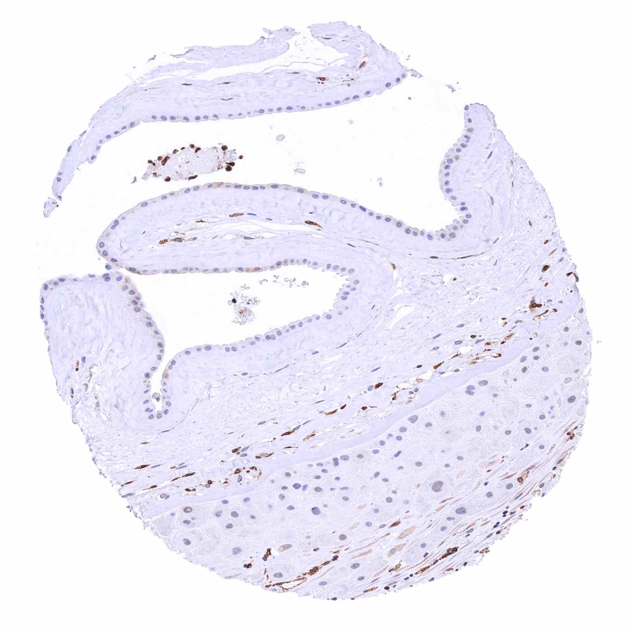
Fallopian tube, mucosa – Strong STING staining of epithelial cells (STING immunohistochemistry))

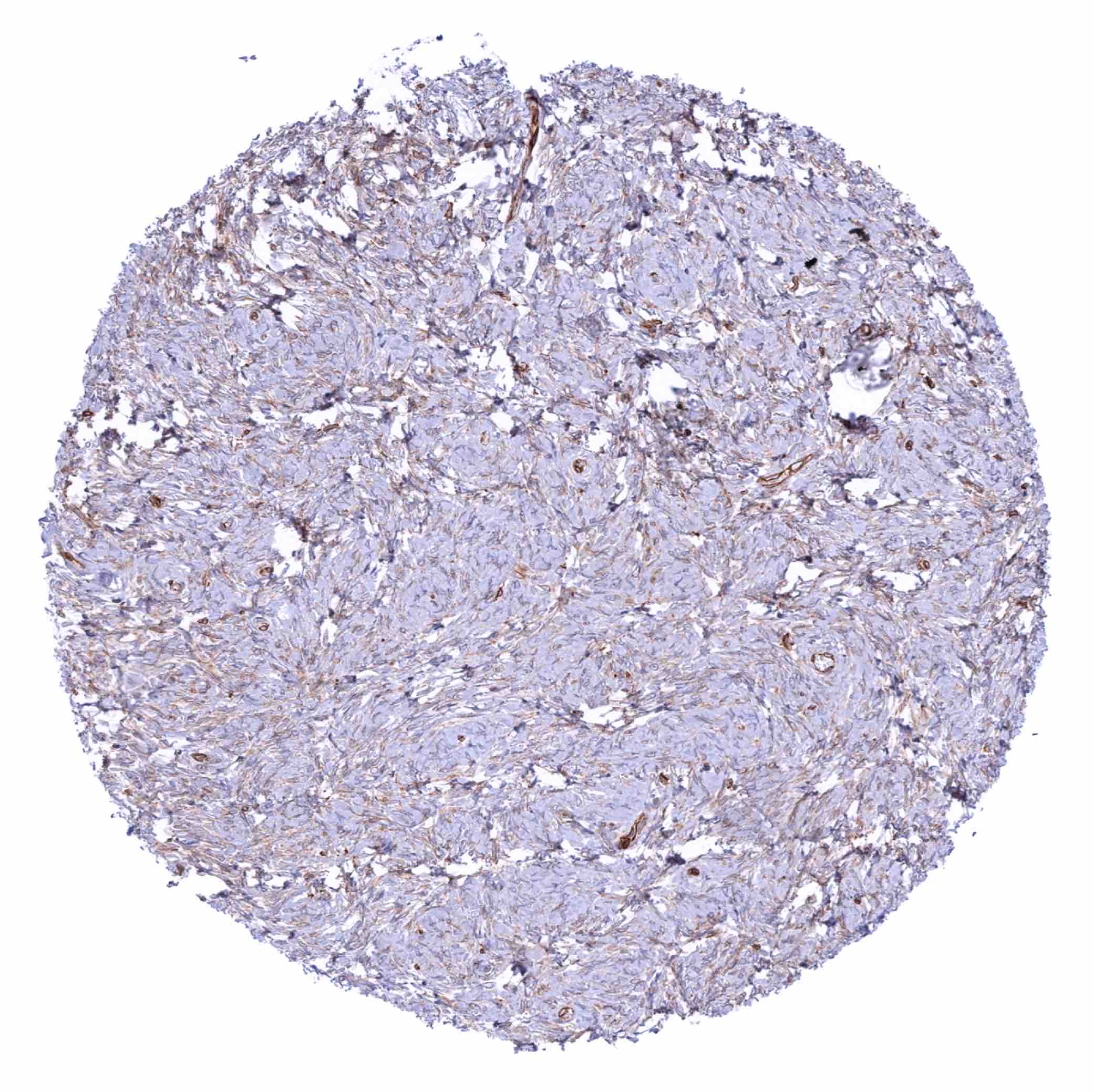
Fat
Gallbladder, epithelium – Complete lack of STING in epithelial cells of this sample
Gallbladder, epithelium – Moderate STING staining of most epithelial cells in this sample (STING immunohistochemistry)
Gallbladder, epithelium – STING staining of only a very small subset of epithelial cells in this sample (STING immunohistochemistry)

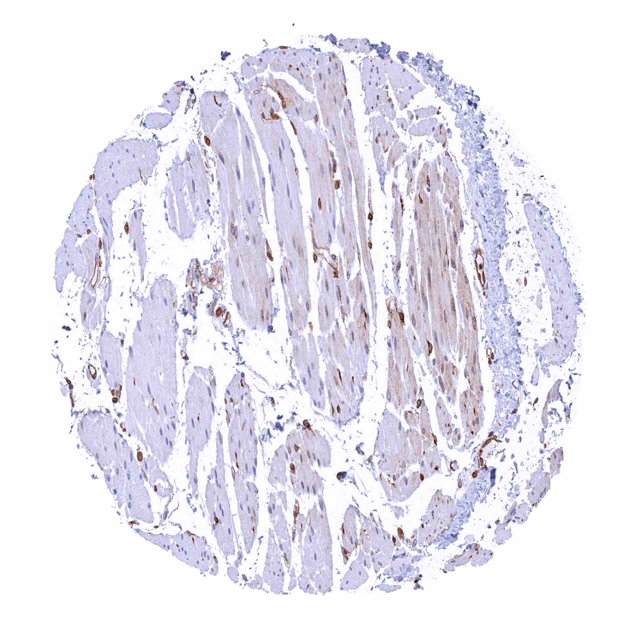
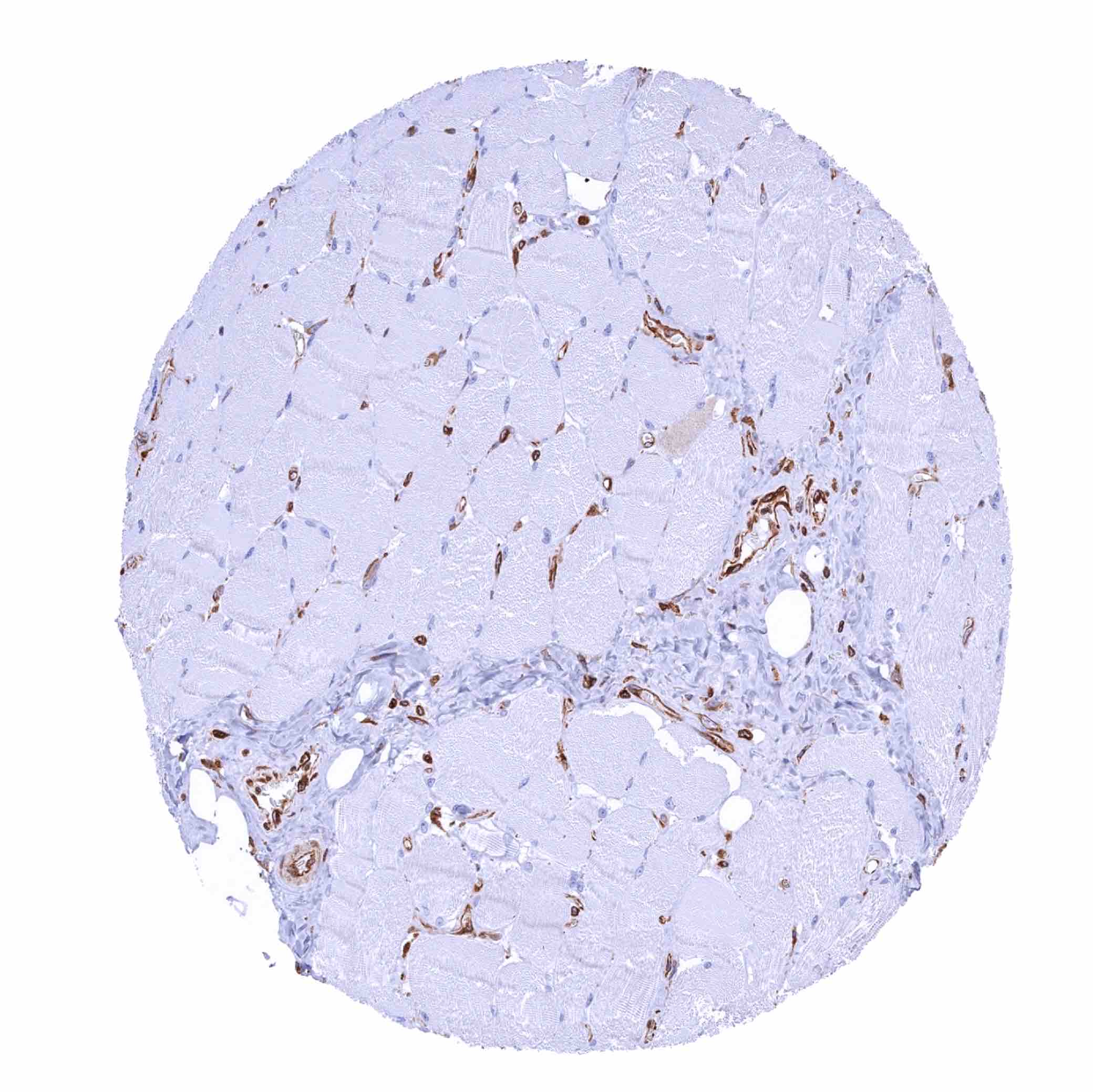
Heart muscle – STING staining of endothelials cells of small capillaries (STING immunohistochemistry)
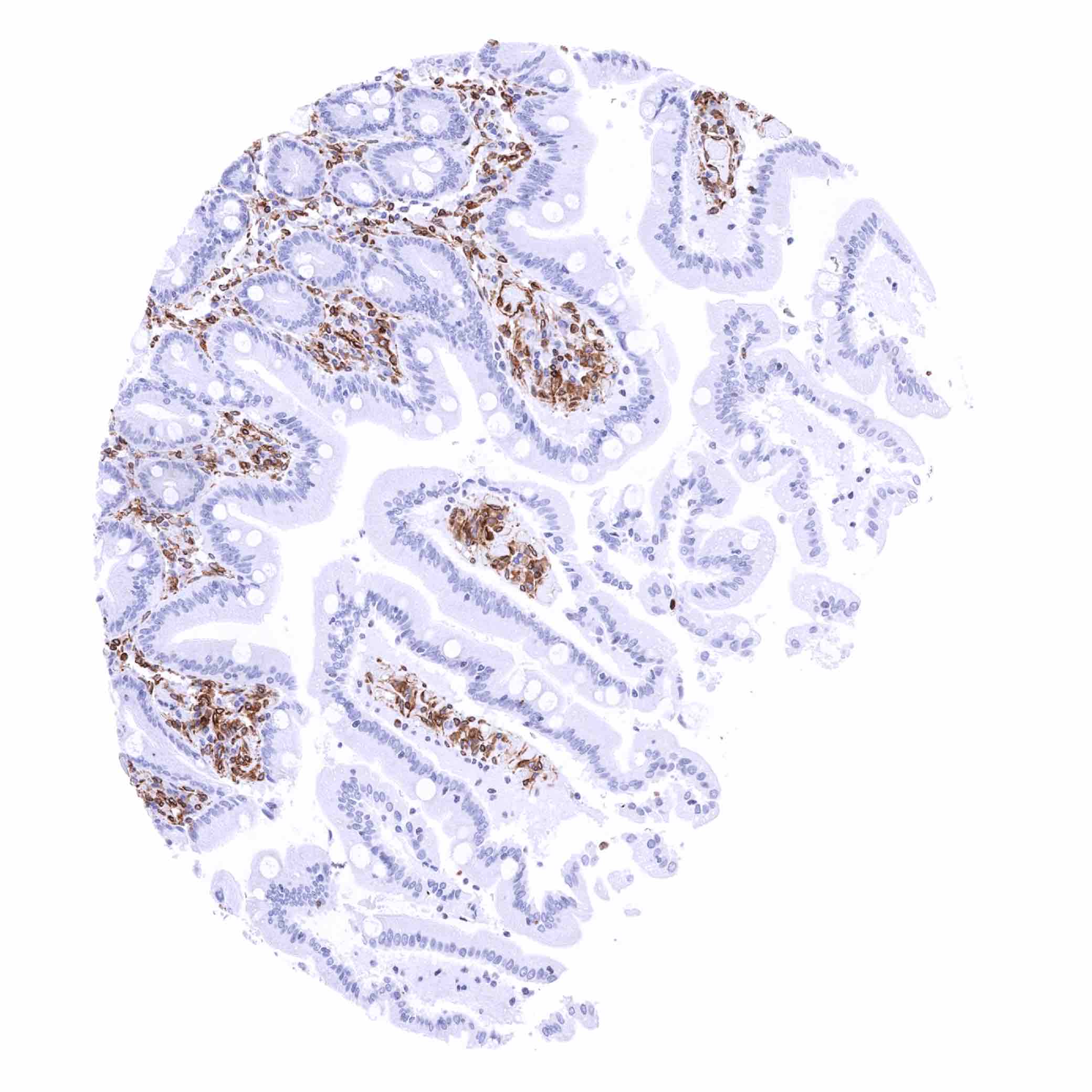
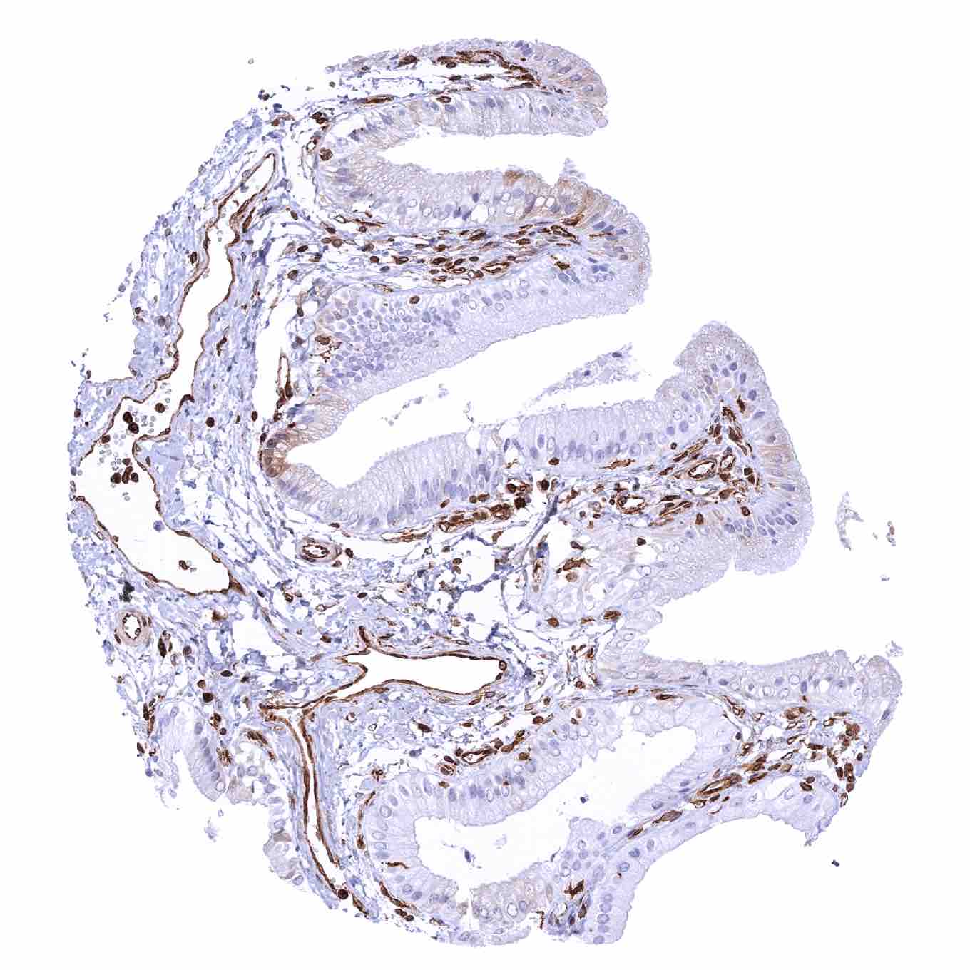
Ileum, mucosa – STING staining of a fraction of endothelial cells and a subset of lymphocytes (STING immunohistochemistry)
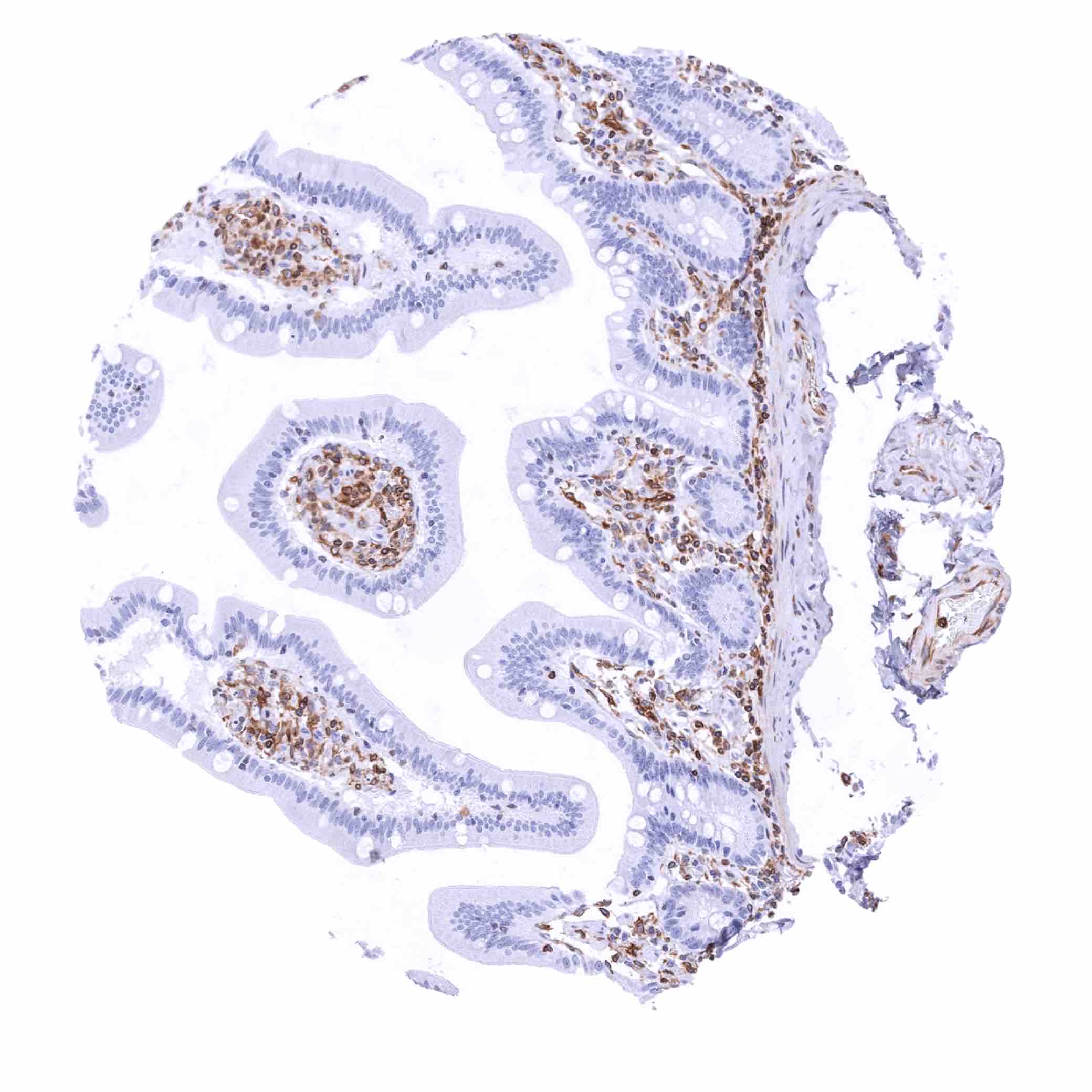
Ileum, mucosa – STING staining of endothelial cells, a subset of lymphocytes and of most epithelial cells (STING immunohistochemistry)
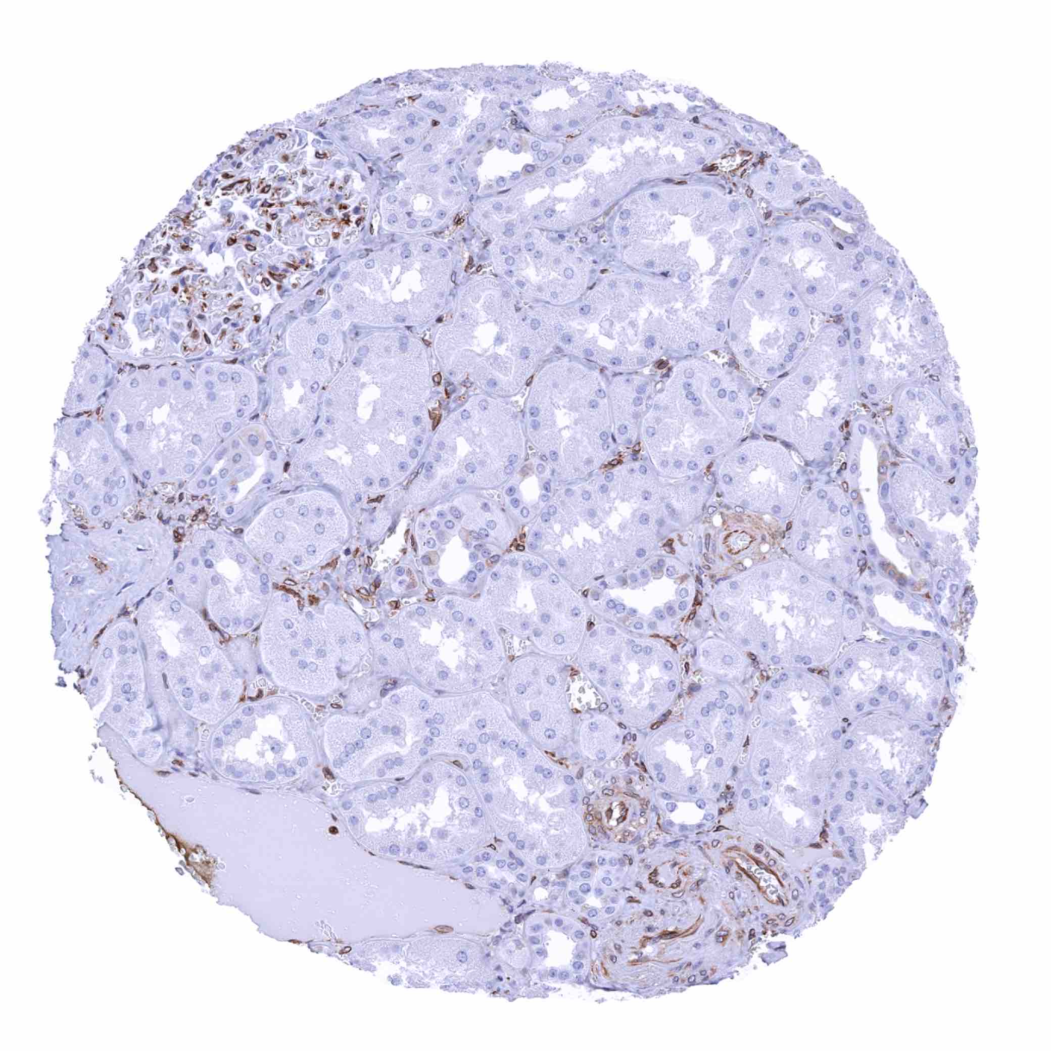
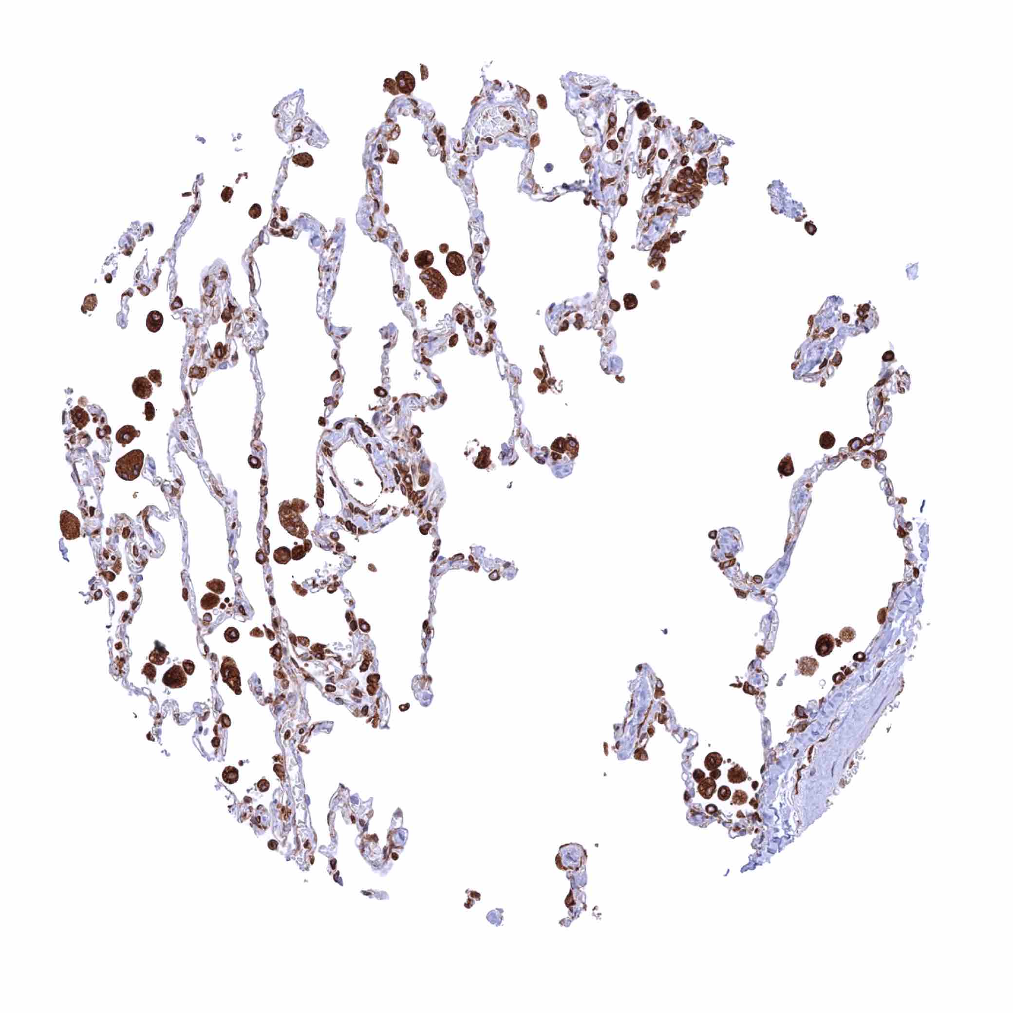
Kidney, cortex – STING staining of endothelial cells including glomeruli (STING immunohistochemistry)
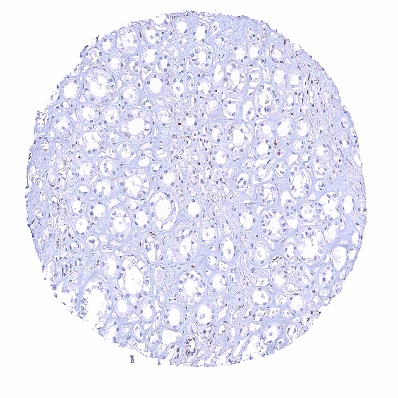
Kidney, medulla
Kidney, pelvis, urothelium – Only a weak basal cell STING staining of the urothelium is seen in this sample (STING immunohistochemistry)
Kidney, pelvis, urothelium – Strong STING staining of the urothelium in this sample (STING immunohistochemistry)
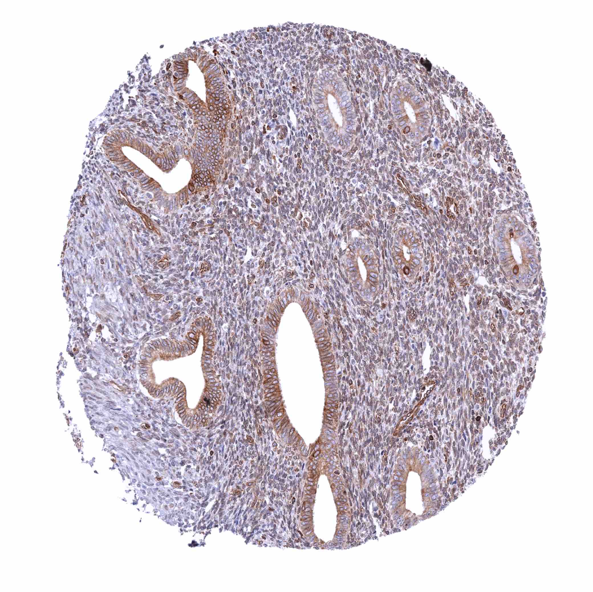
Kidney, pelvis, urothelium – Variable STING staining of the urothelium predominating in the basal cell layers (STING immunohistochemistry)
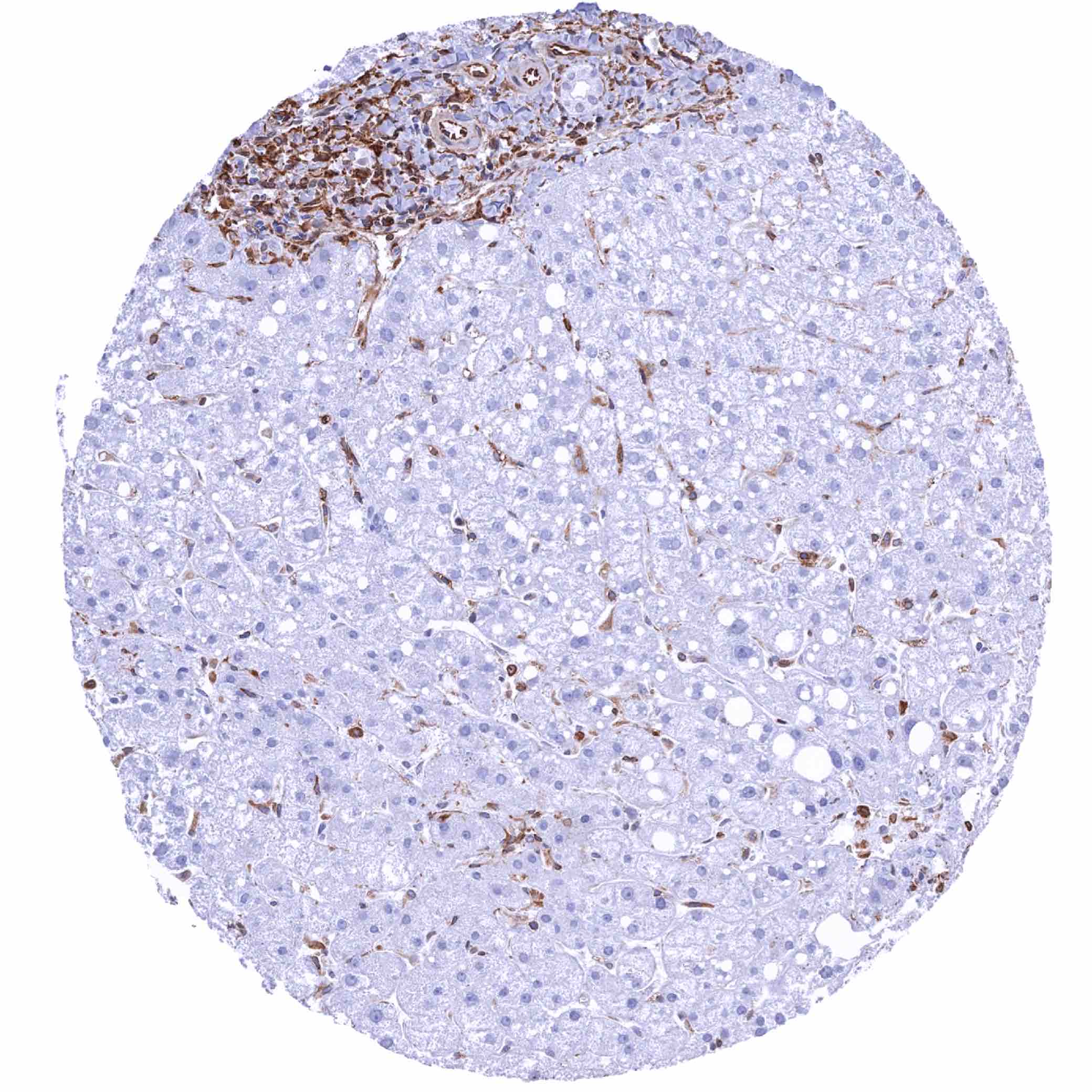
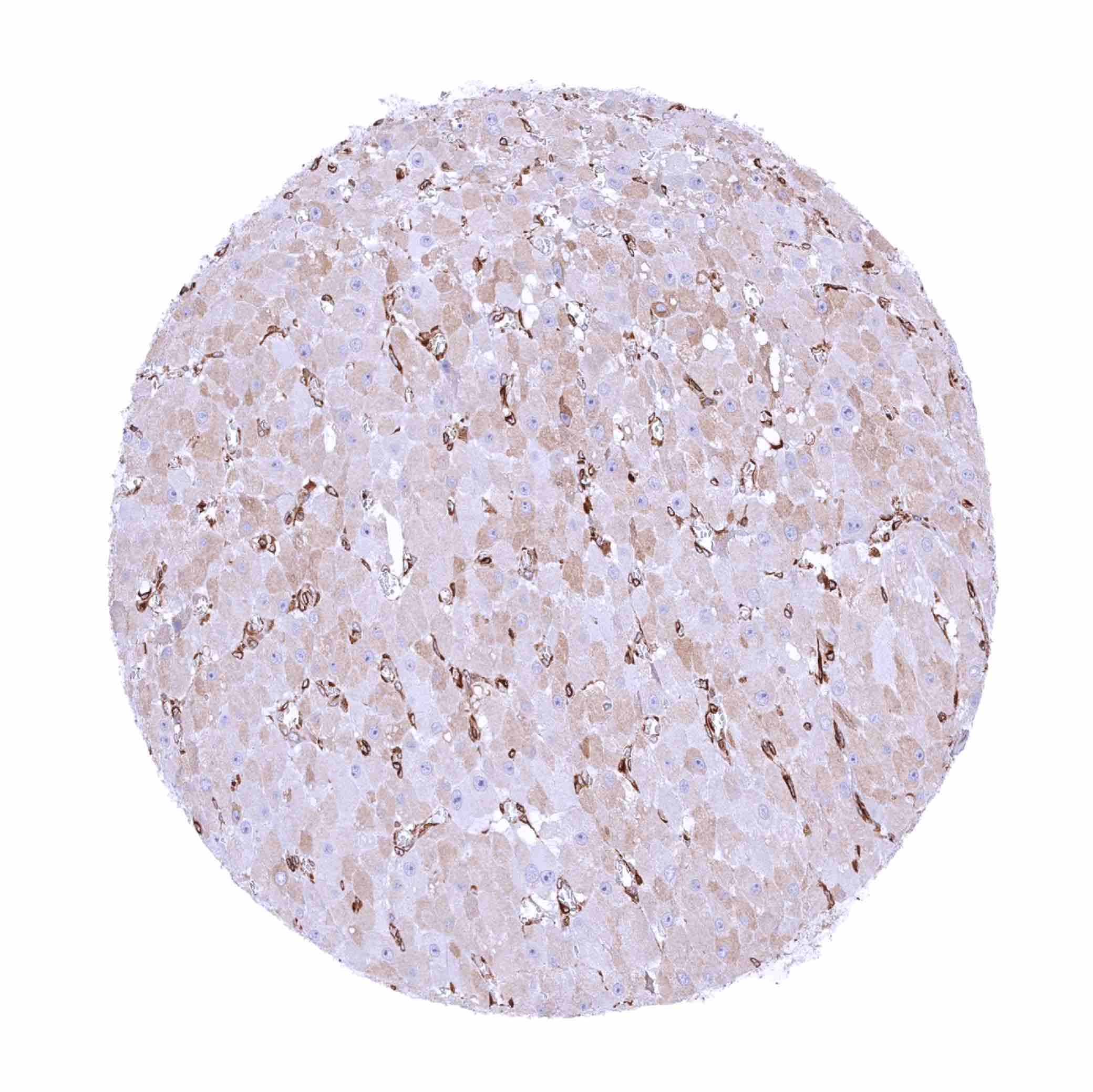
Liver – STING staining of endothelial cells, sinus cells and inflammatory cells (STING immunohistochemistry)
Lung – Strong STING staining of alveolar macrophages (STING immunohistochemistry)
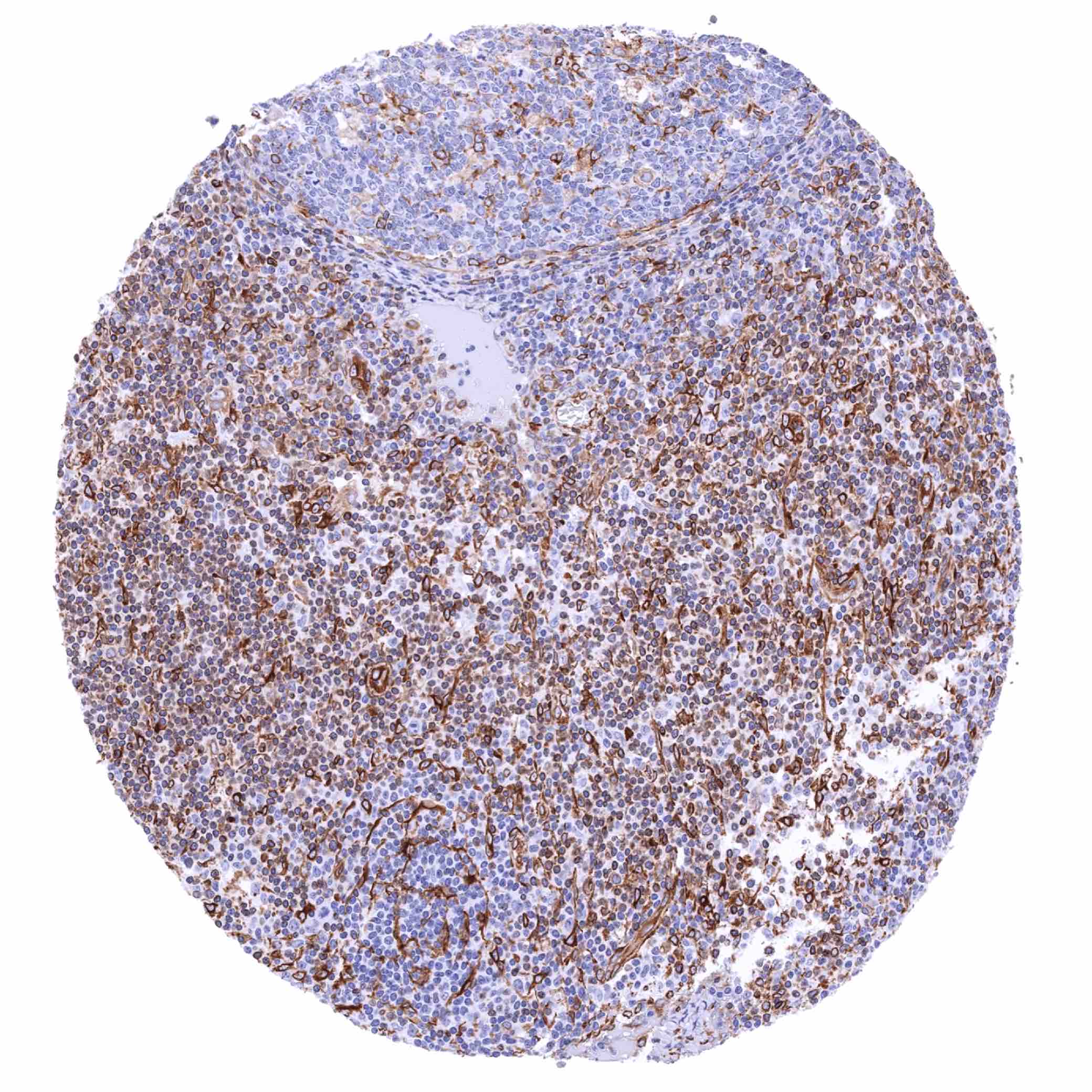
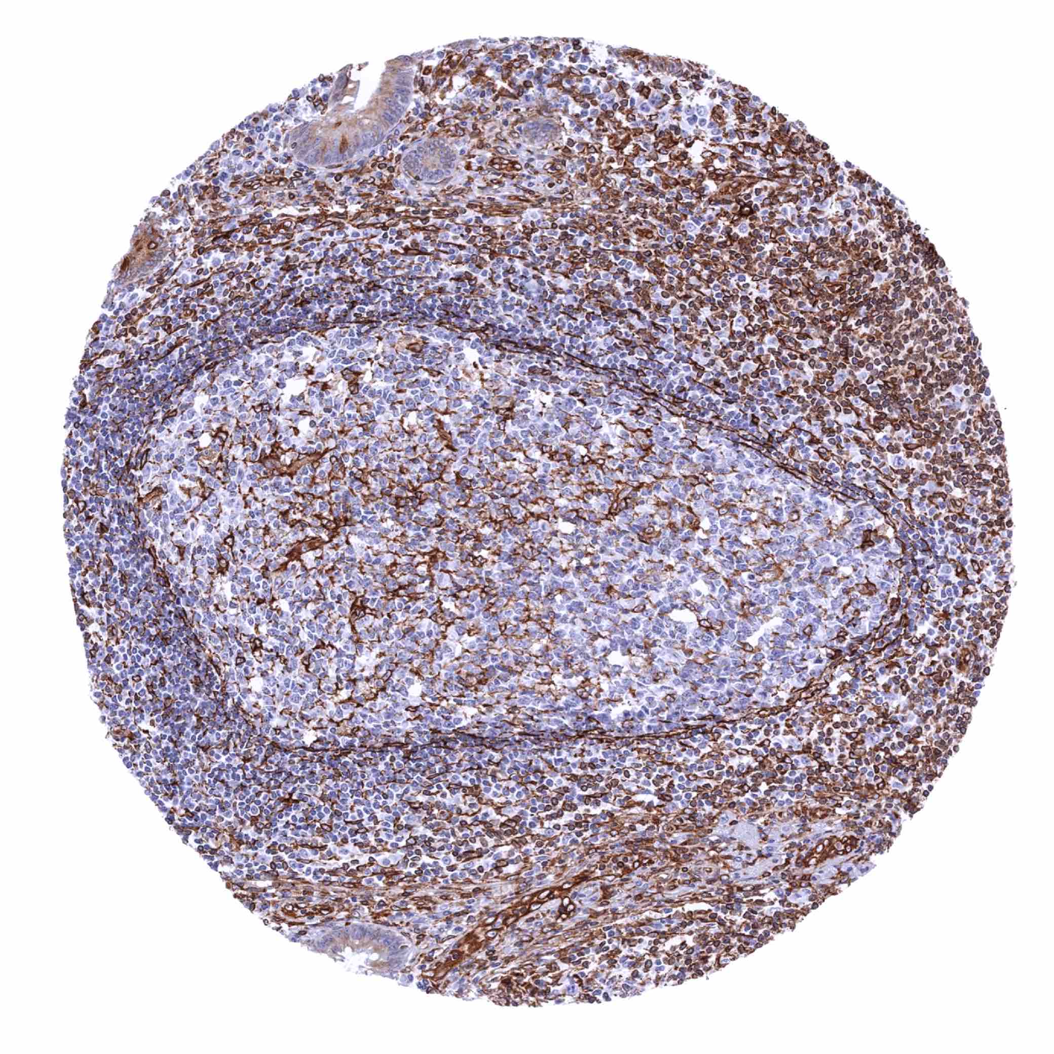
Lymph node – Moderate to strong STING staining of a significant fraction of lymphocytes, mainly in the interfollicular zone (STING immunohistochemistry)
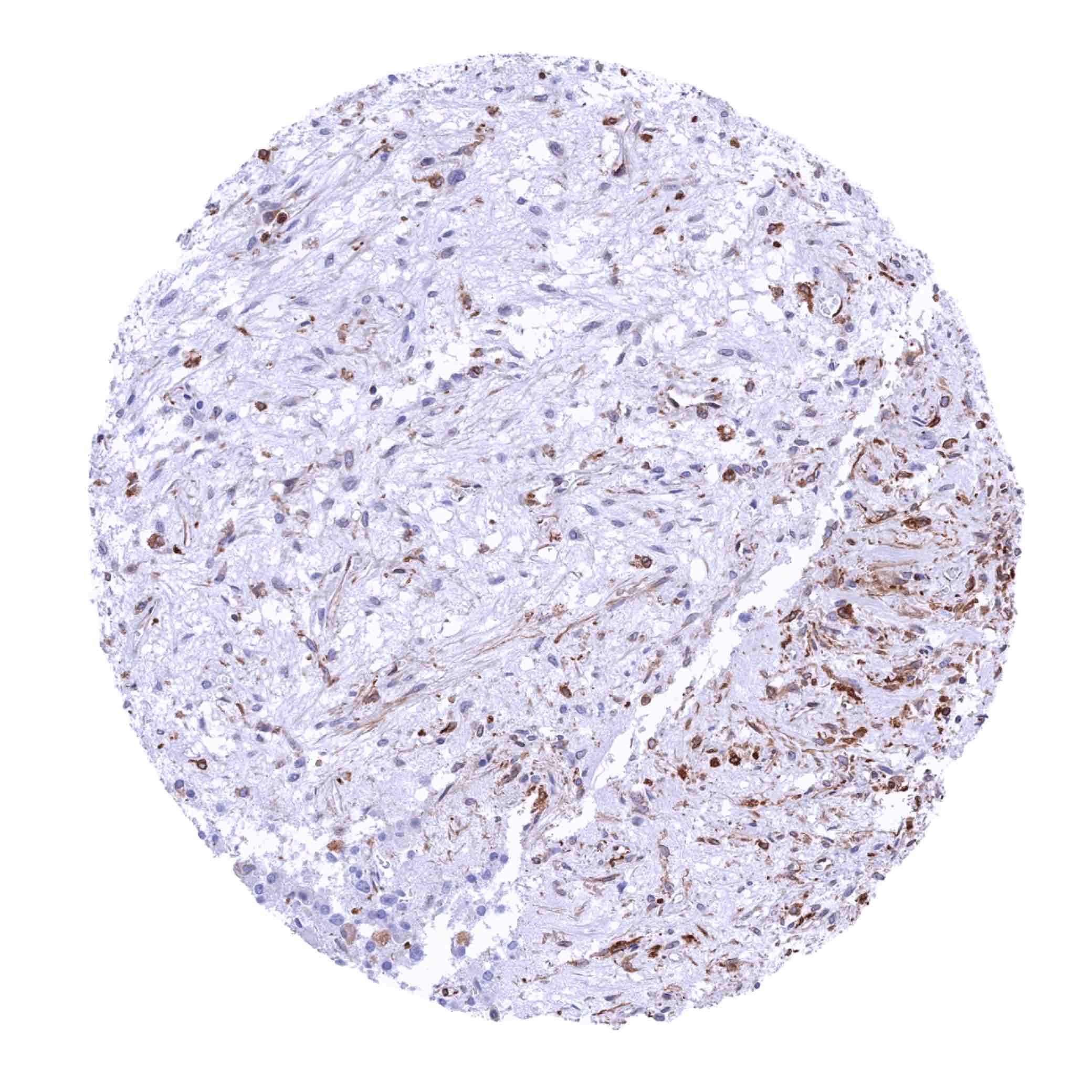
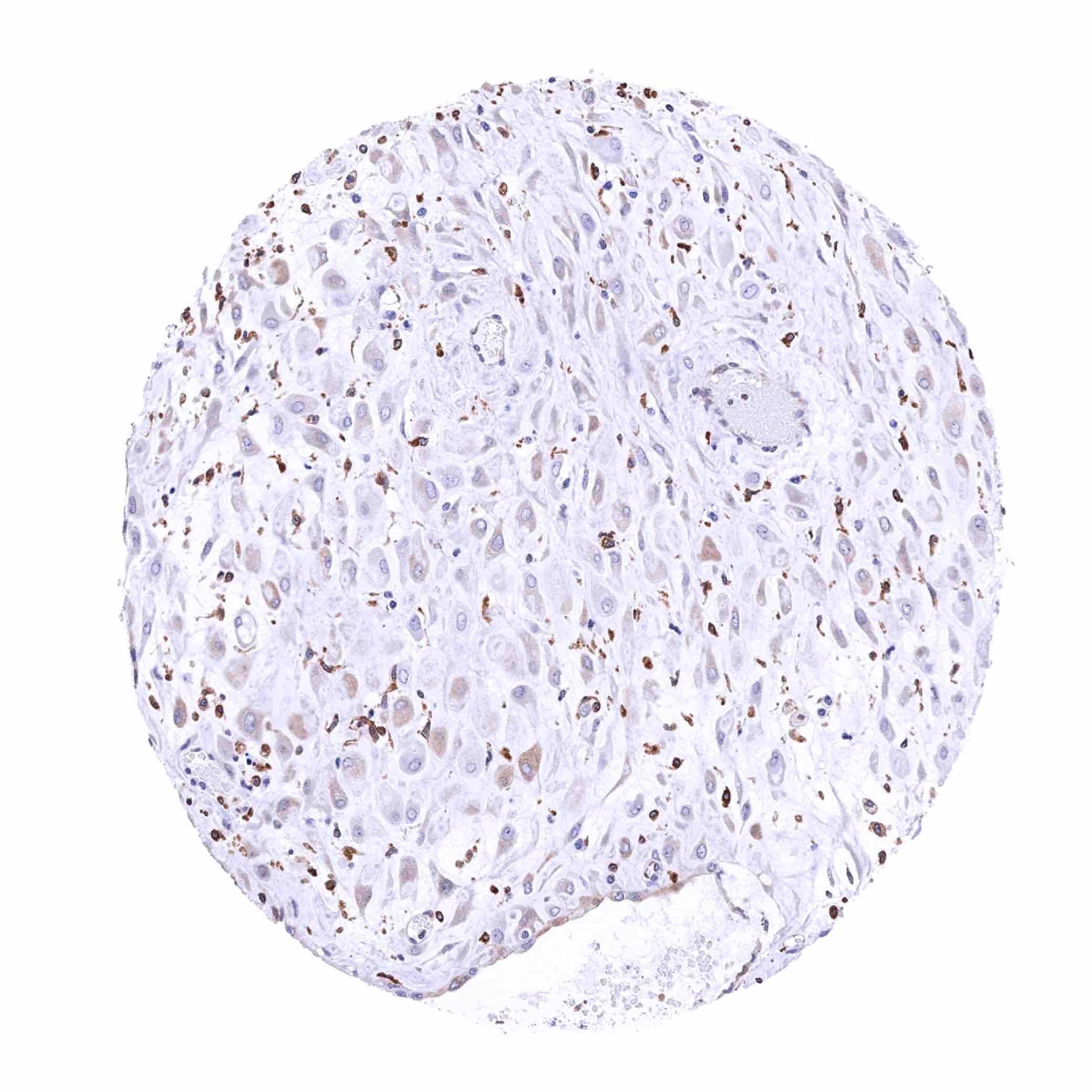
Ovary, corpus luteum – Strong STING staining of inflammatory cells while corpus luteum cells show a rather weak staining (STING immunohistochemistry)
Ovary, stroma – Weak to moderate STING staining in stromal cells (STING immunohistochemistry)
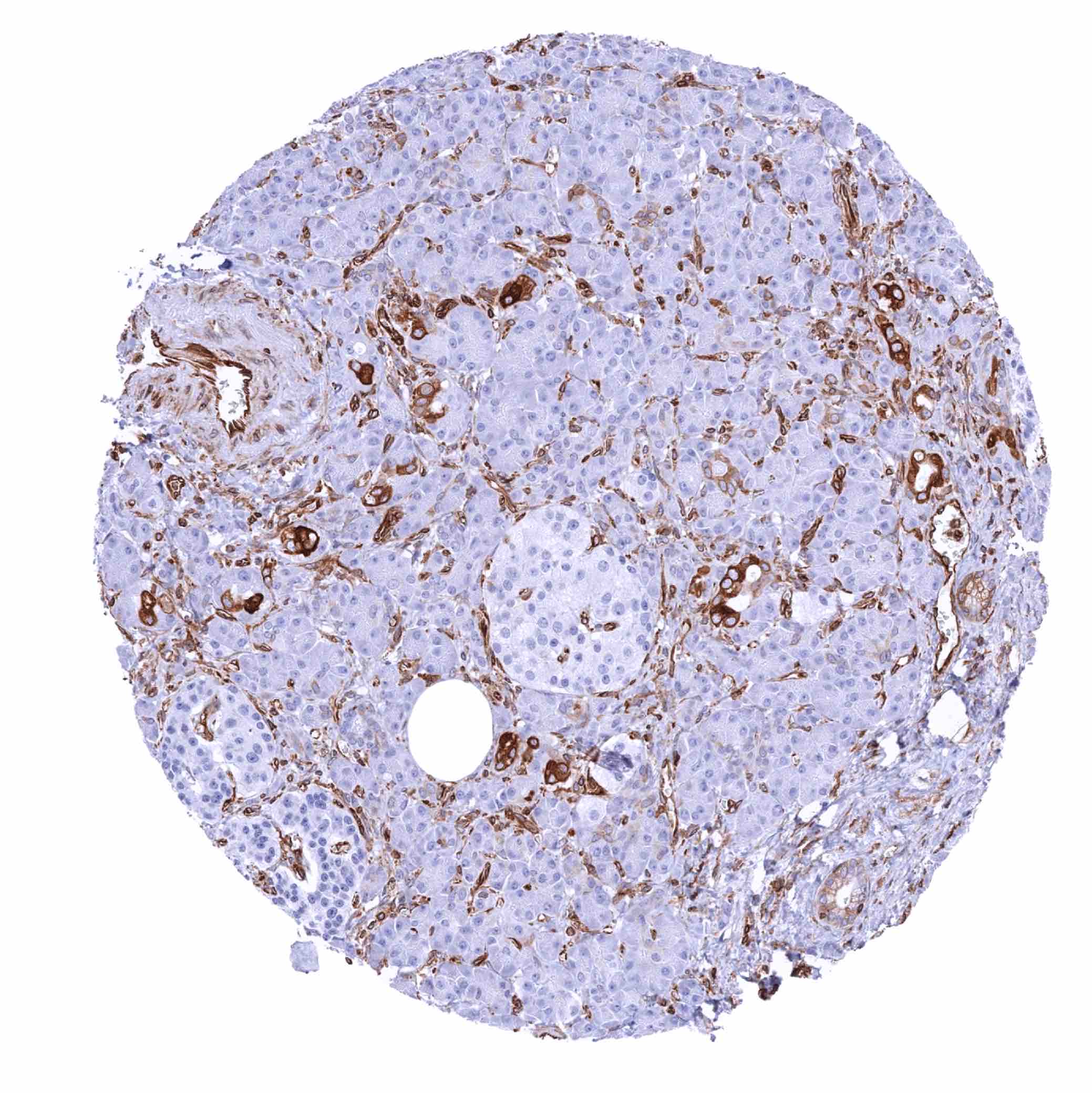
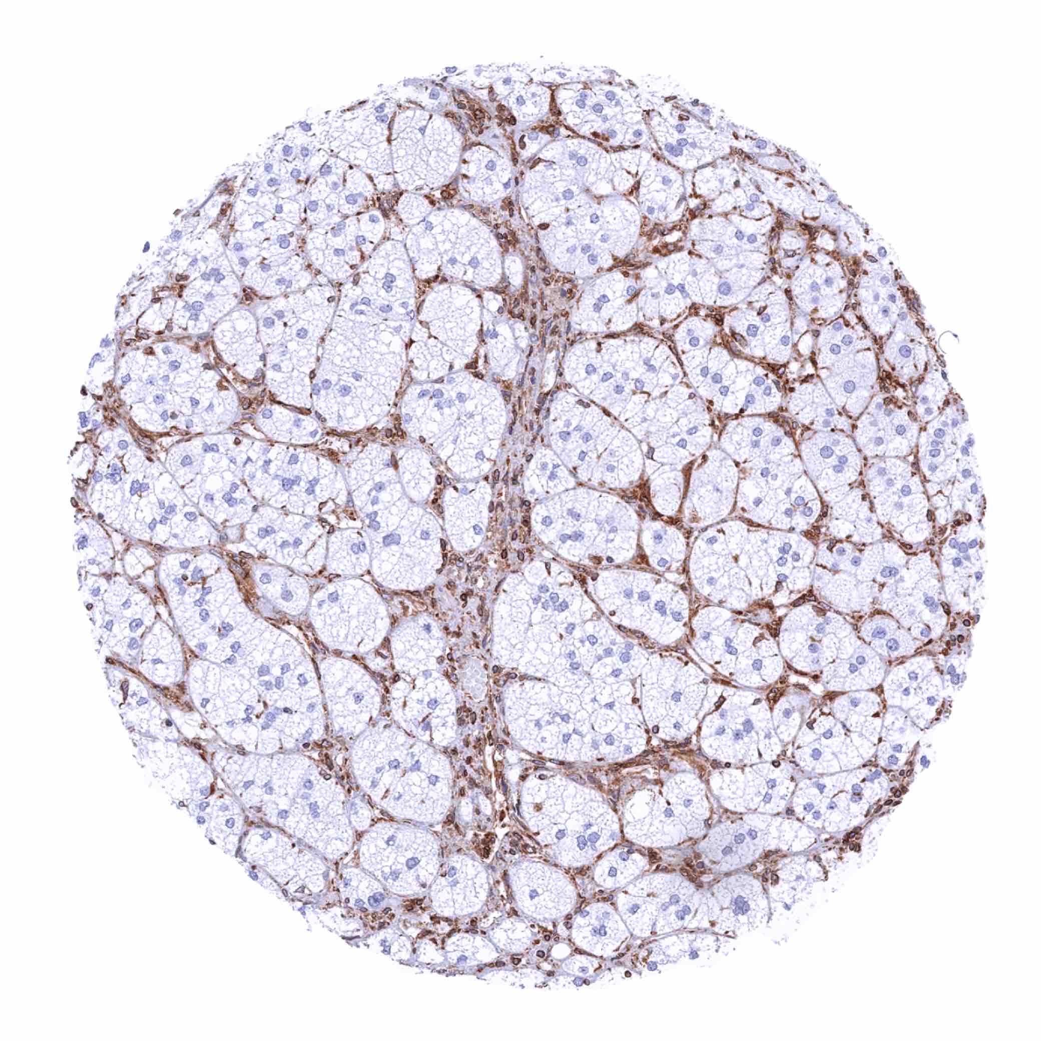
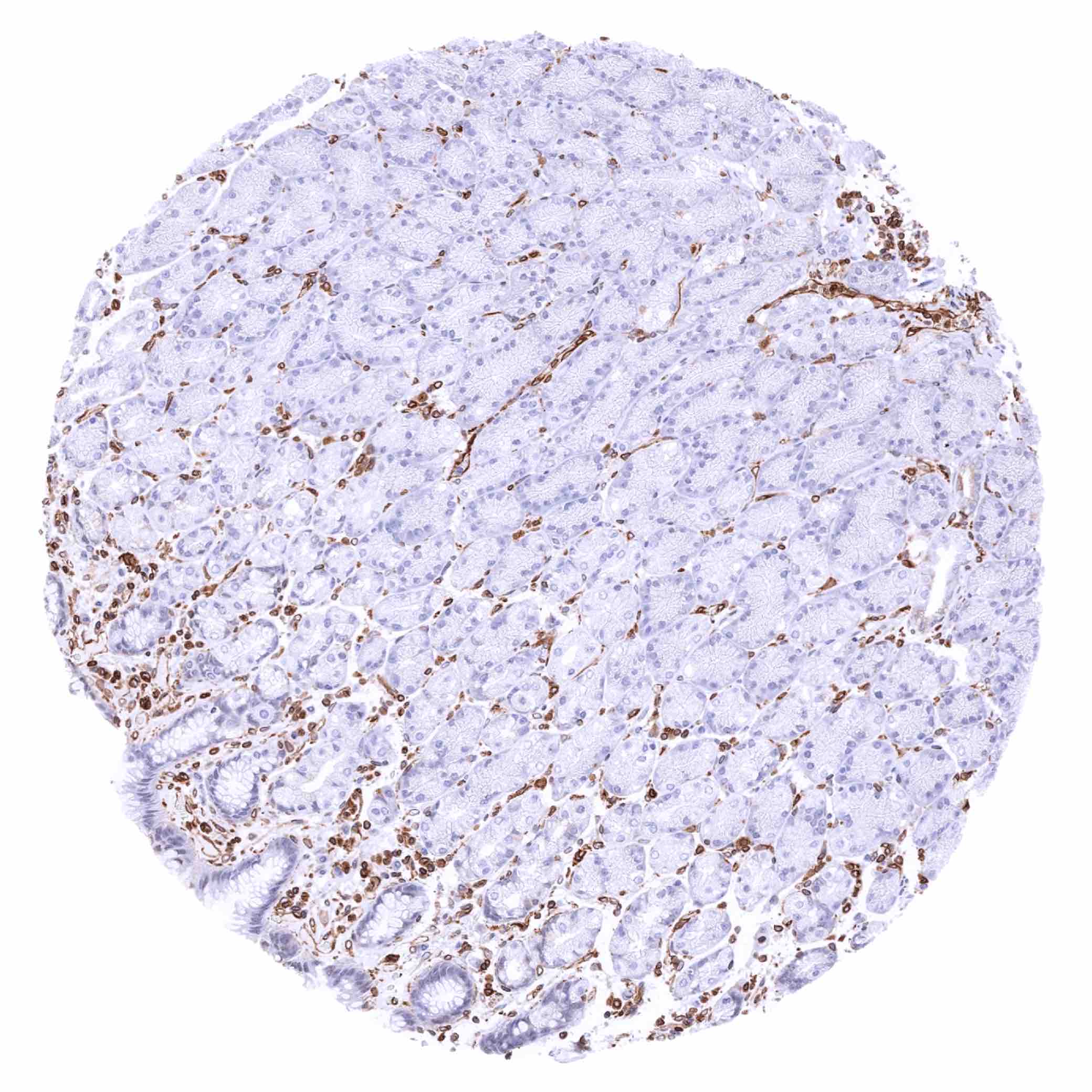
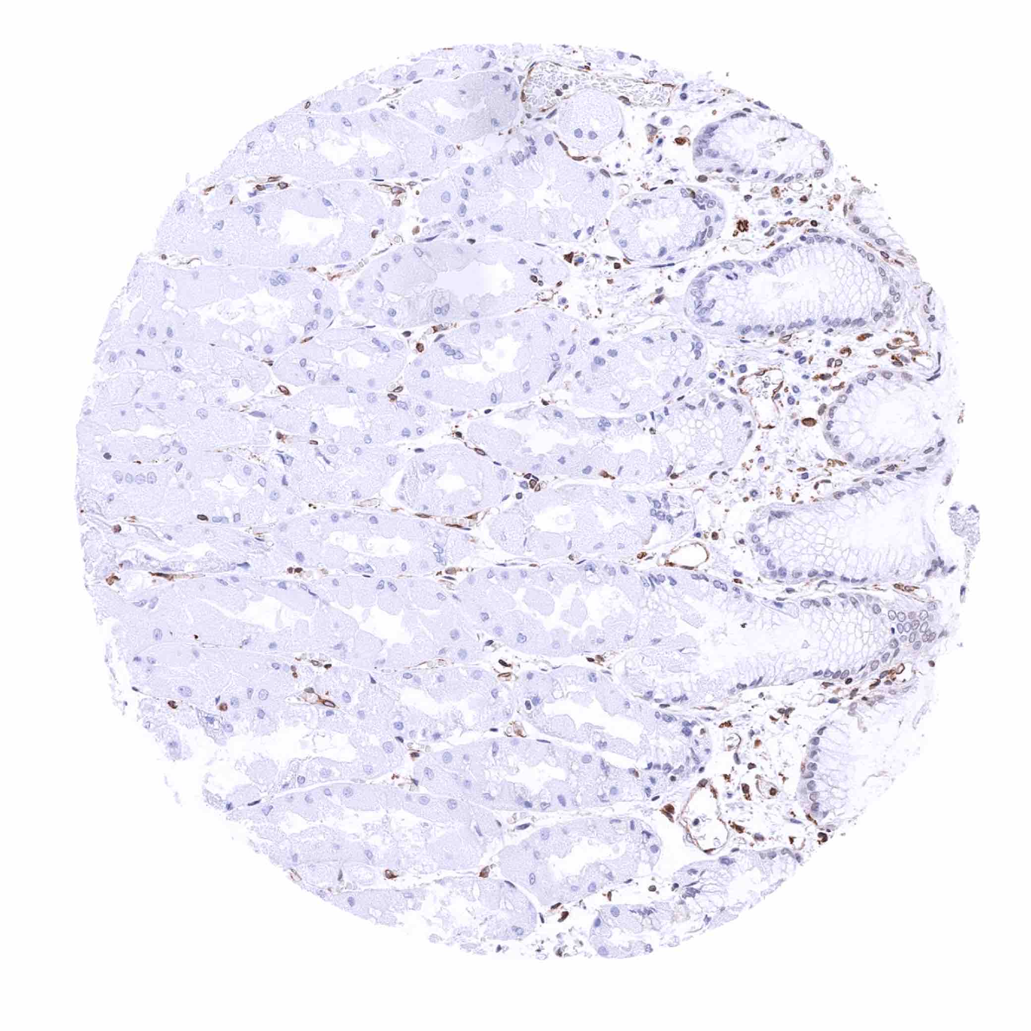
Pancreas – STING staining of endothelial cells, intercalated ducts and excretory ducts (STING immunohistochemistry)

Parathyroid gland – STING staining of endothelial cells (STING immunohistochemistry)
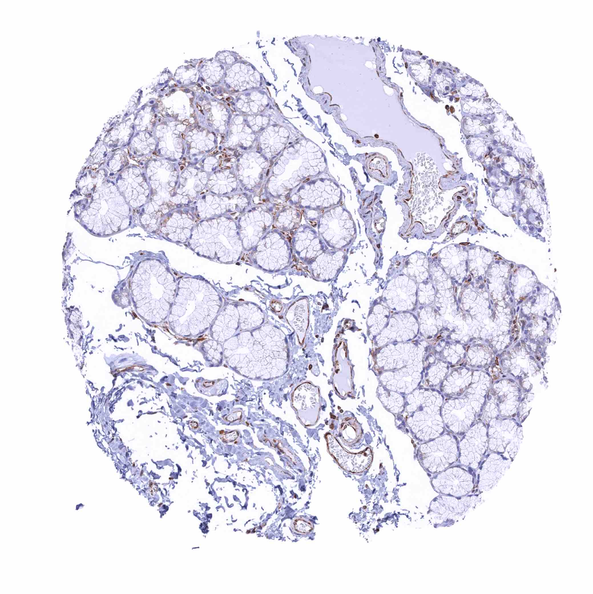
Parotid gland – STING staining of endothelial cells and of basal cells in excretory ducts (STING immunohistochemistry)
Parotid gland – STING staining of some glandular cells in this sample (STING immunohistochemistry)

Pituitary gland, anterior lobe – STING staining of endothelial cells and of a subset of epithelial cells (STING immunohistochemistry)
Pituitary gland, anterior lobe – STING staining of endothelial cells only in this sample (STING immunohistochemistry)
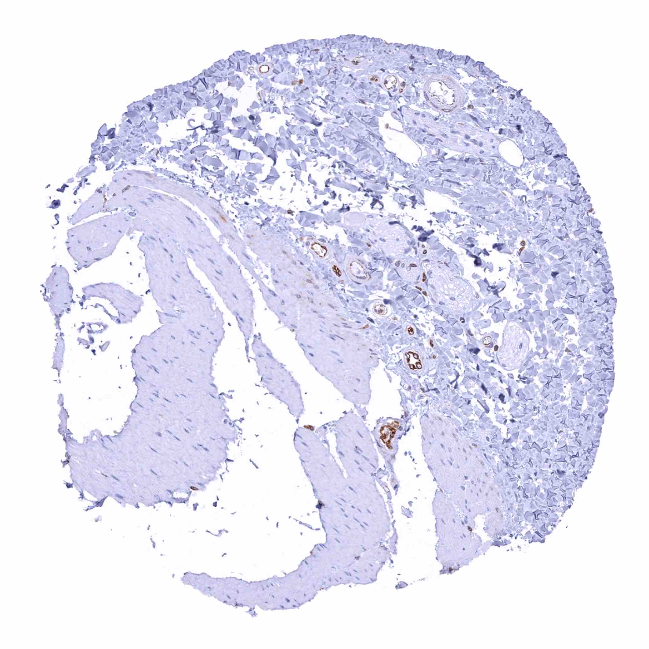
Pituitary gland, posterior lobe – STING staining of endothelial cells and probably of some other fibrous structures (STING immunohistochemistry)
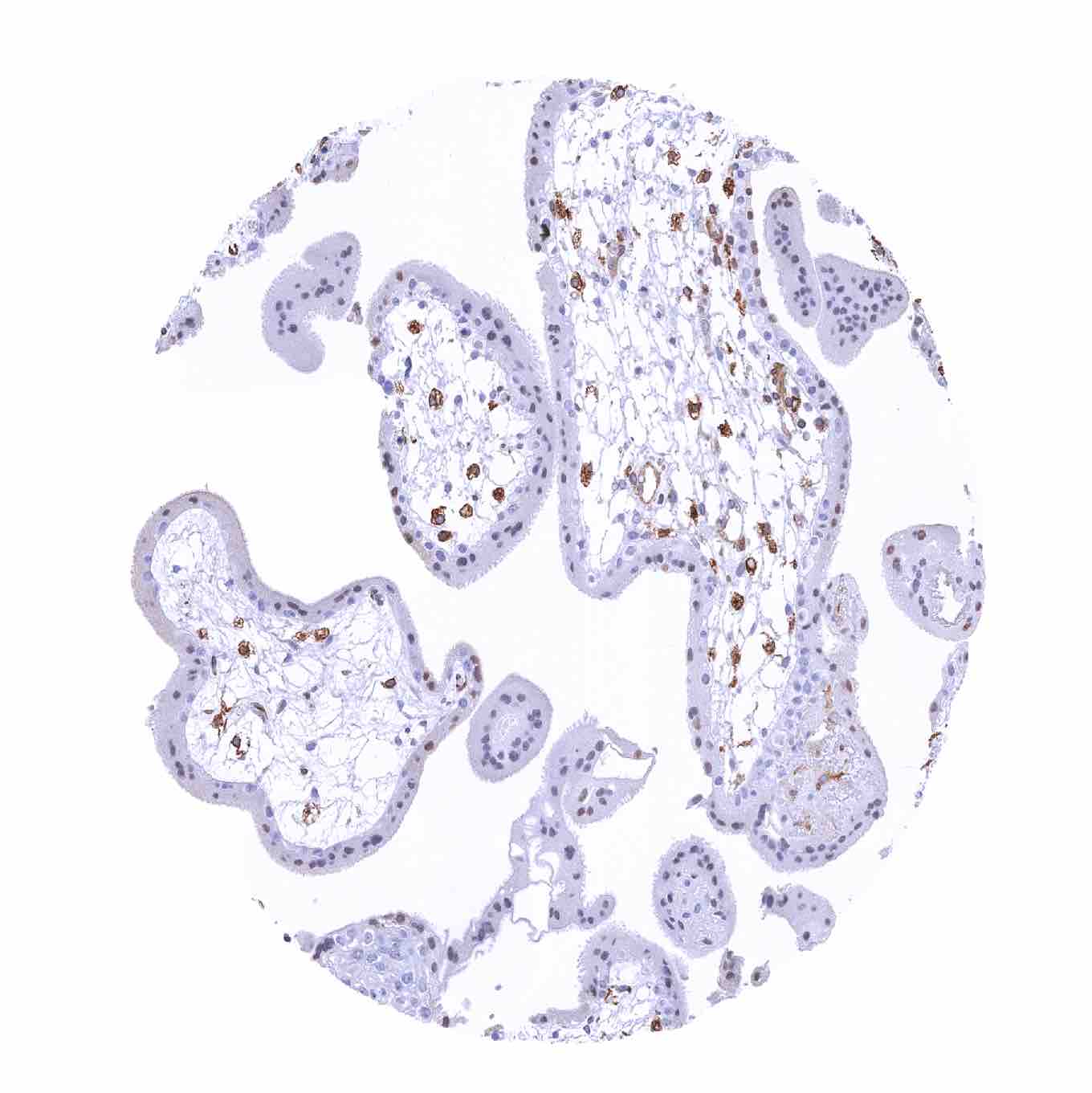
Placenta (amnion and chorion) – STING staining is absent in chorion and amnion cells
Placenta, early – Moderate STING staining of macrophages (STING immunohistochemistry)
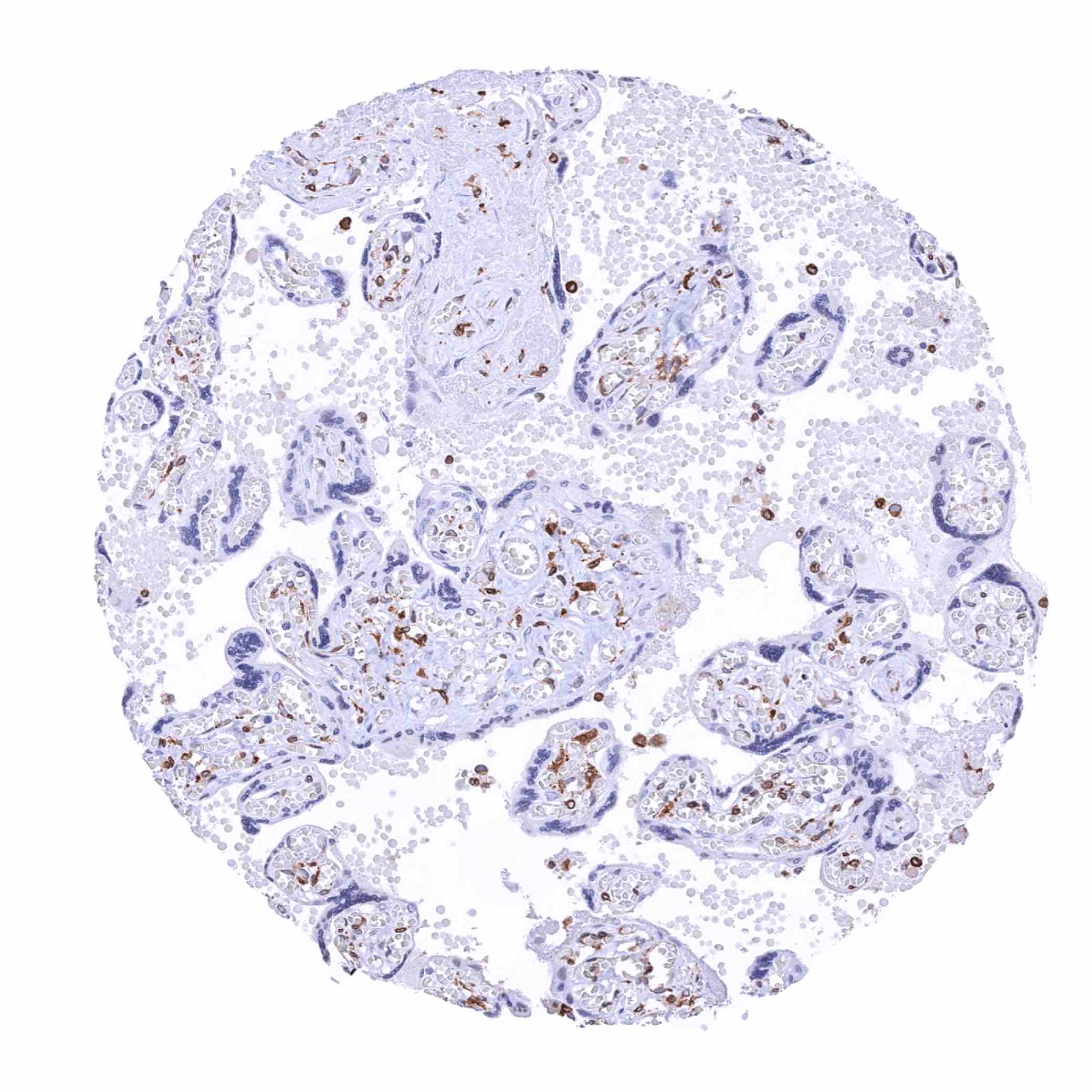
Placenta, mature – Moderate STING staining of macrophages (STING immunohistochemistry)
Prostate – Moderate to strong STING staining of basal cells while only few acinar cells show some positivity (STING immunohistochemistry)

Rectum, mucosa – Moderate STING staining of most epithelial cells in this sample (STING immunohistochemistry)
Seminal vesicle – Moderate to strong STING staining of a scattered fraction of luminal epithelial and basal cells - 2 (STING immunohistochemistry)
Seminal vesicle – Moderate to strong STING staining of a scattered fraction of luminal epithelial and basal cells (STING immunohistochemistry)
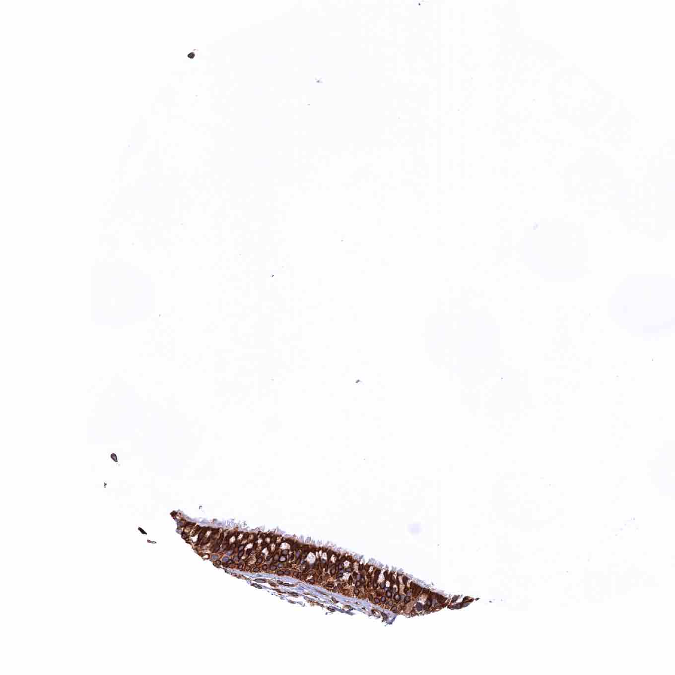
Sinus paranasales – Strong STING staining of the respiratory epithelium (STING immunohistochemistry)
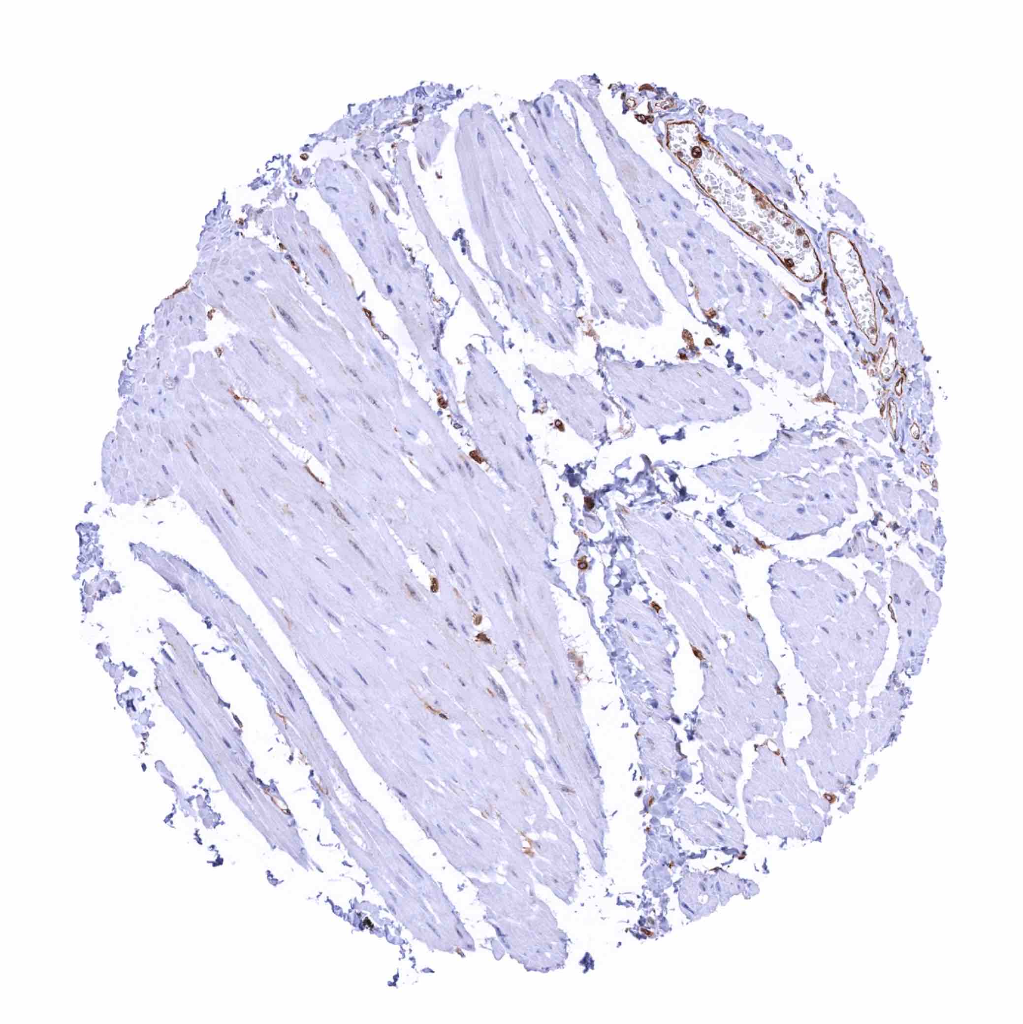
Skeletal muscle – STING staining of endothelial cells of capillaries (STING immunohistochemistry)
Skin – In this sample, a moderate STING staining is seen in the basal cell layer of the epidermis (STING immunohistochemistry)
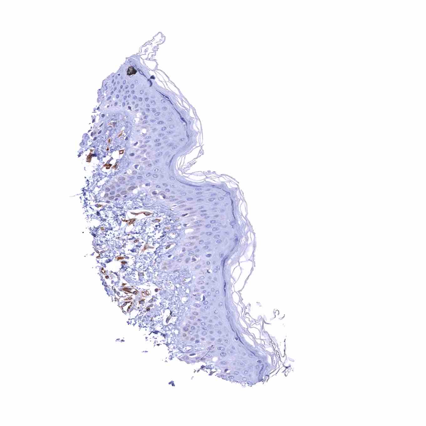
Skin – STING staining is lacking in squamous epithelial cells in this samples
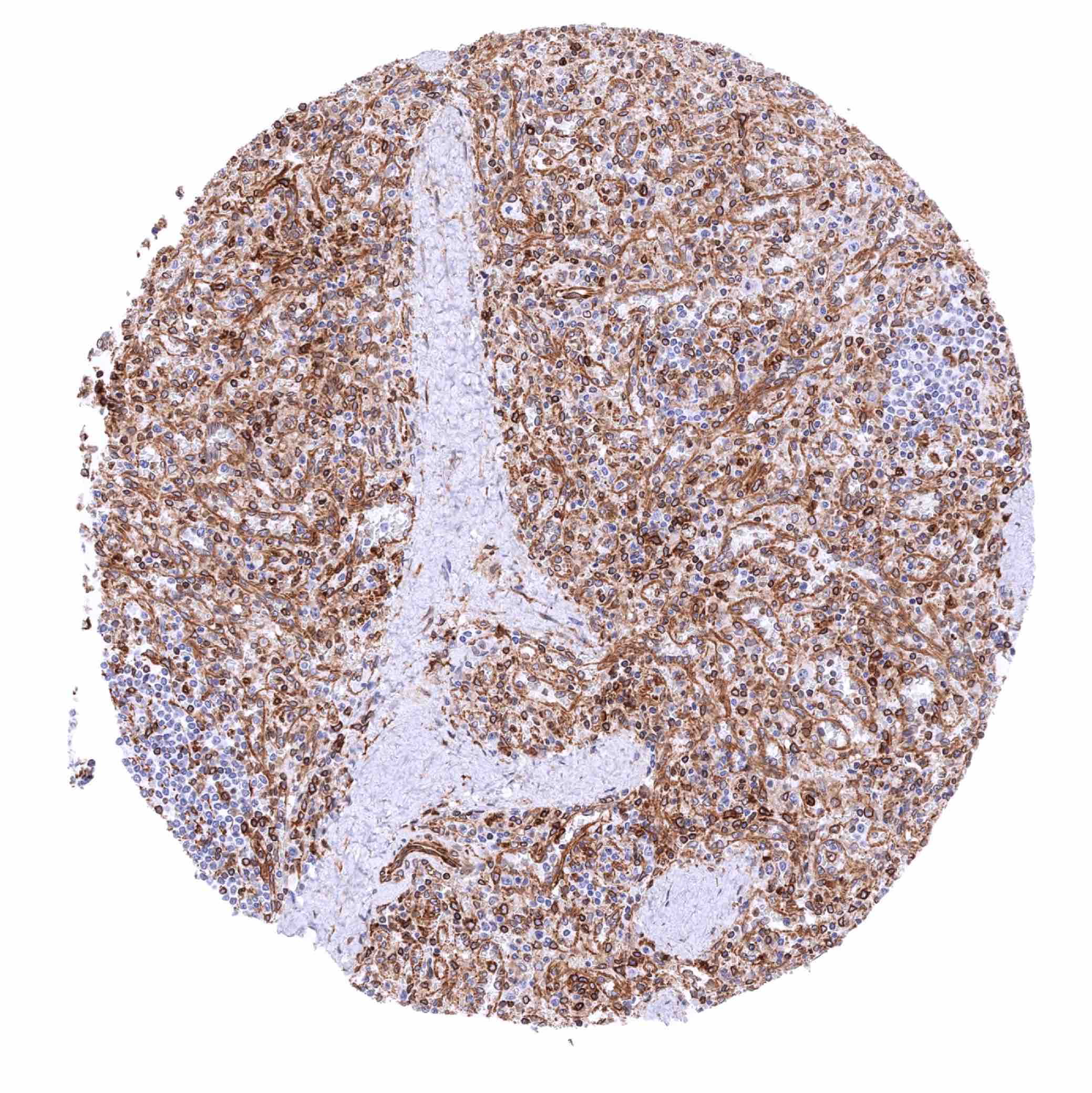
Spleen – Strong STING staining of endothelial cells and a significant fraction of inflammatory cells (STING immunohistochemistry)
Stomach, antrum – STING staining of endothelial cells and a subset of lymphocytes (STING immunohistochemistry)
Stomach, corpus – STING staining of endothelial cells and a subset of lymphocytes (STING immunohistochemistry)
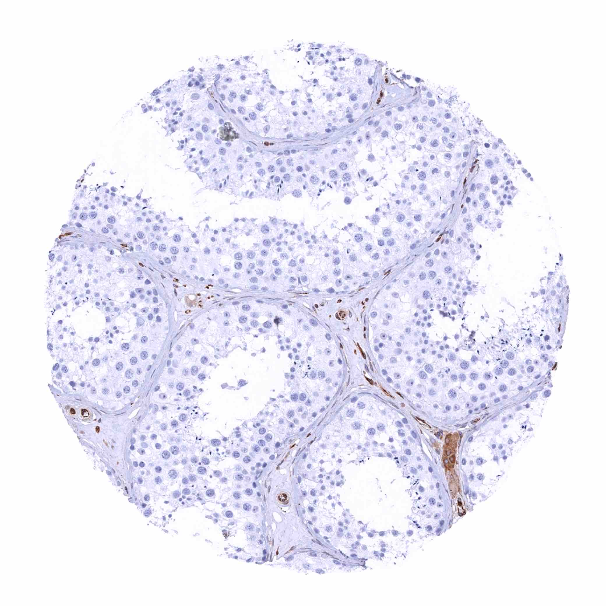
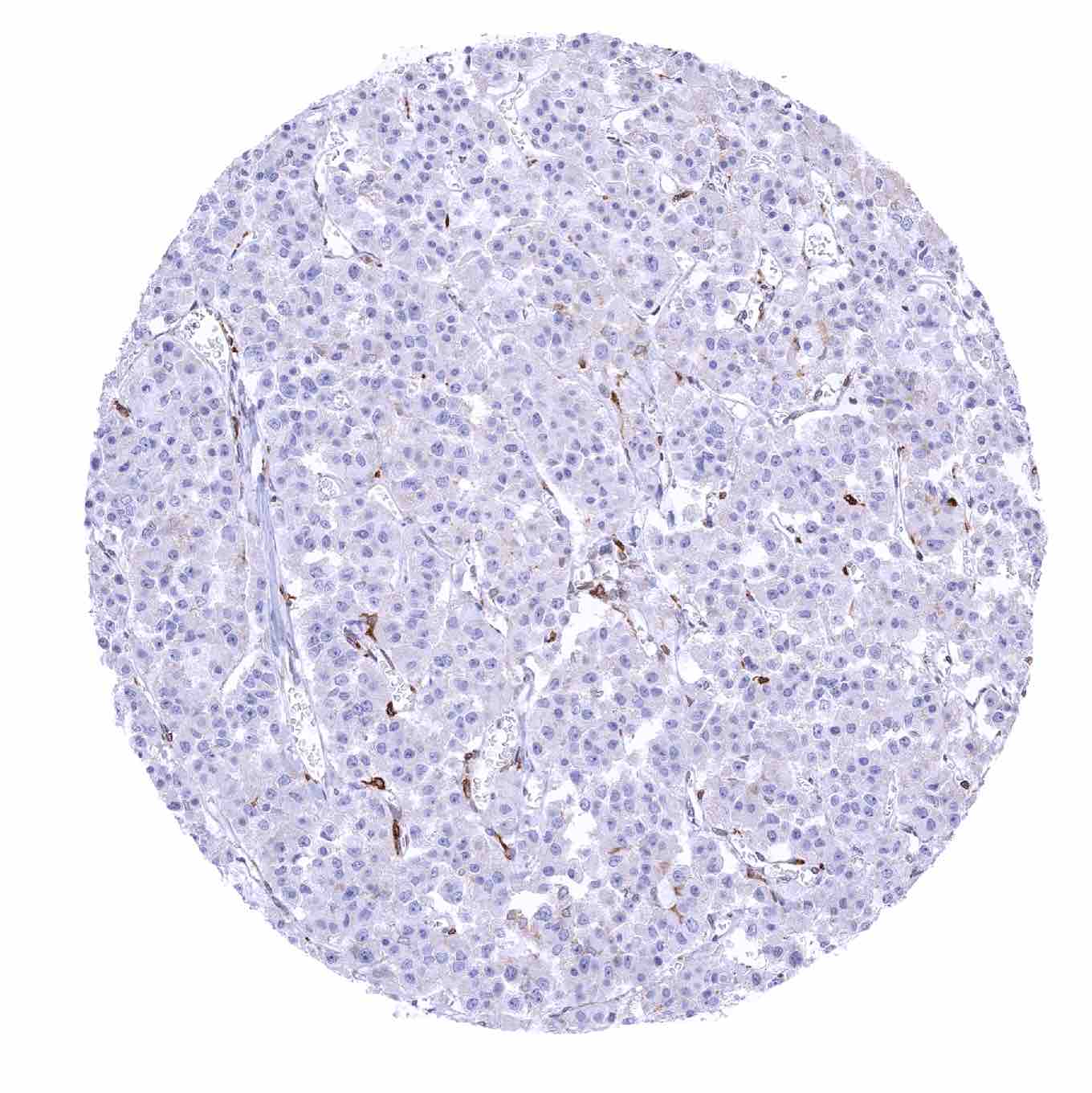
Testis – STING staining of endothelial cells and of Leydig cells (STING immunohistochemistry)

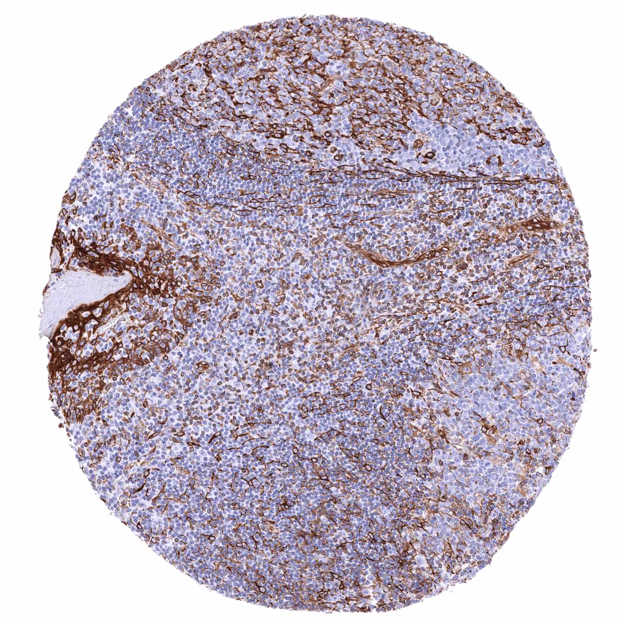
Thymus – Moderate to strong STING staining of a significant fraction of lymphocytes in the medulla but hardly in the cortex (STING immunohistochemistry)
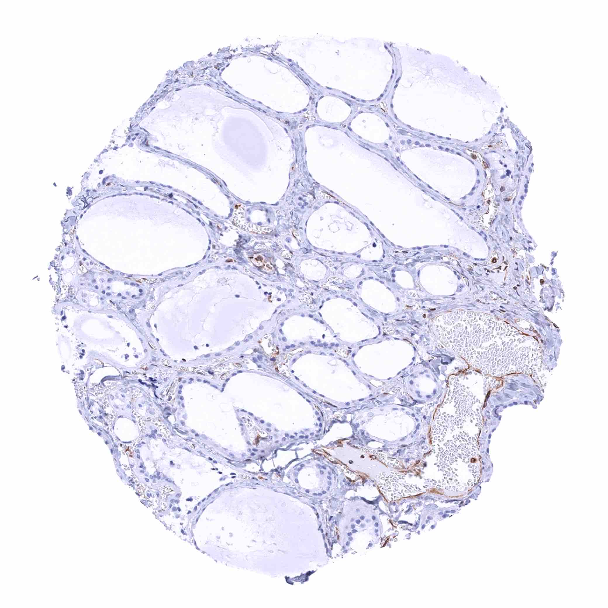
Thyroid gland
Tonsil – Moderate to strong STING staining of a fraction of lymphocytes, endothelial cells and of most cells of crypt epithelium (STING immunohistochemistry)
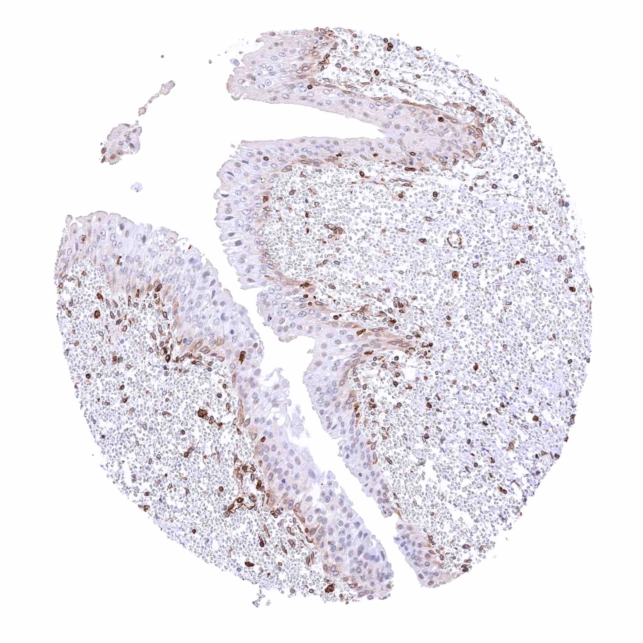
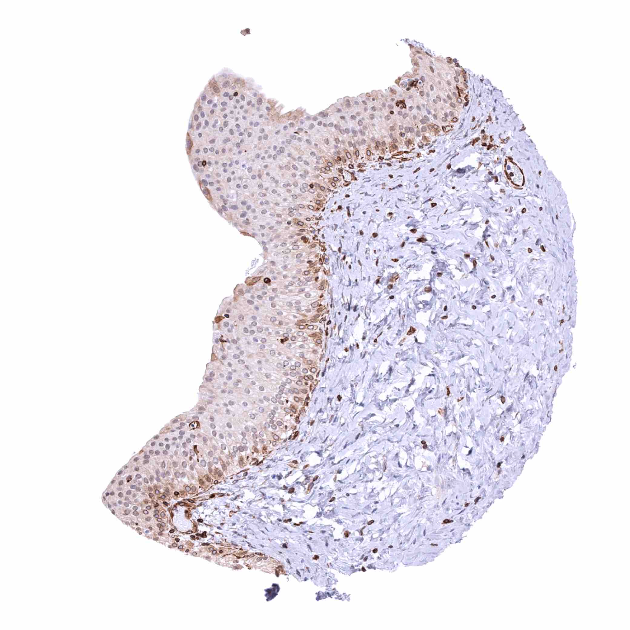
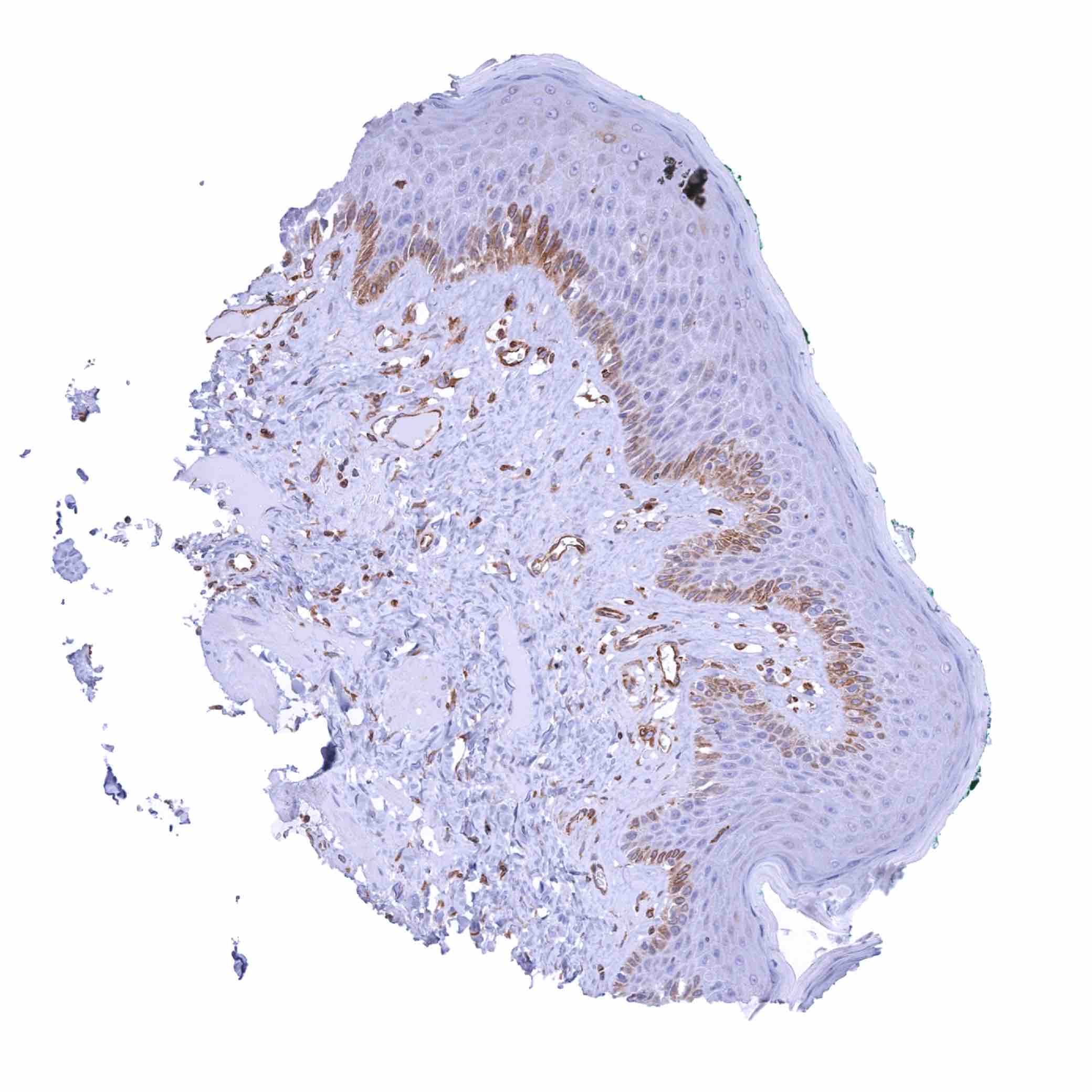
Tonsil, surface epithelium – Moderate to strong STING staining of the bottom 2-3 cell layers of the squamous epithelium (STING immunohistochemistry)

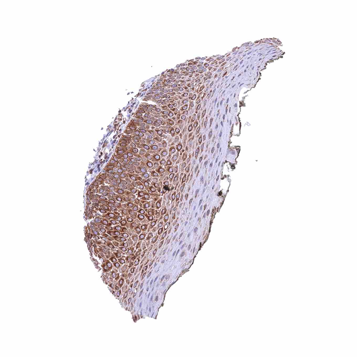
Tonsil, surface epithelium – Strong STING staining of all cell layers of the squamous epithelium (STING immunohistochemistry)
Urinary bladder, muscular wall
Urinary bladder, urothelium – Strong STING staining of the urothelium, especially in the basal 2-3 of the cell layers (STING immunohistochemistry)

Urinary bladder, urothelium – Strong STING staining of the urothelium, especially in umbrella cells (STING immunohistochemistry)
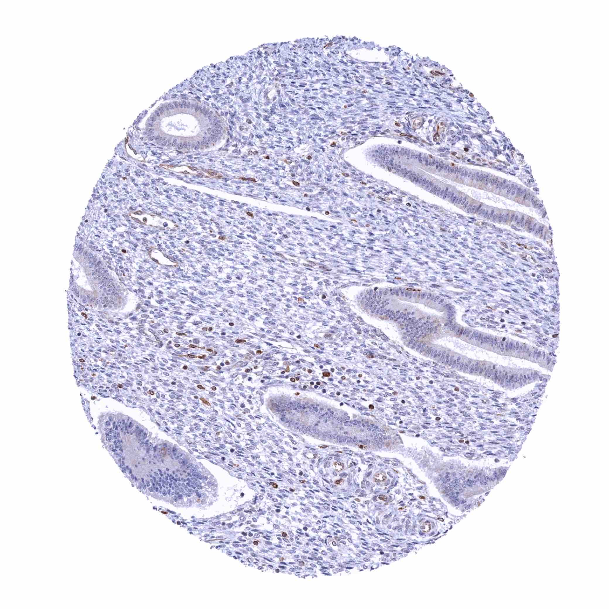
Urinary bladder, urothelium – Weak STING staining of the basal cell layer of the urothelium in this sample (STING immunohistochemistry)

Uterus, ectocervix – Weak STING staining of the basal cell layer of the squamous epithelium (STING immunohistochemistry)
Uterus, endocervix – Significant STING staining of epithelial cells (STING immunohistochemistry)
Uterus, endometrium (pregnancy) – Strong STING staining of inflammatory cells while only few decidua cells show a rather weak staining (STING immunohistochemistry)
Uterus, endometrium (proliferation) – Significant STING staining of epithelial cells and of stroma cells in this sample (STING immunohistochemistry)
Uterus, endometrium (proliferation) – Significant STING staining of inflammatory cells while epithelial and stromal cells are largely negative in this sample (STING immunohistochemistry)
Uterus, endometrium (proliferation) – STING staining of only few epithelial cells but of all stroma cells in this sample (STING immunohistochemistry)

Uterus, endometrium (secretion) – Strong STING staining of only few epithelial cells but moderate staining of most stroma cells in this sample (STING immunohistochemistry)
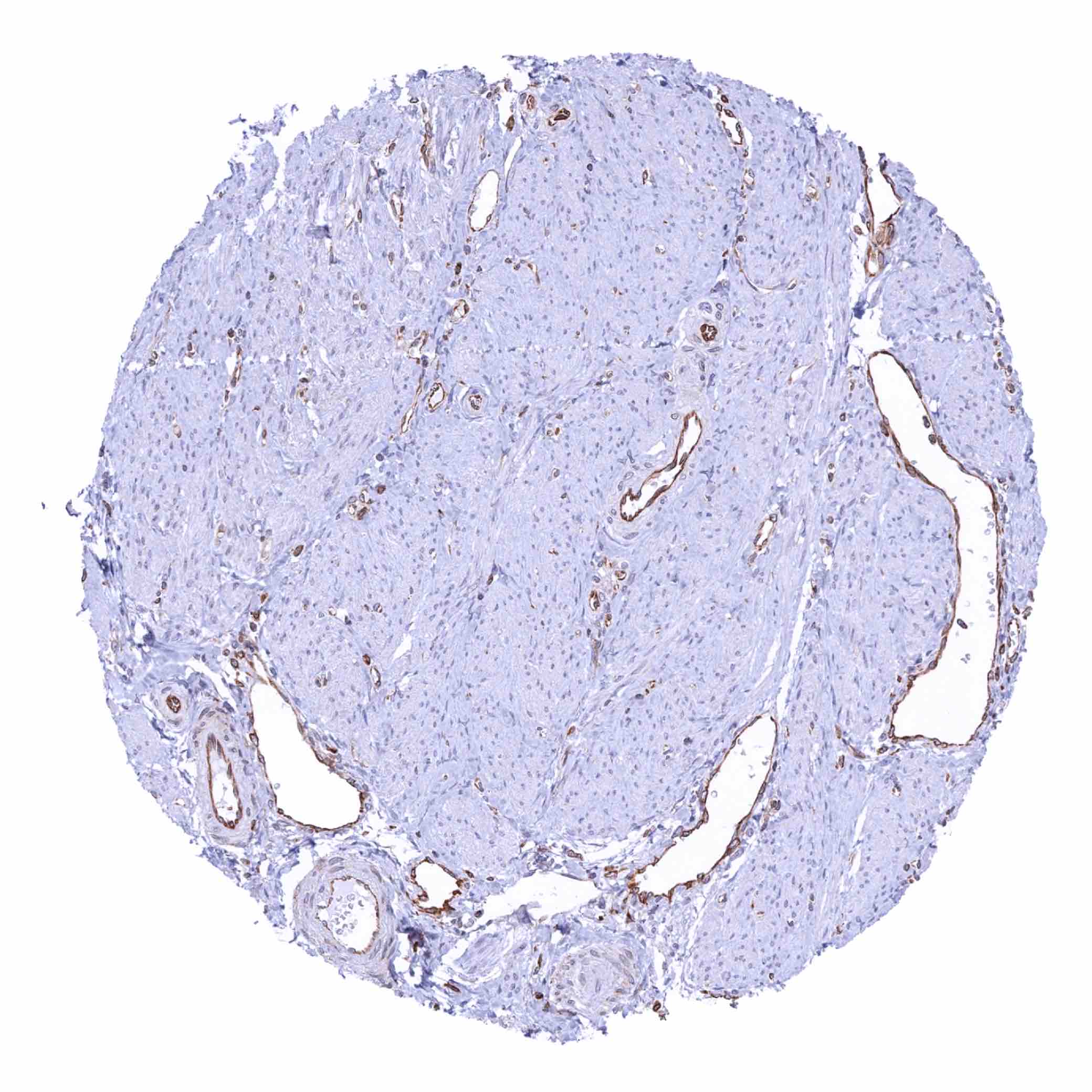
Uterus, myometrium – STING staining of endothelial cells (STING immunohistochemistry)