Adrenal gland
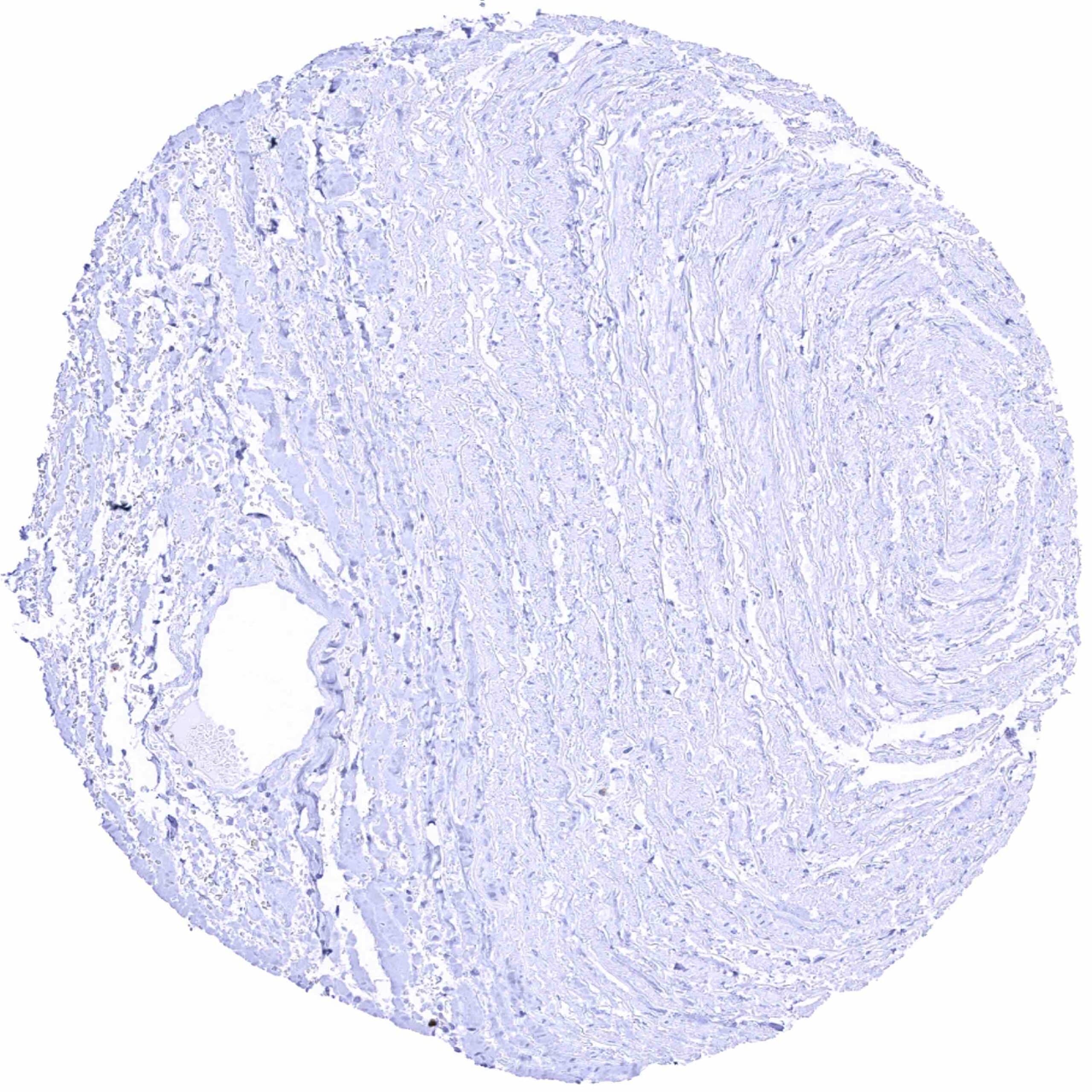
Aorta, media
Appendix, mucosa
Appendix, muscular wall
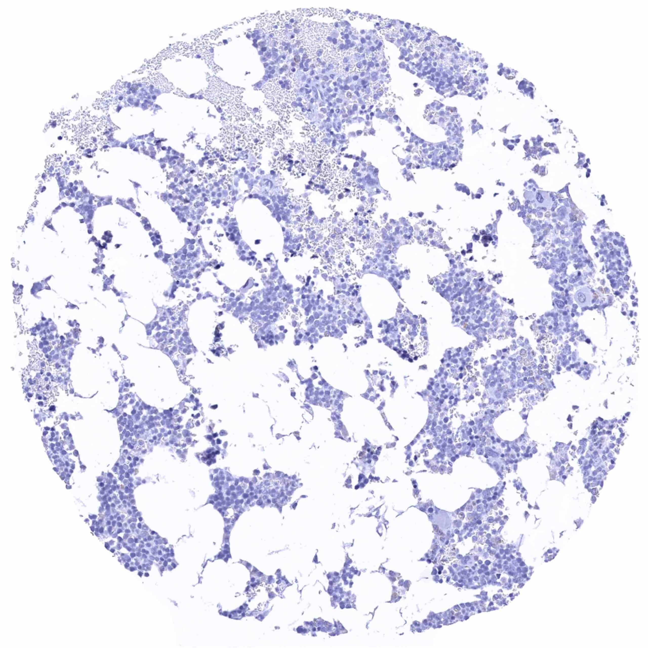
Bone marrow
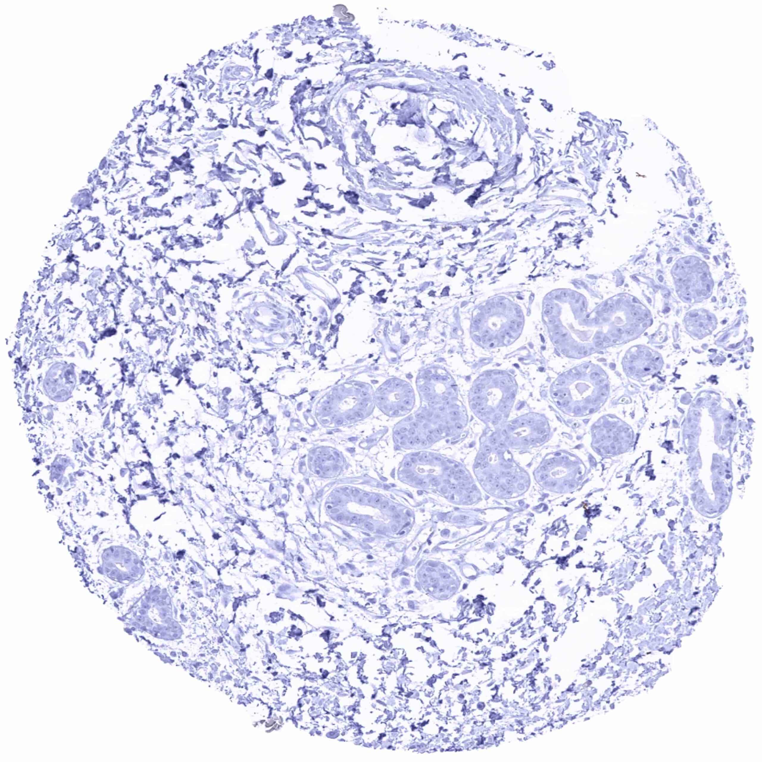
Breast
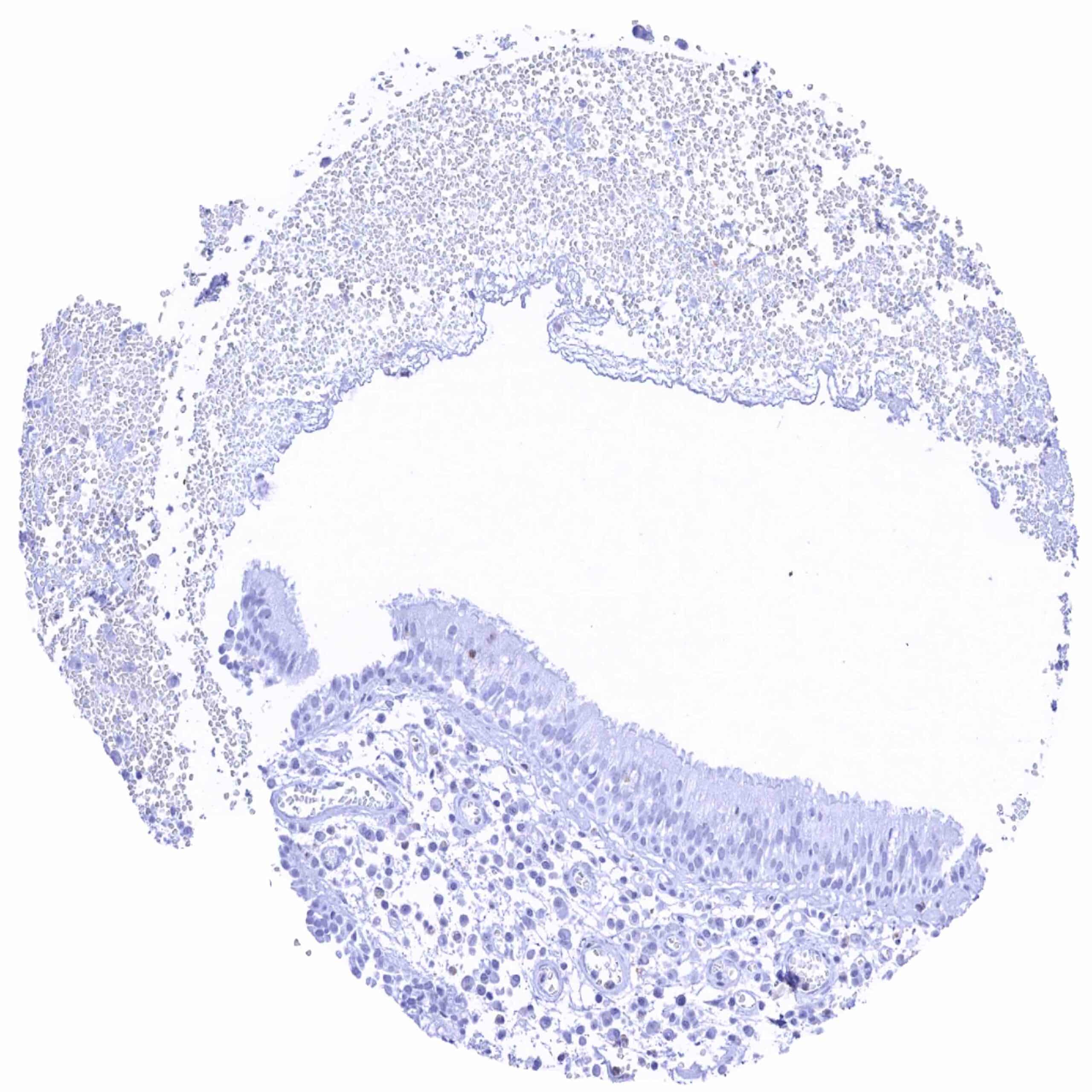
Bronchus, mucosa
Cerebellum (white matter)
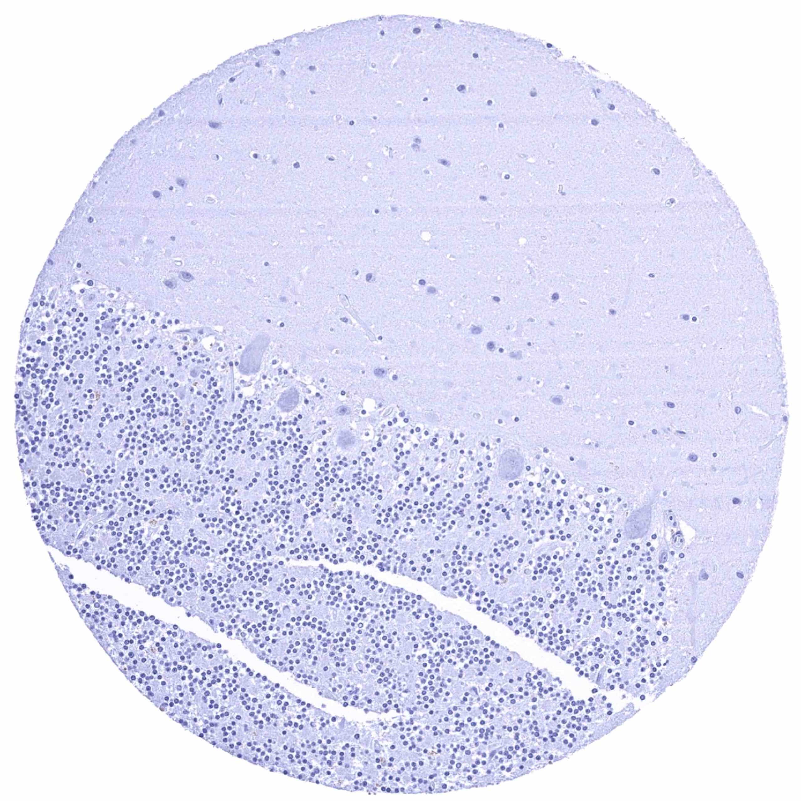
Cerebellum, cortex (molecular layer, Purkinje cell layer, granule cell layer)
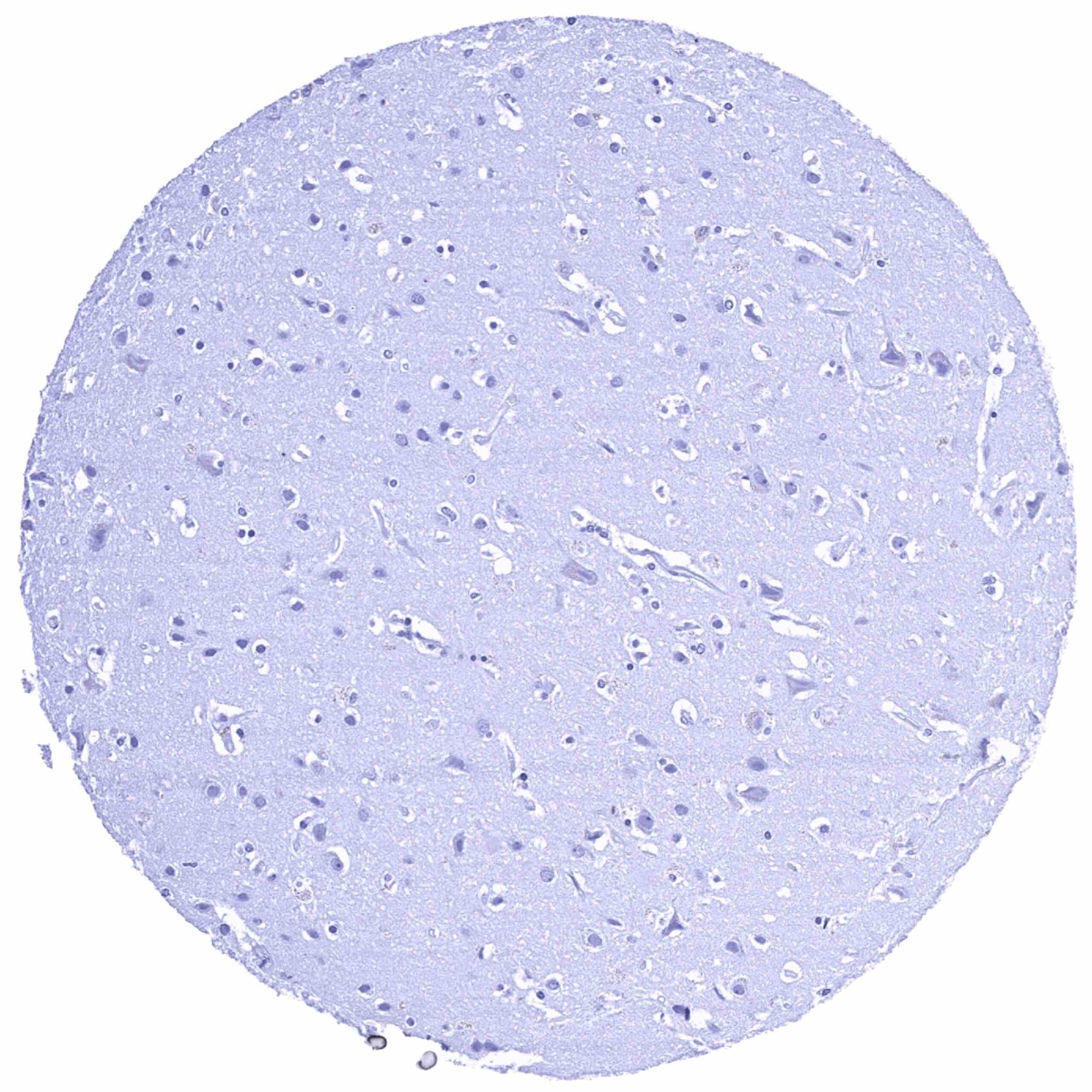
Cerebrum, grey matter
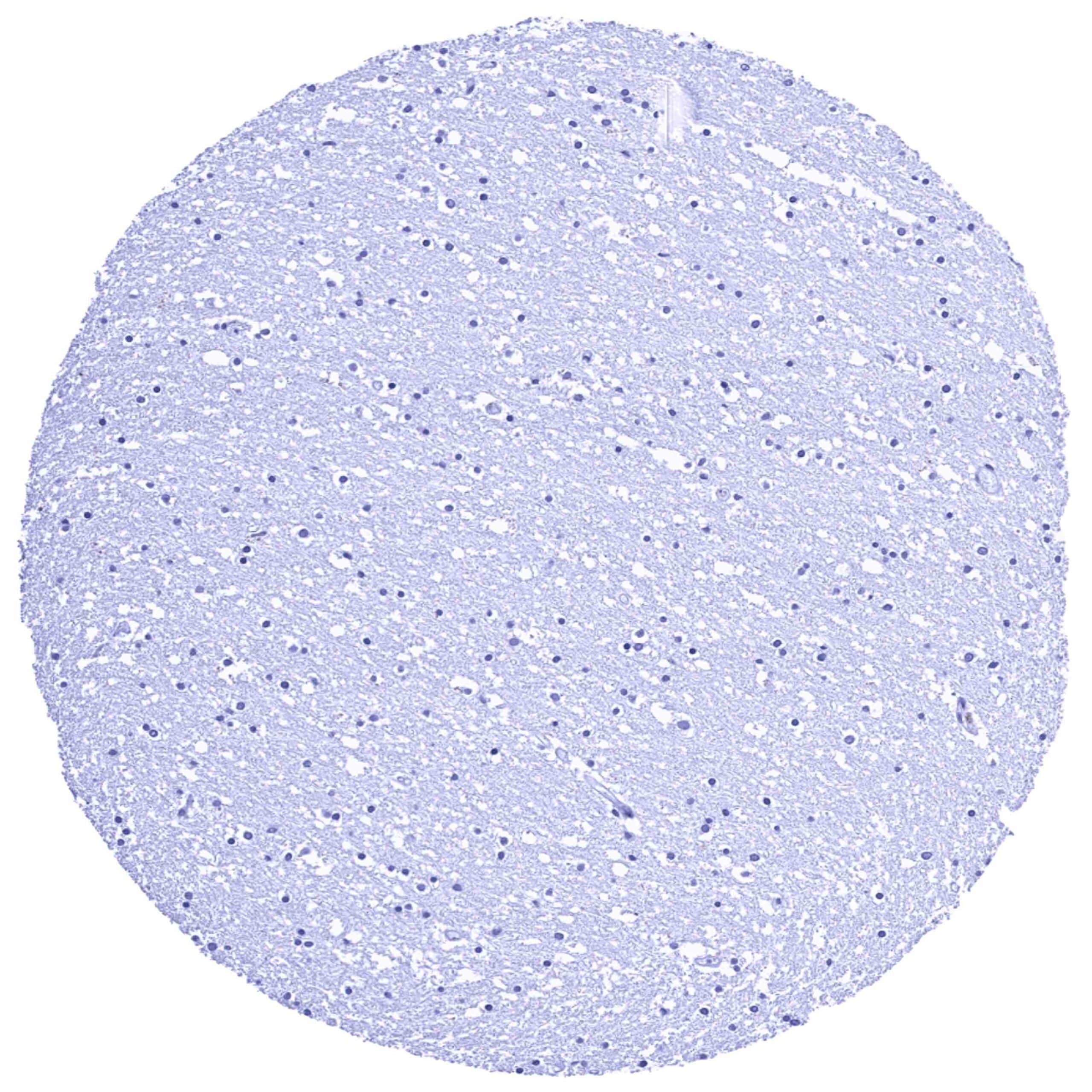
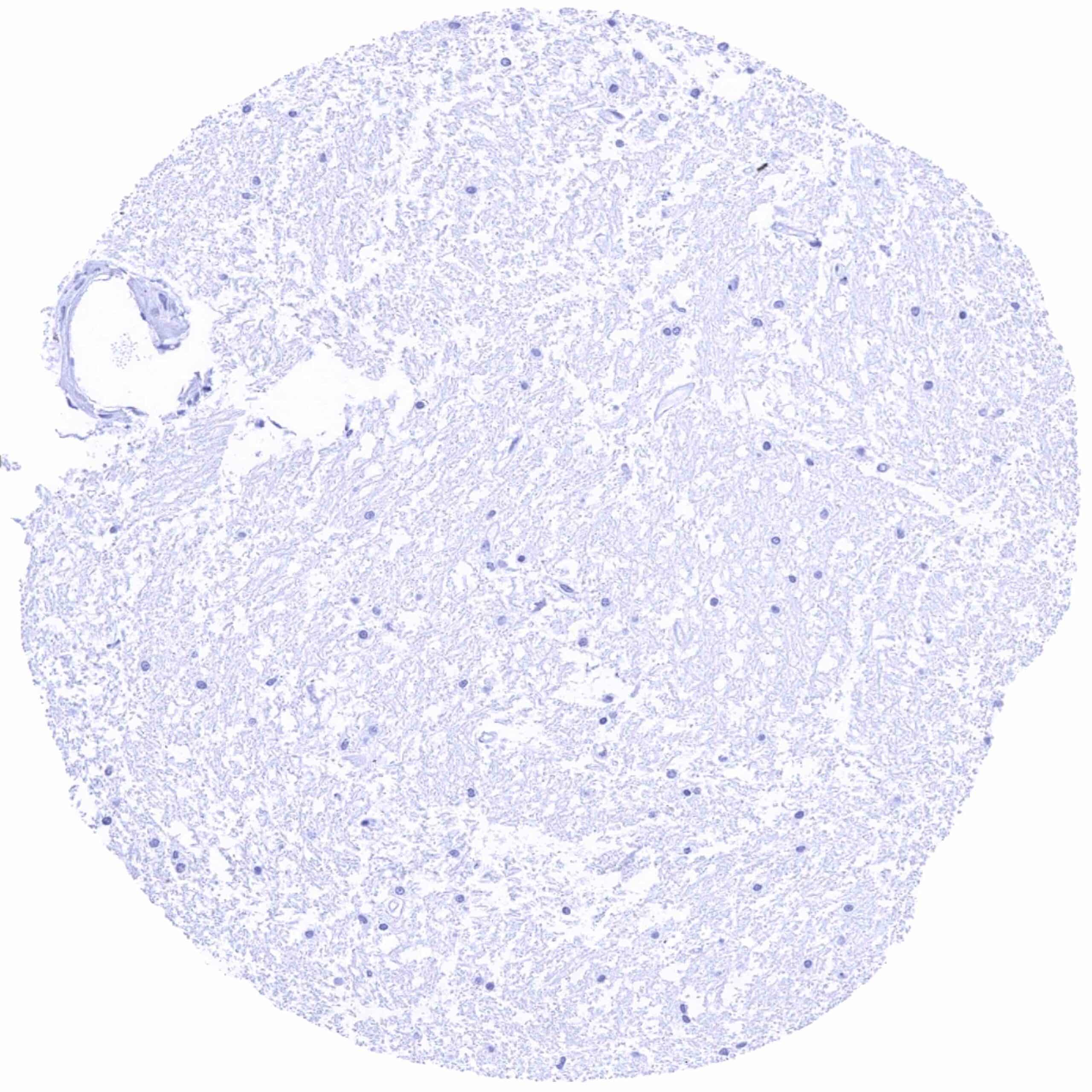
Cerebrum, white matter
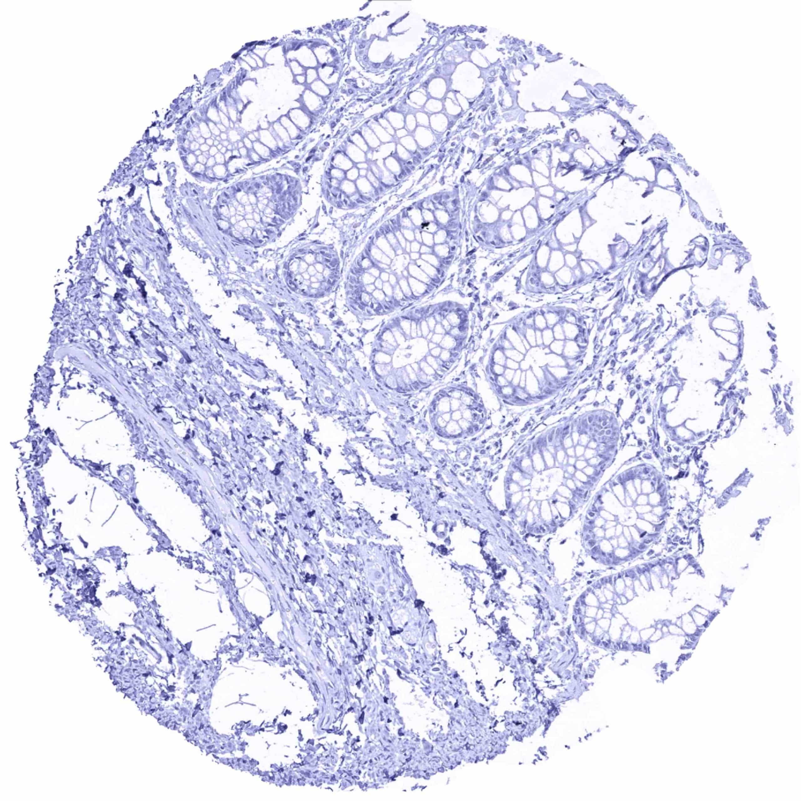
Colon descendens, mucosa
Colon descendens, muscular wall
Duodenum, Brunner gland
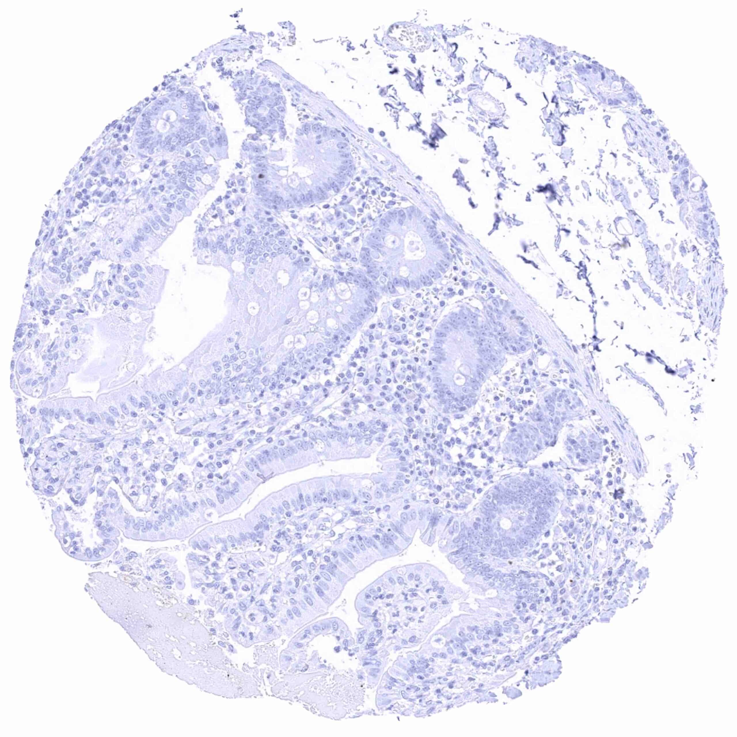
Duodenum, mucosa
Epididymis (Corpus)
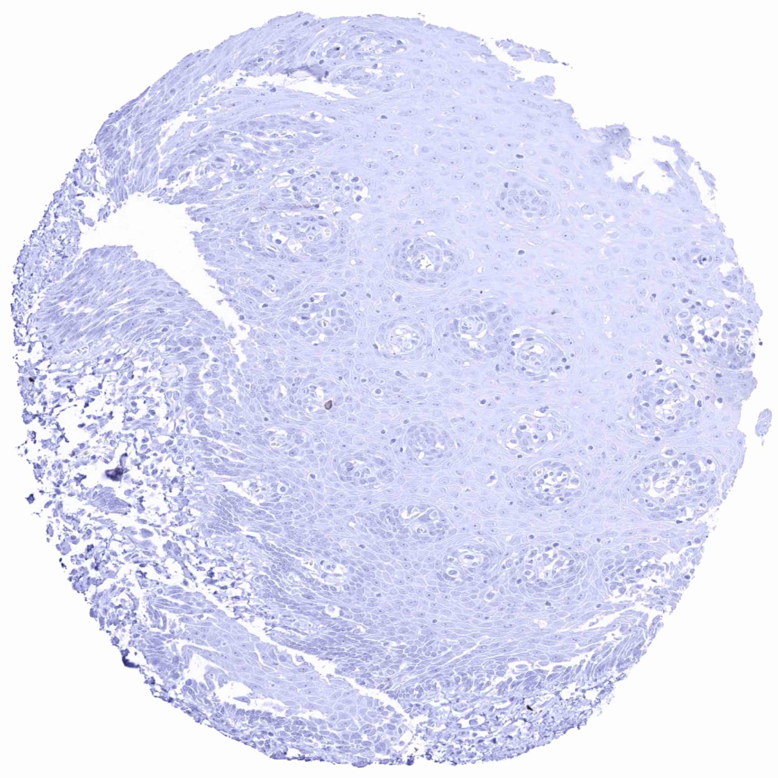
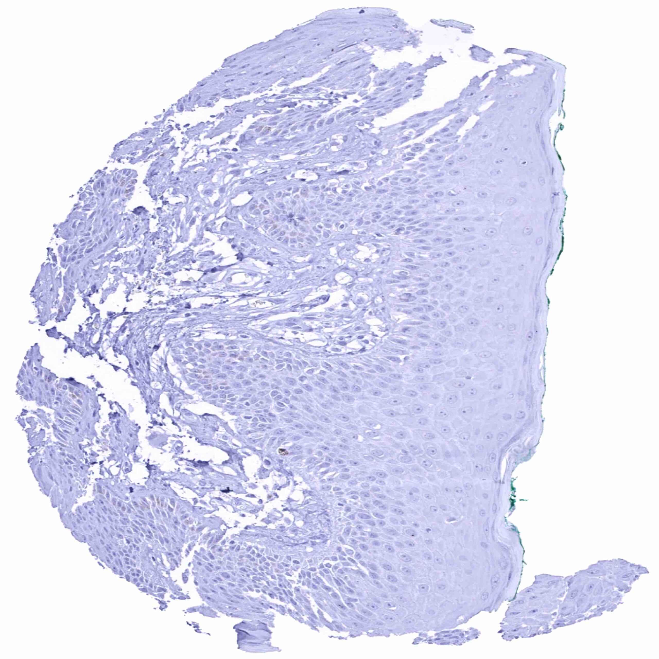
Esophagus, squamous epithelium
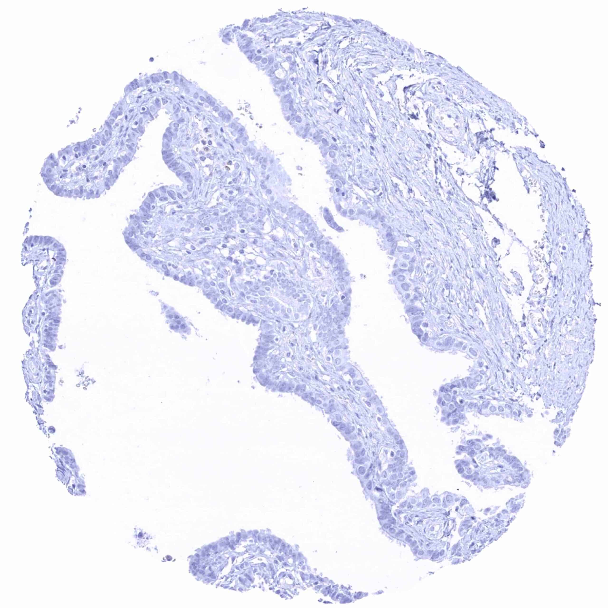
Fallopian tube, mucosa
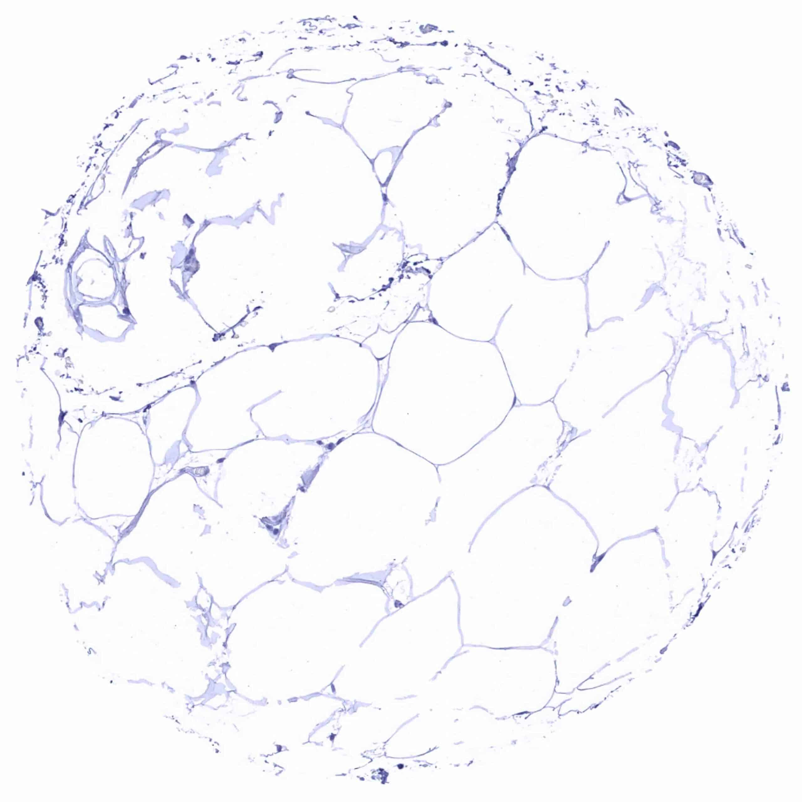
Fat
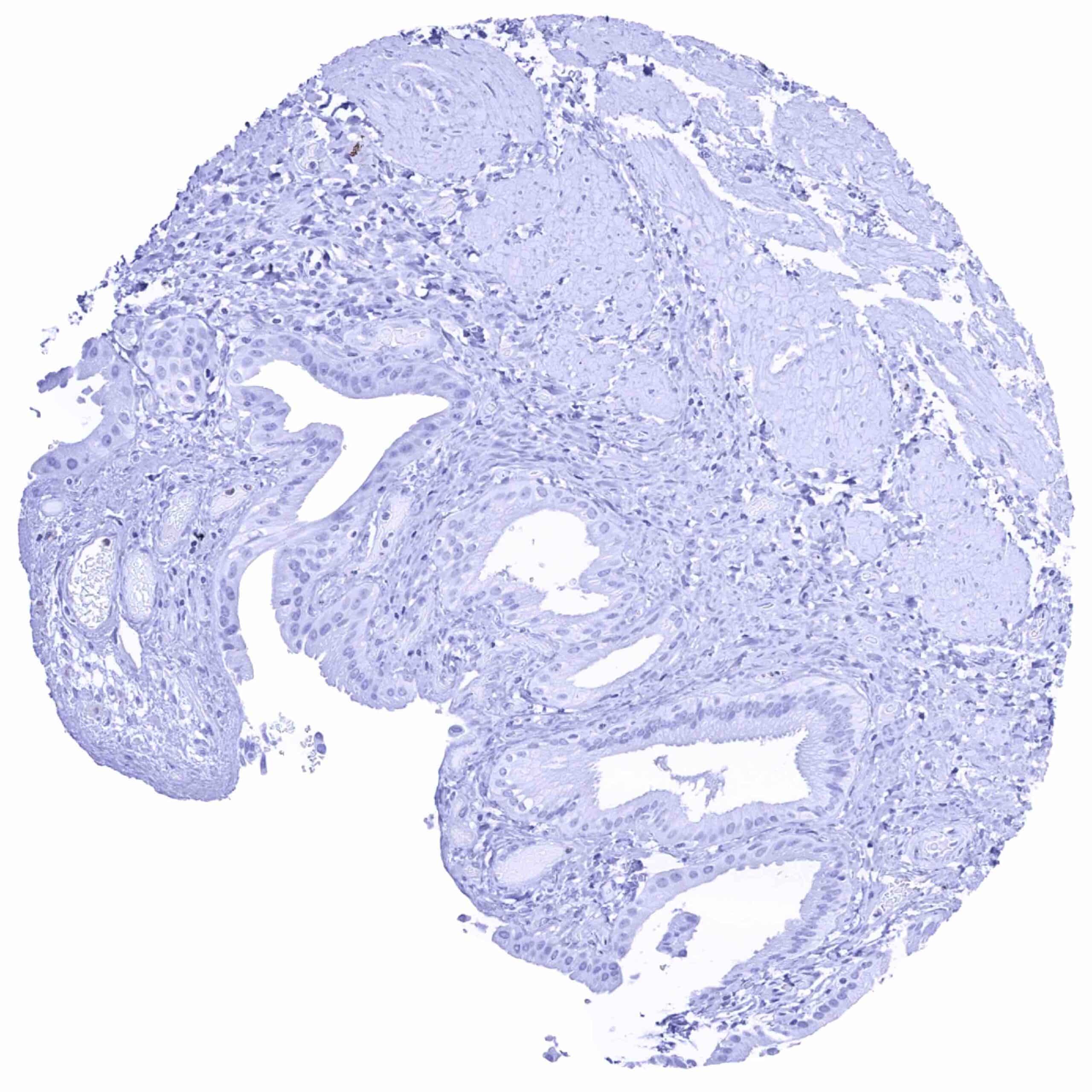
Gallbladder, epithelium
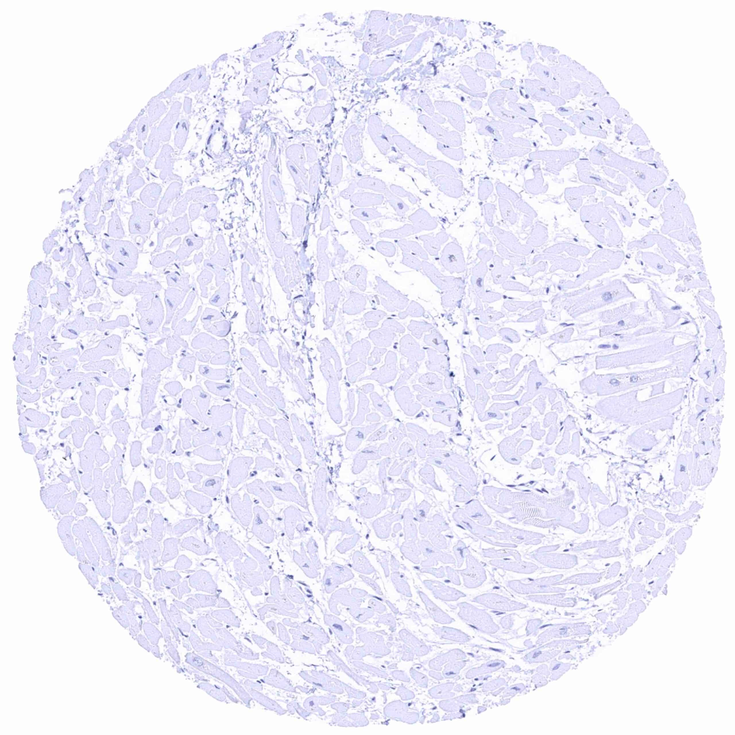
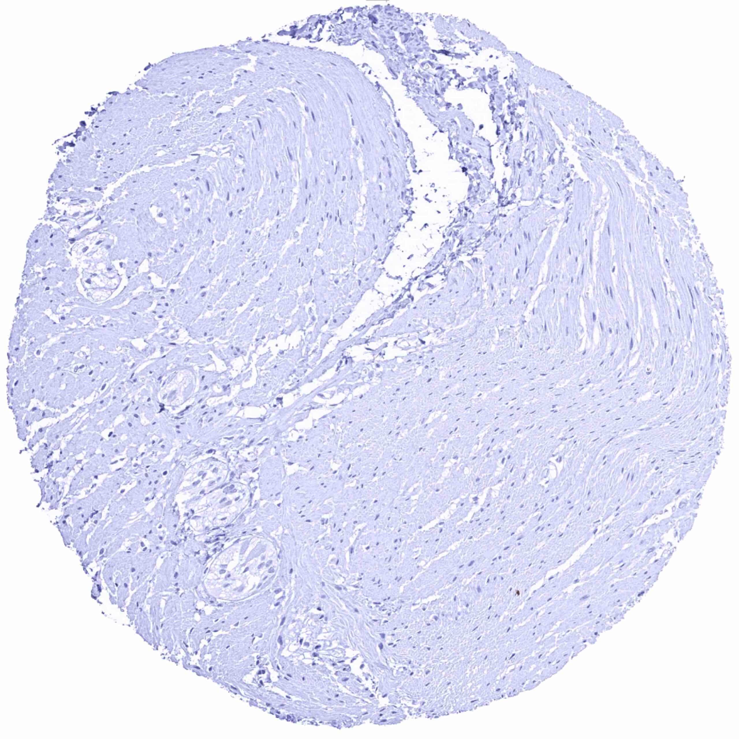
Heart muscle
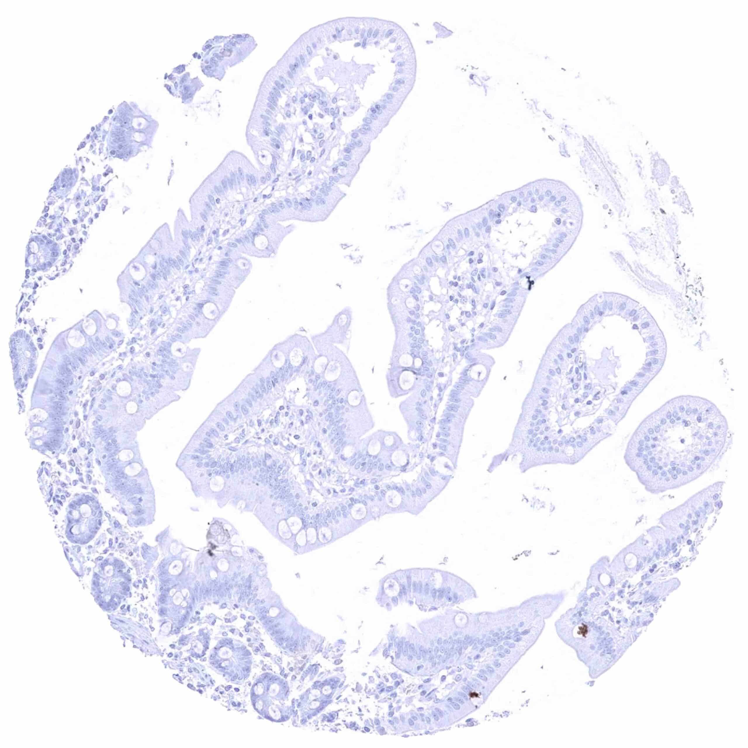
Ileum, mucosa
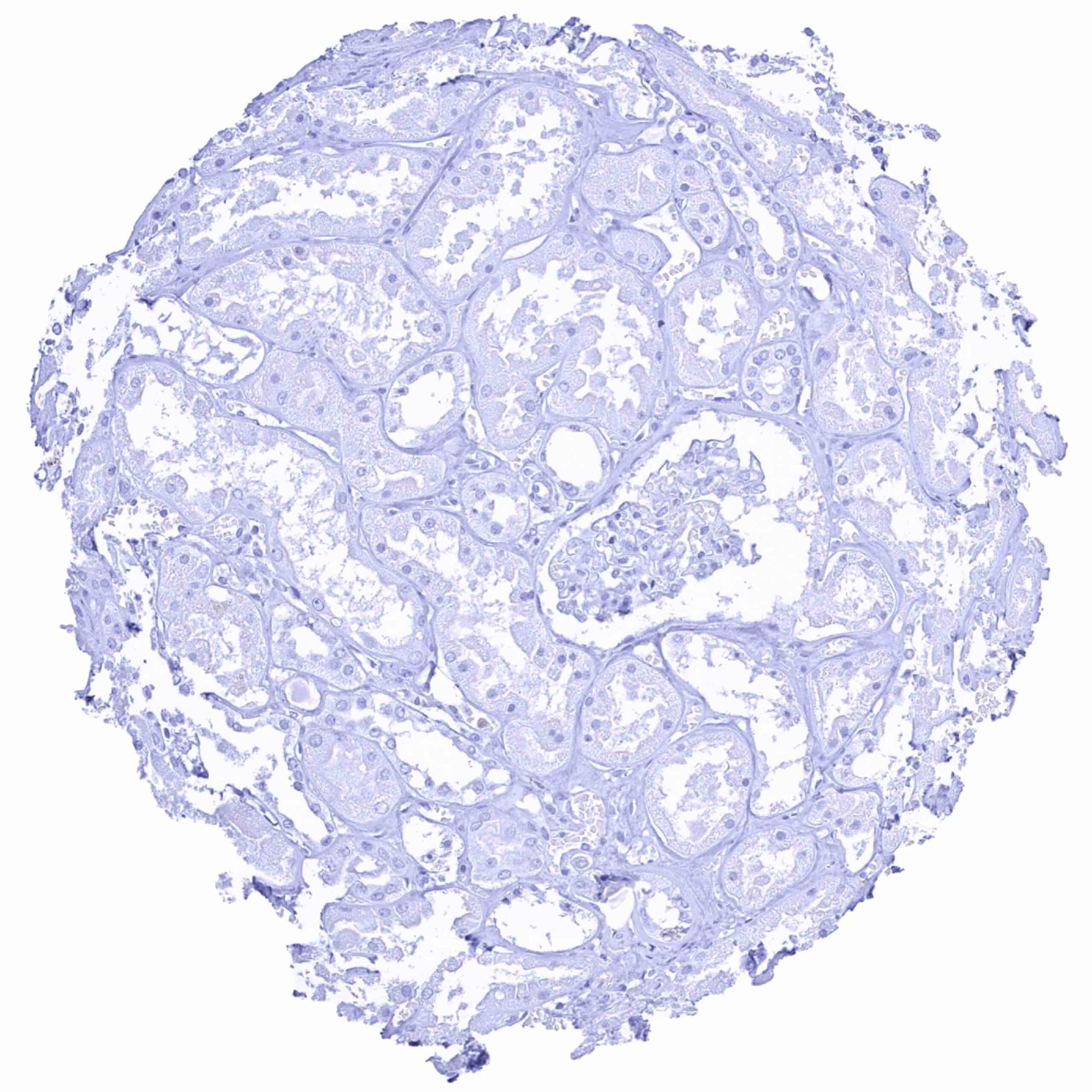
Kidney, cortex
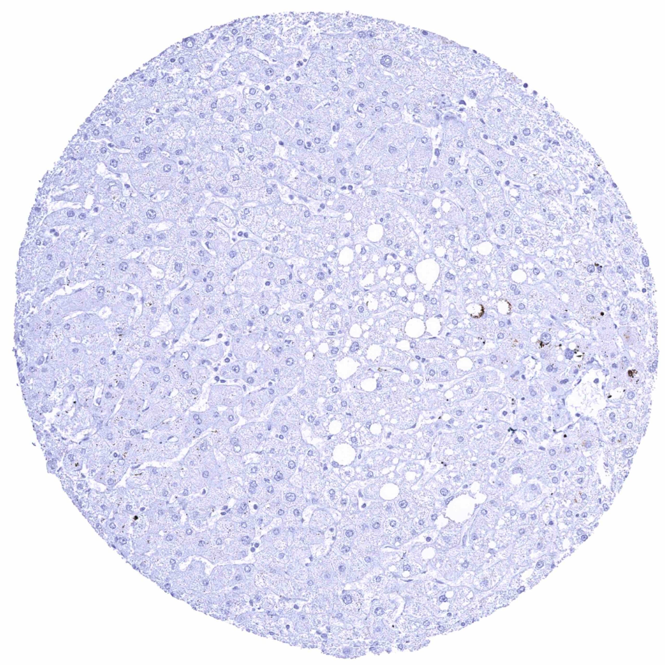
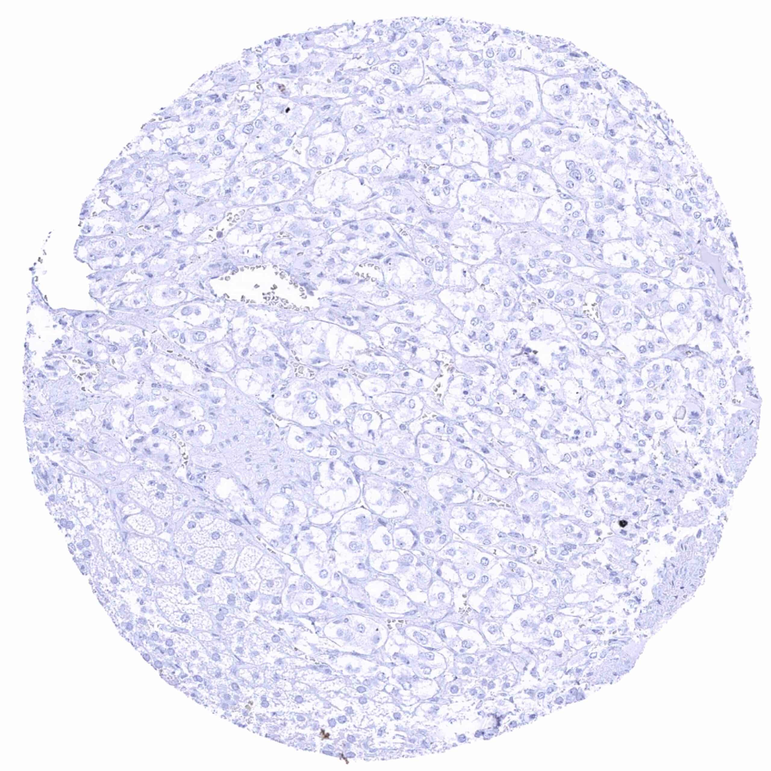
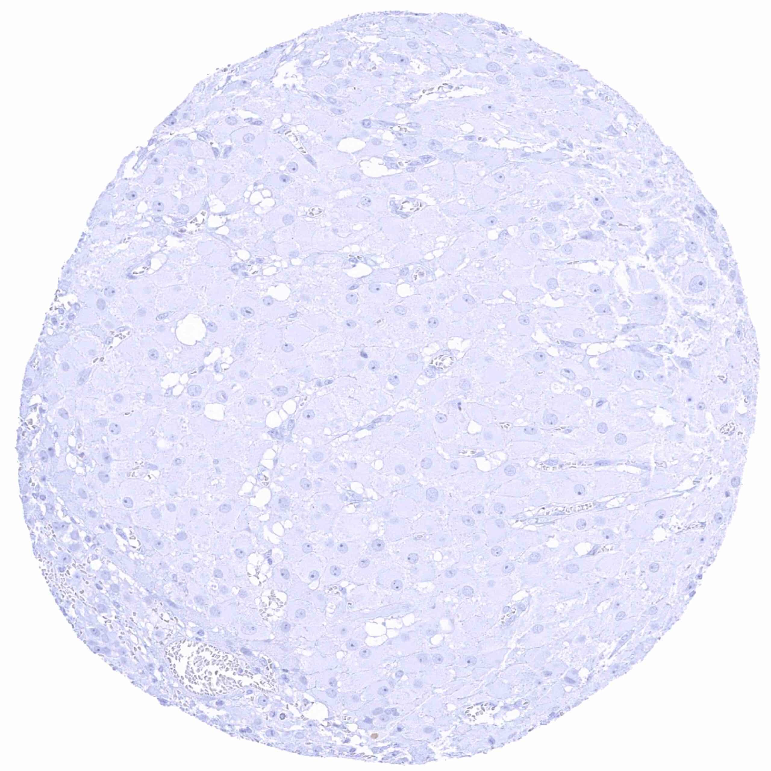
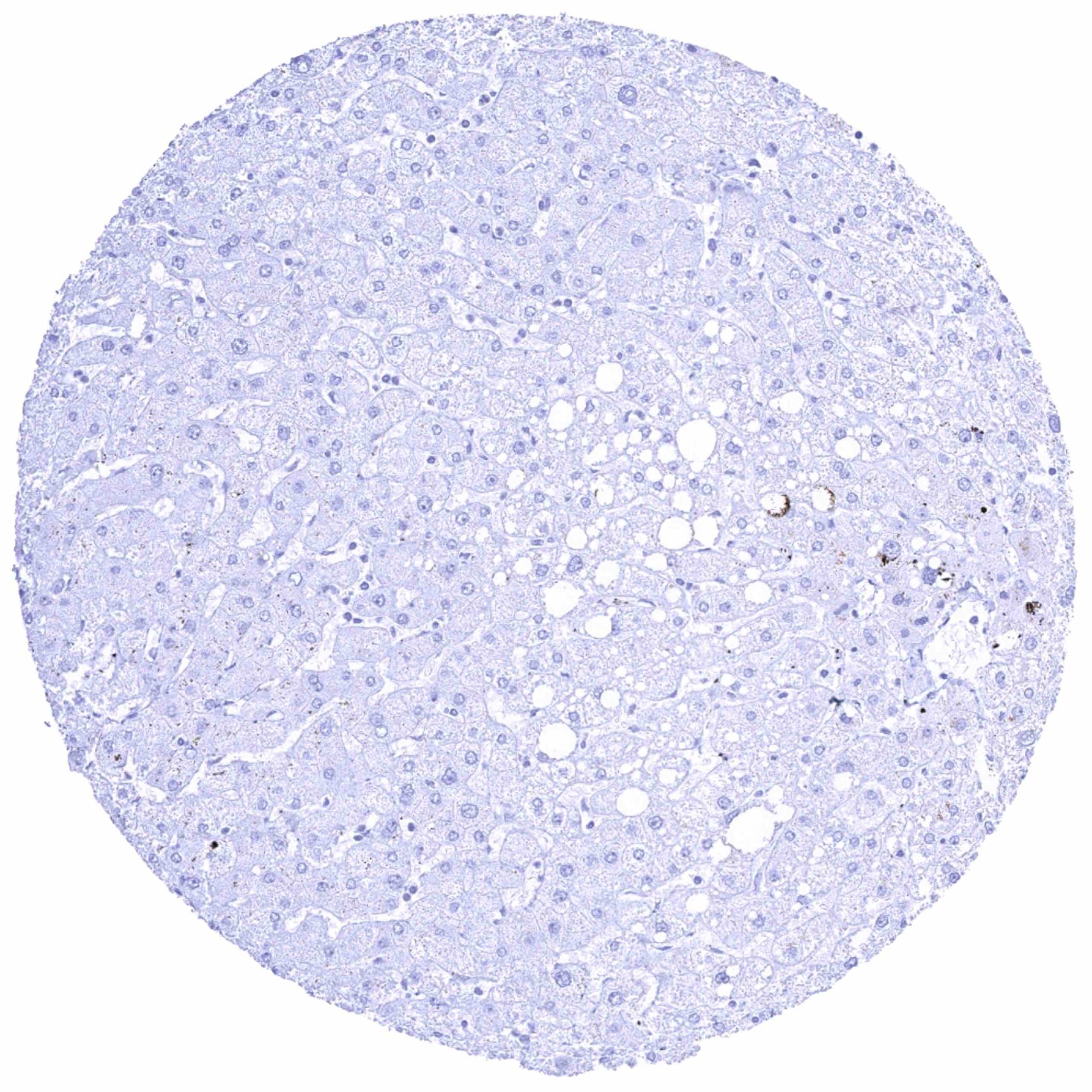
Liver
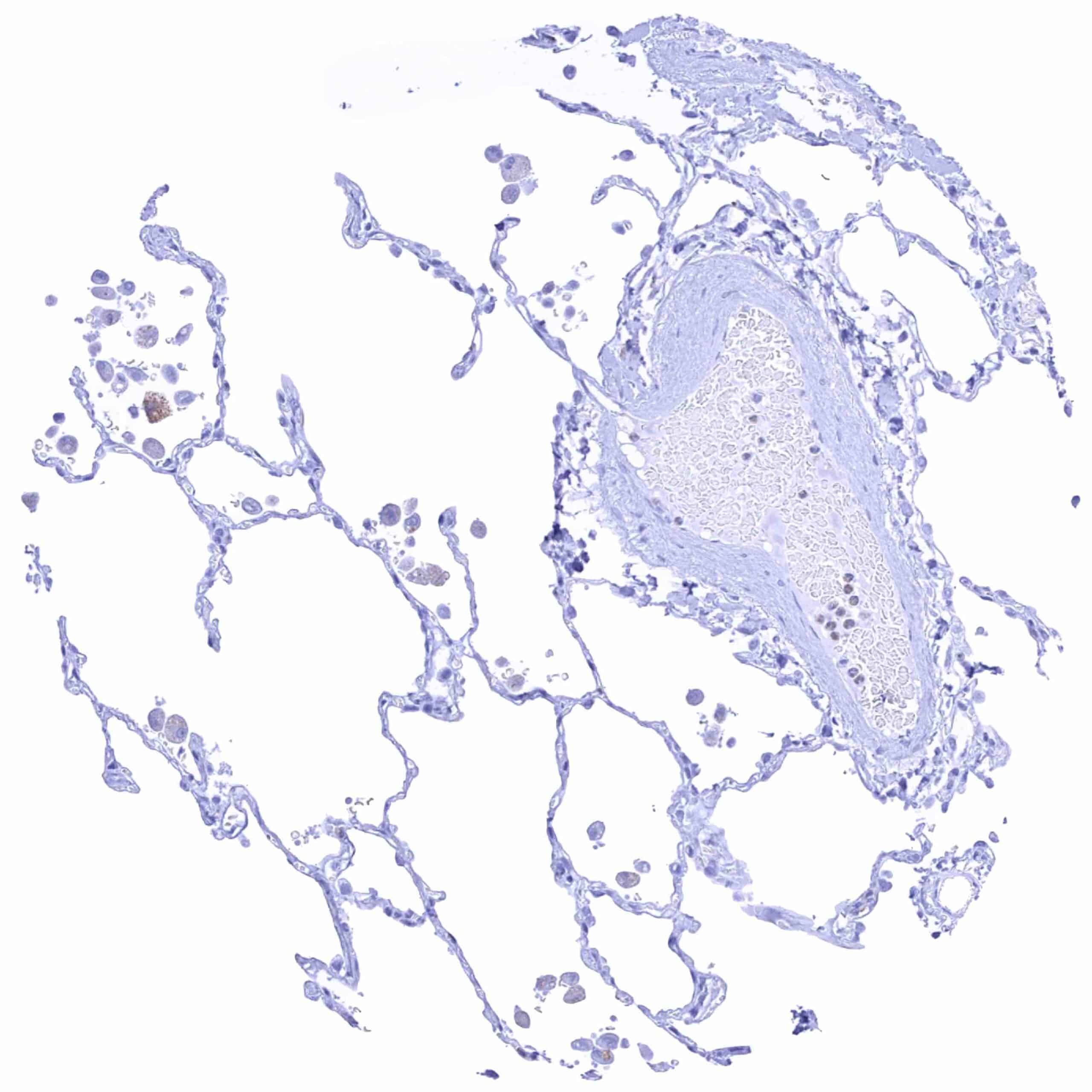
Lung
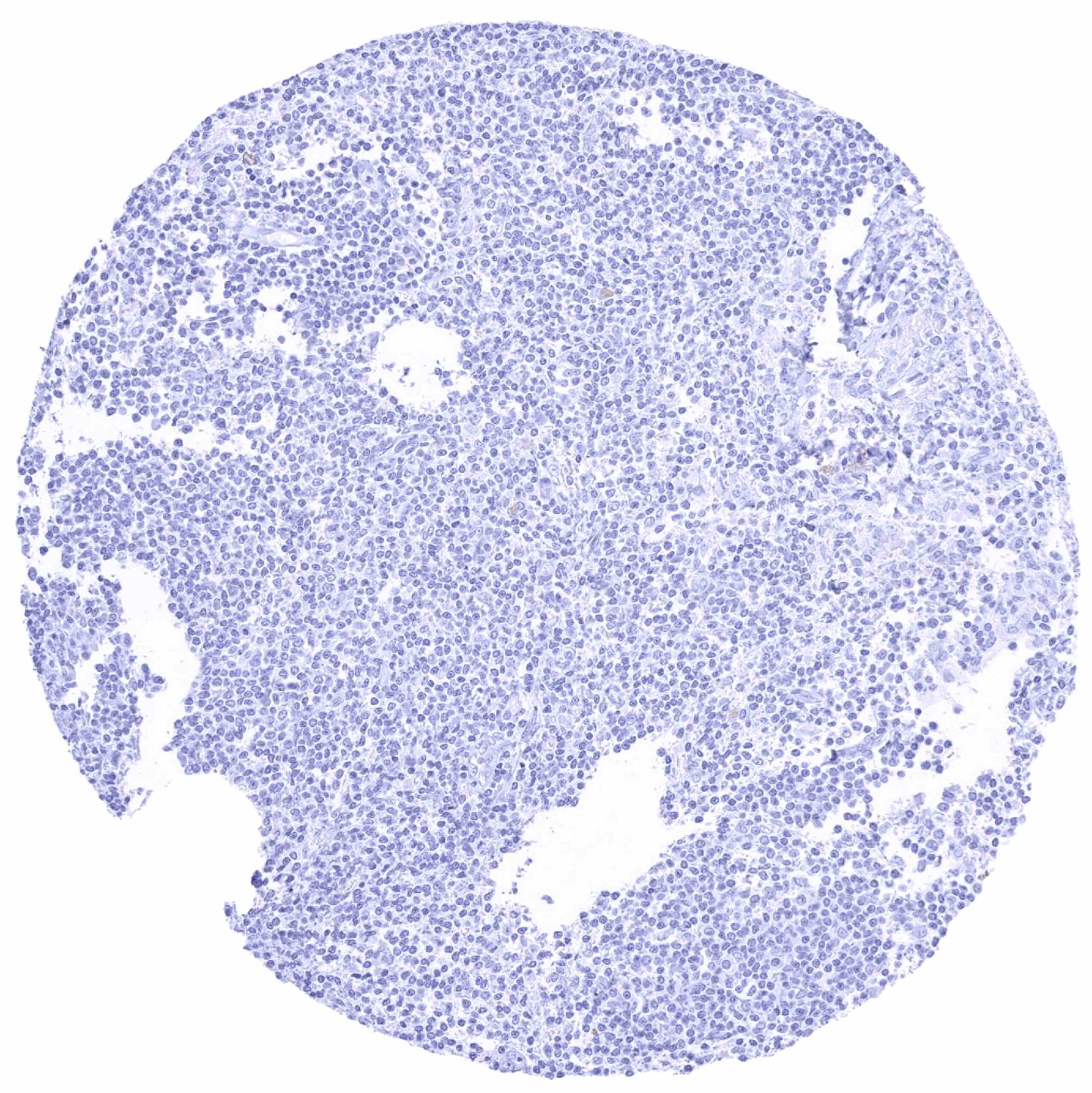
Lymph node
Ovary, corpus luteum
Ovary, stroma
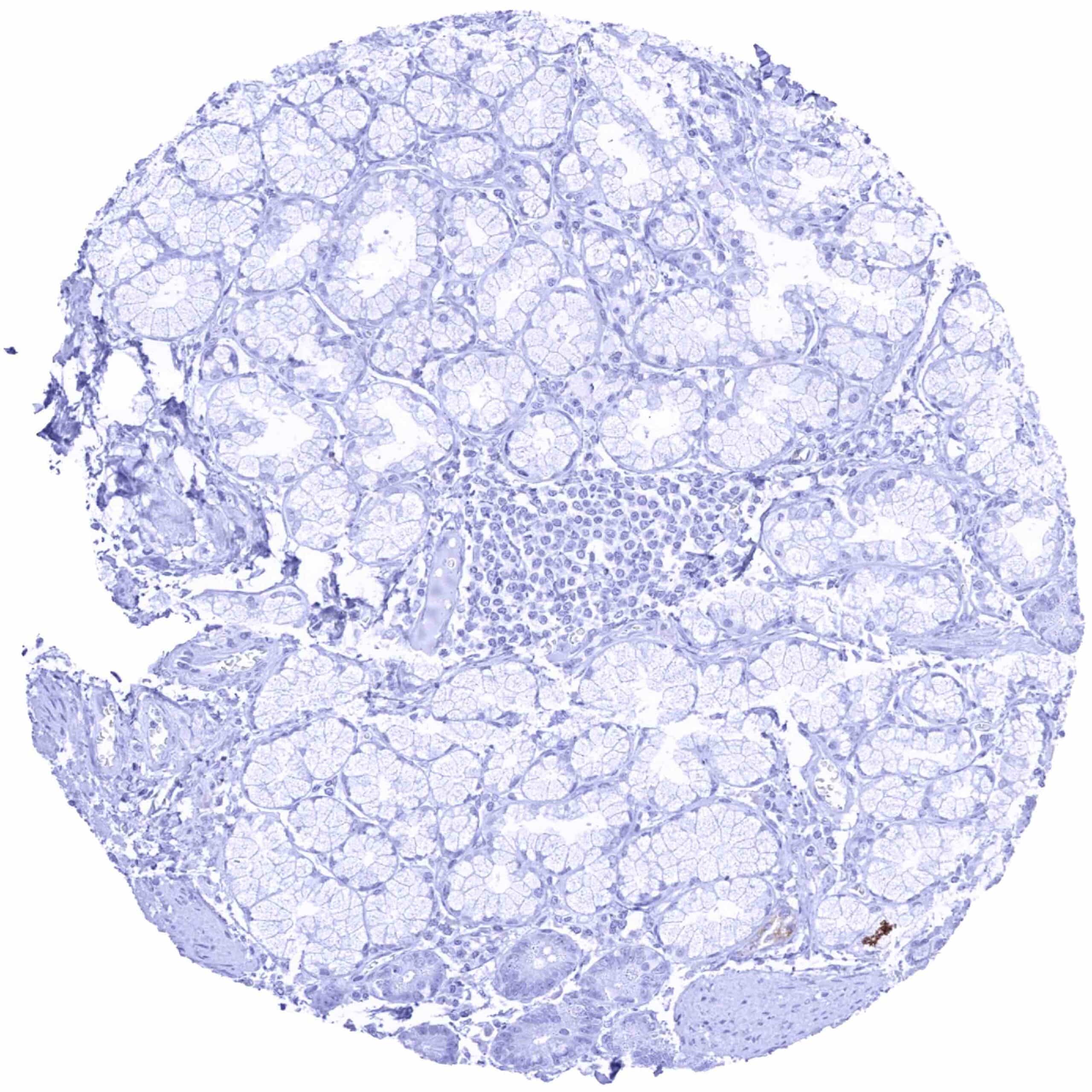
Pancreas
Parathyroid gland
Parotid gland
Pituitary gland, anterior lobe
Pituitary gland, posterior lobe
Placenta (amnion and chorion)
Placenta, early
Placenta, mature
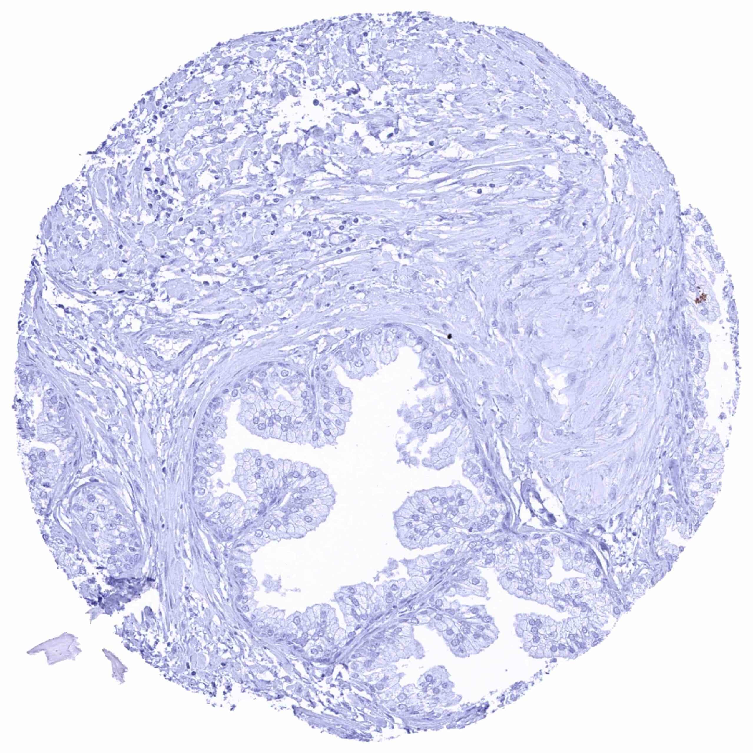
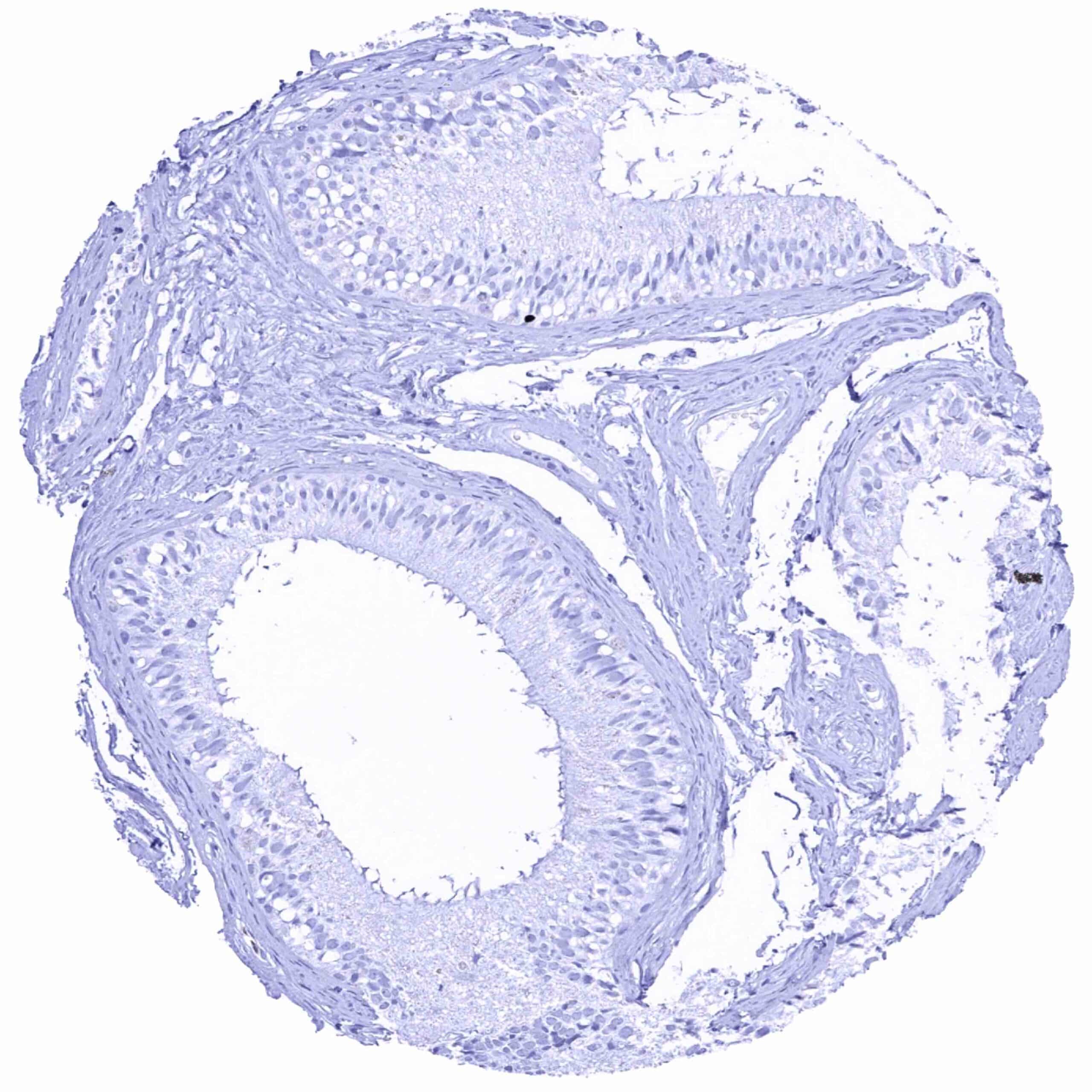
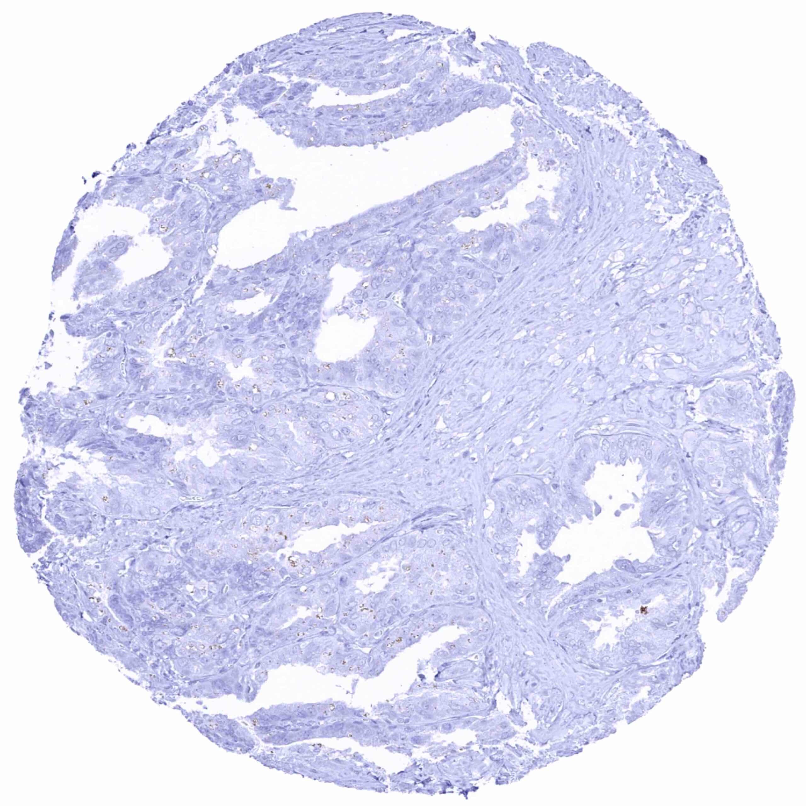
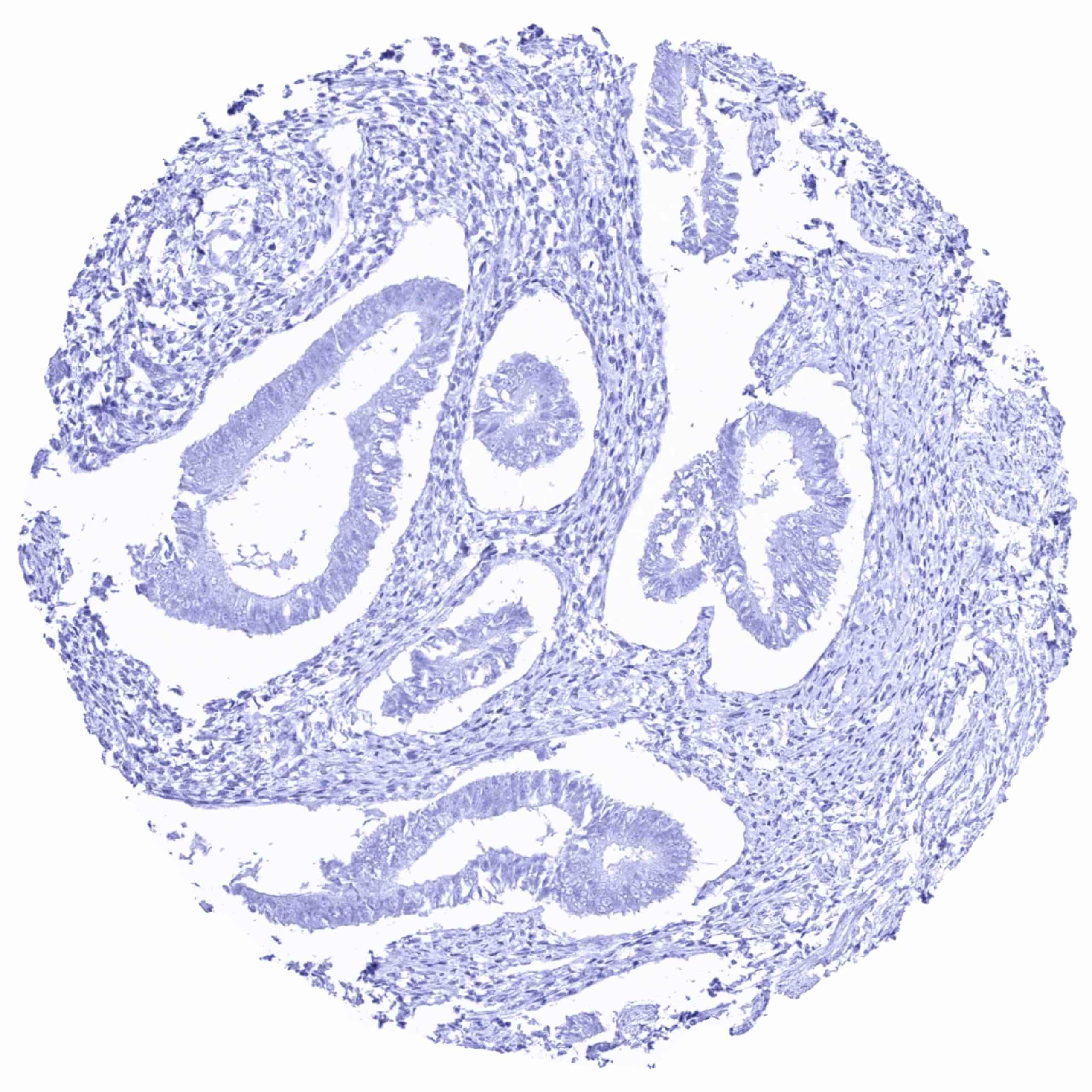
Prostate
Rectum, mucosa
Seminal vesicle.jpeg
Sinus paranasales
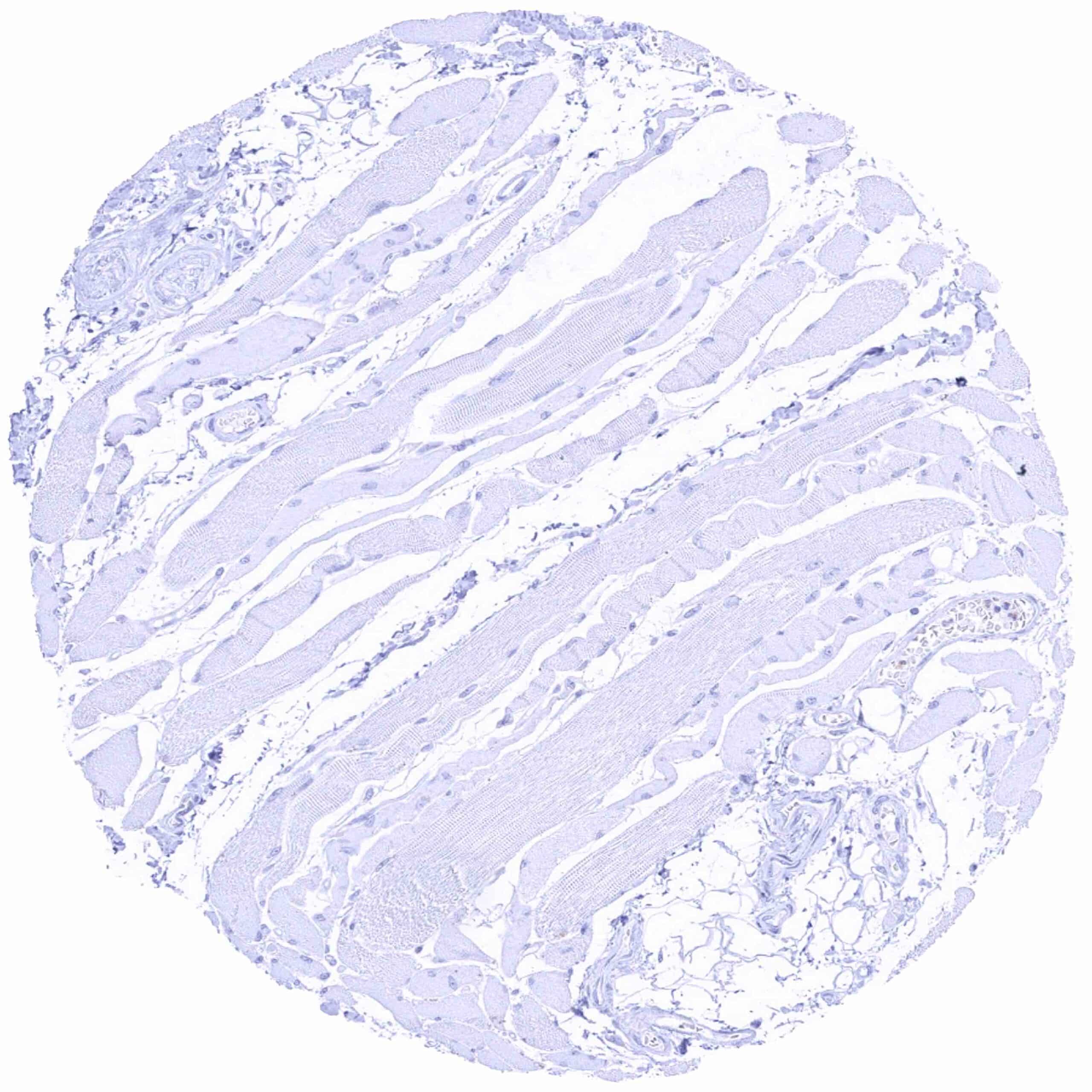
Skeletal muscle
Skin
Spleen
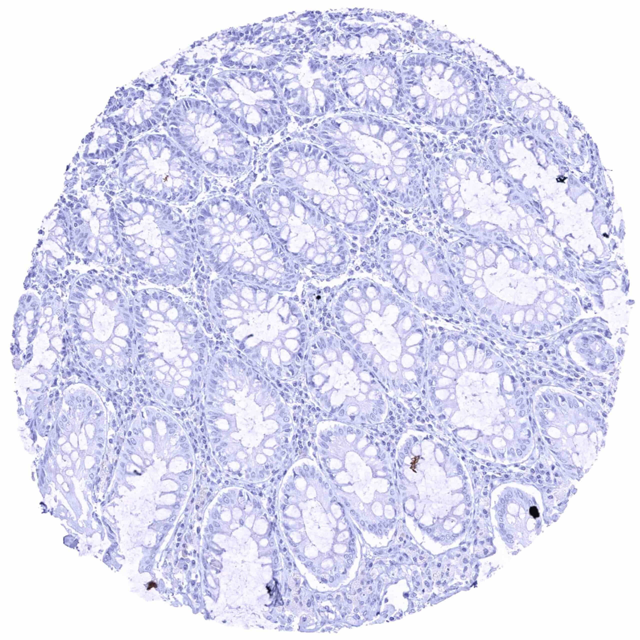
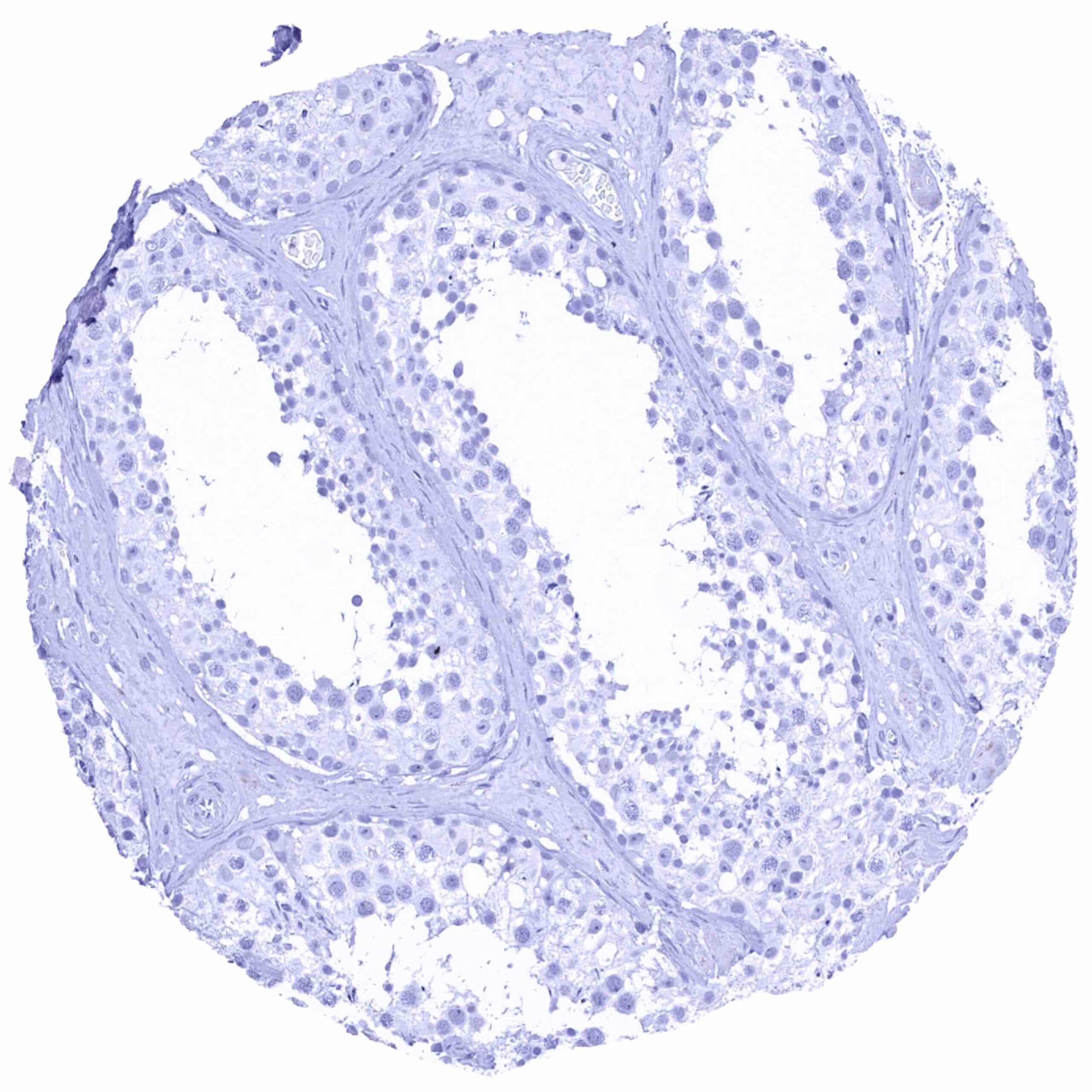
Stomach, antrum – Pepsinogen-I positive chief cells are normally lacking in the antrum of the stomach.jpeg
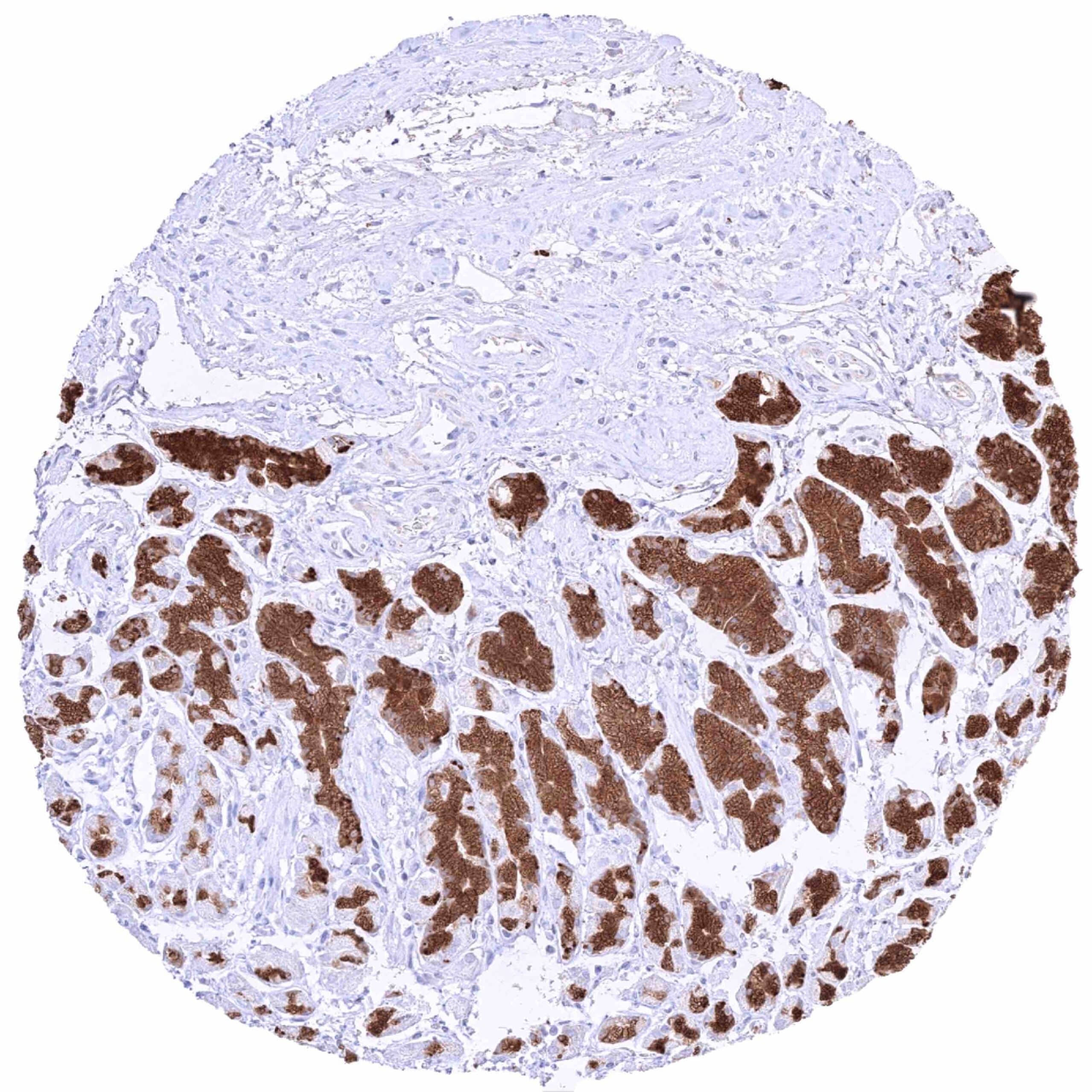
Stomach, antrum – Pepsinogen-I positivity in few glands formed by chief cells in the corpus-antrum transition zone
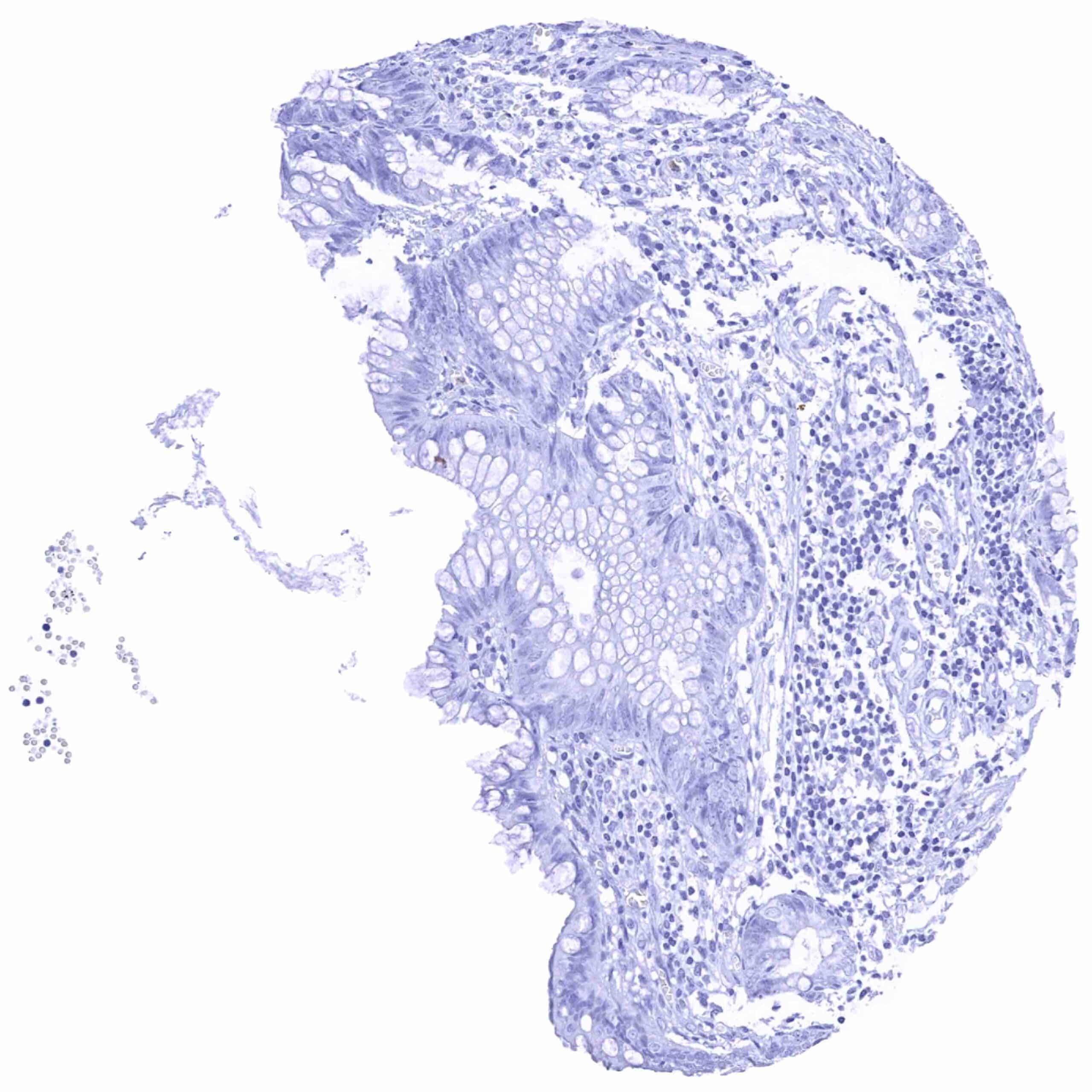
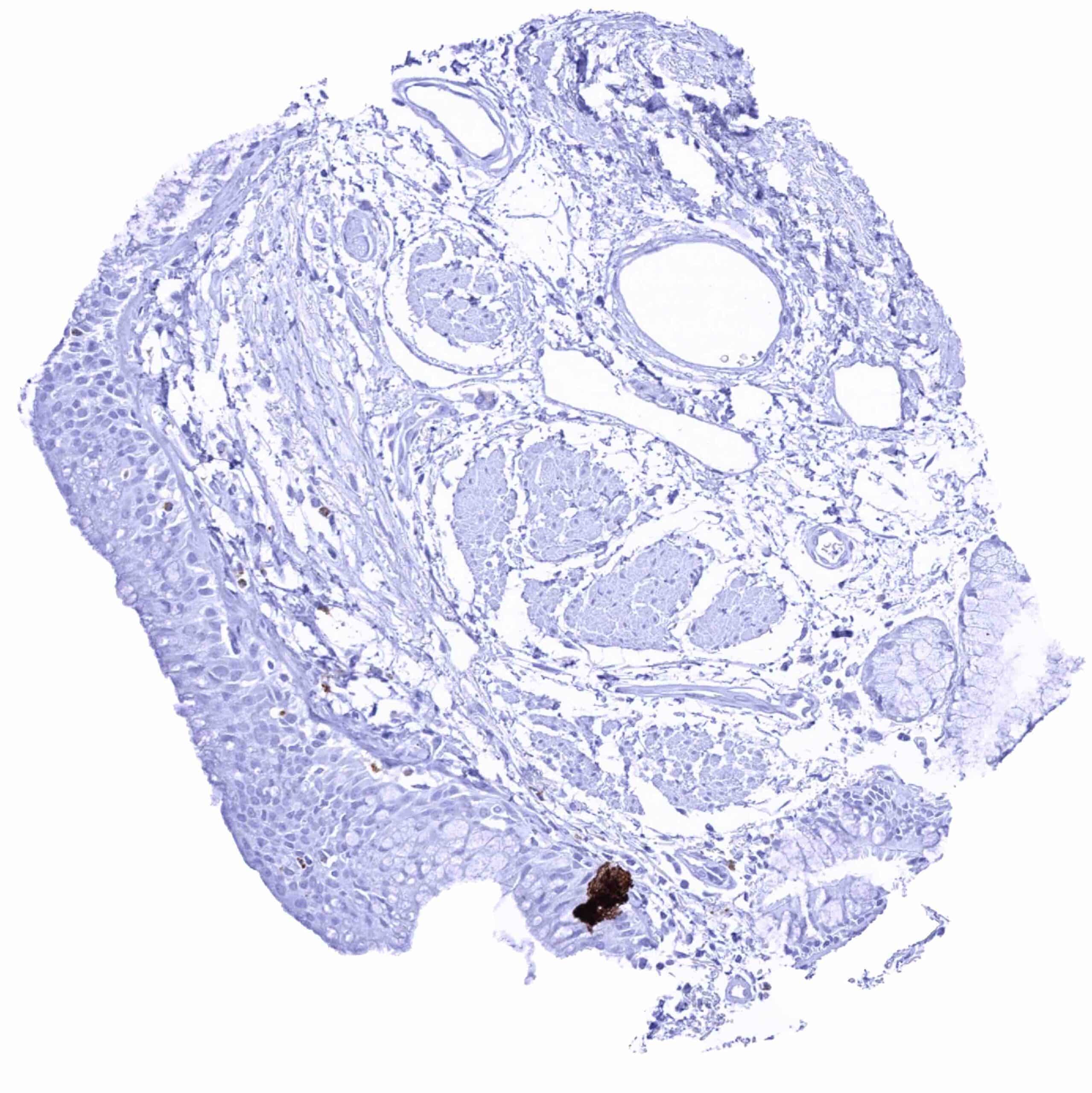
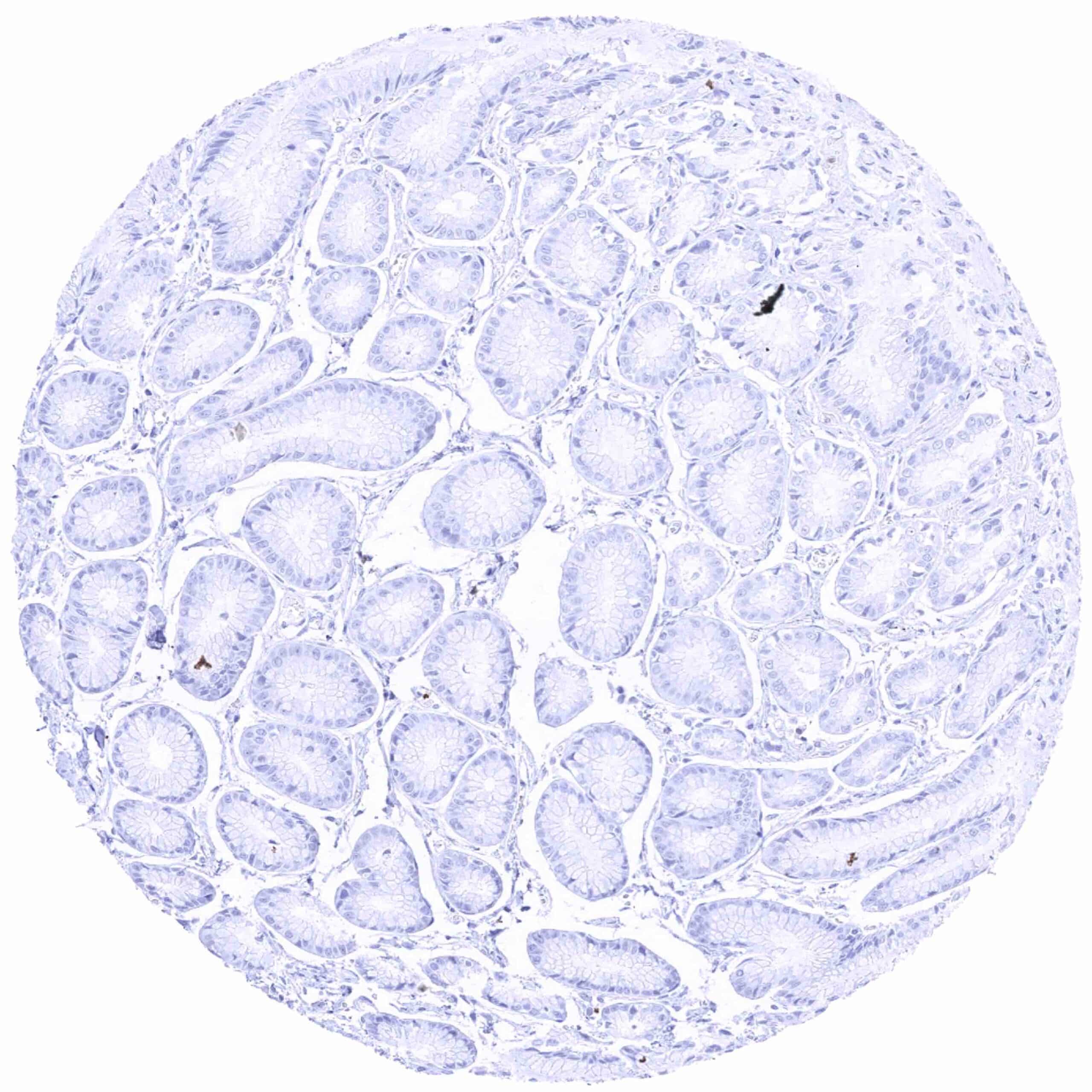
Stomach, antrum
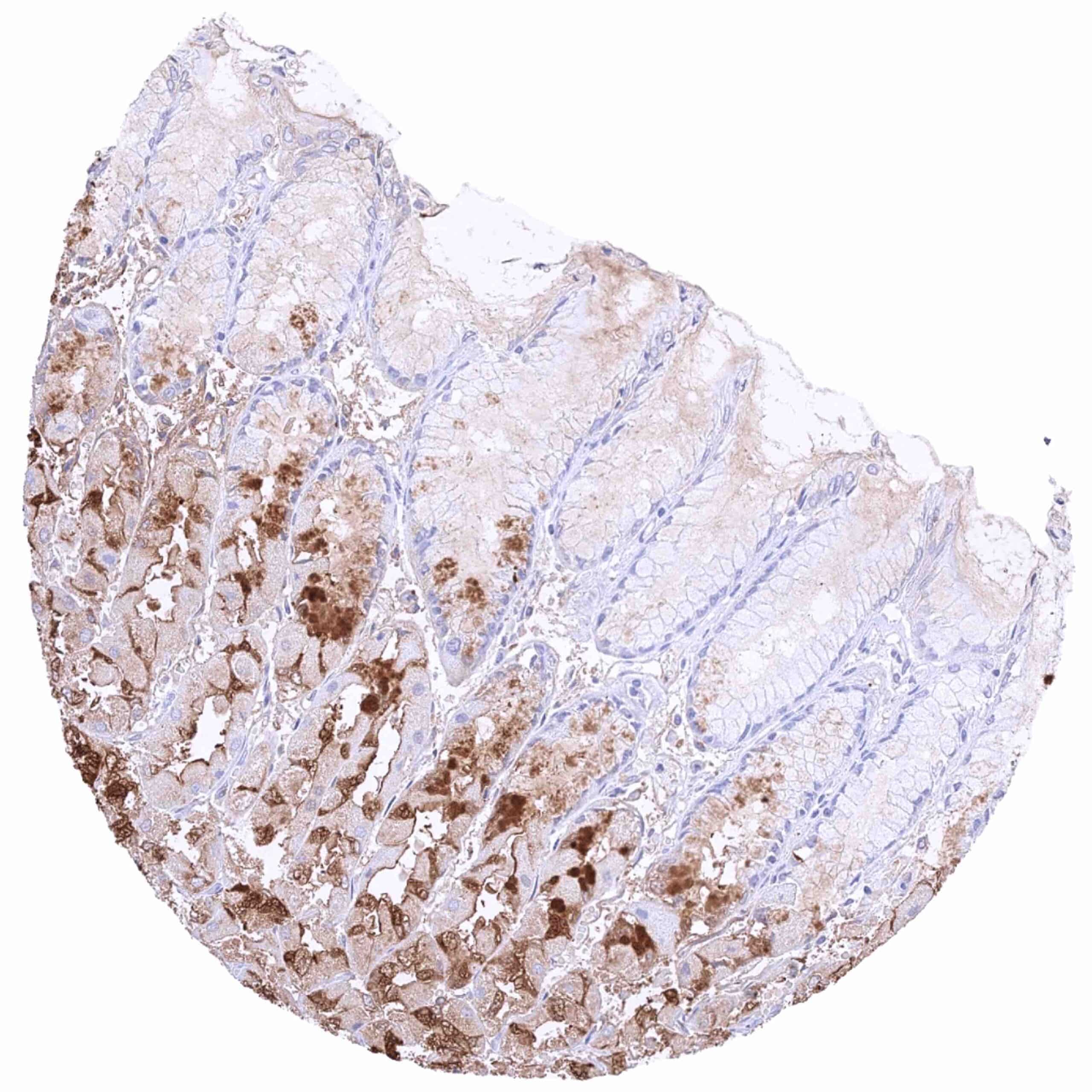
Stomach, corpus – Pepsinogen-I positivity in chief cells of the corpus. Some staining also occurs in adjacent strom and „non-chief cells„ (contamination artifact due to very high levels of pepsinogen I in chief cells)
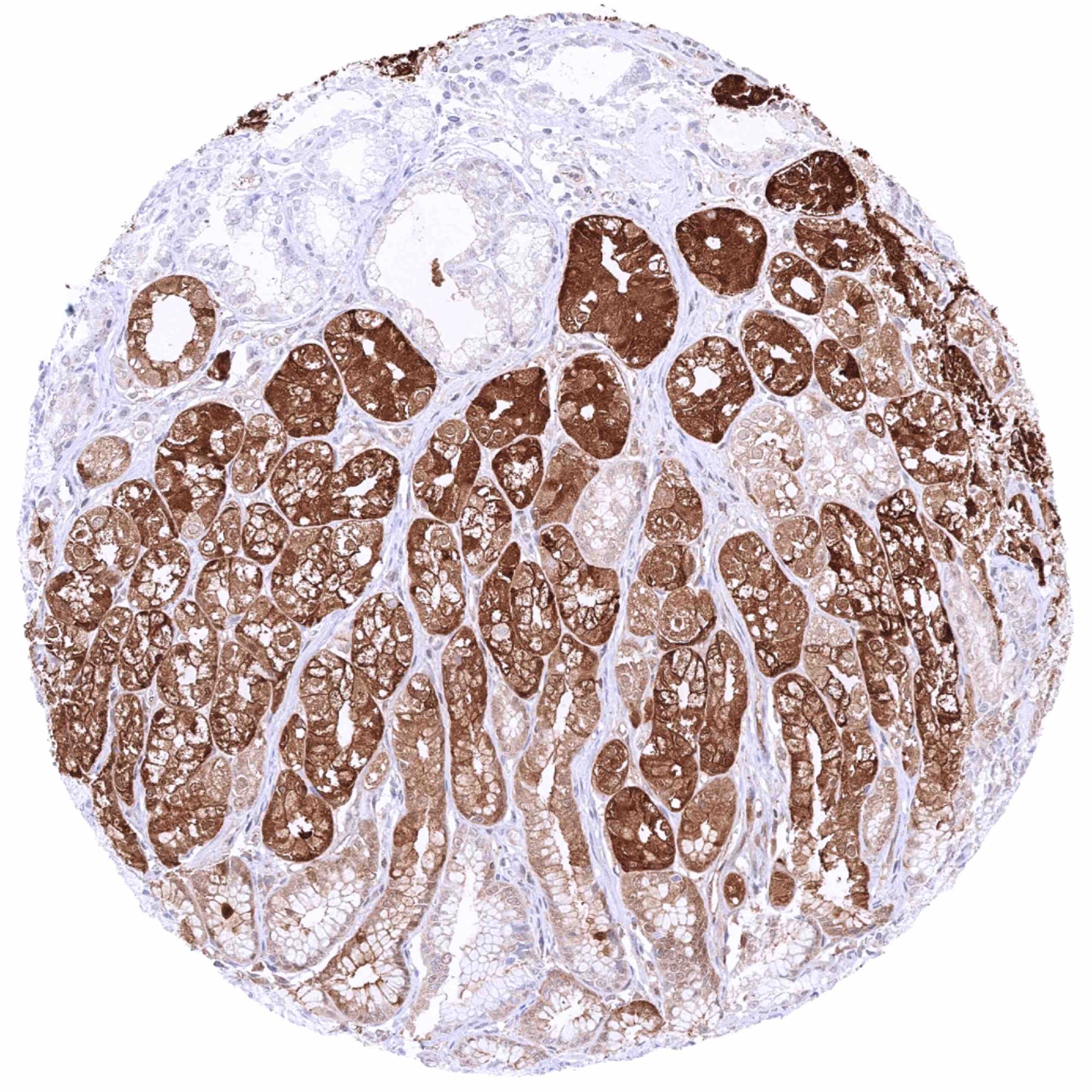
Stomach, corpus – Strong pepsinogen-I positivity in chief cells of corpus glands
Testis
Thymus
Thyroid gland
Tonsil, surface epithelium
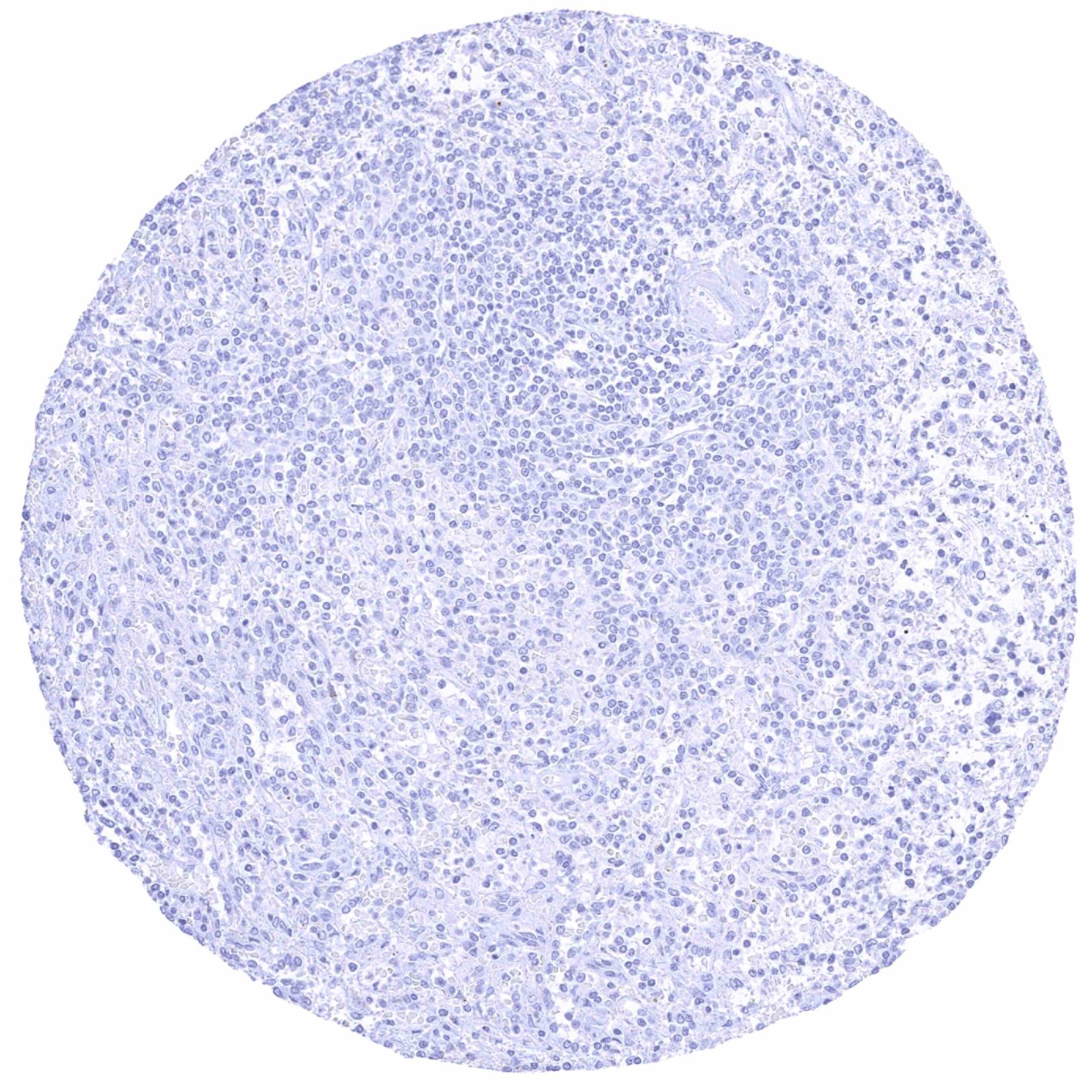
Tonsil
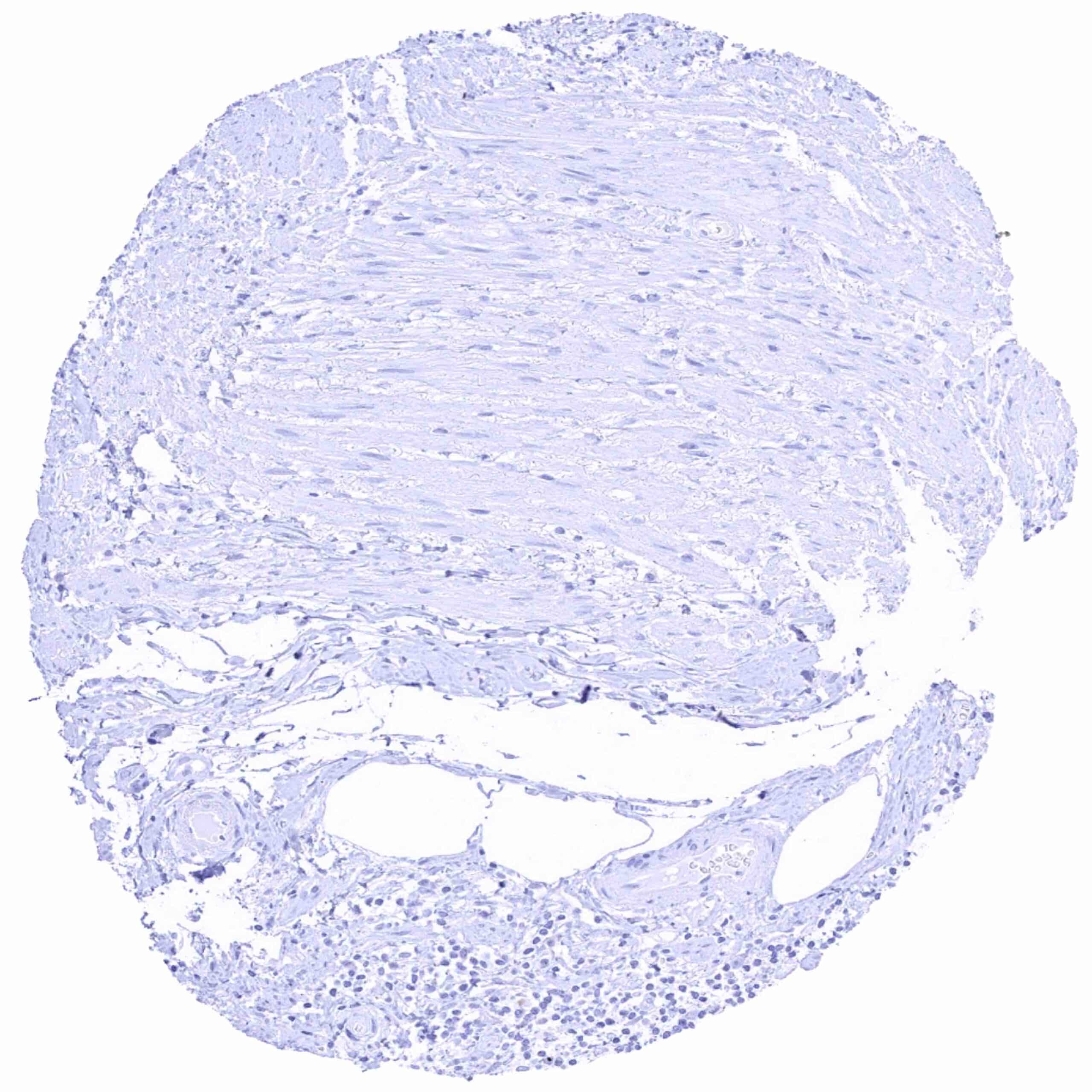
Urinary bladder, muscular wall
Urinary bladder, urothelium
Uterus, ectocervix
Uterus, endocervix (2)
Uterus, endocervix
Uterus, endometrium (pregnancy)
Uterus, endometrium (proliferation)
Uterus, endometrium (secretion)
Uterus, myometrium