
Adrenal gland
Aorta, endothelium
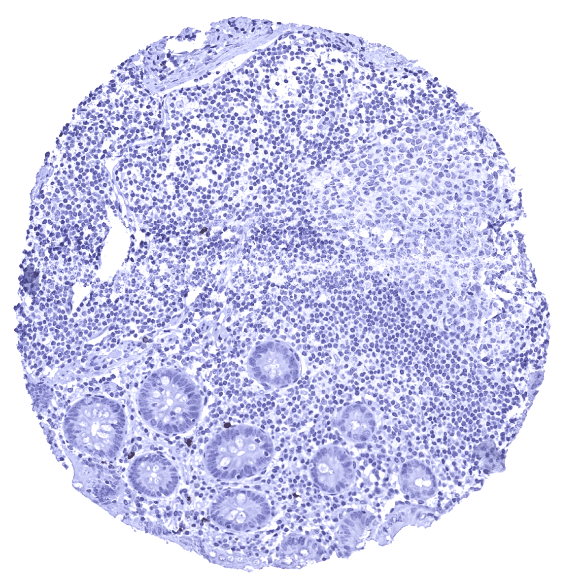
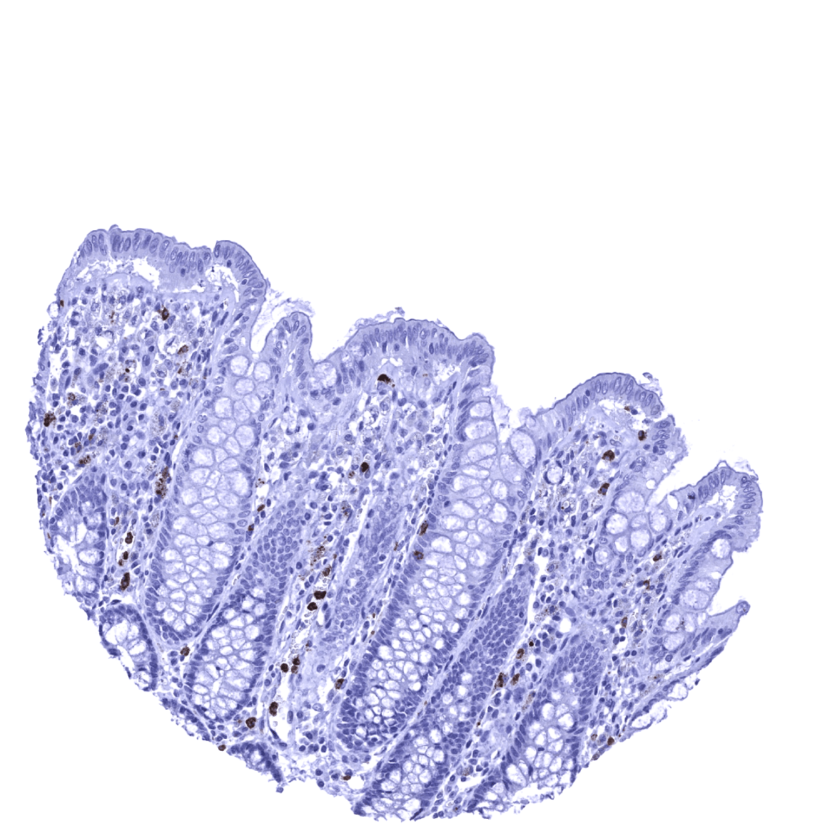
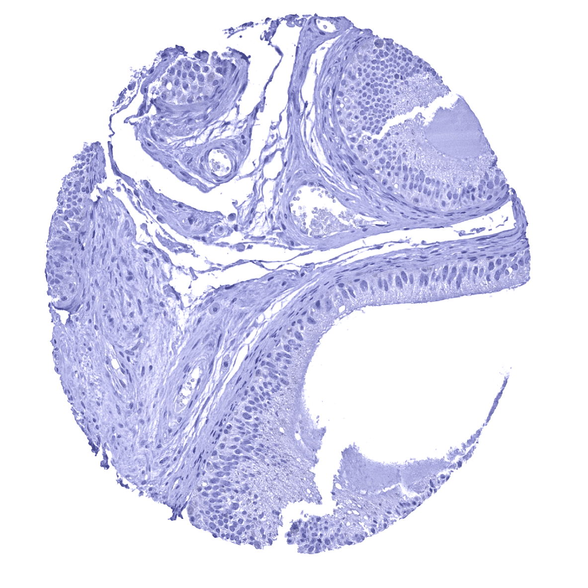
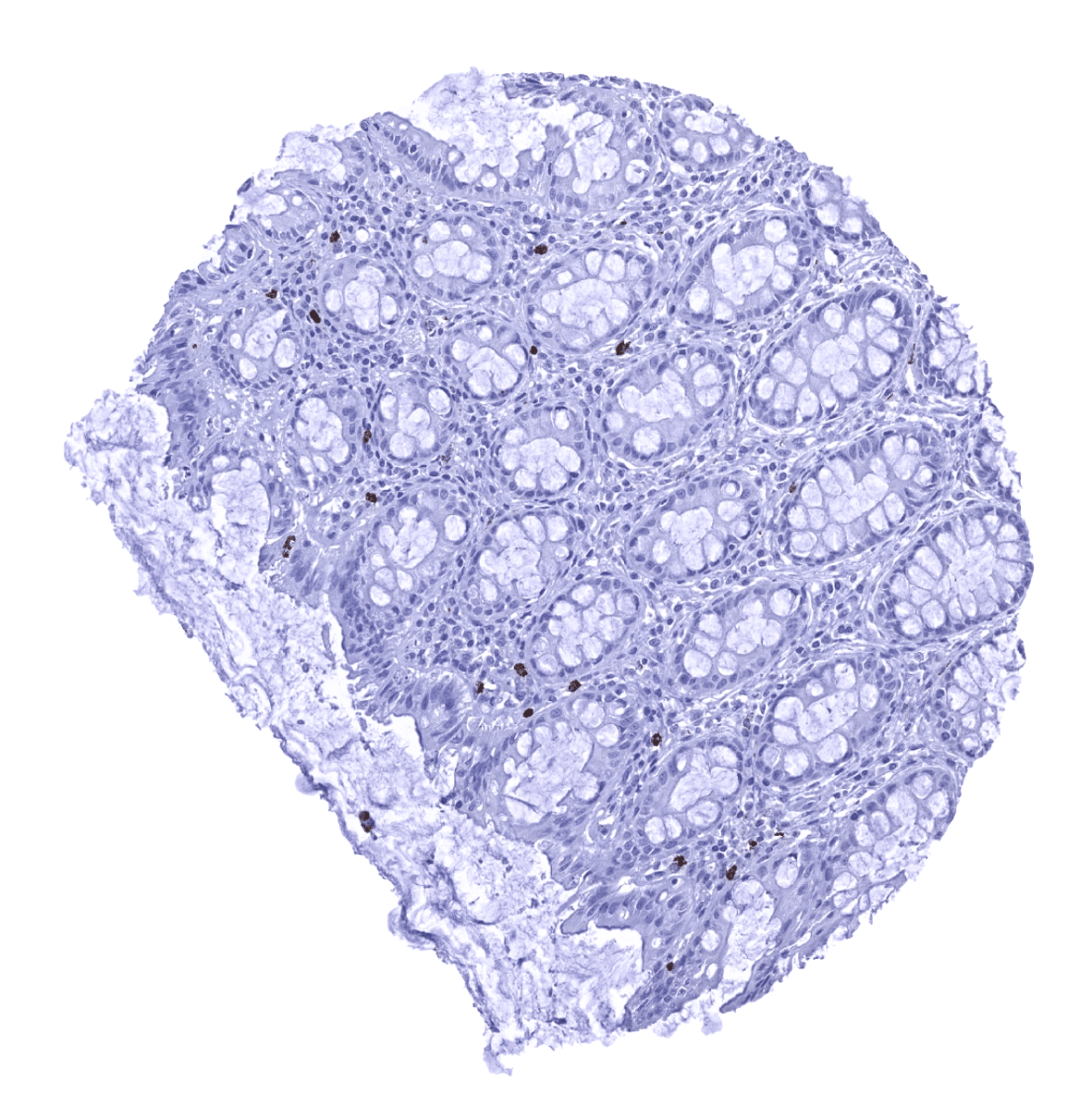
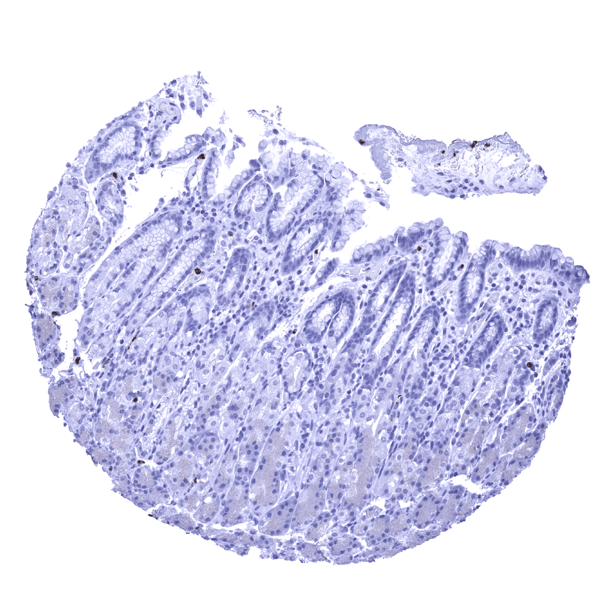
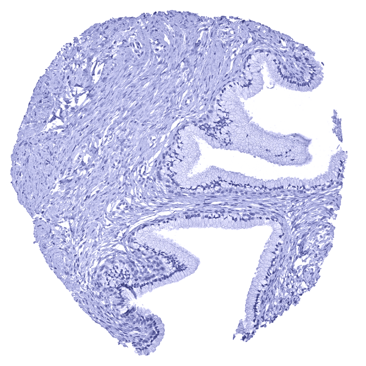
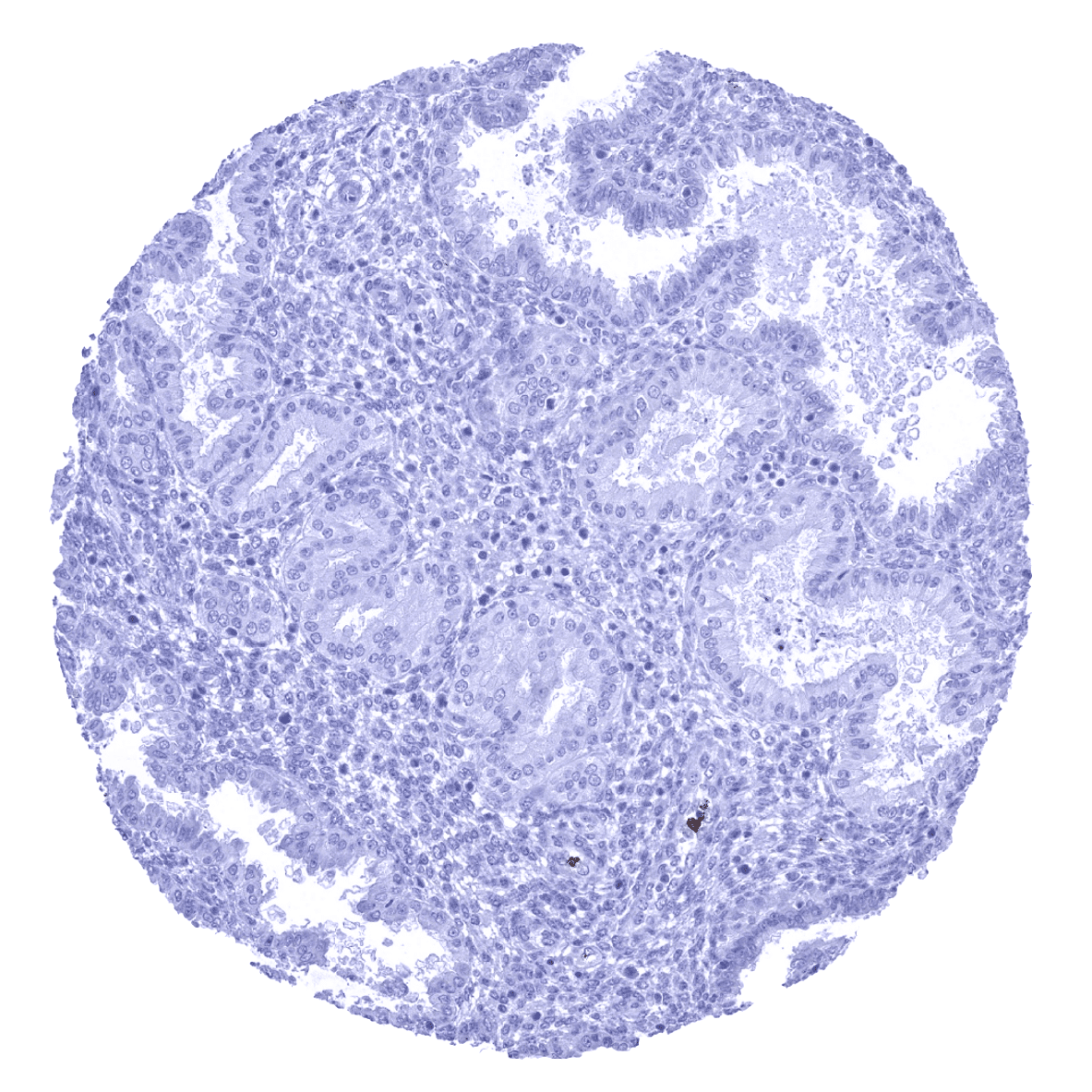
Appendix, mucosa
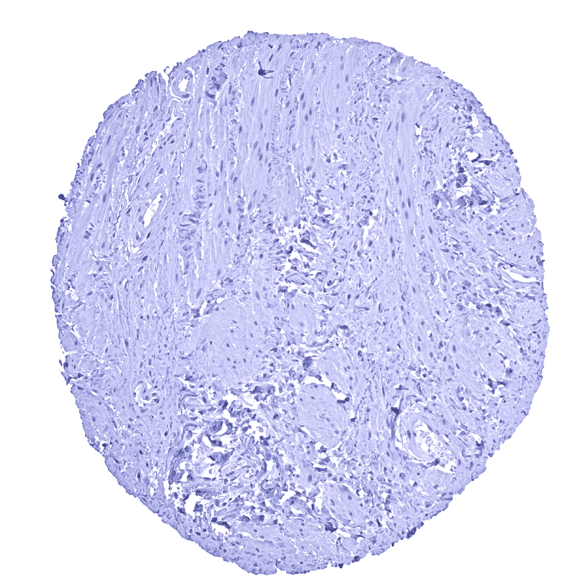
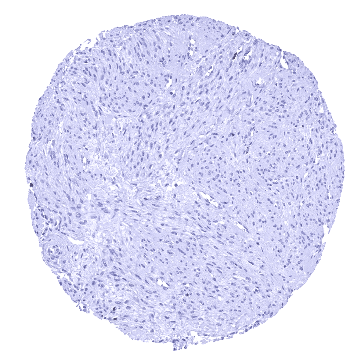
Appendix, muscular wall
Bone marrow - Numerous MPO positive granulocyte precursor cells are seen in the bone marrow.
Breast
Bronchus, mucosa - Numerous MPO positive granulocytes are seen within small capillaries.
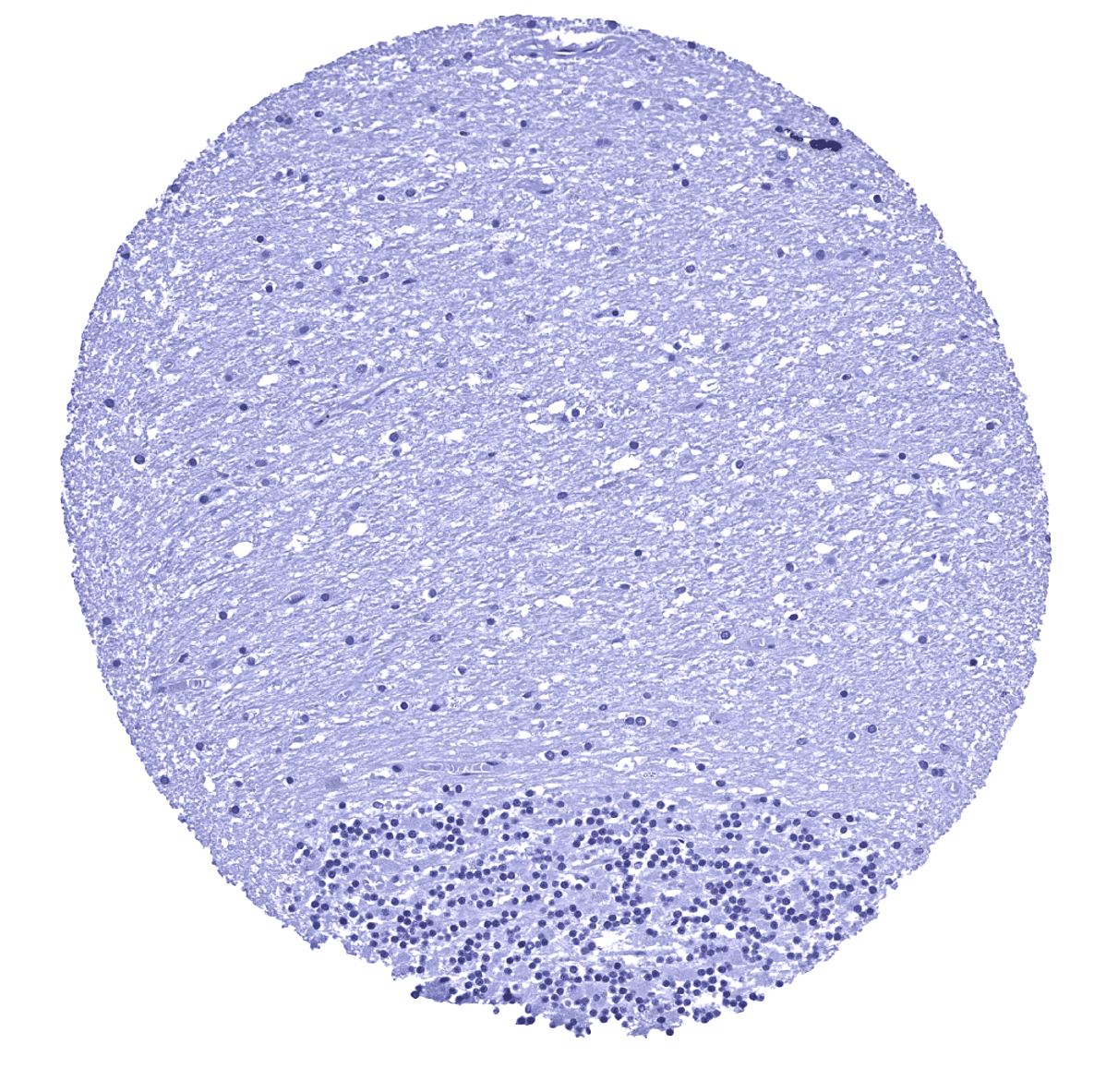
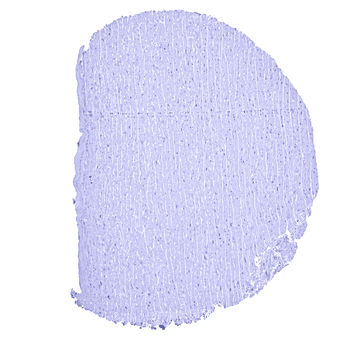
Cerebellum (granule cell layer, white matter)
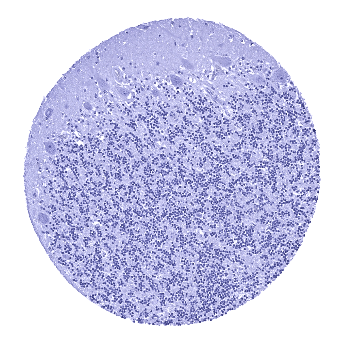
Cerebellum (molecular layer, Purkinje cell layer, granule cell layer)
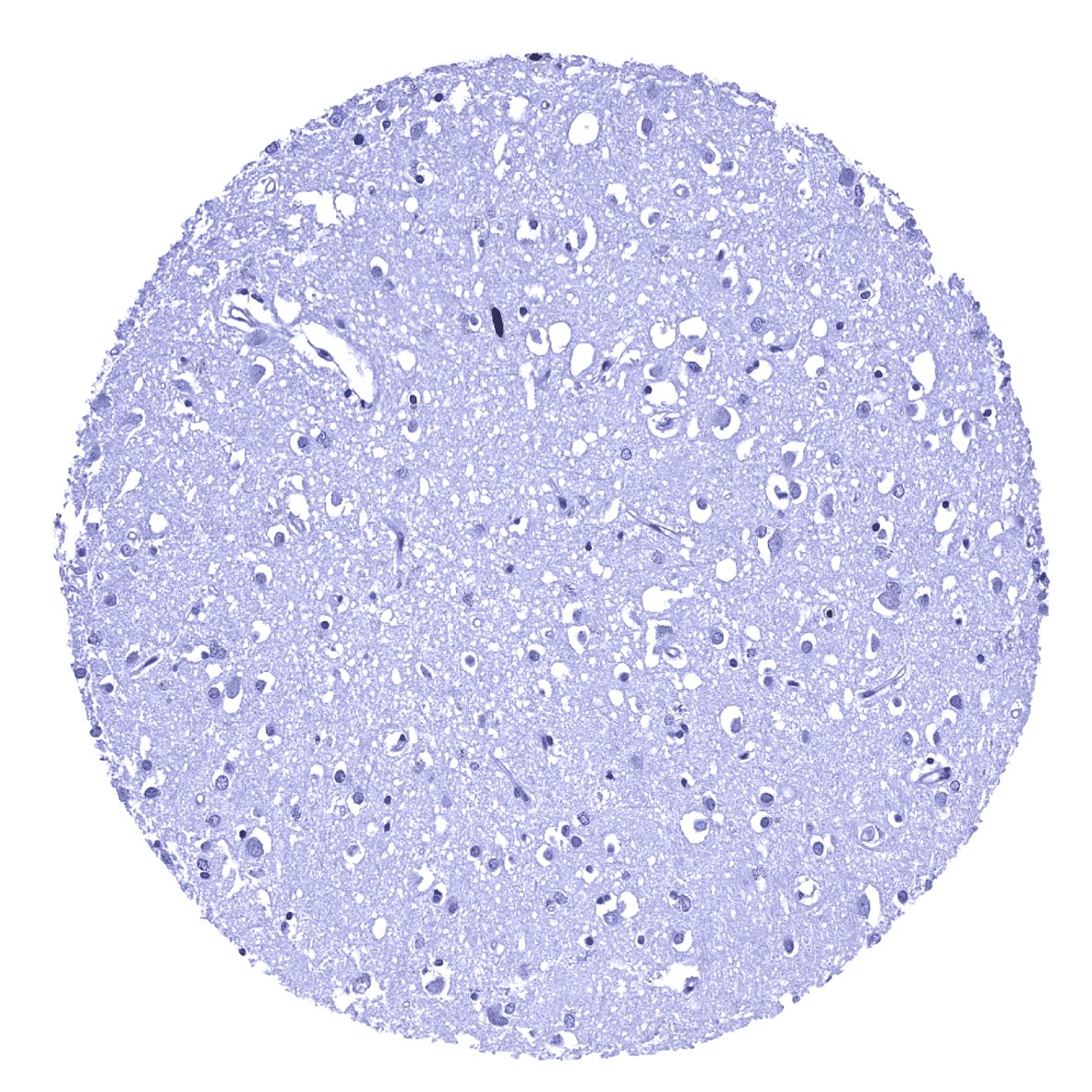
Cerebrum, grey matter
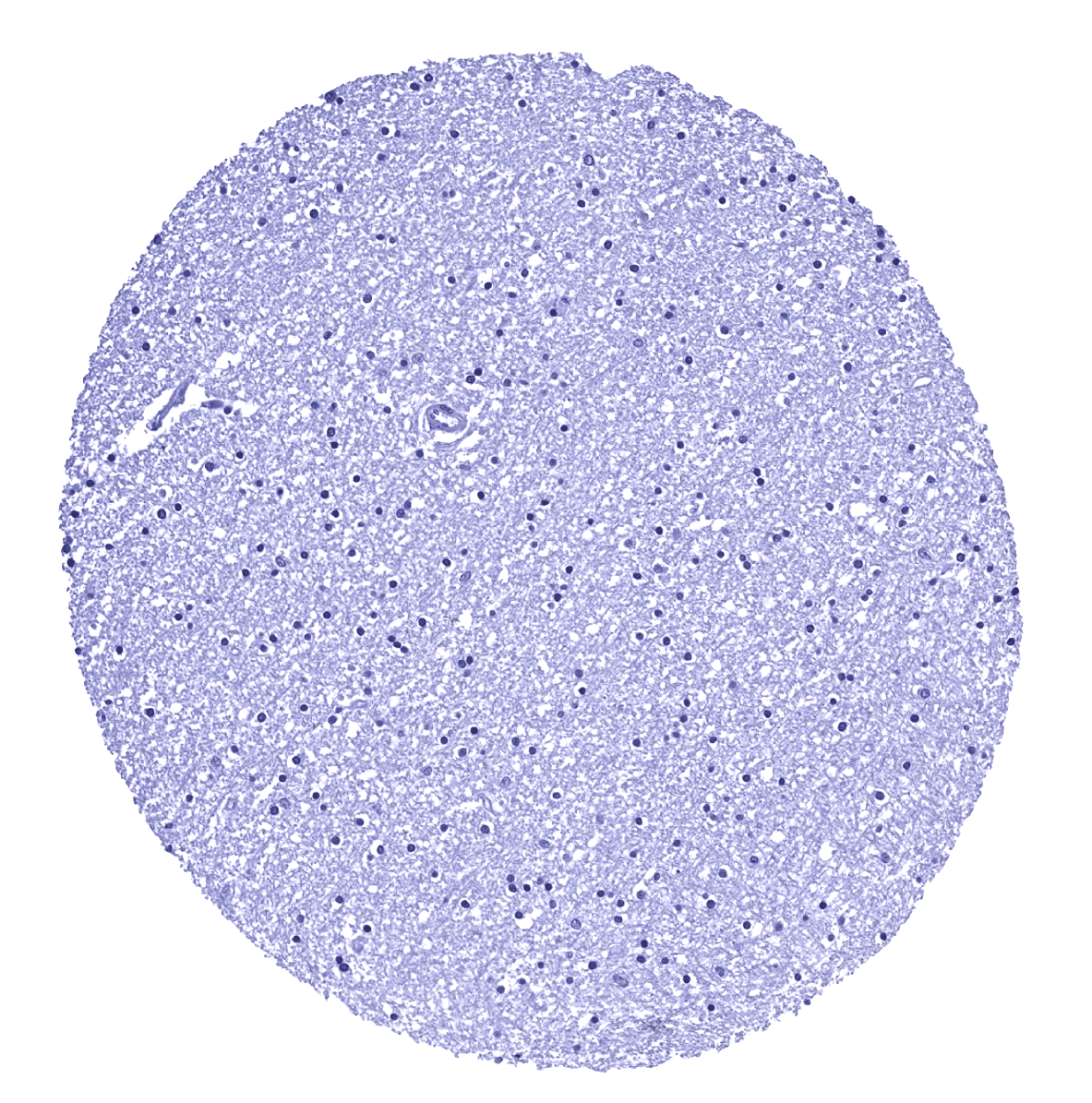
Cerebrum, white matter
Colon descendens, mucosa - MPO positive granulocytes are seen within small capillaries and possibly also the stroma of the lamina propria.

Colon descendens, muscular wall
Duodenum, Brunner gland

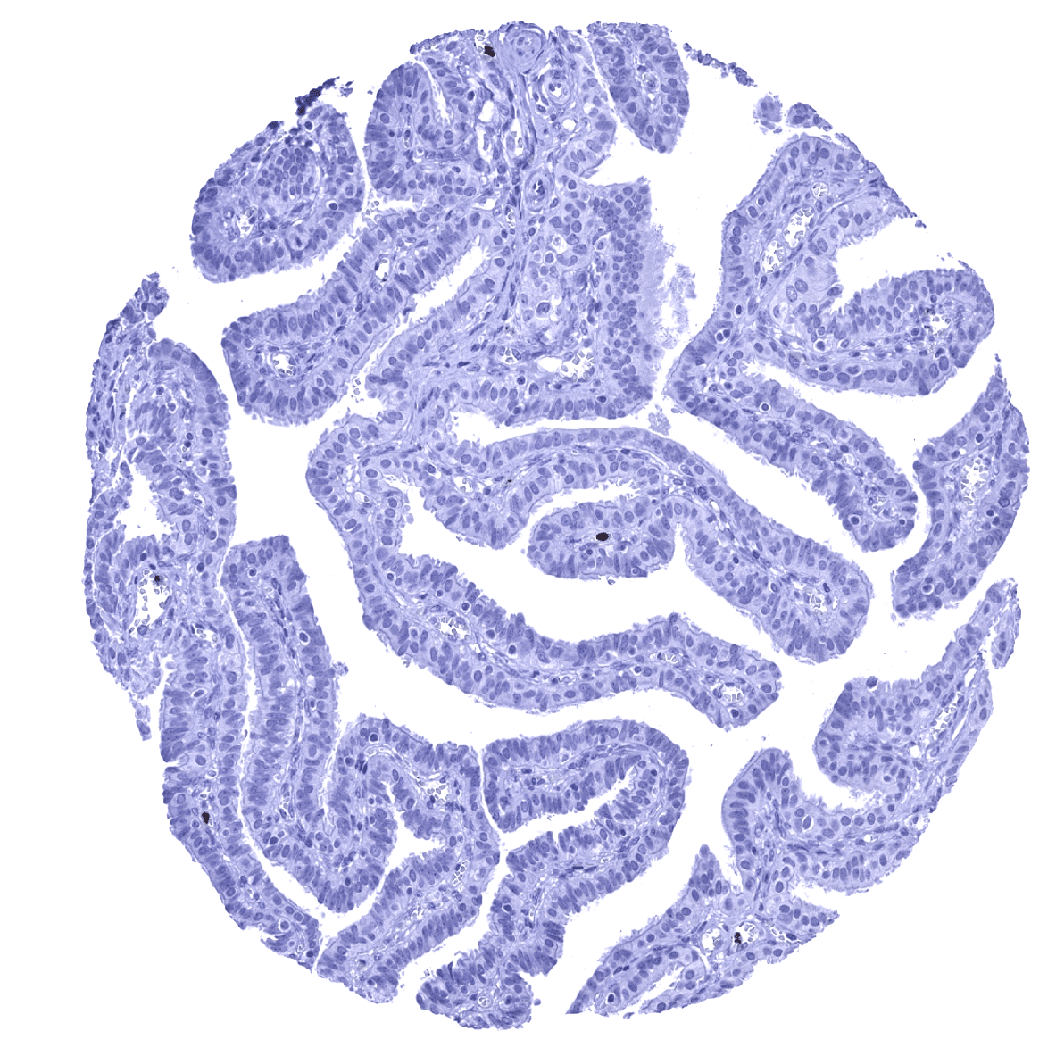
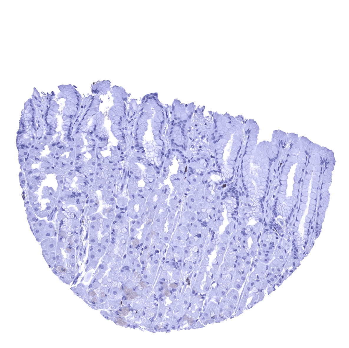
Duodenum, mucosa
Epididymis
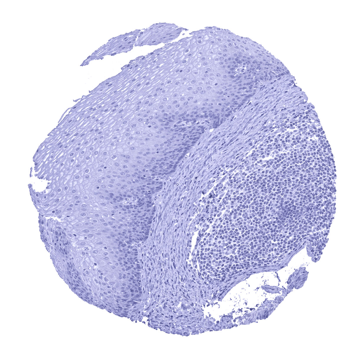
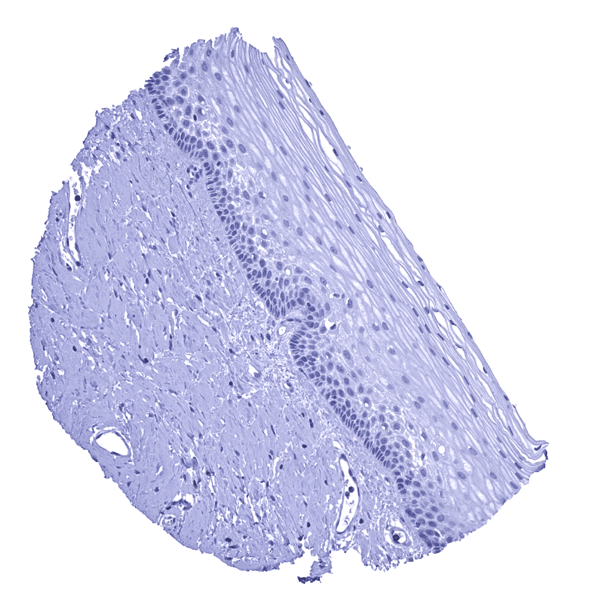
Esophagus, squamous epithelium
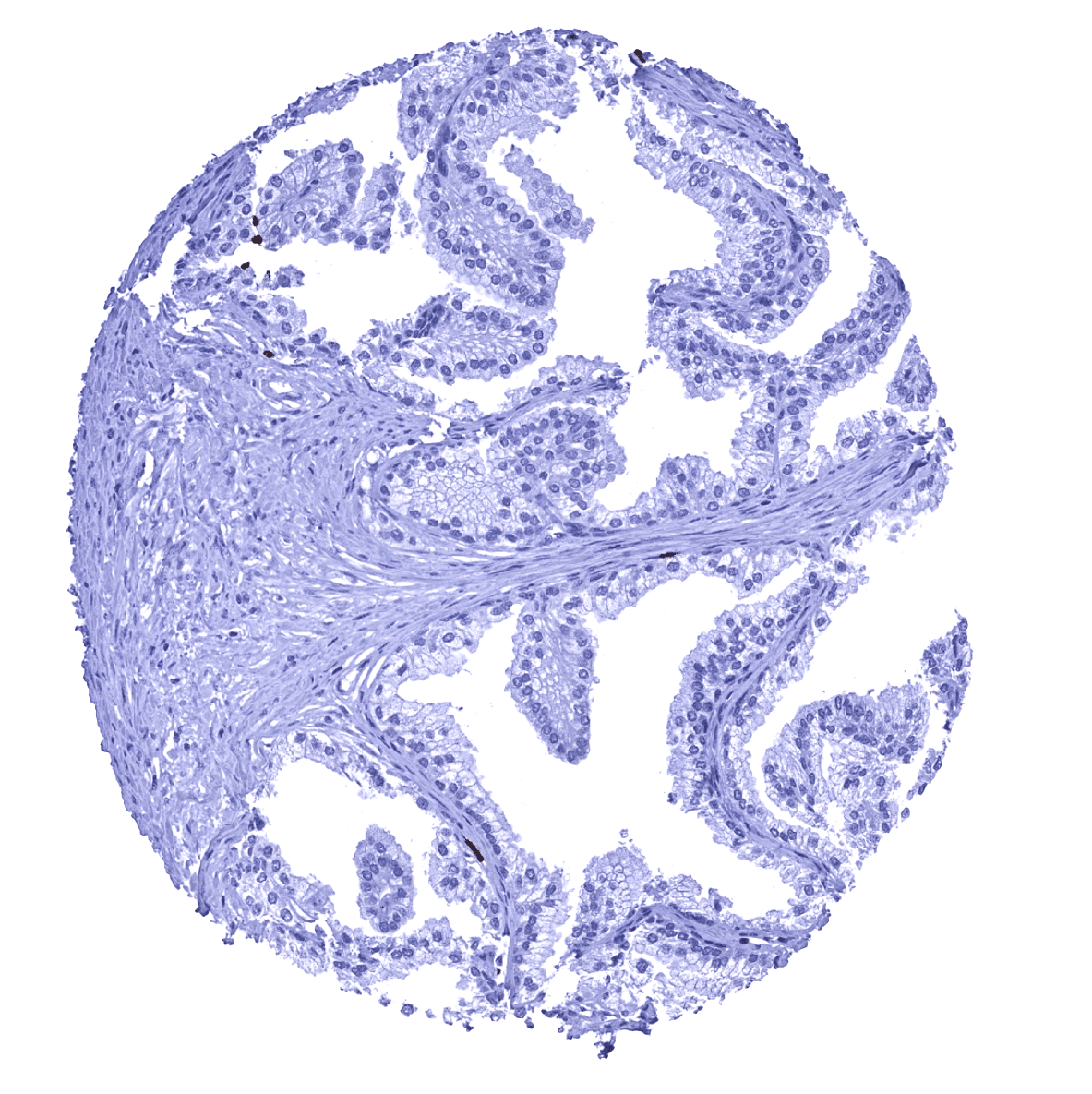
Fallopian tube, mucosa
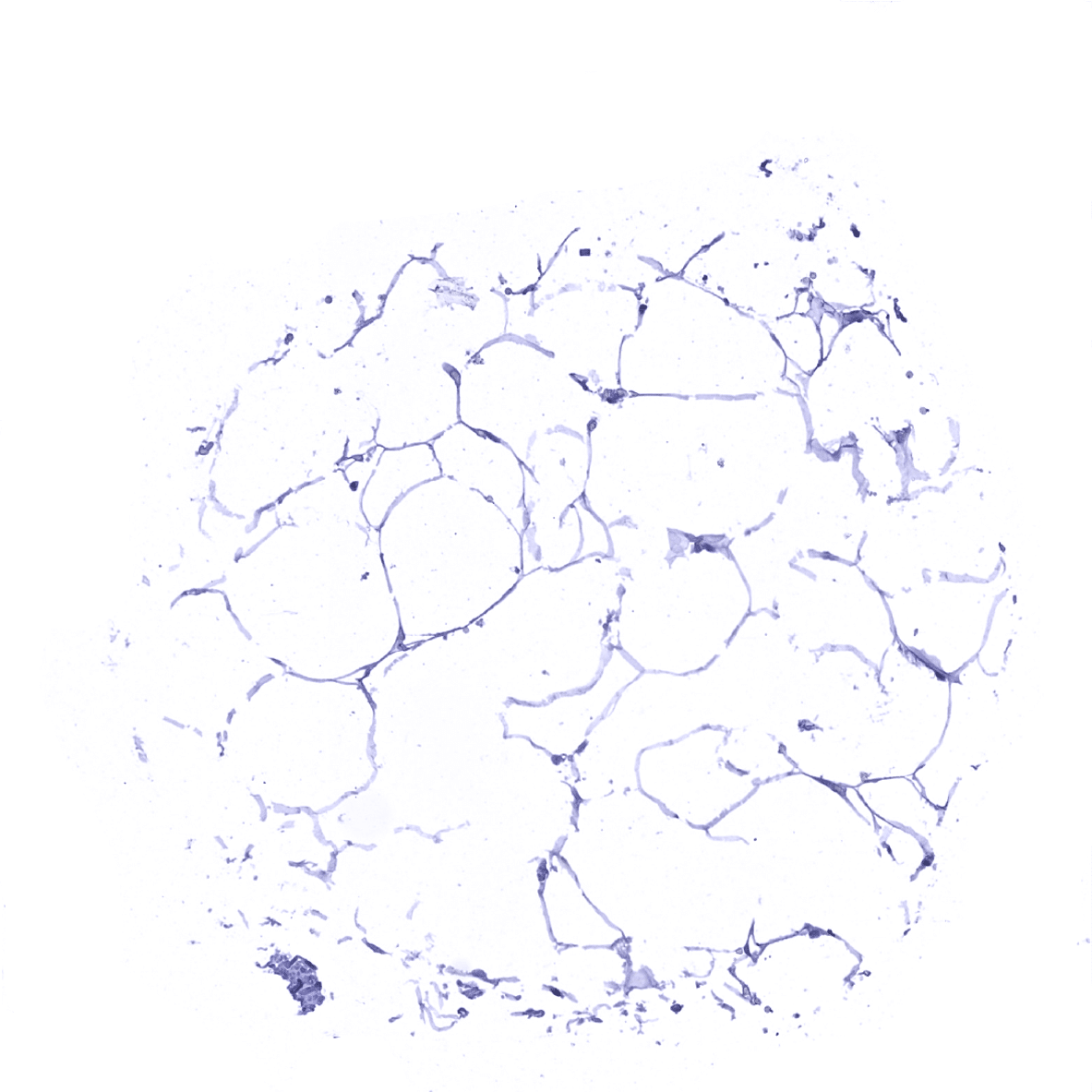
Fat

Gallbladder, epithelium
Heart - MPO positive granulocytes are seen within small capillaries.
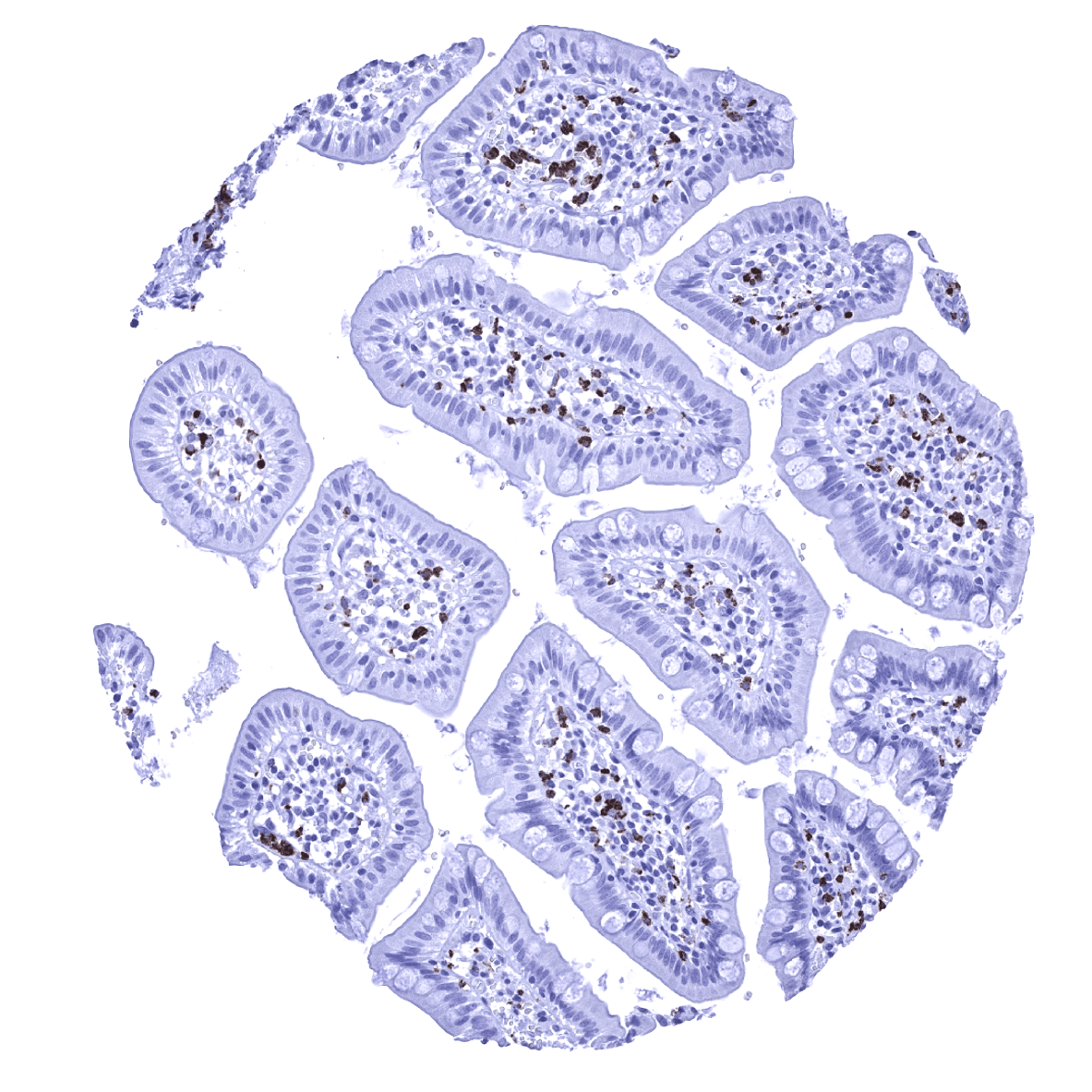
Ileum, mucosa - MPO positive granulocytes are seen within small capillaries and possibly also the stroma of the lamina propria.
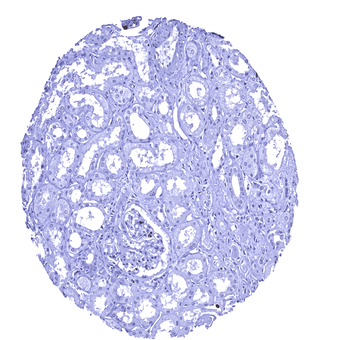
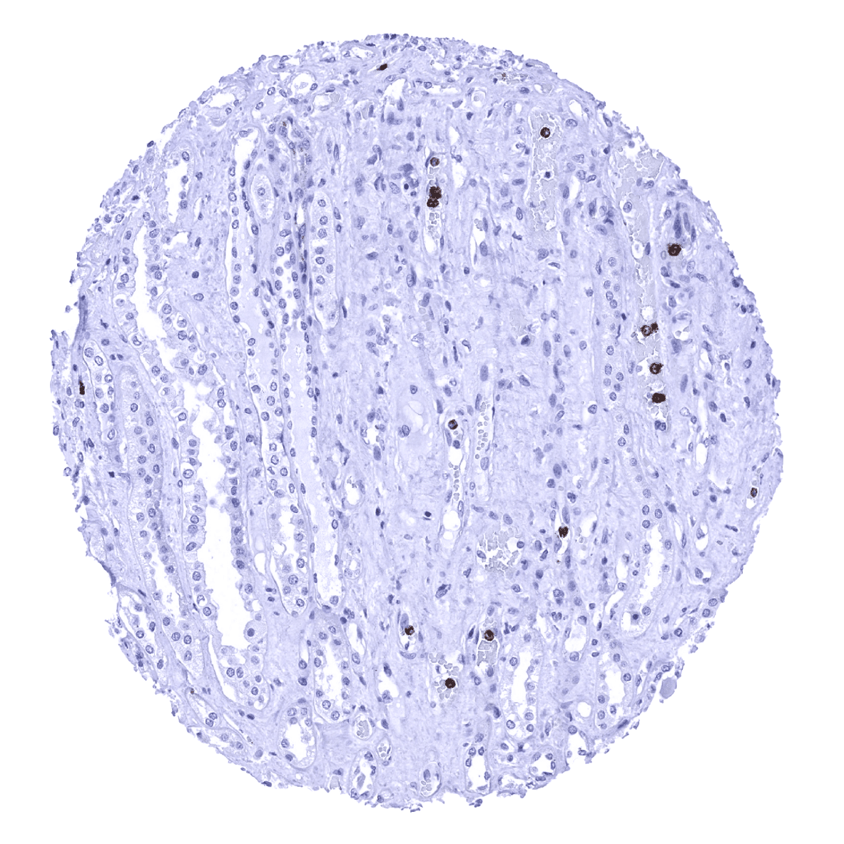
Kidney, cortex
Kidney, medulla - MPO positive granulocytes are seen within small capillaries.

Kidney, pelvis, urothelium
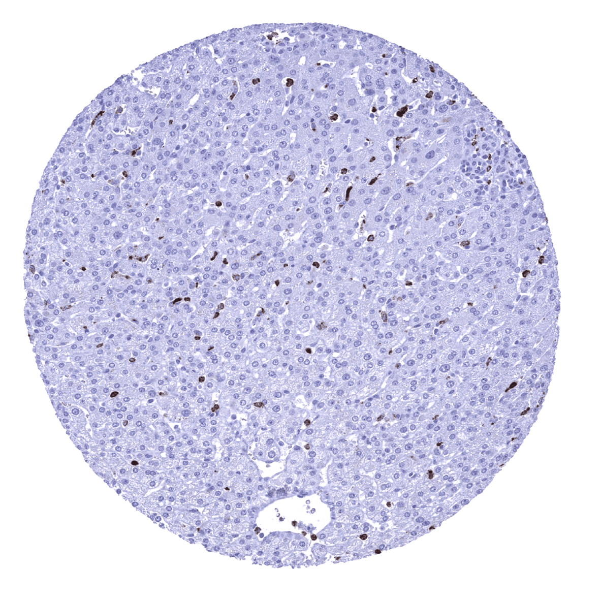
Liver - Few MPO positive granulocytes are seen within the liver sinus.

Lung - MPO positive granulocytes are seen within small blood vessels.
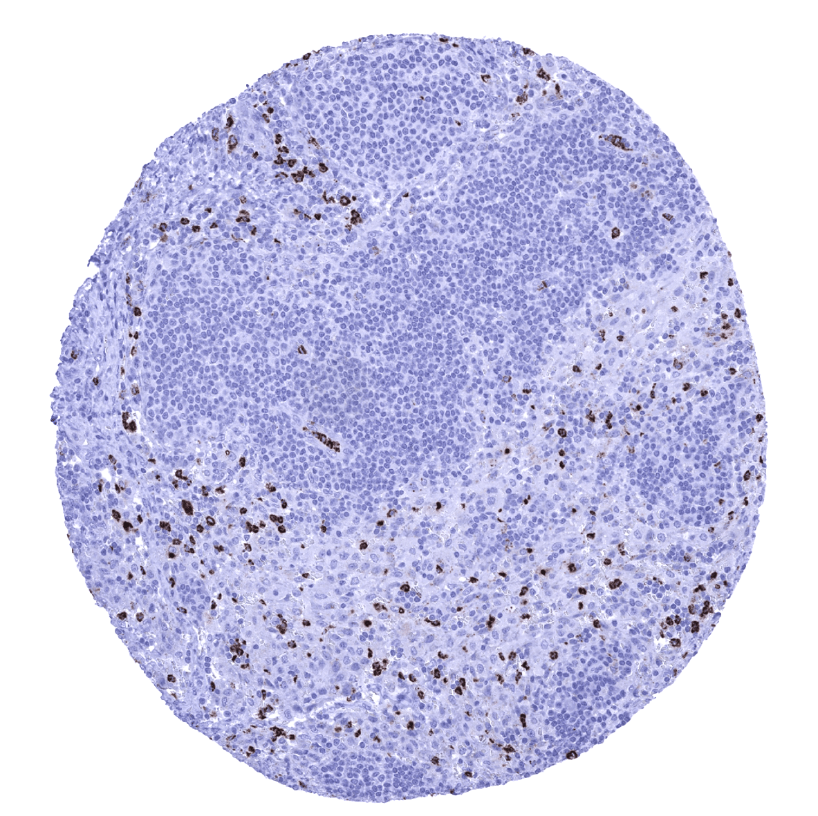
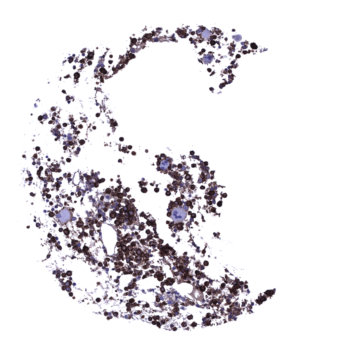
Lymph node - Abundant MPO positive granulocytes are seen within the sinus of a lymph node.
Lymph node
Ovary, stroma
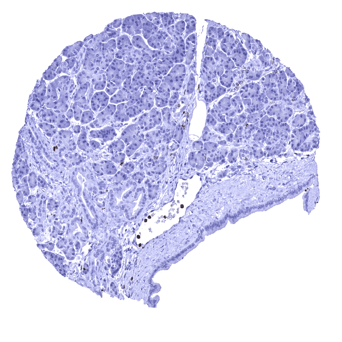
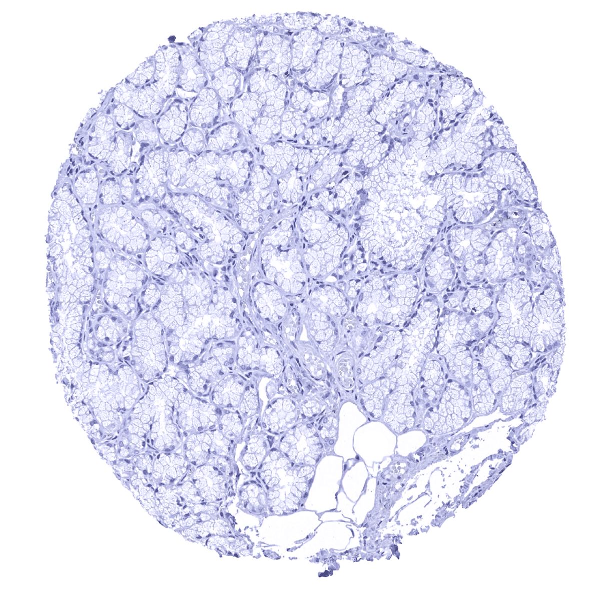
Pancreas

Parathyroid
Parotid gland

Pituitary gland, anterior lobe

Pituitary gland, posterior lobe

Placenta, early
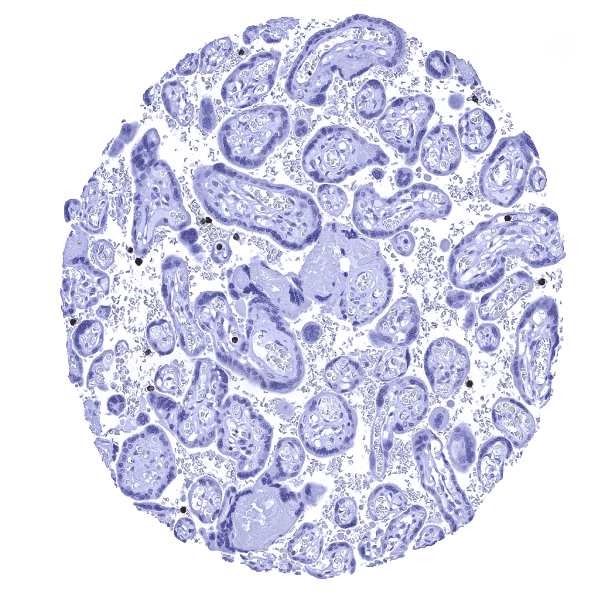
Placenta, mature

Placenta (amnion and chorion)
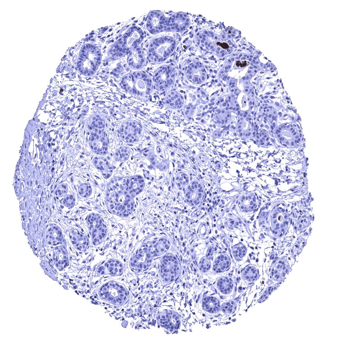
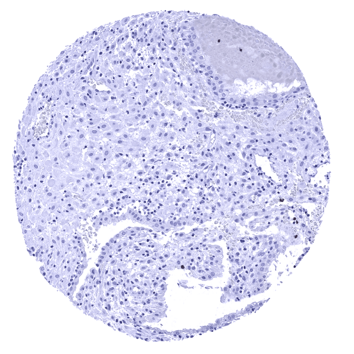
Prostate - MPO positive intraglandular material is often absent in the prostate.
Prostate - MPO positive material can be seen in the lumina of prostatic glands.
Rectum, mucosa
Seminal vesicle
Skin

Spleen - Numerous MPO positive granulocytes are usually seen within the red pulp of the spleen.
Stomach, antrum - A faint cytoplasmic MPO staining in stomach glands is considered a tolerable cross-reactivity of our antibody.

Stomach, antrum
Stomach, corpus - A faint cytoplasmic MPO staining in stomach glands is considered a tolerable cross-reactivity of our antibody.
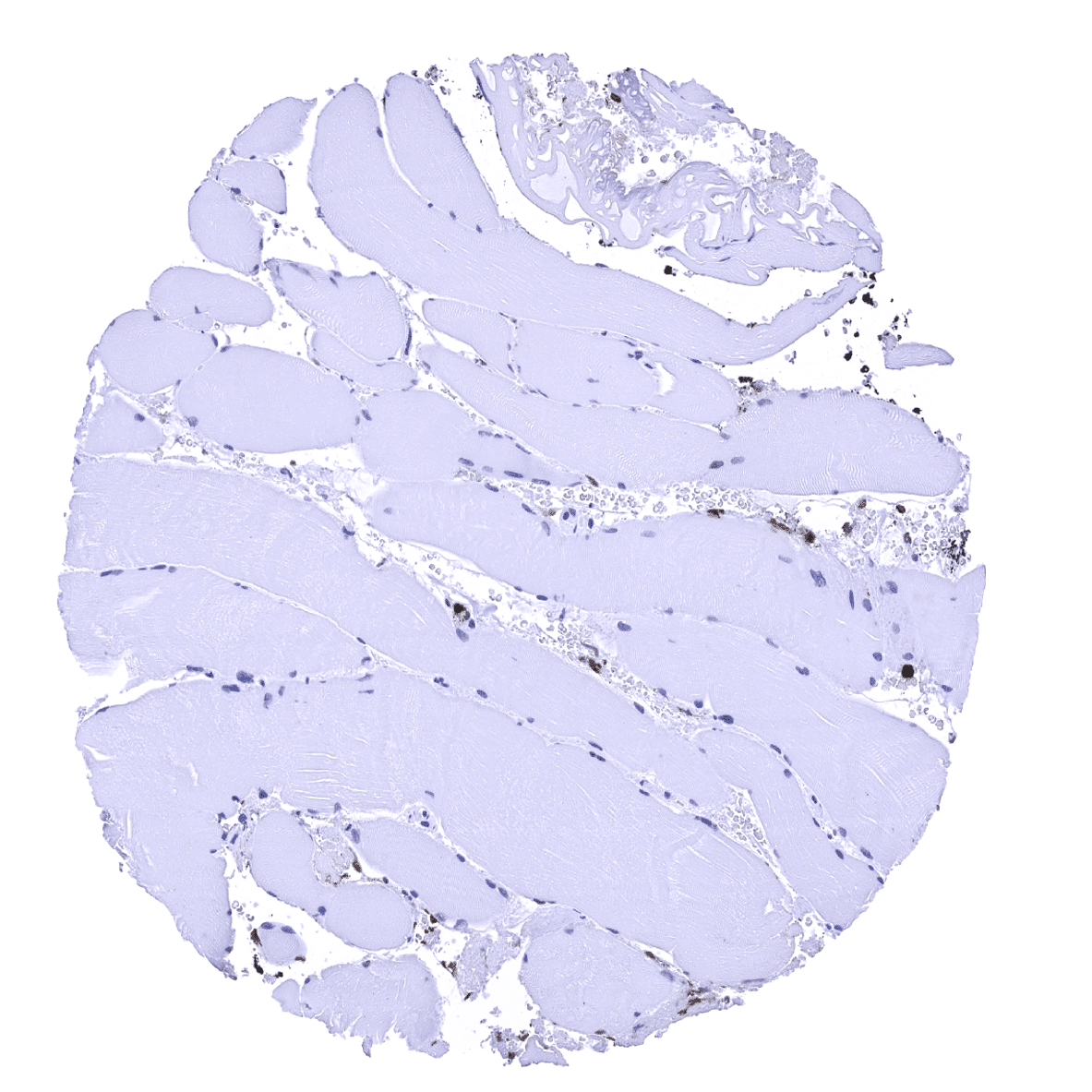
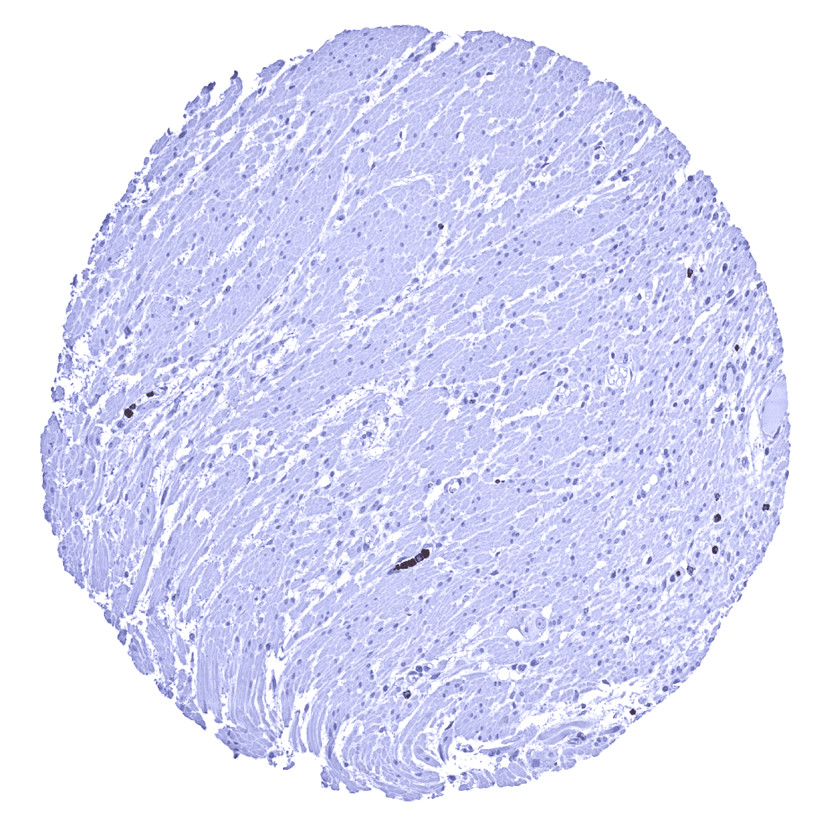
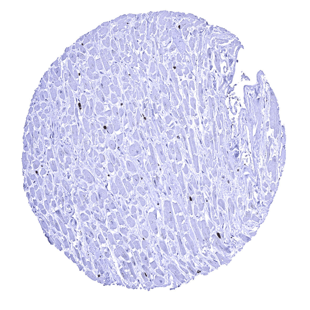
Striated muscle
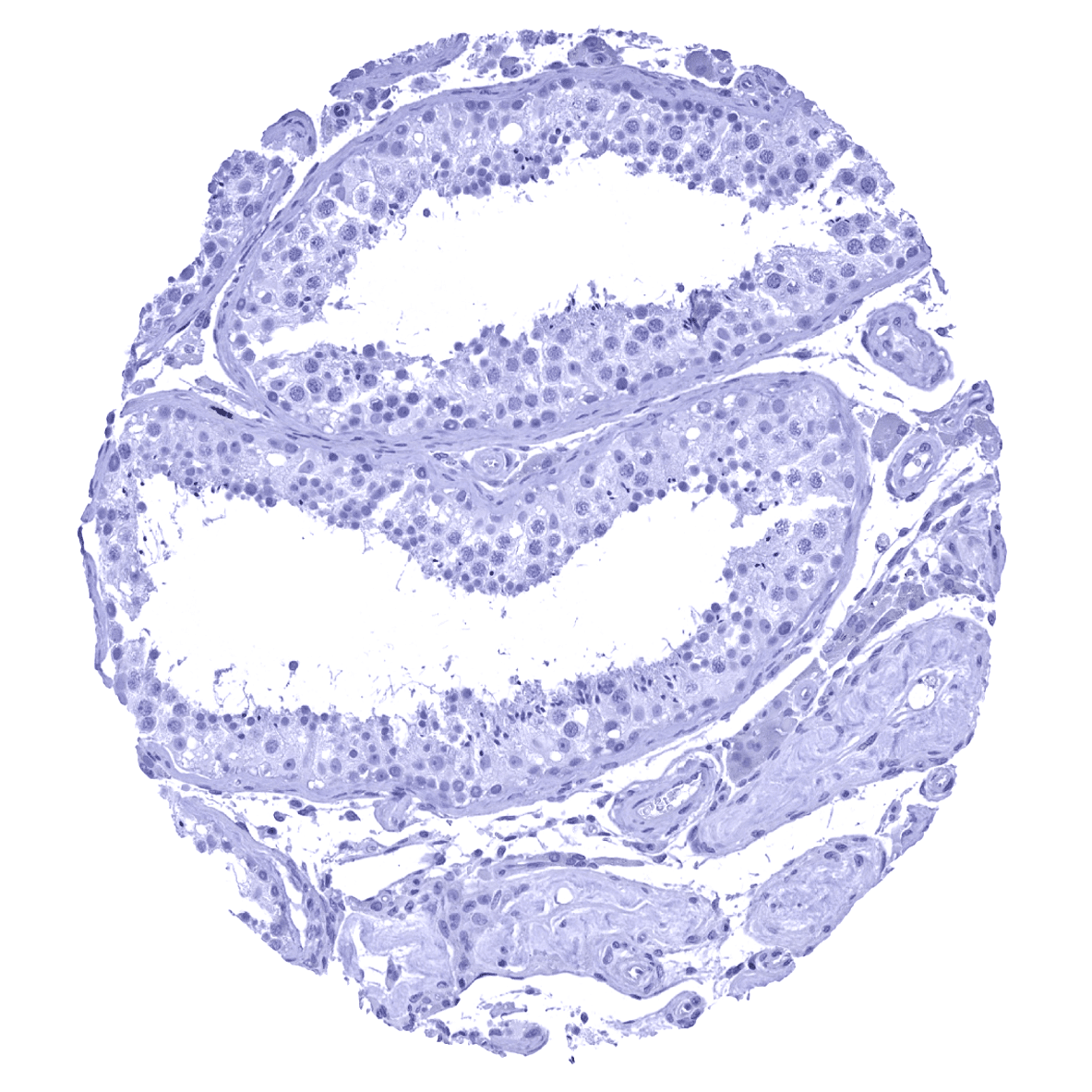
Testis

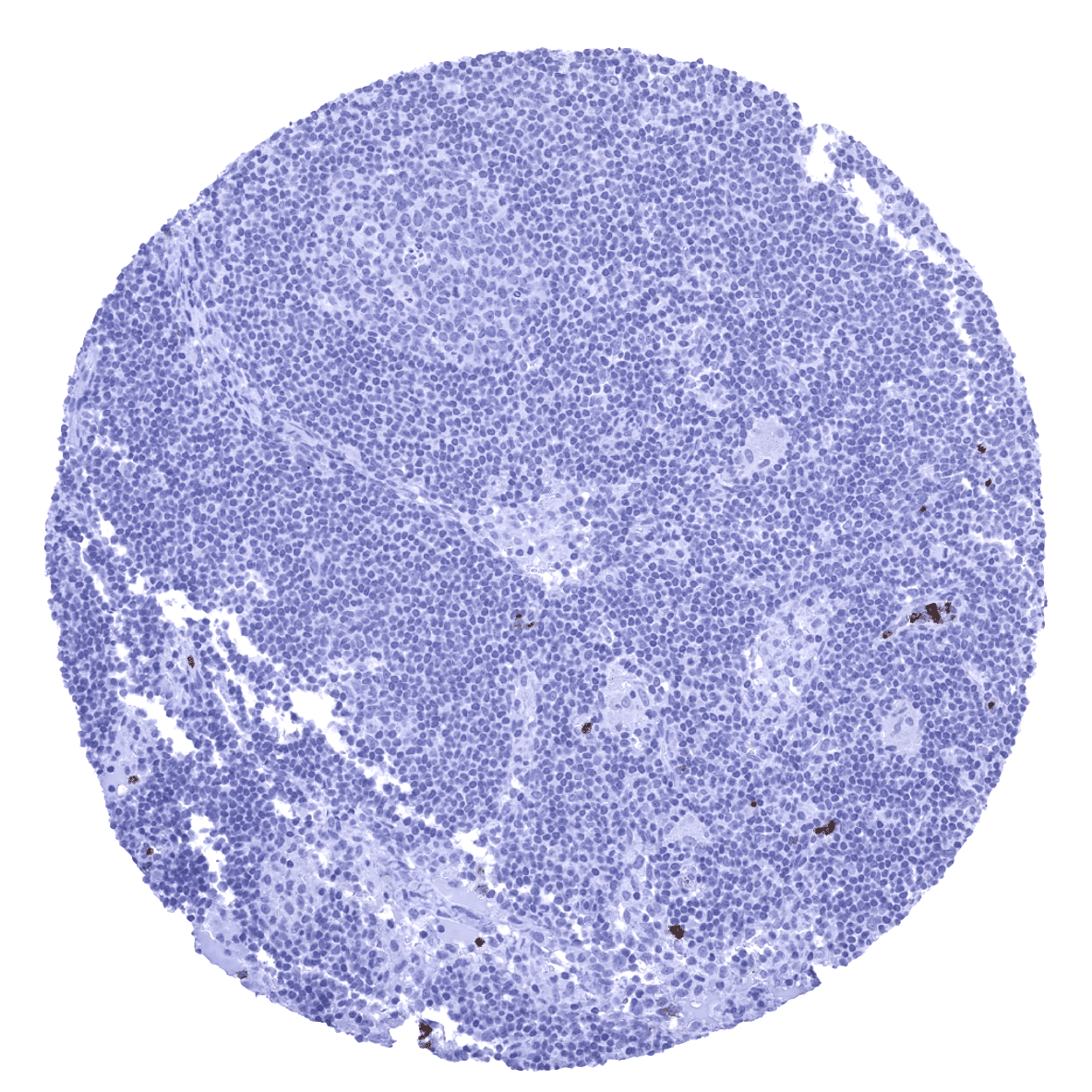
Thymus
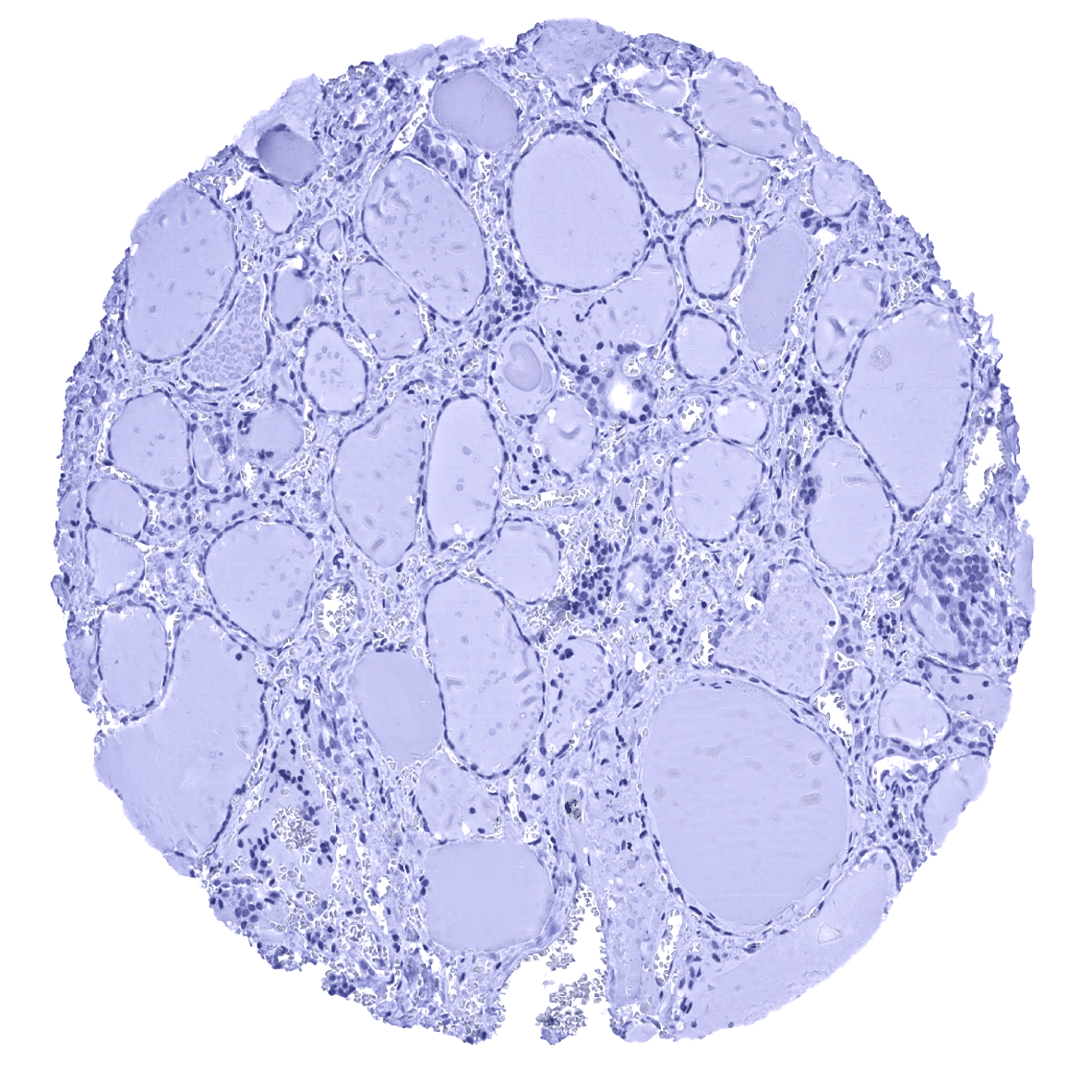
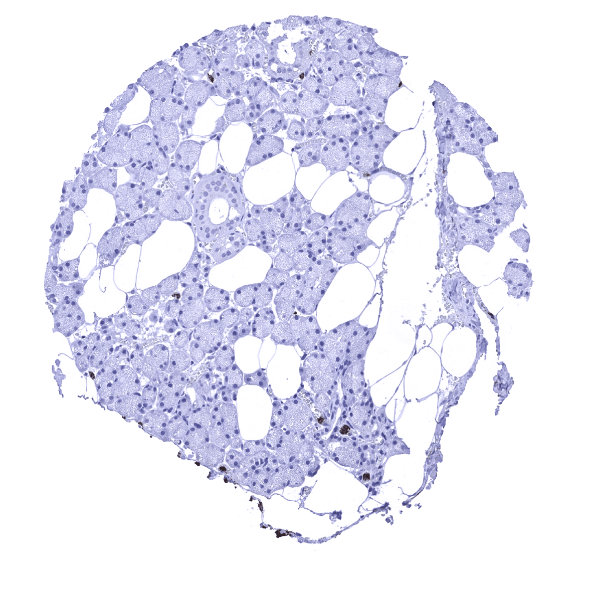
Thyroid gland
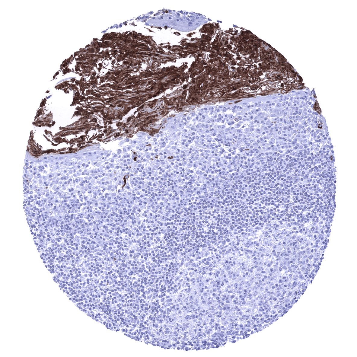
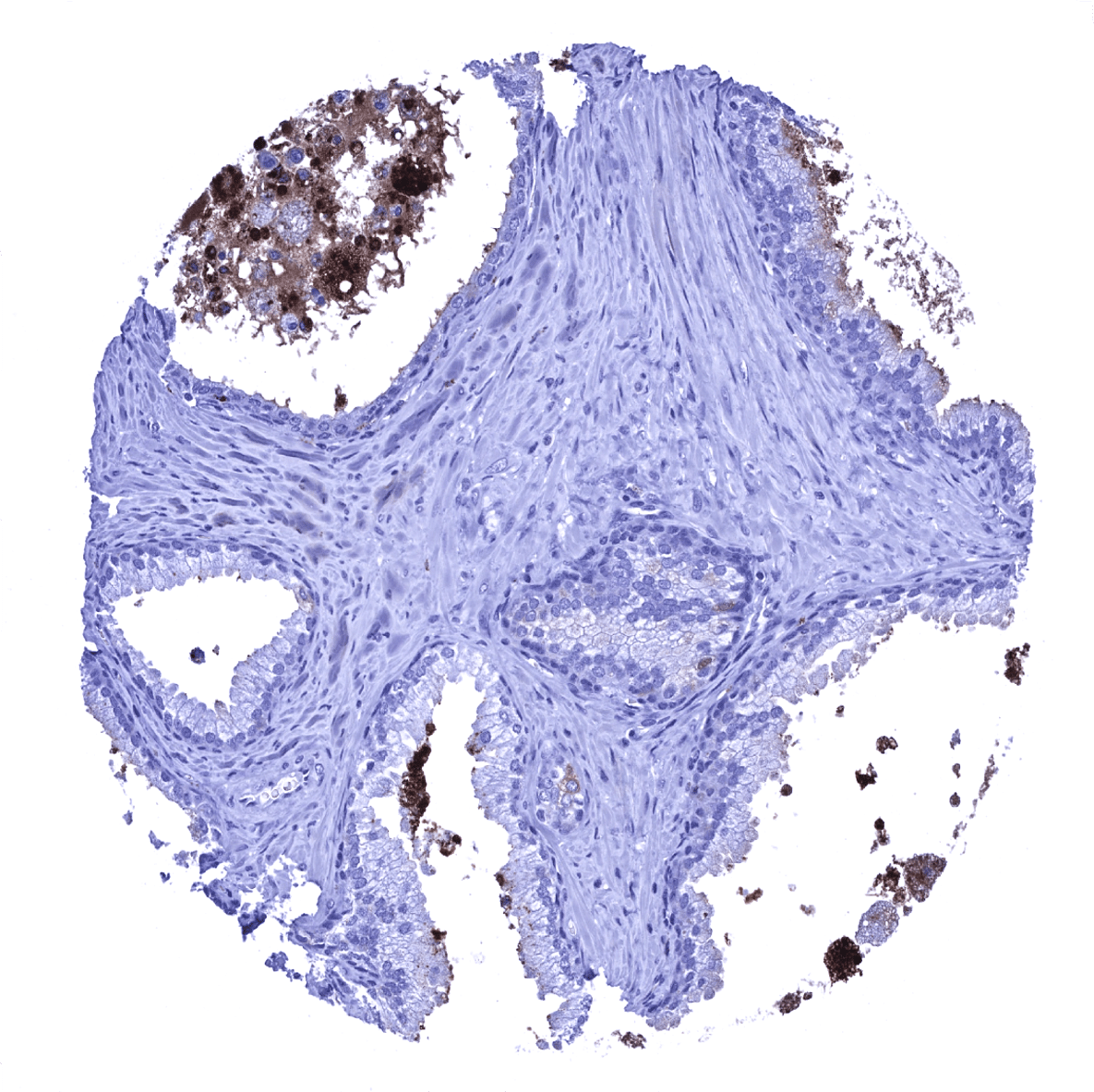
Tonsil - Significant MPO staining is seen in inflamed tonsil crypt epithelium which also contains numerous MPO positive granulocytes.
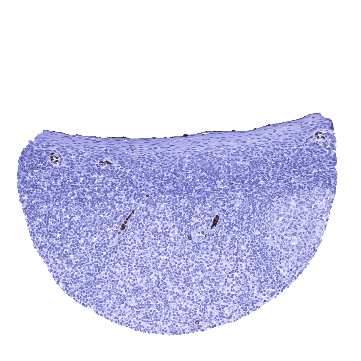
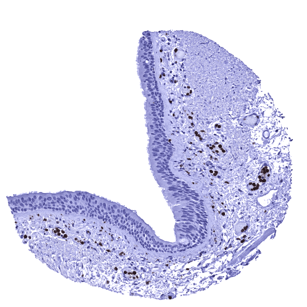
Tonsil, surface epithelium - MPO positive granulocytes are seen within small capillaries
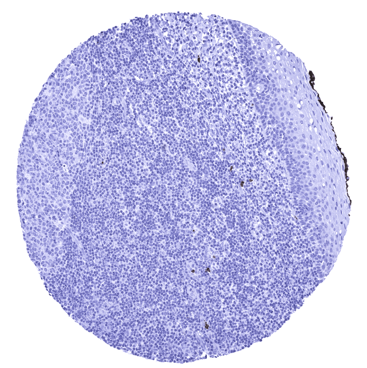
Tonsil, surface epithelium
Urinary bladder, muscular wall
Uterus, ectocervix
Uterus, endocervix
Uterus, endometrium (pregnancy)

Uterus, endometrium (proliferation)
Uterus, endometrium (secretion)
Uterus, myometrium