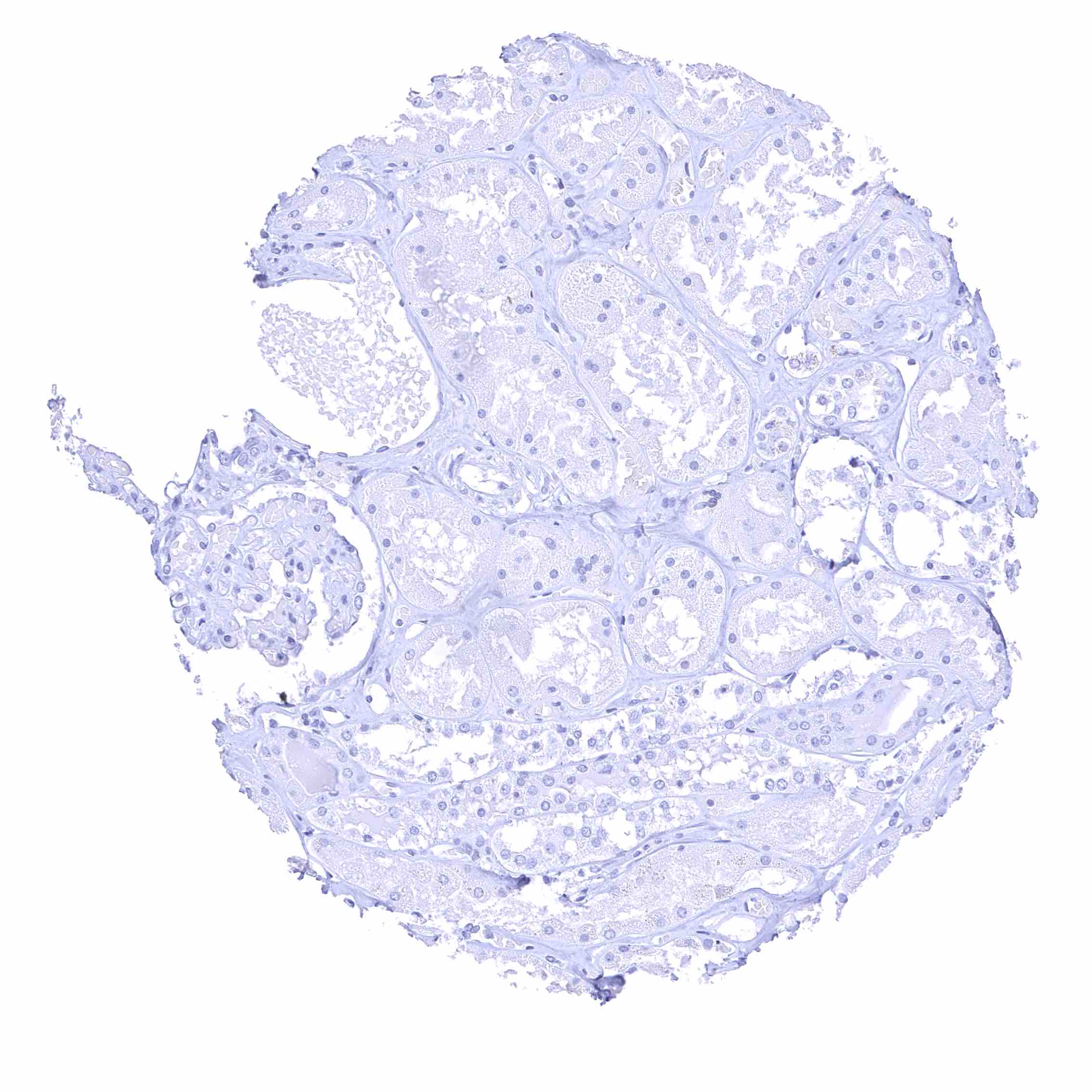
Adrenal gland
Aorta, endothelium
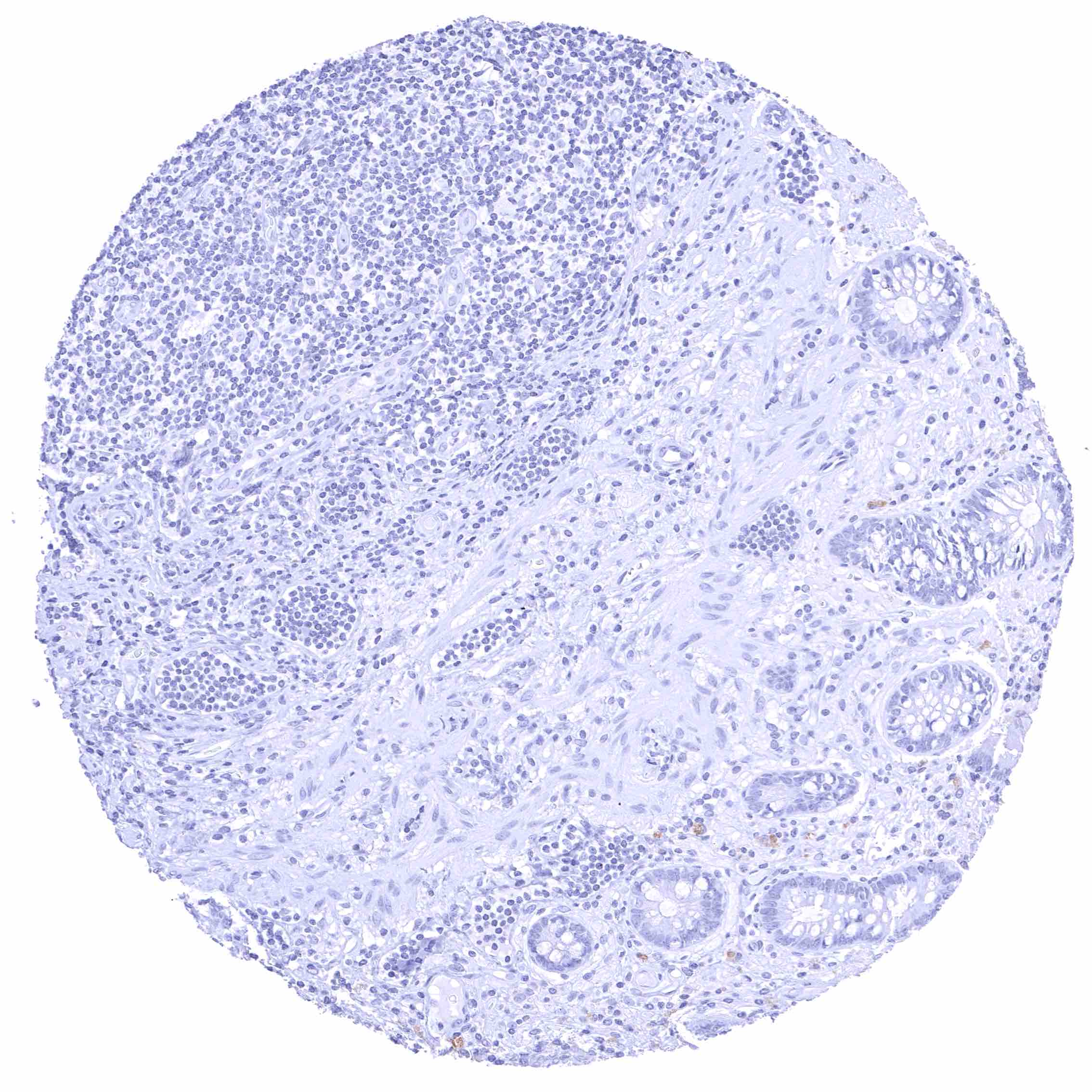
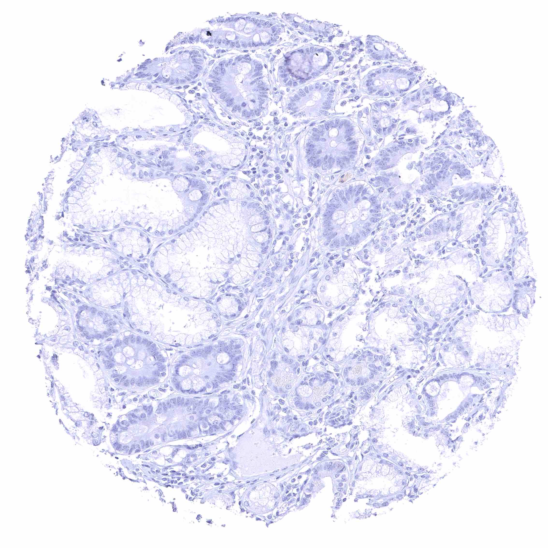
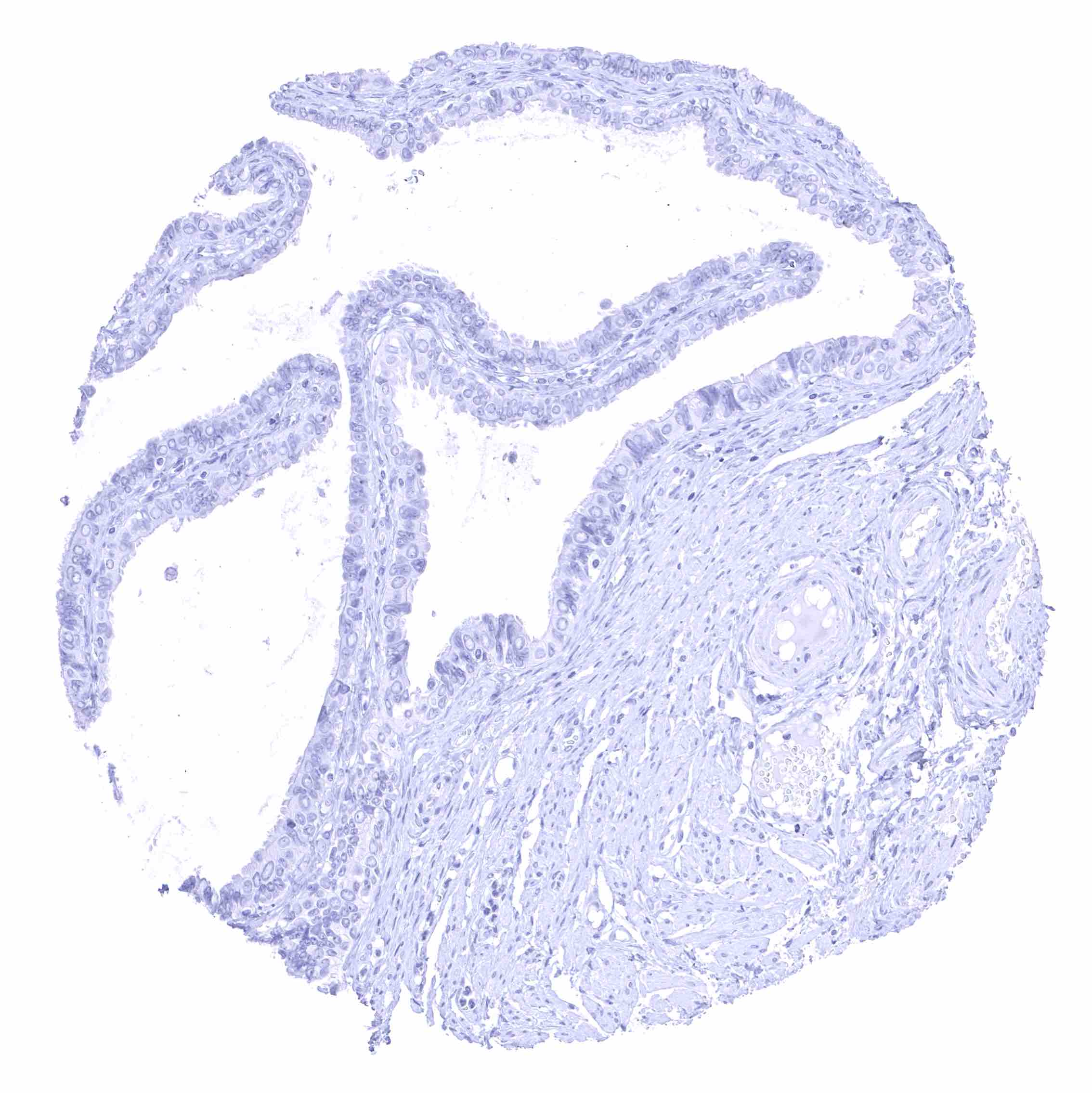
Appendix, mucosa.jpeg
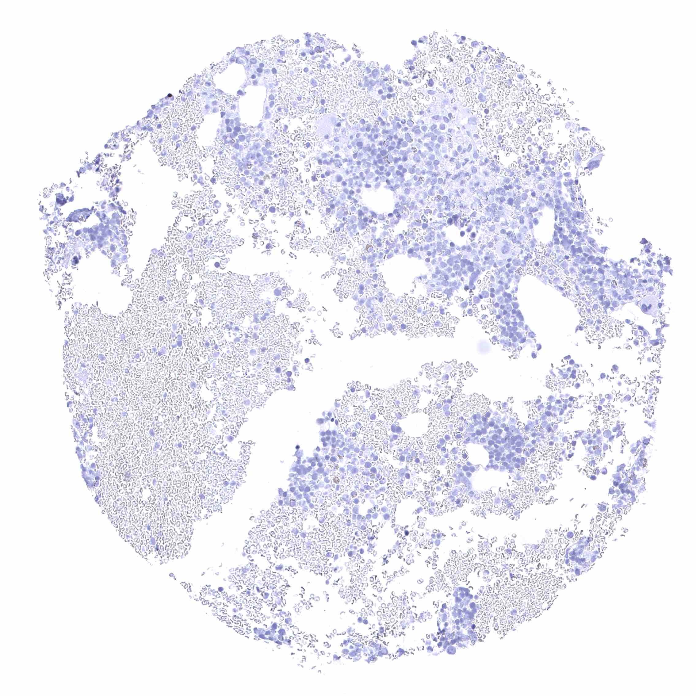
Bone marrow
Breast
Bronchus, mucosa
Cerebellum (white matter)
Cerebellum, cortex (molecular layer, Purkinje cell layer, granule cell layer) .jpeg
Cerebrum, grey matter
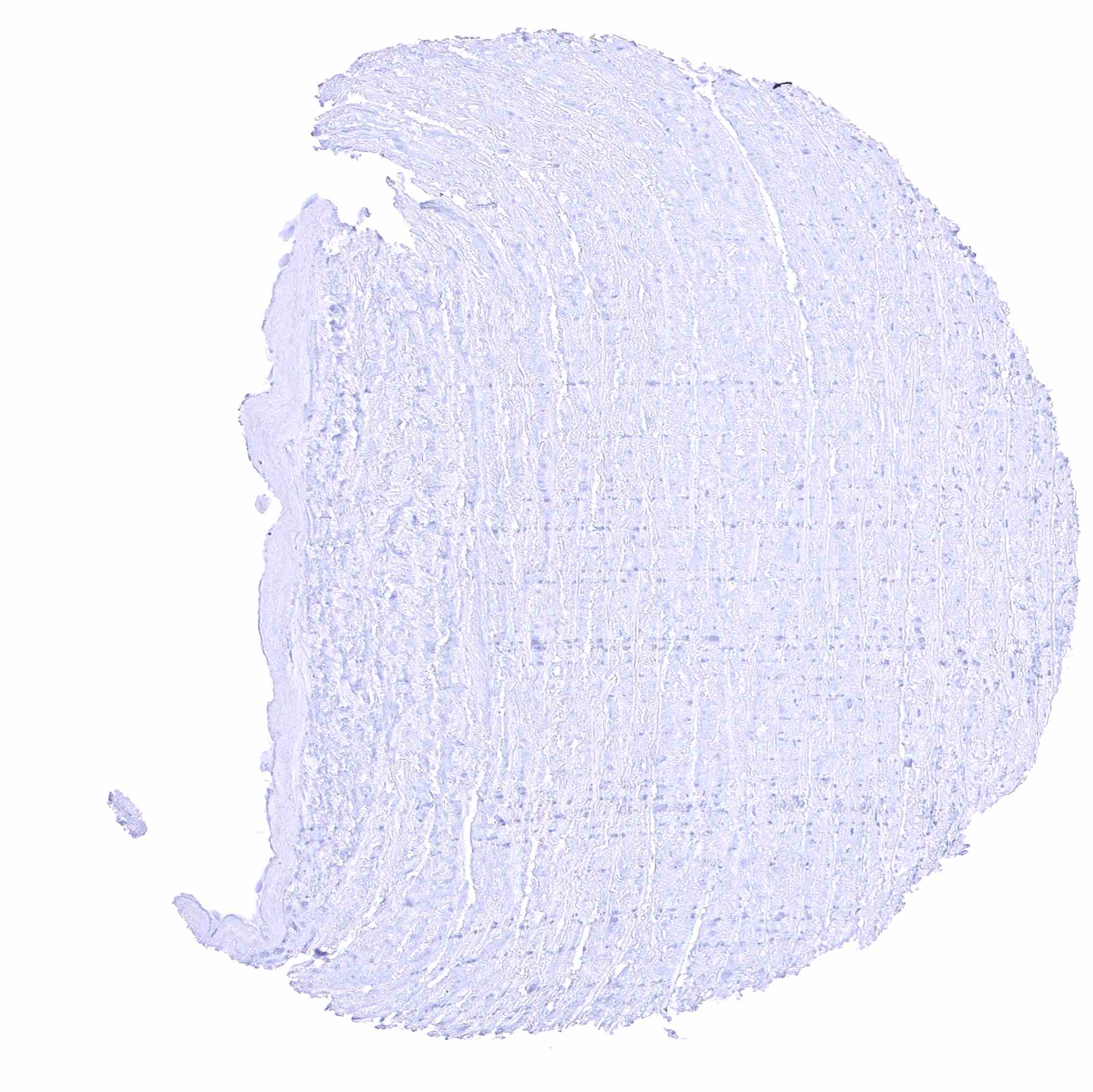
Cerebrum, white matter.jpeg
Colon descendens, mucosa
Duodenum, Brunner gland
Duodenum, mucosa
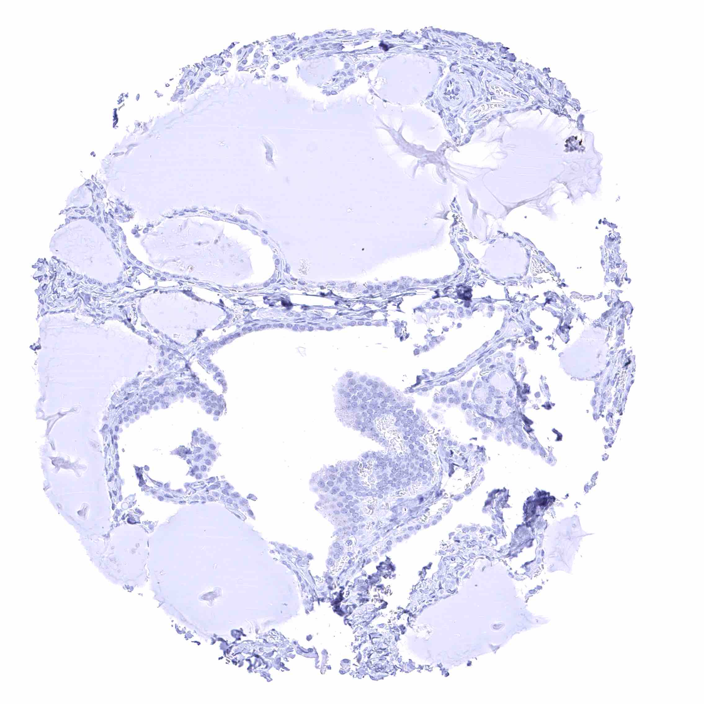
Epididymis (Cauda)
Epididymis (Corpus)
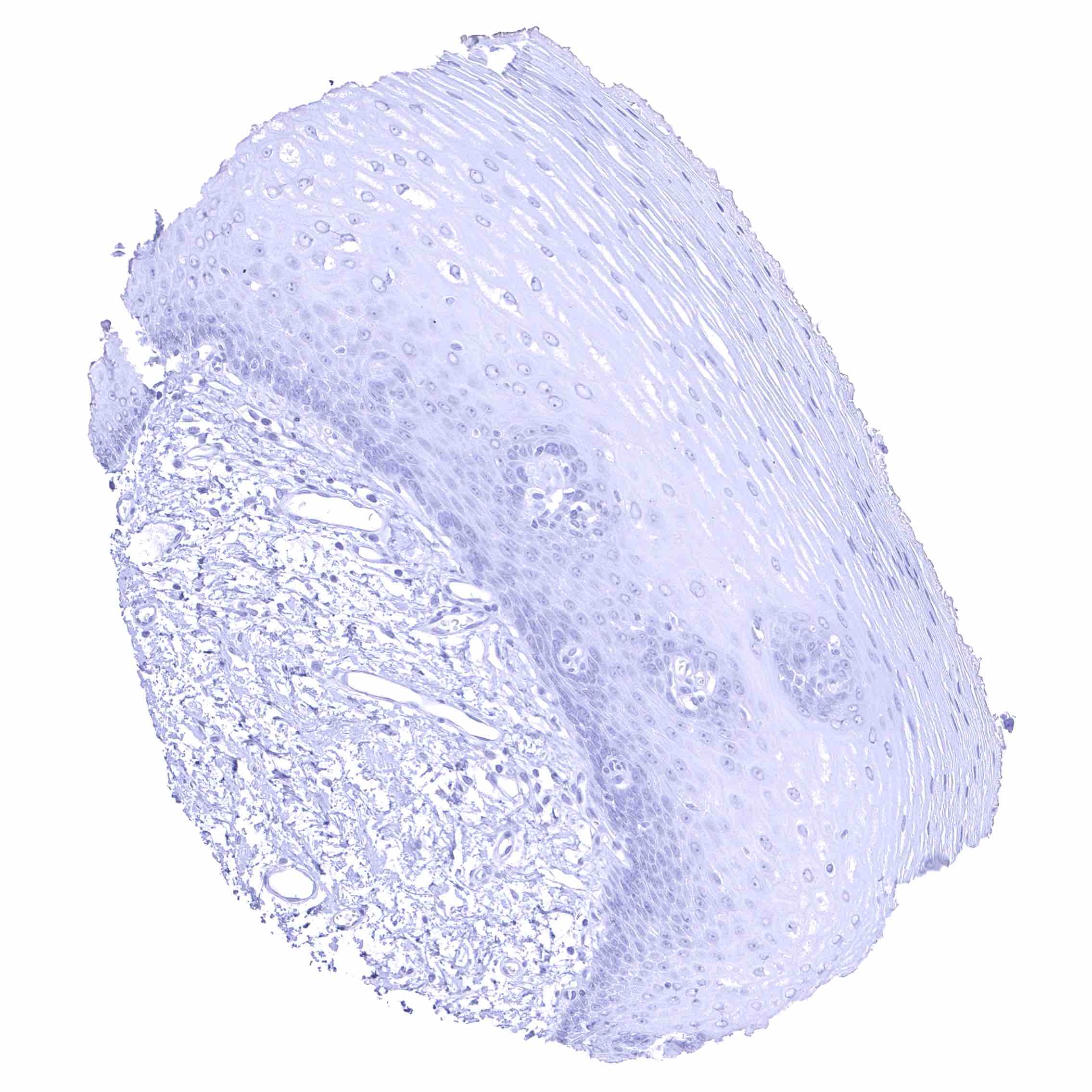
Esophagus, squamous epithelium
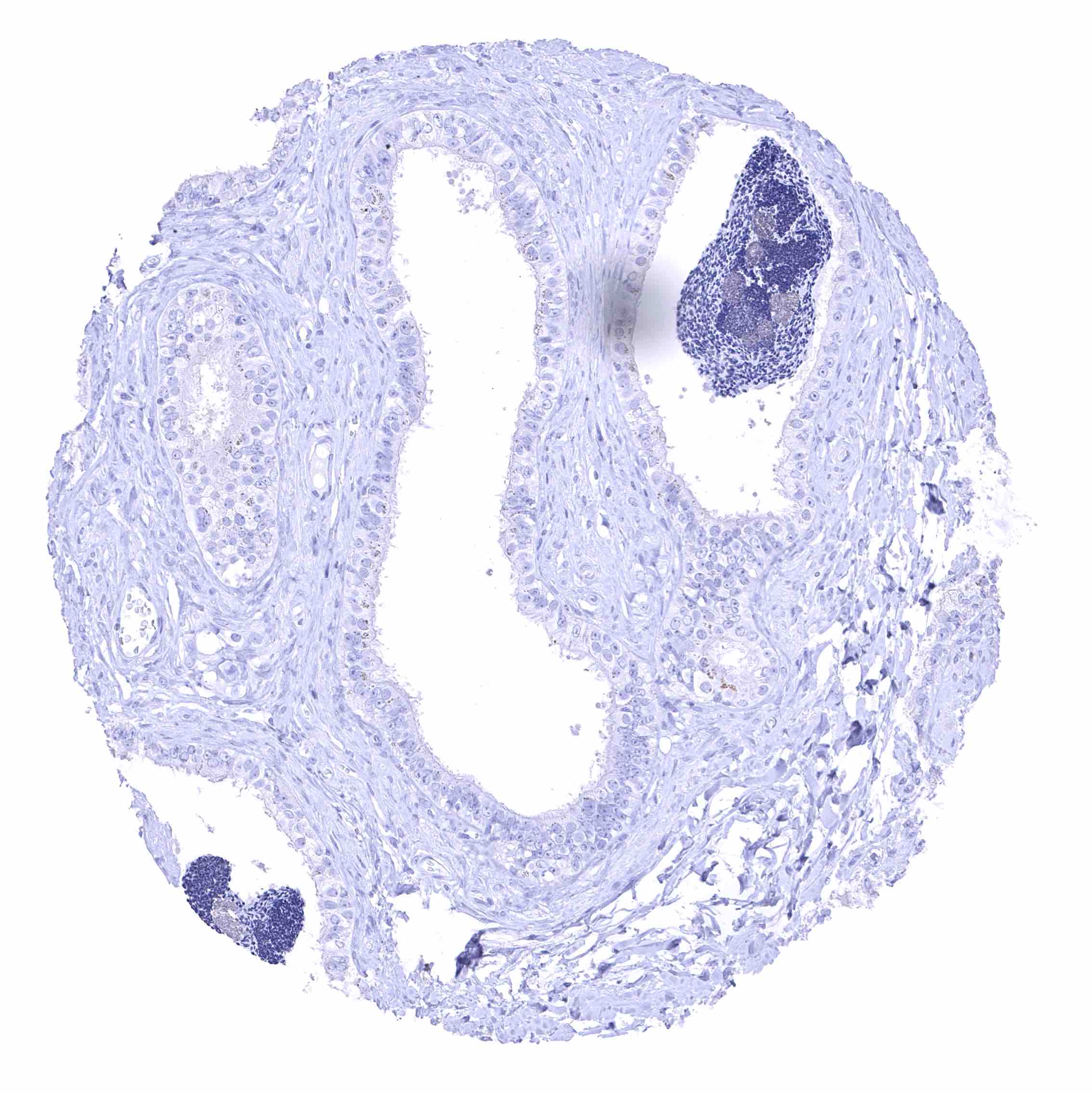
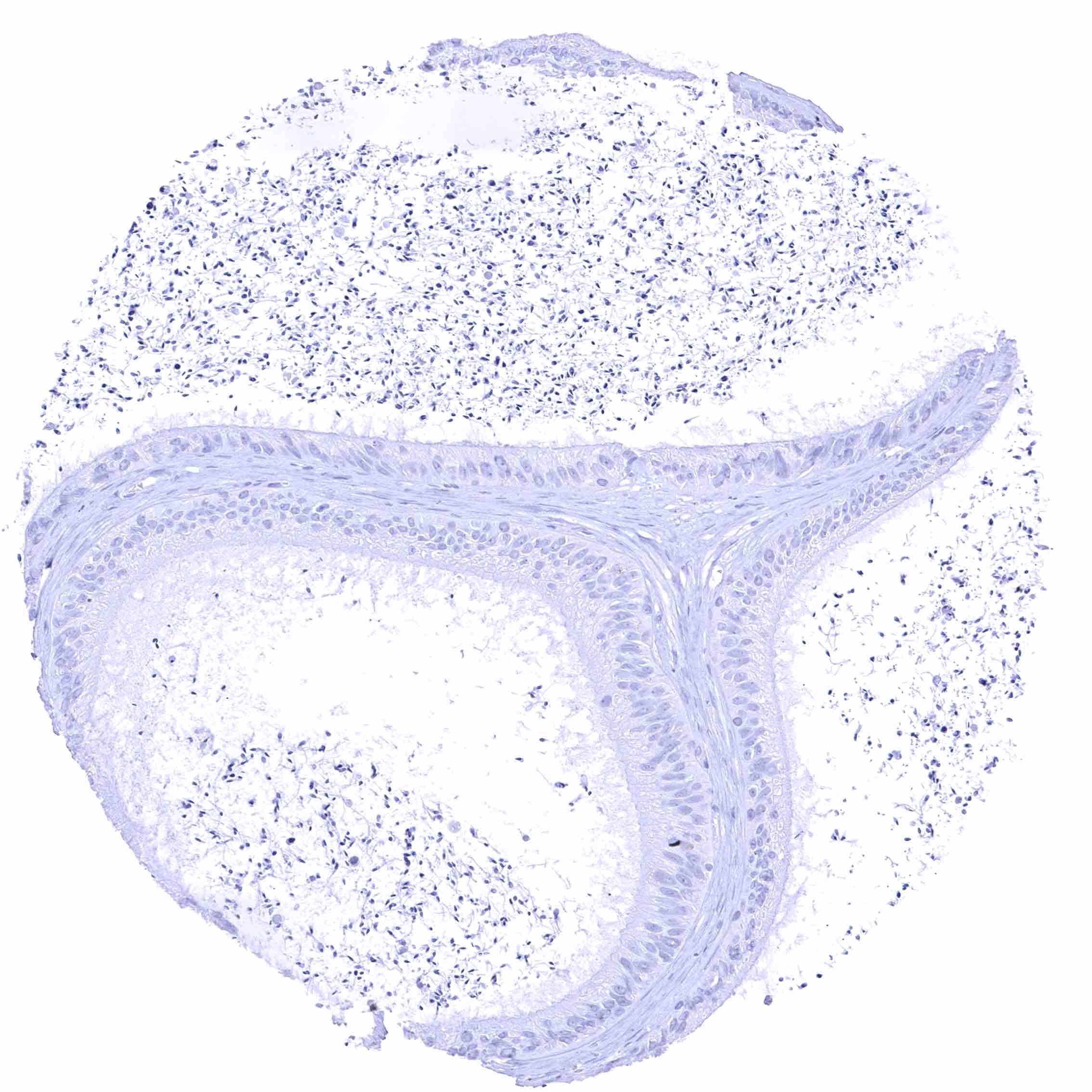
Fallopian tube, mucosa
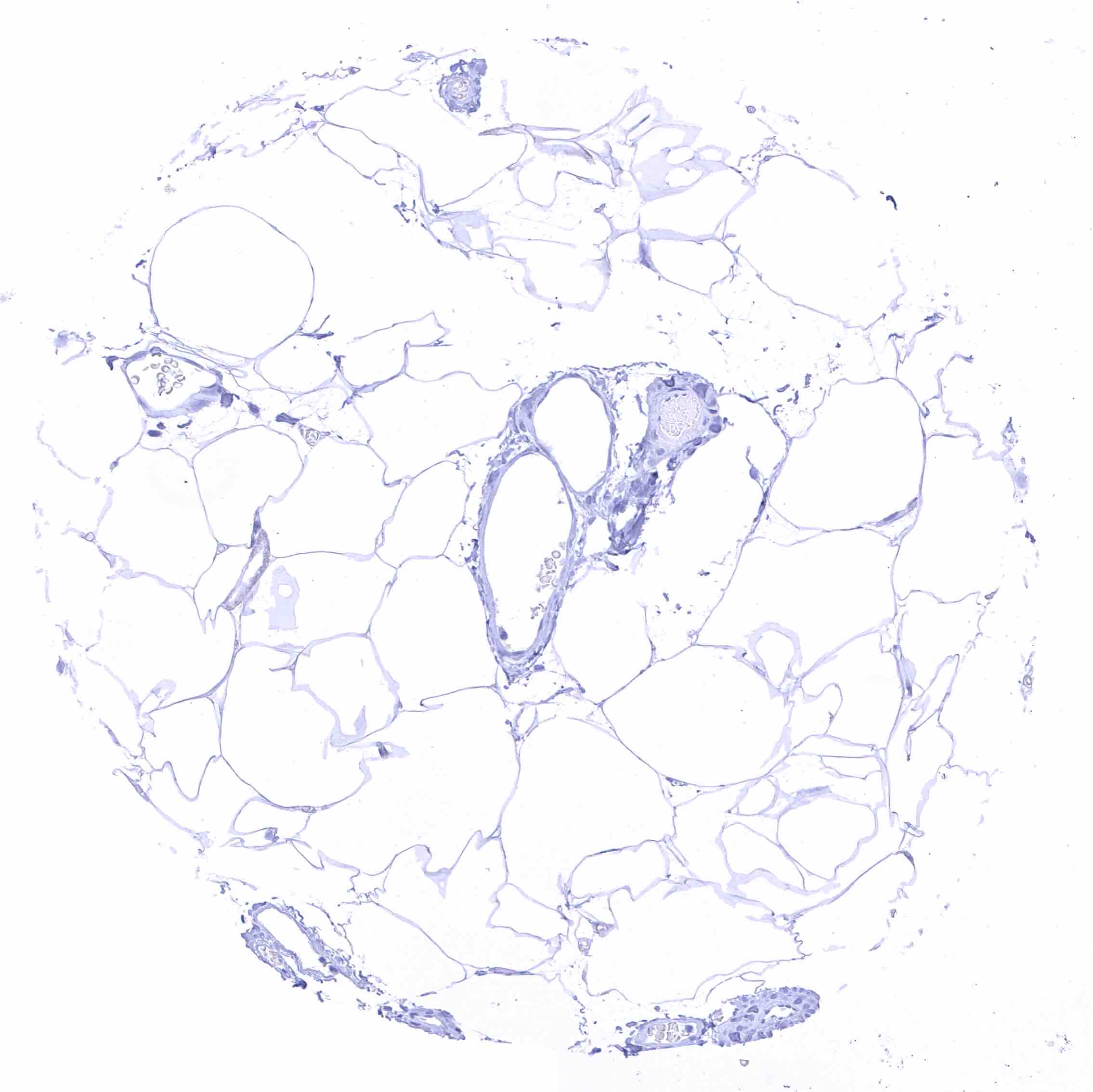
Fat
Gallbladder, epithelium
Heart muscle
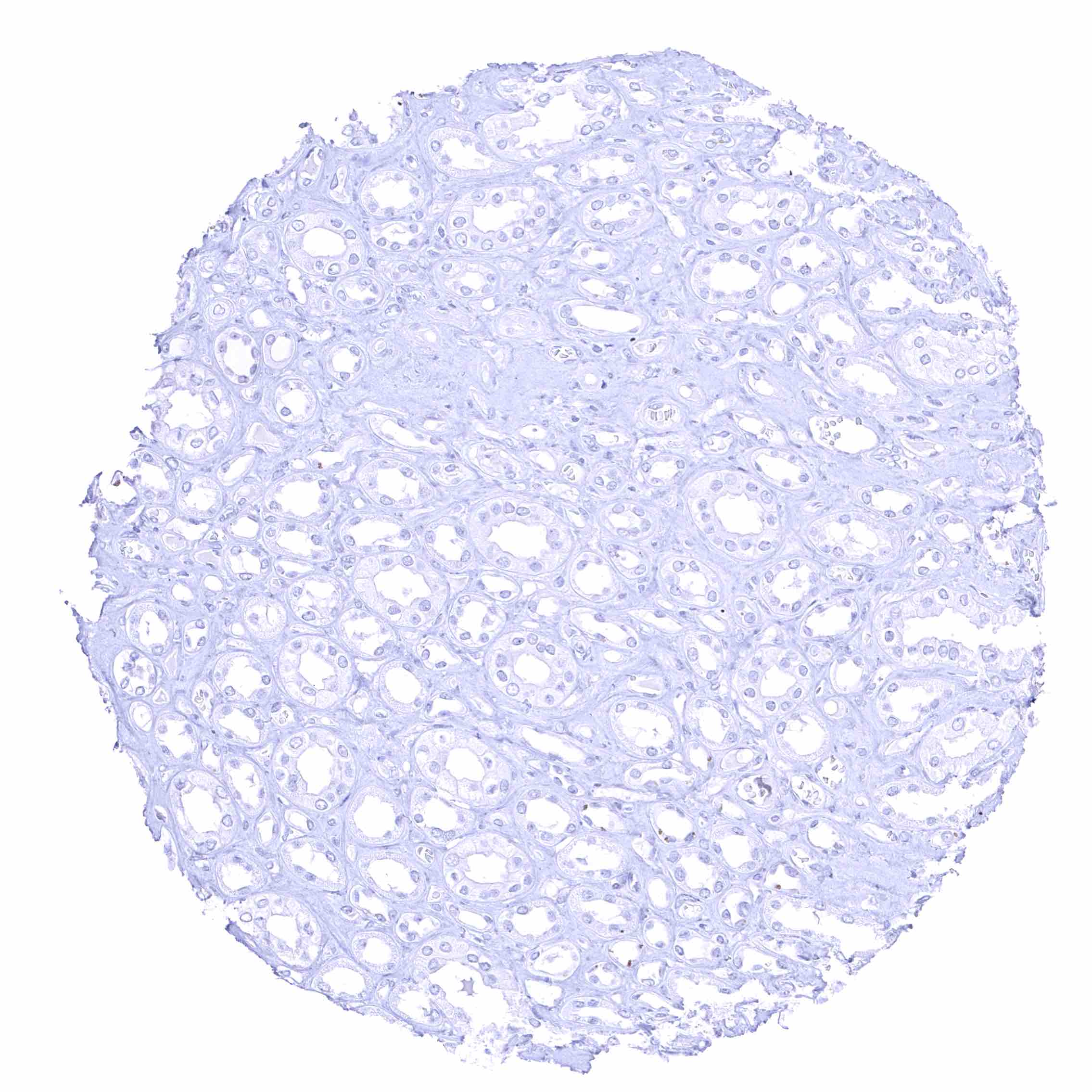
Kidney, cortex
Kidney, medulla
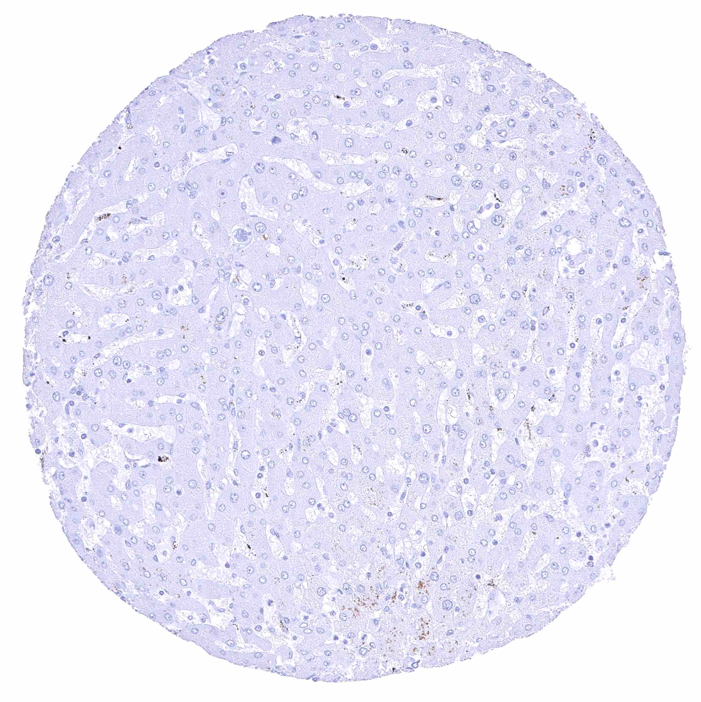
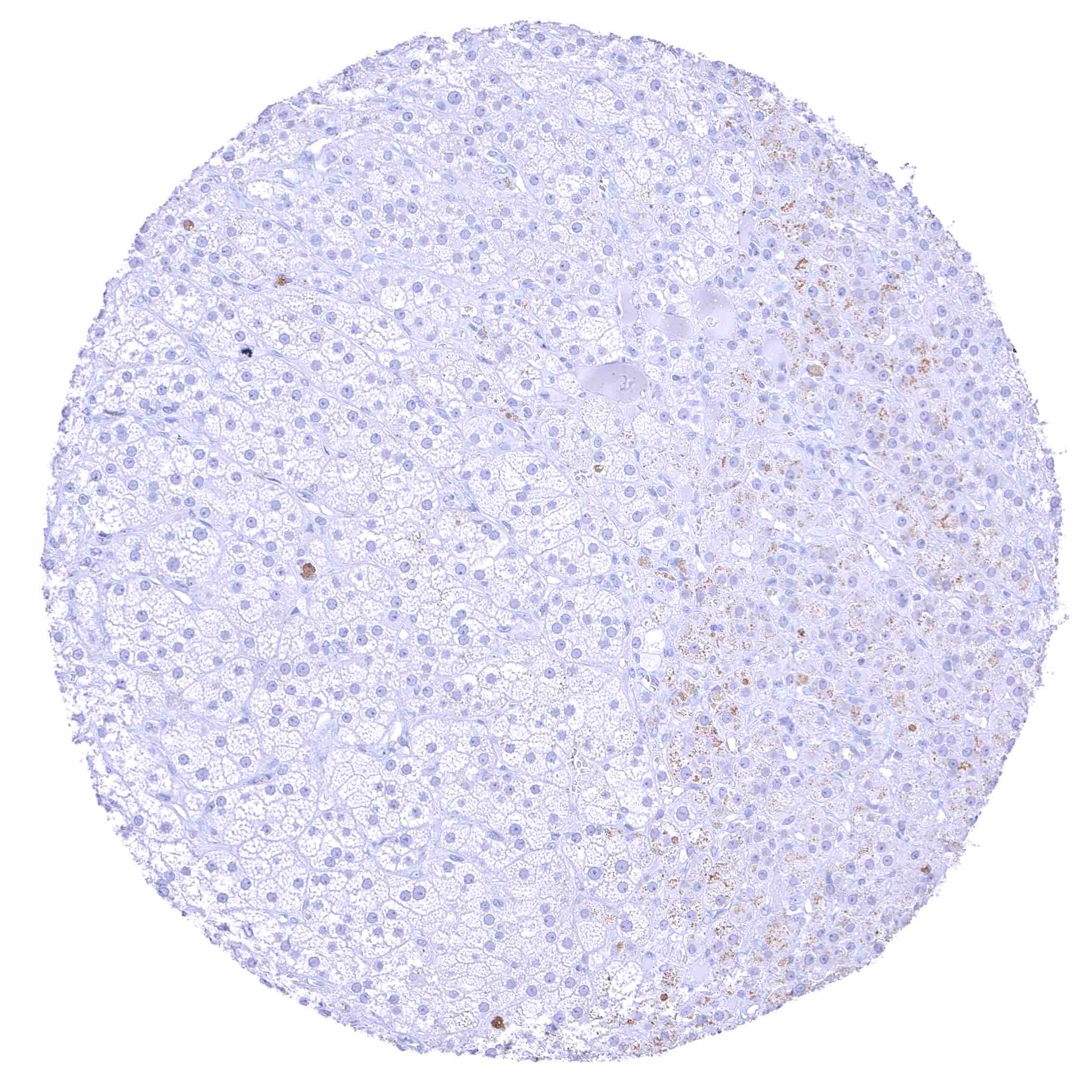
Liver
Lung
Lymph node
Ovary, corpus luteum
Ovary, stroma
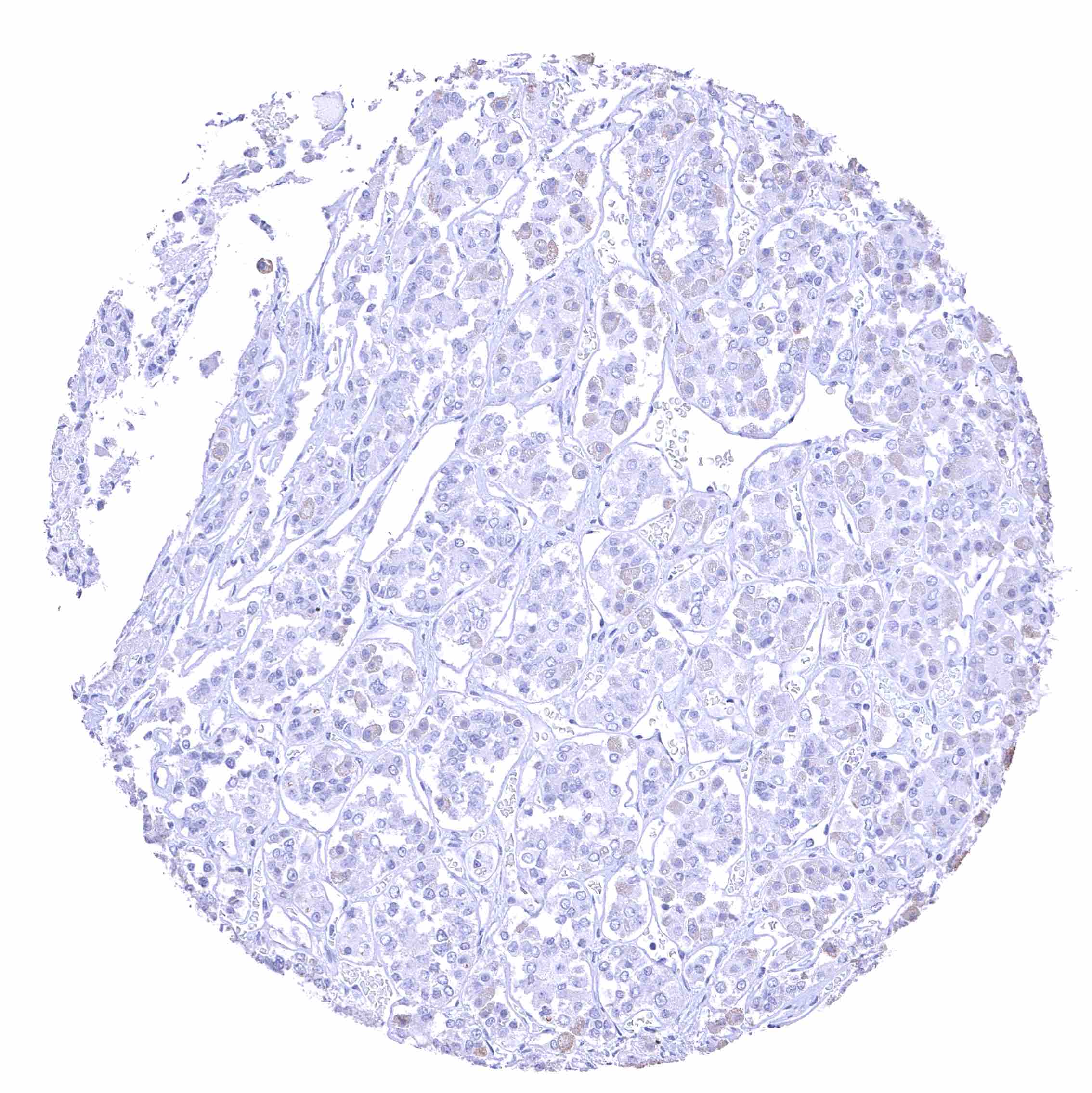
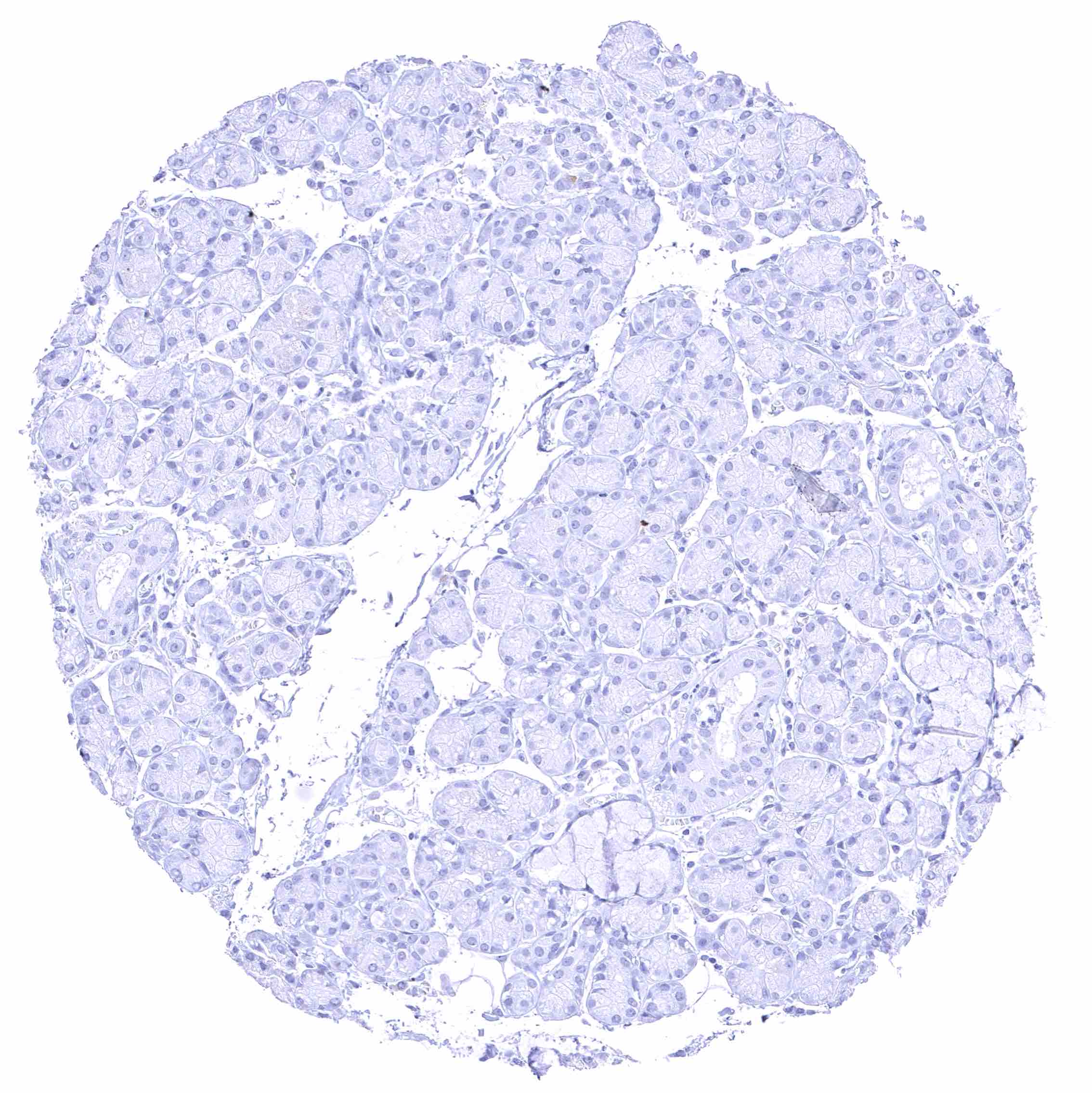
Pancreas
Pancreas
Parathyroid gland
Parotid gland
Pituitary gland, anterior lobe
Pituitary gland, posterior lobe
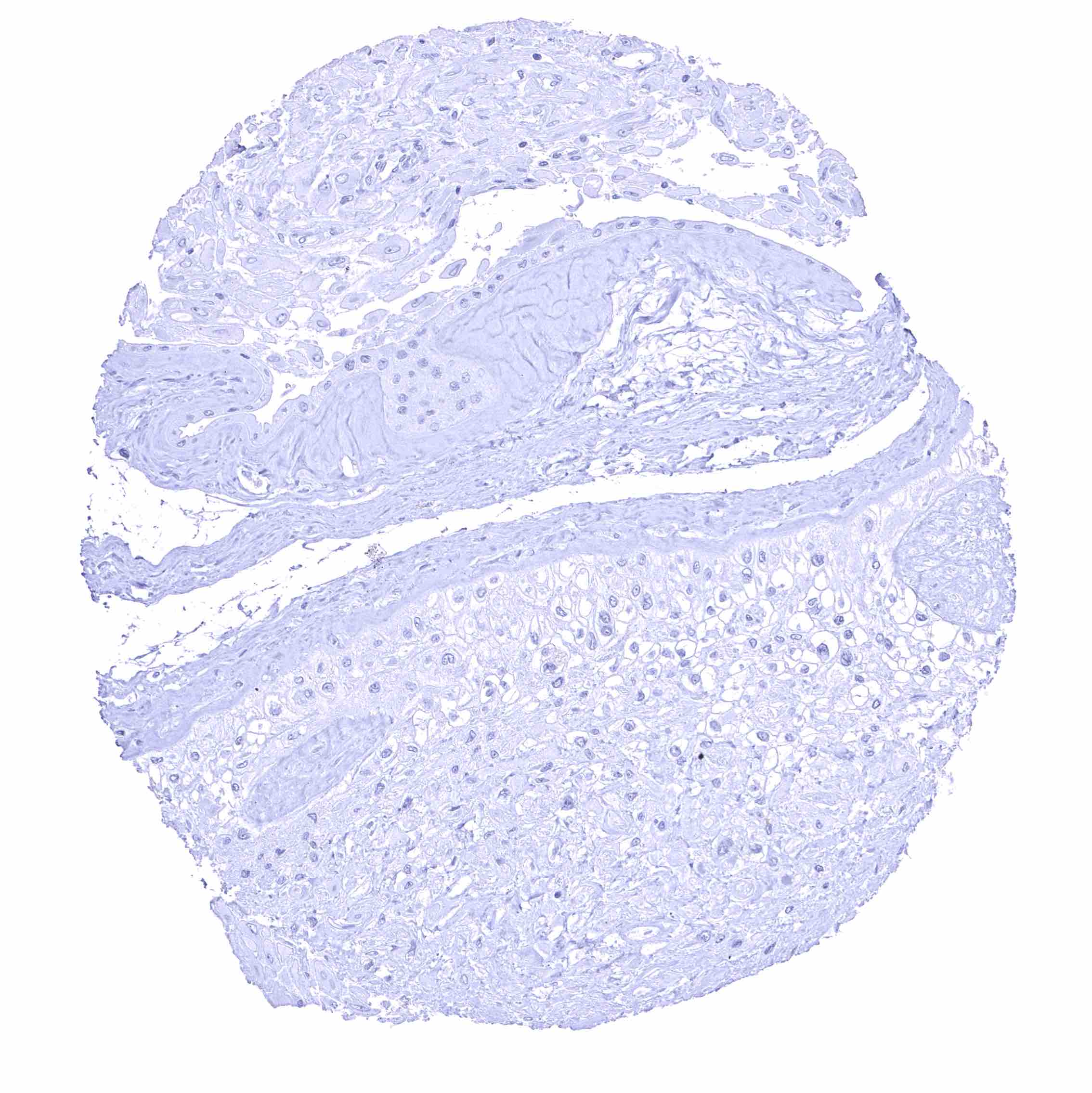
Placenta (amnion and chorion)
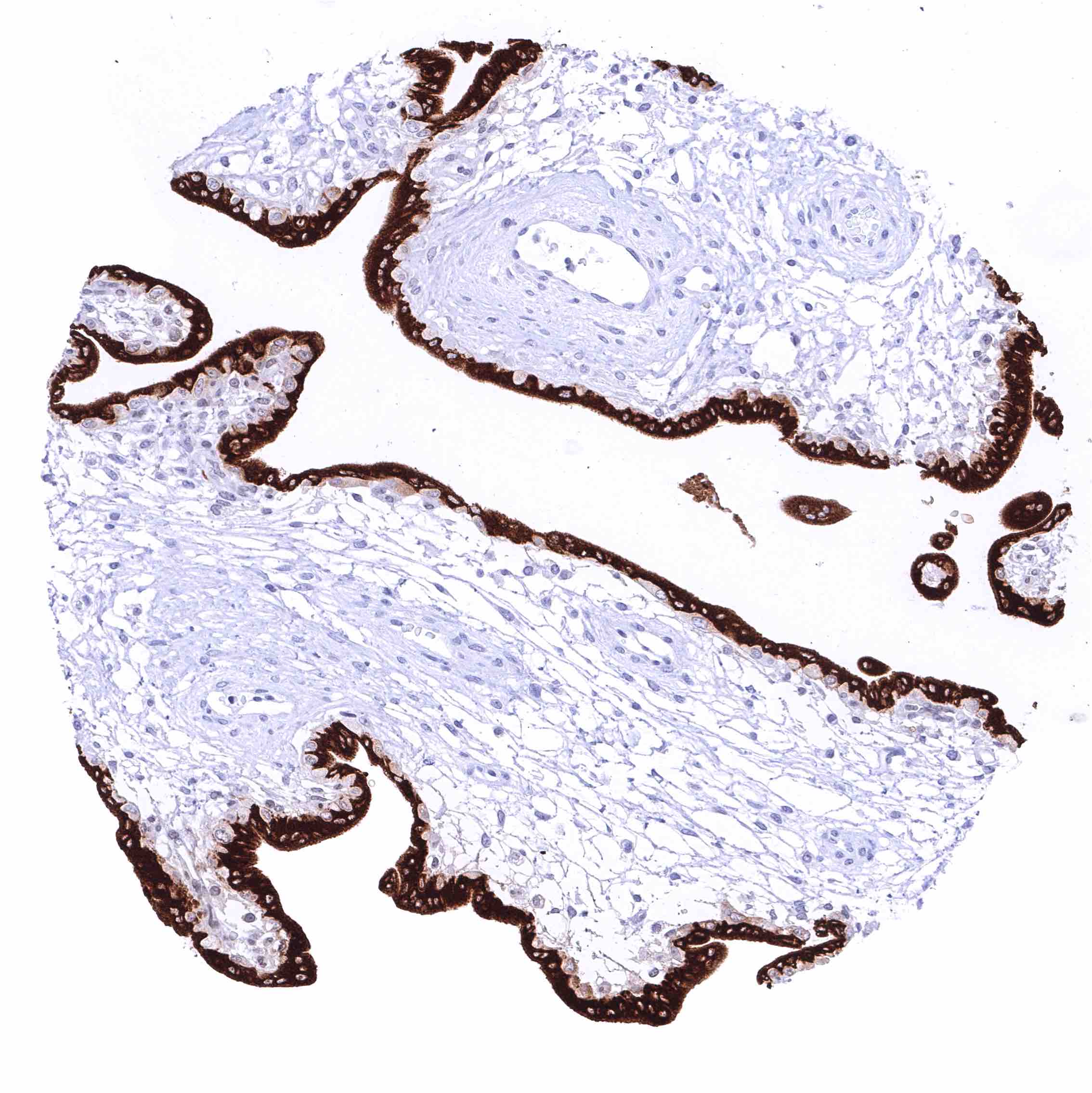
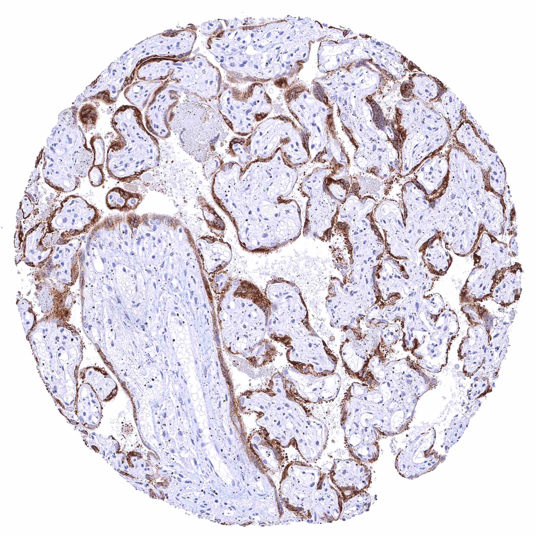
Placenta (first Trimenon) – Intense cytoplasmic KISS1 staining of the syncytiotrophoblast while cytotrophoblast and other cells remain KISS1 negative.
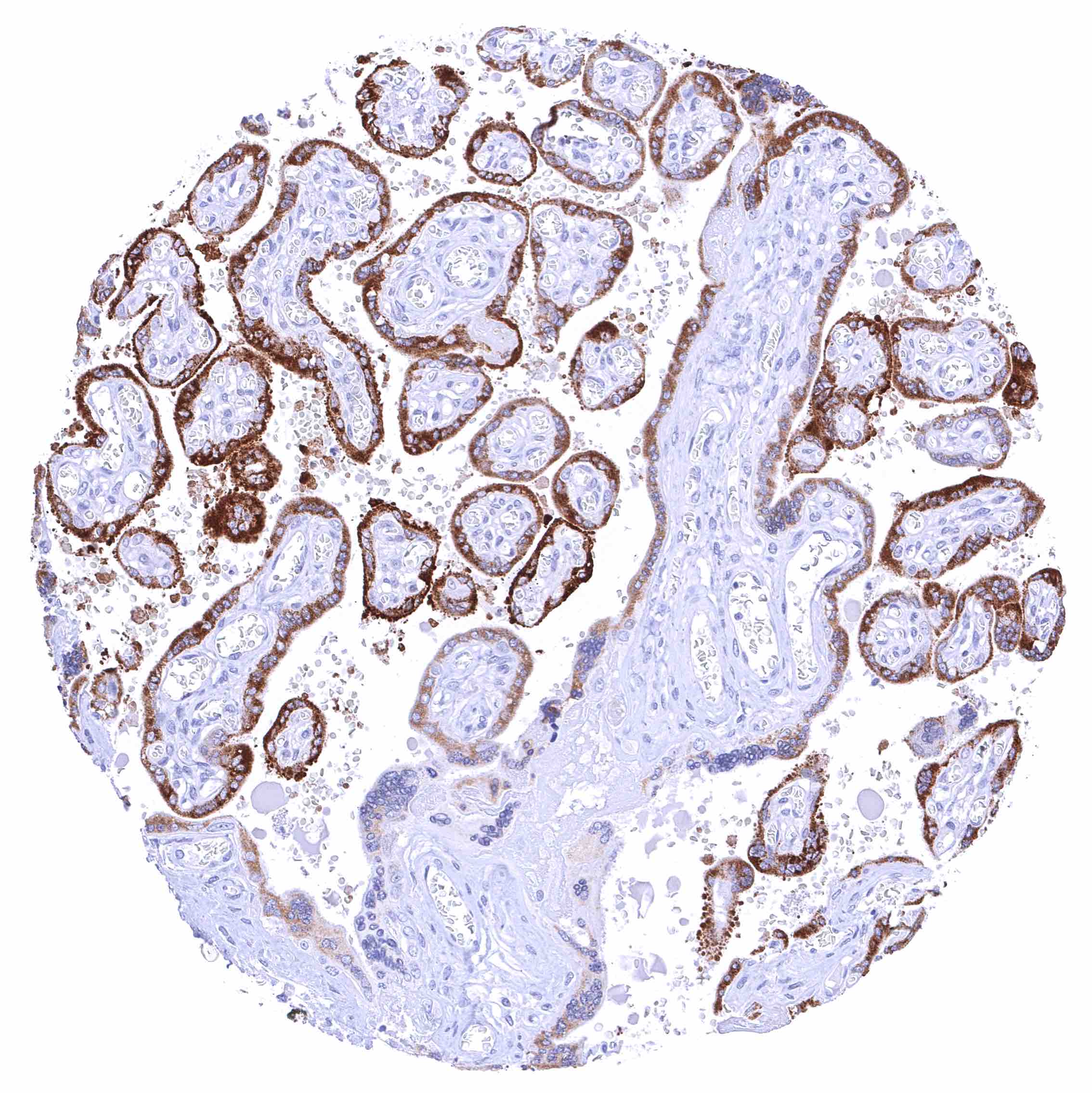
Placenta, mature – Strong cytoplasmic KISS1 staining of most cells of the syncytiotrophoblast.
Placenta, mature – Strong cytoplasmic KISS1 staining of the majority of the syncytiotrophoblast cells.
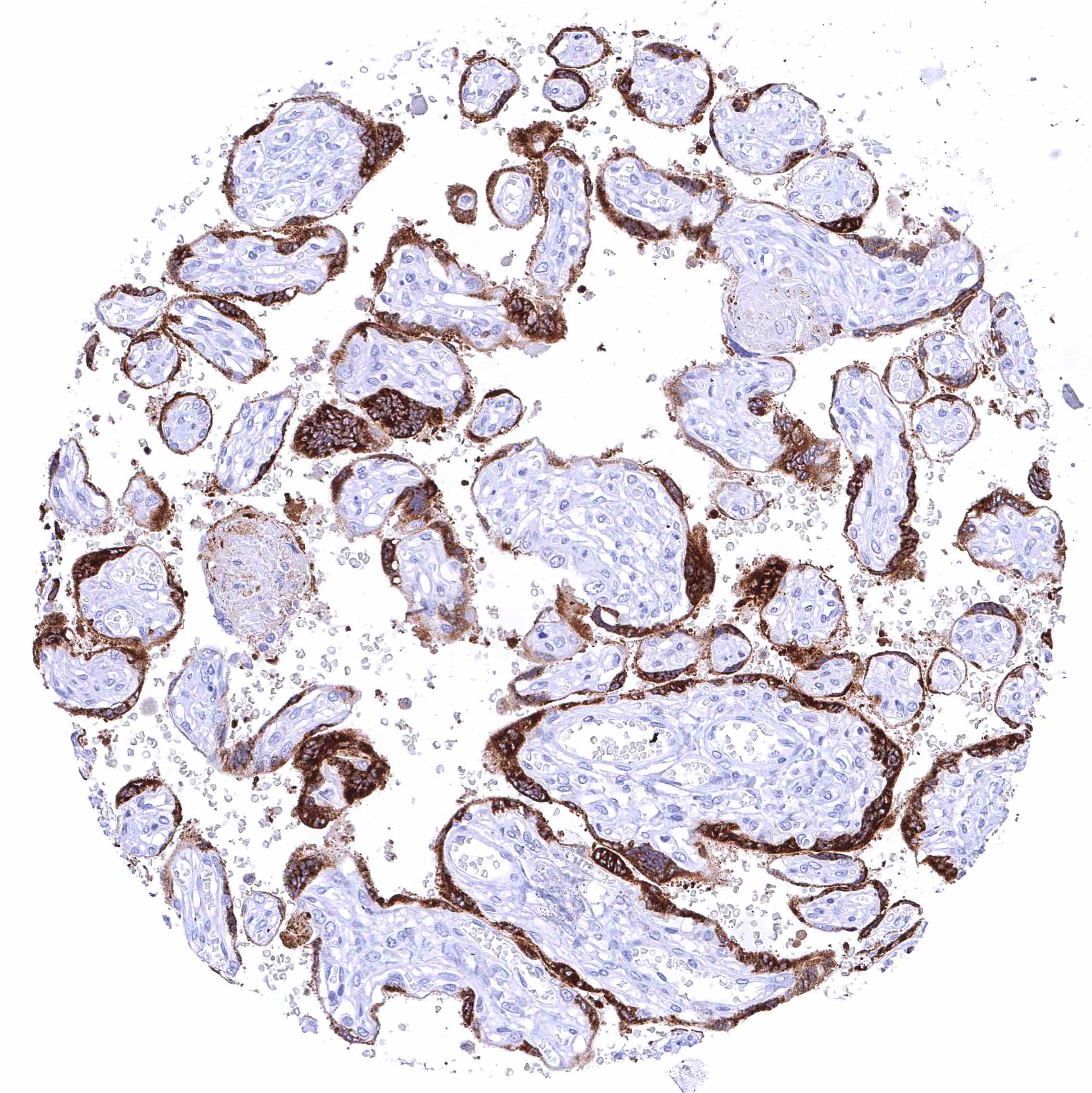
Placenta, mature – Strong cytoplasmic KISS1 staining of the syncytiotrophoblast.
Prostate
Rectum, mucosa
Seminal vesicle
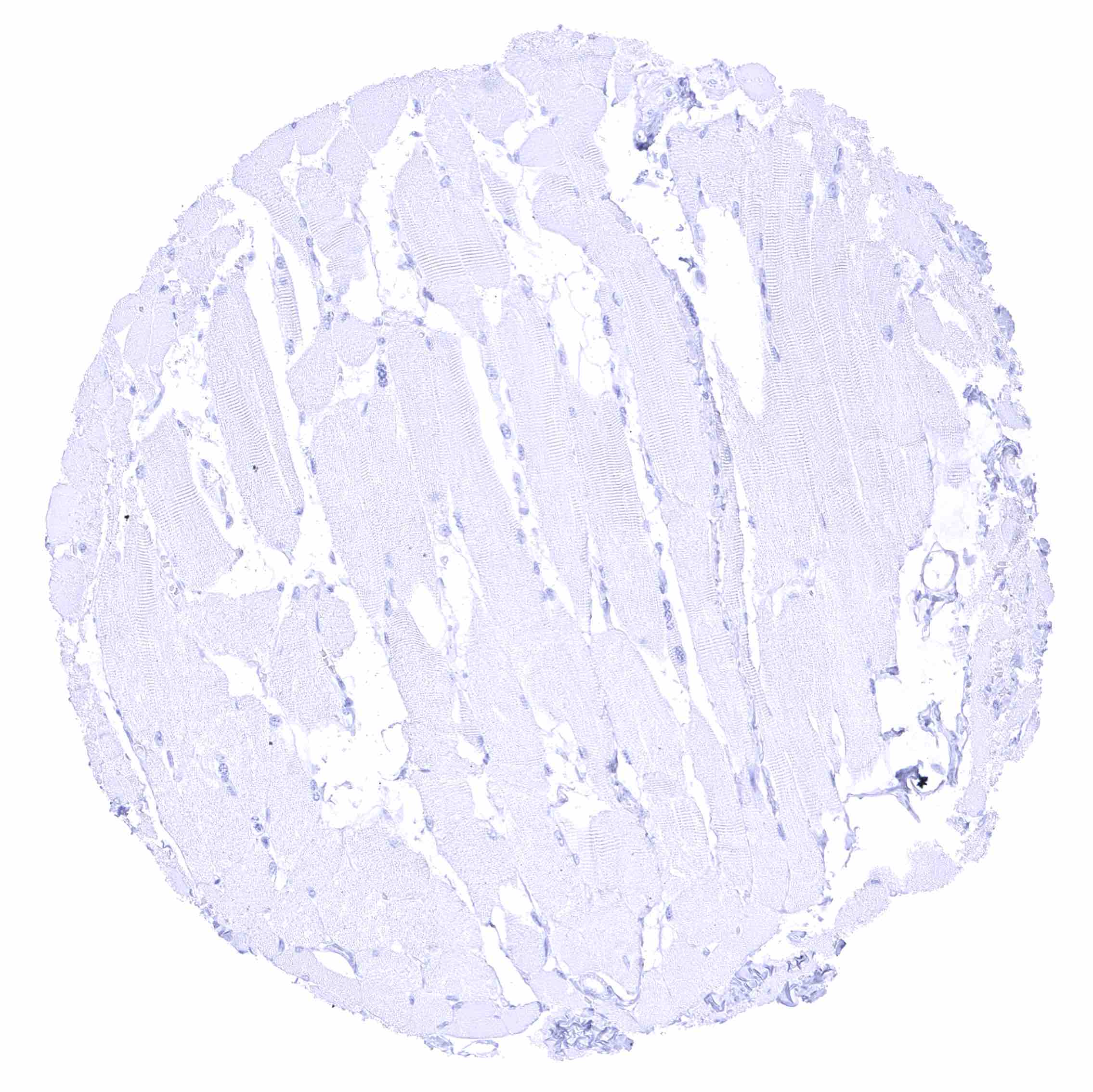
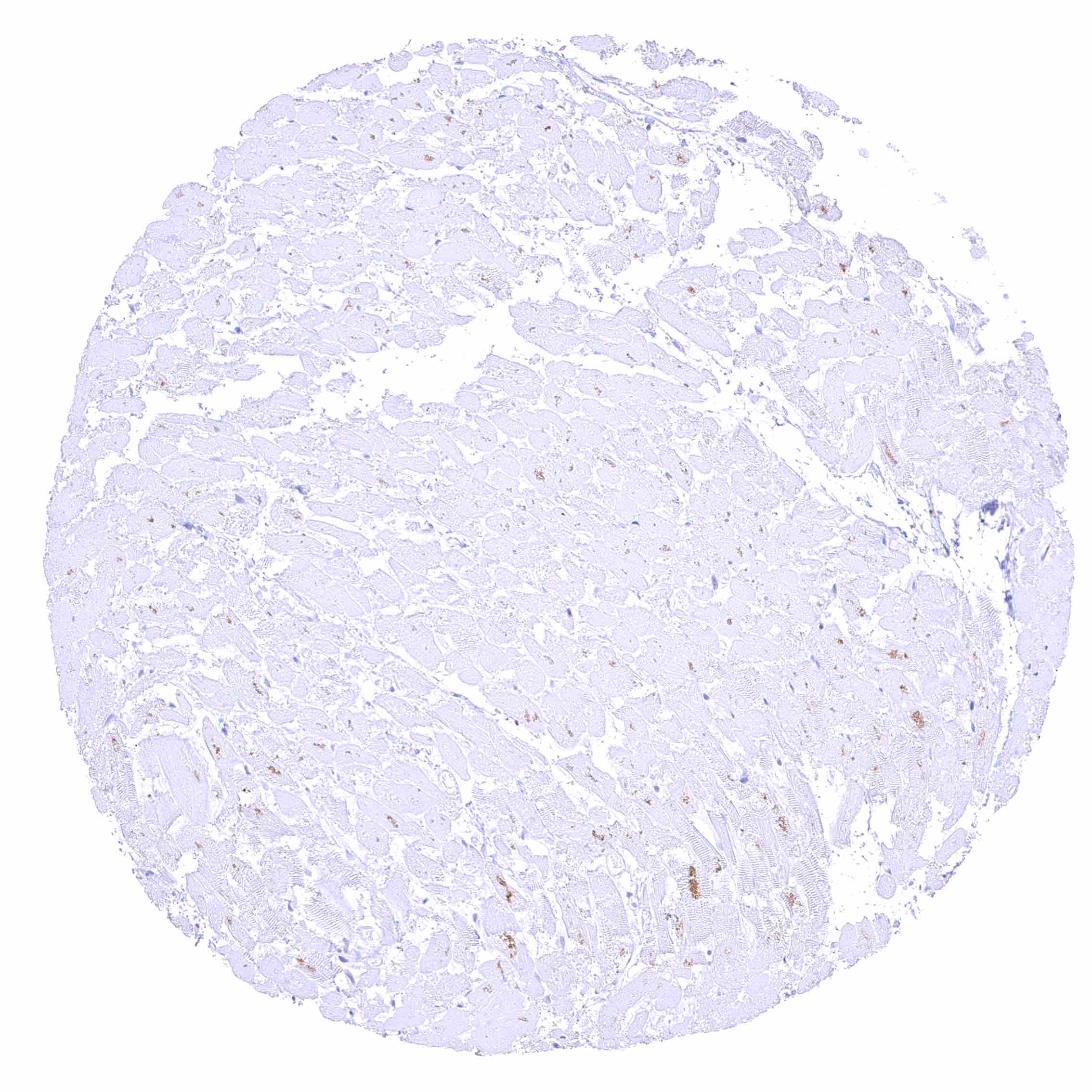
Skeletal muscle
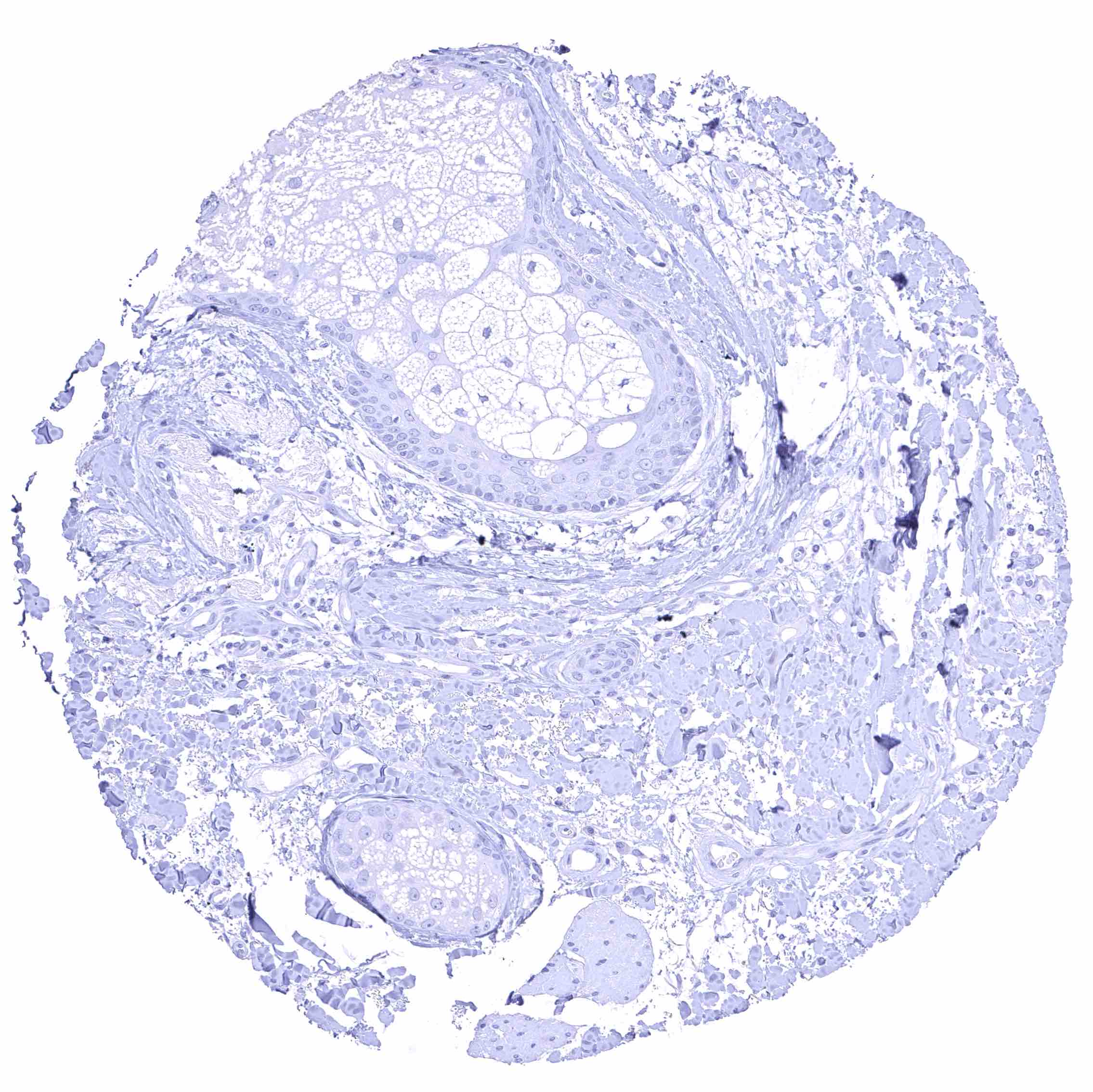
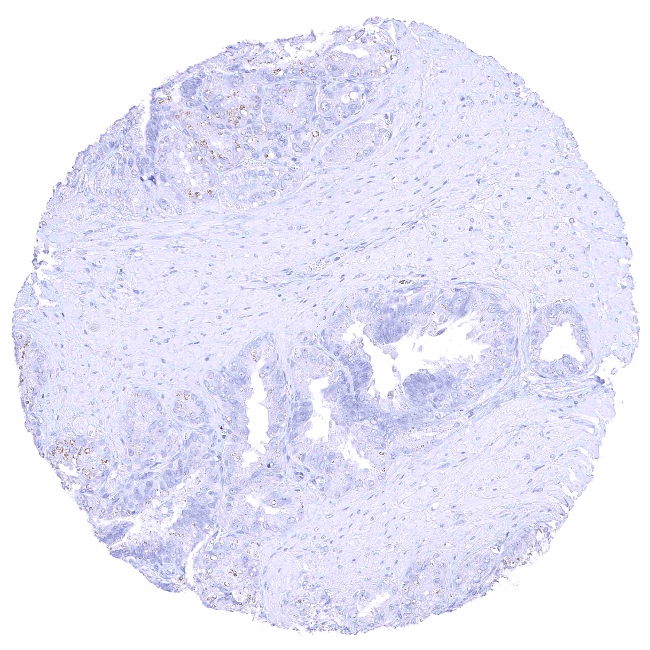
Skin, sebaceous gland
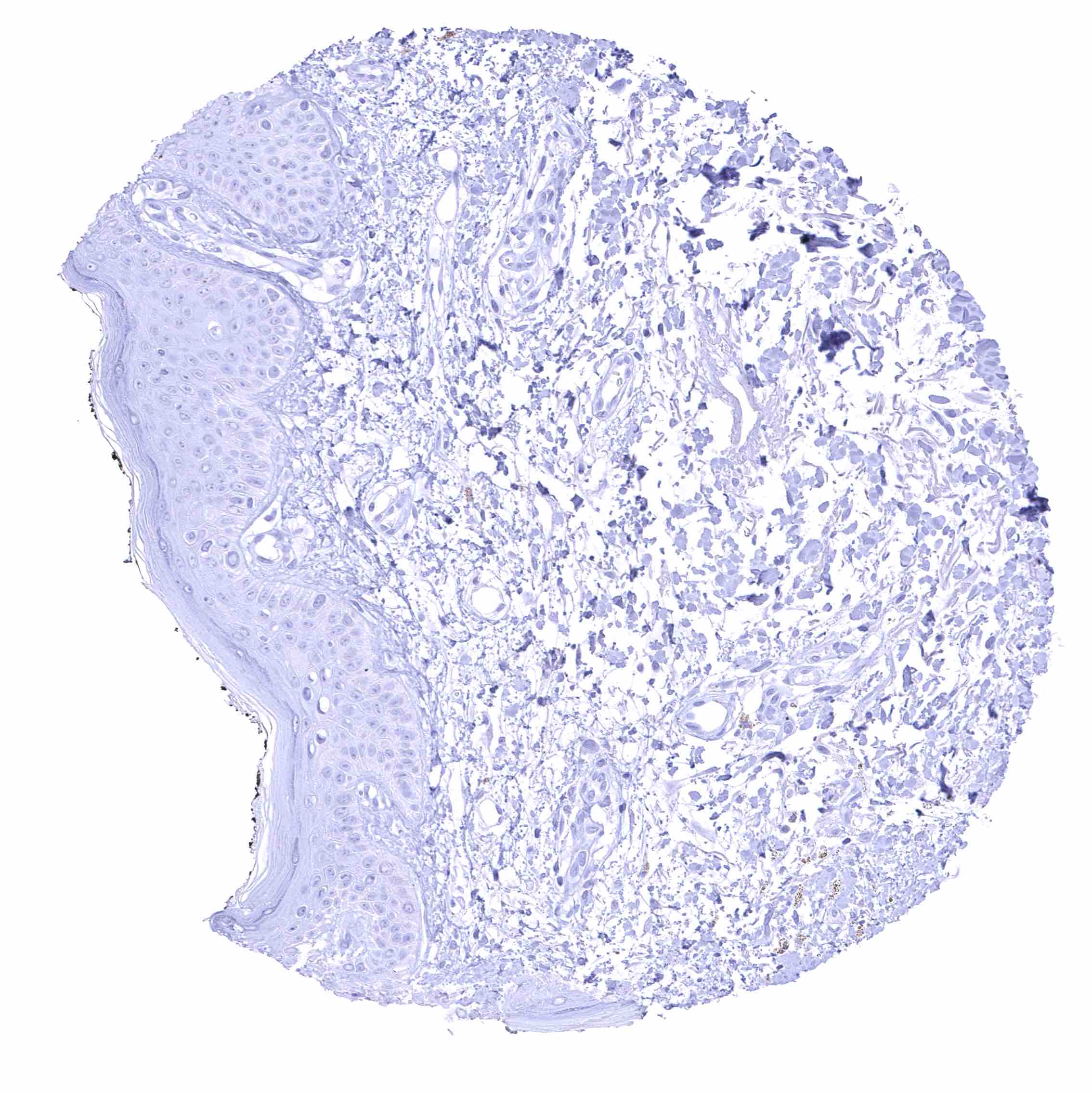
Skin
Spleen
Stomach, antrum
Stomach, corpus
Stomach, muscular wall
Submandibular gland
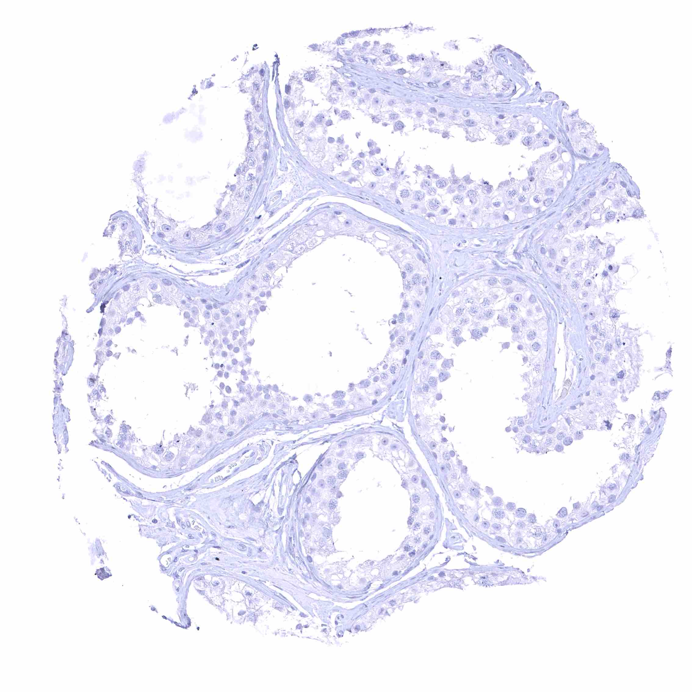
Testis

Thymus
Thyroid gland
Tonsil, surface epithelium
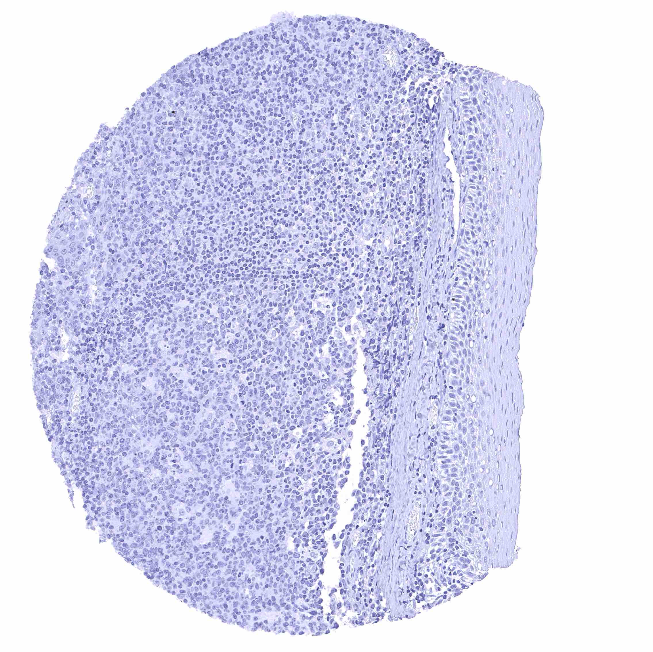
Tonsil
Urinary bladder, muscular wall
Urinary bladder, urothelium
Uterus, endocervix
Uterus, endometrium (pregnancy)
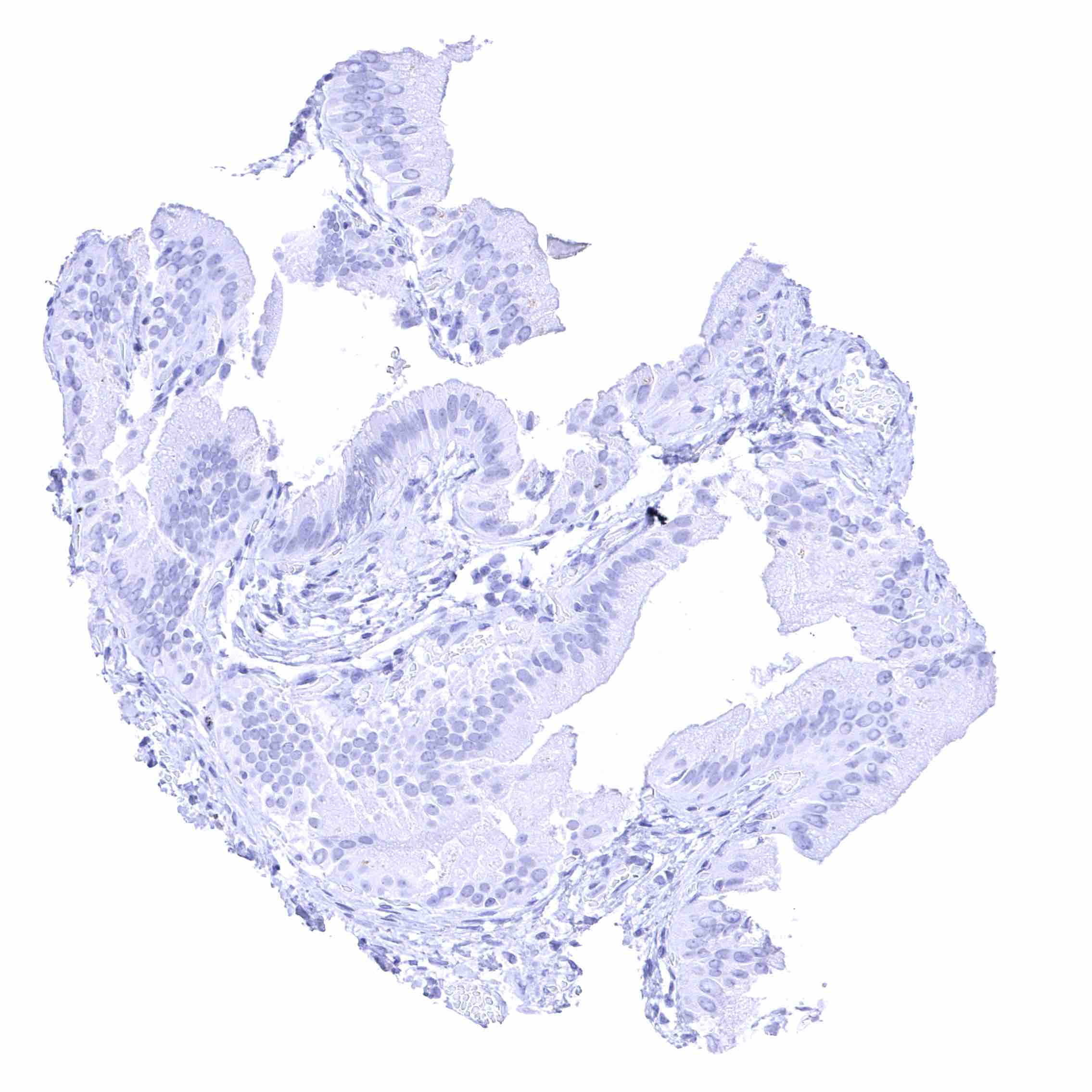
Uterus, endometrium (proliferation).jpeg
Uterus, endometrium (secretion)

Uterus, myometrium