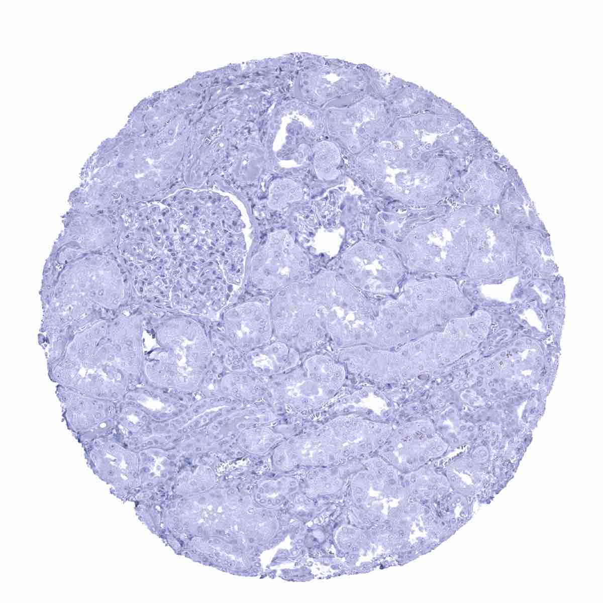
Adrenal gland

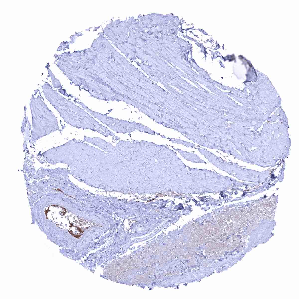
Aorta, media
Appendix, mucosa - IgA positive plasma cells are numerous in the stroma. IgA also accumulates in the cytoplasm of the crypt epithelium.
Appendix, muscular wall

Bone marrow - IgA positive plasma cells are rather rare.
Breast - IgA positive secreted liquid is seen in breast glands in this sample.
Breast - IgA staining is lacking in this sample.
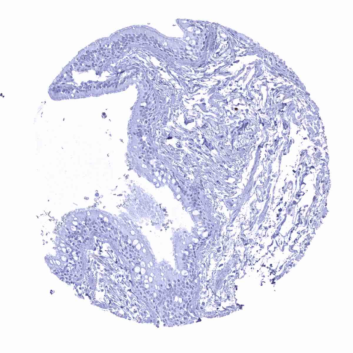
Bronchus, mucosa
Cerebellum, cortex (Stratum moleculare)

Cerebellum, grey (Stratum neuronorum)
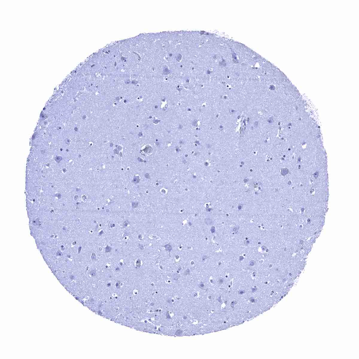
Cerebrum, grey matter

Cerebrum, white matter
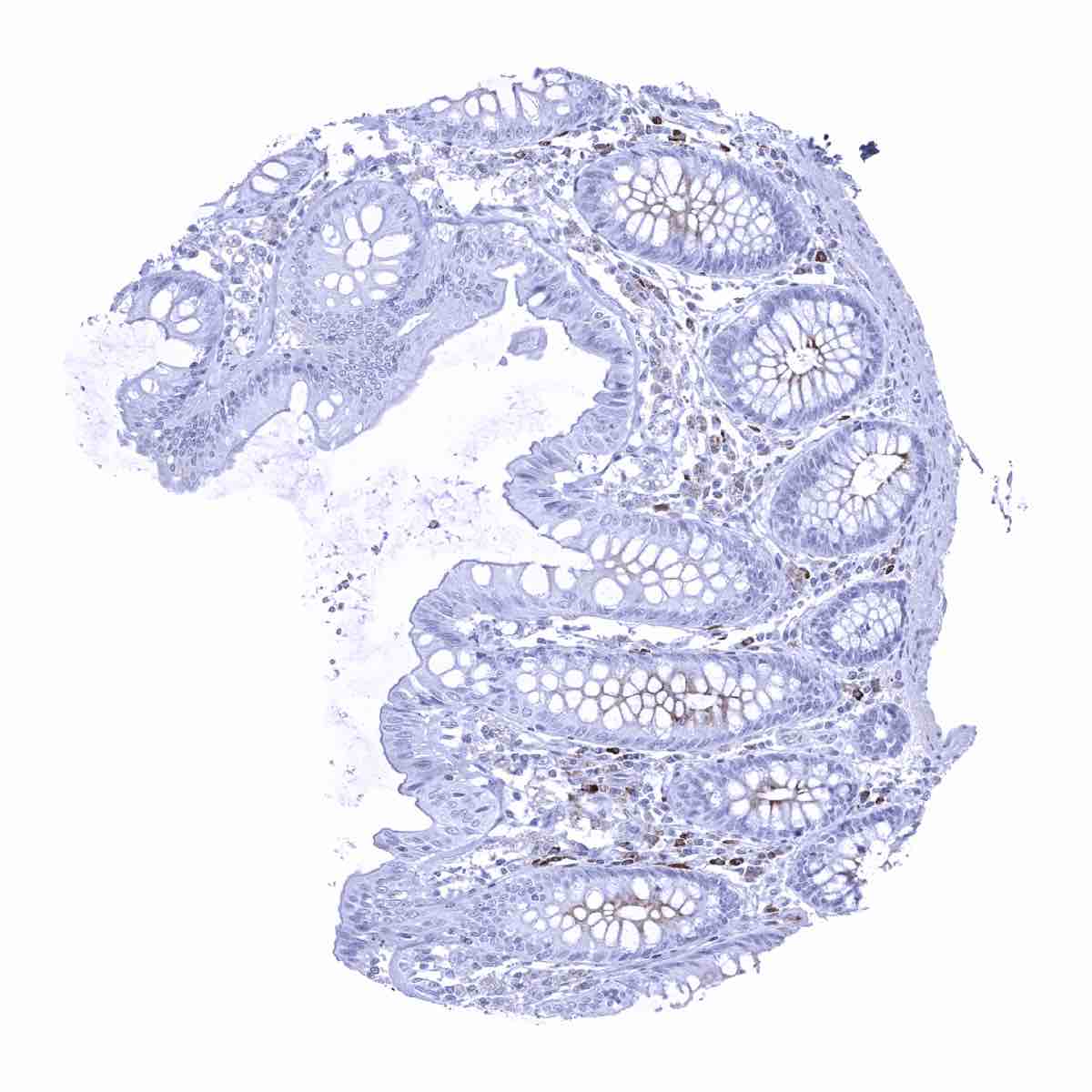
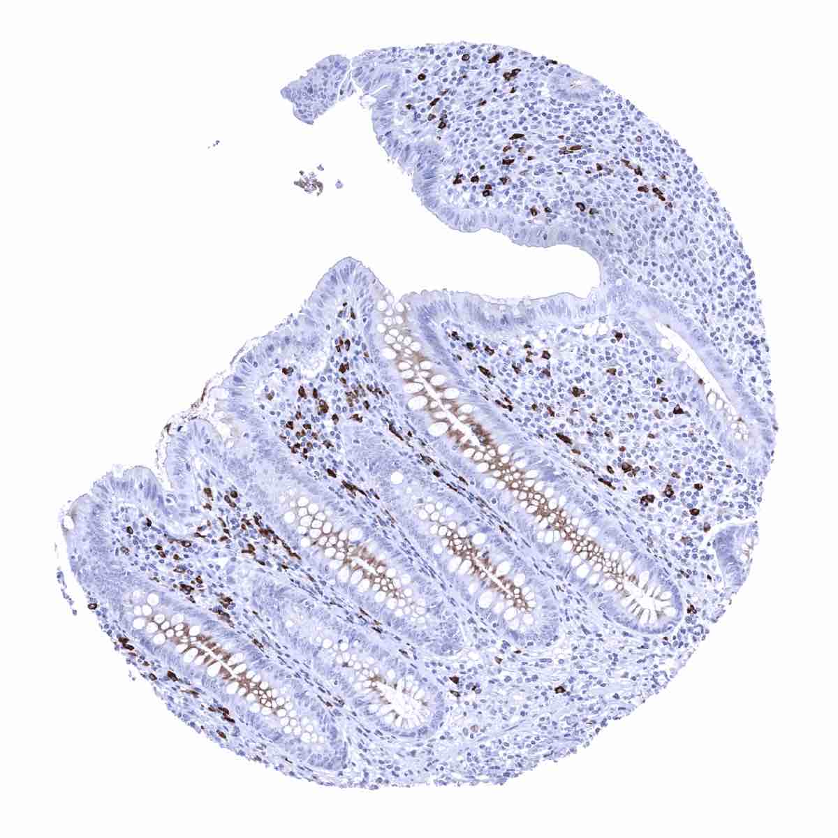
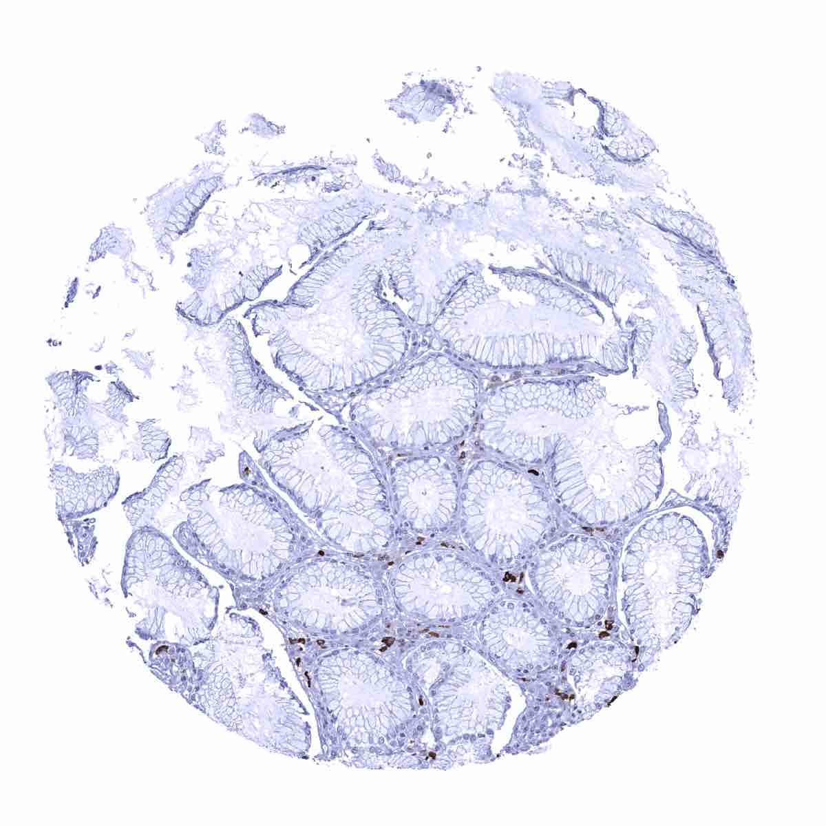
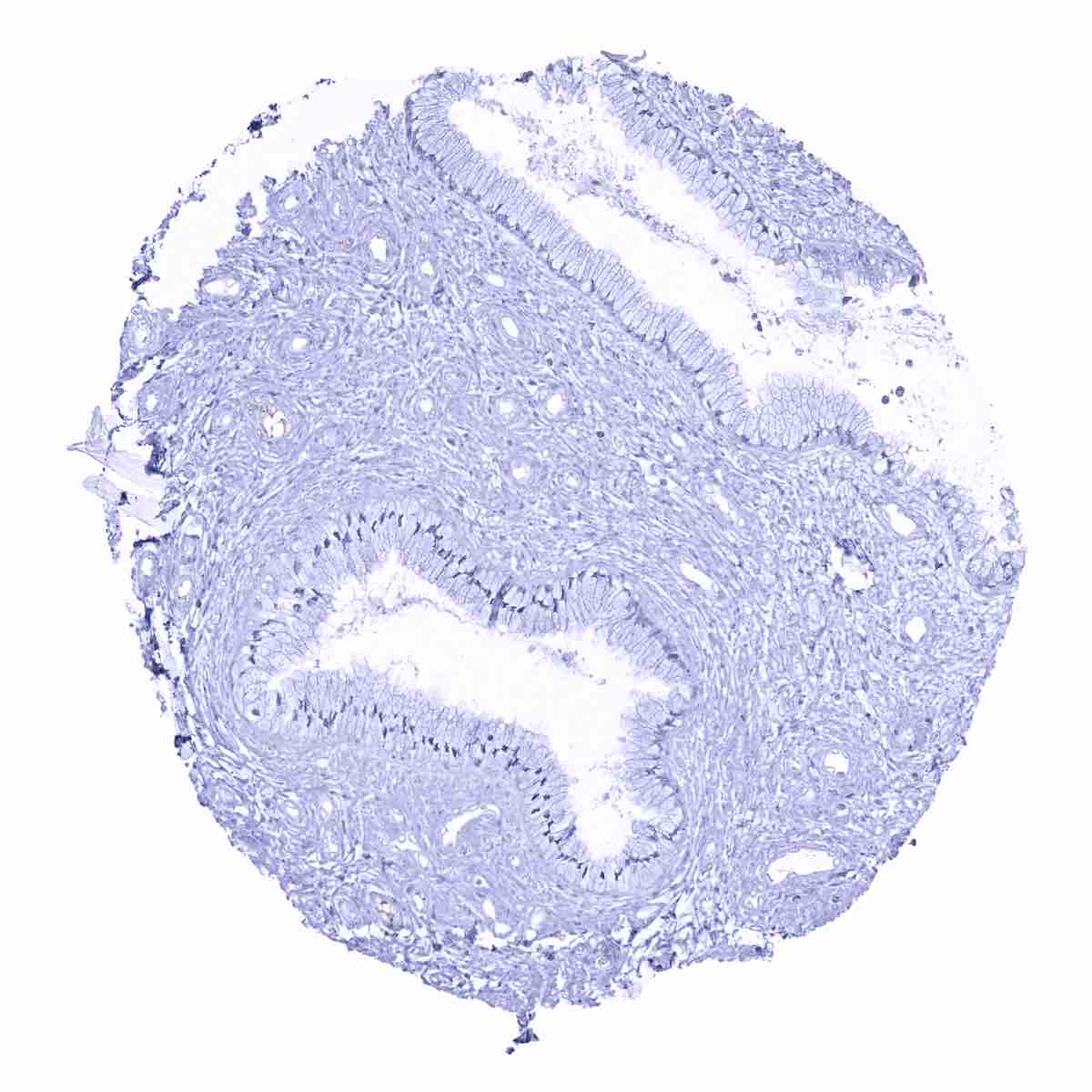
Colon descendens, mucosa

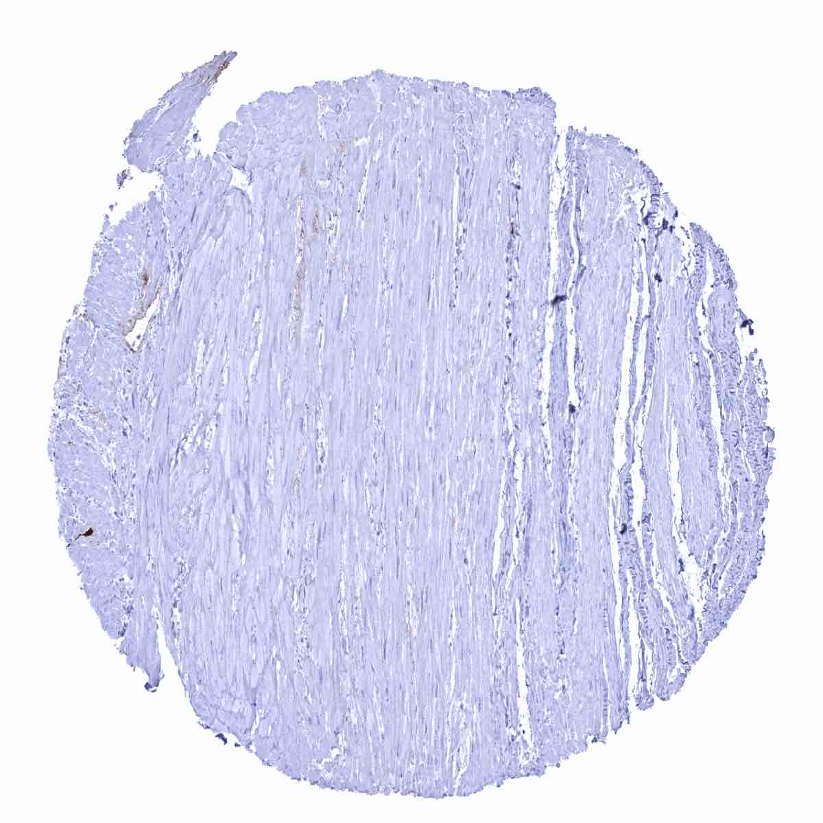
Colon descendens, muscular wall
Duodenum, Brunner gland
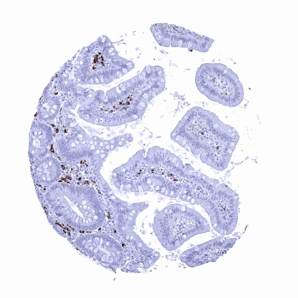
Duodenum, mucosa - IgA positive plasma cells are abundant in the stroma.
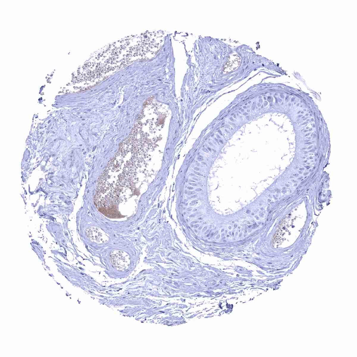
Epididymis - IgA positive plasma cells are lacking in the epididymis but some IgA staining occurs in blood serum.
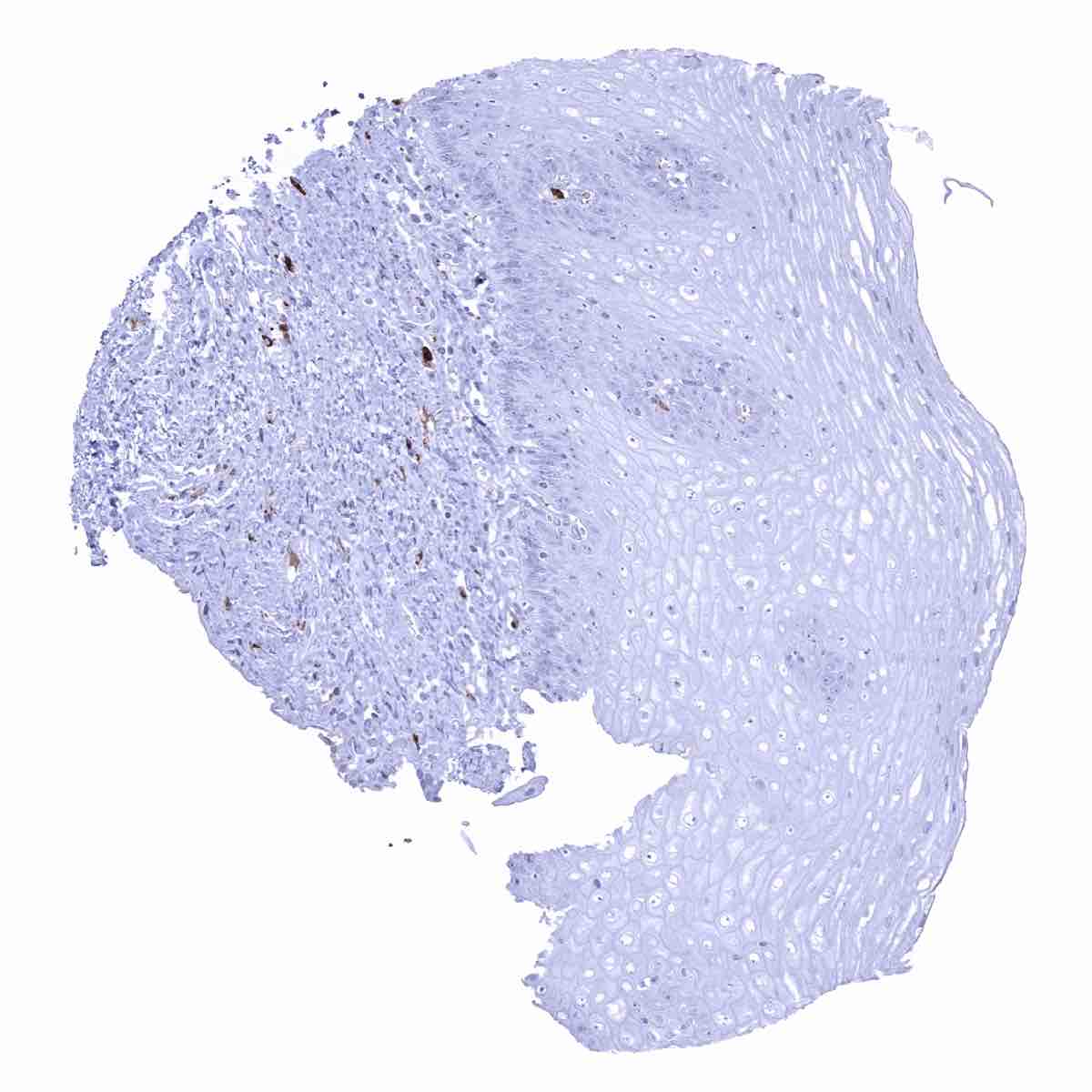
Esophagus, squamous epithelium - Few IgA positive plasma cells are seen.

Fallopian tube, mucosa
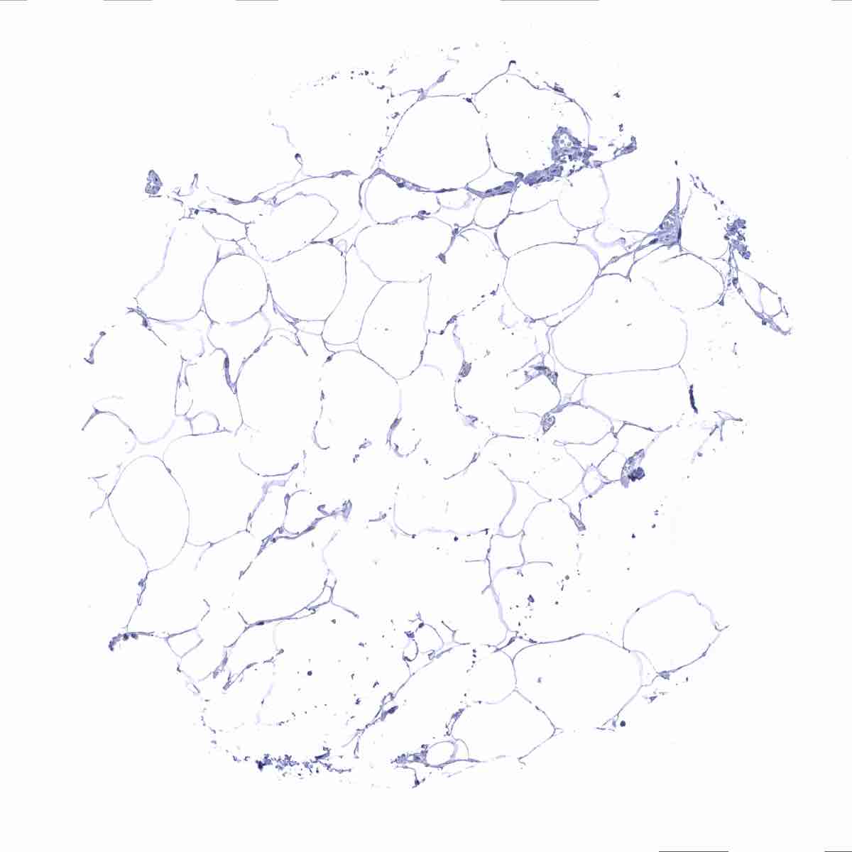
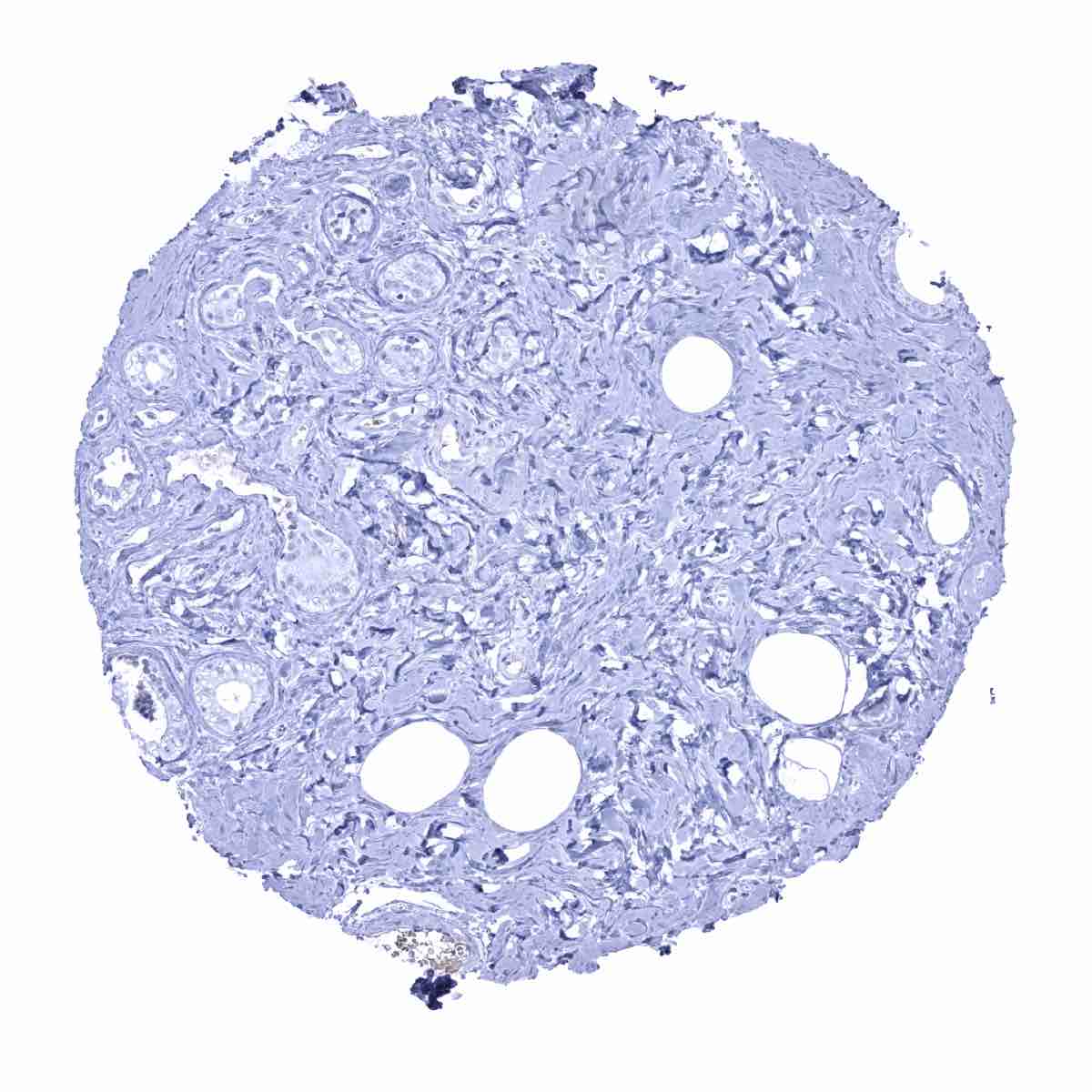
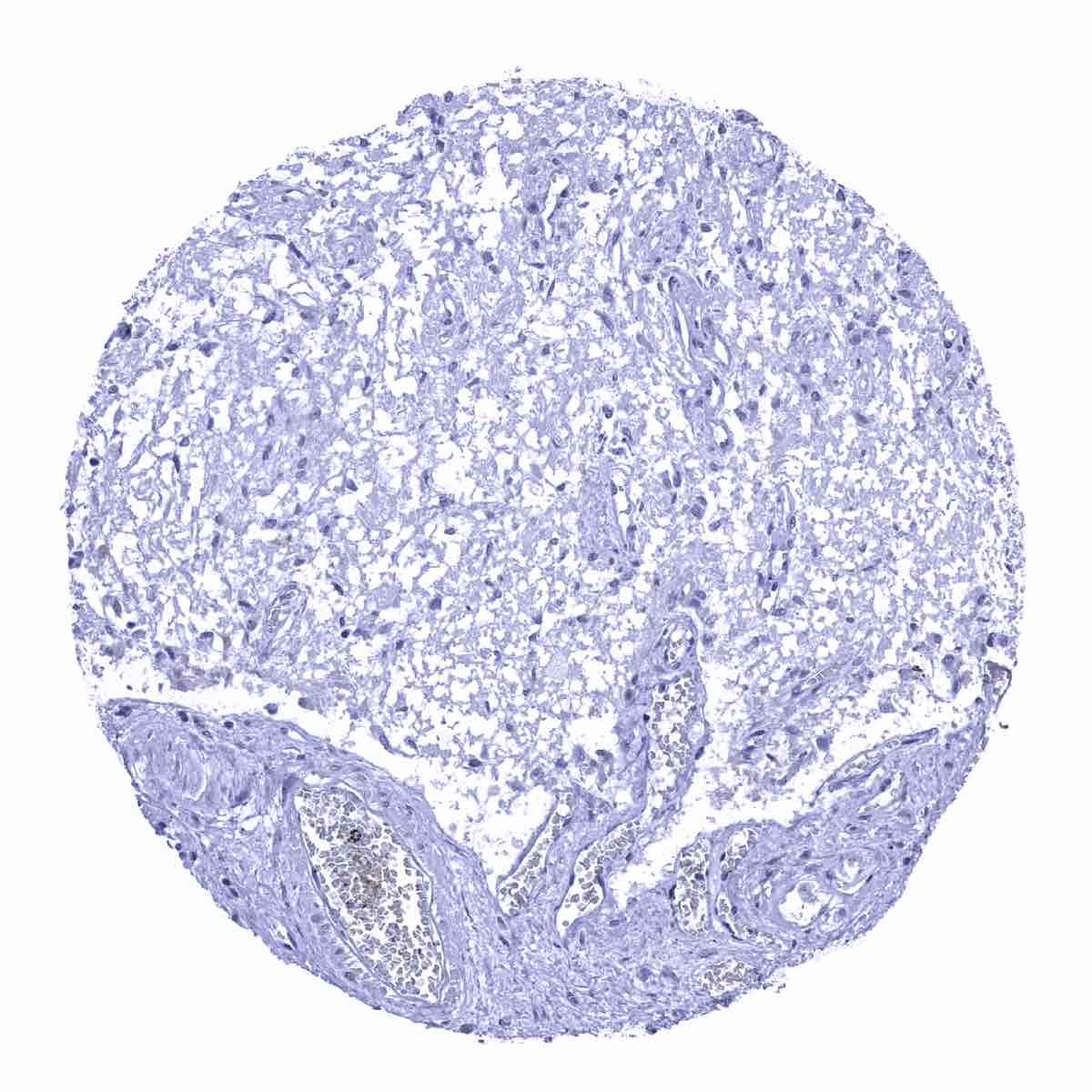
Fat

Gallbladder, epithelium
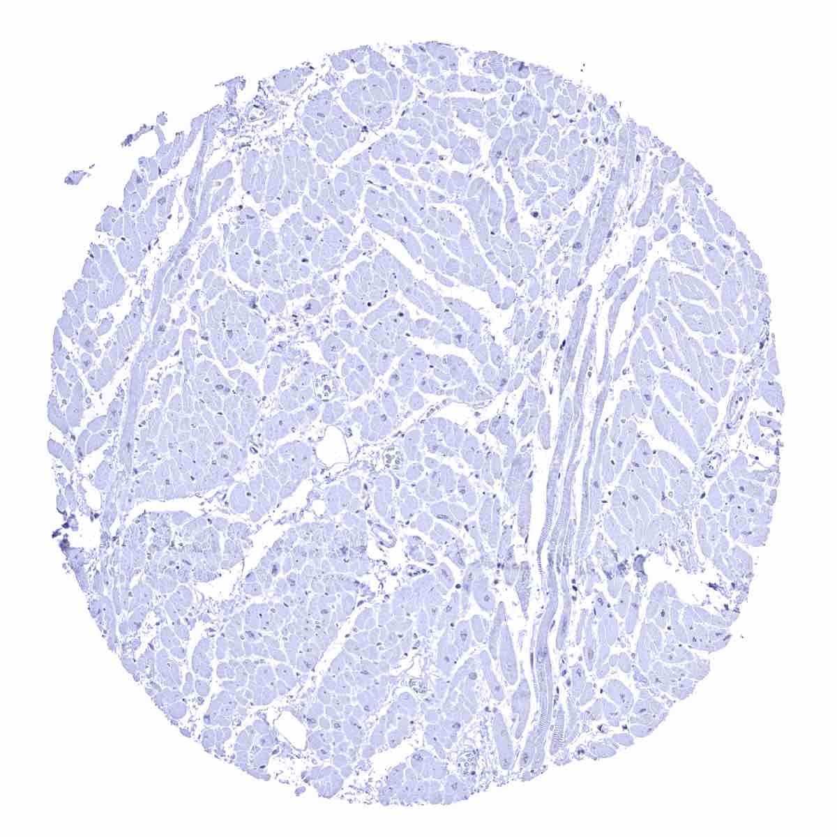
Heart muscle
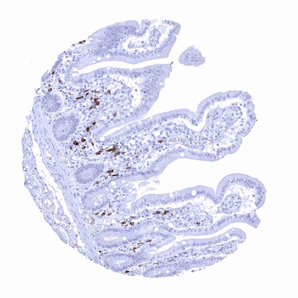
Ileum, mucosa - Numerous IgA positive plasma cells in the stroma
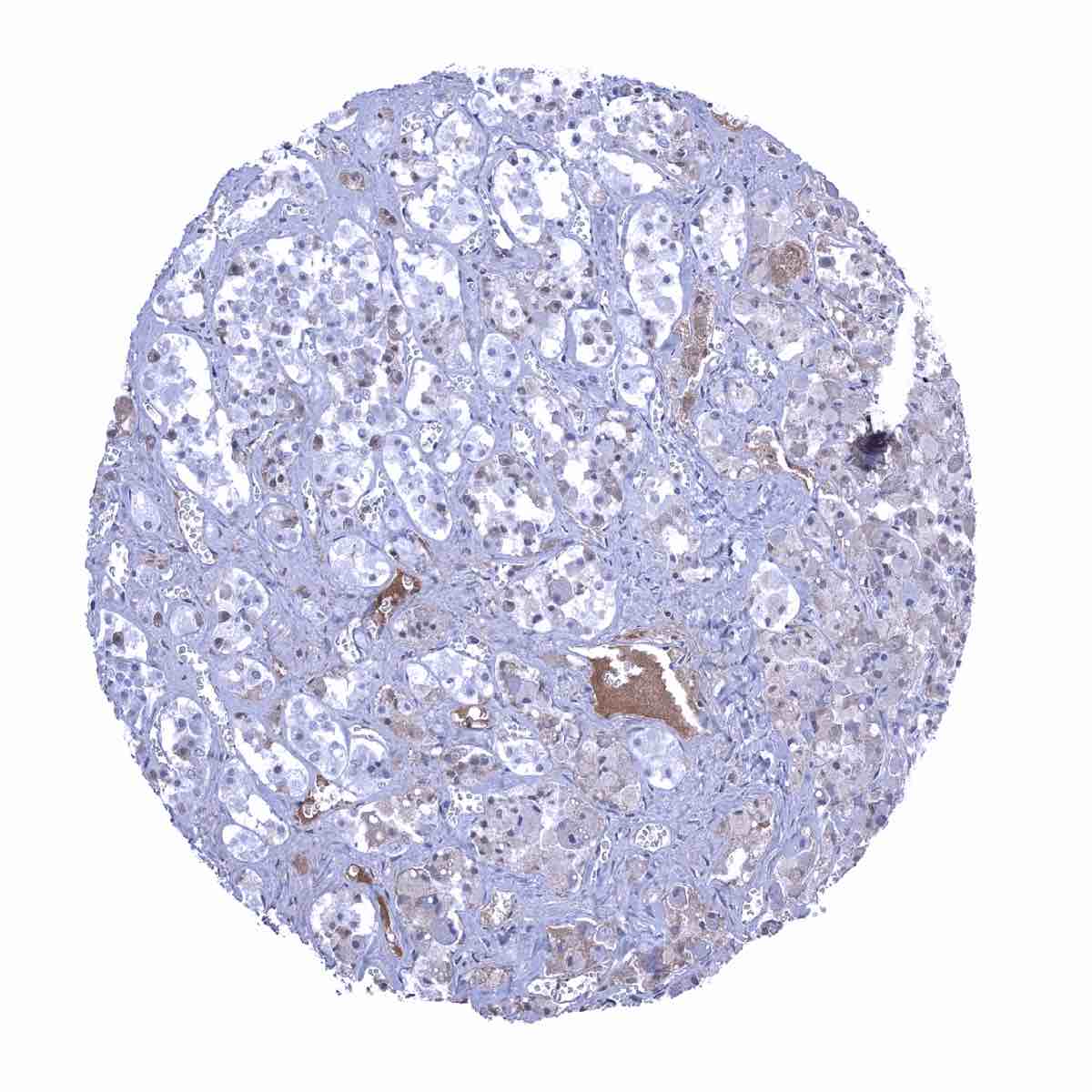
Kidney, cortex
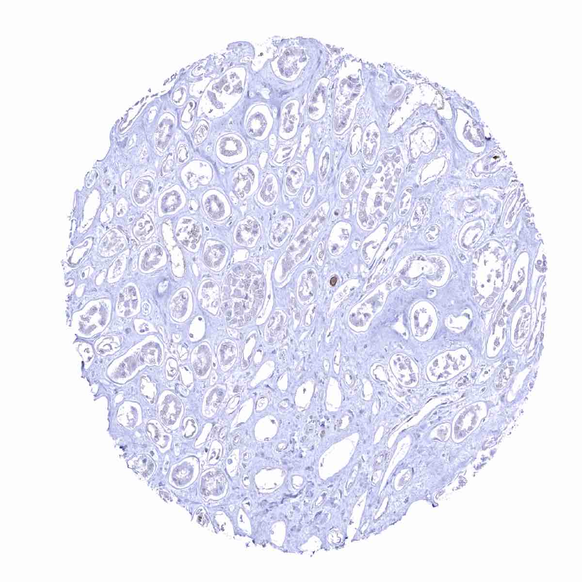
Kidney, medulla
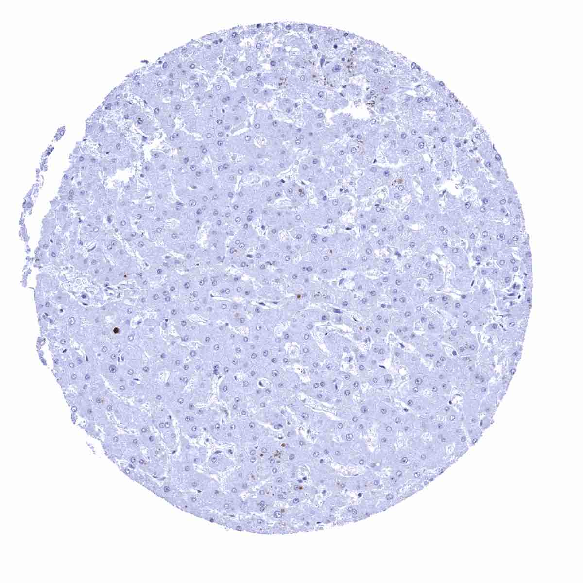
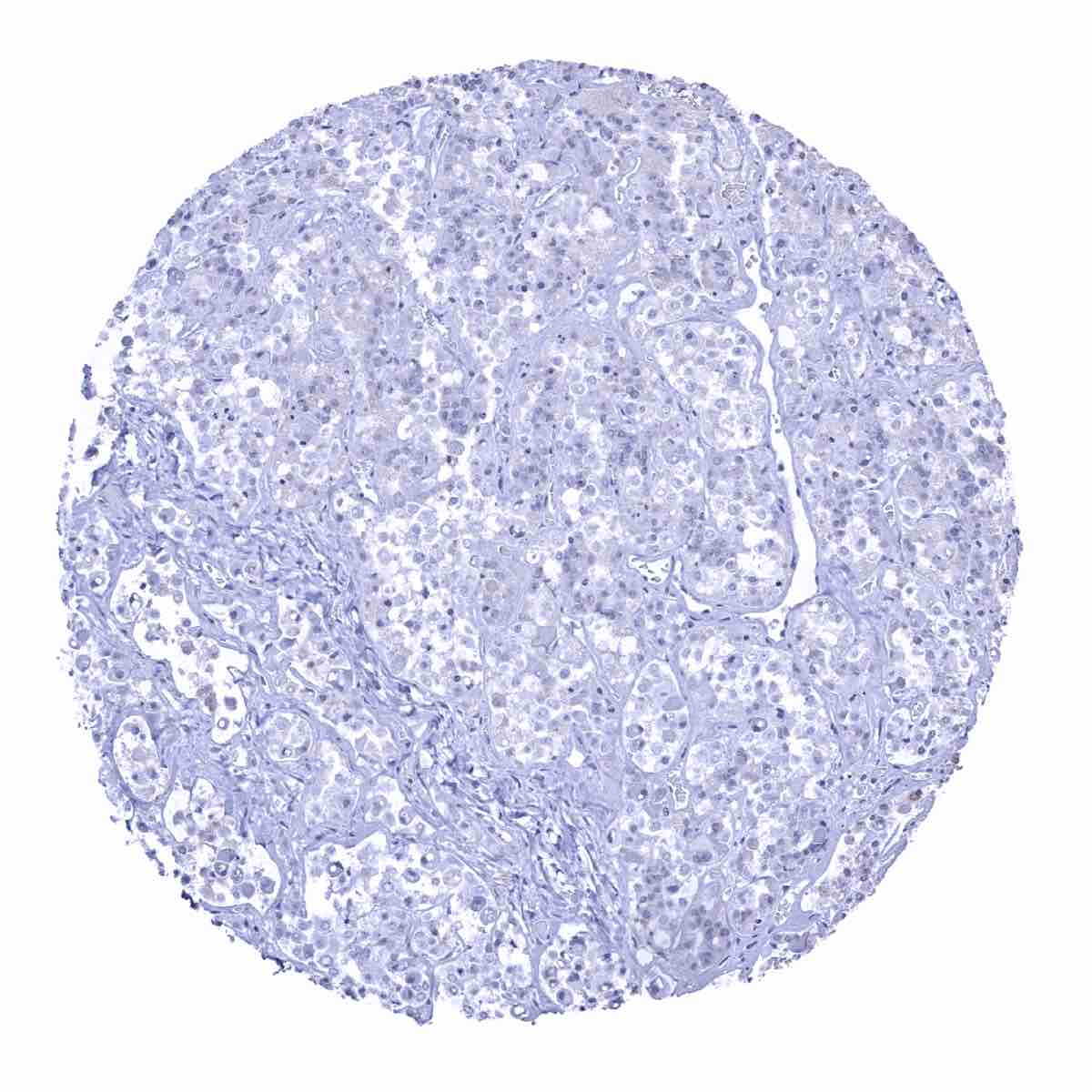
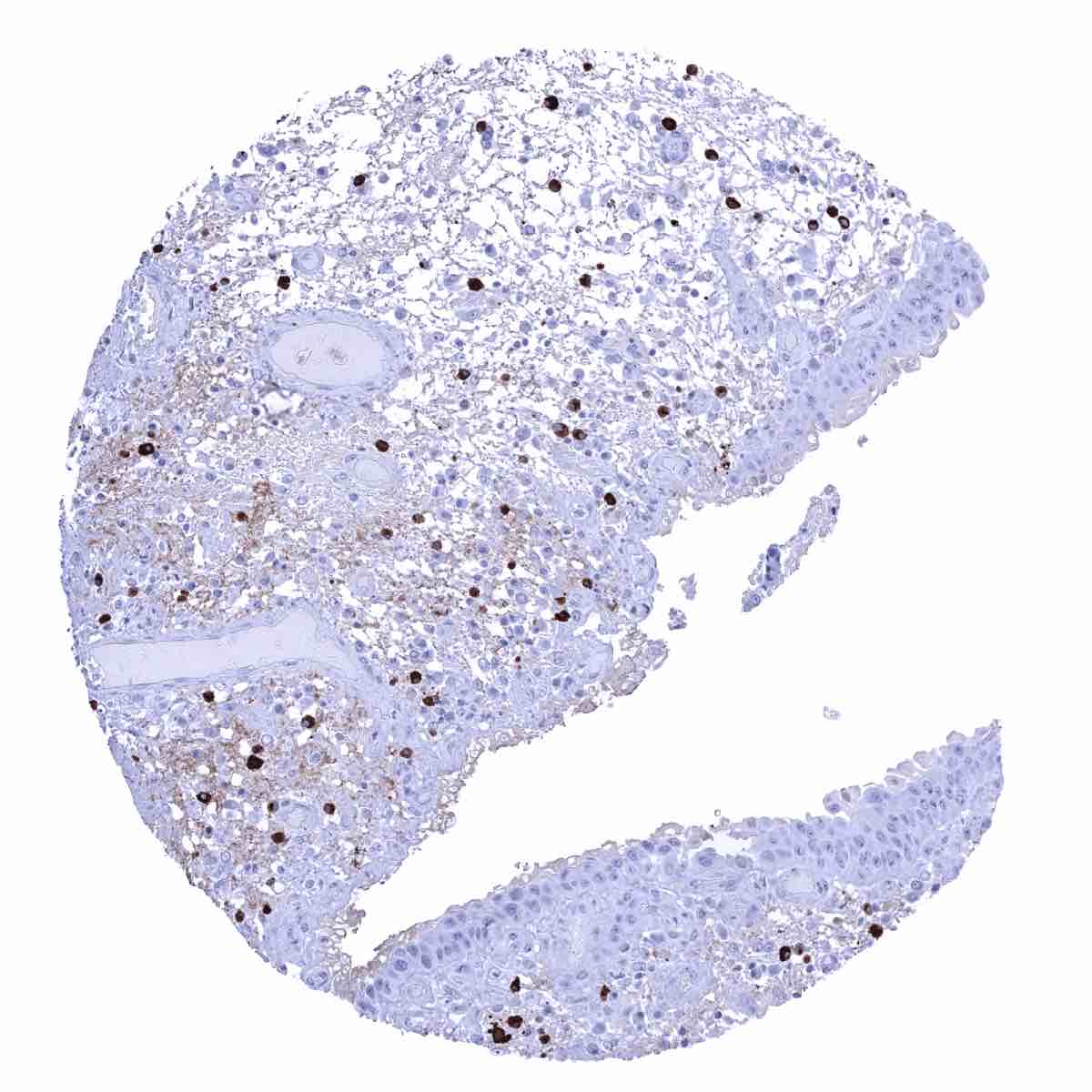
Liver
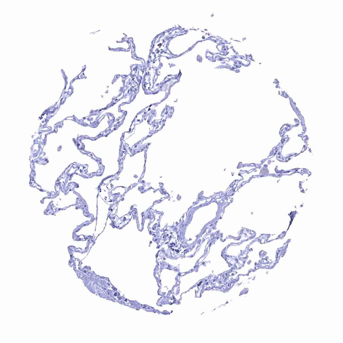
Lung
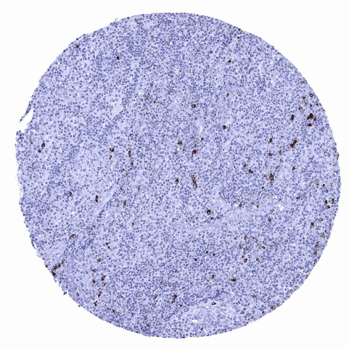
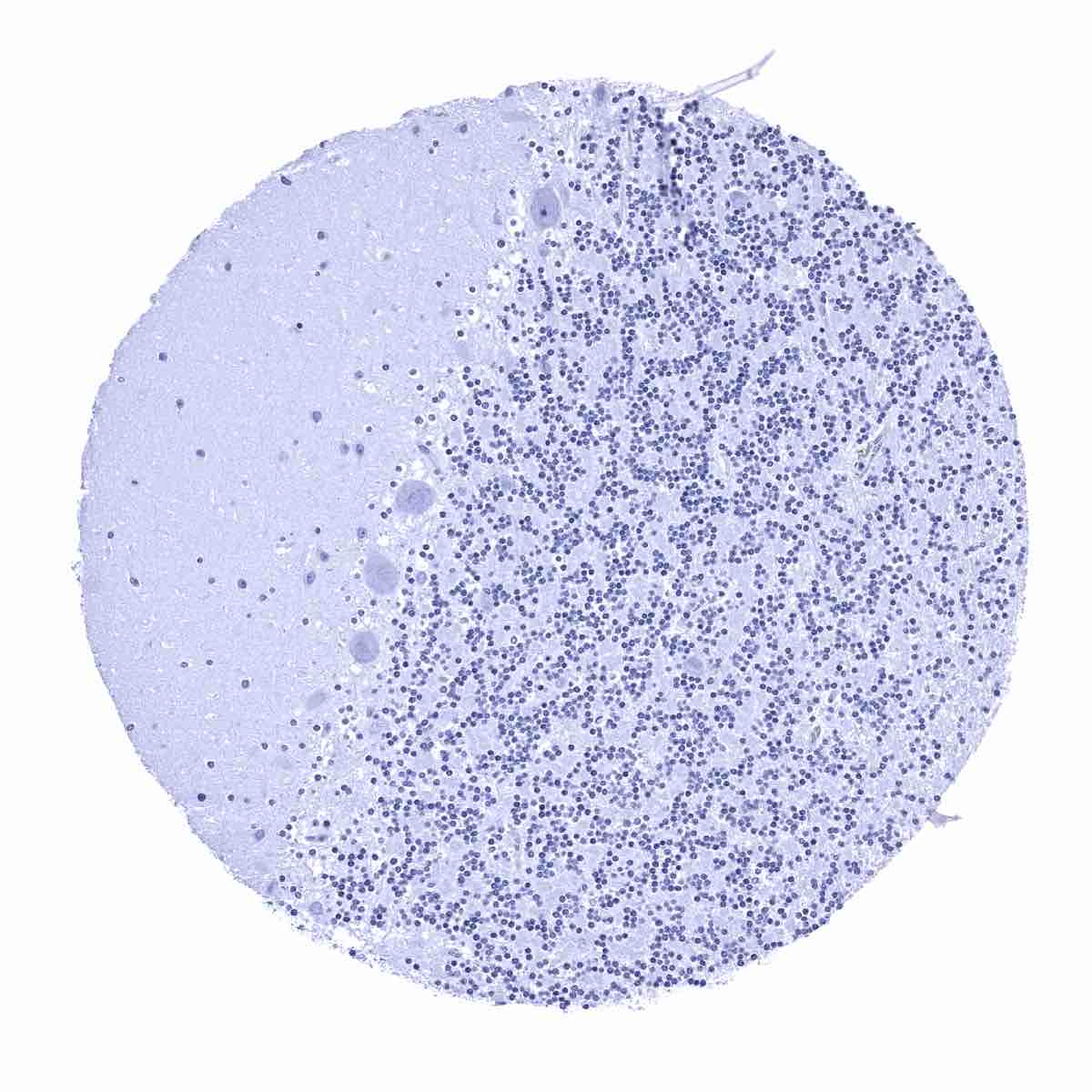
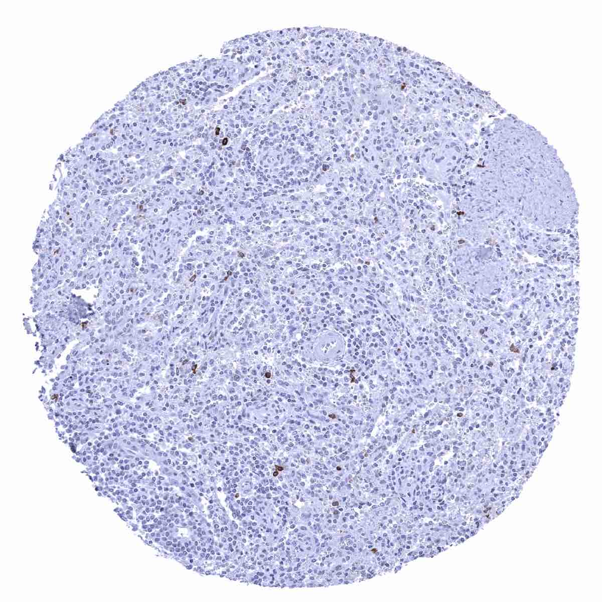
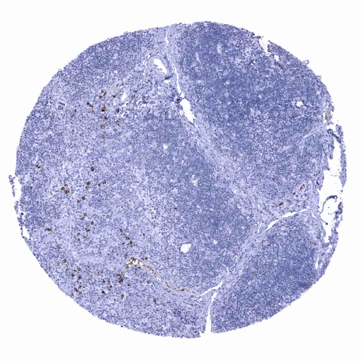
Lymph node - A fraction of plasma cells are IgA positive.

Ovary, stroma
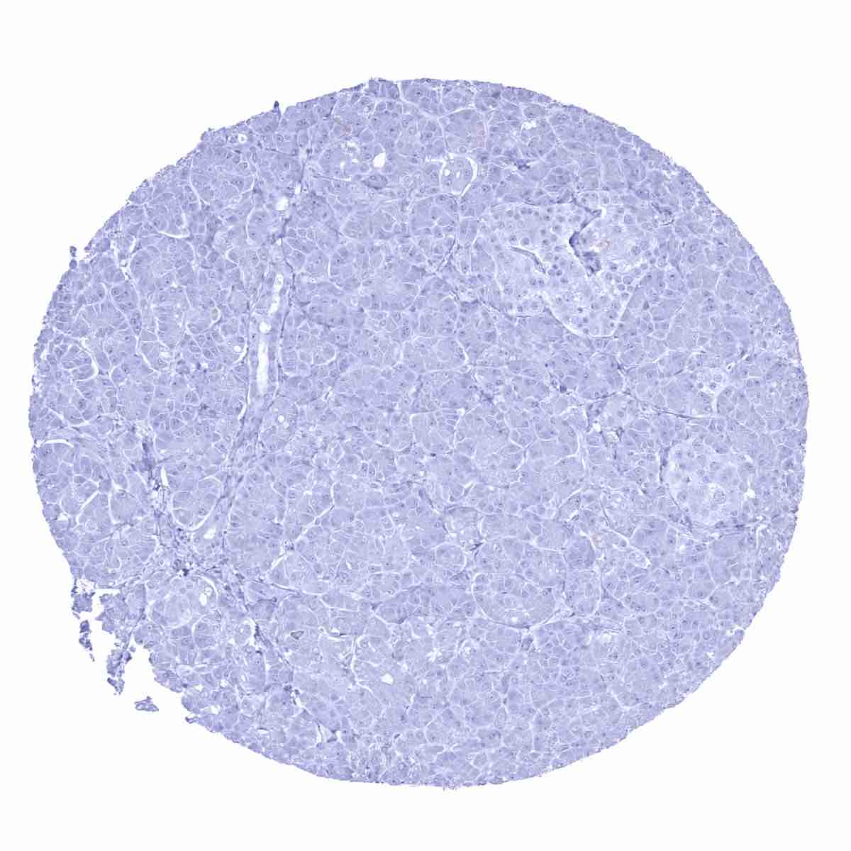
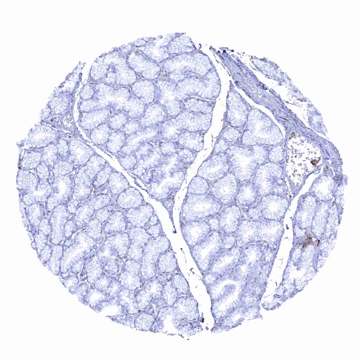
Pancreas

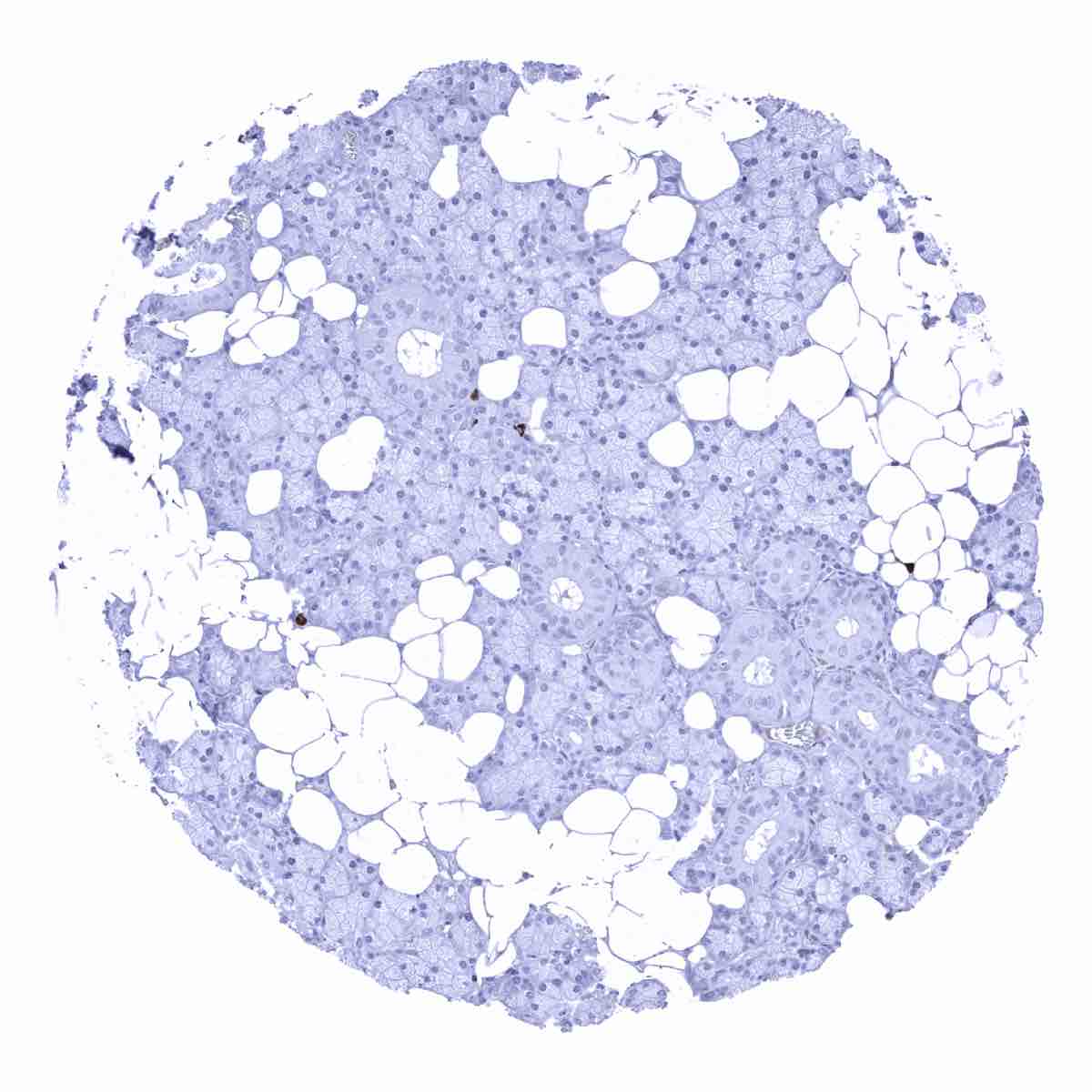
Parathyroid gland
Parotid gland
Pituitary gland, anterior lobe - Some IgA staining can occurs in the adenohypophysis.
Pituitary gland, anterior lobe
Pituitary gland, posterior lobe
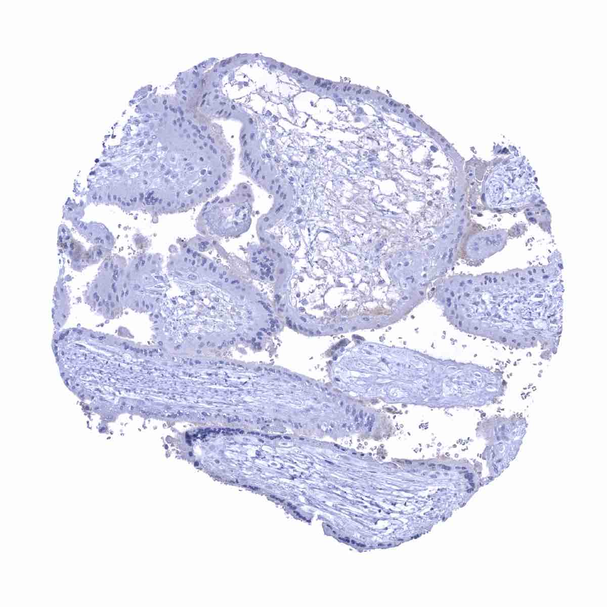
Placenta (amnion and chorion) - Stromal IgA staining is occasionally seen – probably caused by IgA from the blood.
Placenta (amnion and chorion)
Placenta, early - Some IgA staining occurs in the cytotrophoblast.
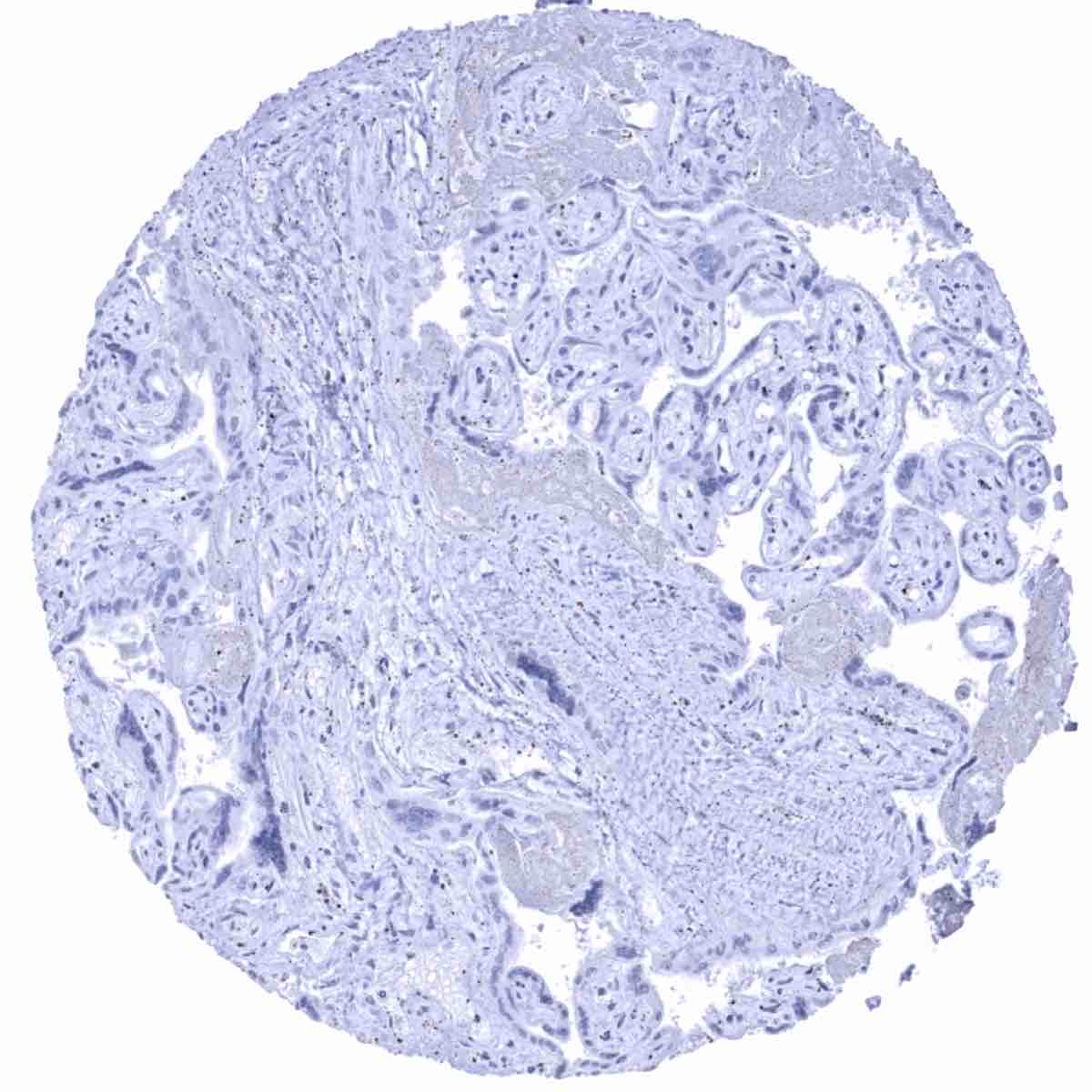
Placenta, mature
Prostate
Seminal vesicle

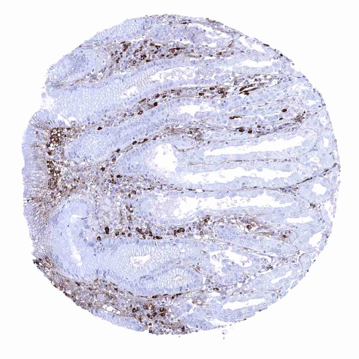
Sinus paranasales - Abundance of IgA positive plasma cells.
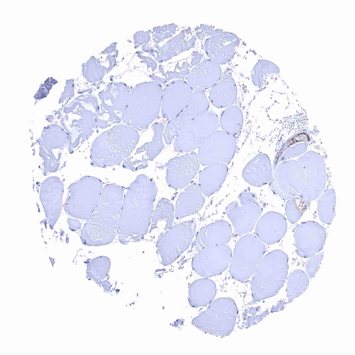
Skeletal muscle
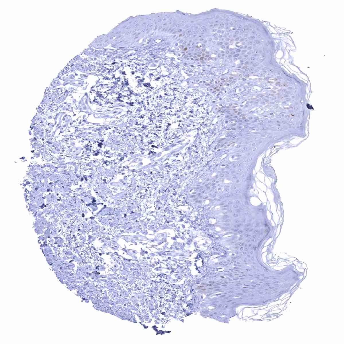
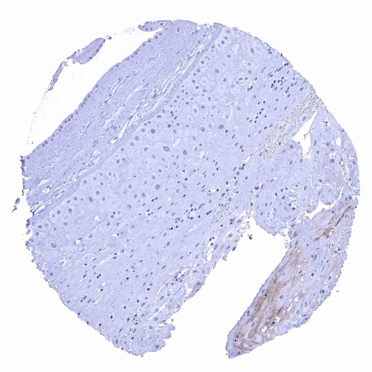
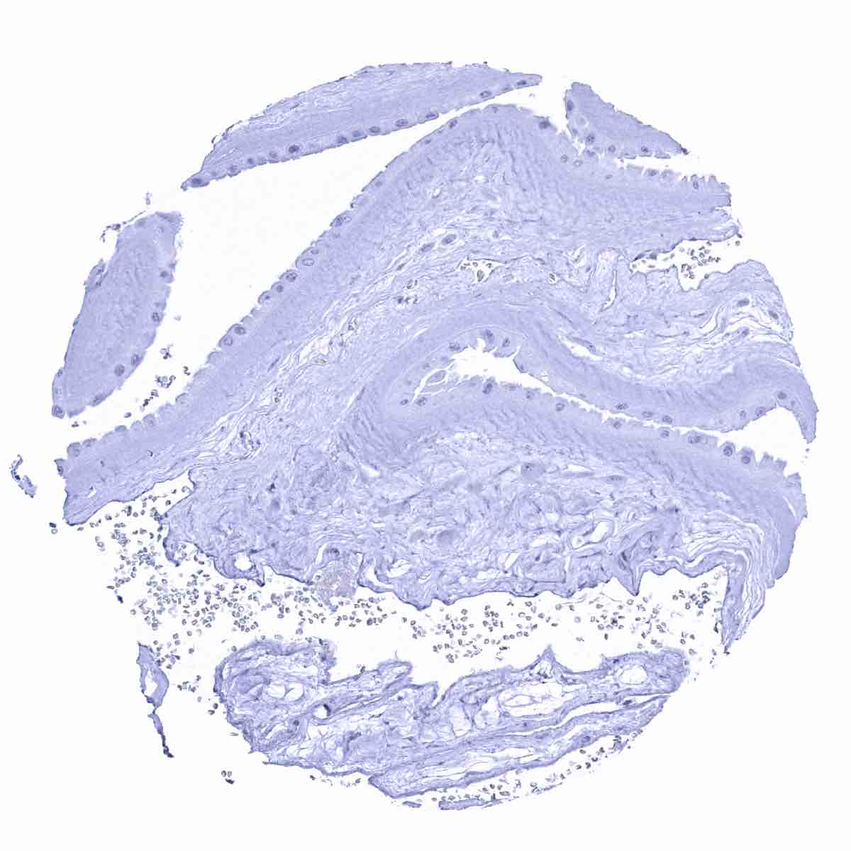
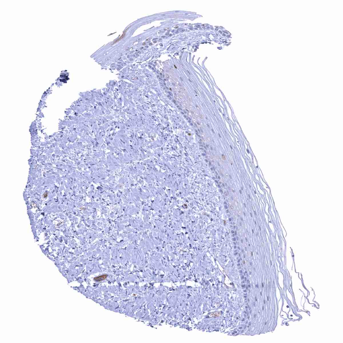
Skin
Spleen - Few IgA positive plasma cells are seen
Stomach, antrum - Few IgA positive plasma cells are seen.
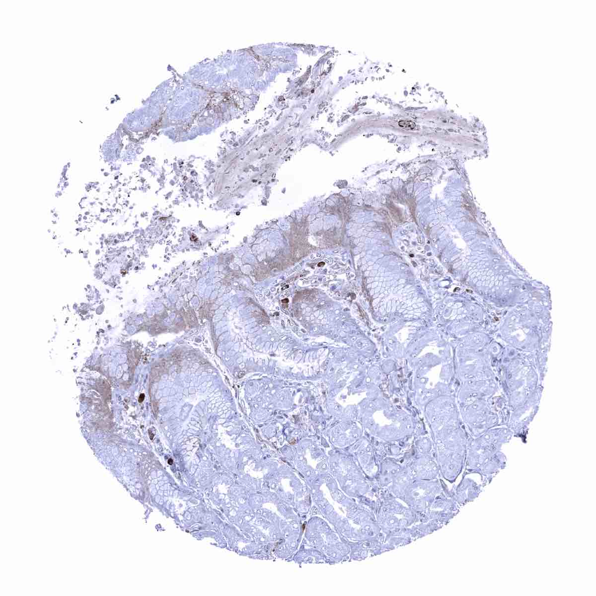
Stomach, antrum - IgA positive plasma cells are occur in the stroma. IgA also accumulates in the cytoplasm of superficial epitheial cell layers.
Stomach, corpus - IgA positive plasma cells are abundant in the stroma. IgA also accumulates in the cytoplasm of superficial epitheial cell layers.

Testis

Testis
Thymus - Few IgA positive plasma cells are seen.
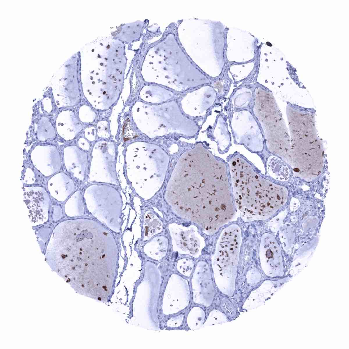
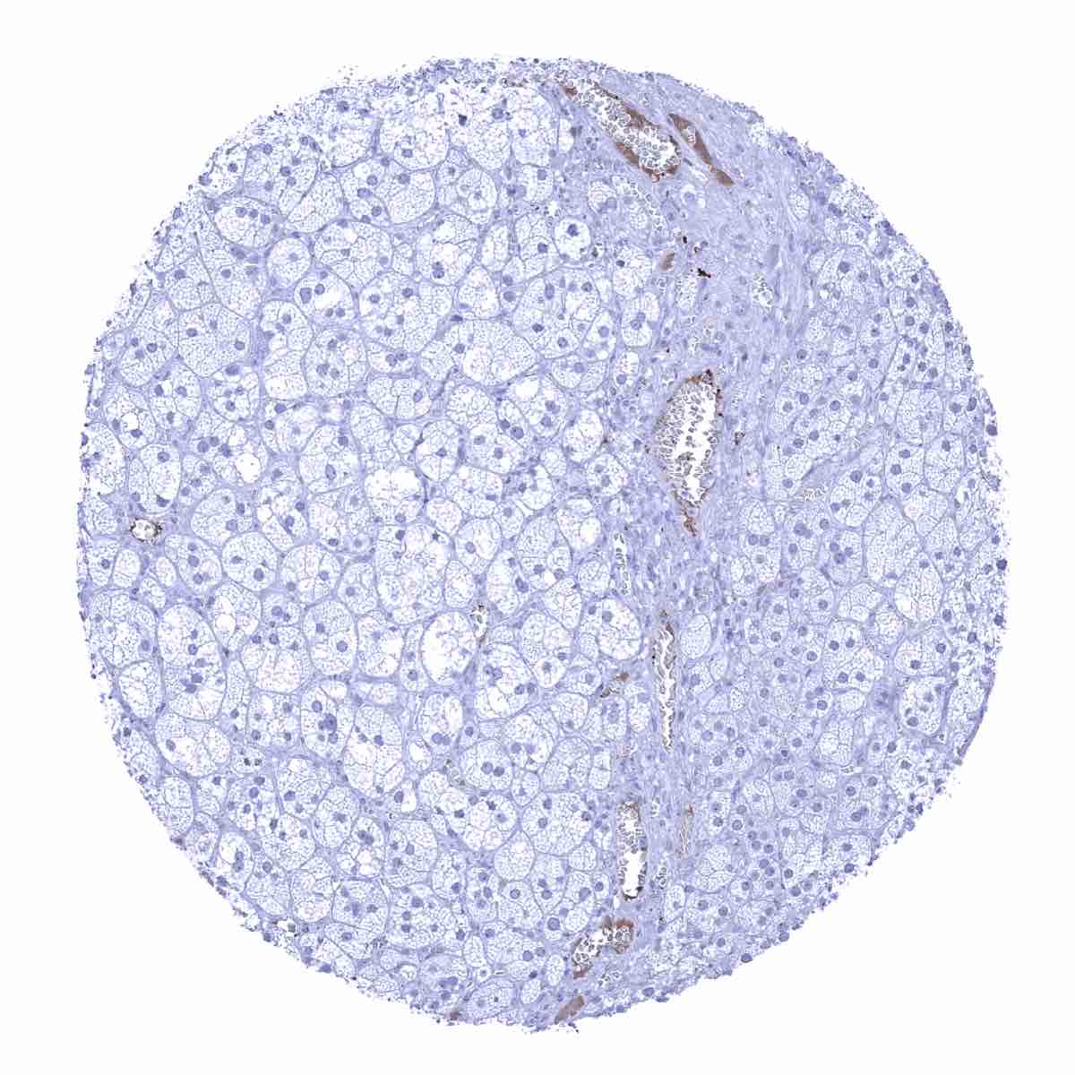
Thyroid gland - Some IgA staining can occurs in the colloid of some follicles
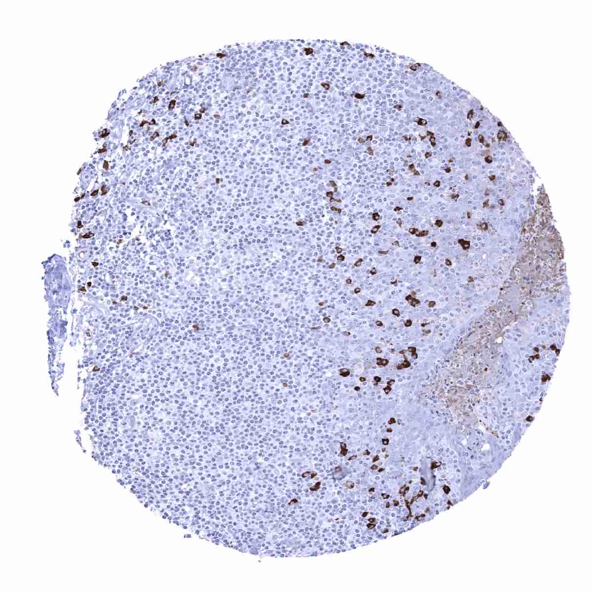
Tonsil - Numerous IgA positive plasma cells are seen. Week staining is also seen in the crypts as a result of IgA secretion
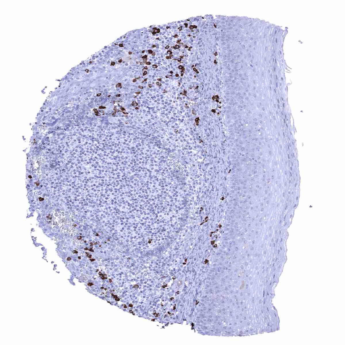
Tonsil, surface epithelium - IgA positive plasma cells are abundant.
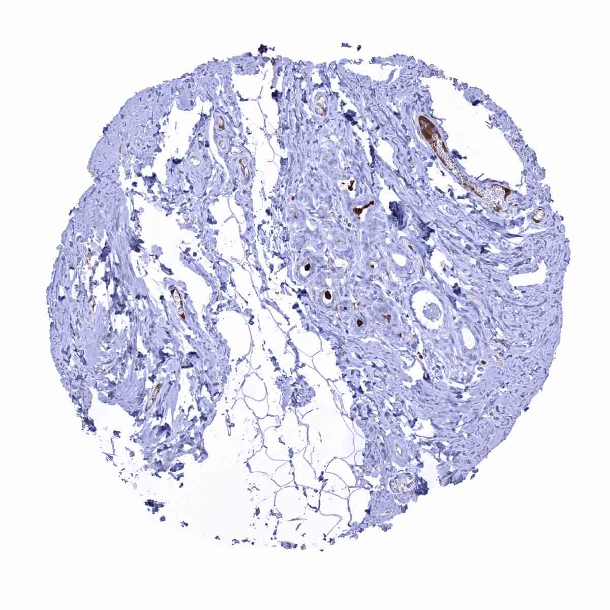
Urinary bladder, muscular wall

Urinary bladder, urothelium - IgA positive plasma cells are abundant in the stroma. IgA accumulates in the superficial layers of the urothelium.
Urinary bladder, urothelium - IgA positive plasma cells are abundant in this sample. A faint IgA staining also occurs in the superficial urothelial cell layer.
Uterus, ectocervix
Uterus, endocervix
Uterus, endometrium (pregnancy)

Uterus, endometrium (secretion)
Uterus, myometrium