Adrenal gland

Aorta, endothelium
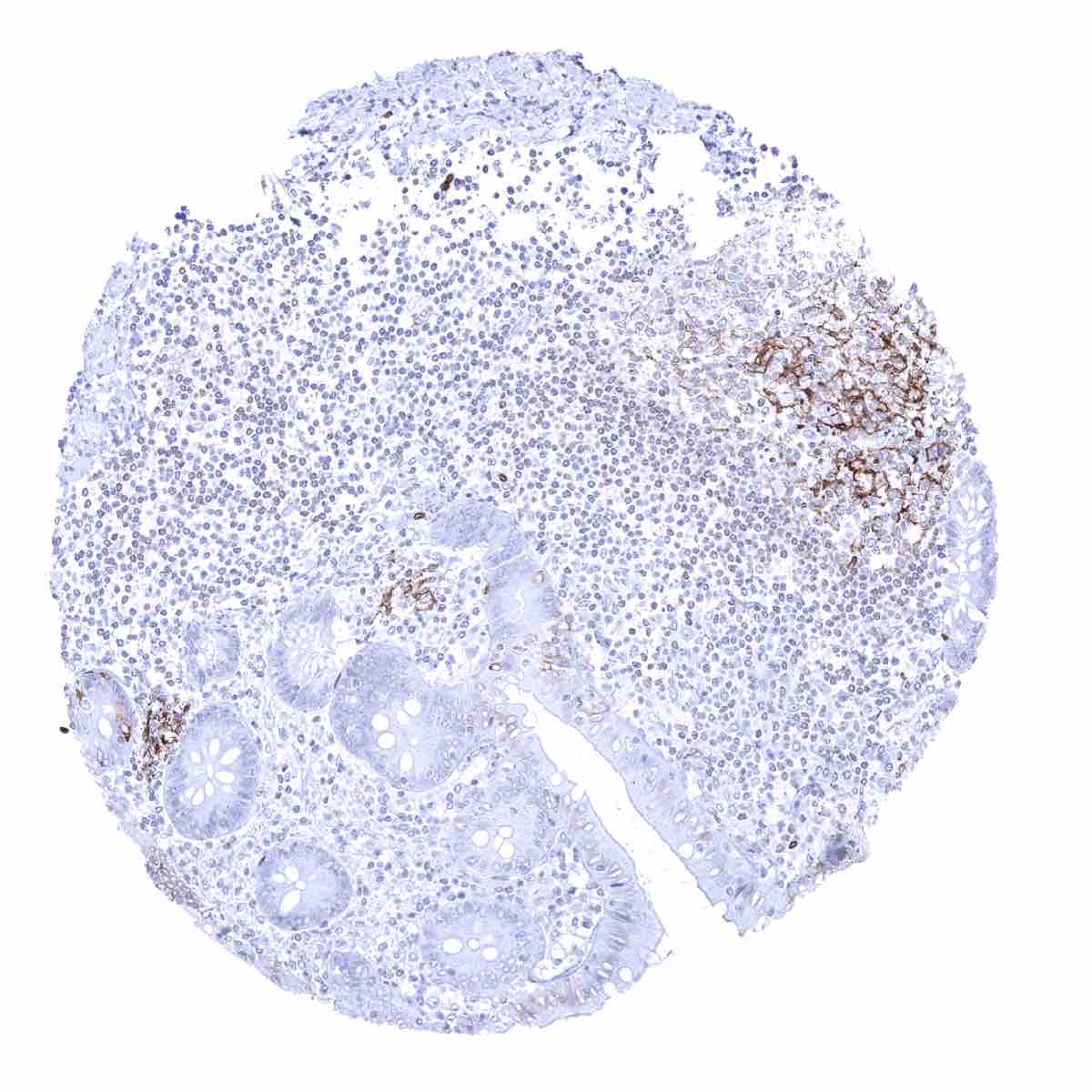
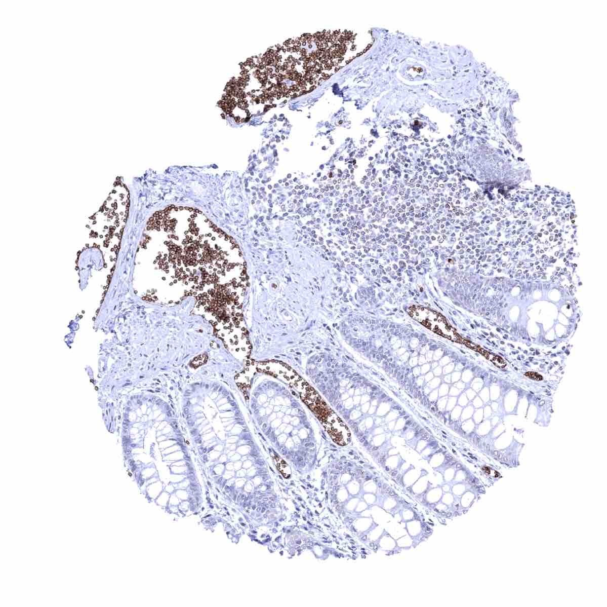
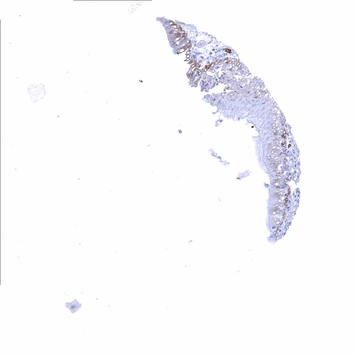
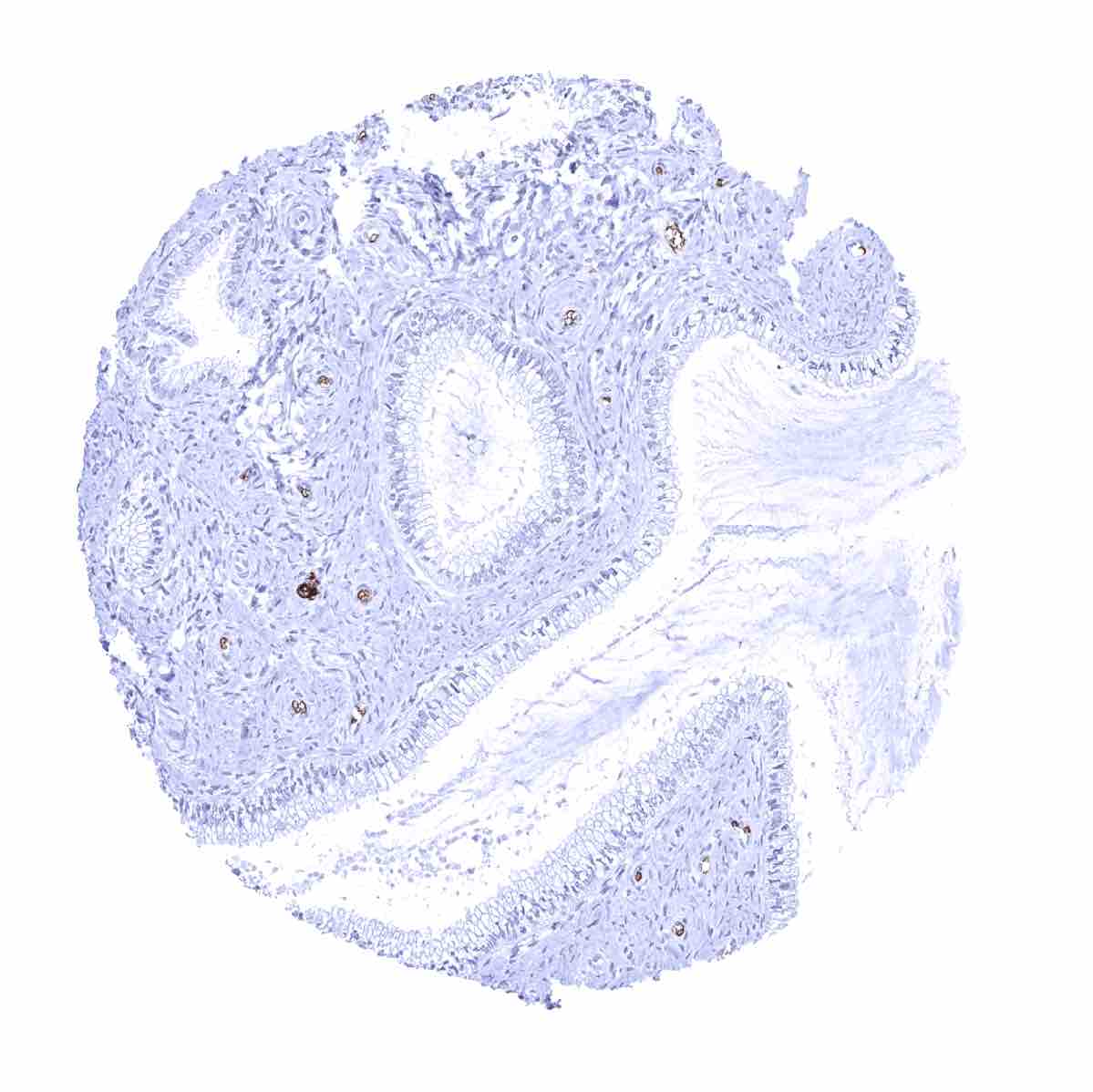
Appendix, mucosa - Focal faint epithelial cell GLUT1 staining. Staining is more intense in follicular dendritic cells of a germinal centre
Appendix, muscular wall
Bone marrow - Erythrocytes and their precursor cells stain positive
Breast - Faint cytoplasmic staining of acinar cells of the breast
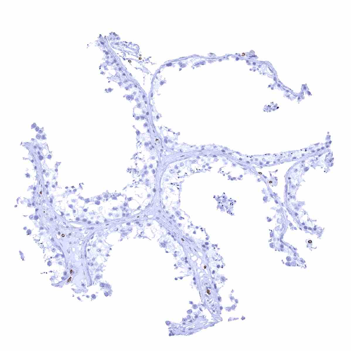
Bronchus, mucosa - Faint cytoplasmic GLUT1 staining occurs in (non-basal) cells of the respiratory epithelium
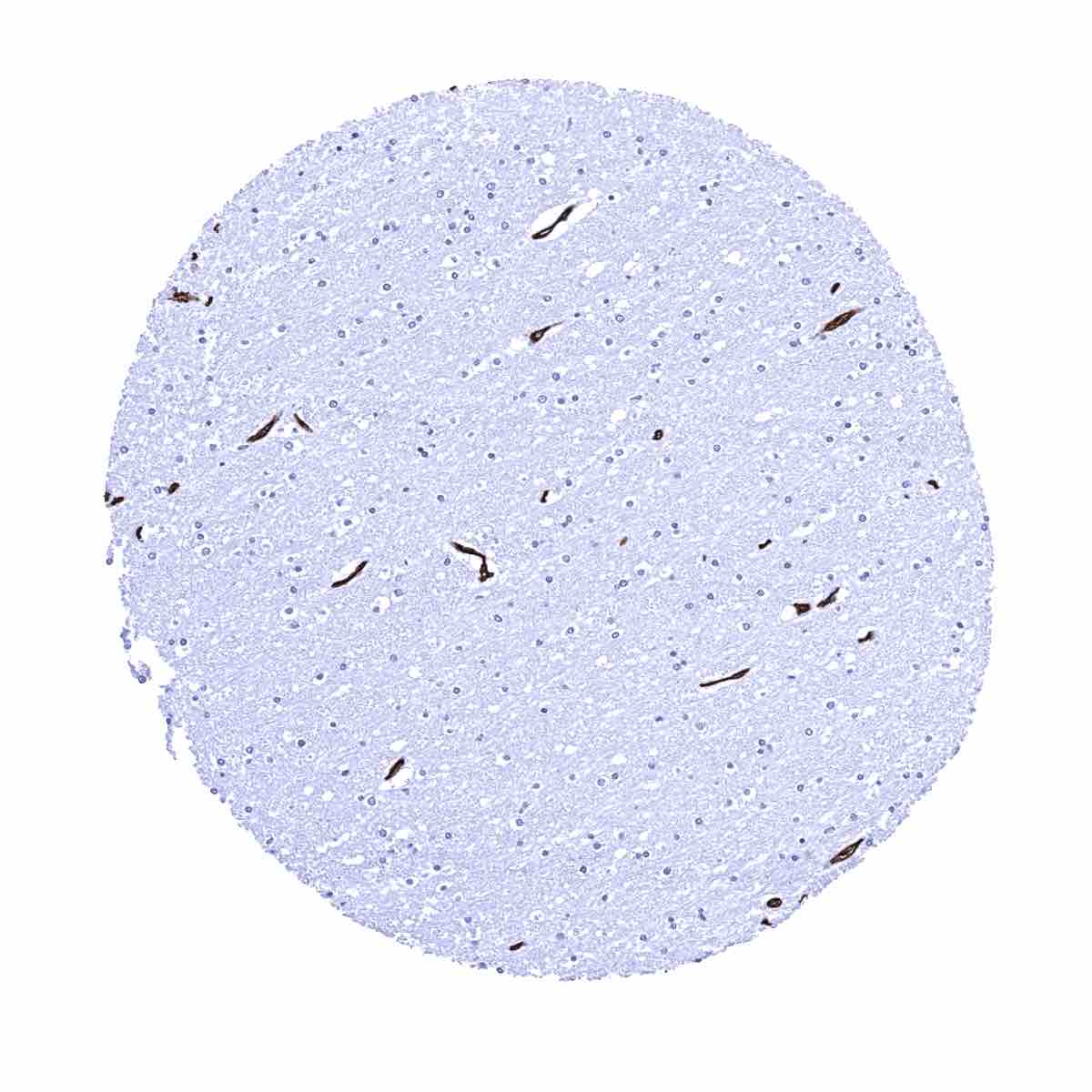
Cerebellum (granule cell layer, white matter)
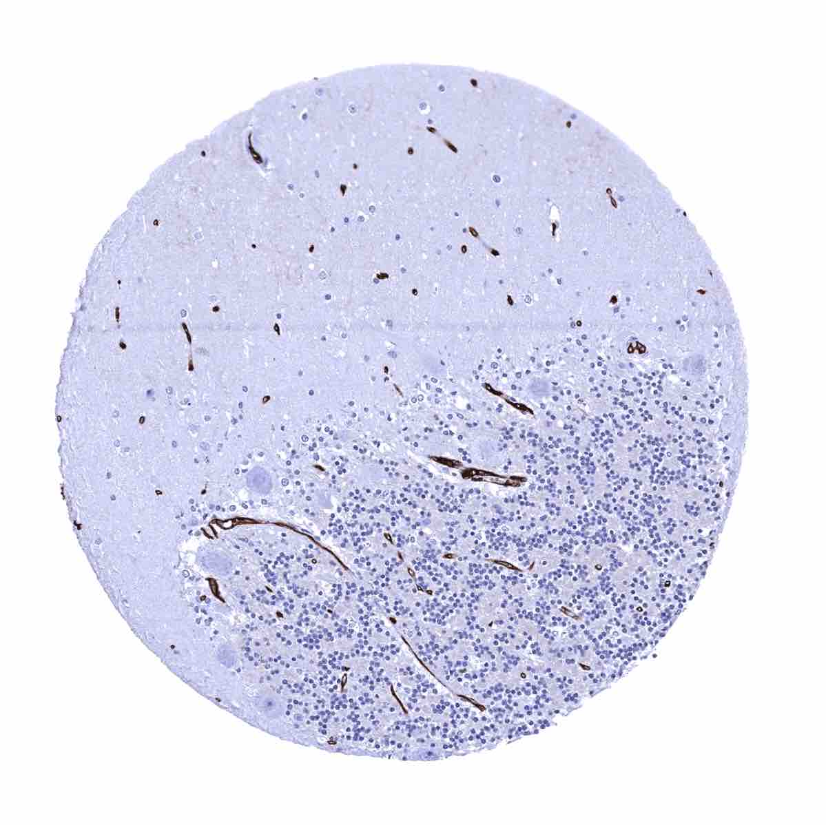
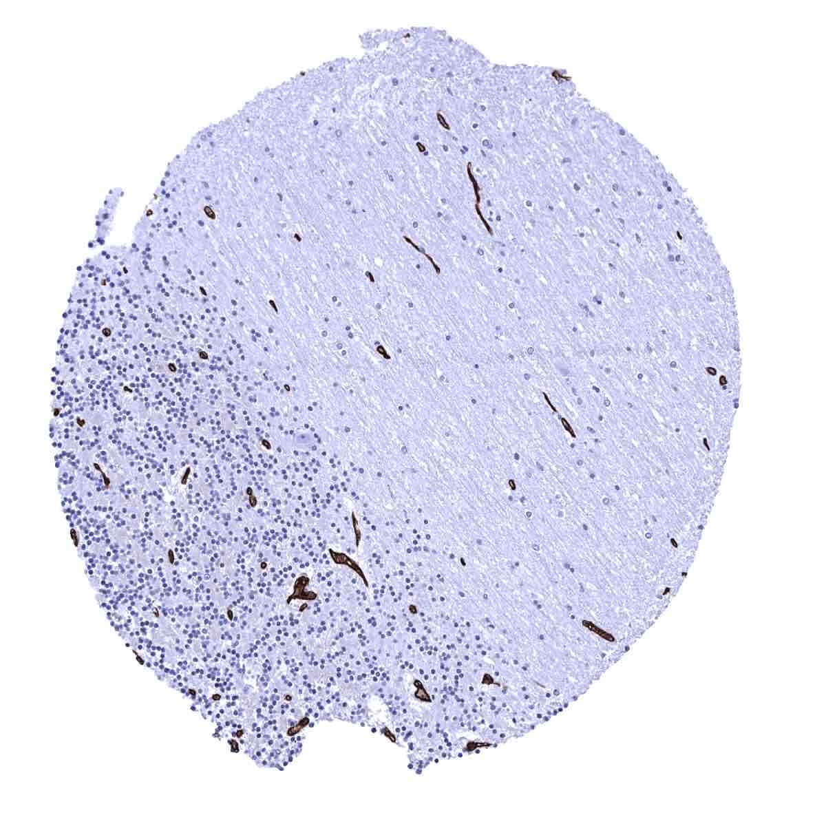
Cerebellum (molecular layer, Purkinje cell layer, granule cell layer) - GLUT1 staining in endothelial cells is strongest in the brain
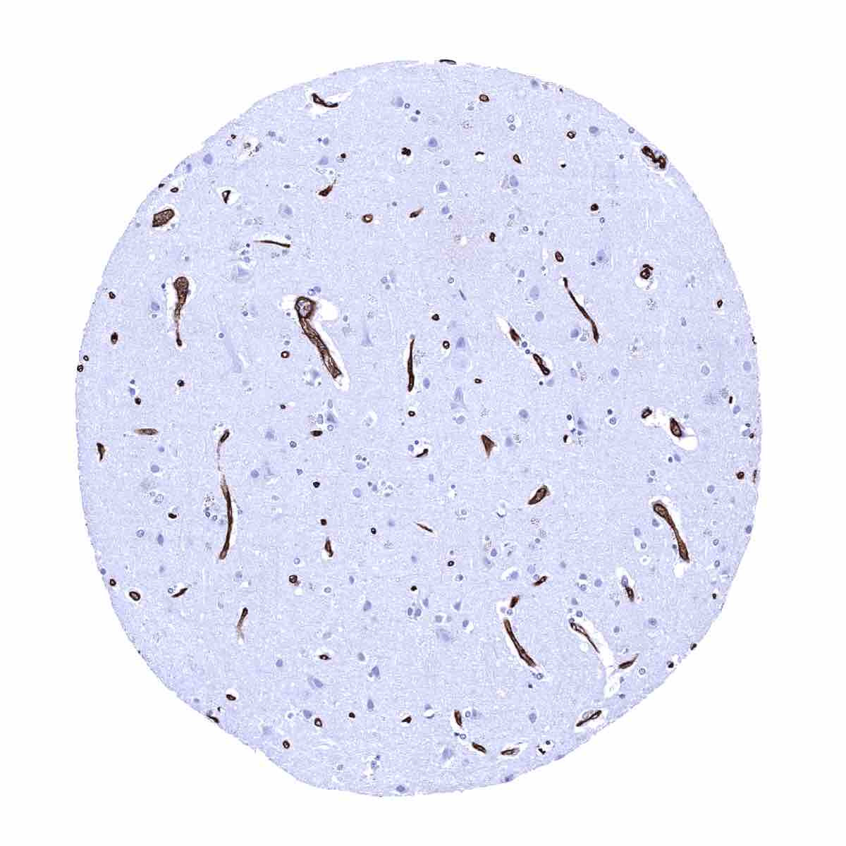
Cerebrum, grey matter - A particularly strong GLUT1 staining of endothelial cells is seen in the brain
Cerebrum, white matter
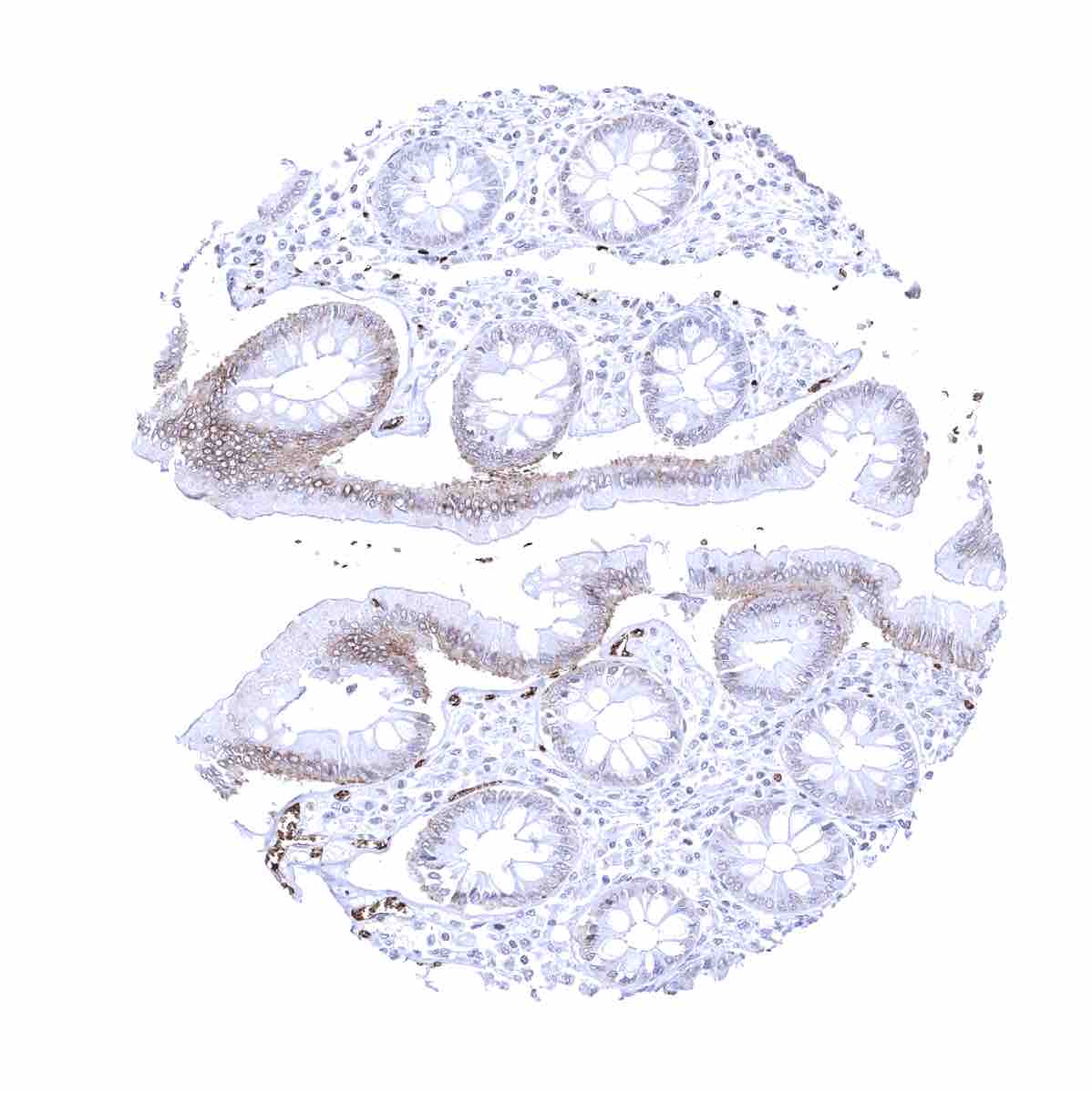
Colon descendens, muscular wall
Colon descendes, mucosa - Signficant GLUT1 staining of endothelial cells of most vessels
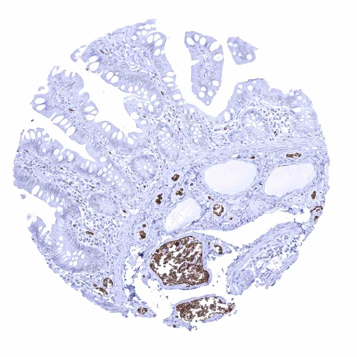
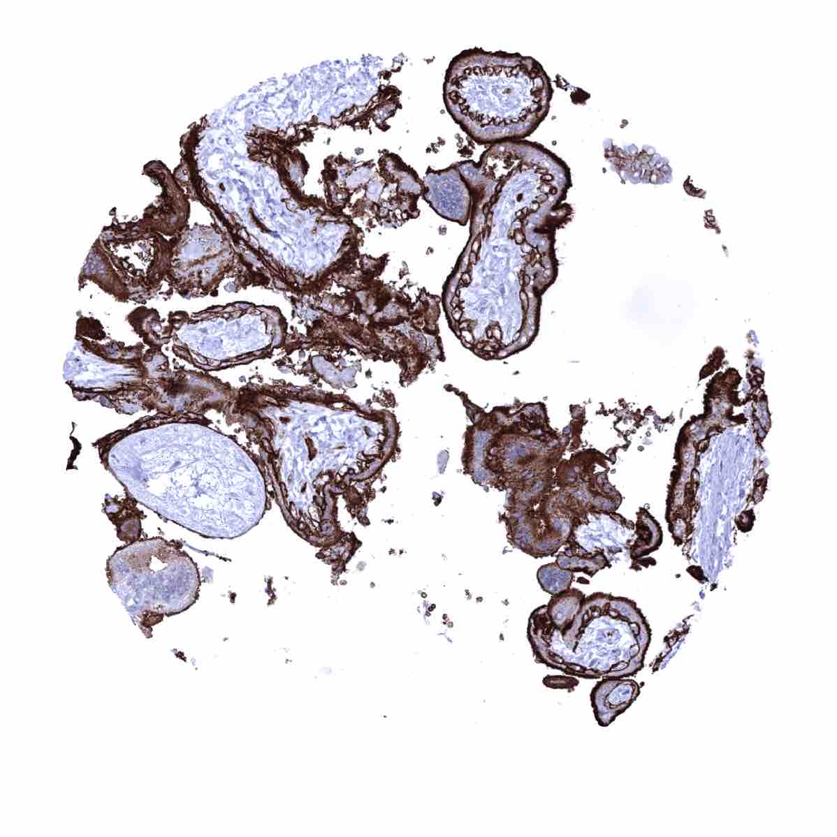
Duodenum, Brunner gland
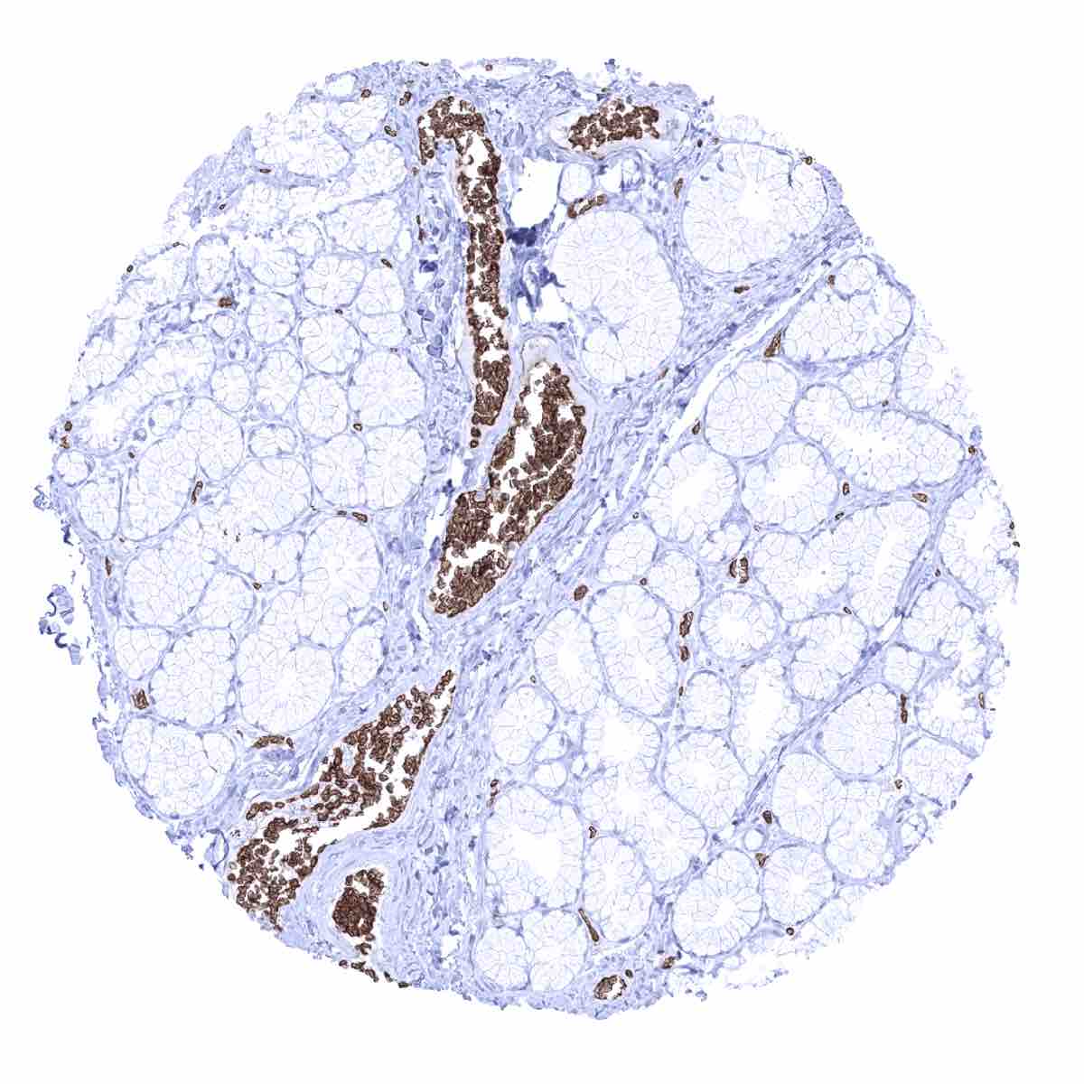
Duodenum, mucosa - Strong GLUT1 staining of endothelial cells of most small vessels
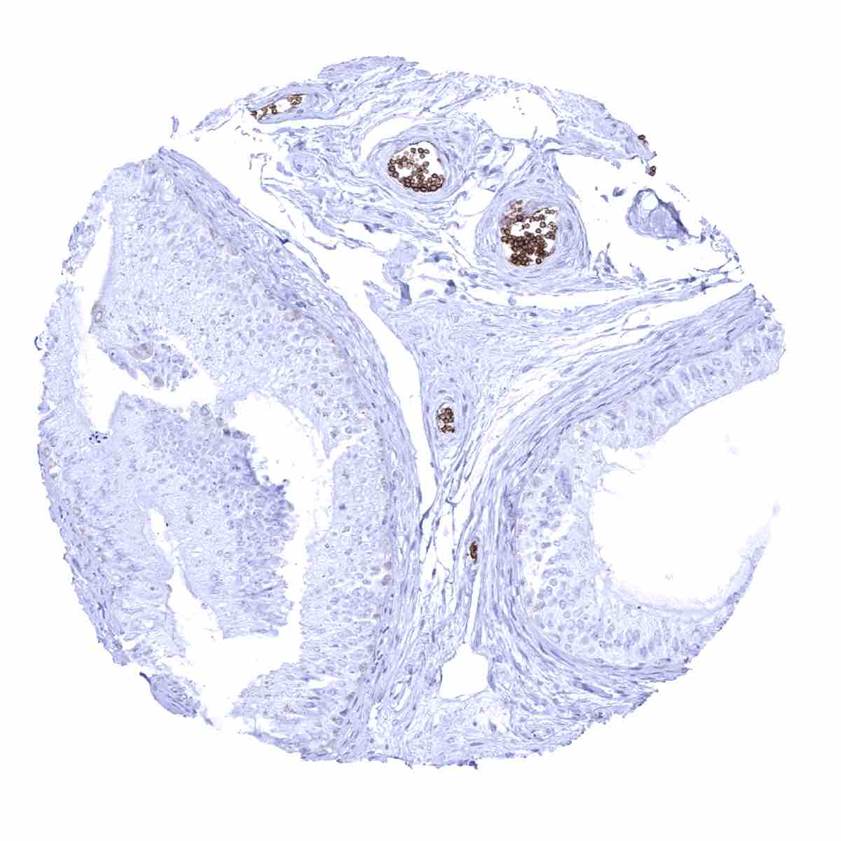
Epididymis
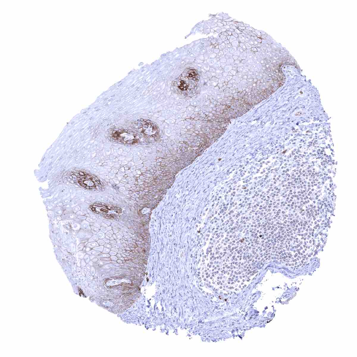
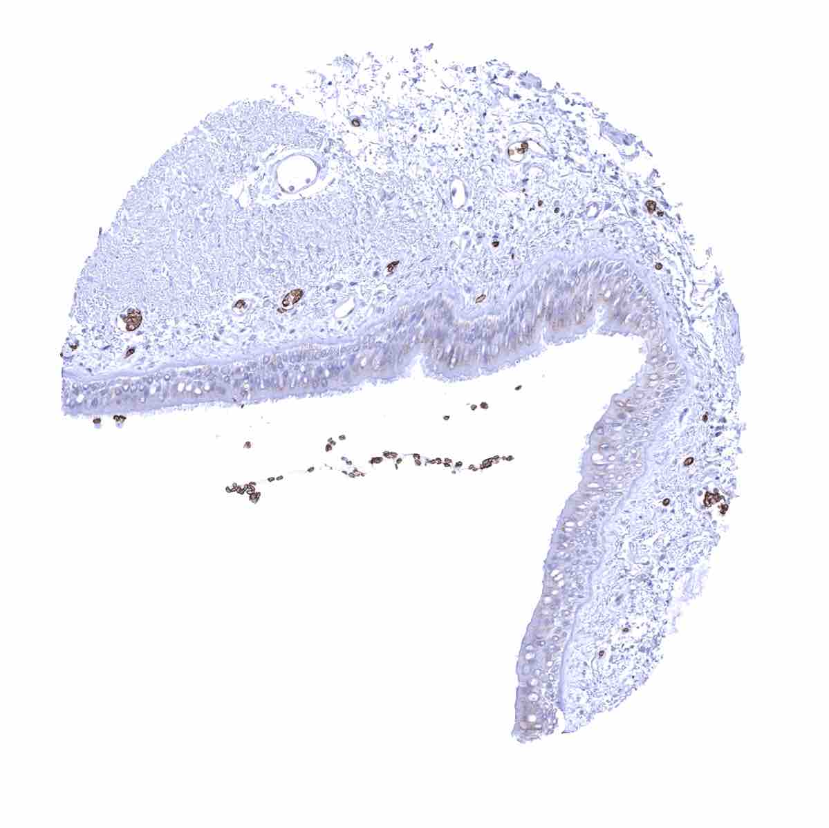
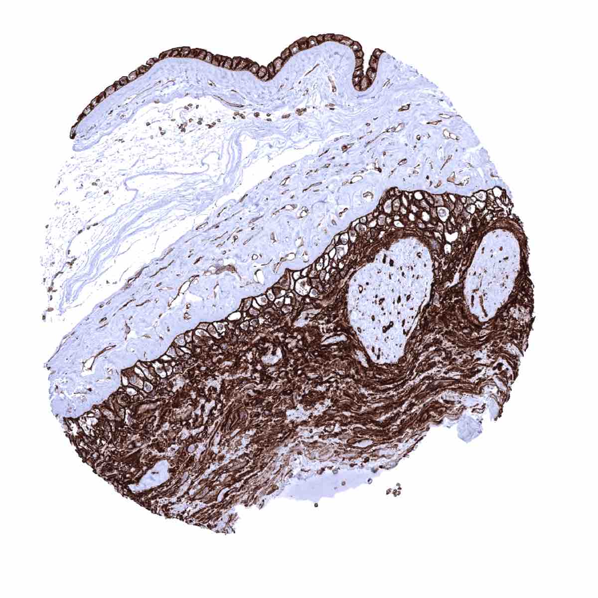
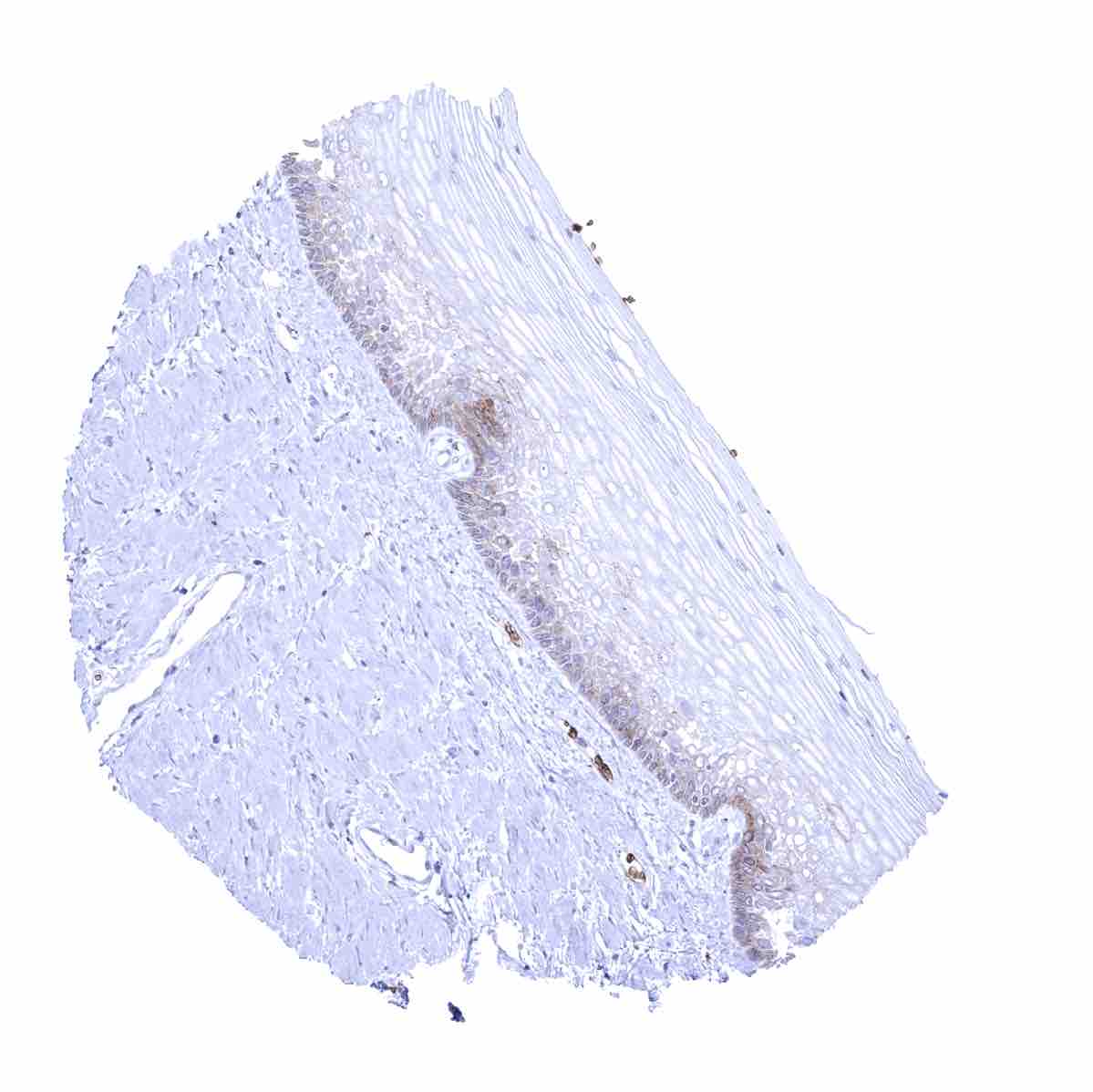
Esophagus, squamous epithelium - Weak to moderate membranous GLUT1 staining in the lower third of the squamous epithelium
Fallopian tube, mucosa - A weak to moderate cytoplasmic staining of epithelial cells occurs in the fallopian tube

Gallbladder, epithelium - Epithelial cell staining is absent in this sample

Gallbladder, epithelium - Focal epithelial cell staining of variable intensity

Heart muscle - Moderate to strong GLUT1 staining of small capillaries
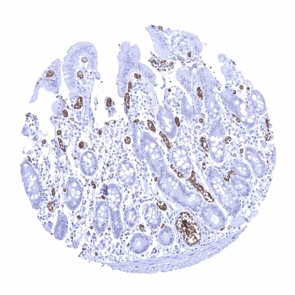
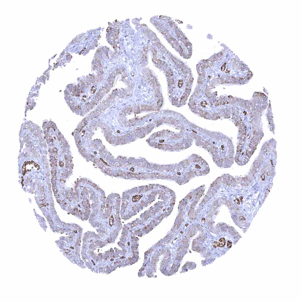
Ileum, mucosa
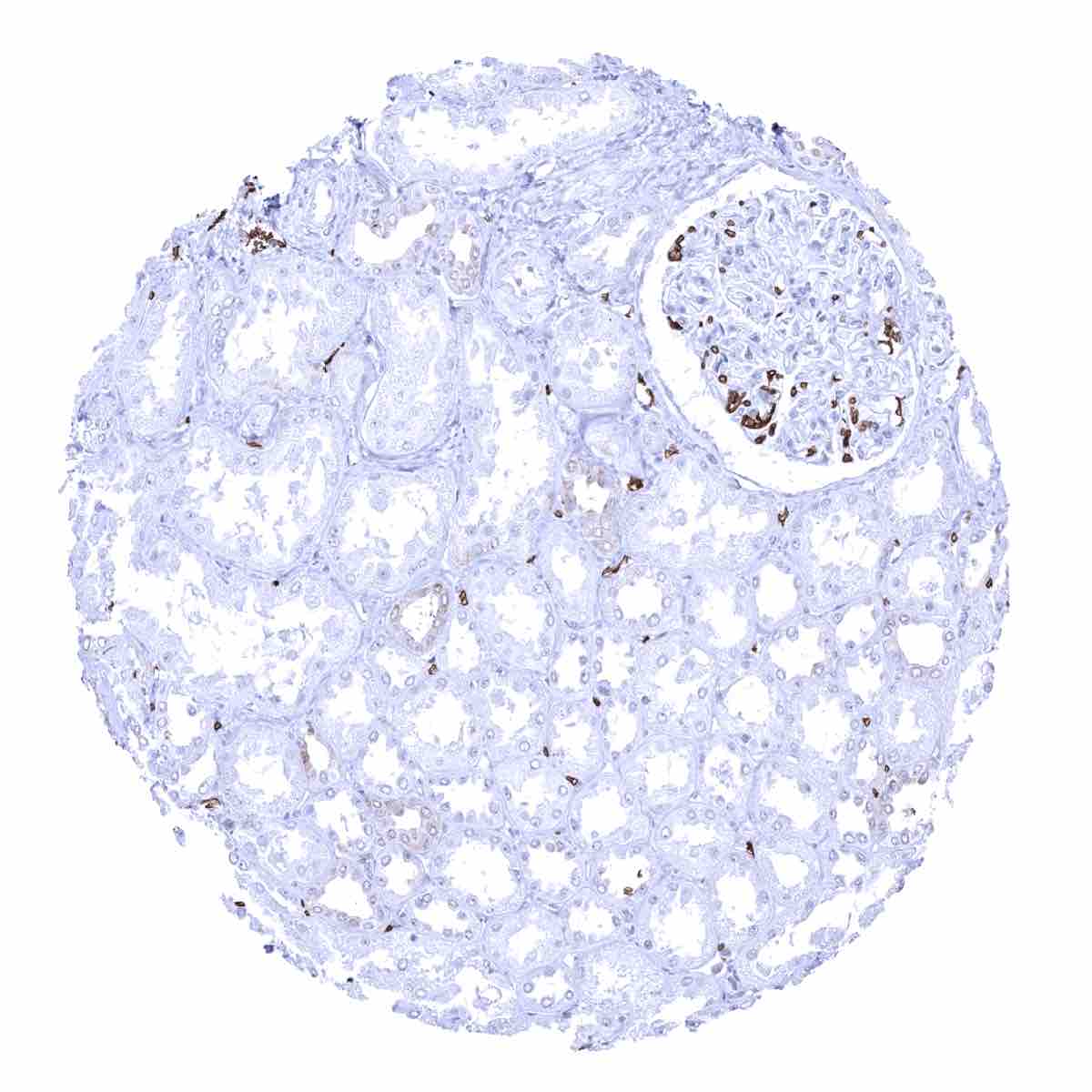
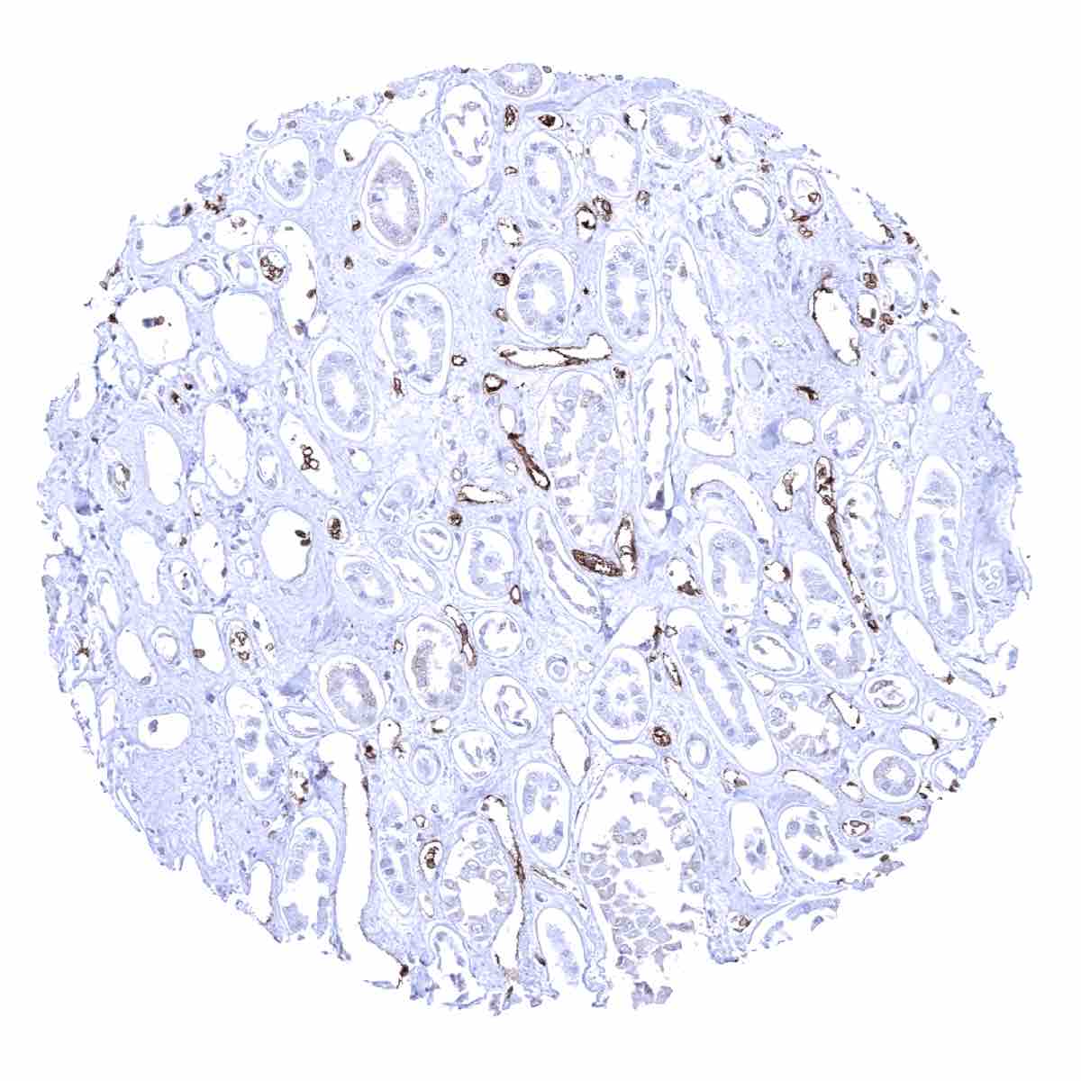
Kidney, cortex - Few tubuli or collecting ducts show weak GLUT1 staining

Kidney, medulla - Few collecting ducts show weak to moderate cytoplasmic GLUT1 staining
Kidney, medulla - GLUT1 staining is strongest in capillaries. Few tubuli or collecting ducts show weak cytoplasmic GLUT1 staining
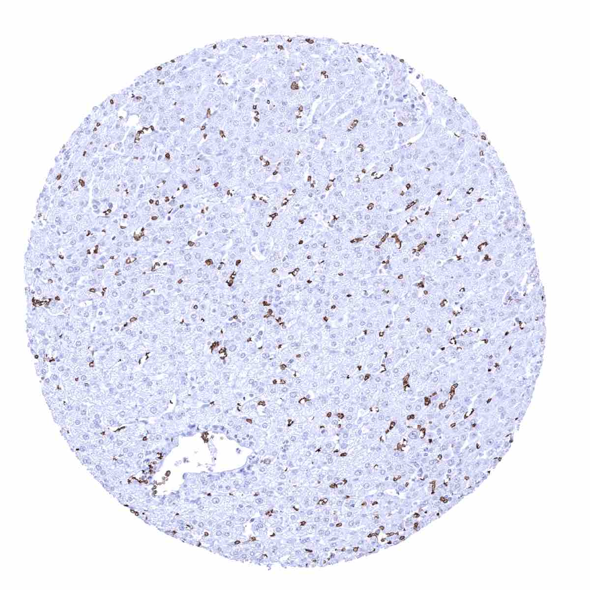
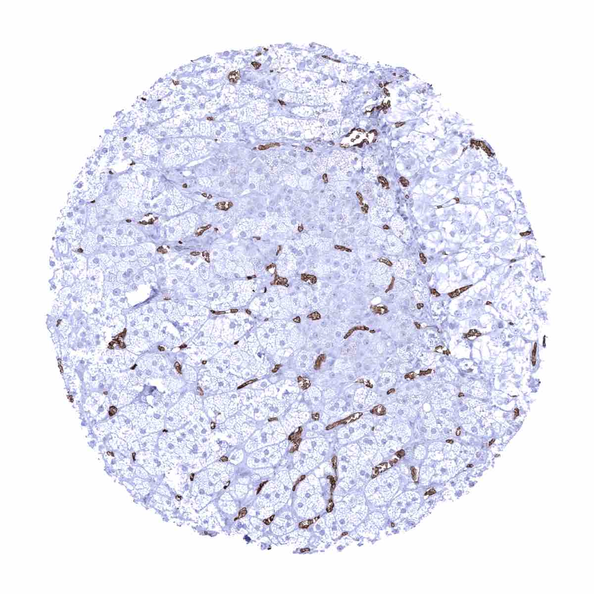
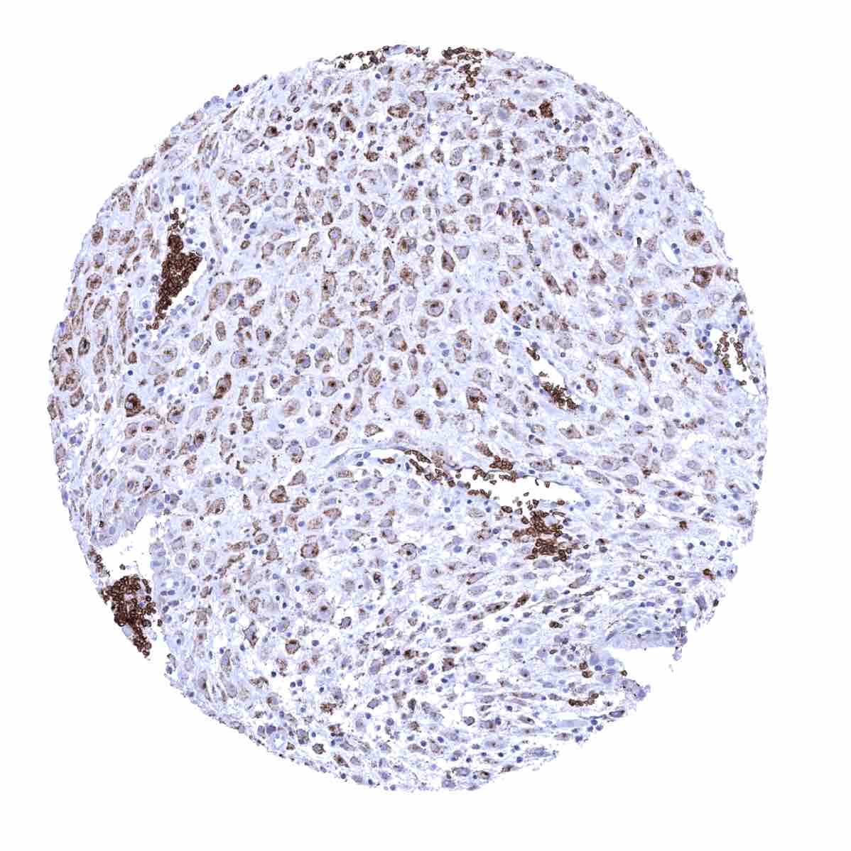
Liver - Strong GLUT1 staining of erythrocytes while sinusoidal cells lack staining
Lung - GLUT1 staining is largely limited to erythrocytes. Endothelial cells hardly stain positive
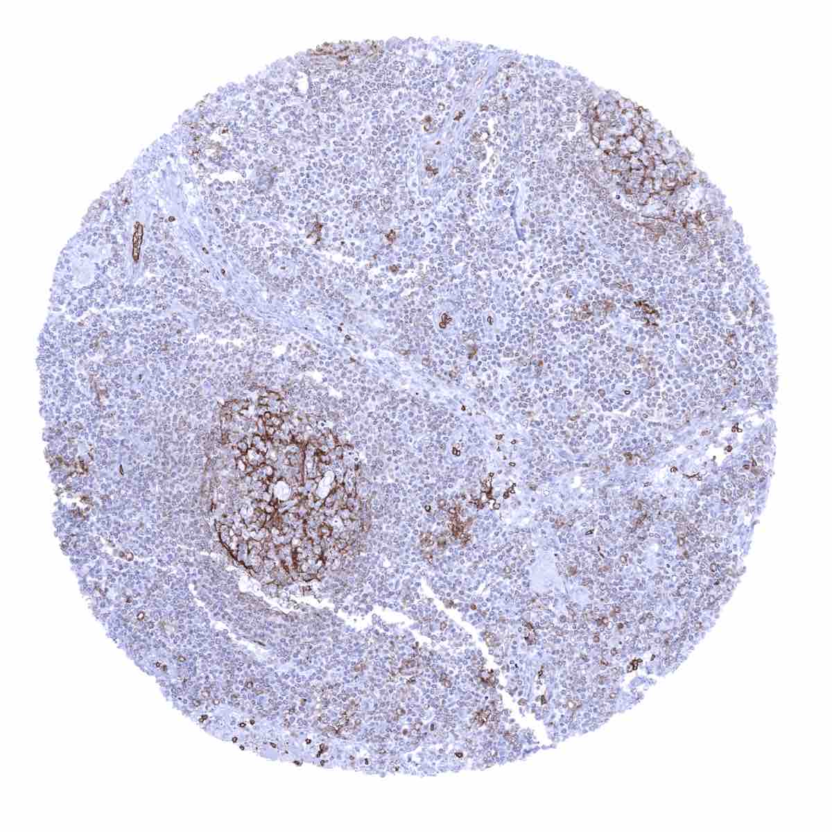
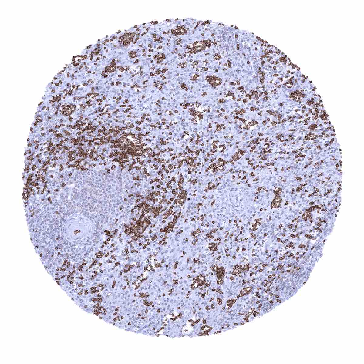
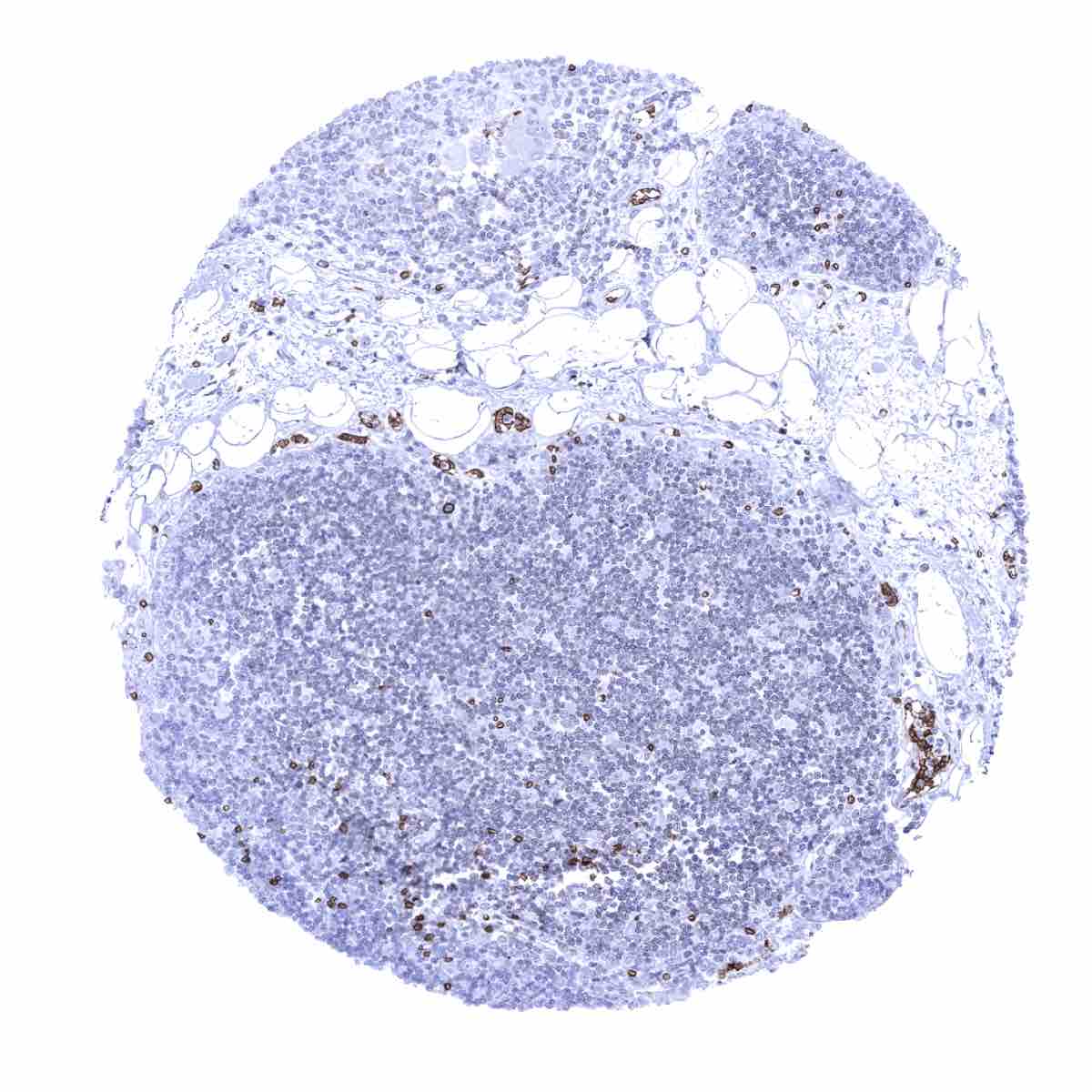
Lymph node - Prominent GLUT1 staining in follicular dendritic cells of germinal centres and in endothelial cells
Lymph node - Prominent GLUT1 staining in follicular dendritic cells of germinal centres and in endothelial cells. Staining is weaker in a fraction of interfollicular cells

Ovary stroma - Strong GLUT1 staining of endothelial cells of some small blood vessels
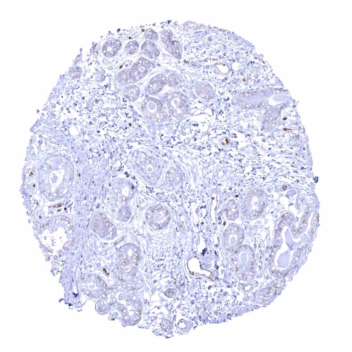
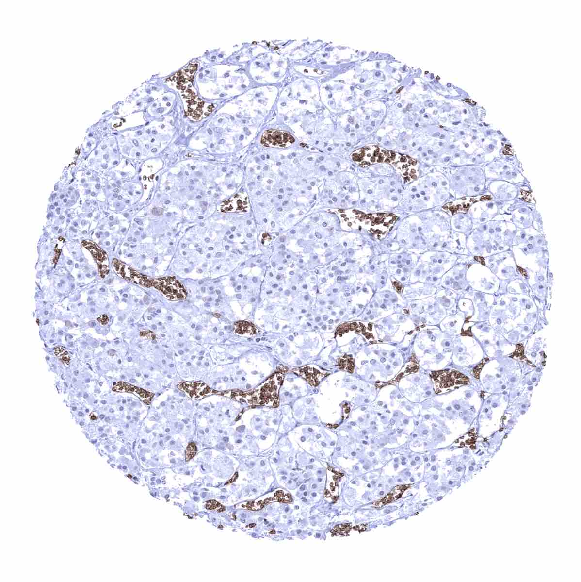
Pancreas - A focal membranous staining of acinar epithelial cells is seen in this sample from the pancreas

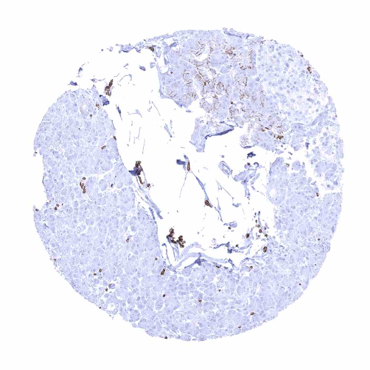
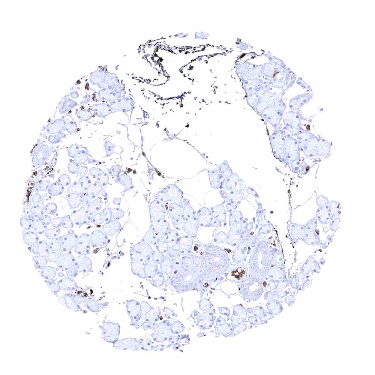
Pancreas - GLUT1 staining is limited to erythrocytes in this sample

Parathyroid gland
Parotid gland
Pituitary gland, anterior lobe
Pituitary gland, posterior lobe - Strong GLUT1 staining of endothelial cells
Placenta (amnion and chorion) - Membranous GLUT1 staining is very intense in amnion and chorion cells. Endothelial cells also show a particularly strong staining
Placenta, early - Very intense, predominantly membranous GLUT1 staining of syncytio- and cytotrophoblast cells

Placenta, mucosa - Very intense, predominantly membranous GLUT1 staining of syncytio- and cytotrophoblast cells. Endothelial cells also show a strong positivity

Prostate - Weak GLUT1 staining in basal cells
Rectum, mucosa - A weak membranous and cytoplasmic, preferentially basolateral GLUT1 staining in the surface epithelium of this rectum sample
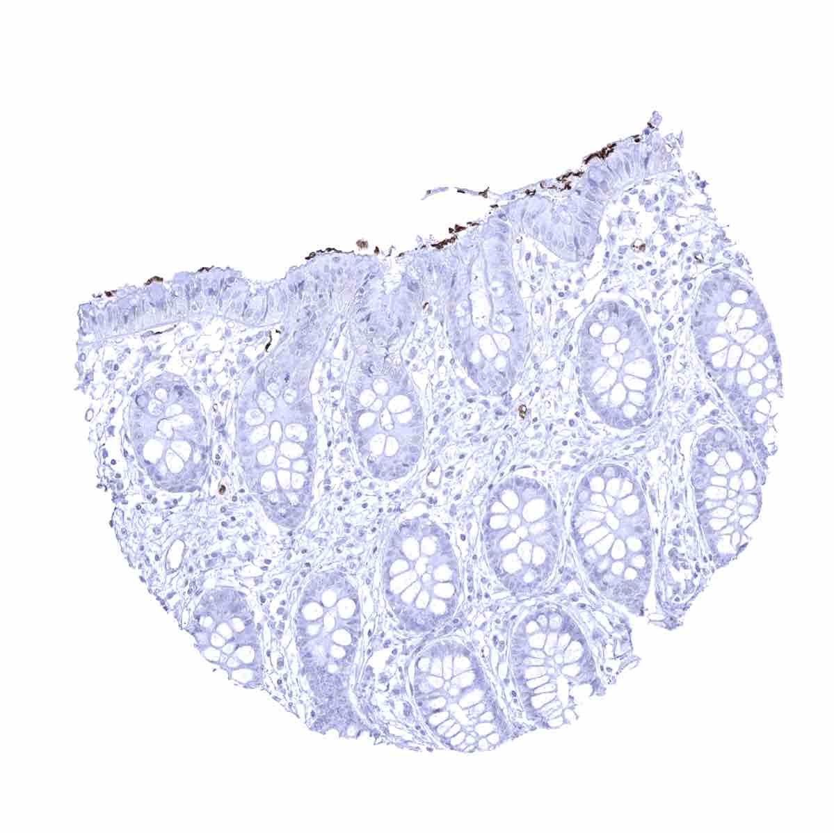
Rectum, mucosa - GLUT1 staining largely absent in this rectum sample

Seminal vesicle - A predominantly basolateral membranous GLUT1 positivity occurs in a fraction of epithelial cells
Sinus paranasales
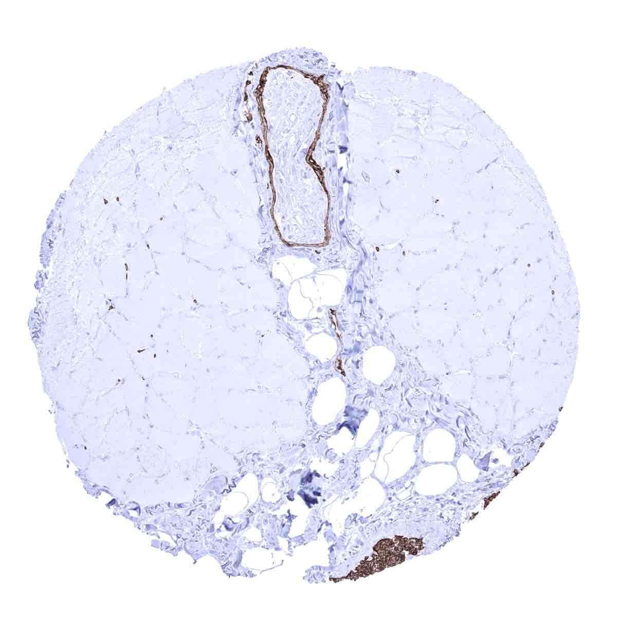
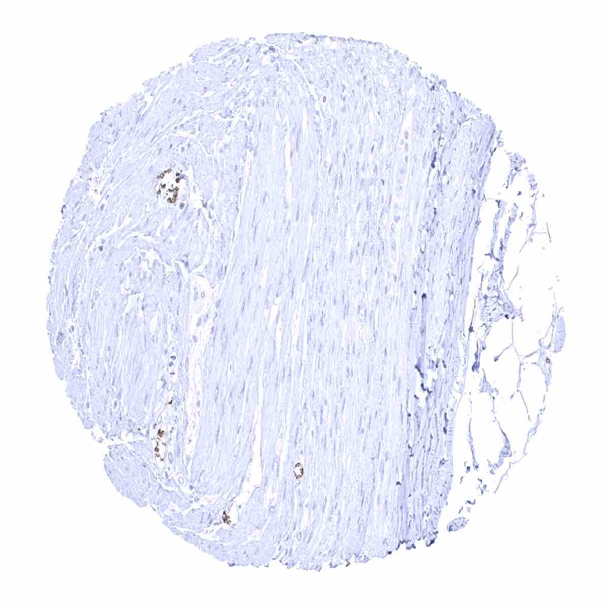
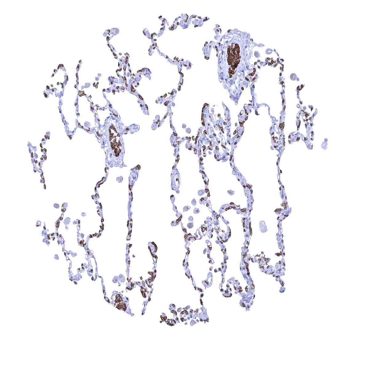
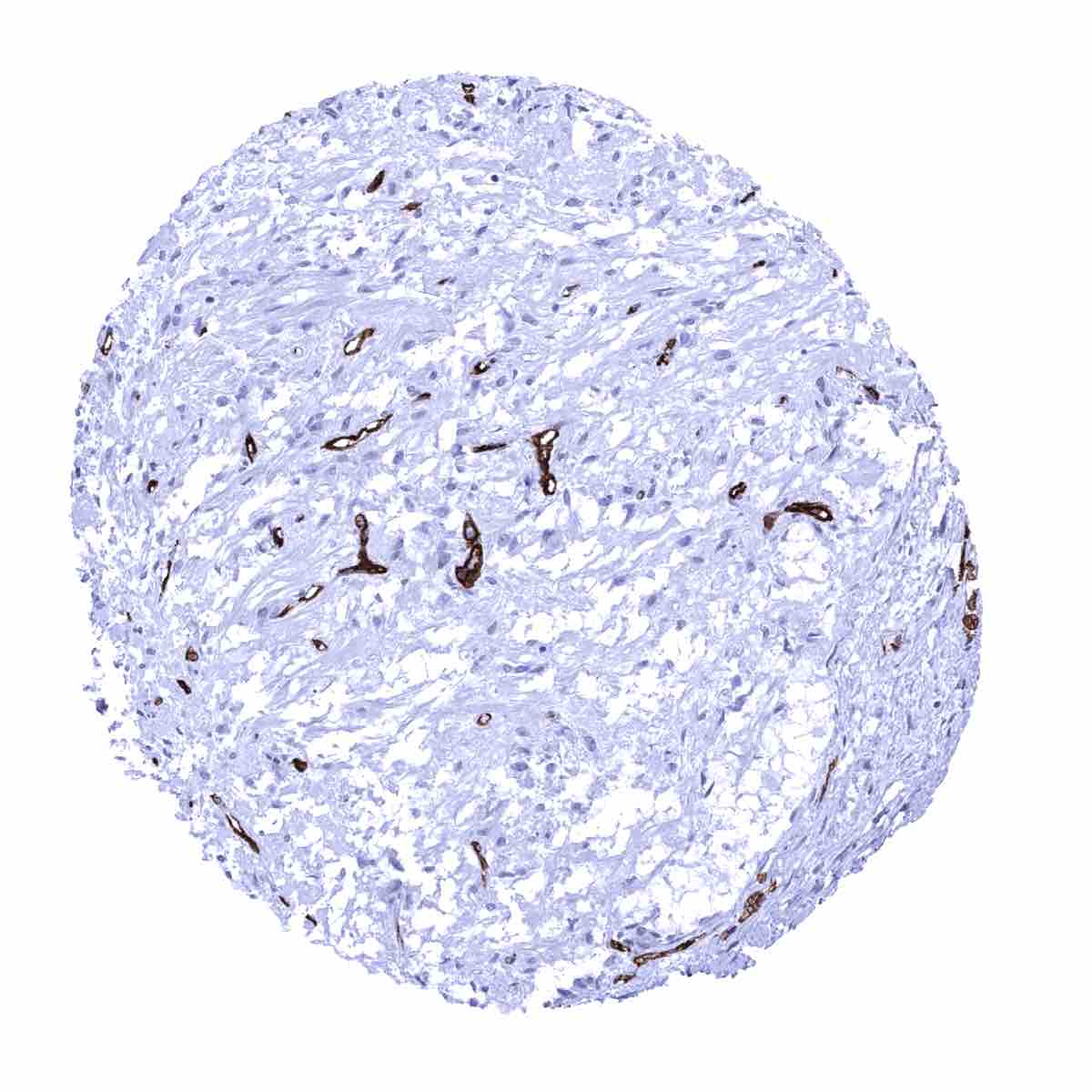
Skeletal muscle - Strong GLUT1 staining perineurium while endothelial cells of small capillaries lack staining
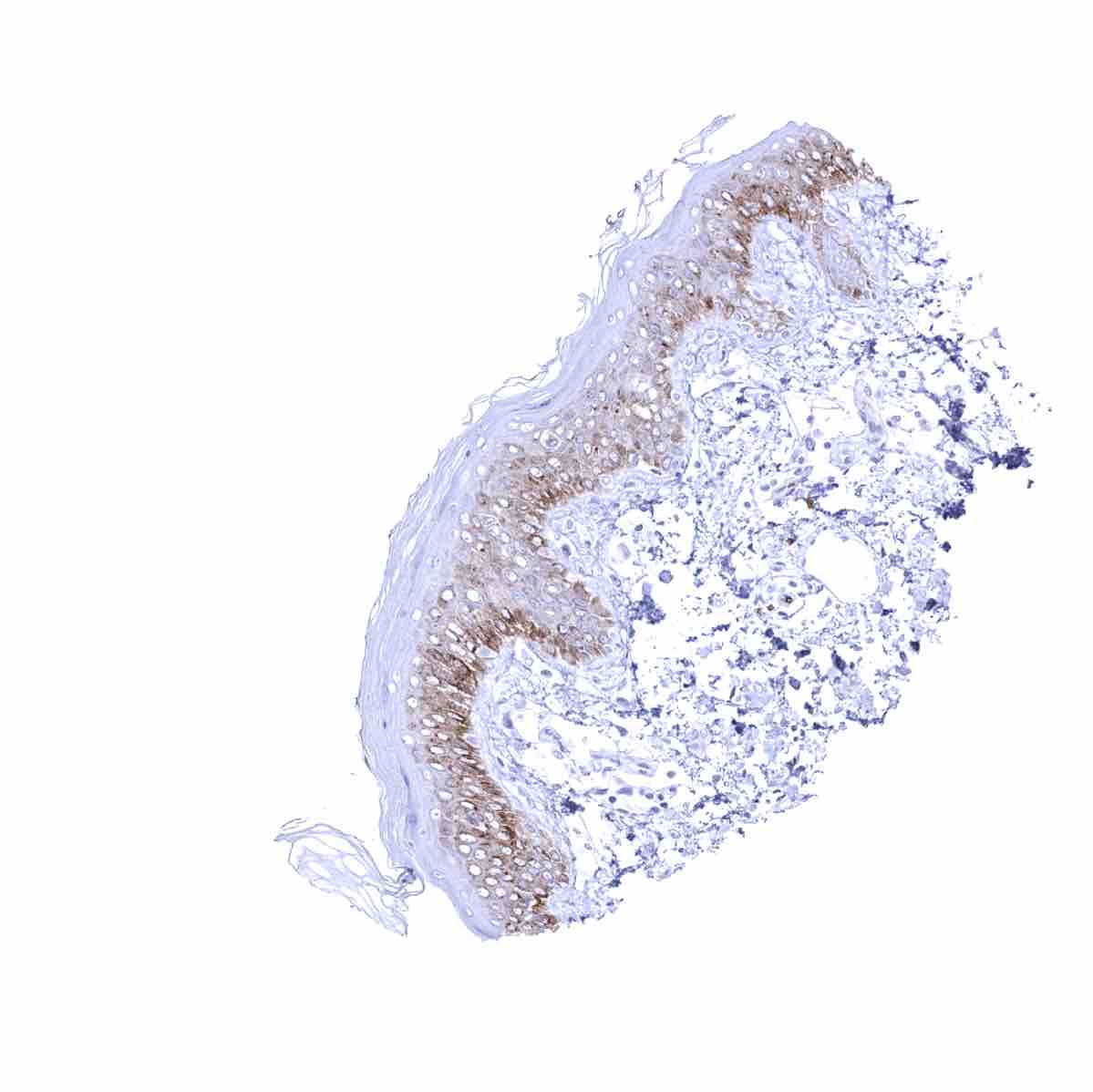
Skin - Weak to moderate cytoplasmic GLUT1 staining, preferentially in the lower third of the squamous epithelium
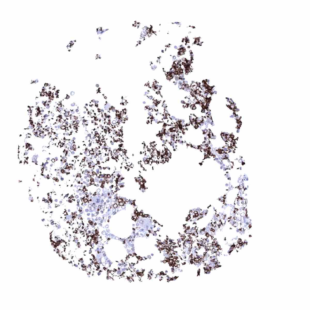
Spleen - Strong GLUT1 staining of erythrocytes

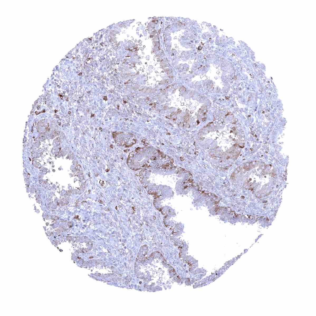
Stomach, antrum - A weak membranous and cytoplasmic, preferentially basolateral GLUT1 staining occurs in the surface epithelium of this sample

Stomach, antrum - GLUT1 staining is limited to erythrocytes in this stomach sample
Testis
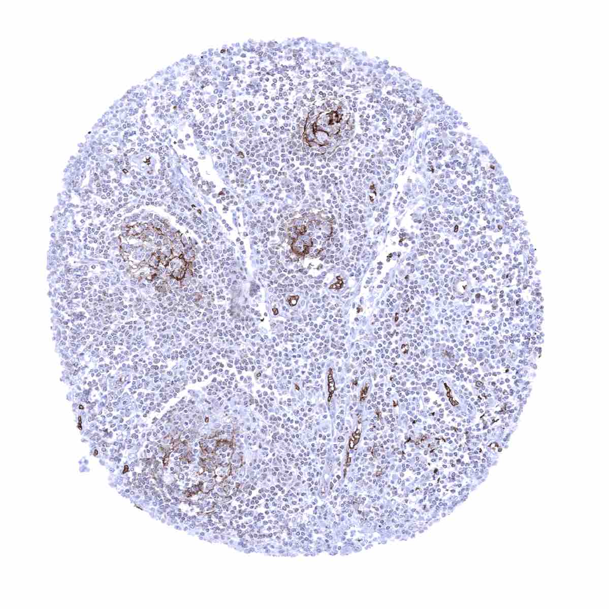
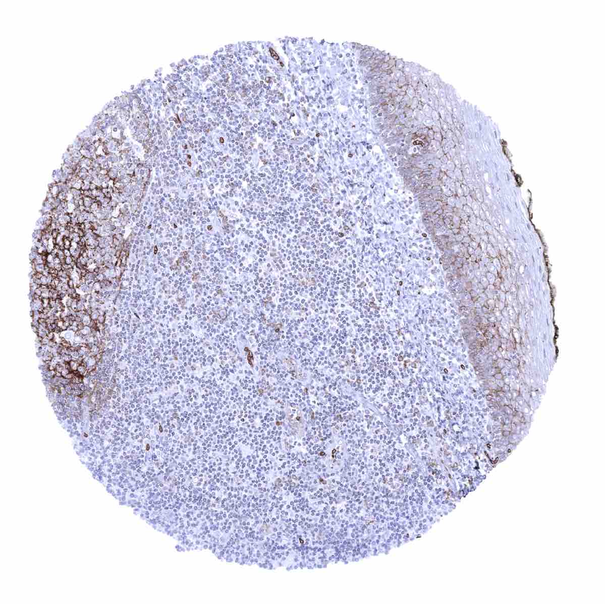
Thymus
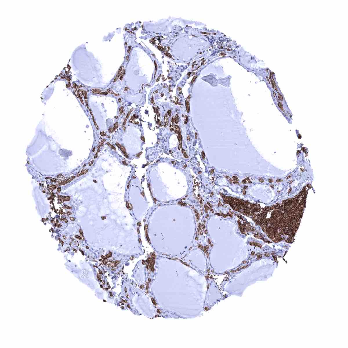
Thyroid gland - Epithelial cells are GLUT1 negative
Tonsil, surface epithelium - Weak to moderate membranous GLUT1 staining in the lower half of the squamous epithelium. Moderate to strong GLUT1 staining of follicular dendritic cells in germinal centres while staining is markedly weaker in a fraction of interfollicular cells.
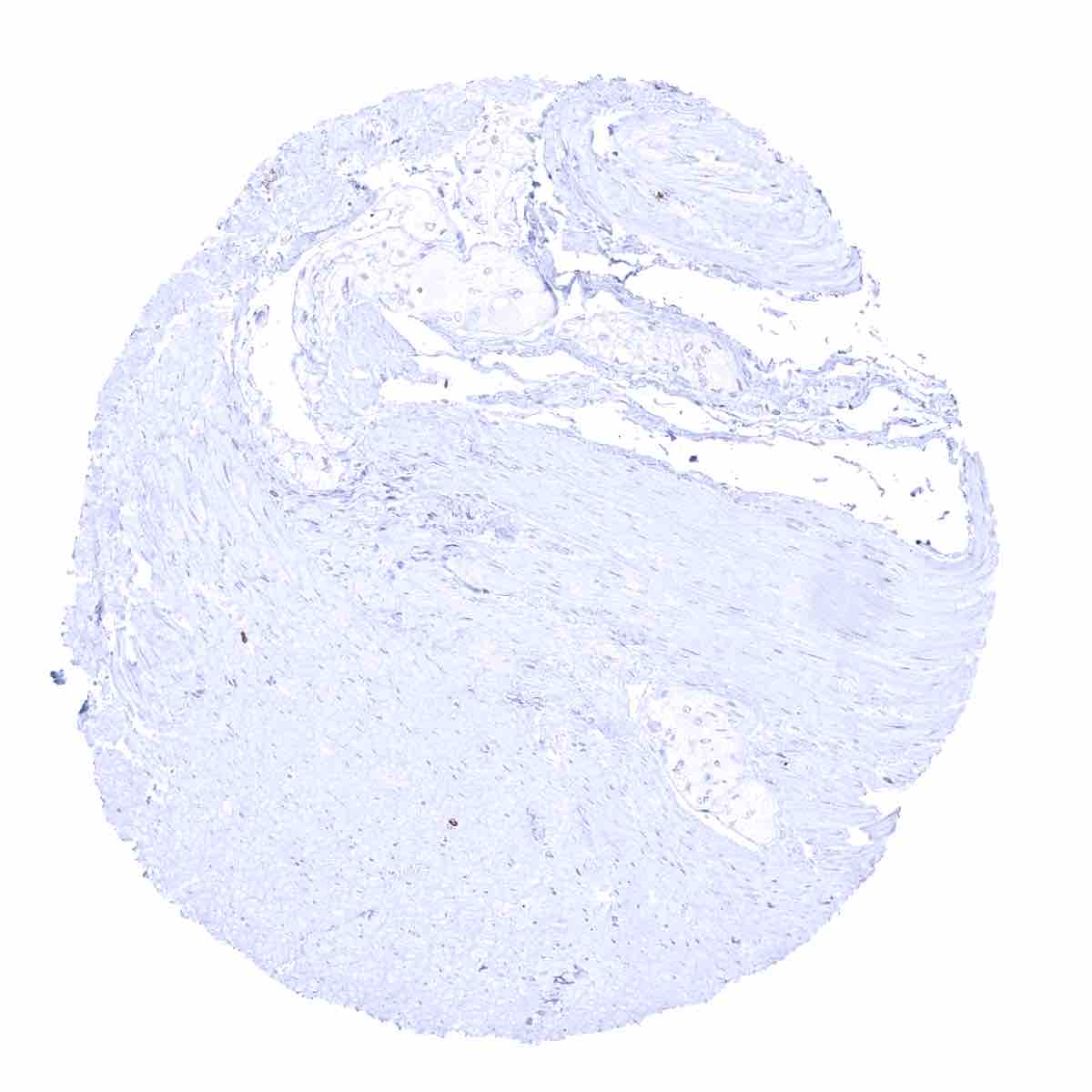
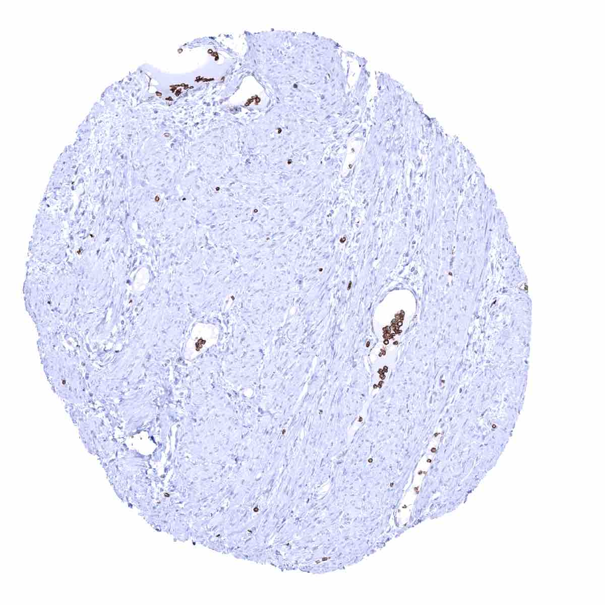
Urinary bladder, muscular wall

Urinary bladder, urothelium - A weak to moderate membranous staining occurs in the urothelium with decreasing intensity from the basal to the superficial cell layers
Uterus, ectocervix
Uterus, endocervix
Uterus, endometrium (pregnancy) - Moderate intensity, predominantly membranous GLUT1 staining of decidua cells

Uterus, endometrium (proliferation) - Prominent GLUT1 staining of small vessels in this sample

Uterus, endometrium (proliferation) - Weak GLUT1 staining of both epithelial and stromal cells
Uterus, endometrium (secretion) - Weak to moderate GLUT1 staining of both epithelial and stromal cells
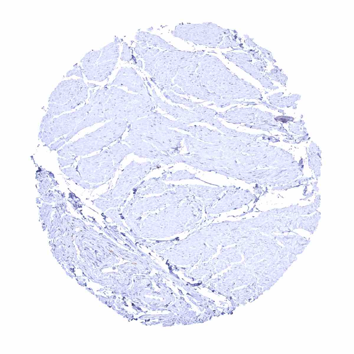
Uterus, myometrium