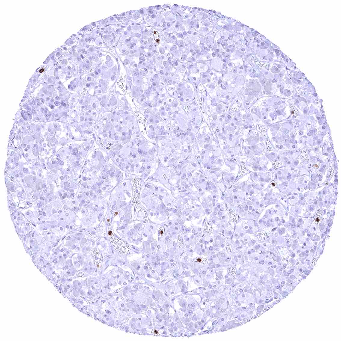
Adrenal gland
Aorta, endothelium.jpeg
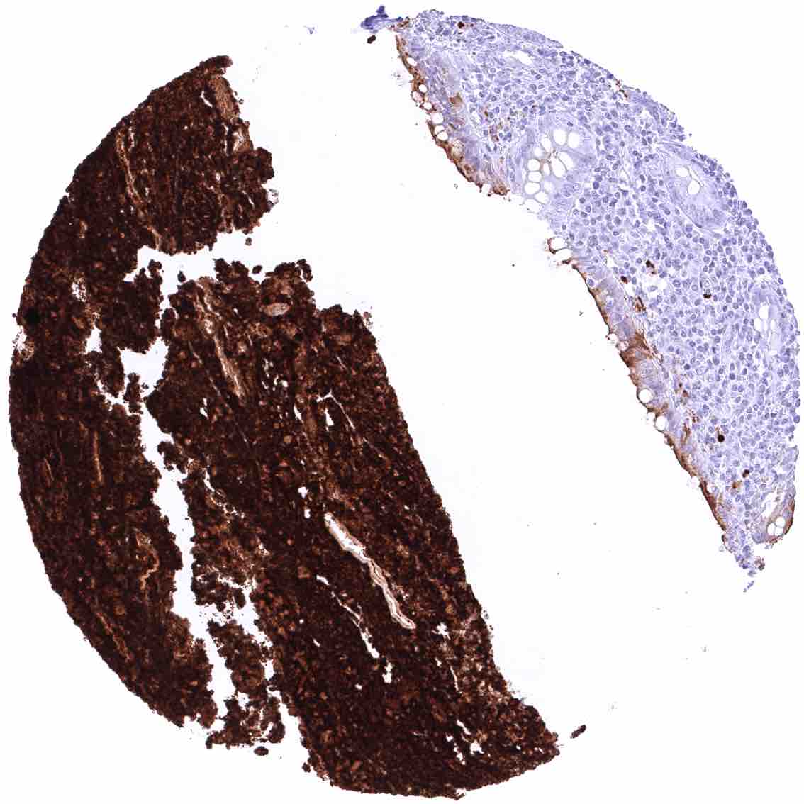
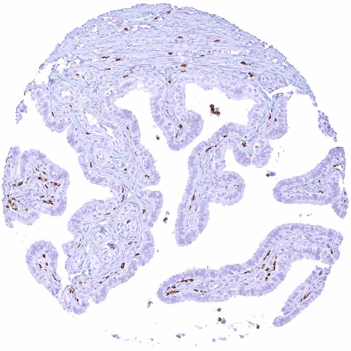
Appendix, mucosa – Moderate apical membranous CEACAM6 staining of at least a fraction of surface epithelial cells. The CEACAM6 staining is weaker in glandular cells. .jpeg
Appendix, muscular wall.jpeg
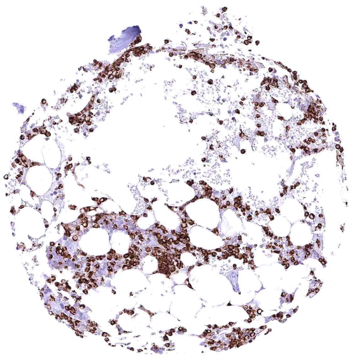
Bone marrow – Strong CEACAM6 staining of a subset of cells, probably of the myeloid cell line. .jpeg
Breast – Apical membranous CEACAM6 staining of a fraction of luminal epithelial cells. .jpeg
Bronchus, mucosa – Moderate membranous CEACAM6 staining of a fraction of respiratory epithelial cells.
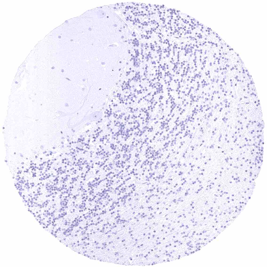
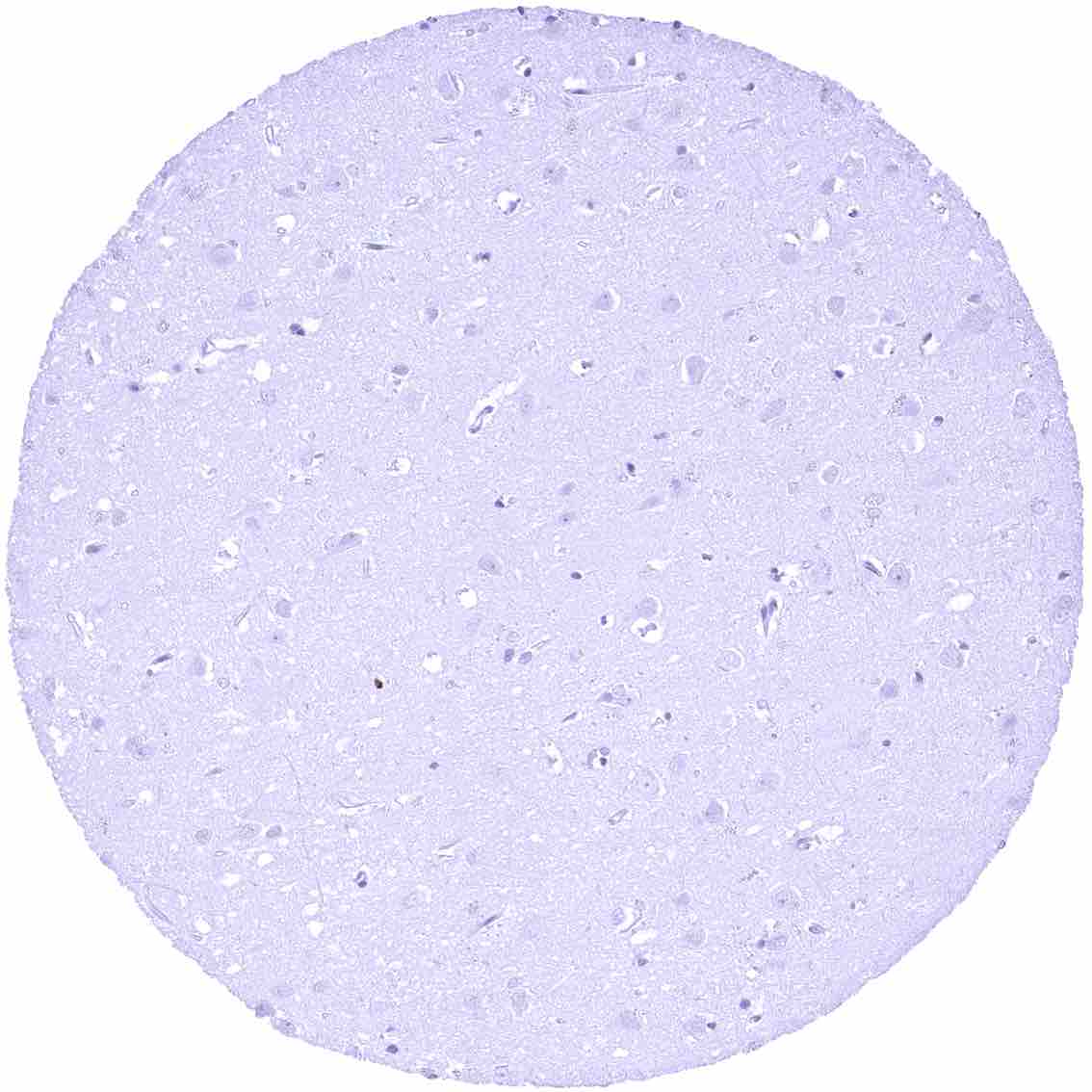
Cerebellum (molecular layer, Purkinje cell layer, granule cell layer)
Cerebellum (white matter)
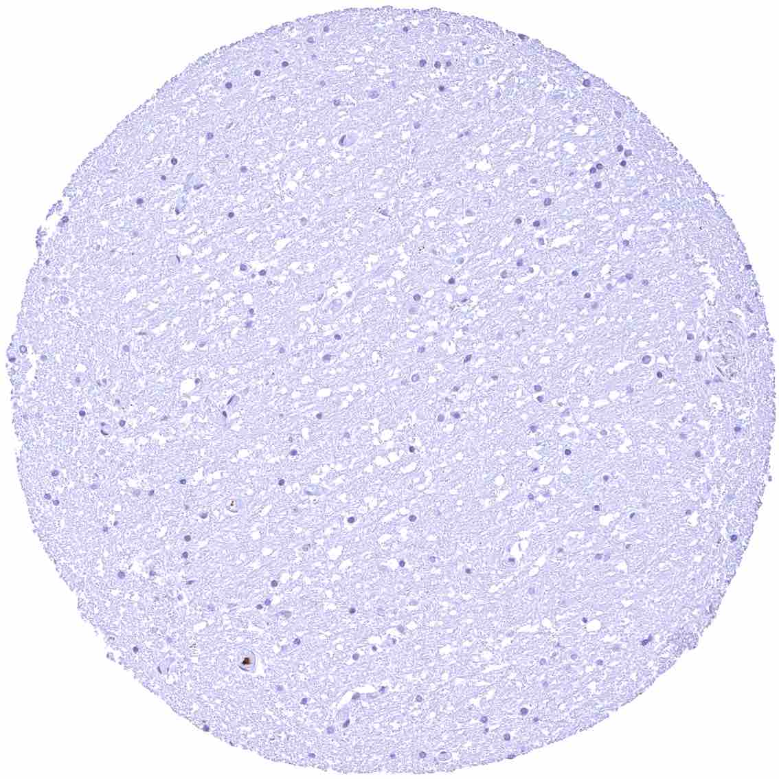
Cerebrum (grey matter)
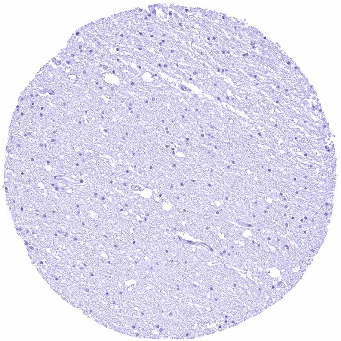
Cerebrum (white matter)
Colon descendens, muscular wall
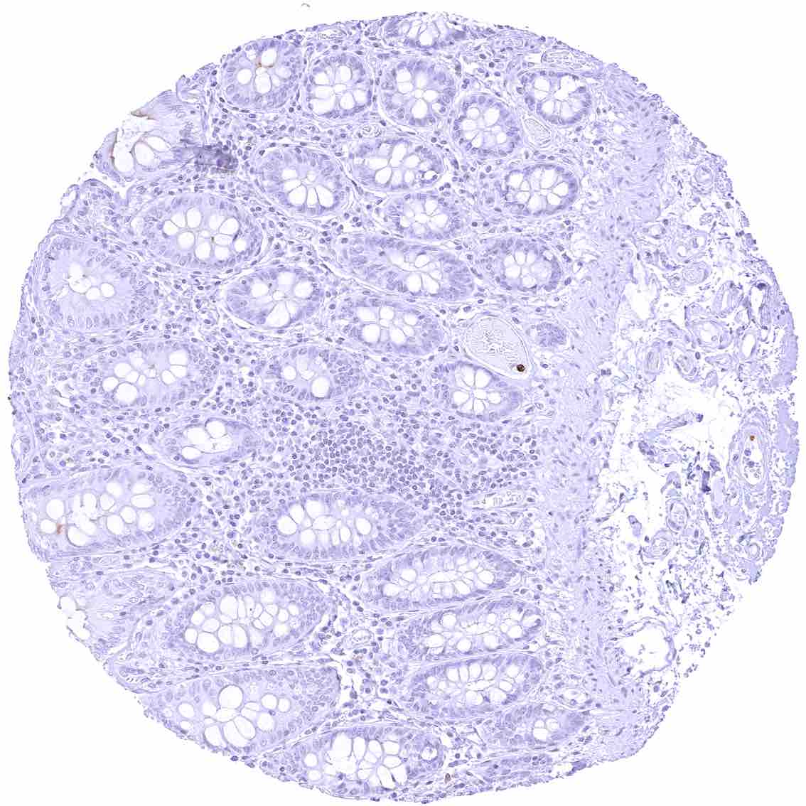
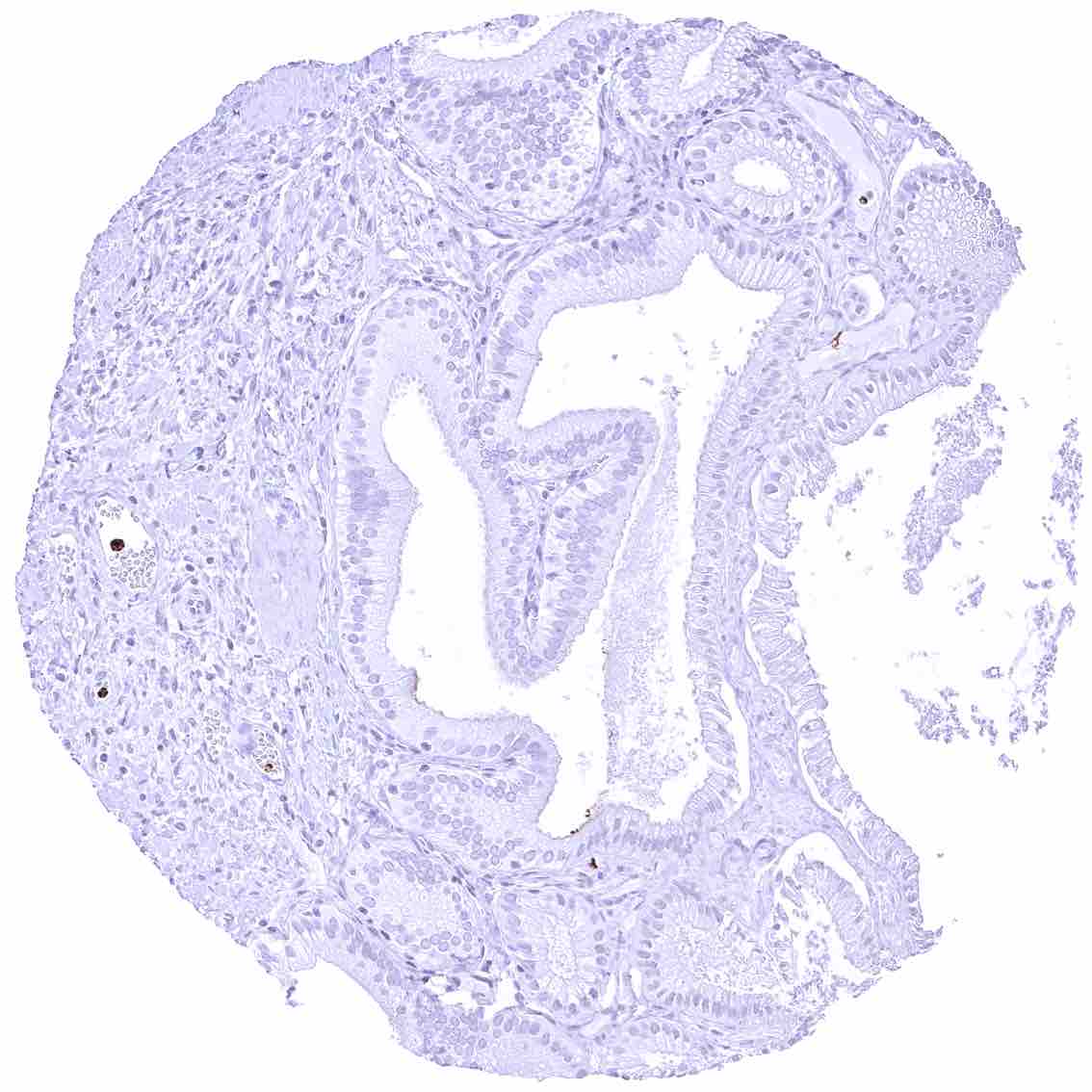
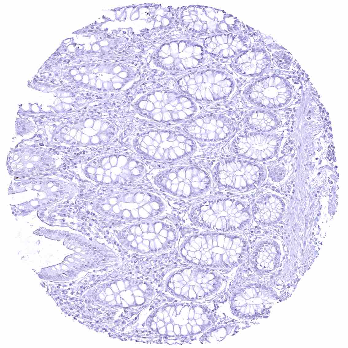
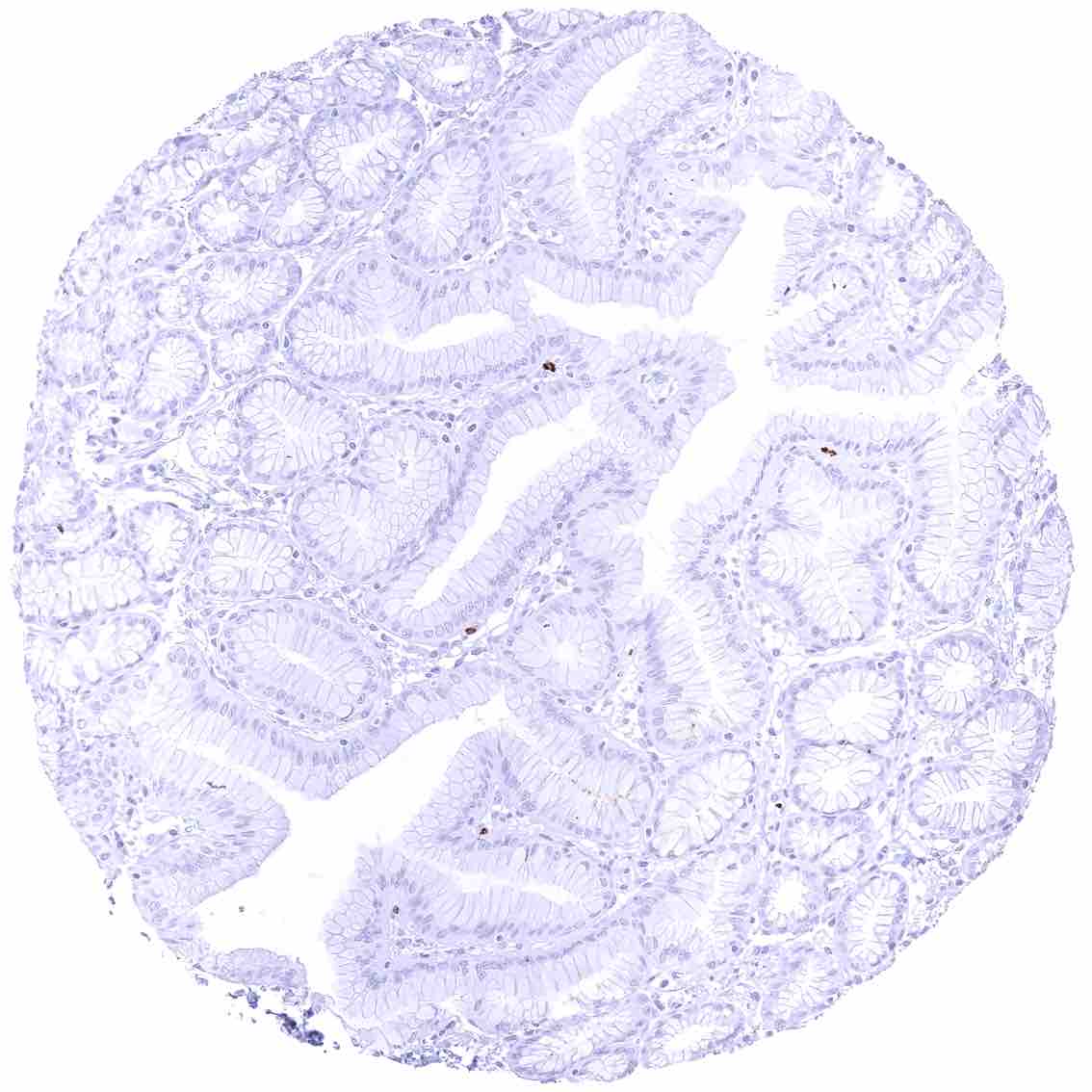
Colon descendens, mucosa – Faint apical membranous CEACAM6 staining of at least a fraction of epithelial cells.
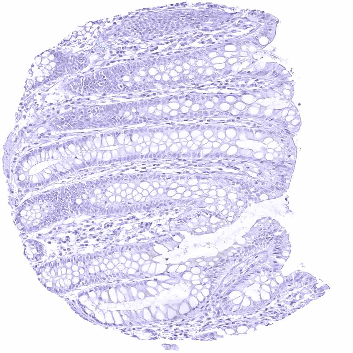
Colon descendens, mucosa.jpeg
Duodenum, mucosa
Duodenum, Brunner gland
Epididymis (Caput)
Esophagus, muscular wall
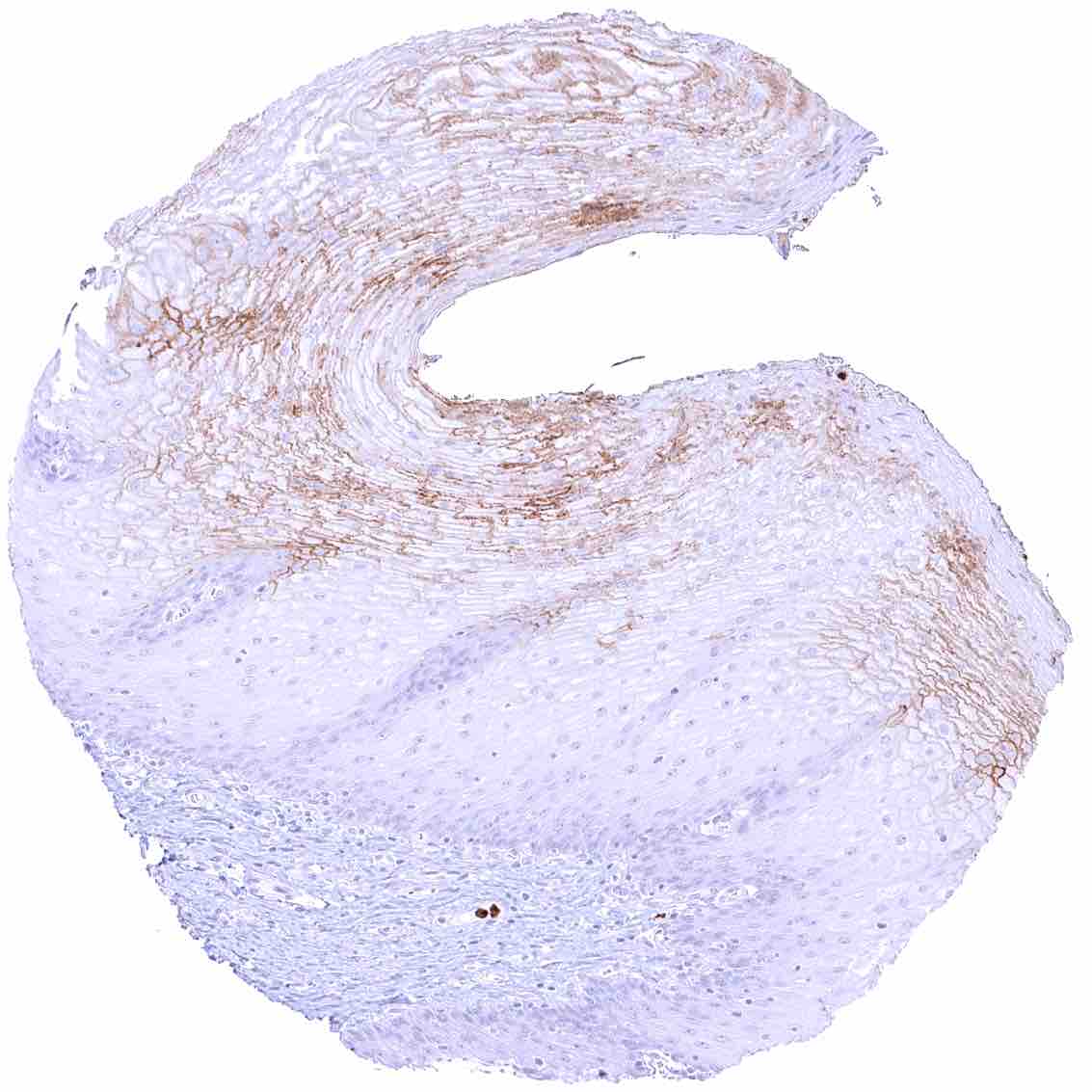
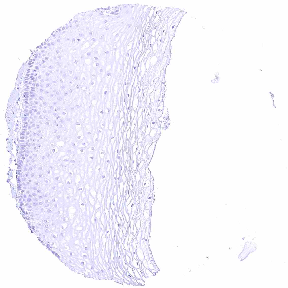
Esophagus, squamous epithelium – Weak to moderate membranous CEACAM6 staining of a large fraction of squamous epithelial cells especially of the upper half of the epithelium.
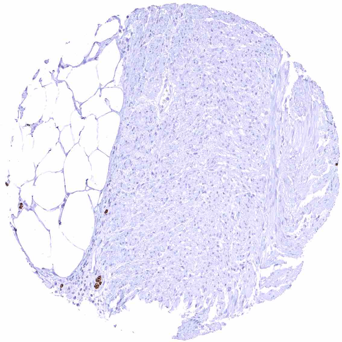
Fallopian tube, mucosa – CEACAM6 staining is limited to inflammatory cells.

Fat
Gallbladder, epithelium – Faint apical membranous CEACAM6 staining of some epithelial cells.
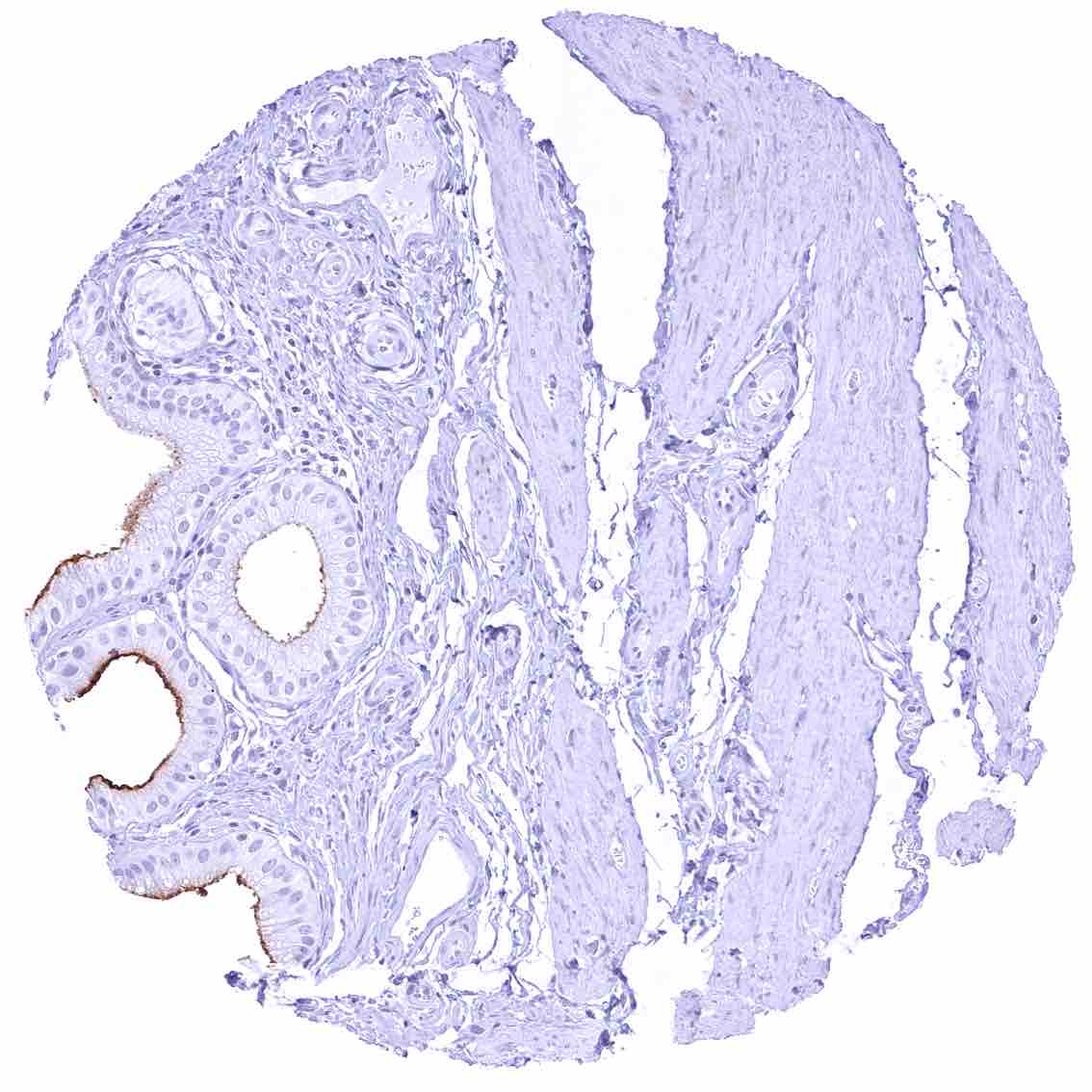
Gallbladder, epithelium – Moderate to strong apical membranous CEACAM6 staining of some epithelial cells.
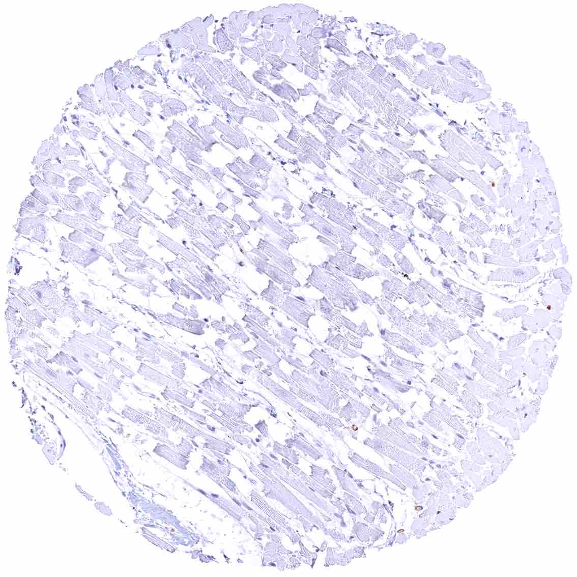
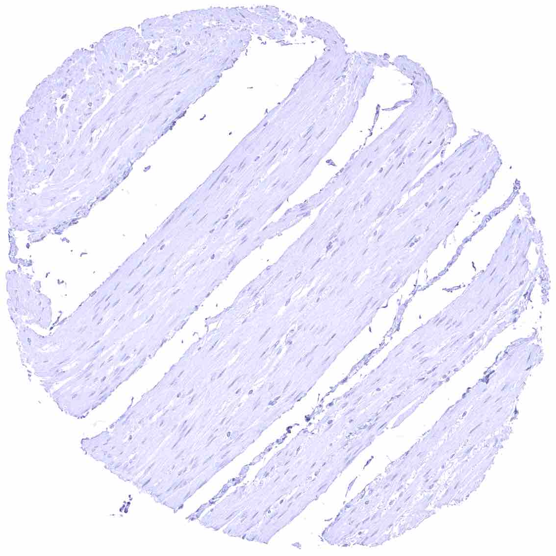
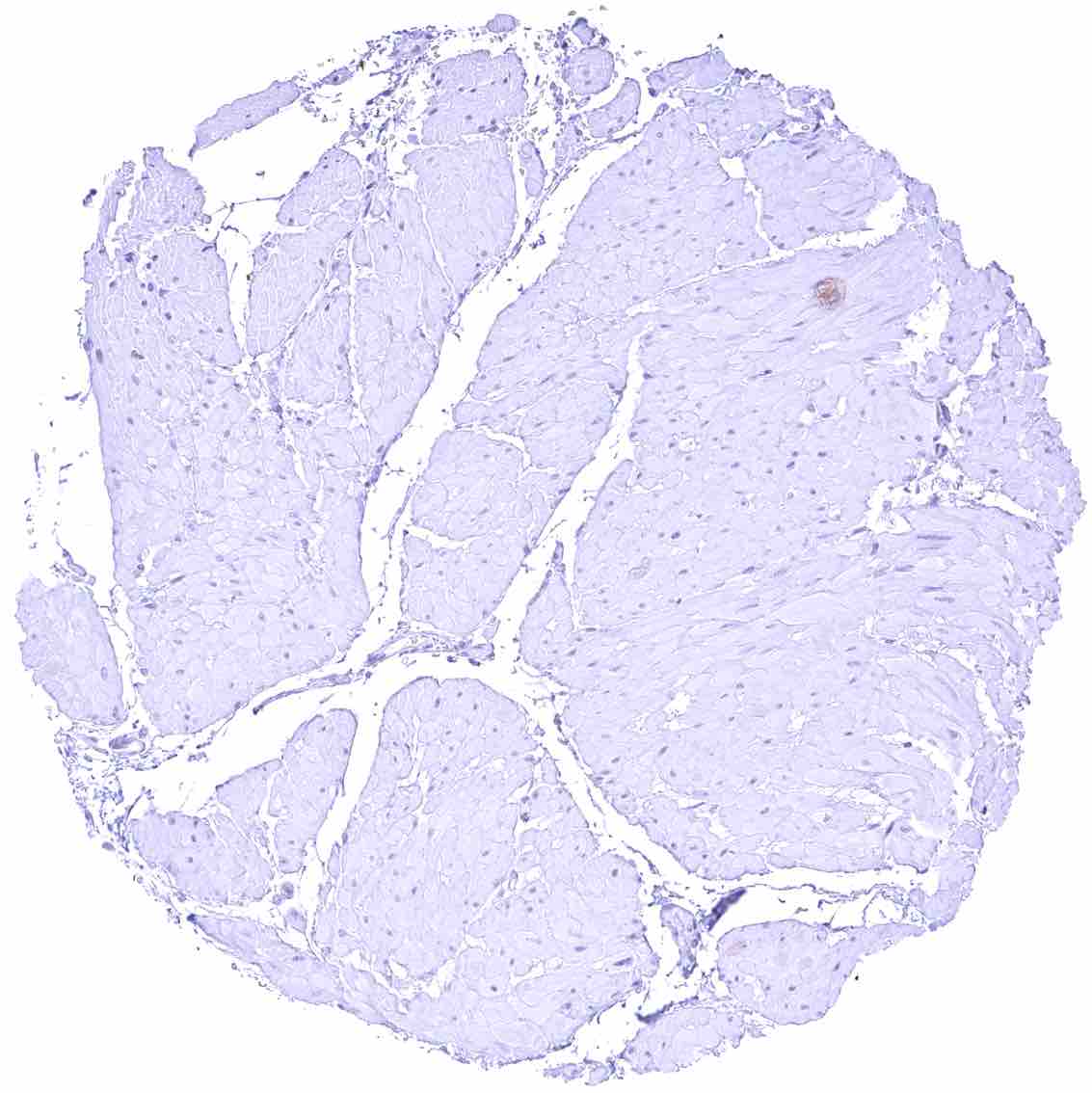
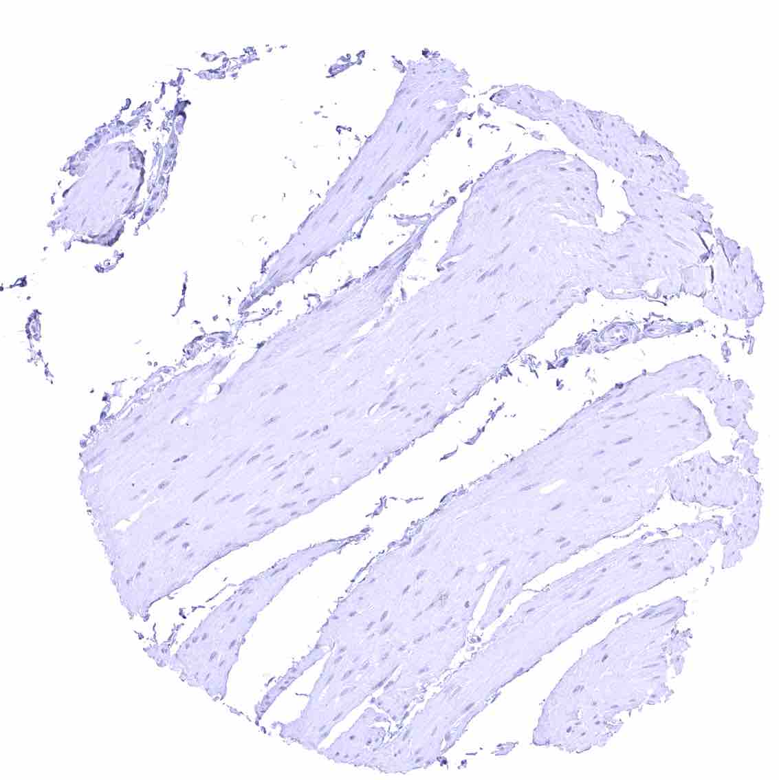
Heart muscle.jpeg
Ileum, muscular wall
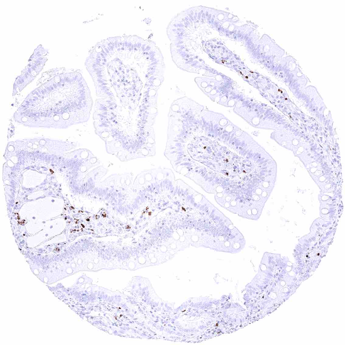
Ileum, mucosa
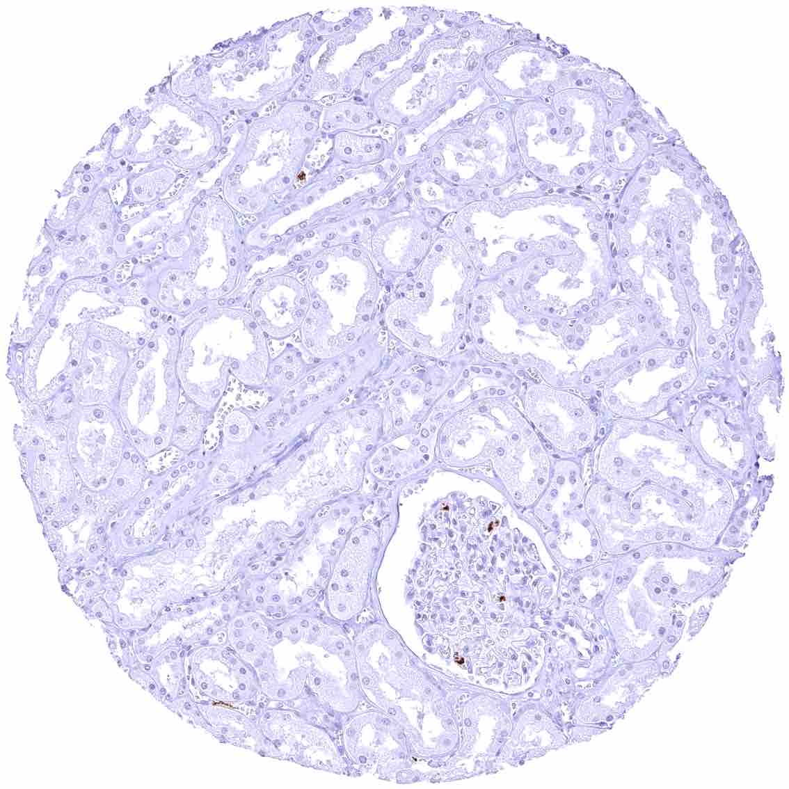
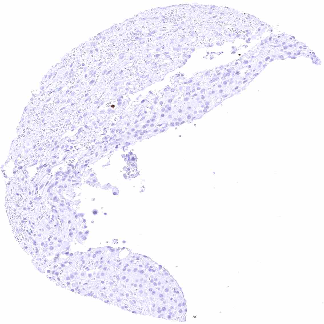
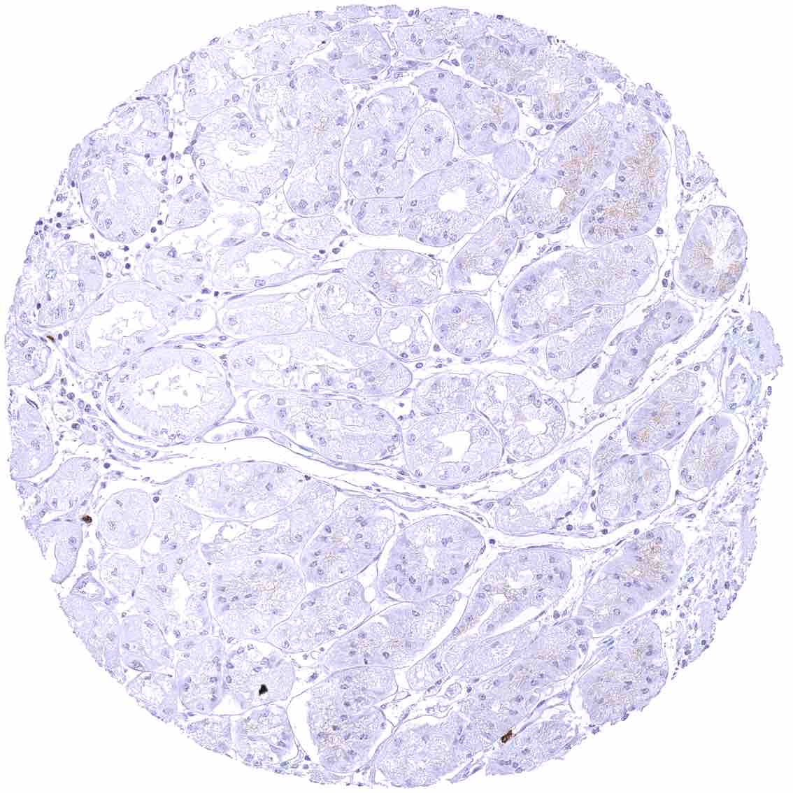
Kidney, cortex – CEACAM6 staining is limited to inflammatory cells.
Kidney, medulla
Kidney, pelvis, muscular wall
Kidney, pelvis, urothelium
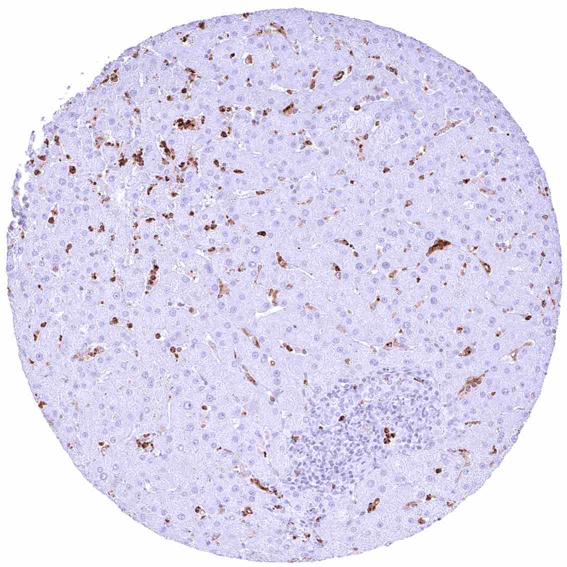
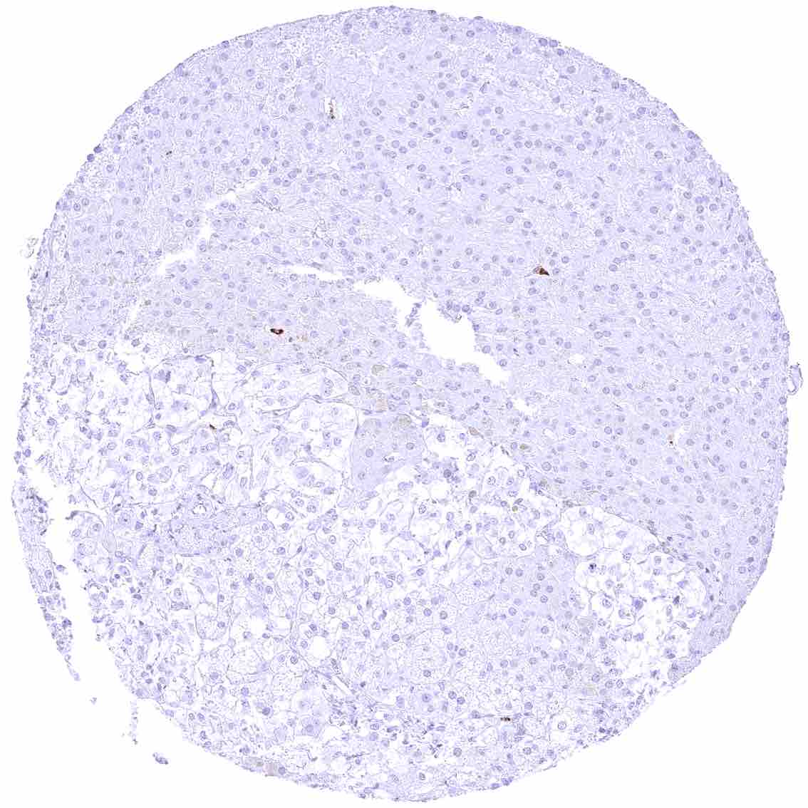
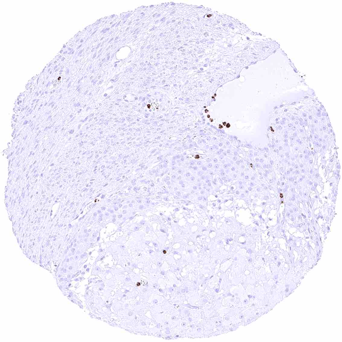
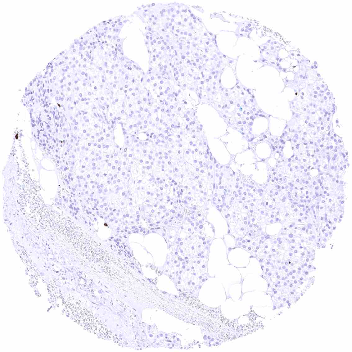
Liver – Strong membranous and cytoplasmic CEACAM6 staining of many intravascular inflammatory cells.
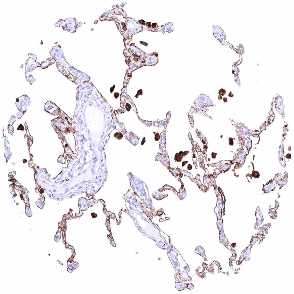
Lung – Strong membranous CEACAM6 staining of all alveolar pneumocytes .jpeg
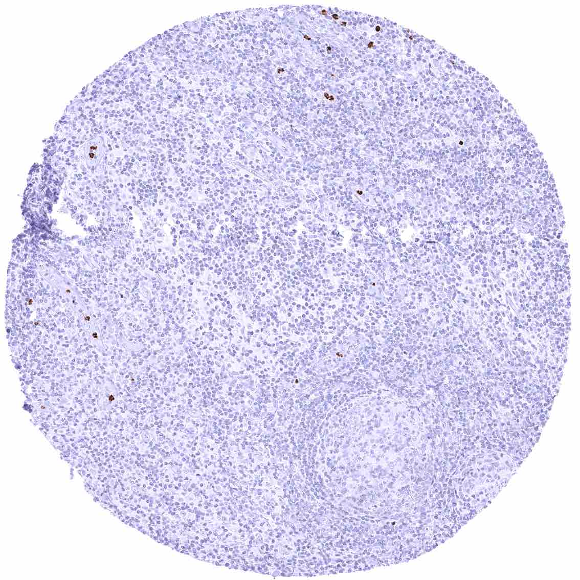
Lymph node – Strong CEACAM6 staining of few inflammatory cells.
Ovary, corpus luteum – CEACAM6 staining is limited to inflammatory cells.
Ovary, follicular cyst
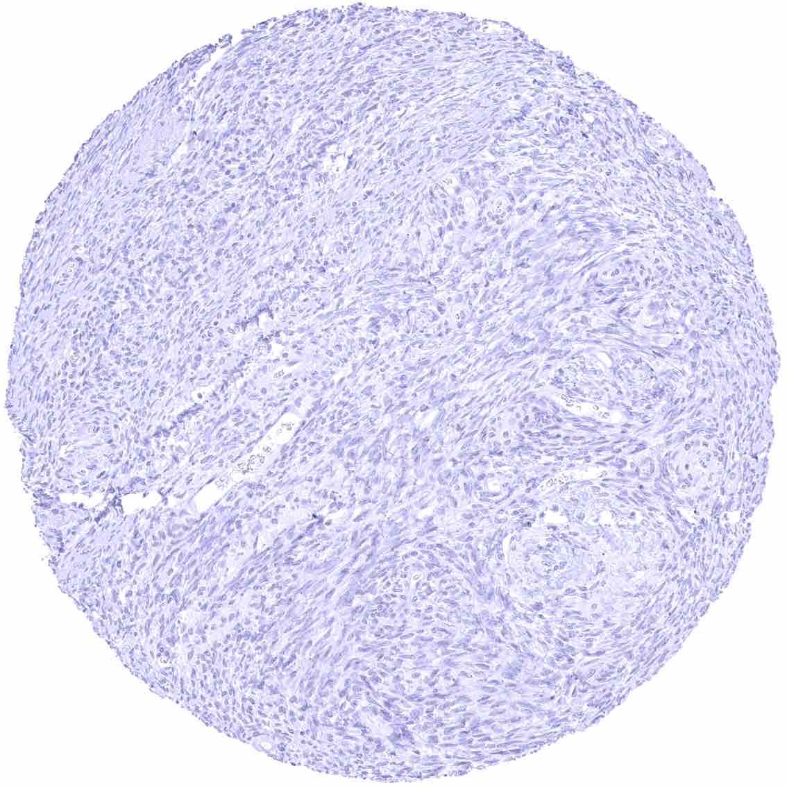
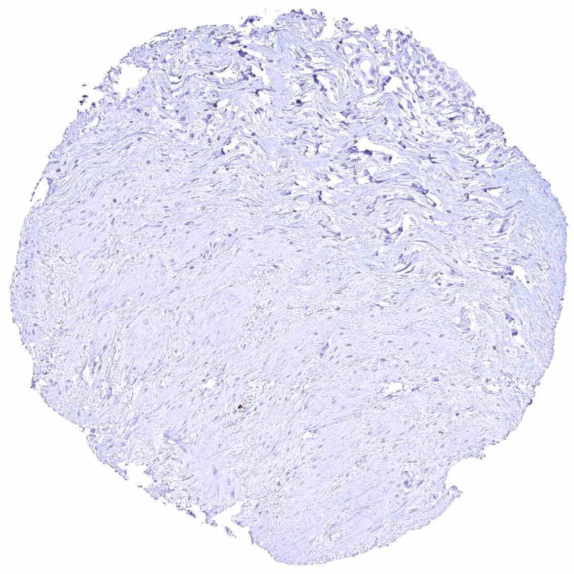
Ovary, stroma
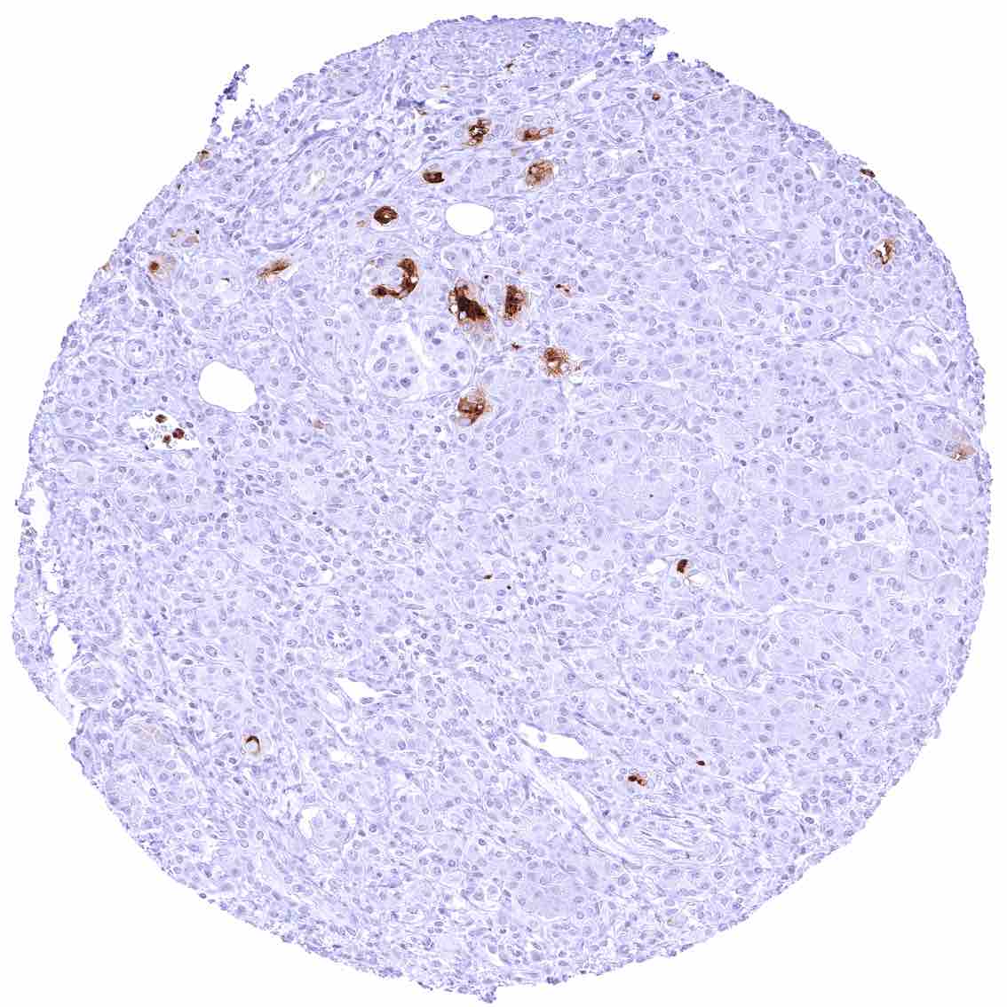
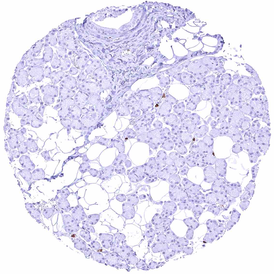
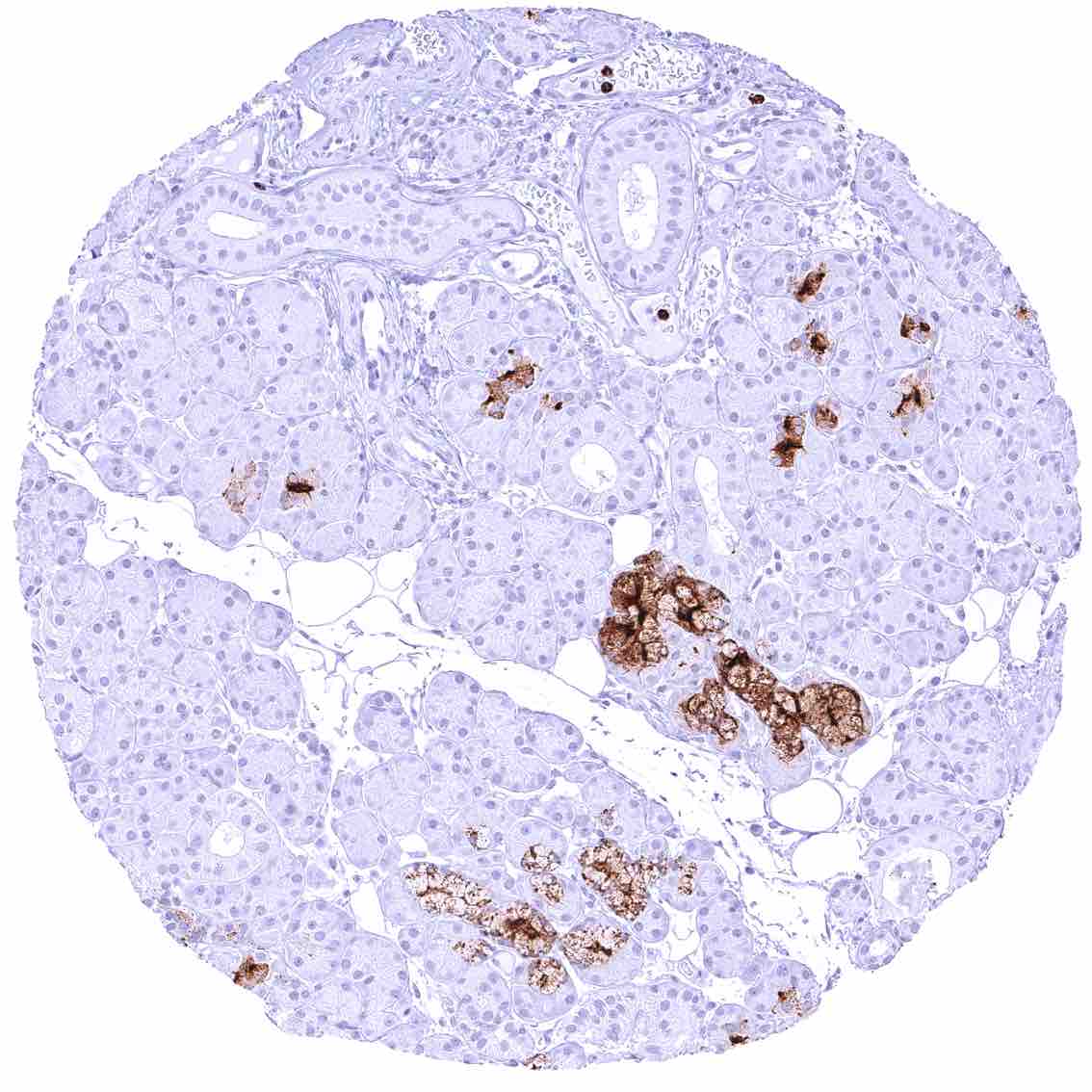
Pancreas – Strong membranous and cytoplasmic CEACAM6 staining of a fraction of small excretory ducts.
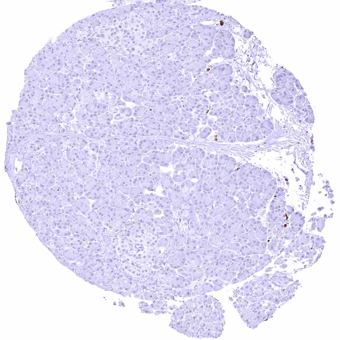
Pancreas
Parathyroid gland
Parotid gland – CEACAM6 staining is limited to inflammatory cells.
Pituitary gland, anterior lobe – CEACAM6 staining is limited to inflammatory cells.
Pituitary gland, posterior lobe
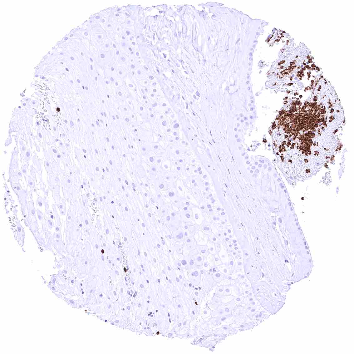
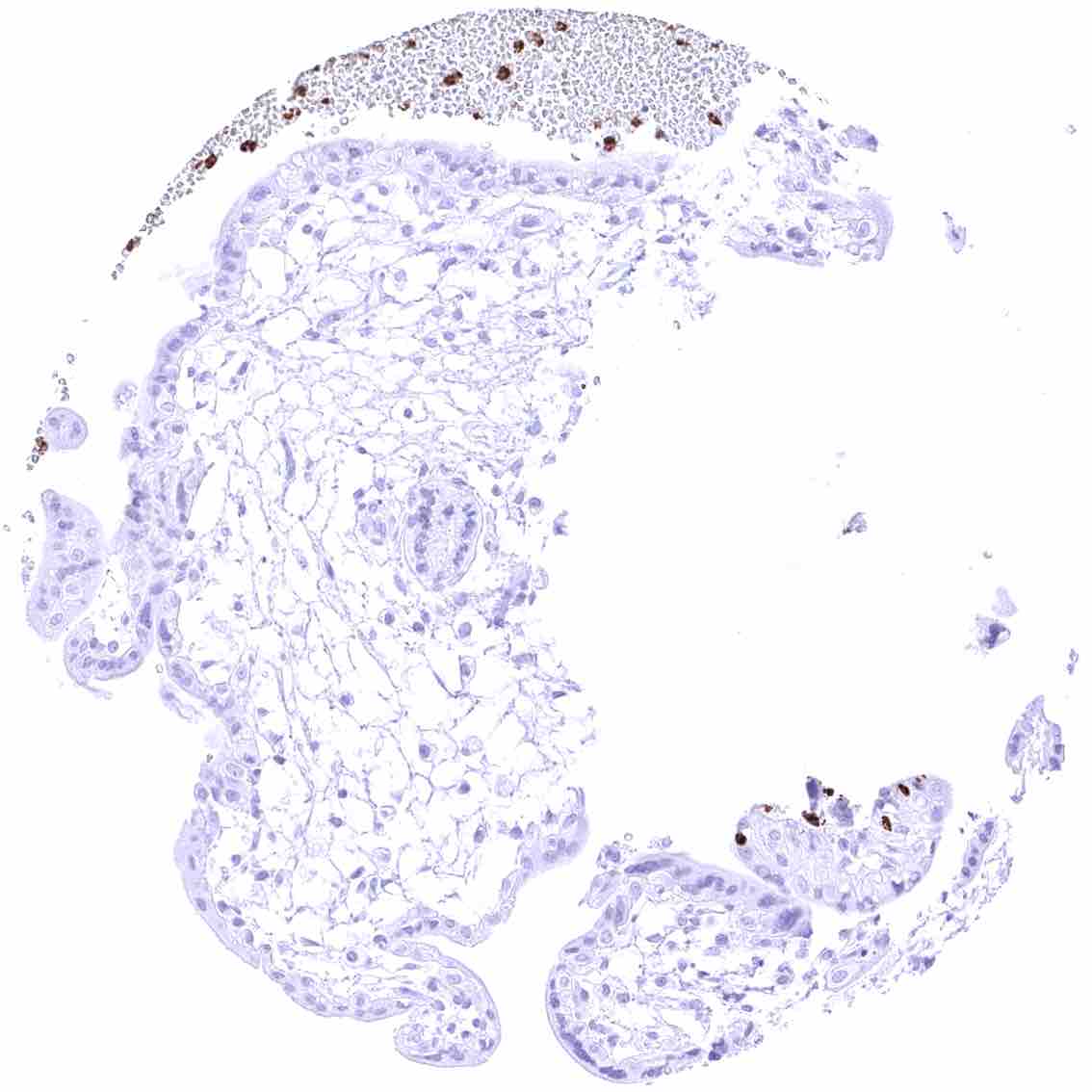
Placenta (amnion and chorion) – Strong CEACAM6 staining of a group of inflammatory cells.
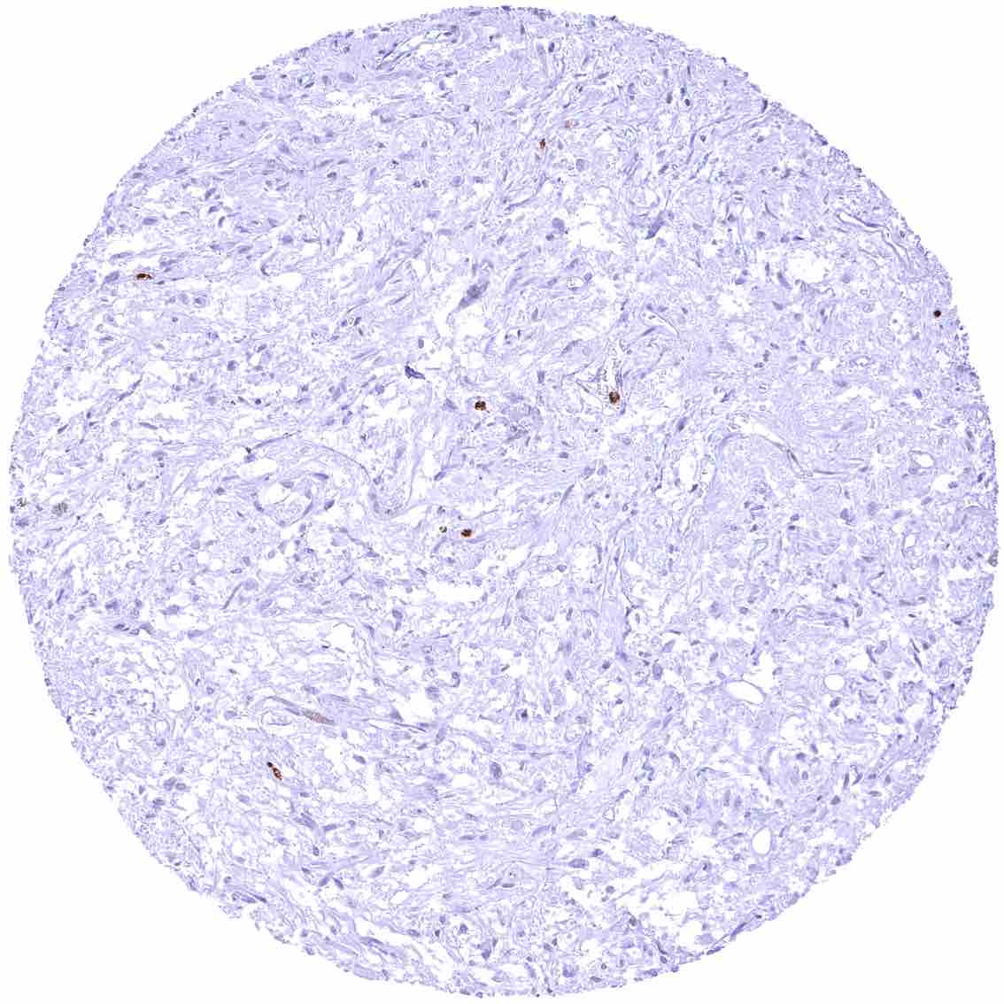
Placenta, early – CEACAM6 staining is limited to inflammatory cells.
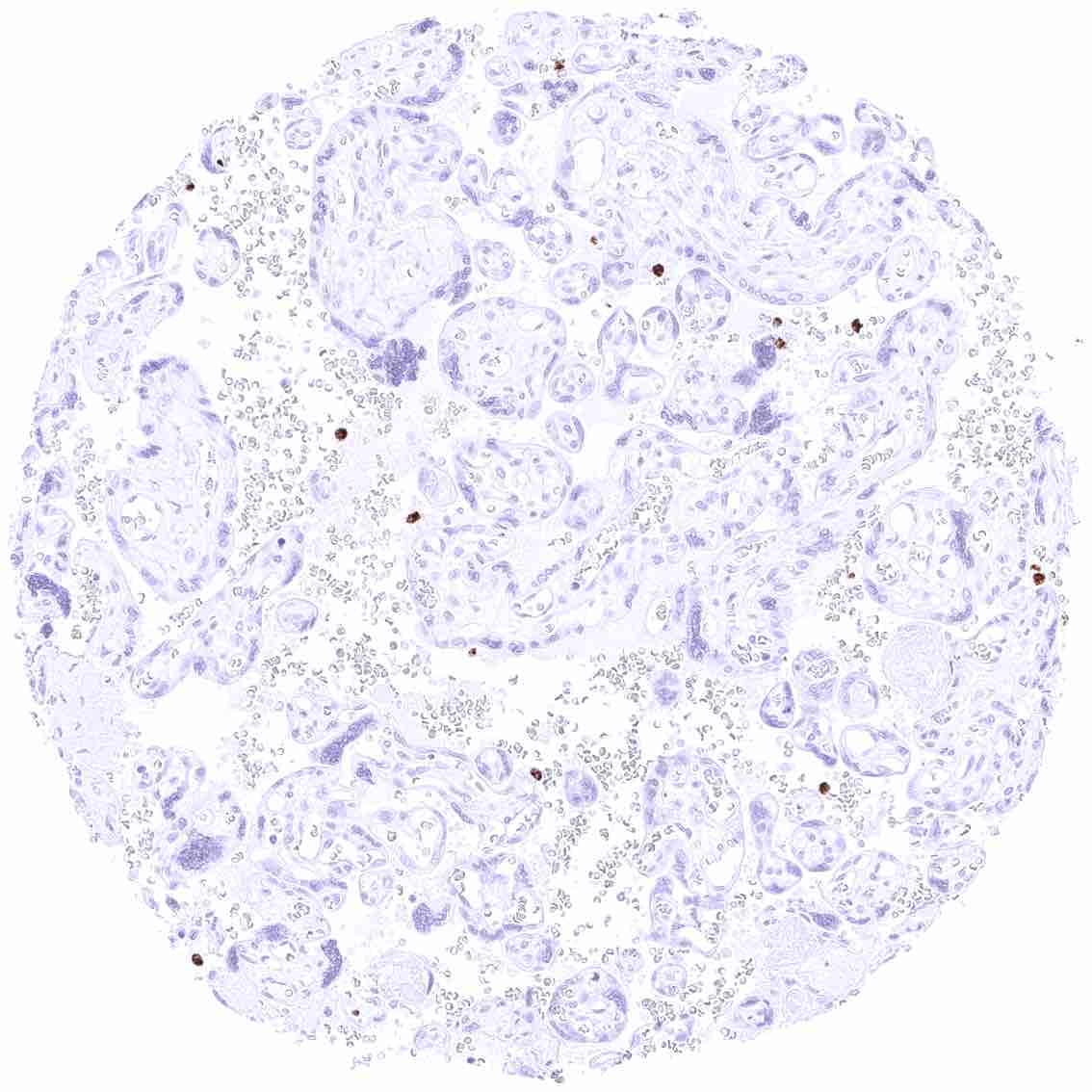
Placenta, mature – CEACAM6 staining is limited to inflammatory cells.
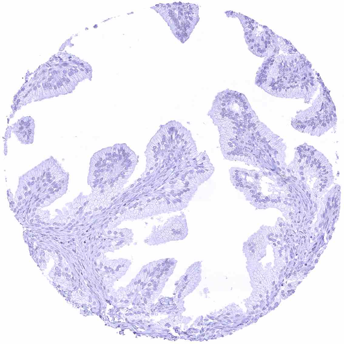
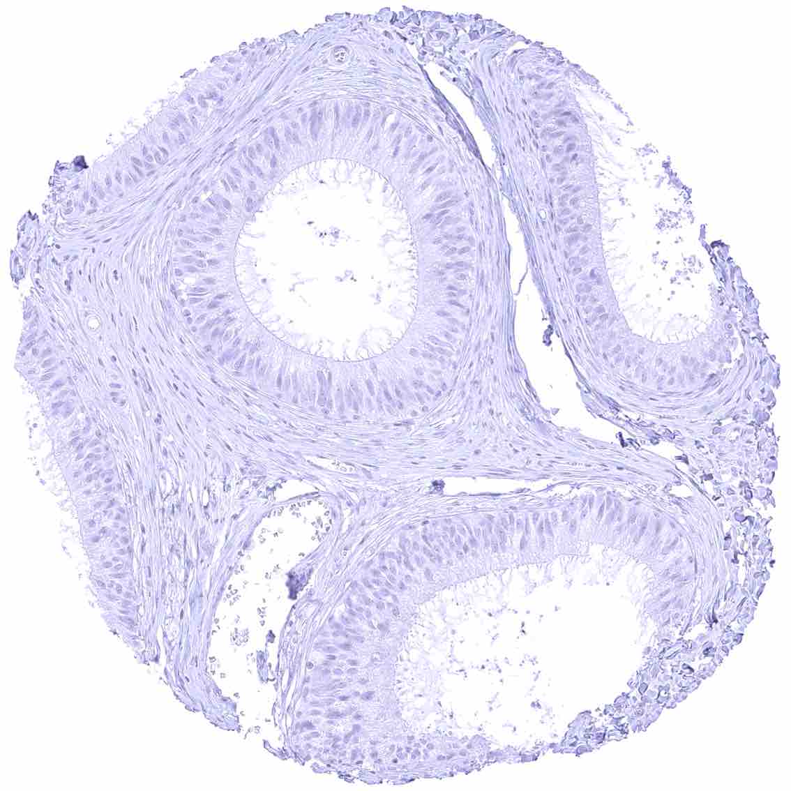
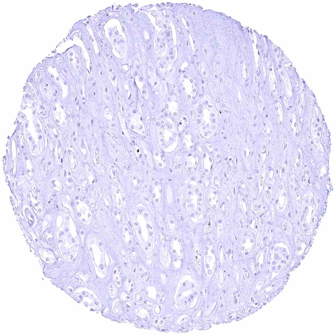
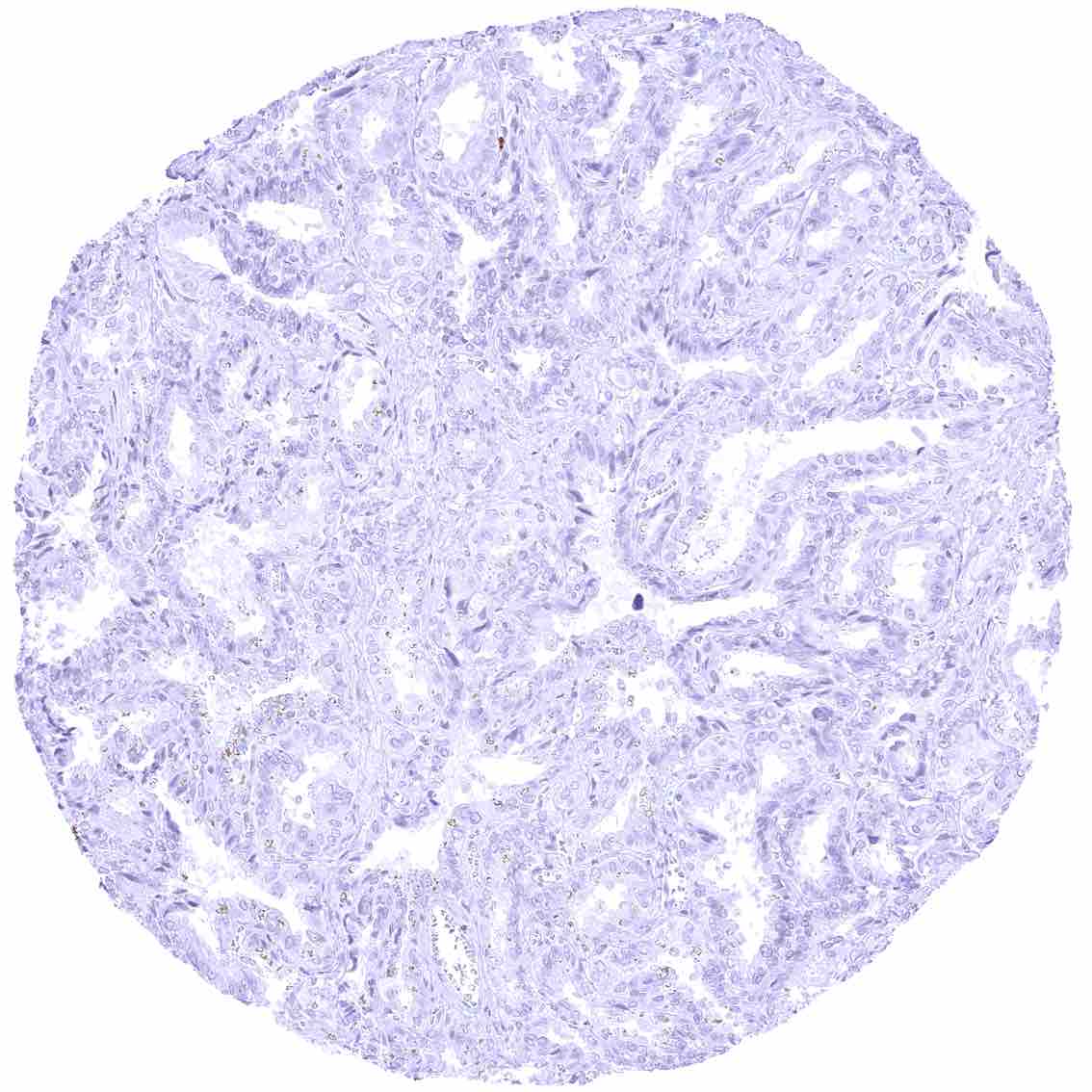
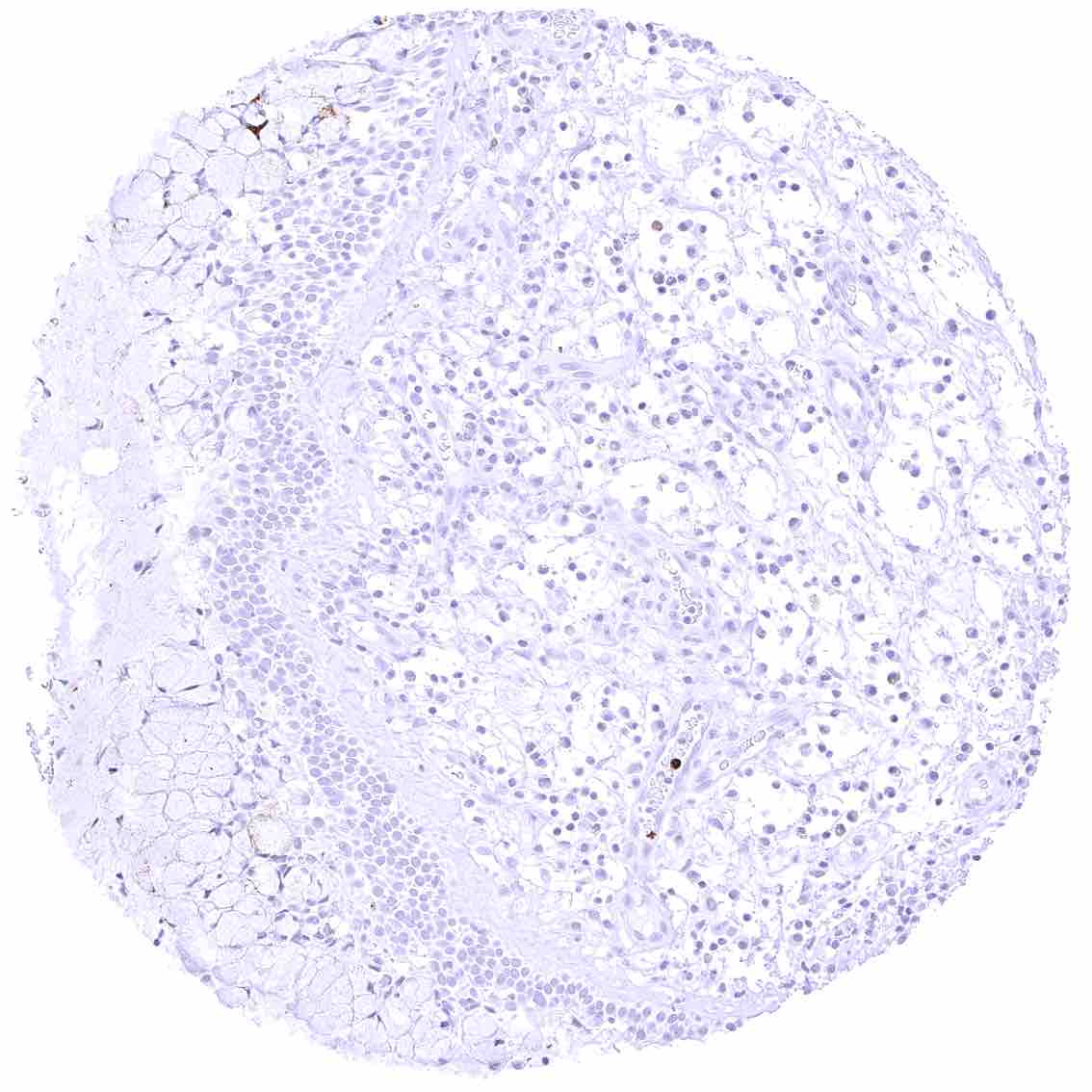
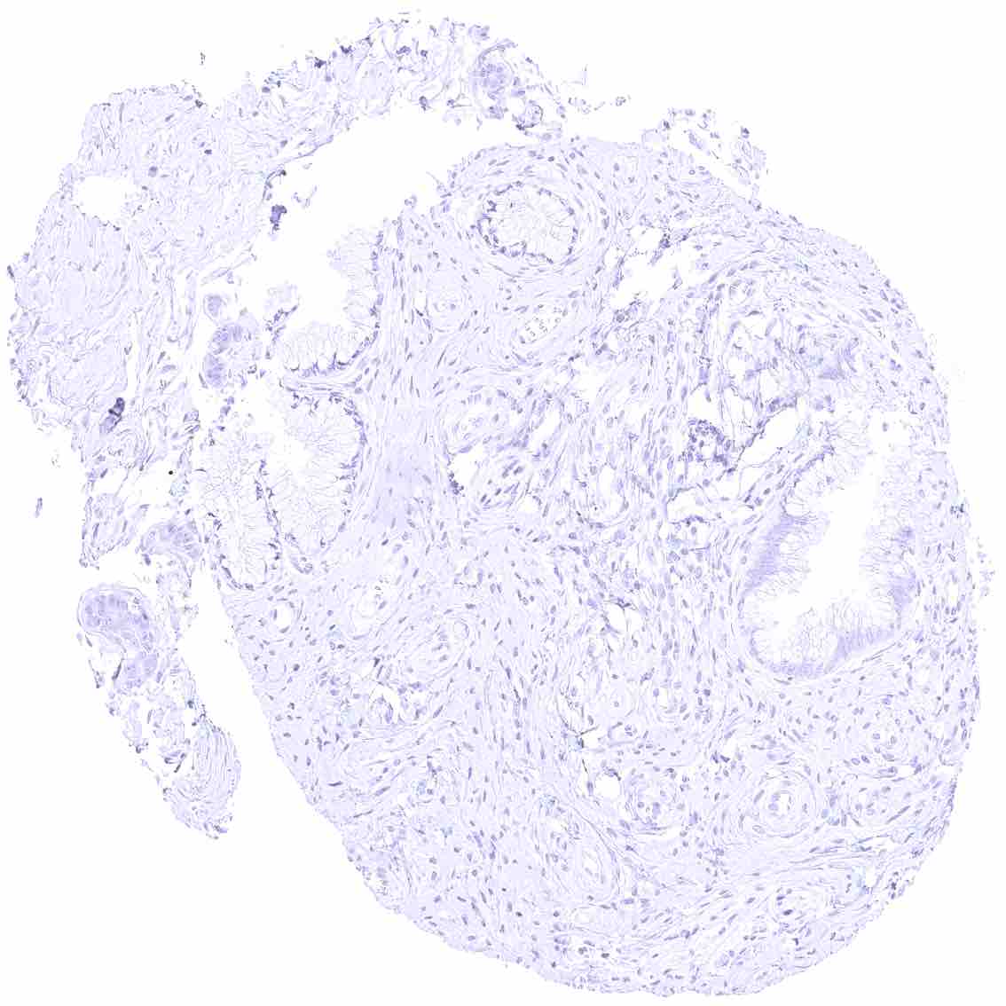
Prostate
Rectum, mucosa
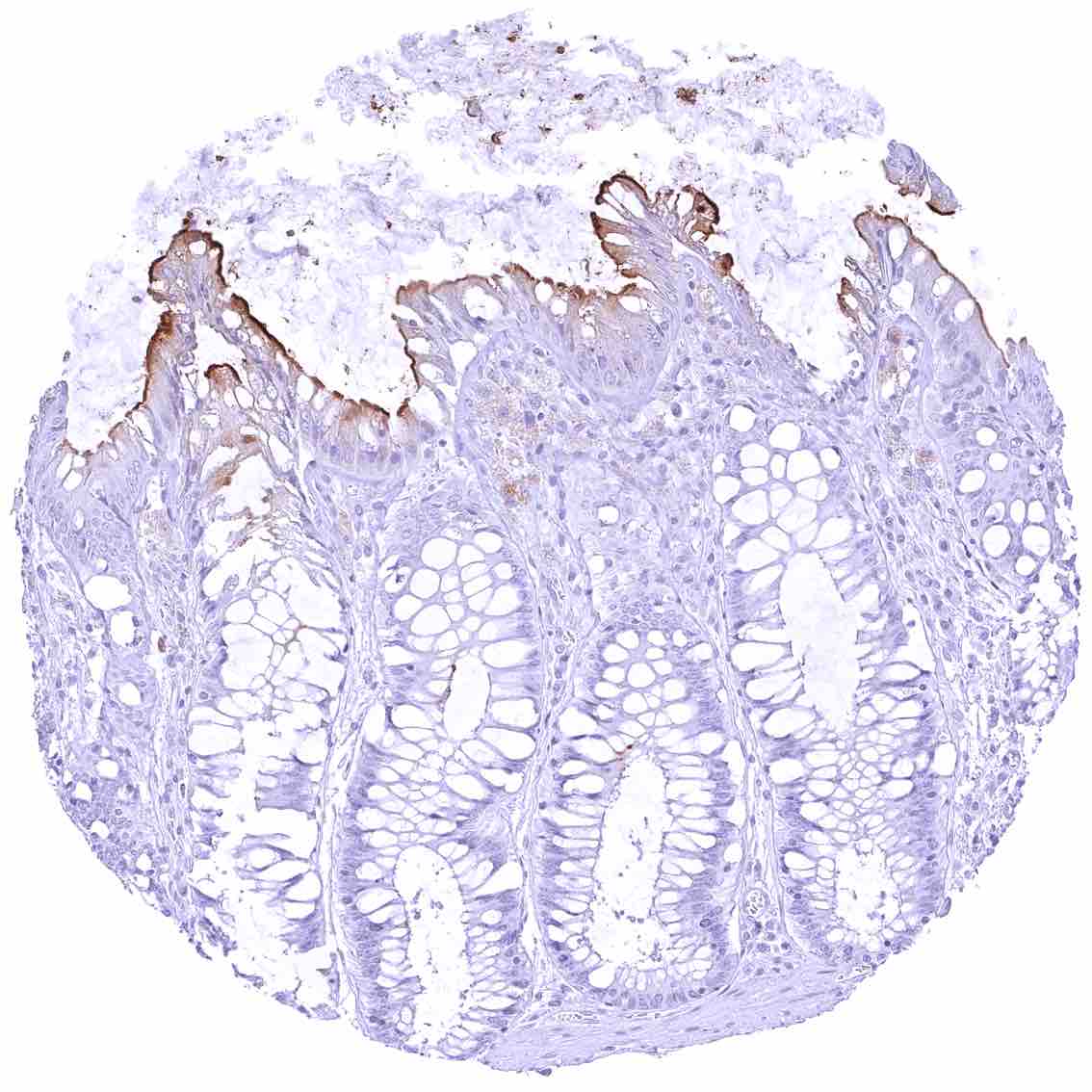
Rectum, mucosa – Strong apical membranous CEACAM6 staining of surface epithelial cells
Seminal vesicle
Sinus paranasales – Respiratory epithelial cells are CEACAM6 negative in this sample.
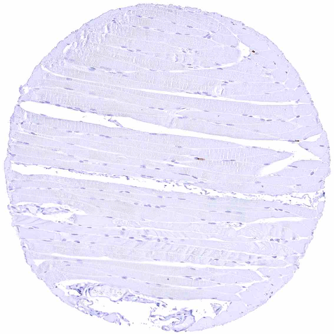
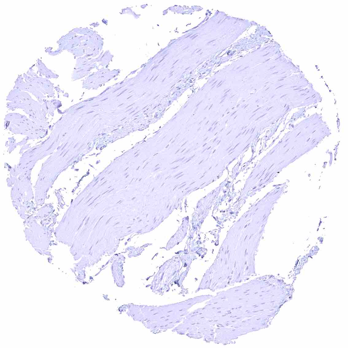
Skeletal muscle
Skin, hairfollicel and sebaceous glands – Strong apical membranous and apical cytoplasmic staining of eccrine glands. Sebaceous cells and hair follicles are CEACAM6 negative.
Skin
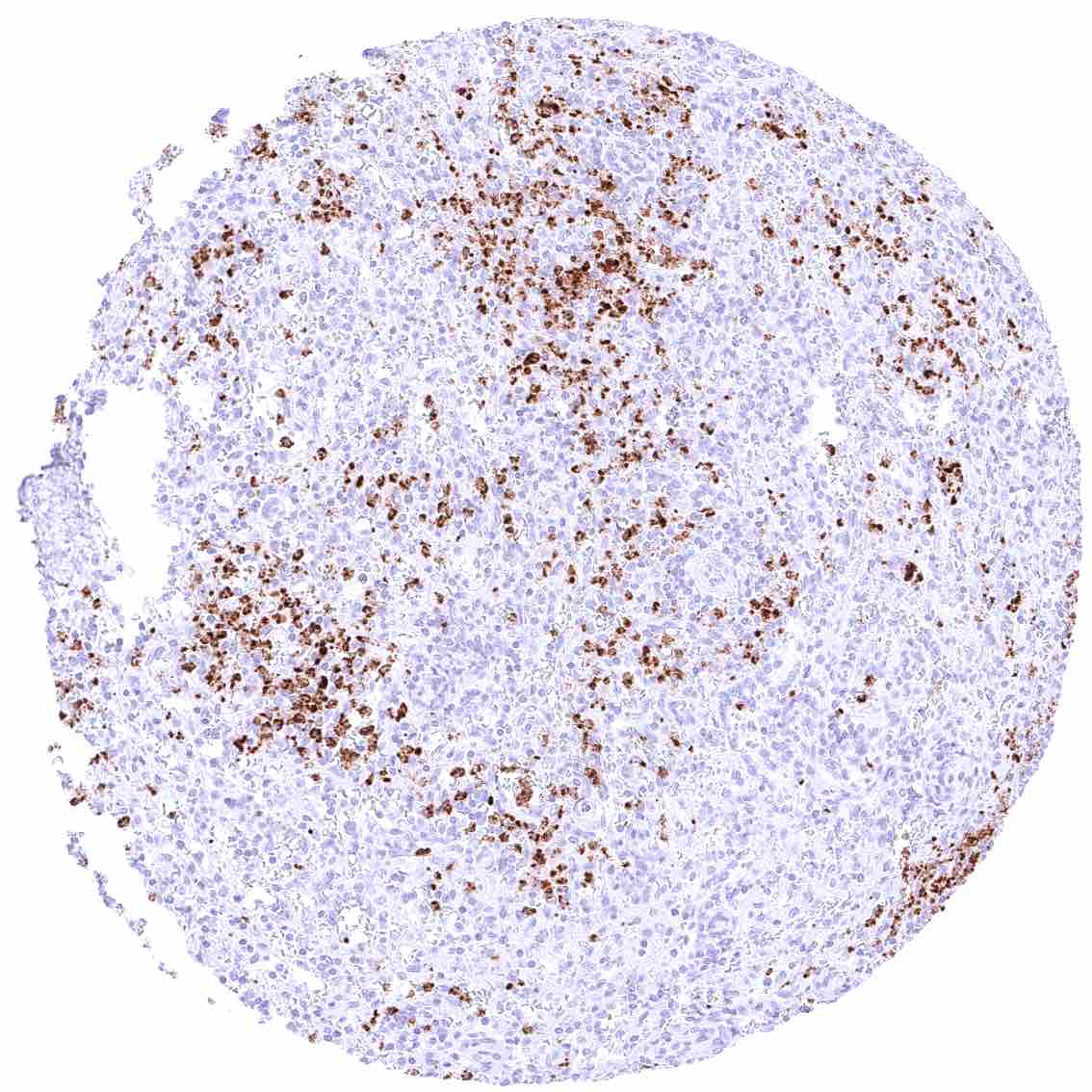
Spleen – Strong CEACAM6 staining of a large fraction of inflammatory cells.
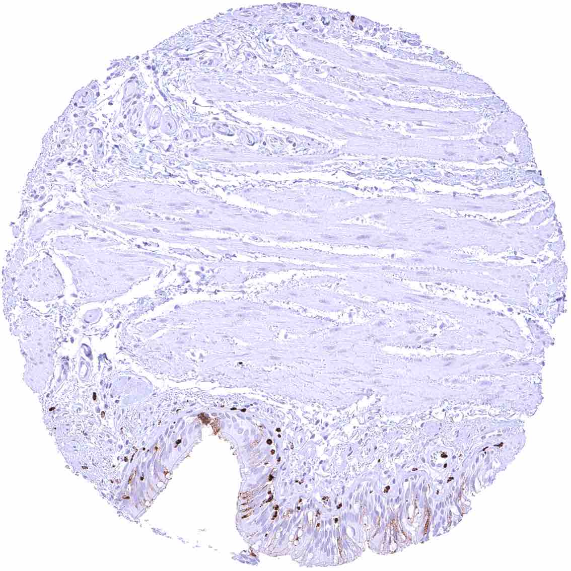
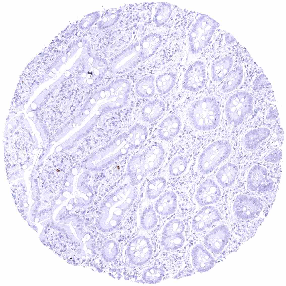
Stomach, antrum
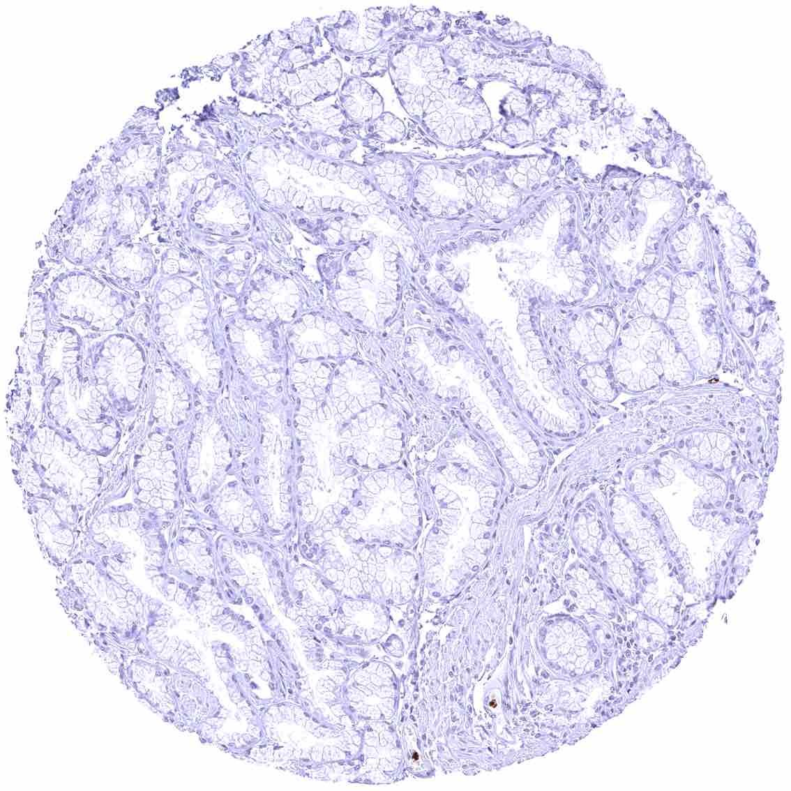
Stomach, corpus
Stomach, muscular wall
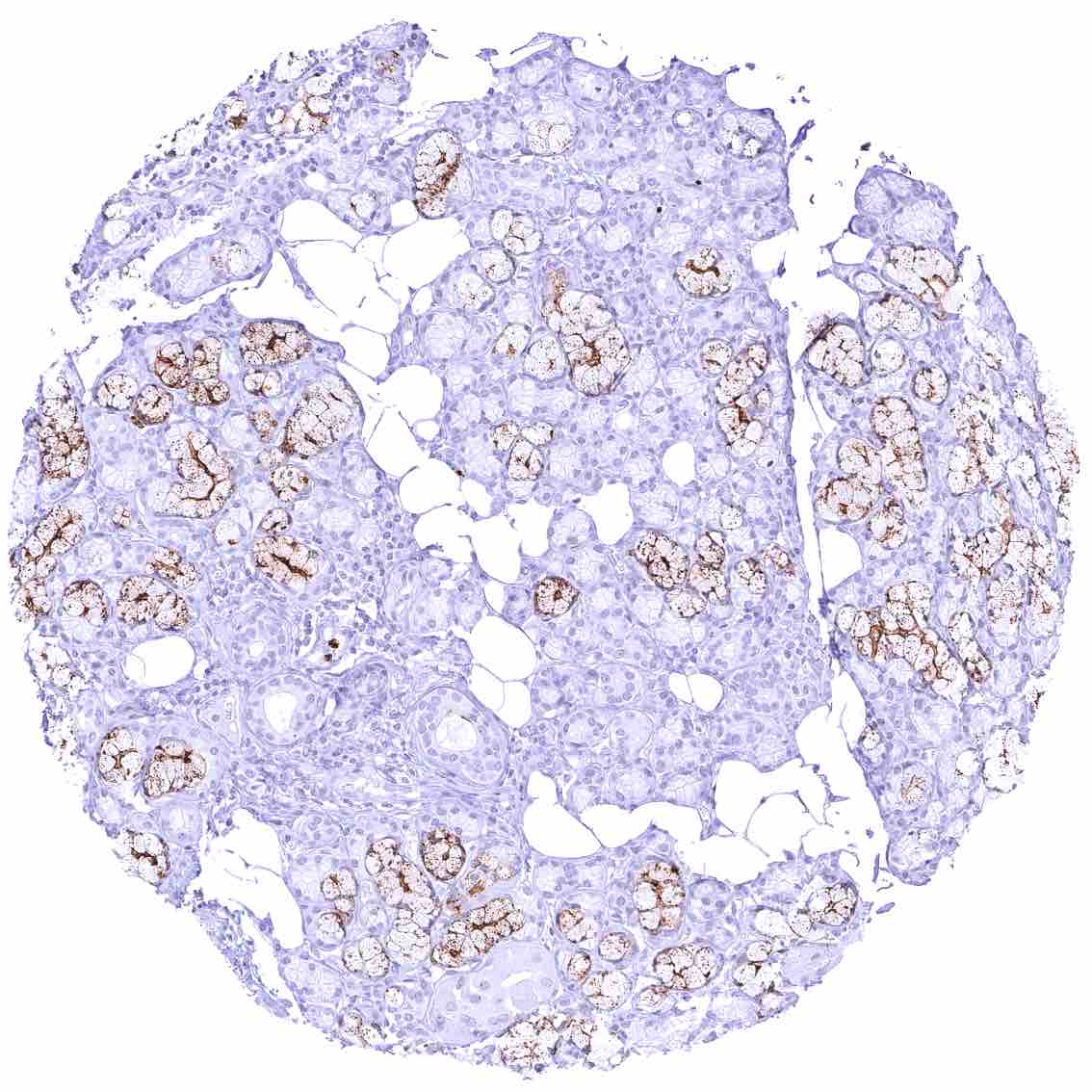
Sublingual gland – Distinct membranous and cytoplasmic CEACAM6 staining of mucinous glandular cells.
Submandibular gland – Distinct membranous and cytoplasmic CEACAM6 staining of mucinous glandular cells.
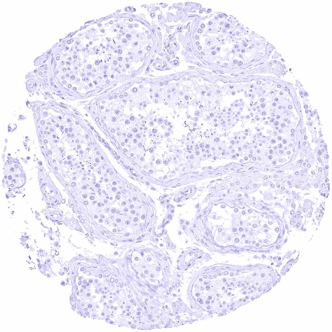
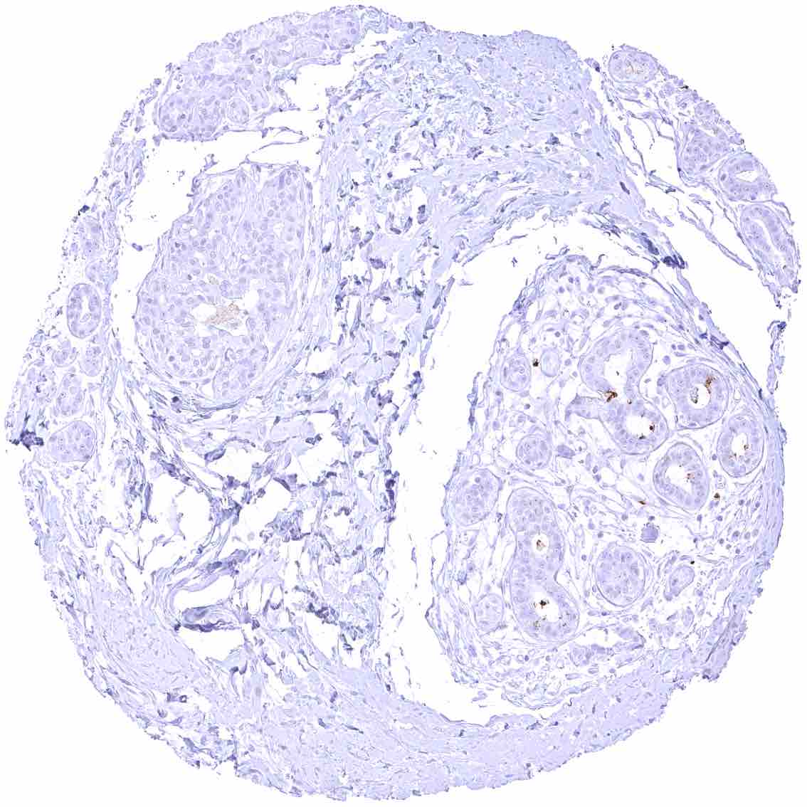
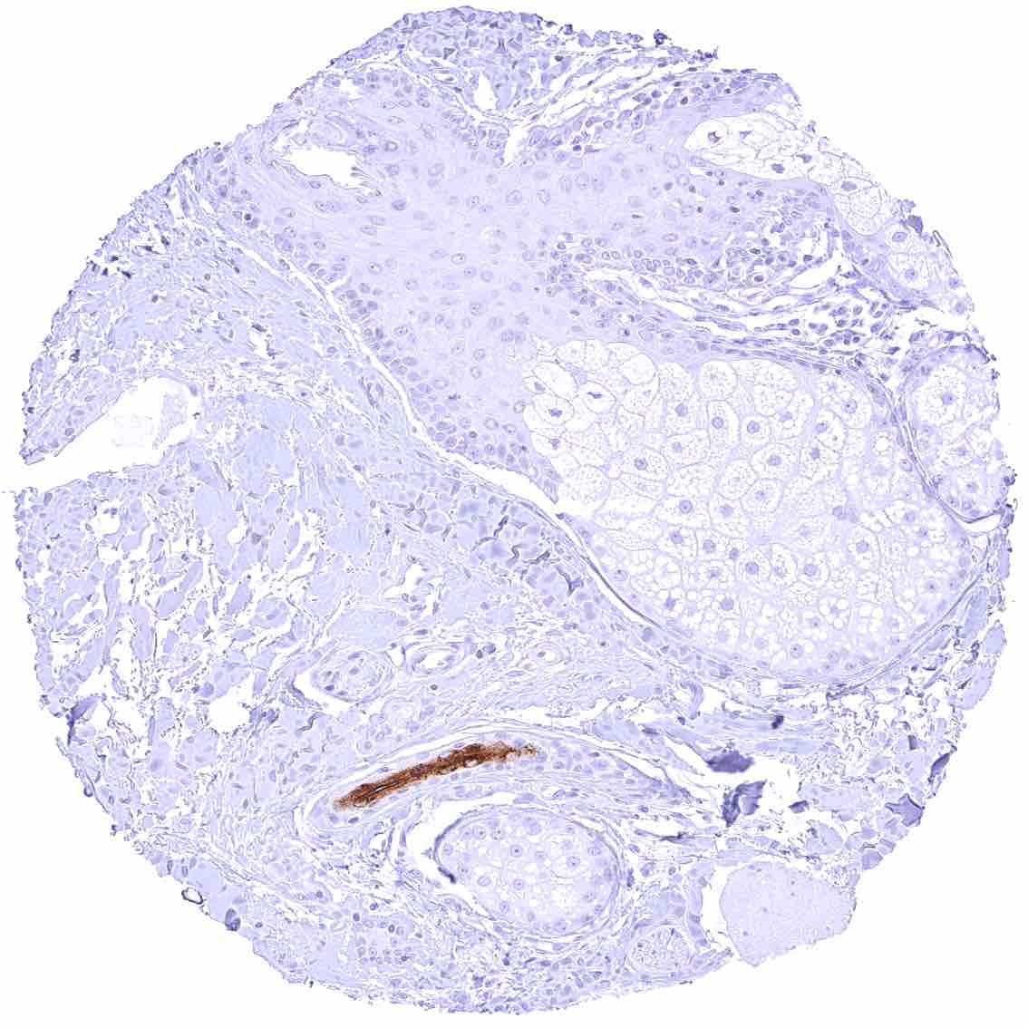
Testis
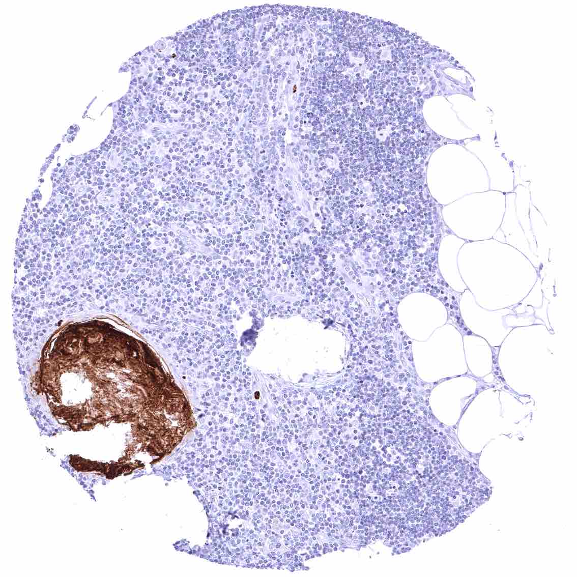
Thymus – Strong CEACAM6 staining of a corpuscle of Hassall’s.
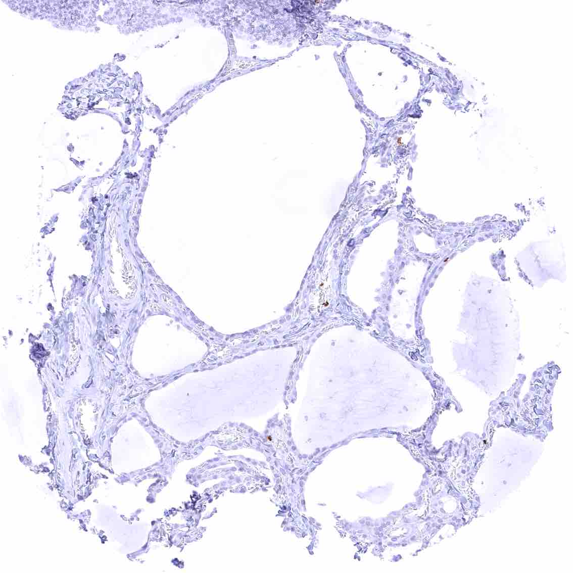
Thyroid gland
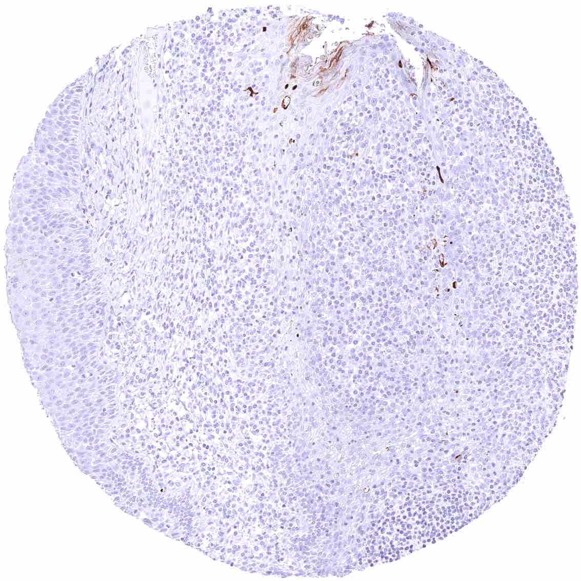
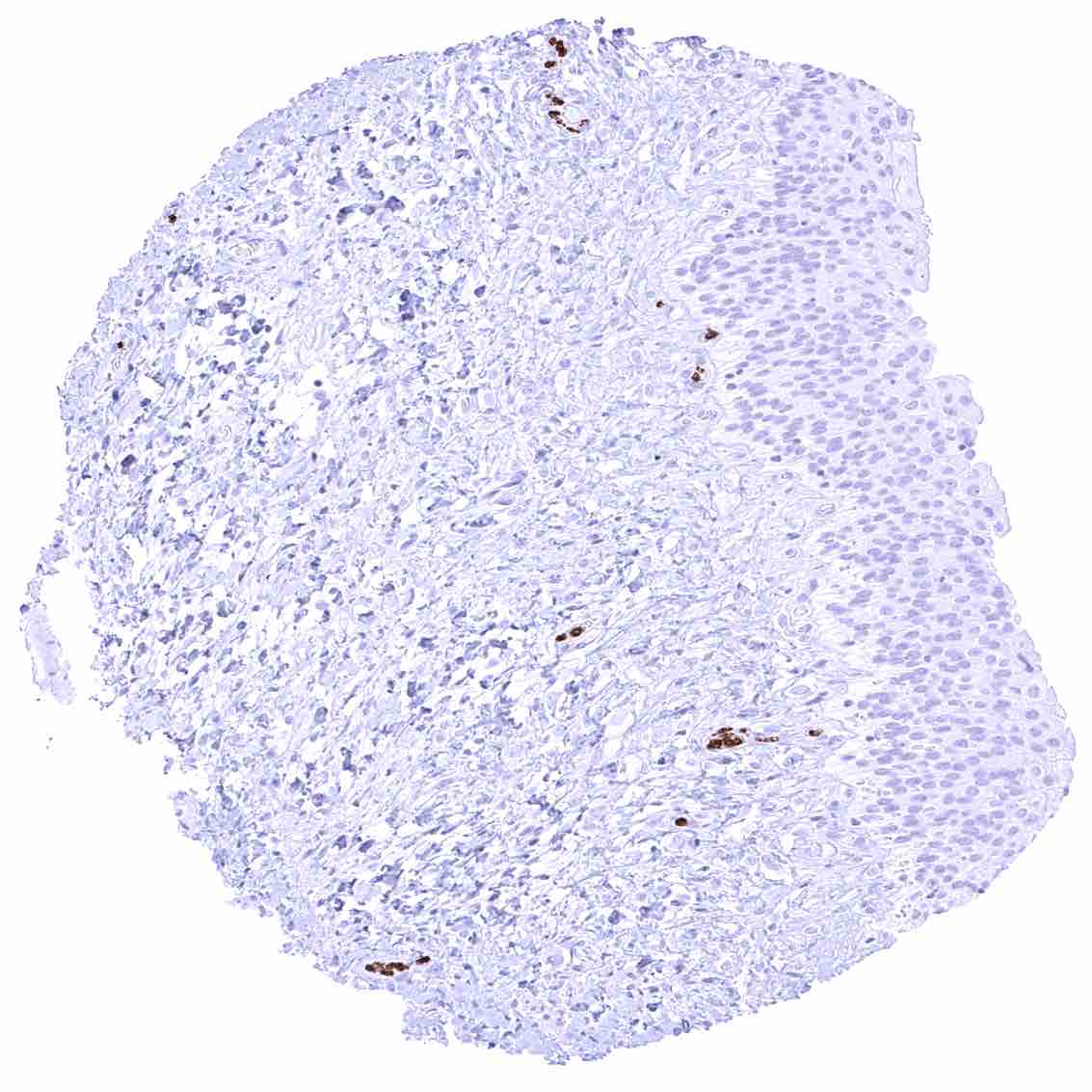
Tonsil – Weak to moderate membranous CEACAM6 staining of a fraction of squamous epithelial cells especially of the upper half of the epithelium.
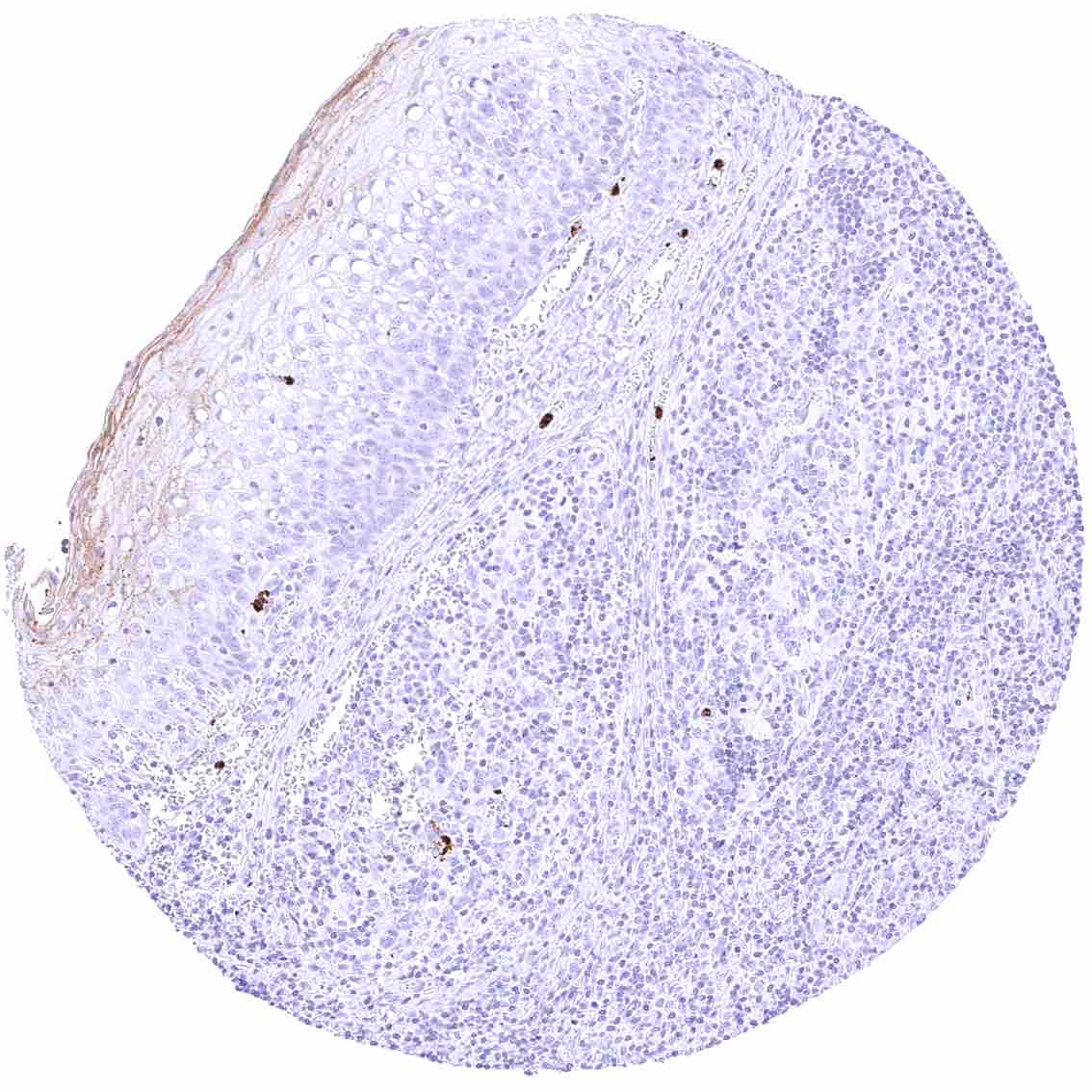
Tonsil, surface epithelium – Weak to moderate membranous CEACAM6 staining of a fraction of squamous epithelial cells especially of the upper half of the epithelium.
Urinary bladder, muscular wall
Urinary bladder, urothelium – Strong CEACAM6 staining of few inflammatory cells.
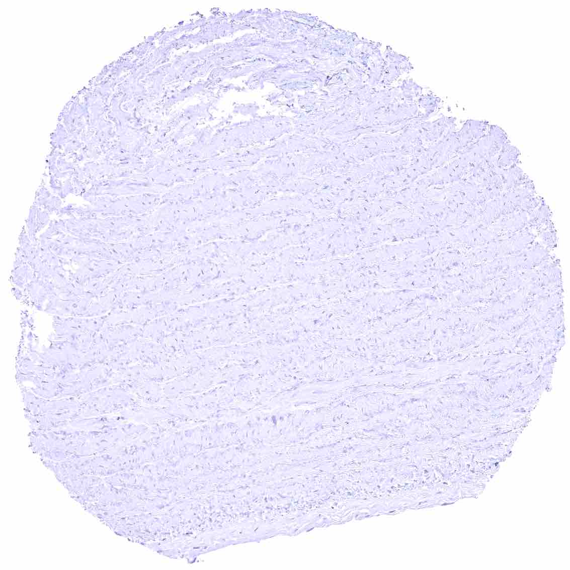
Uterus, myometrium
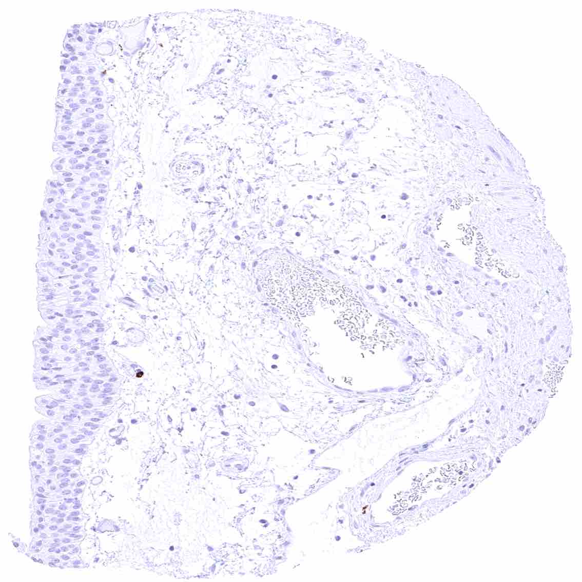
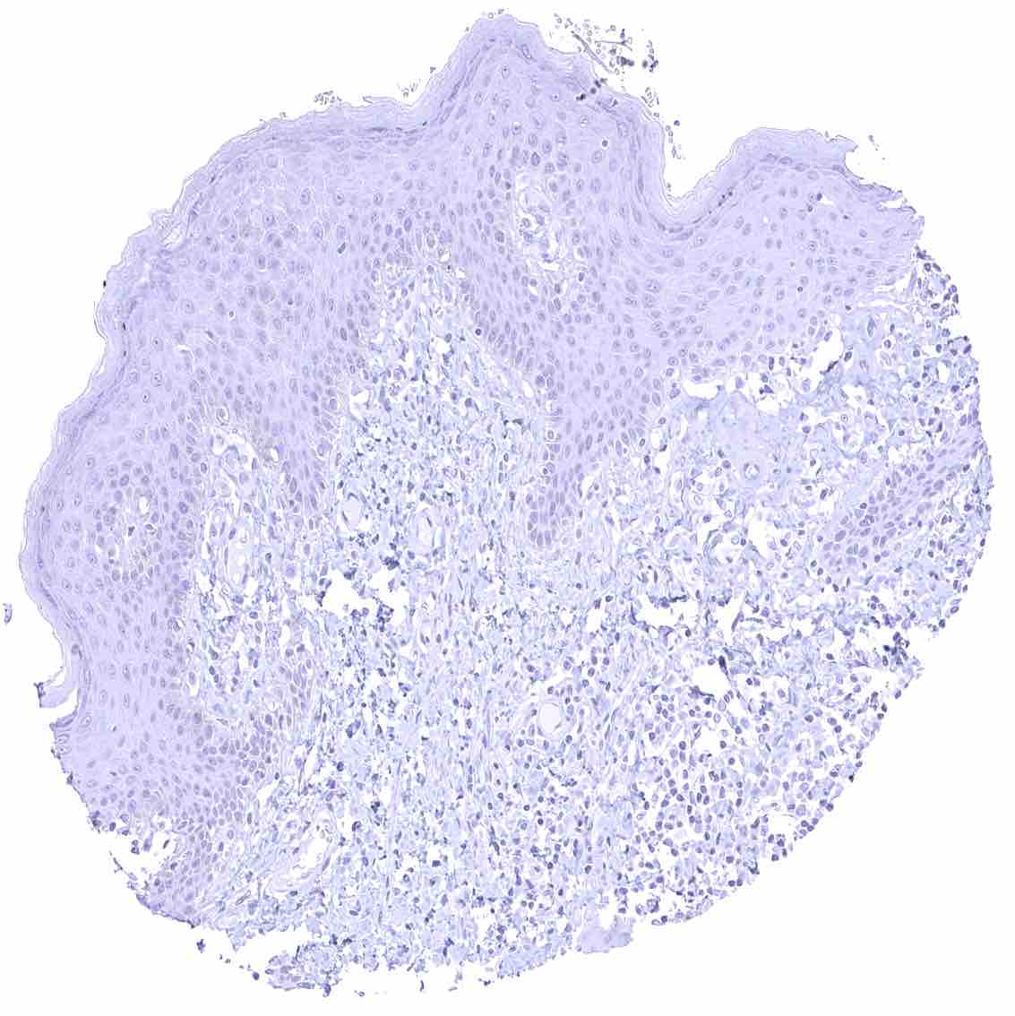
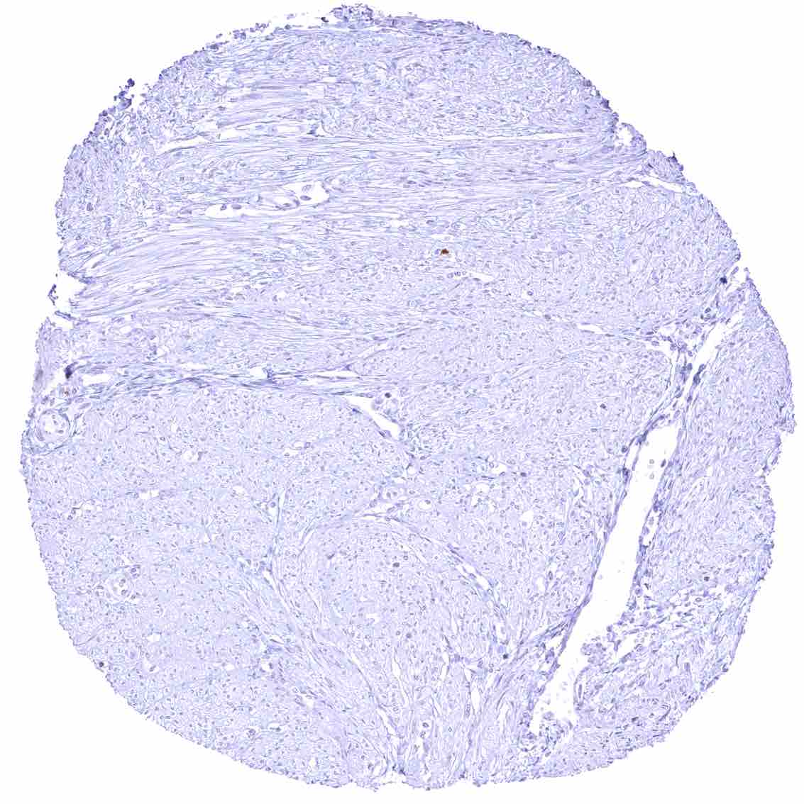
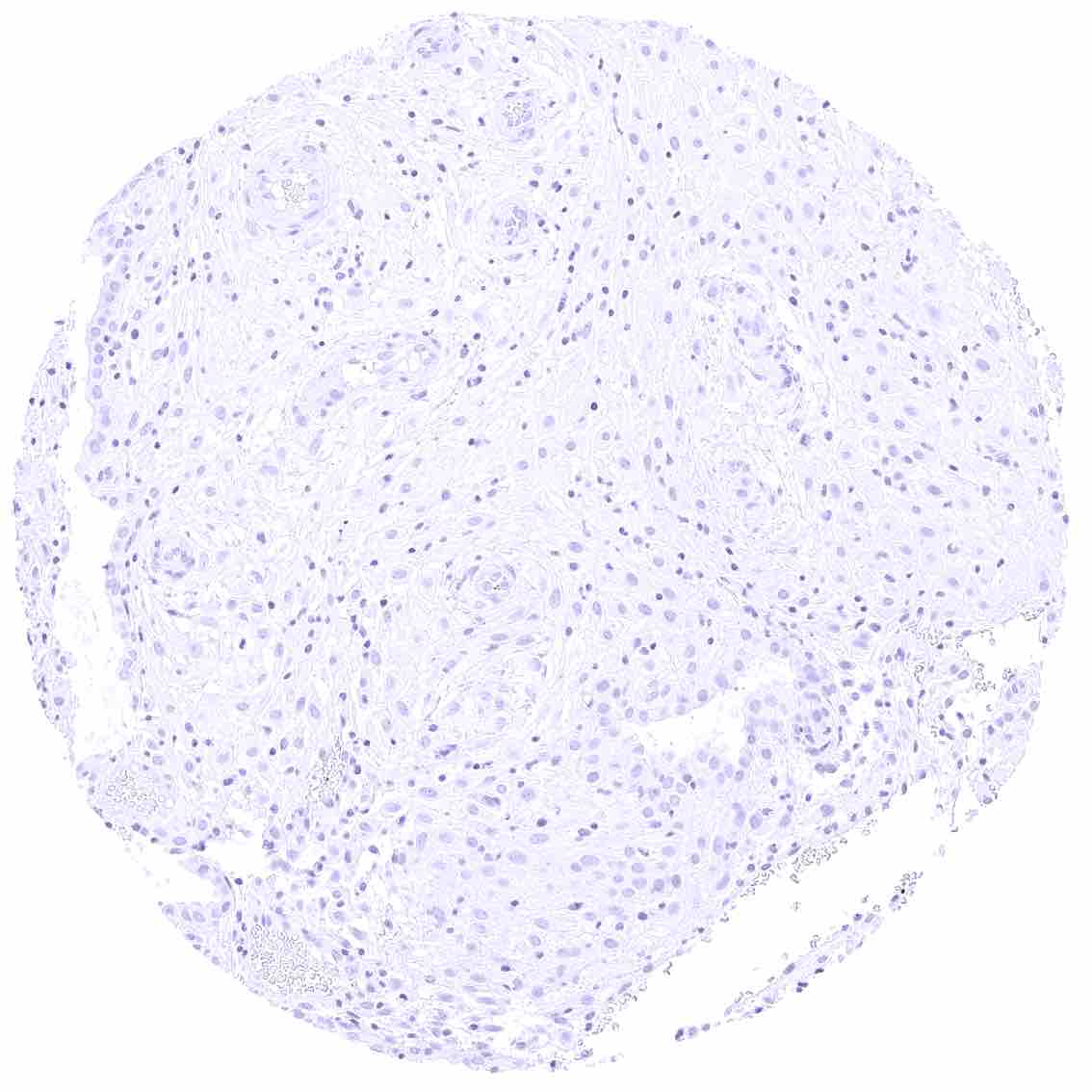
Uterus, ectocervix – Absence of CEACAM6 staining in this sample.
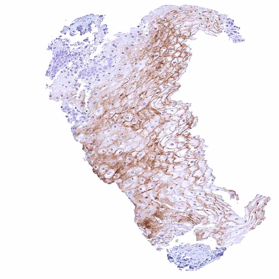
Uterus, ectocervix – Strong membranous CEACAM6 staining of squamous epithelial cells in this sample.
Uterus, endocervix
Uterus, endometrium (pregnancy)
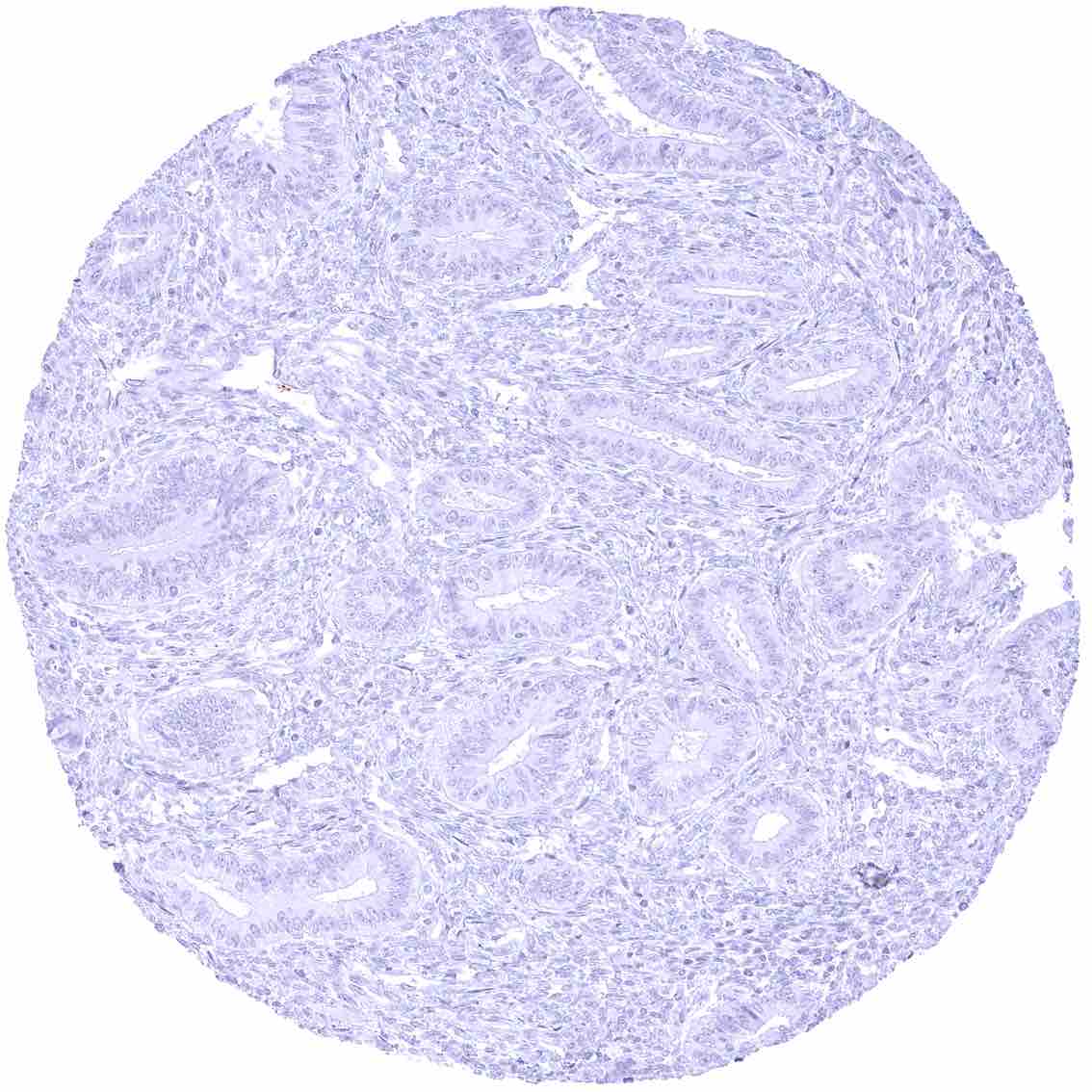
Uterus, endometrium (proliferation)