
Adrenal gland - Membranous CD56 immunostaining is prominent in the medulla but also occurs – to a lesser degree - in cortical cells.
Aorta, media

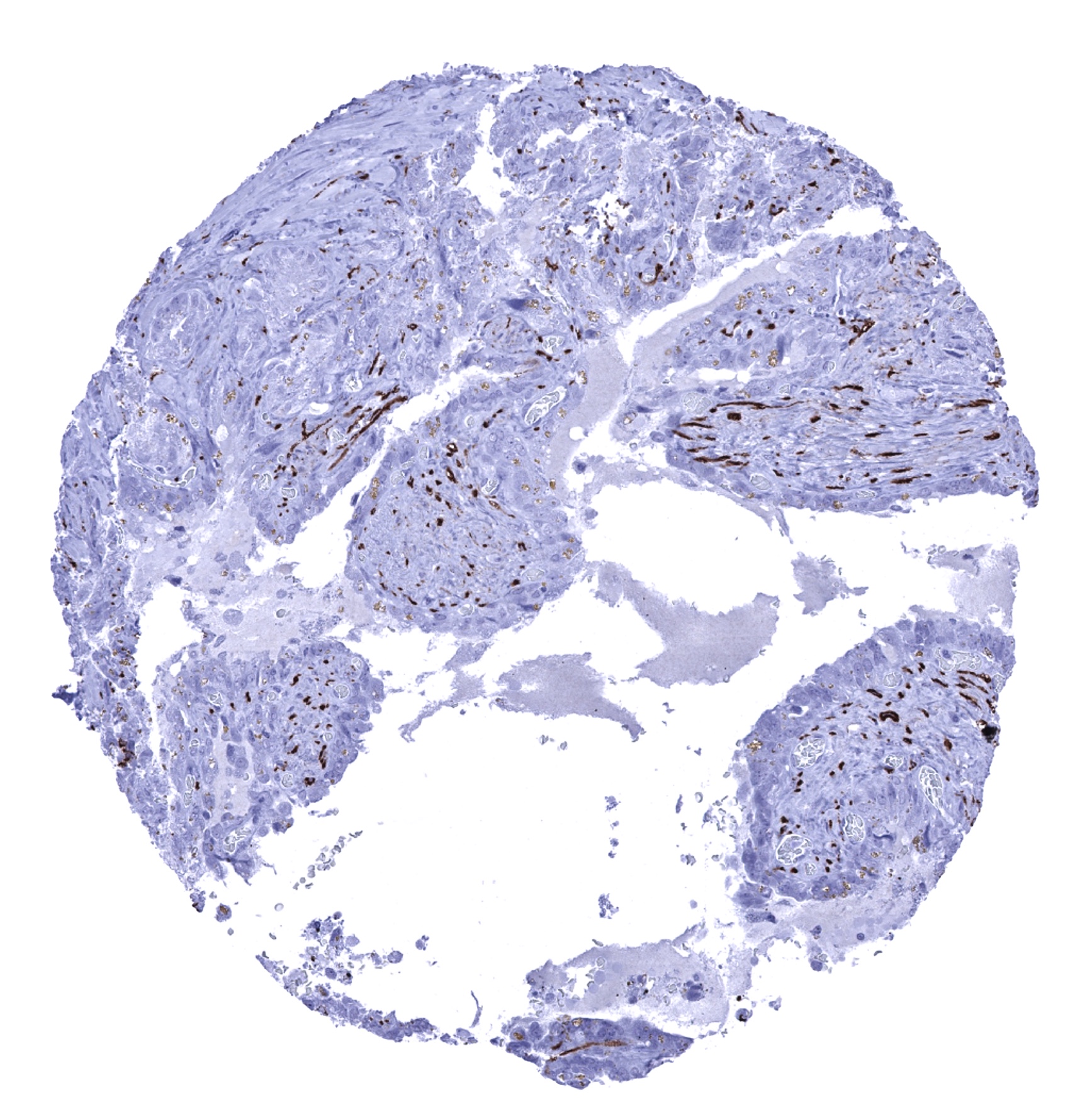
Appendix, mucosa - Intense CD56 immunostaining of nerve fibres.

Appendix, muscular wall - Nerve fibres and ganglia show strong CD56 immunostaining.

Bone marrow
Breast
Bronchus, mucosa
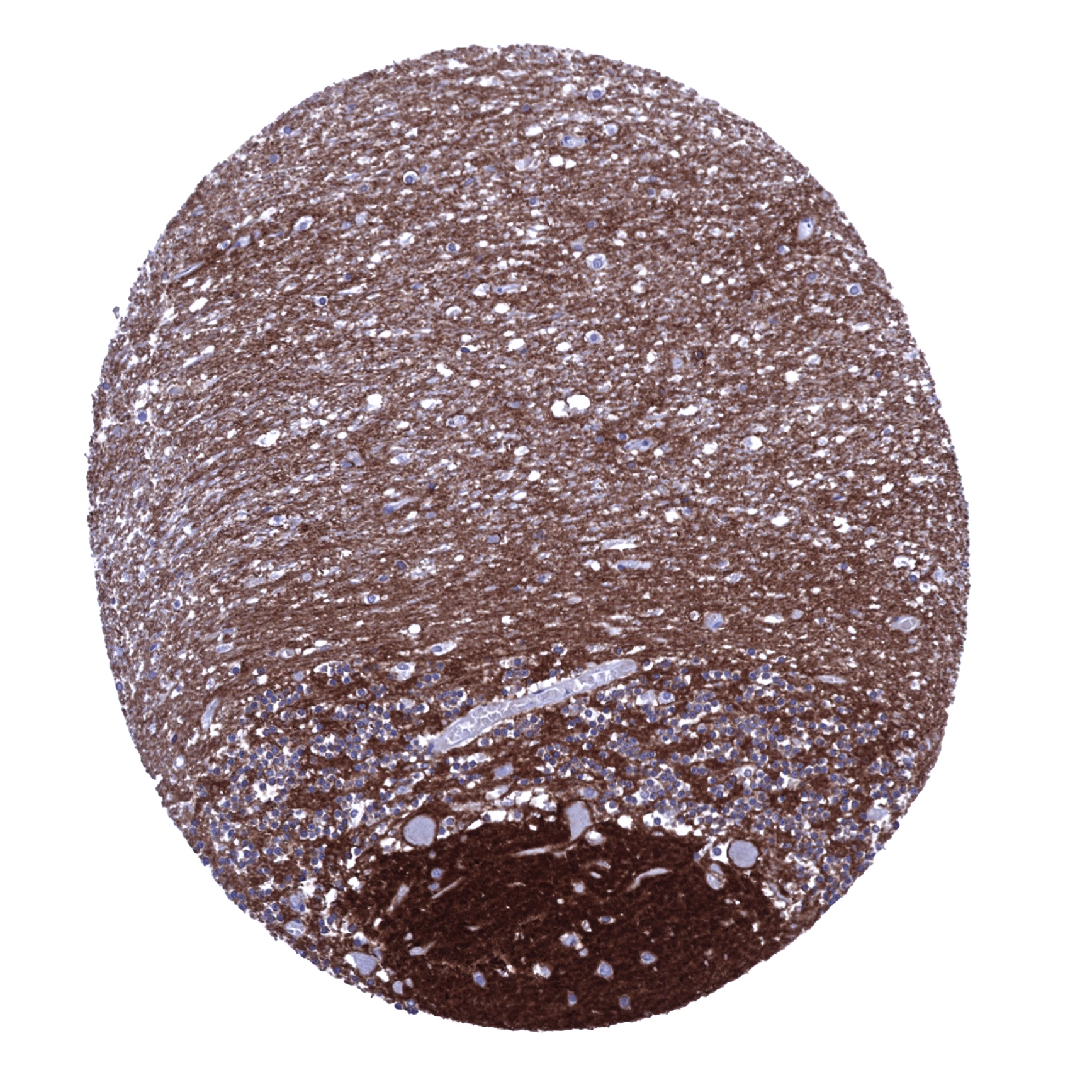
Cerebellum (molecular layer, Purkinje cell layer, granule cell layer, white matter) - CD56 immunostaining is particularly strong in neurons, axons and glia cells of the brain.
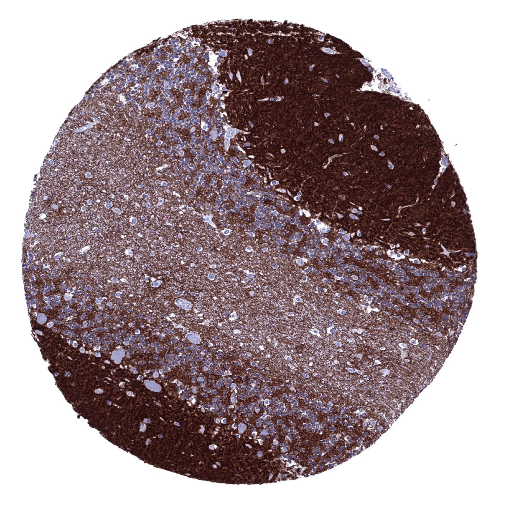
Cerebellum (molecular layer, Purkinje cell layer, granule cell layer, white matter) - CD56 immunostaining is particularly strong in neurons, axons and glia cells of the brain.
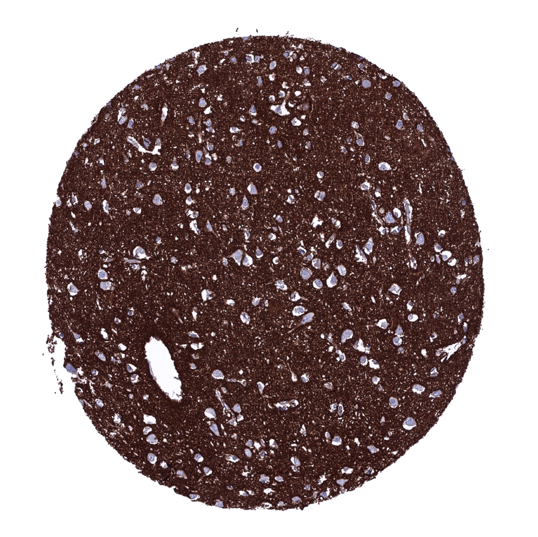
Cerebrum, grey matter - CD56 immunostaining is particularly strong in neurons, axons and glia cells of the brain.
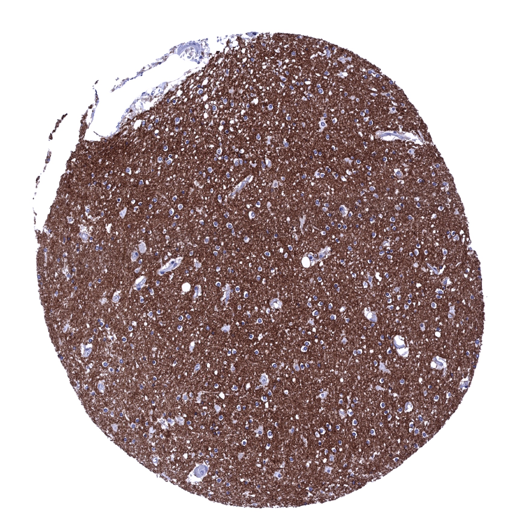
Cerebrum, white matter - CD56 immunostaining is particularly strong in neurons, axons and glia cells of the brain.

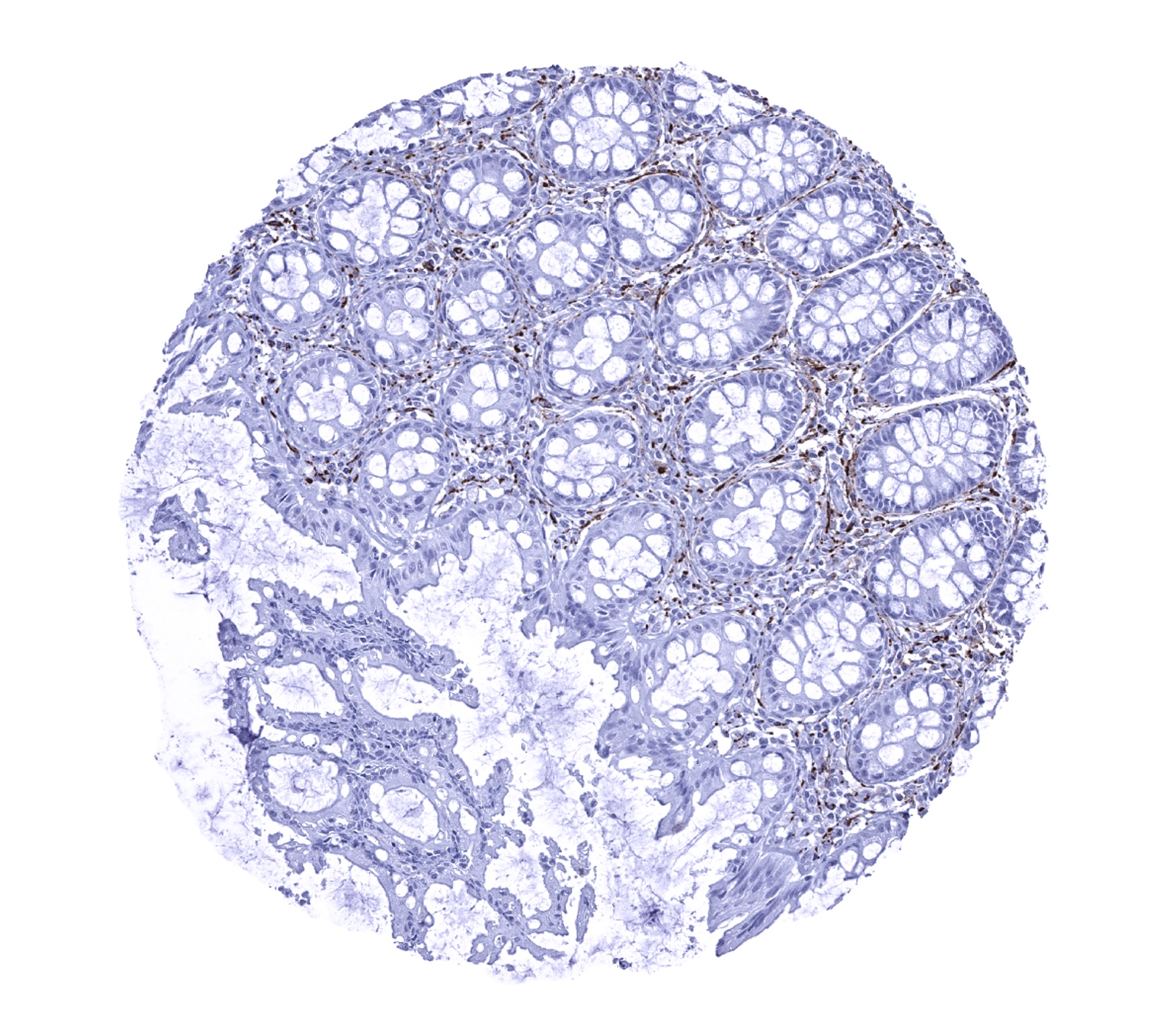
Colon descendens, mucosa - Variable intensity CD56 immunostaining of nerve fibres.

Colon descendens, muscular wall - Nerve fibres and ganglia exhibit strong CD56 immunostaining.

Duodenum, Brunner gland - A moderate CD56 immunostaining of basolateral membranes of epithelial cells of Brunner glands can be seen.

Duodenum, mucosa - Nerve fibres stain CD56 positive in the mucosa.
Epididymis - A membranous CD56 immunostaining occurs in the cauda (and not the corpus) epididymis_
Epididymis
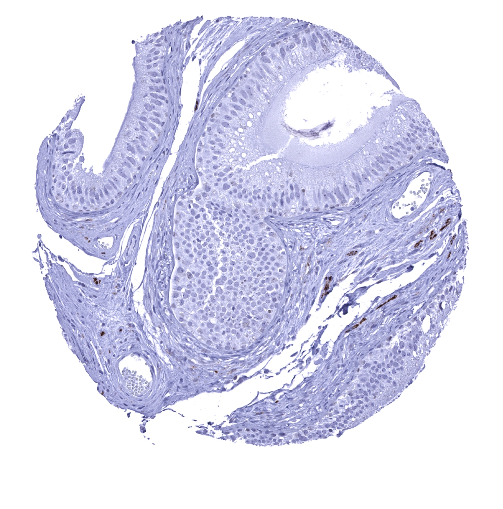
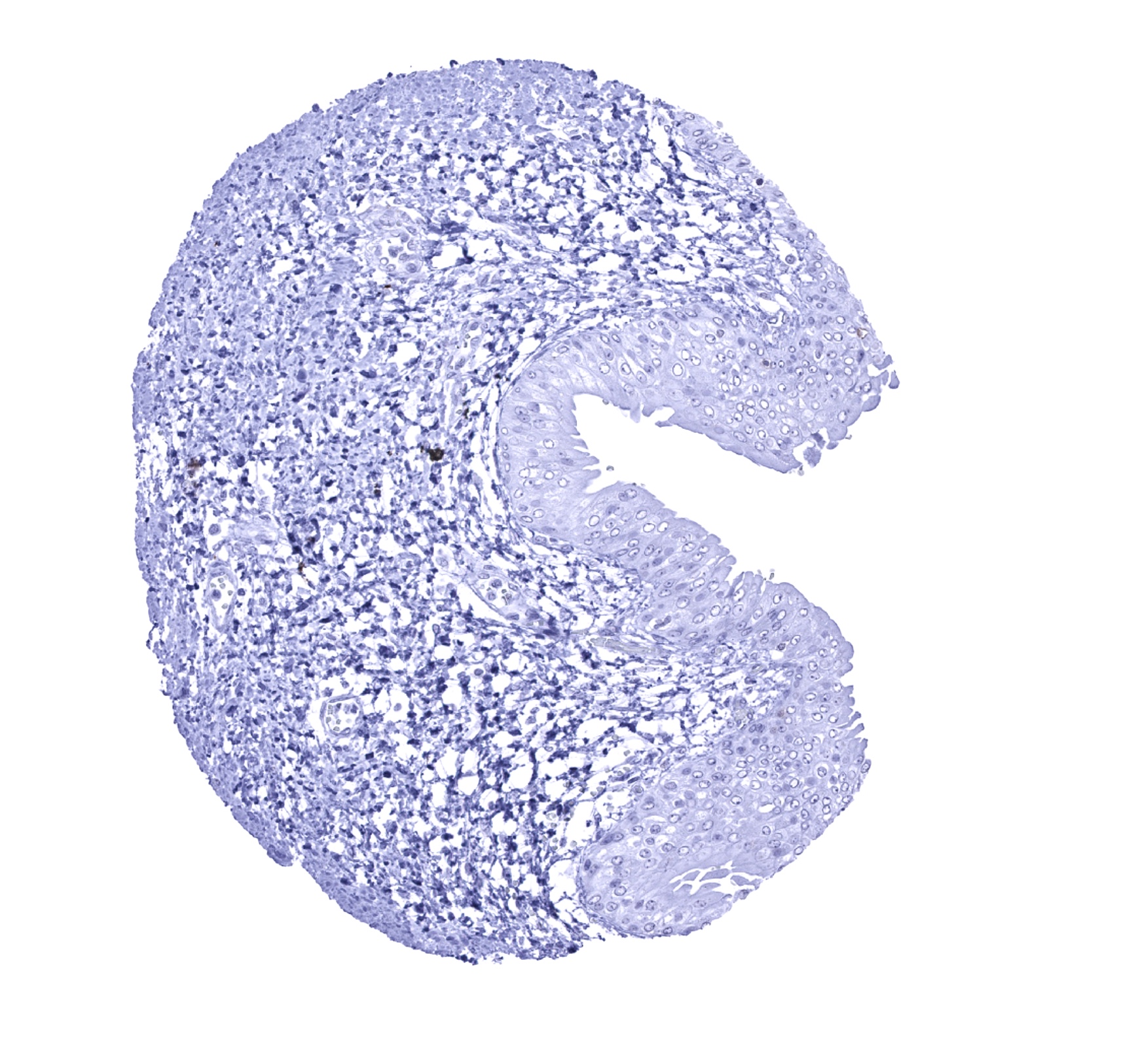
Esophagus, squamous epithelium - Scattered CD56 positive lymphocytes can be seen.
Fallopian tube, mucosa

Fat
Gallbladder, epithelium - CD56 immunostaining of nerve fibres.
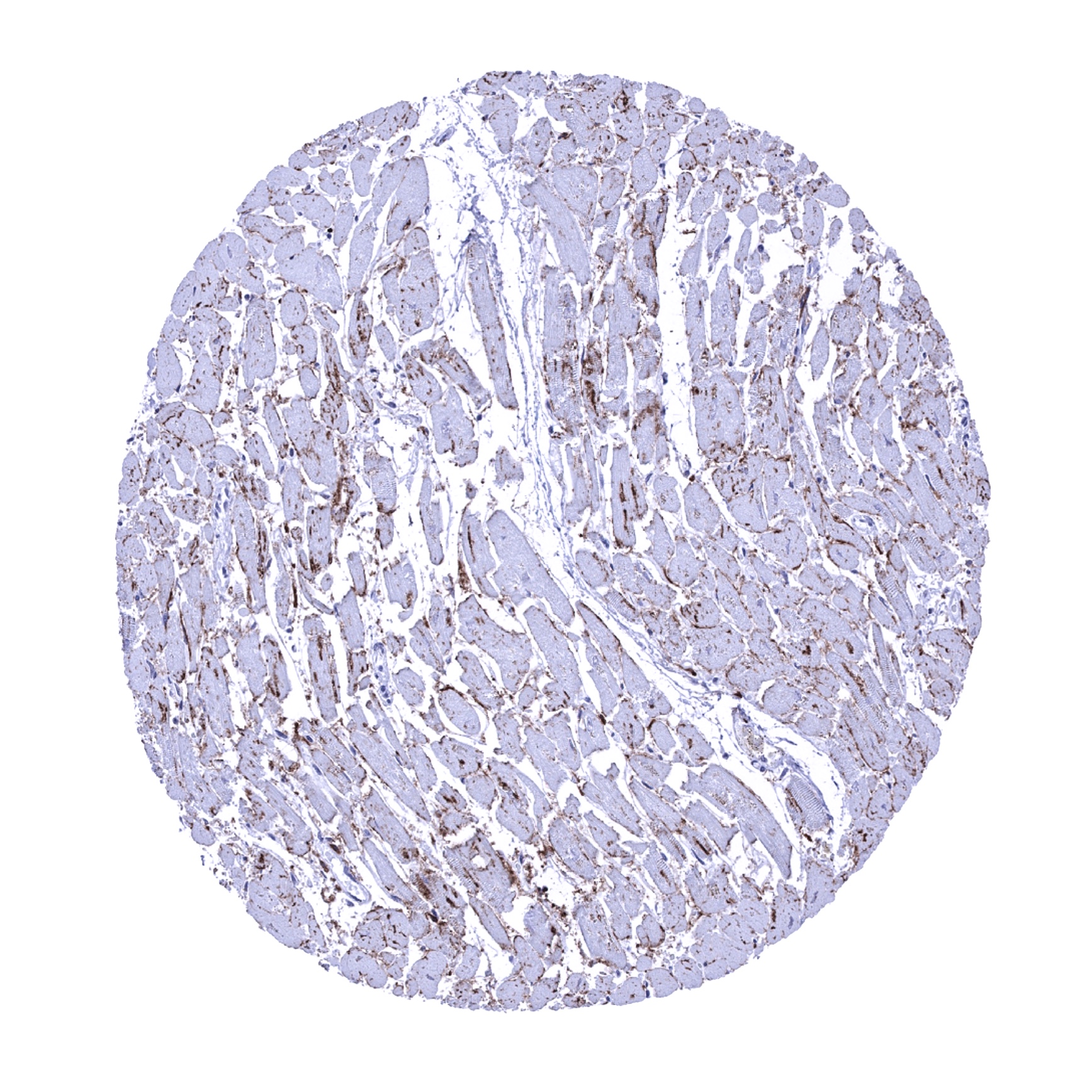
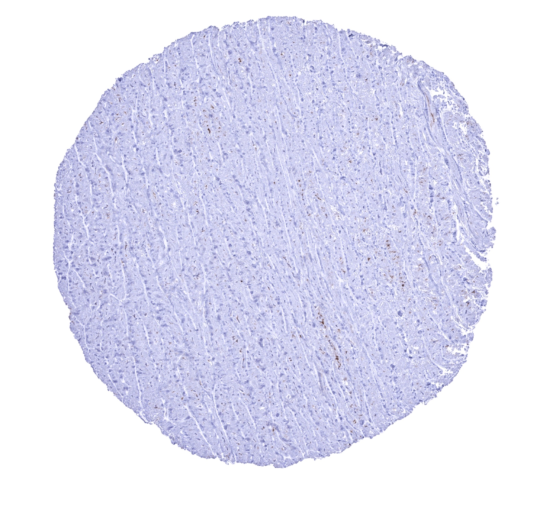
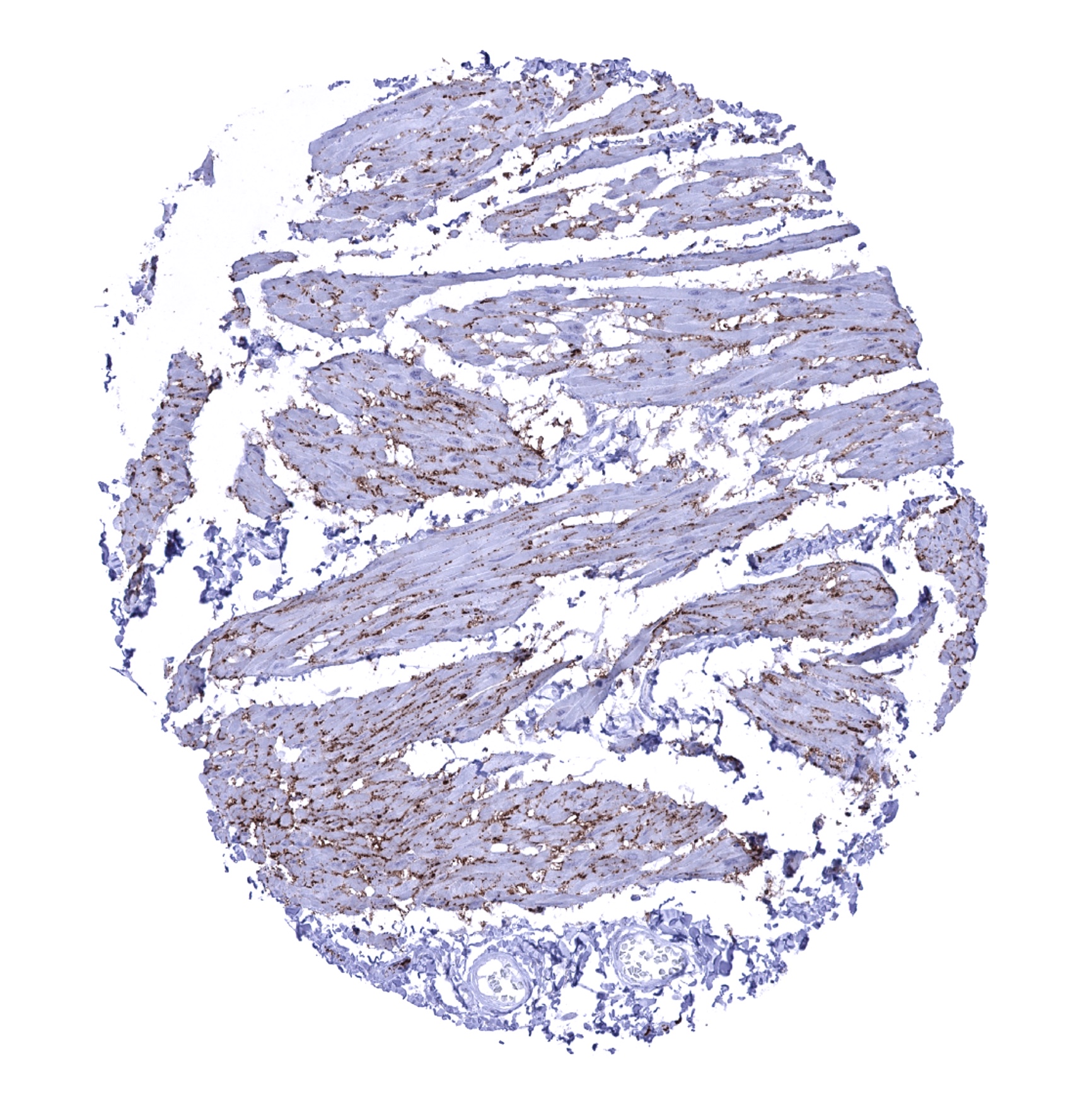
Heart muscle - CD56 immunostaining of membreans of heart muscle is not seen by other CD56 antibodies and thus considered an (tolerable) cross-reactivity.
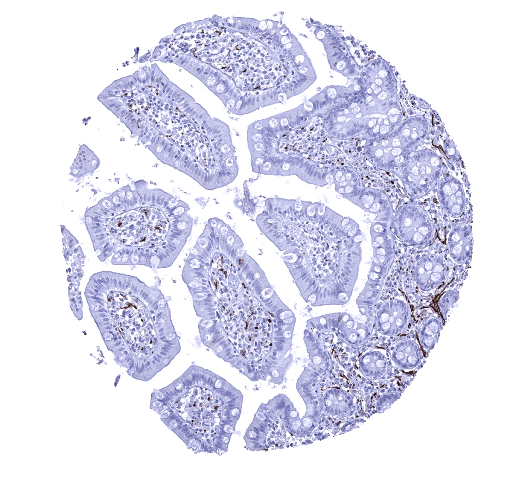
Ileum, mucosa - Nerve fibres stain CD56 positive in the mucosa.

Ileum, mucosa - Nerve fibres stain CD56 positive in the mucosa.
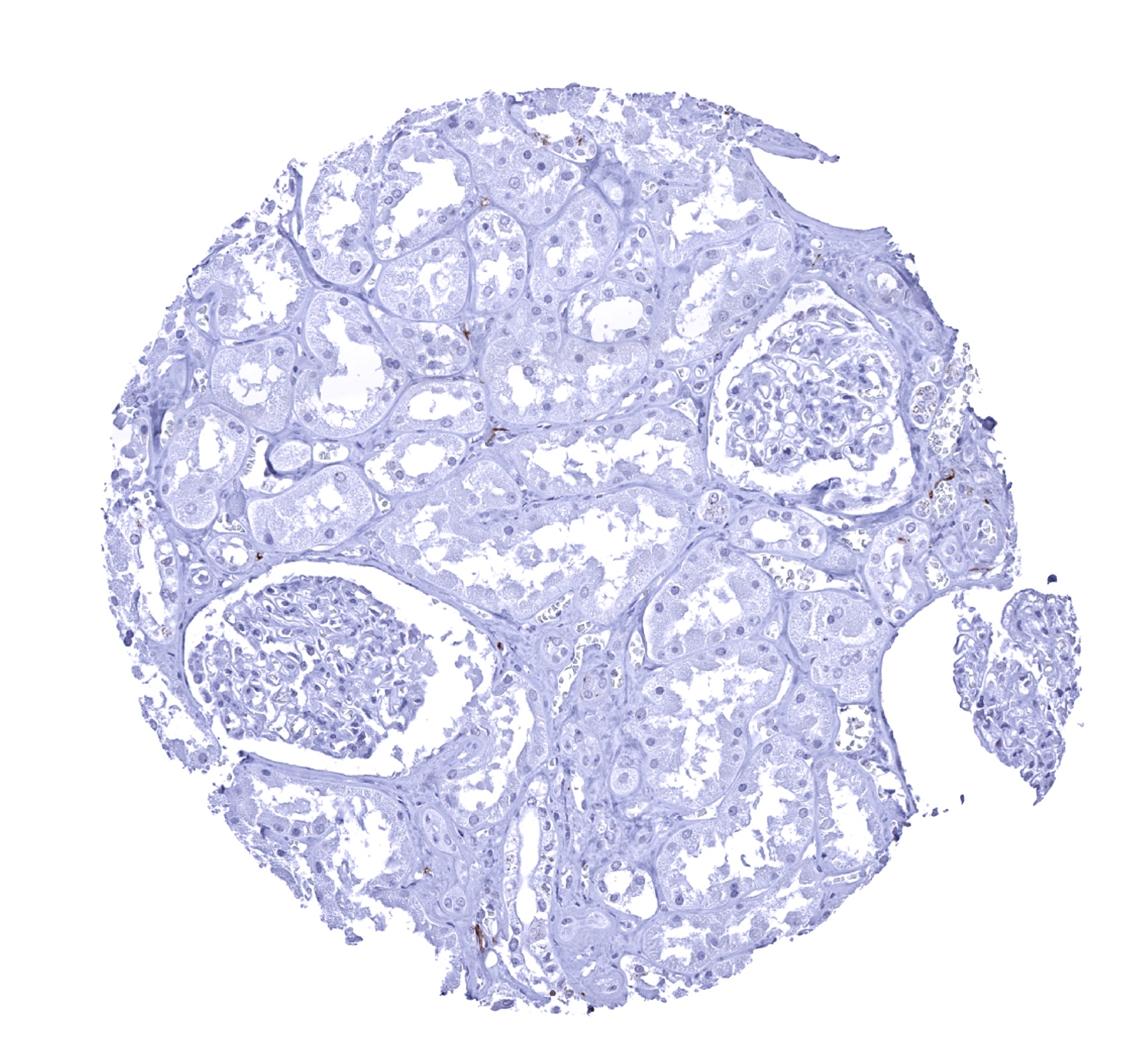
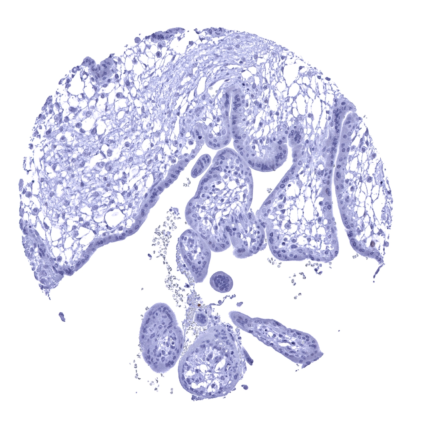
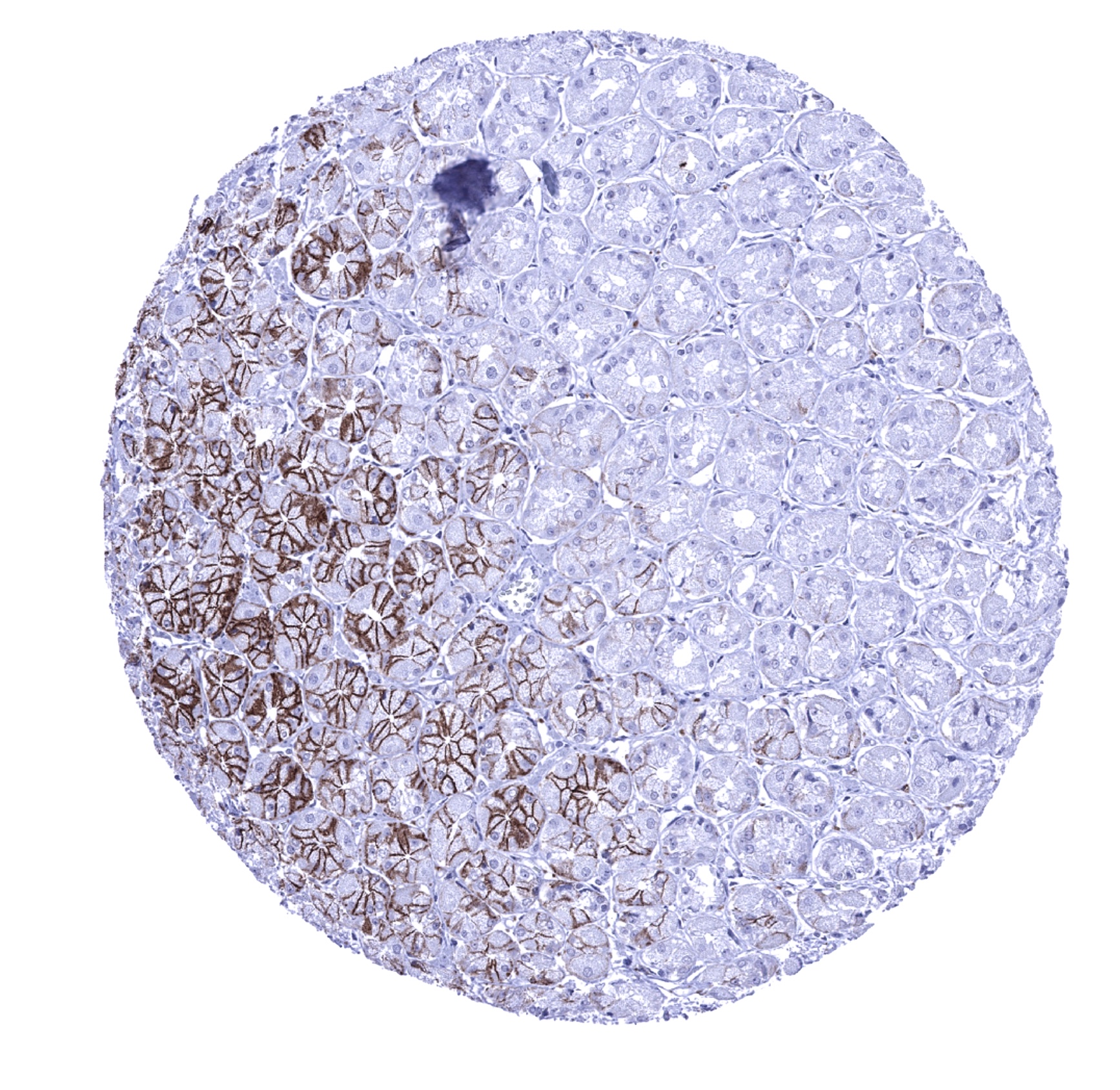
Kidney, cortex
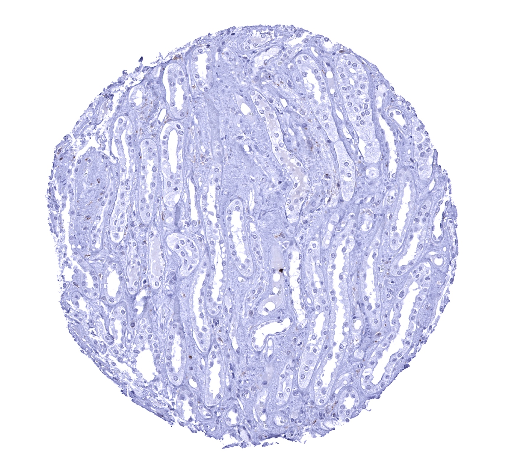
Kidney, medulla
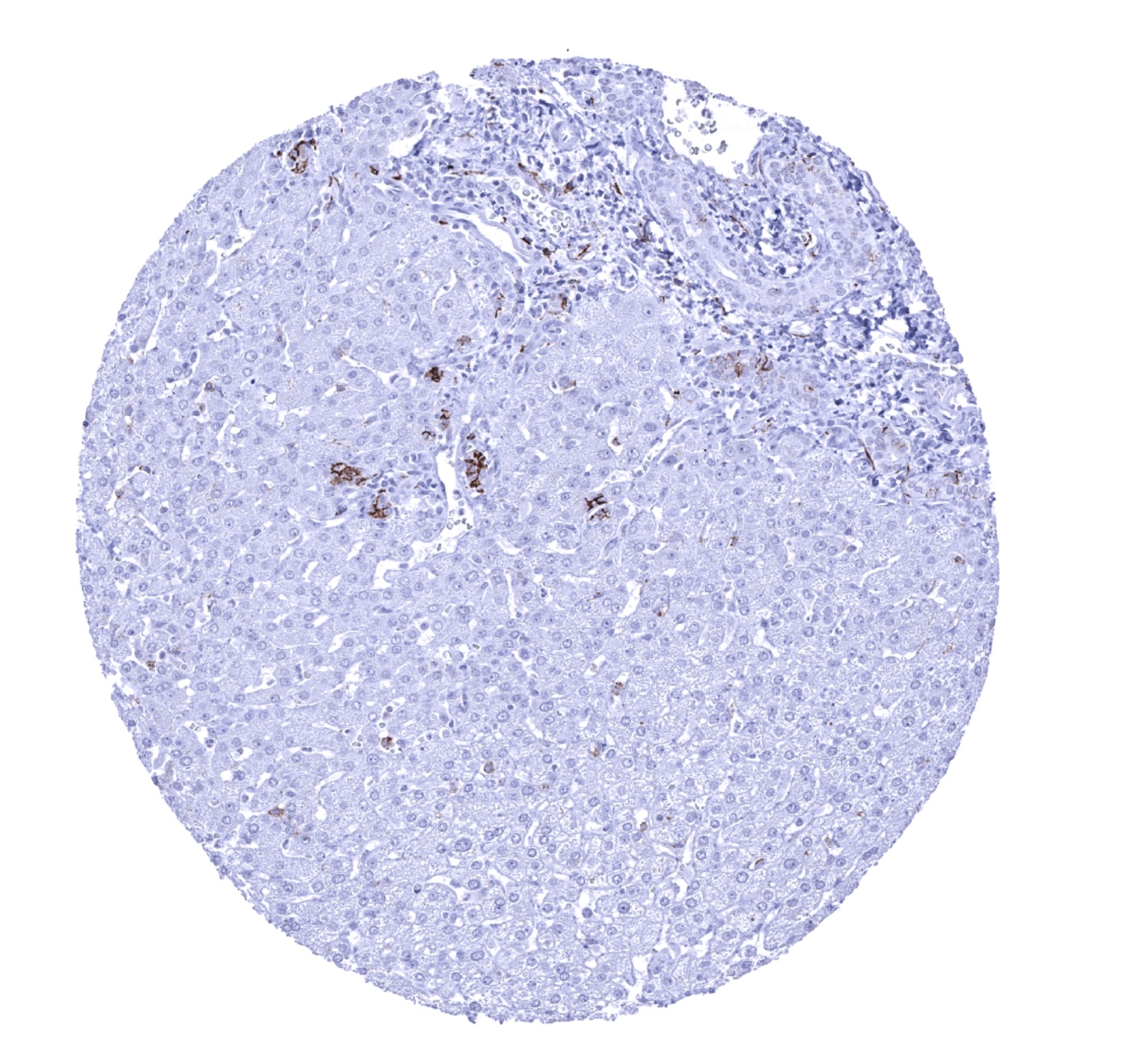
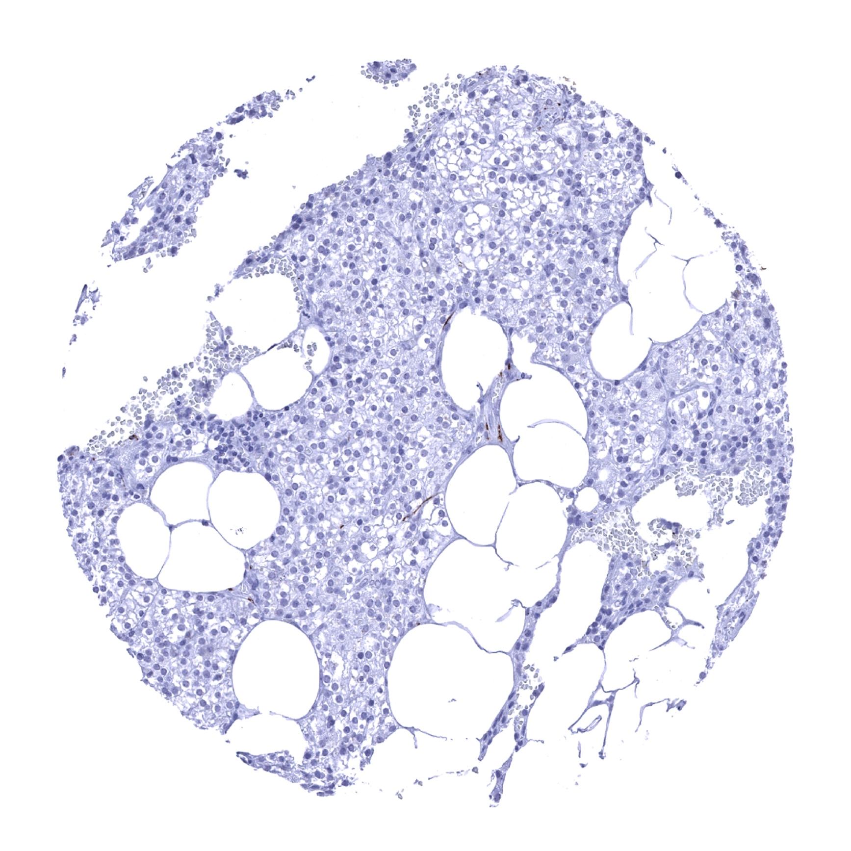
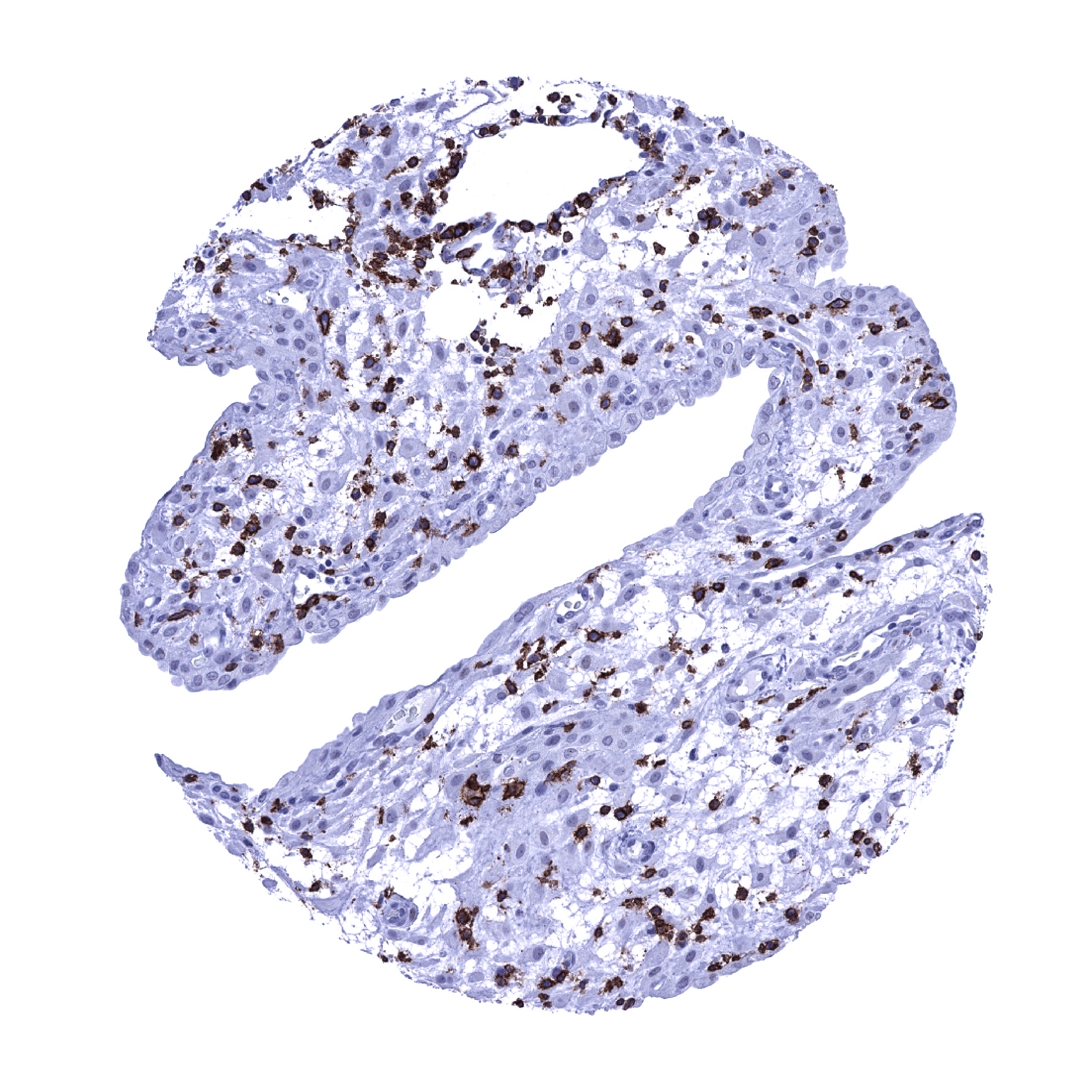
Liver - A moderate CD56 staining can occur in small periportal and to a lesser degree in portal bile ducts of the liver.
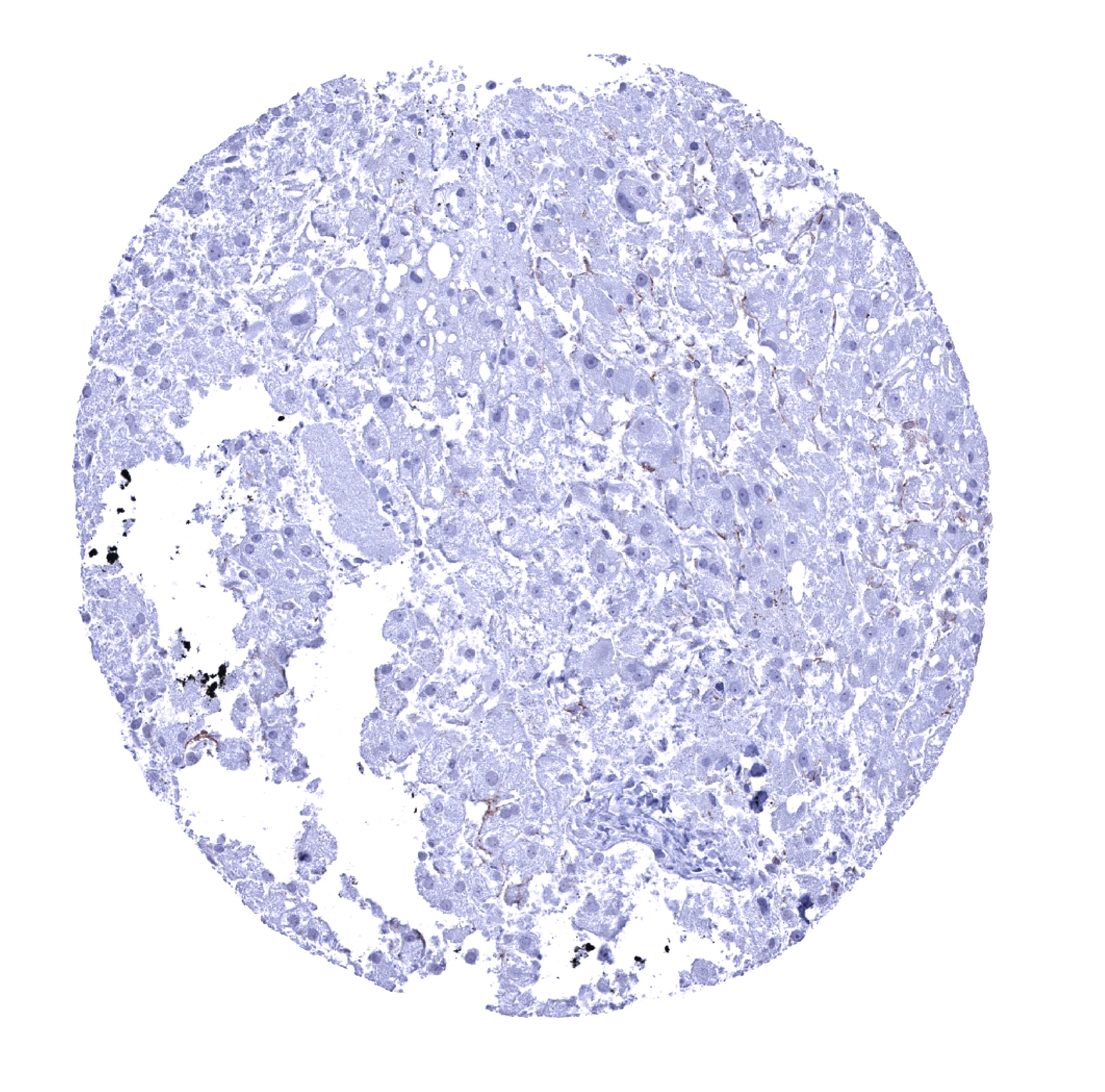
Liver - A weak to moderate CD56 immunostaining can be seen in cells lining sinusoids.
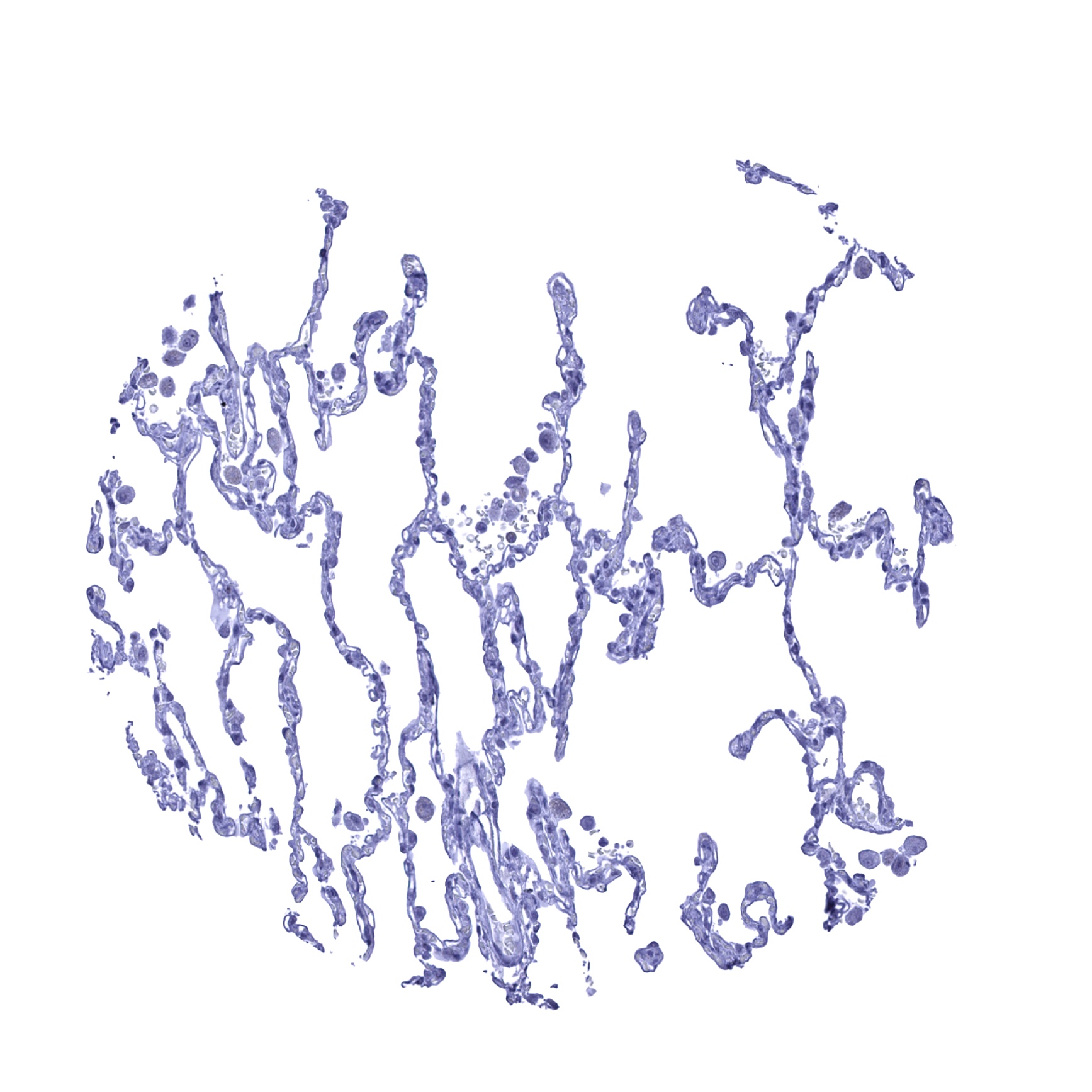
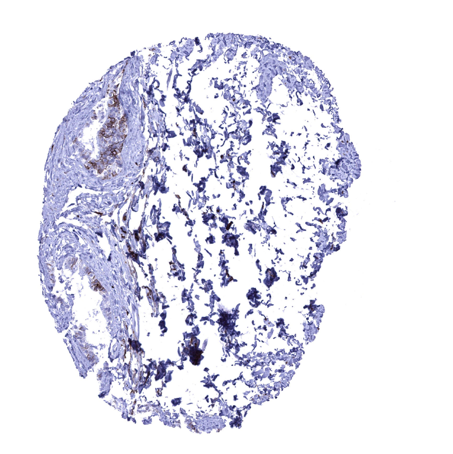
Lung

Lymph node - Scattered CD56 positive lymphocytes can be seen in lymph nodes.
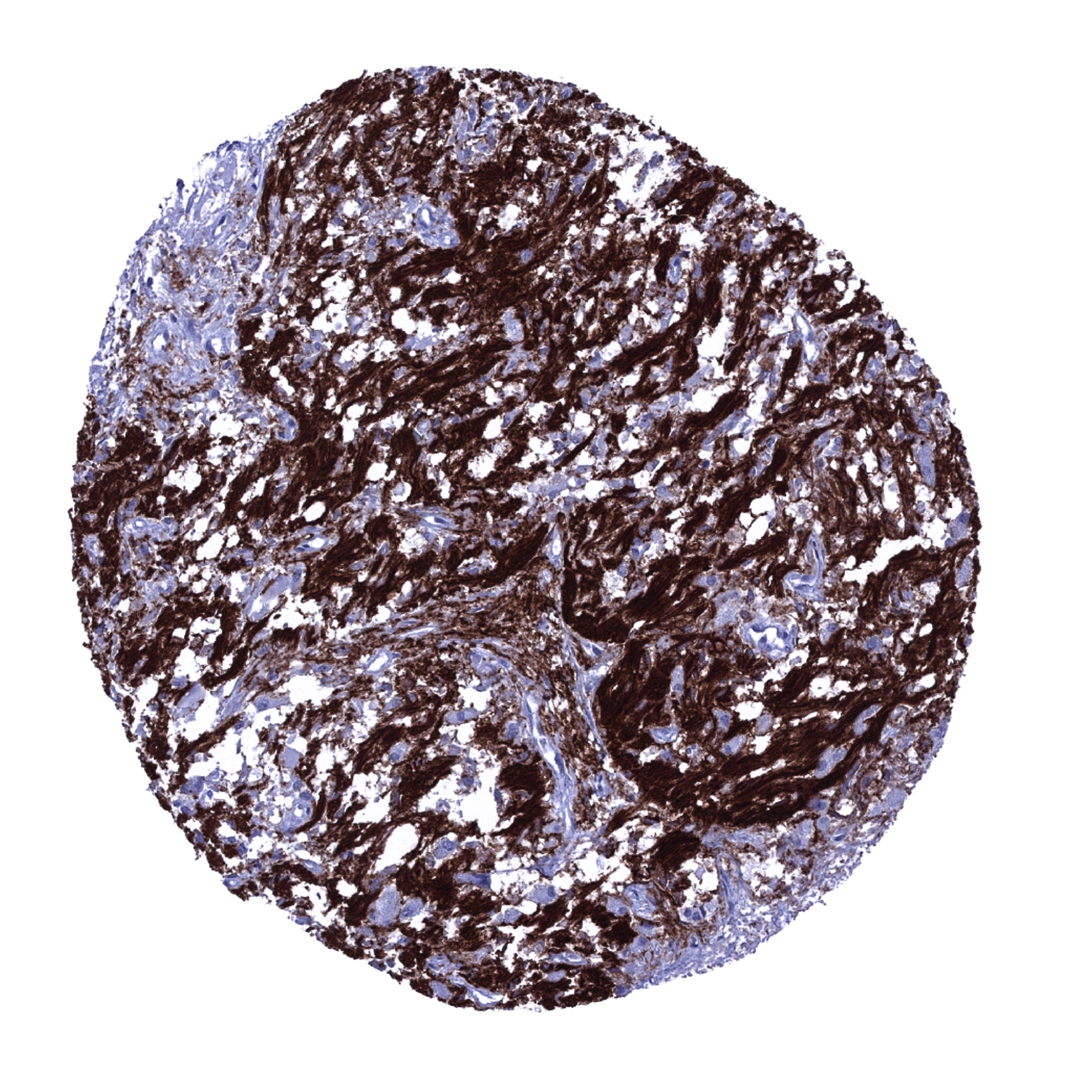
Ovary, stroma - Stroma cells show a strong membranous CD56 immunostaining.
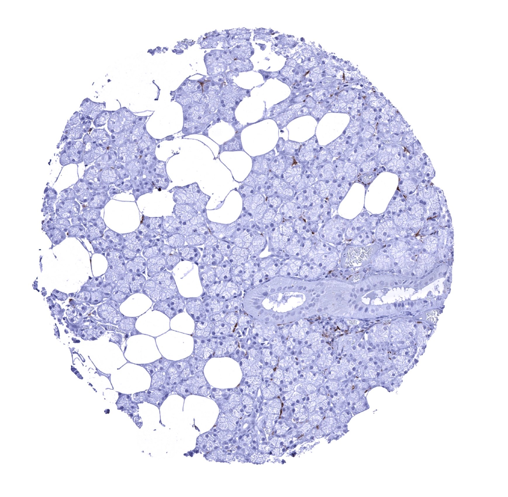
Pancreas - Few small intraparenchymal ducts and a fraction of the islet cells show weak to moderate membranous CD56 immunostaining.
Parathyroid gland
Parotid gland - Nerve fibres stain CD56 positive.
Pituitary gland, posterior lobe - A strong CD56 immunostaining is seen in the neurohypophysis_
Pituitary, anterior lobe

Placenta (amnion and chorion)
Placenta, early

Placenta, mature
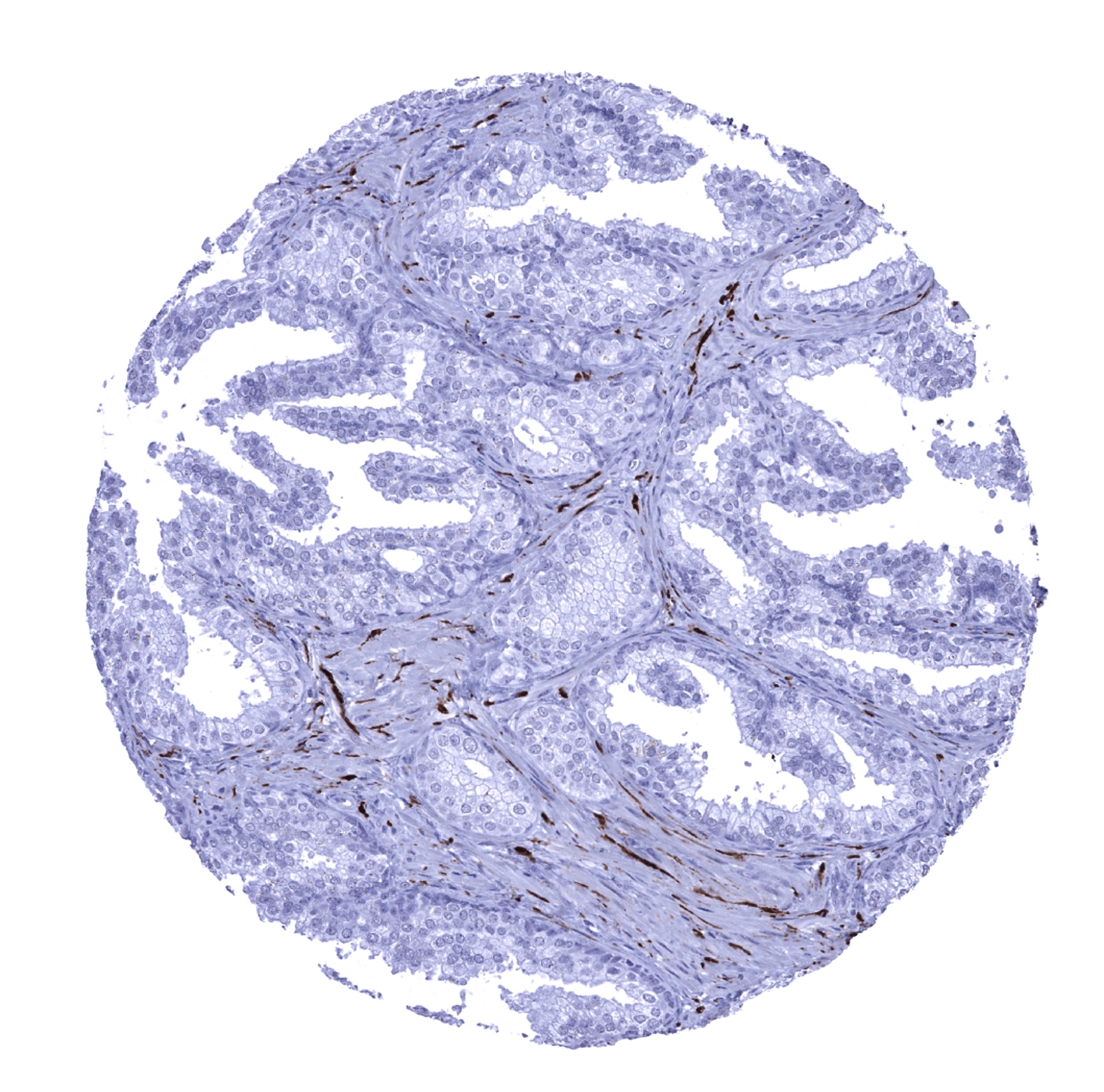
Prostate - Nerve fibres show CD56 immunostaining.
Rectum, mucosa - CD56 positivity of nerve fibres.
Seminal vesicle - Nerve fibres show CD56 immunostaining.
Sinus paranasales - A moderate to strong membranous CD56 immunostaining occurs in a fraction of (non-basal) cells of the respiratory epithelium.
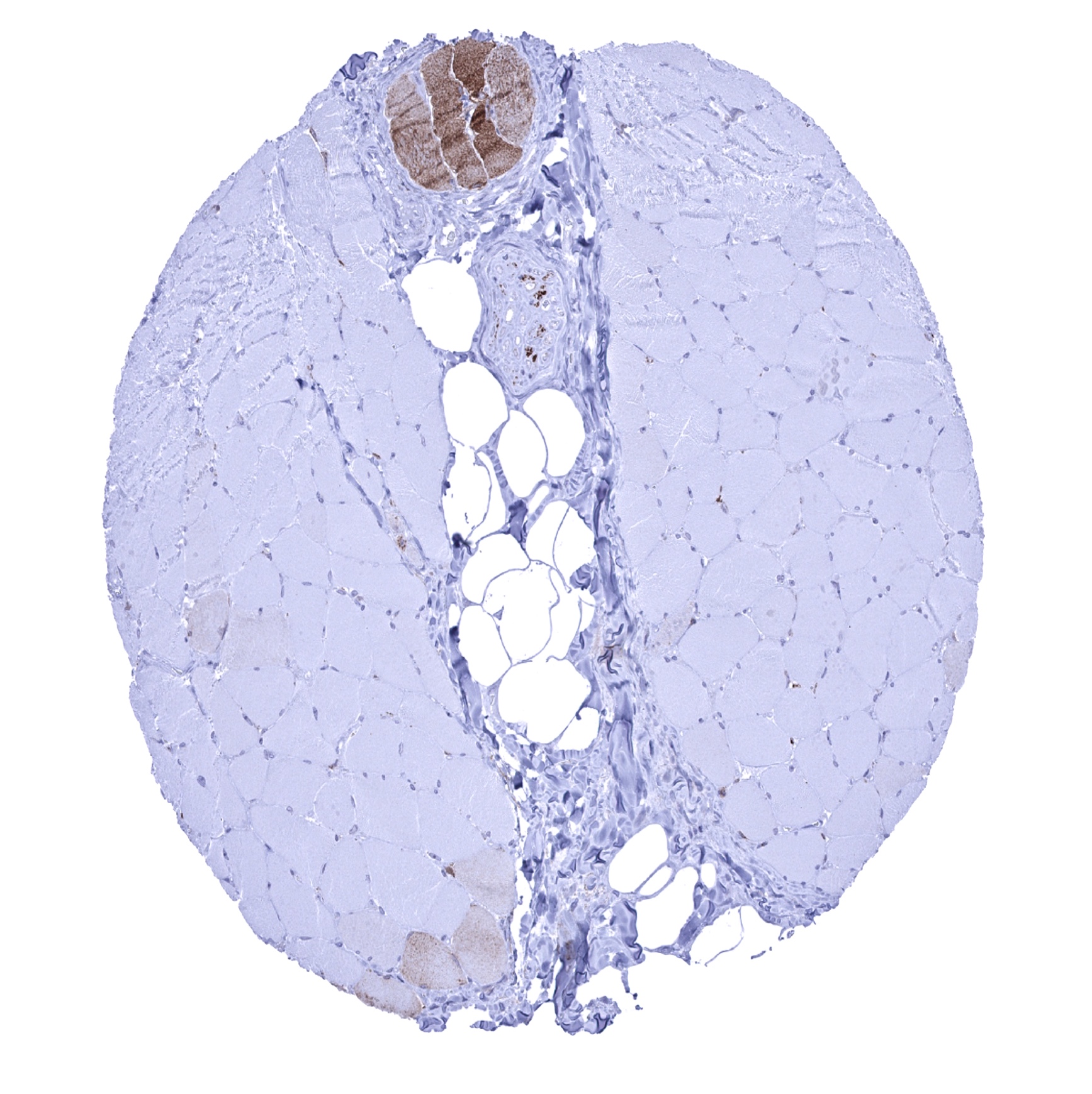
Skeletal muscle - A cytoplasmic staining of skeletal muscle cells can occasionally be seen and is considered a (tolerable) cross-reactivity.
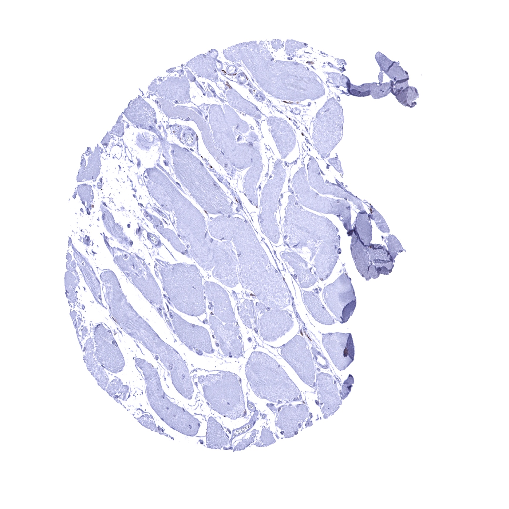
Skeletal muscle
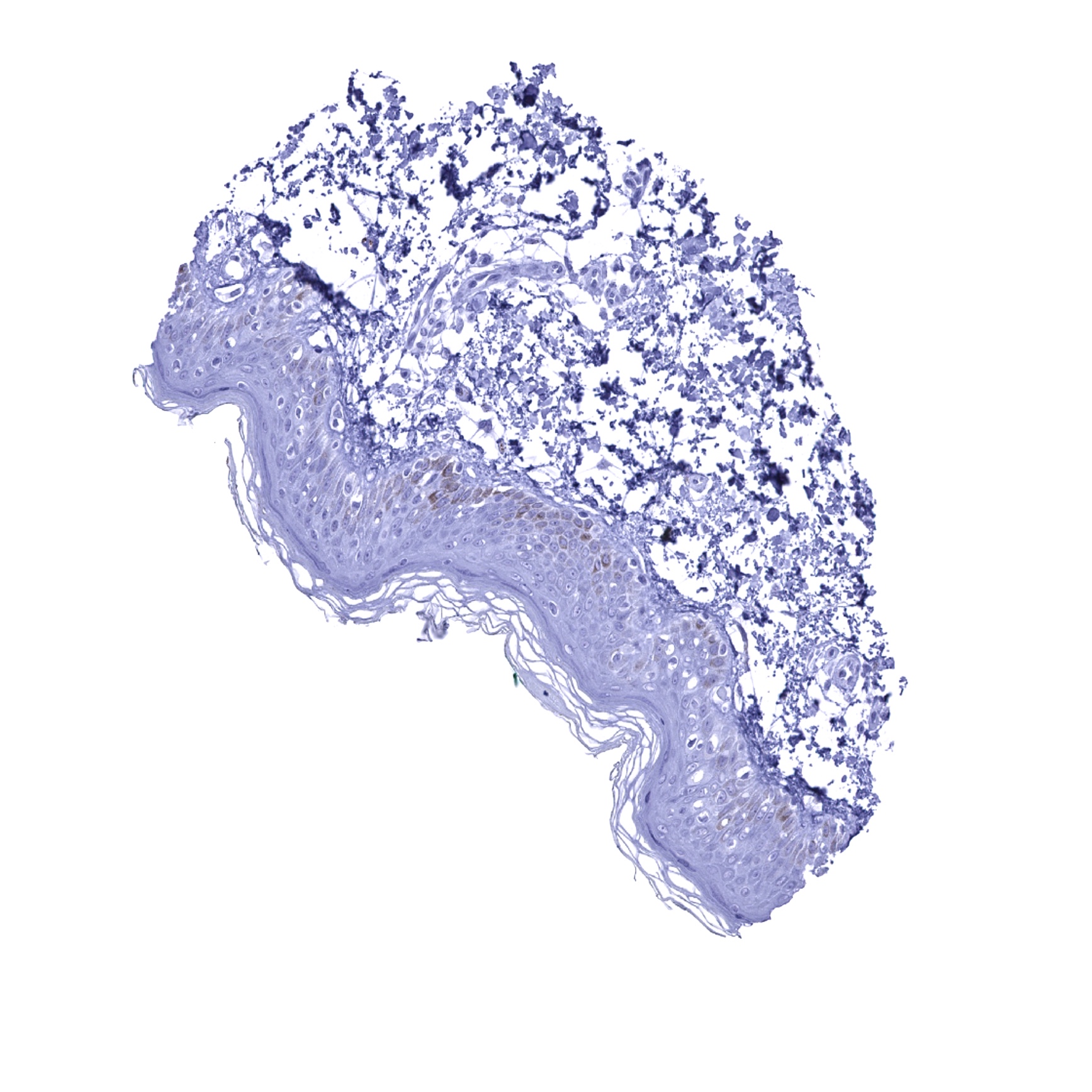
Skin
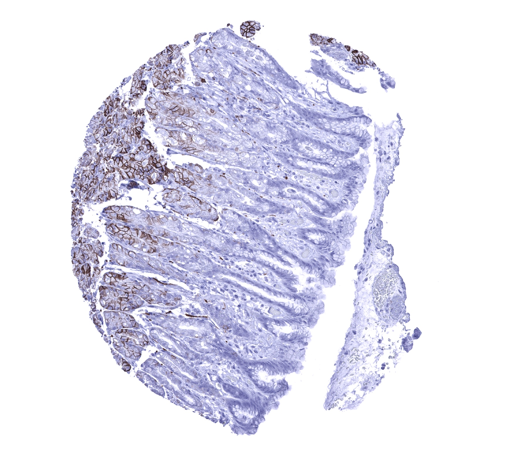
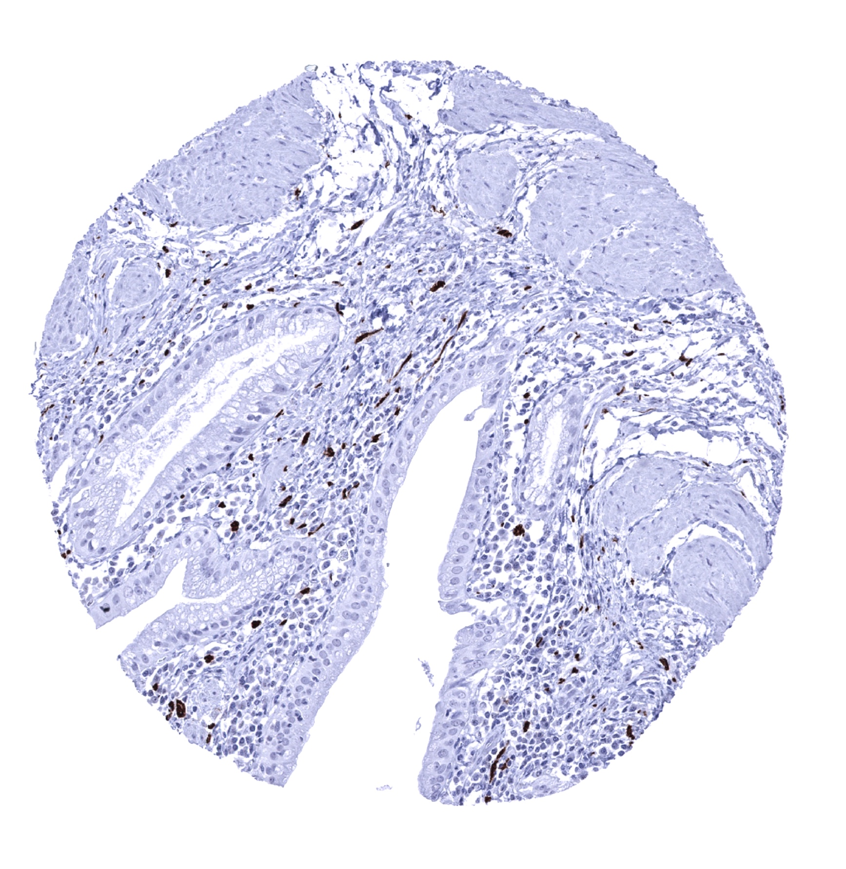
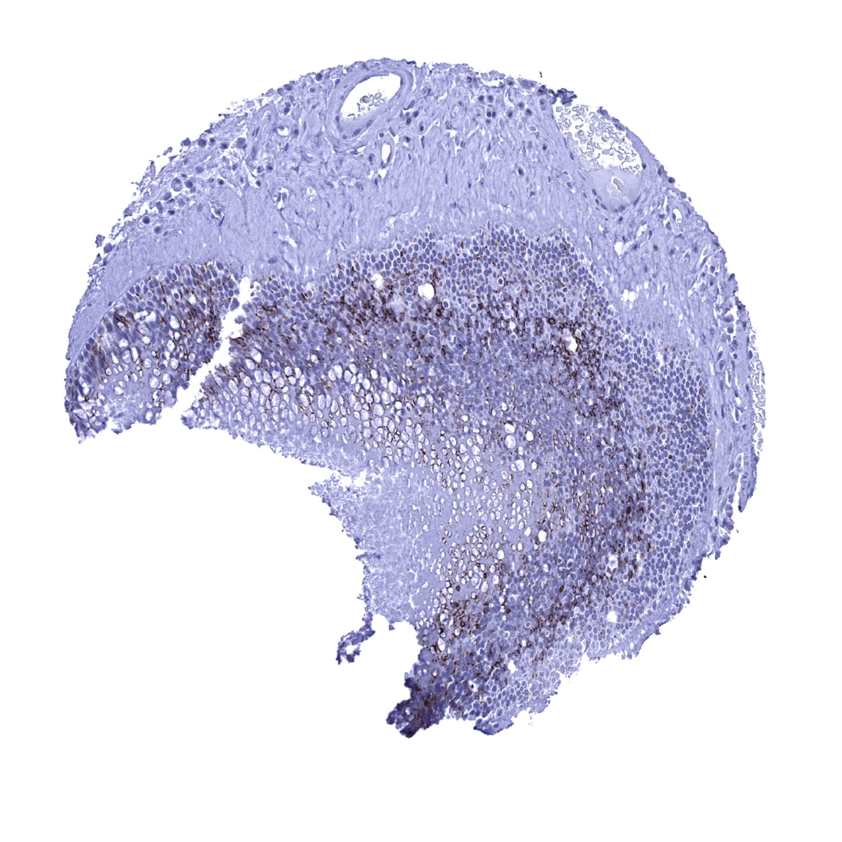
Somach, antrum - A moderate to strong membranous CD56 immunostaining occurs in most glandular cells of the stomach.
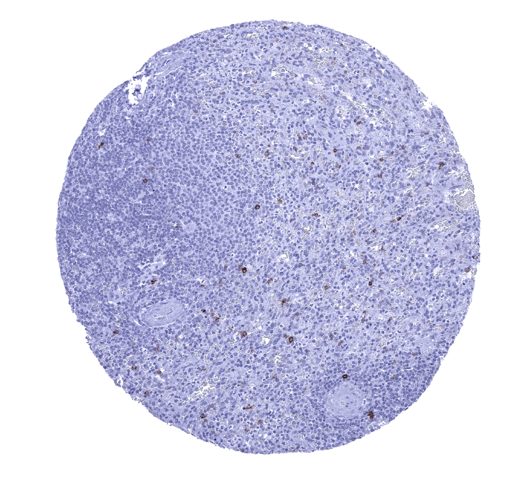
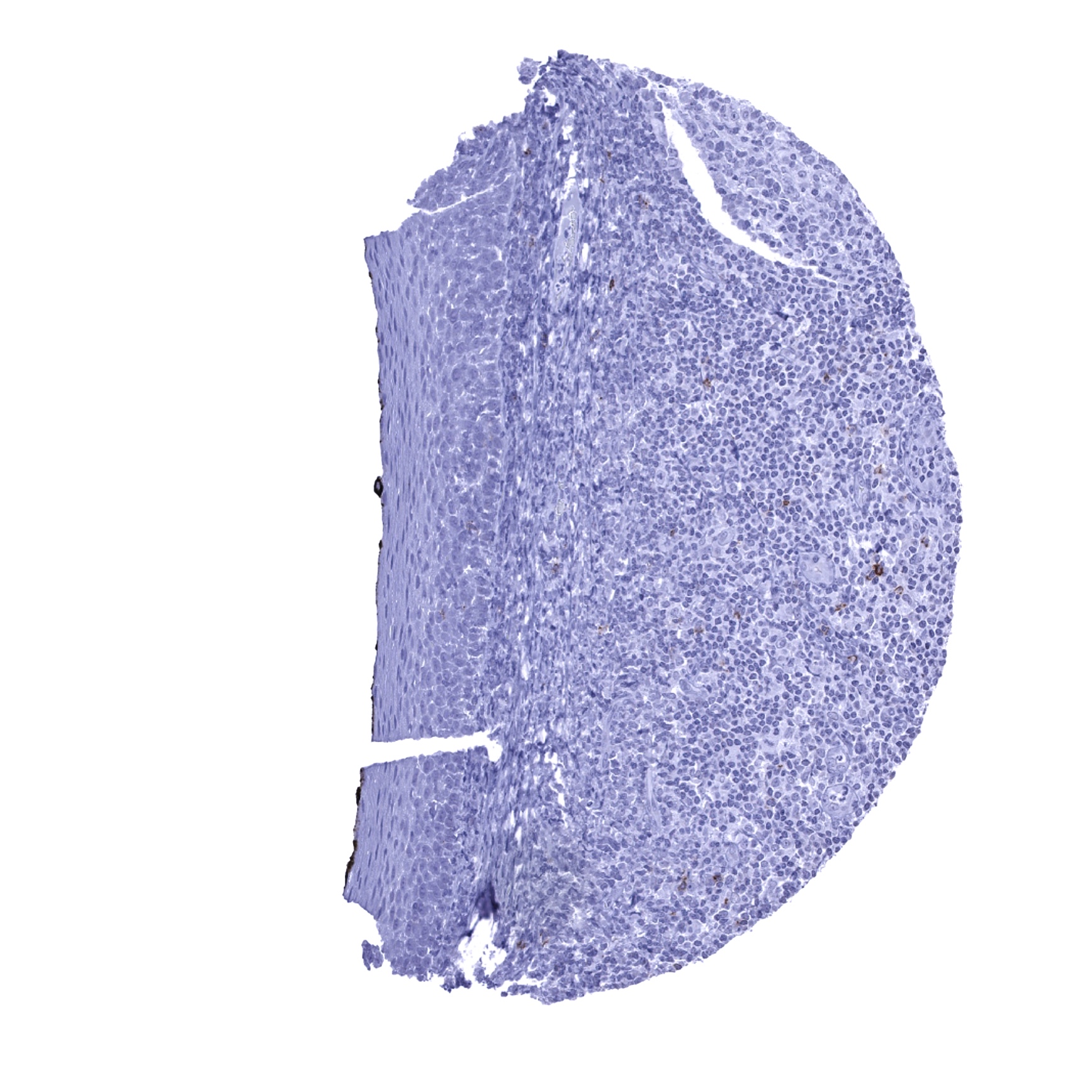
Spleen - Scattered CD56 positive lymphocytes can be seen in the spleen.
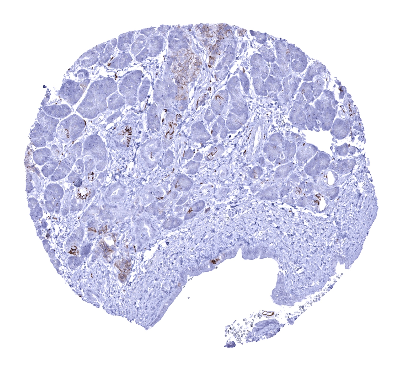
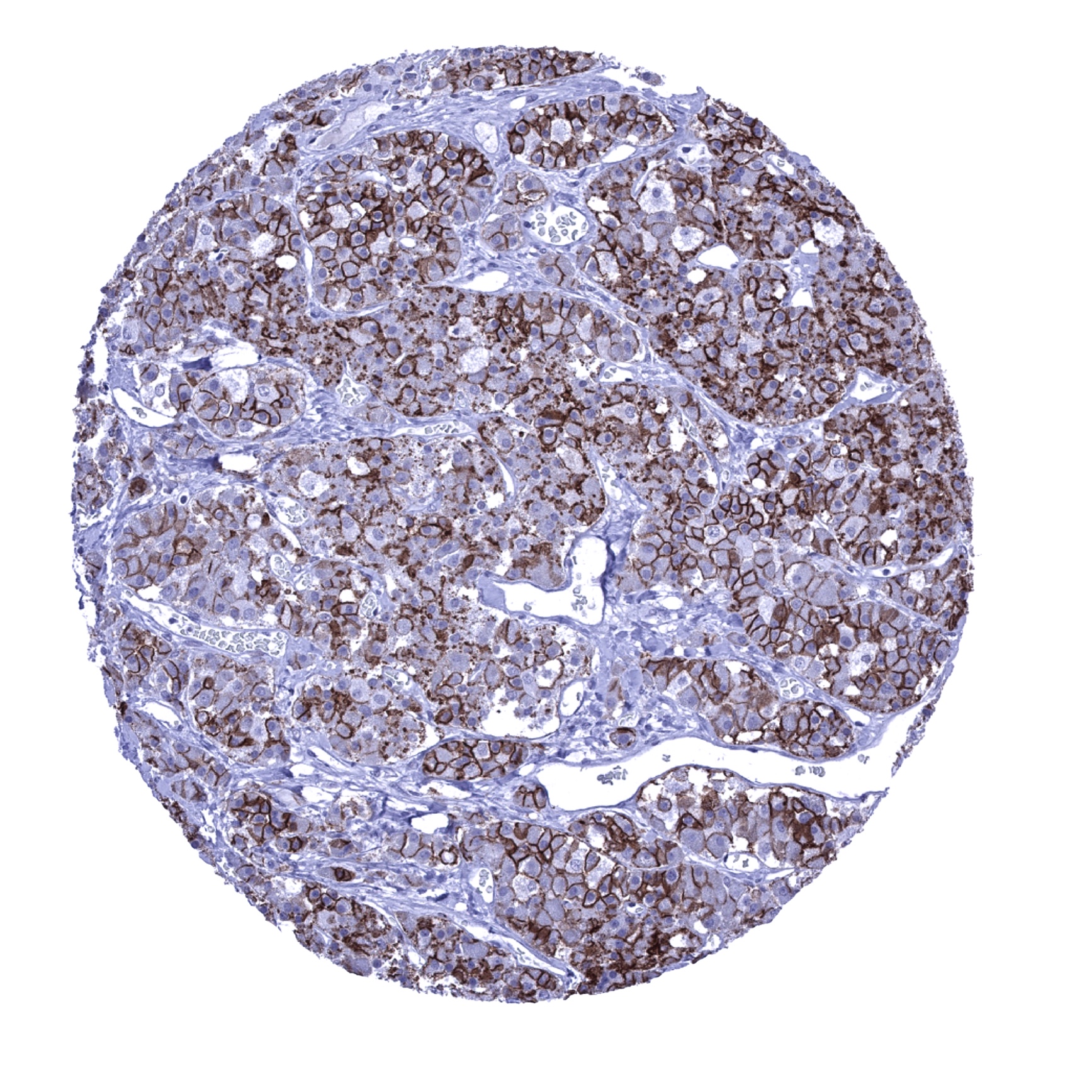
Stomach, corpus - A moderate to strong membranous CD56 immunostaining occurs in most glandular cells of the stomach.
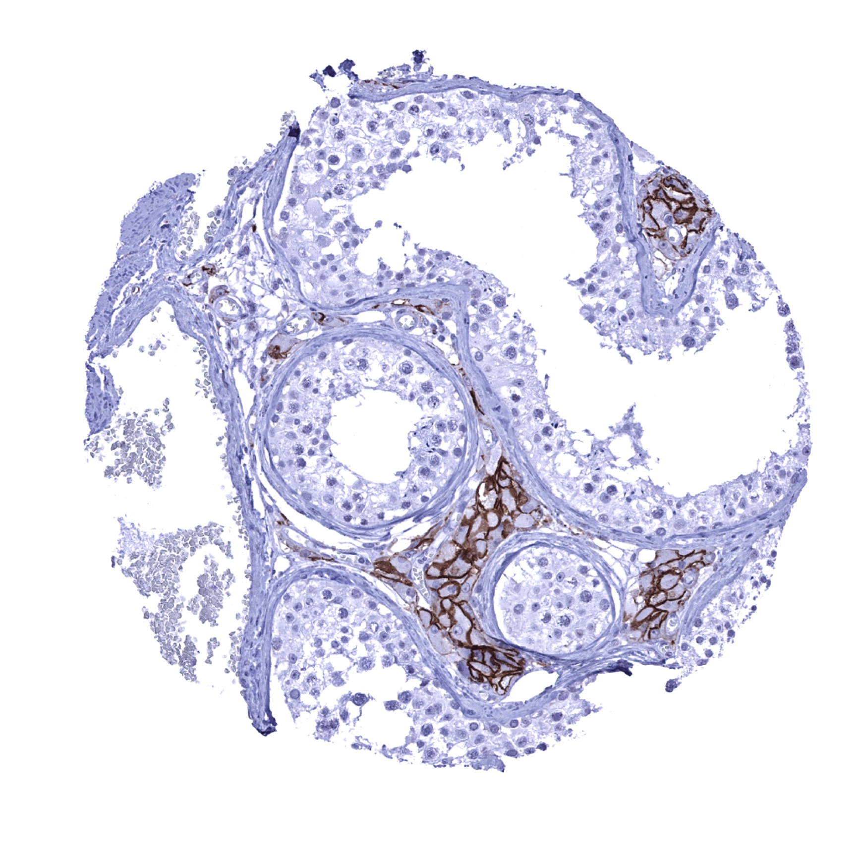
Testis - Strong membranous CD56 immunostaining of Leydig cells.

Thymus

Thyroid gland - A strong membranous CD56 immunostaining of follicle cells is seen in the thyroid.
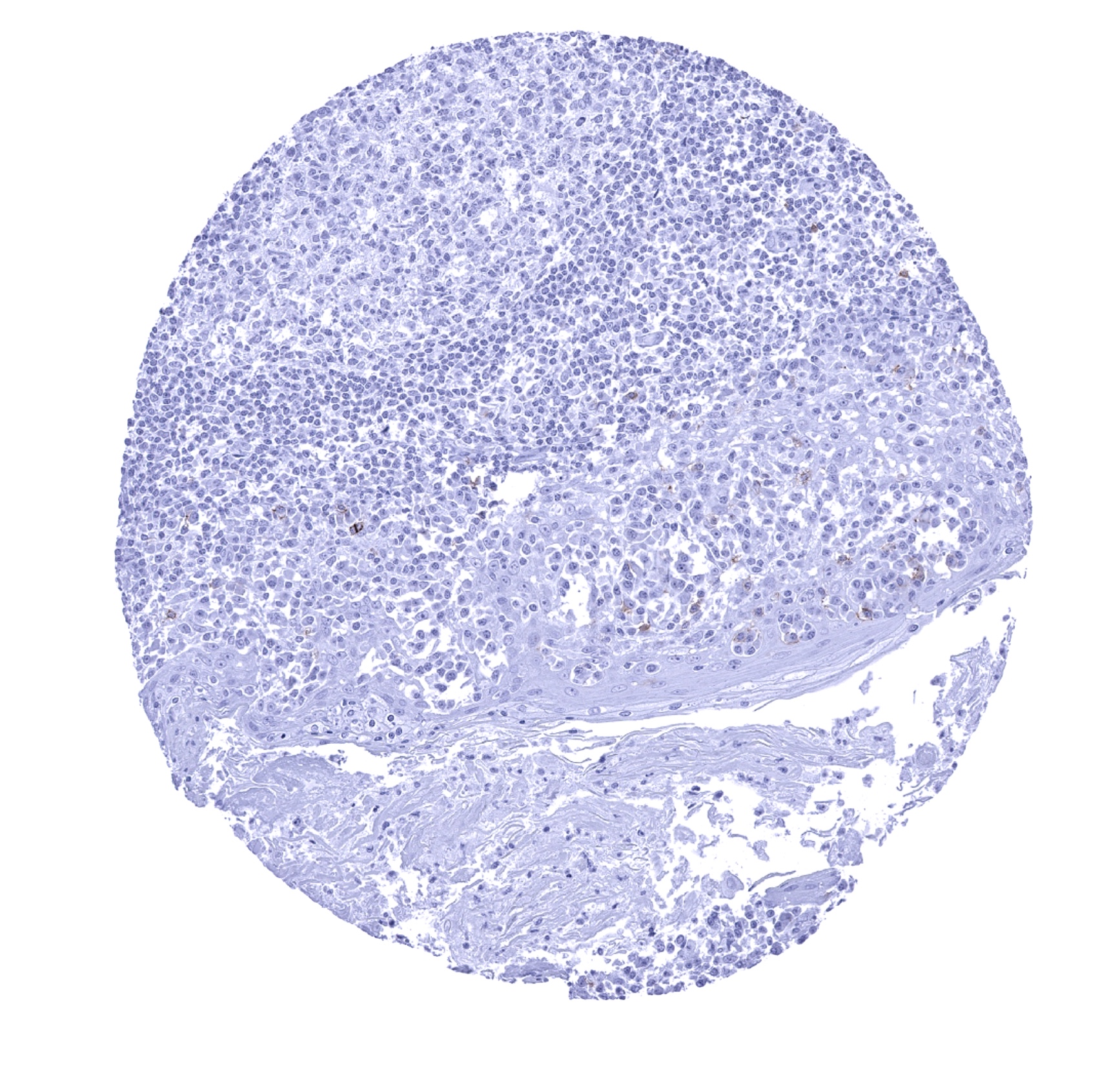
Tonsil - Scattered CD56 positive lymphocytes can be seen in in the tonsil.
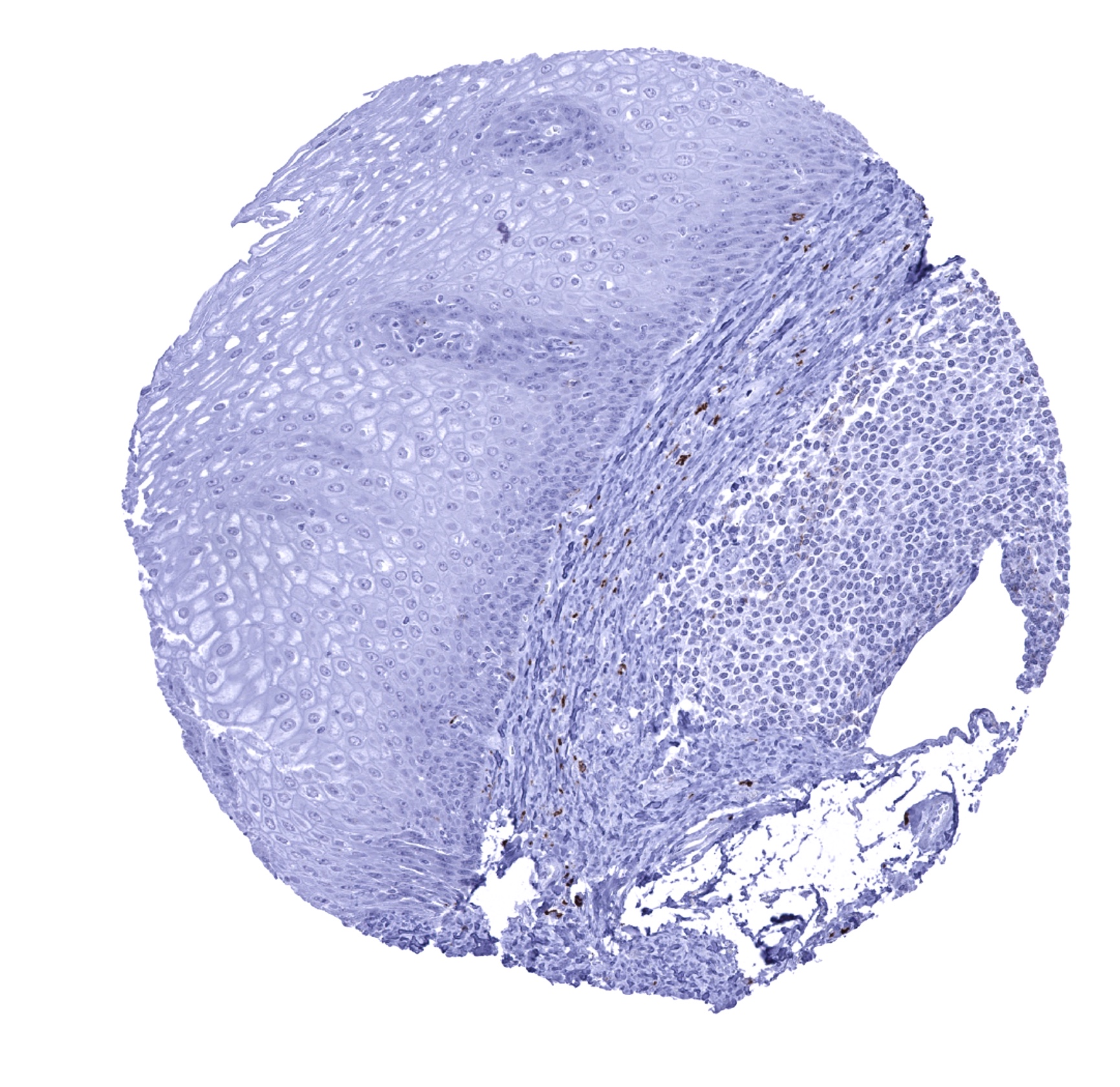
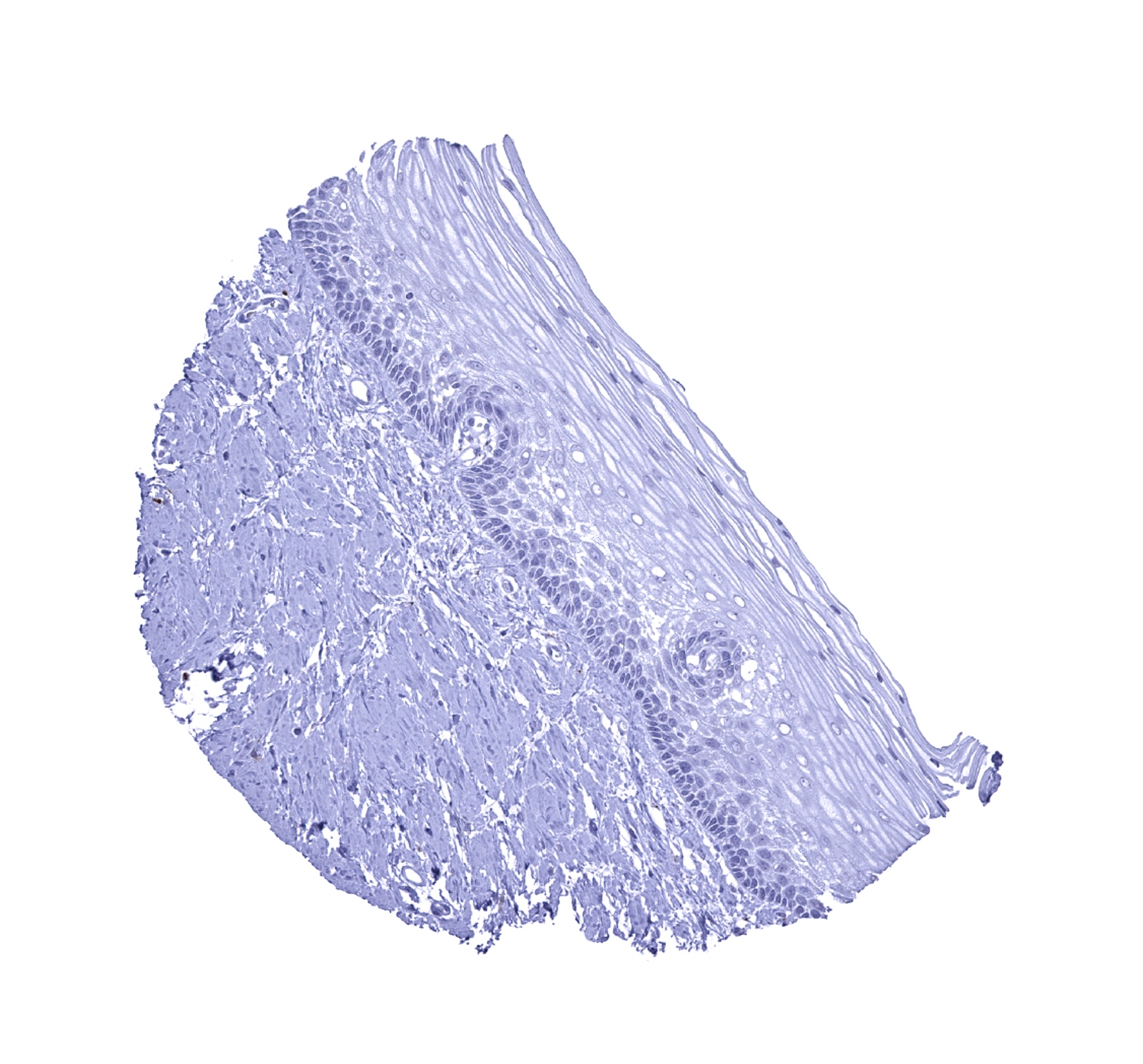
Tonsil, surface epithelium
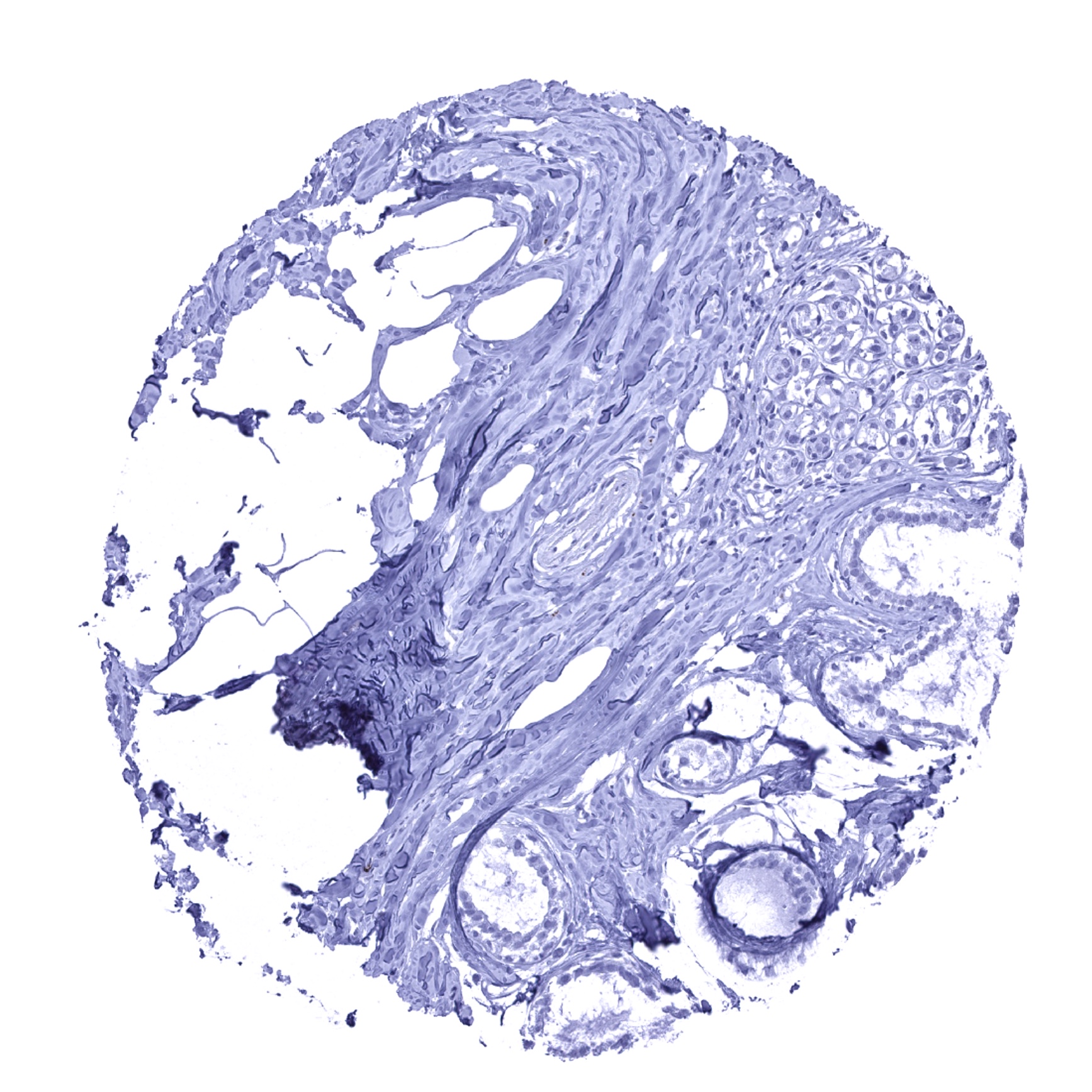
Urinary bladder, muscular wall - A dense net of CD56 positive nerve fibres is seen.
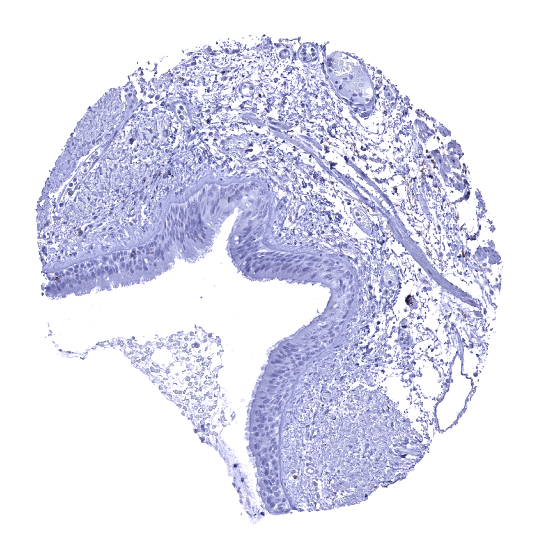
Urinary bladder, urothelium
Uterus, ectocervix
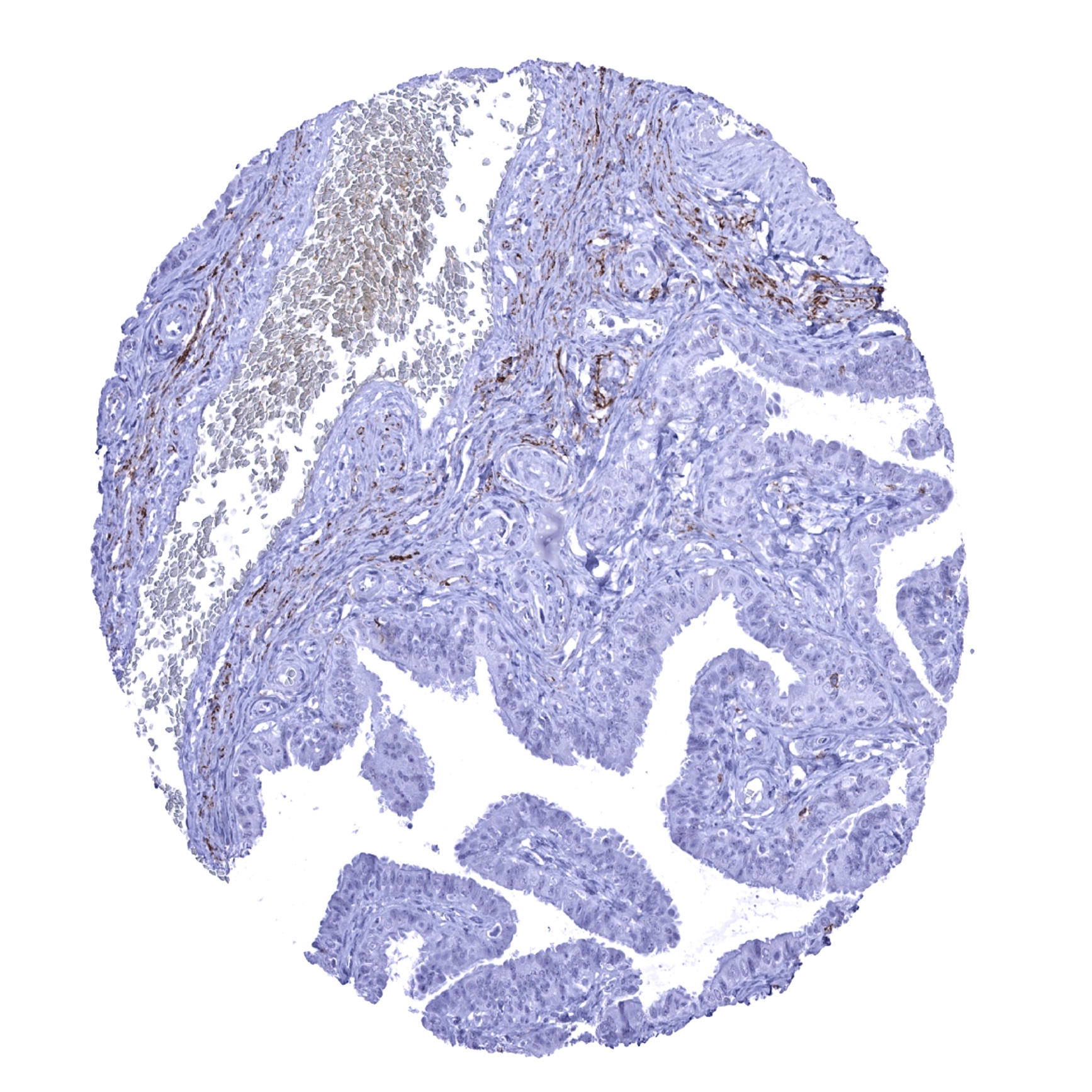
Uterus, endocervix
Uterus, endometrium (pregnancy) - CD56 positive NK-cells are particularly frequent in the endometrium_
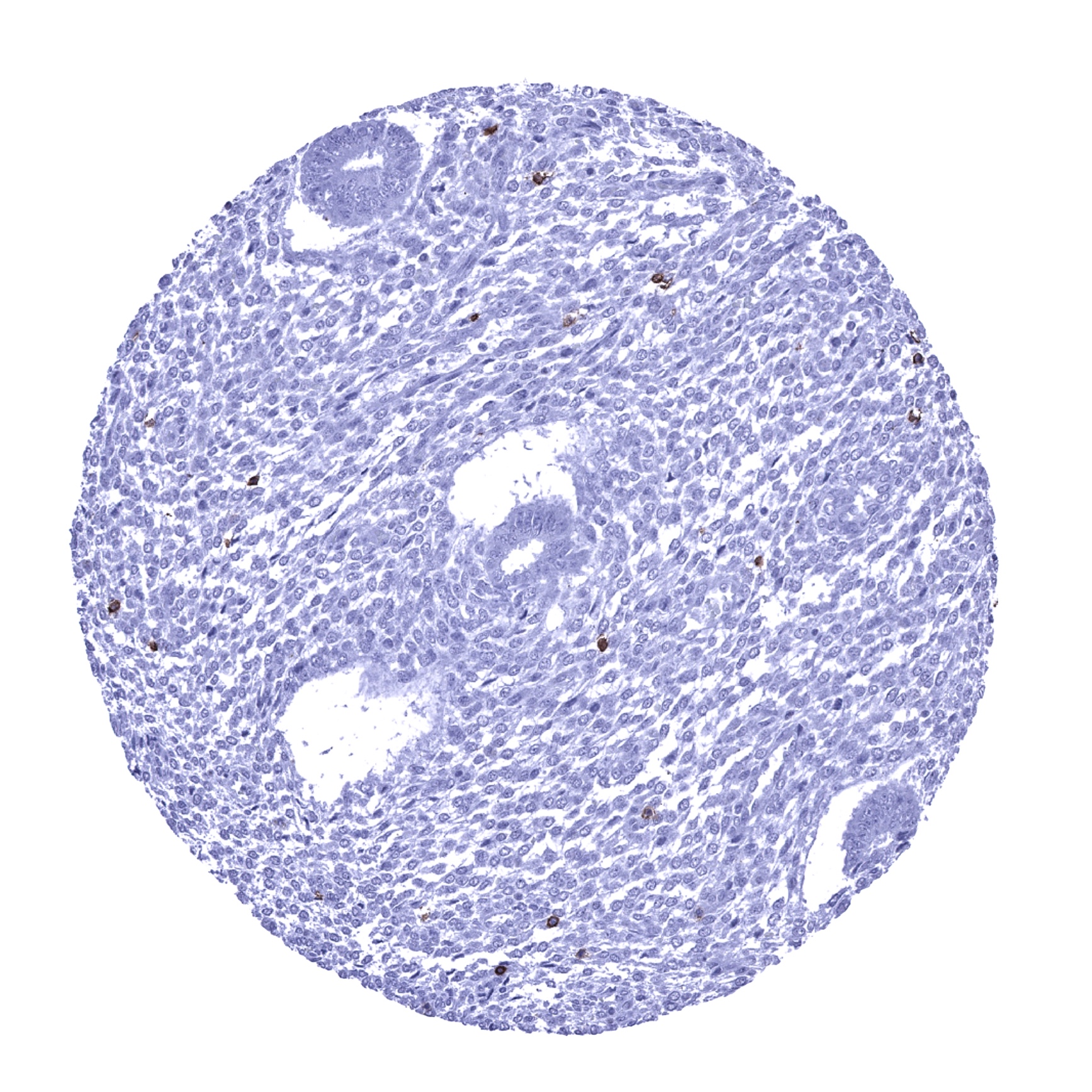
Uterus, endometrium (proliferation) - CD56 positive NK-cells regularly occur in the endometrium.
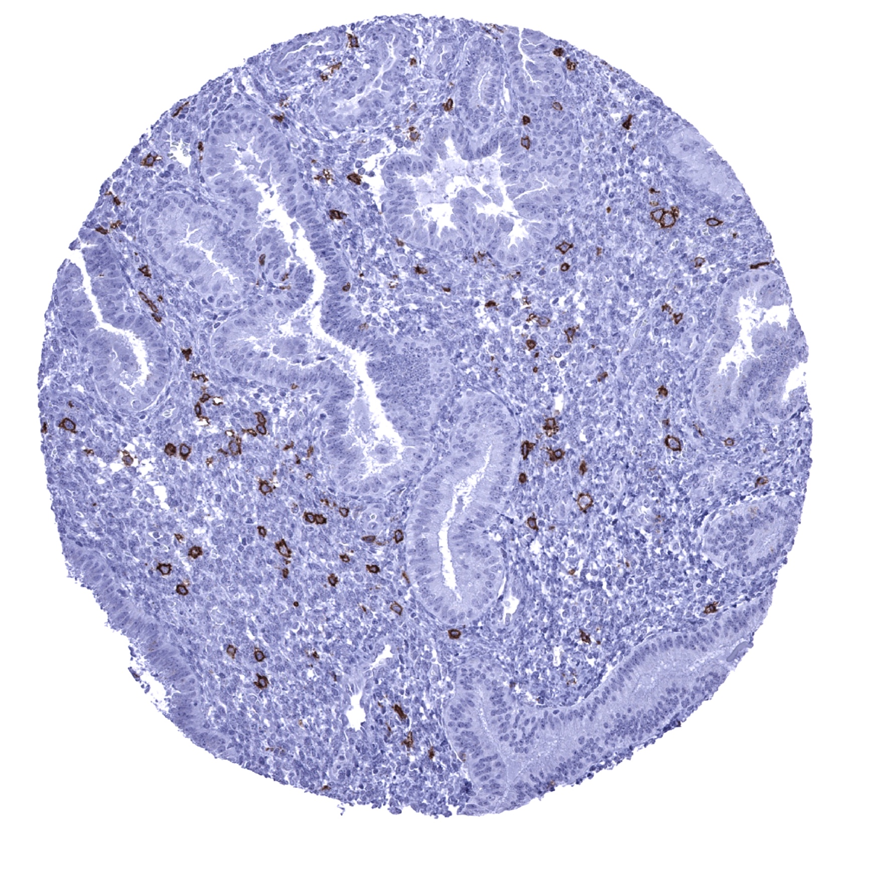
Uterus, endometrium (secretion) - CD56 positive NK-cells are particularly frequent in the endometrium.

Uterus, myometrium - A weak to moderate CD56 immunostaining occurs in muscle fibres of the myometrium