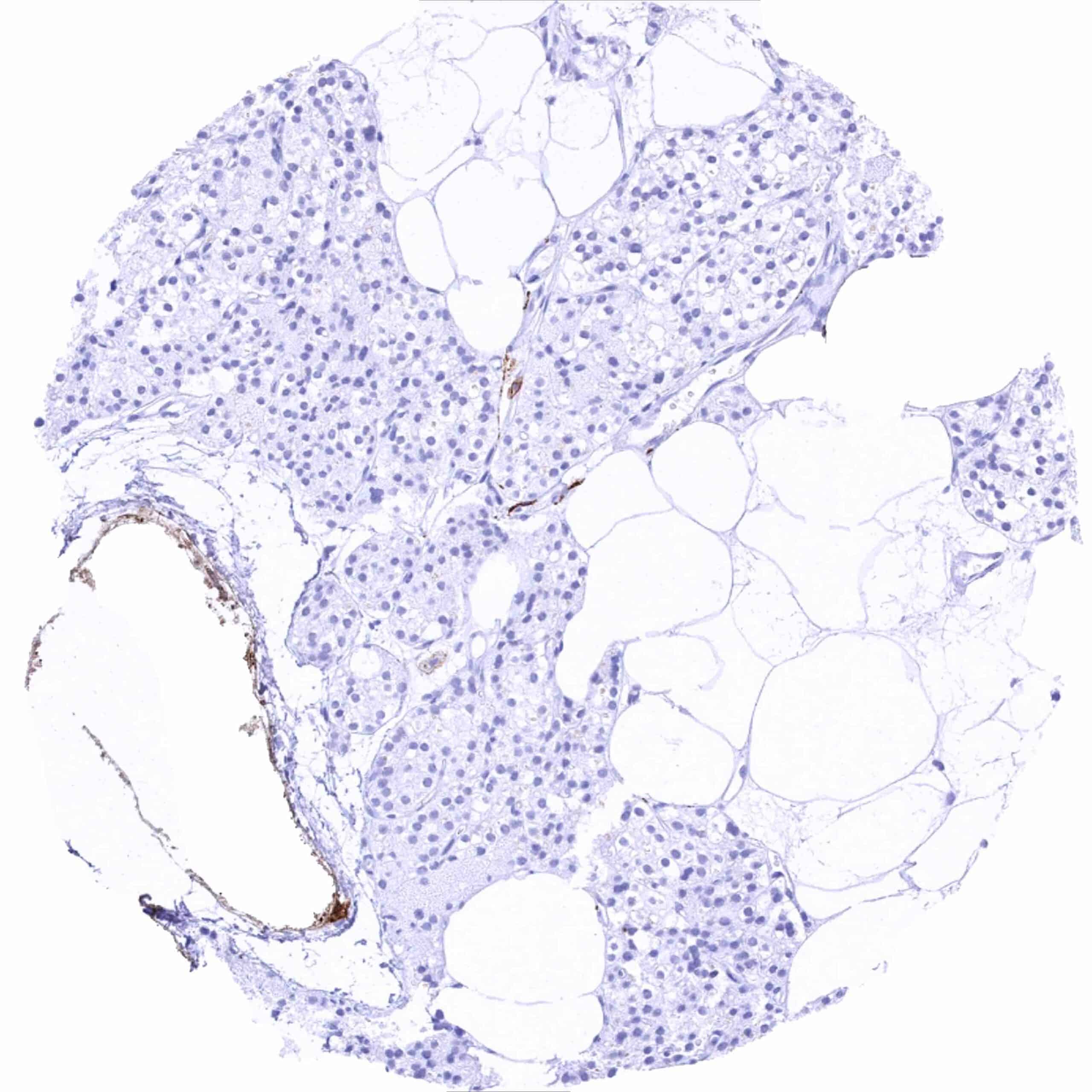
Adrenal gland – Strong, predominantly membranous L1CAM staining of a medullary cells
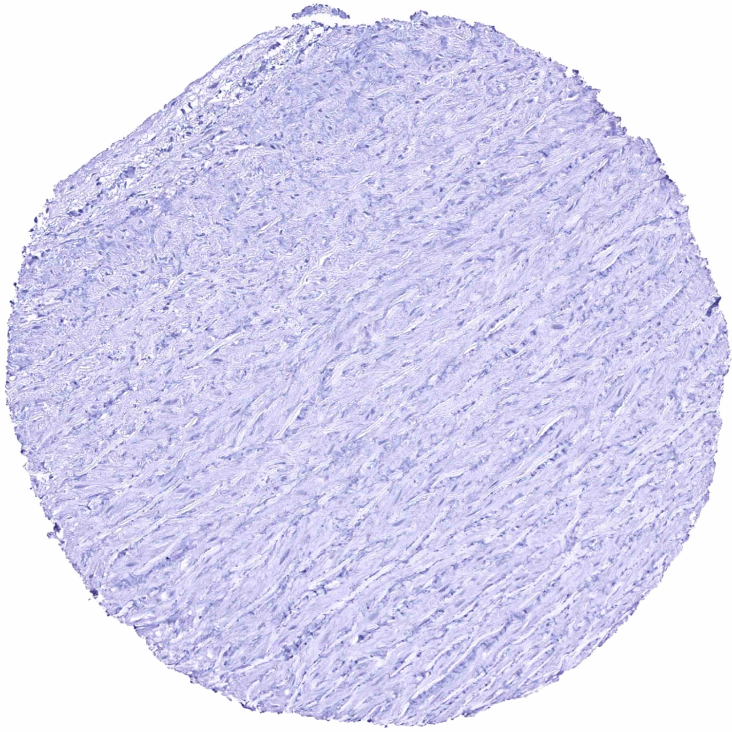
Aorta, mediaAorta, media
Appendix, mucosa – Distinct L1CAM staining of nerve fibres
Appendix, muscular wall – Strong L1CAM staining of nerve fibres and ganglia
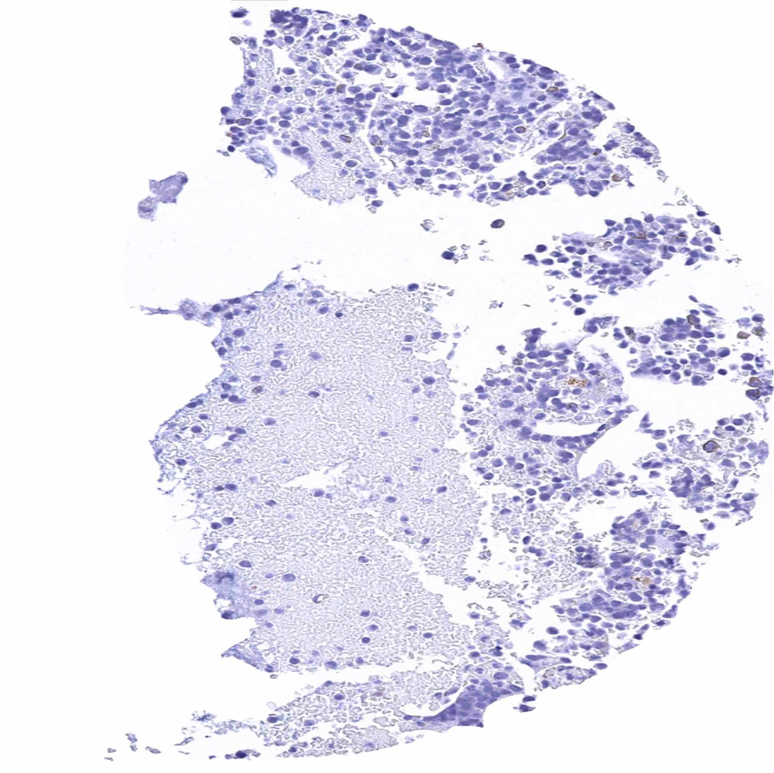
Bone marrow
Breast
Bronchus, mucosa
Cerebellum (white matter)
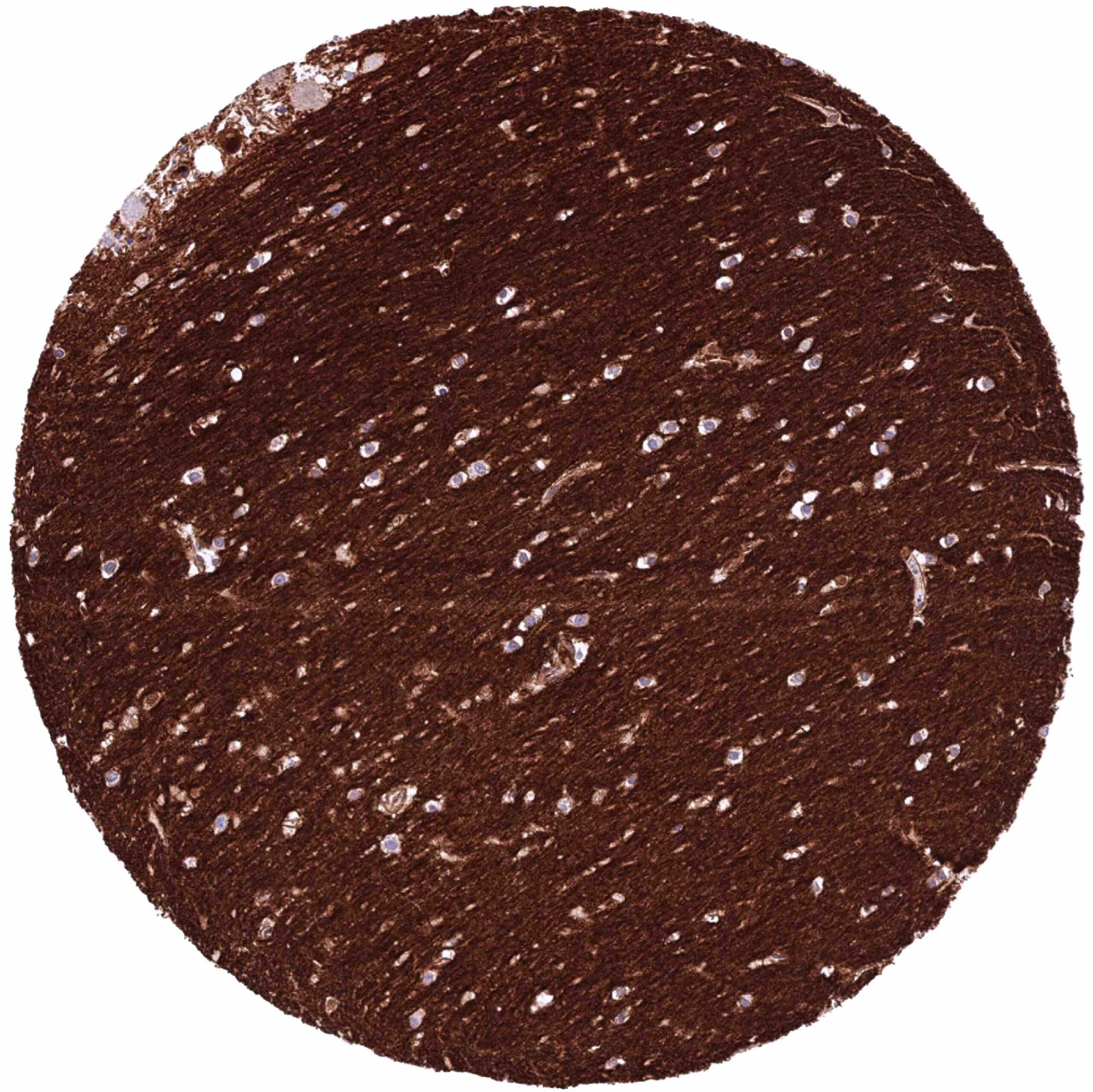
Cerebellum, cortex (molecular layer) – Intense L1CAM staining, especially of nerve fibres
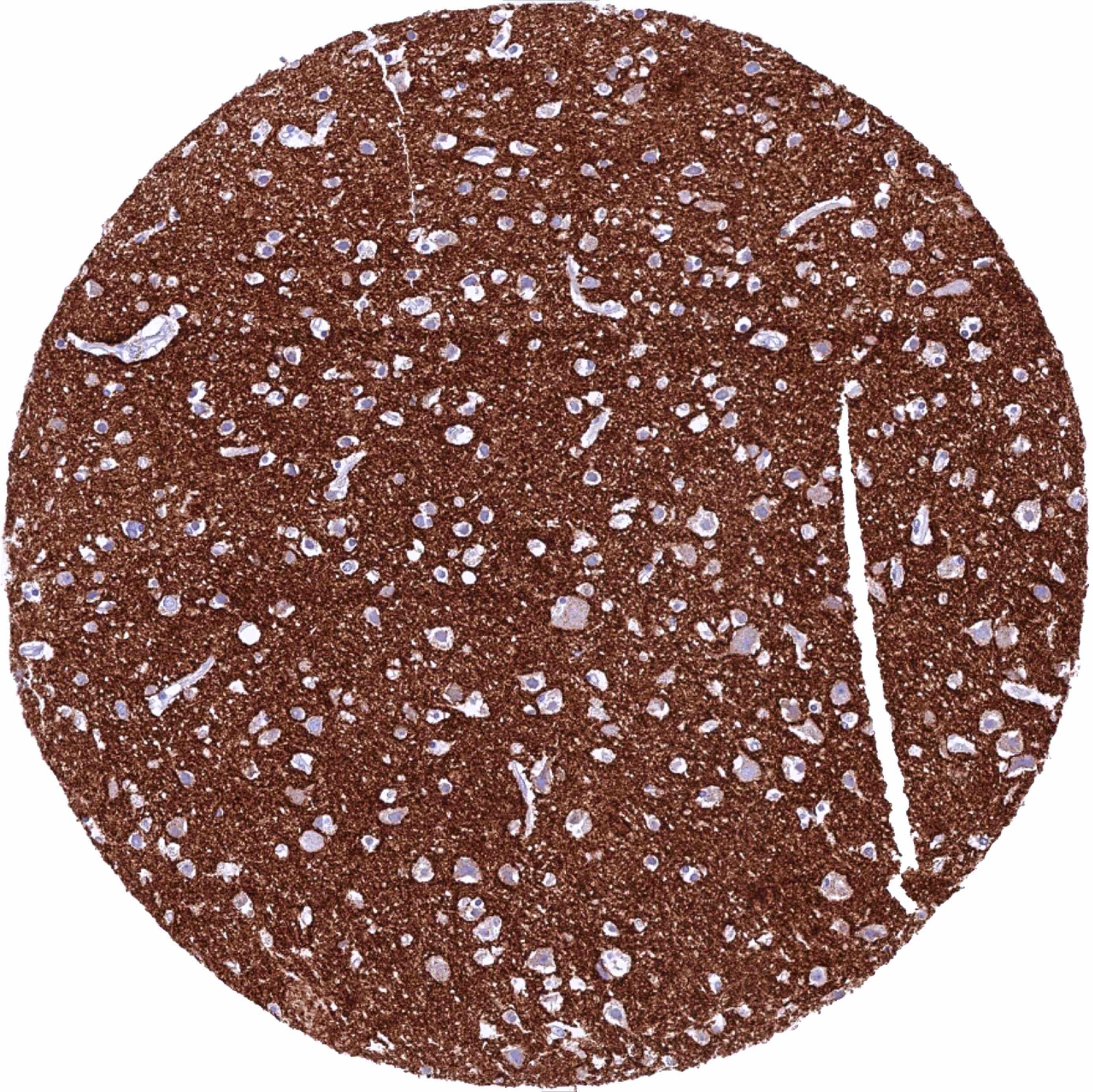
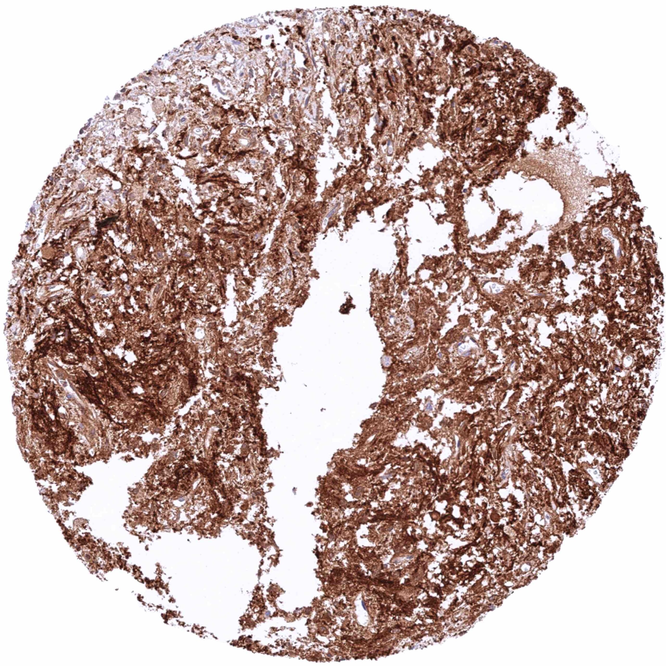
Cerebrum, grey matter – Intense L1CAM staining, especially of nerve fibres
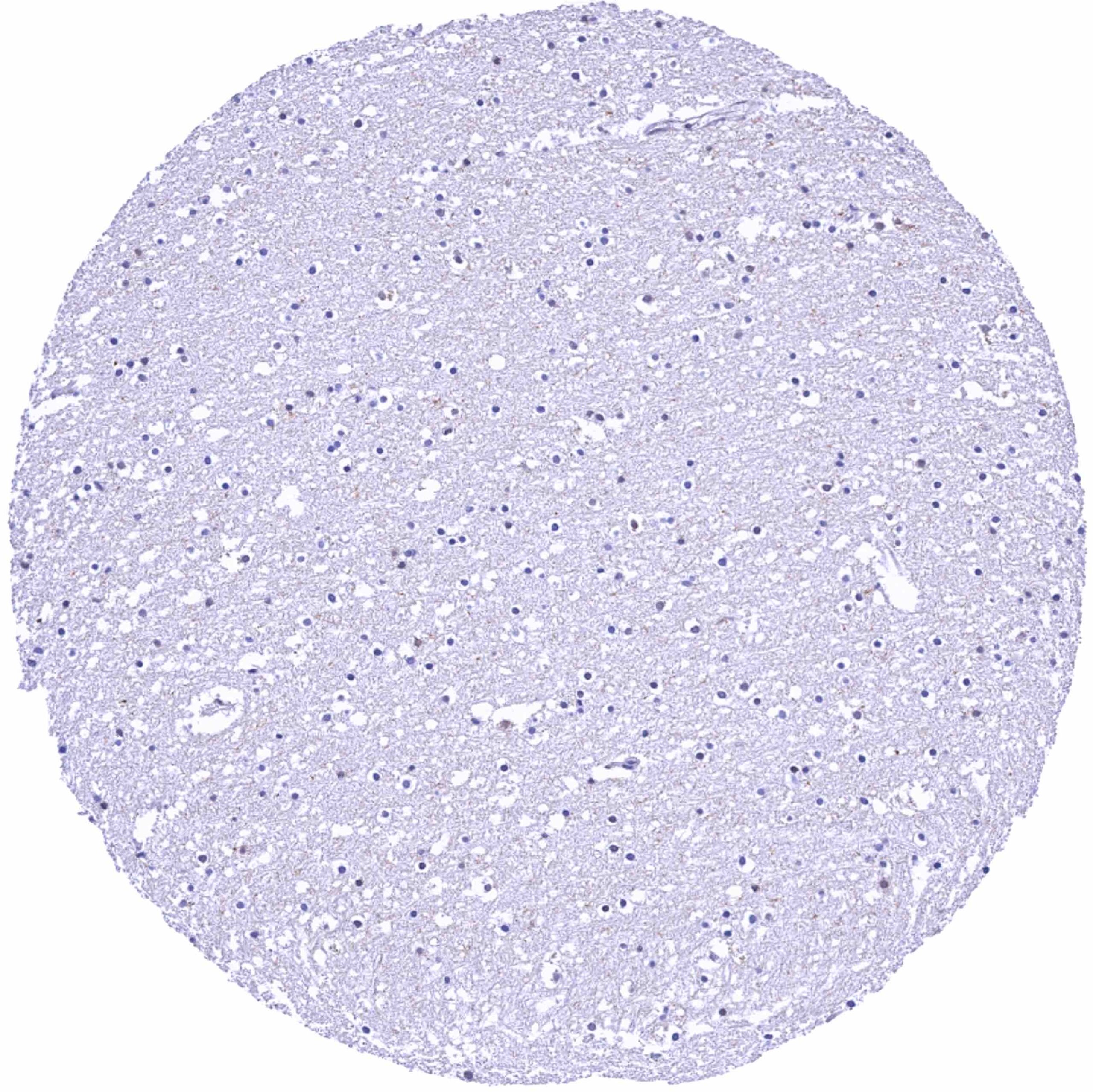
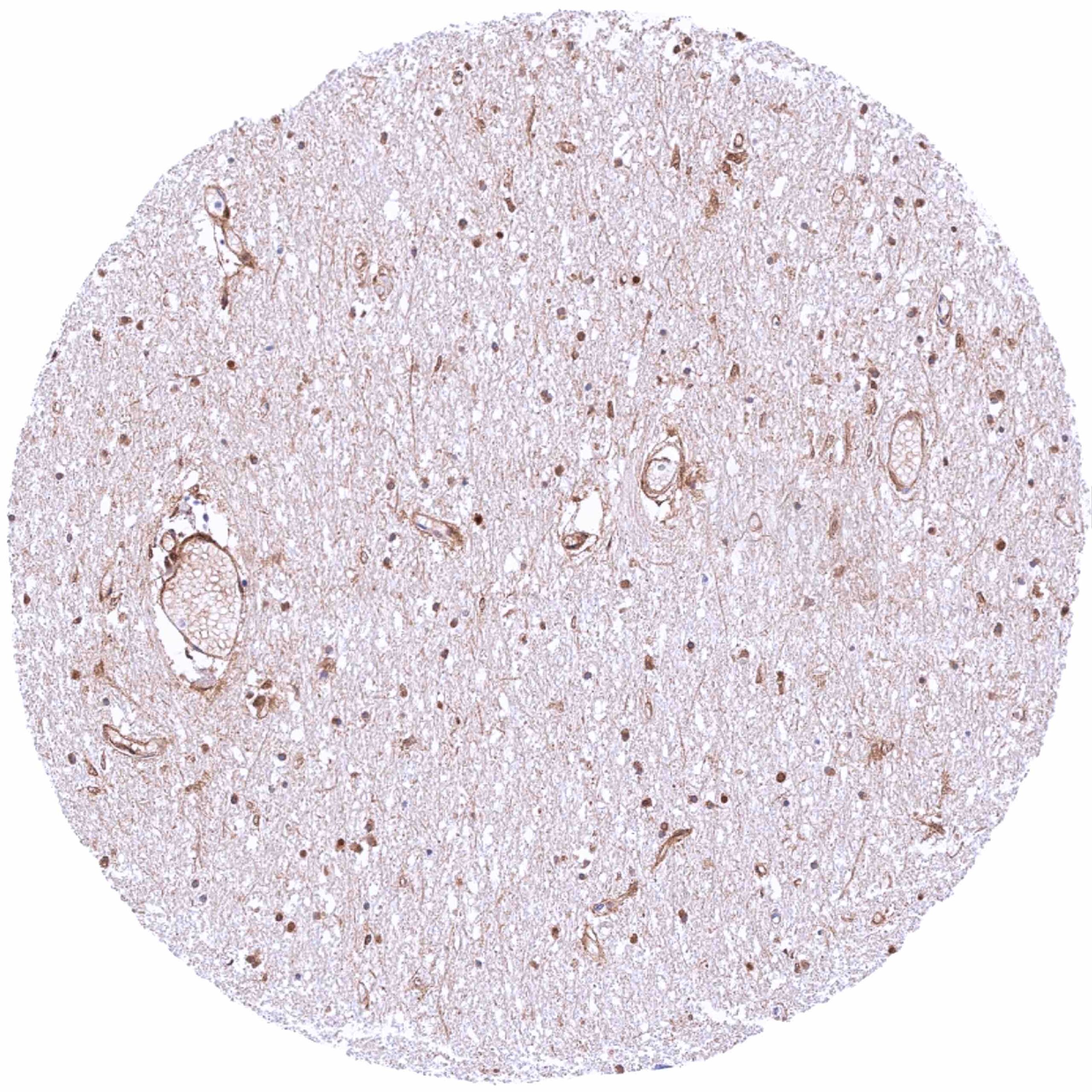
Cerebrum, white matter
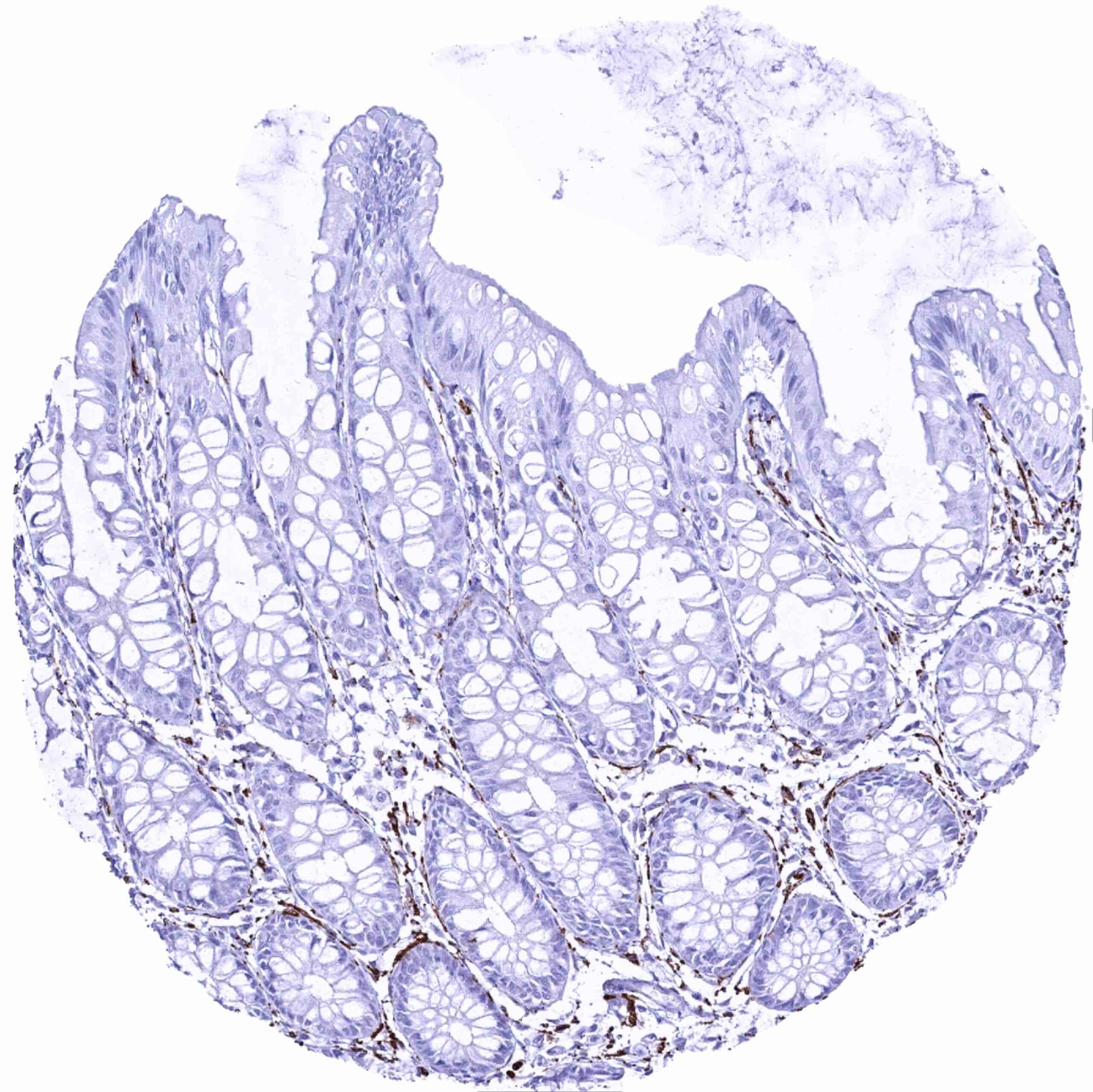
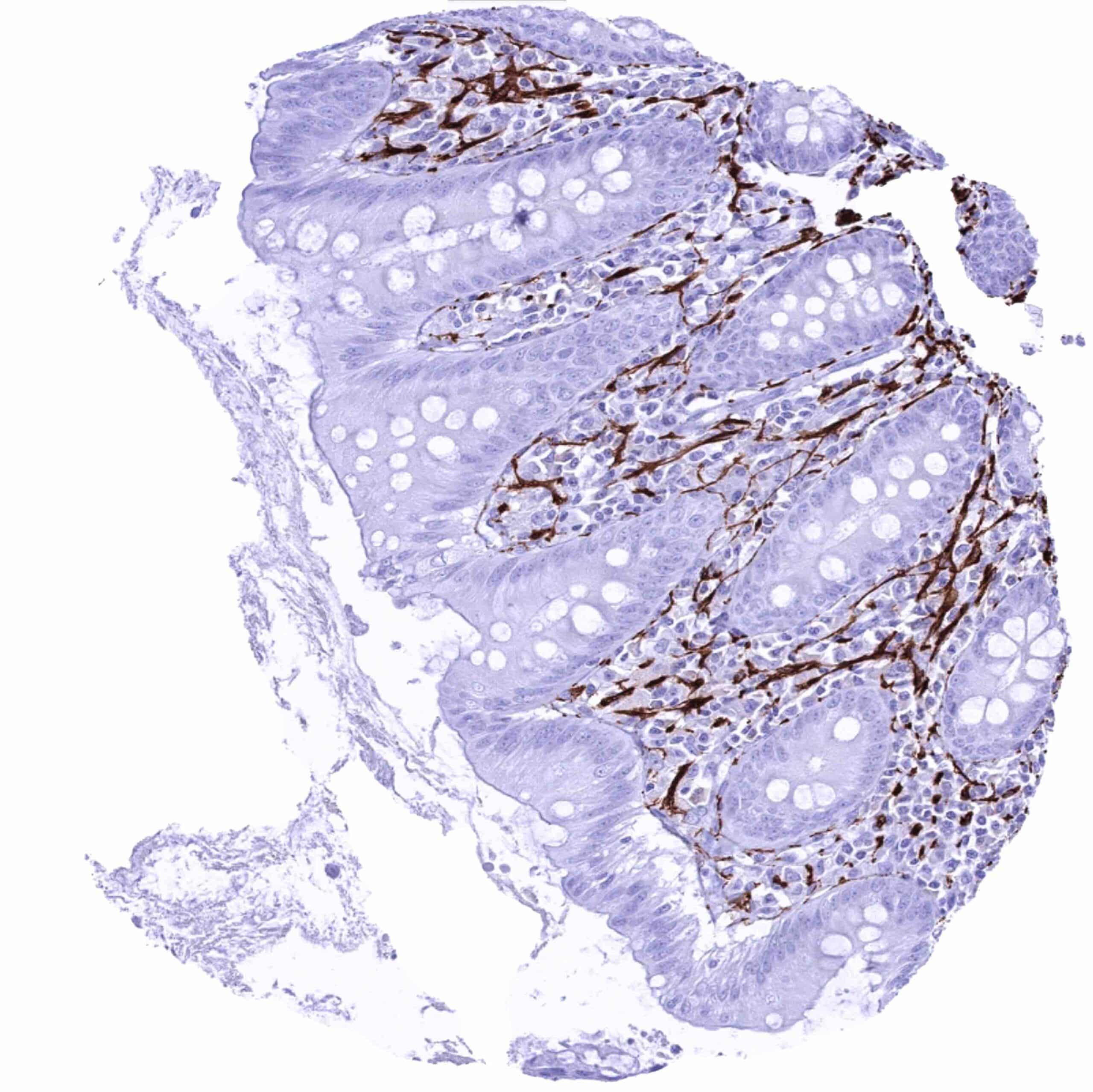
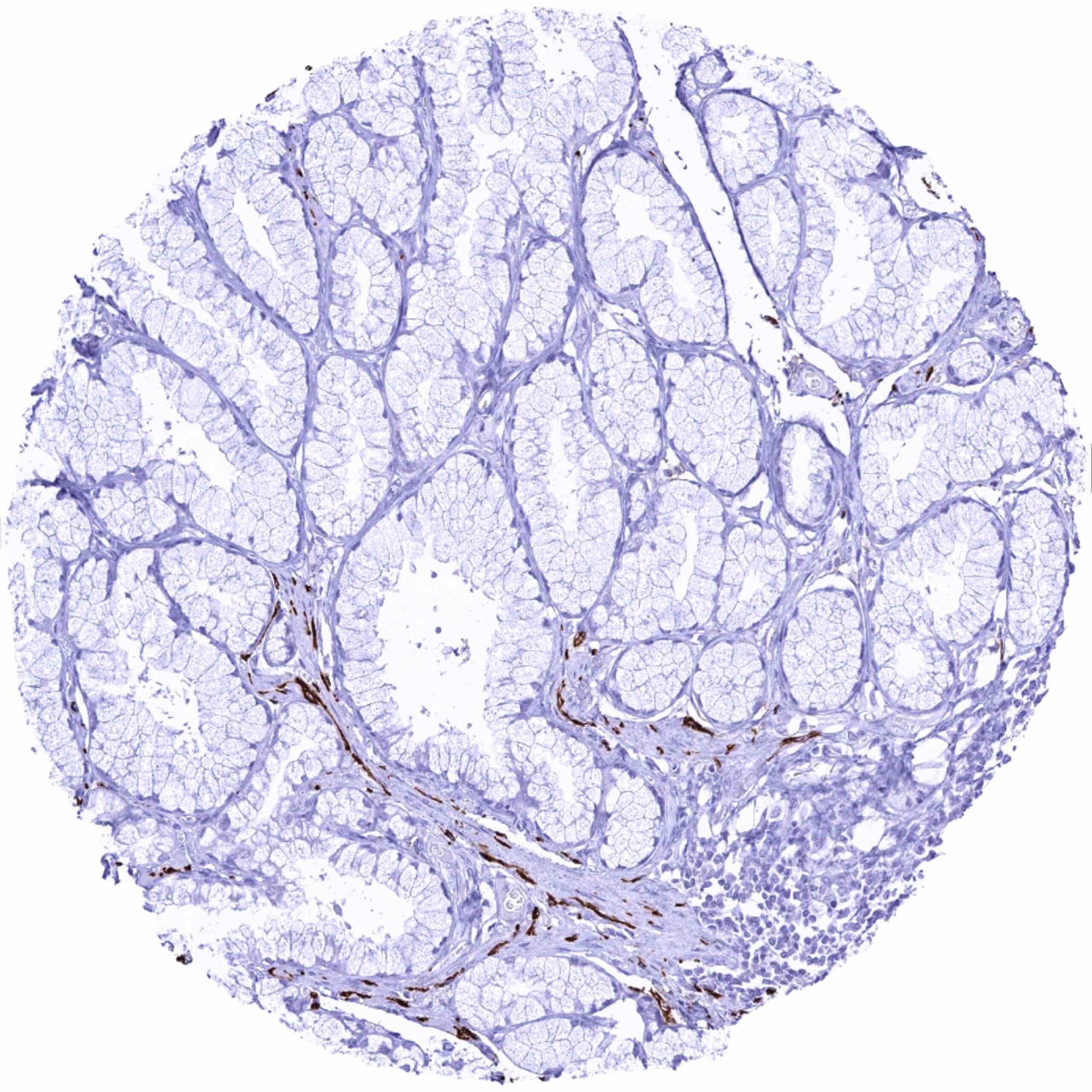
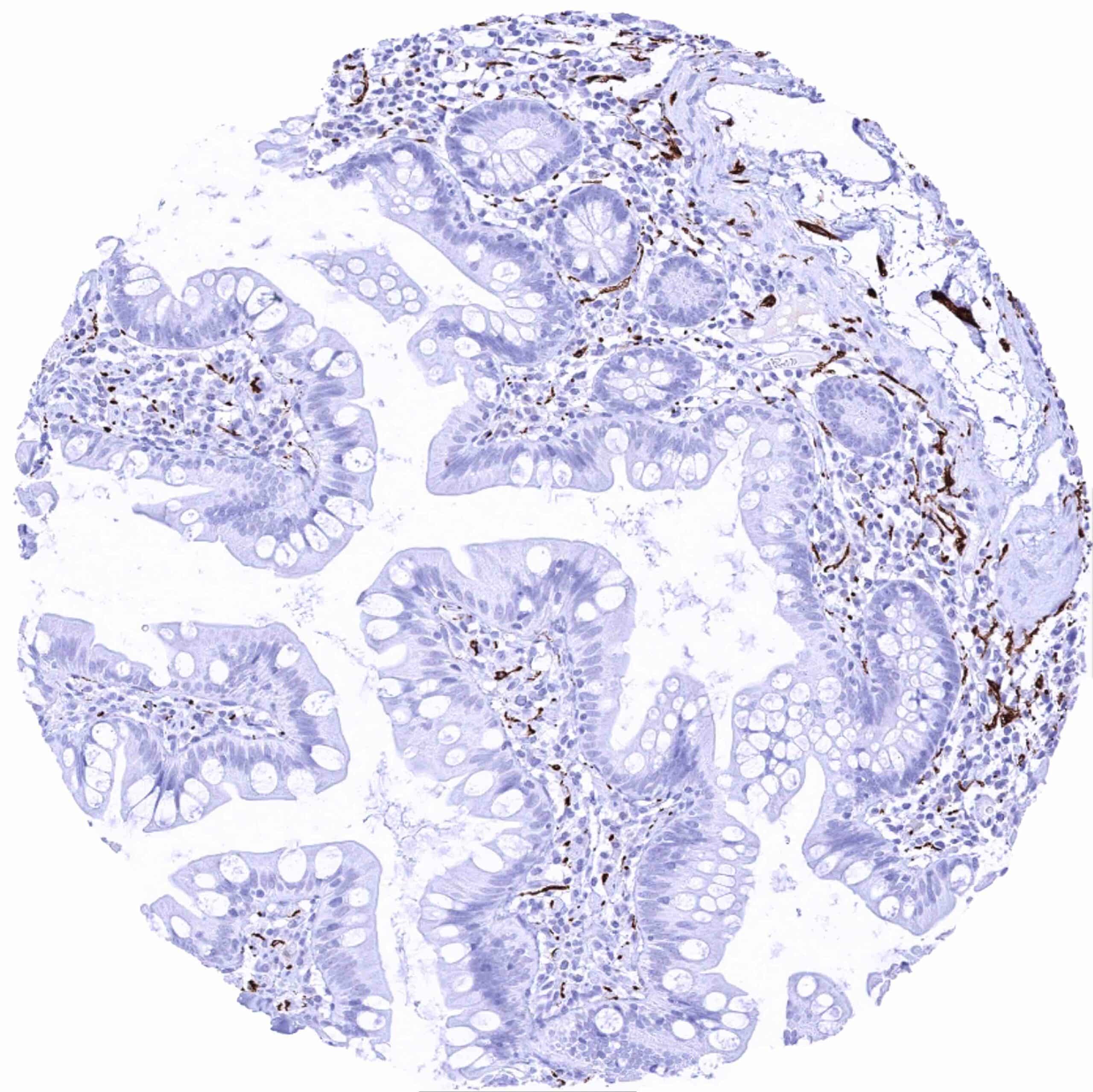
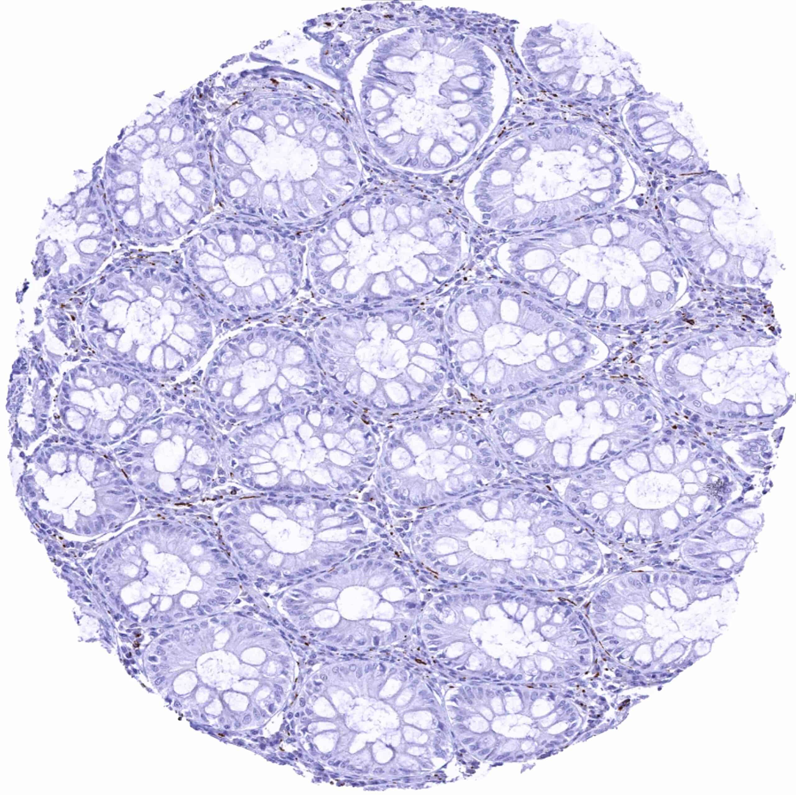
Colon descendens, mucosa – Distinct L1CAM staining of nerve fibres
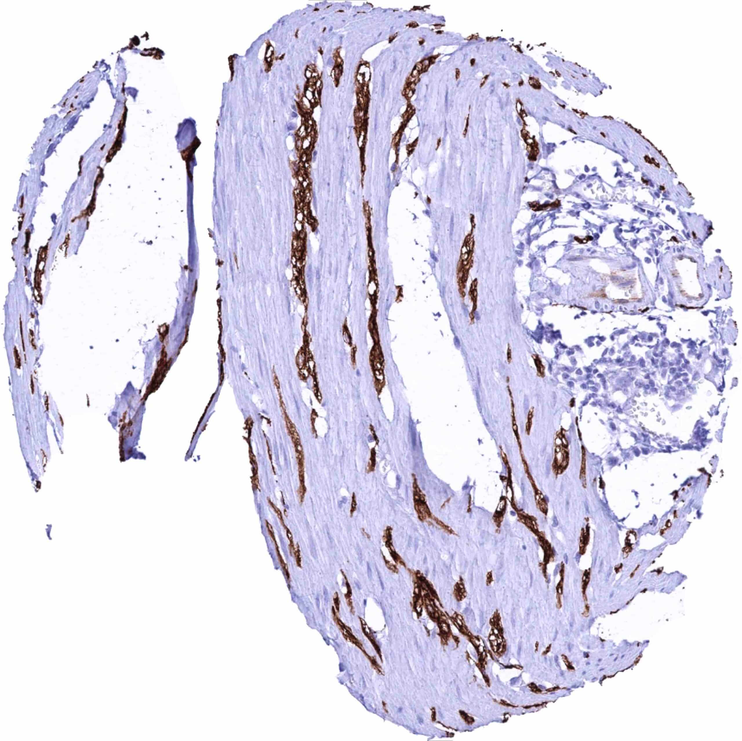
Colon descendens, muscular wall – Strong L1CAM staining of nerve fibres and ganglia
Duodenum, Brunner gland – Distinct L1CAM staining of nerve fibres
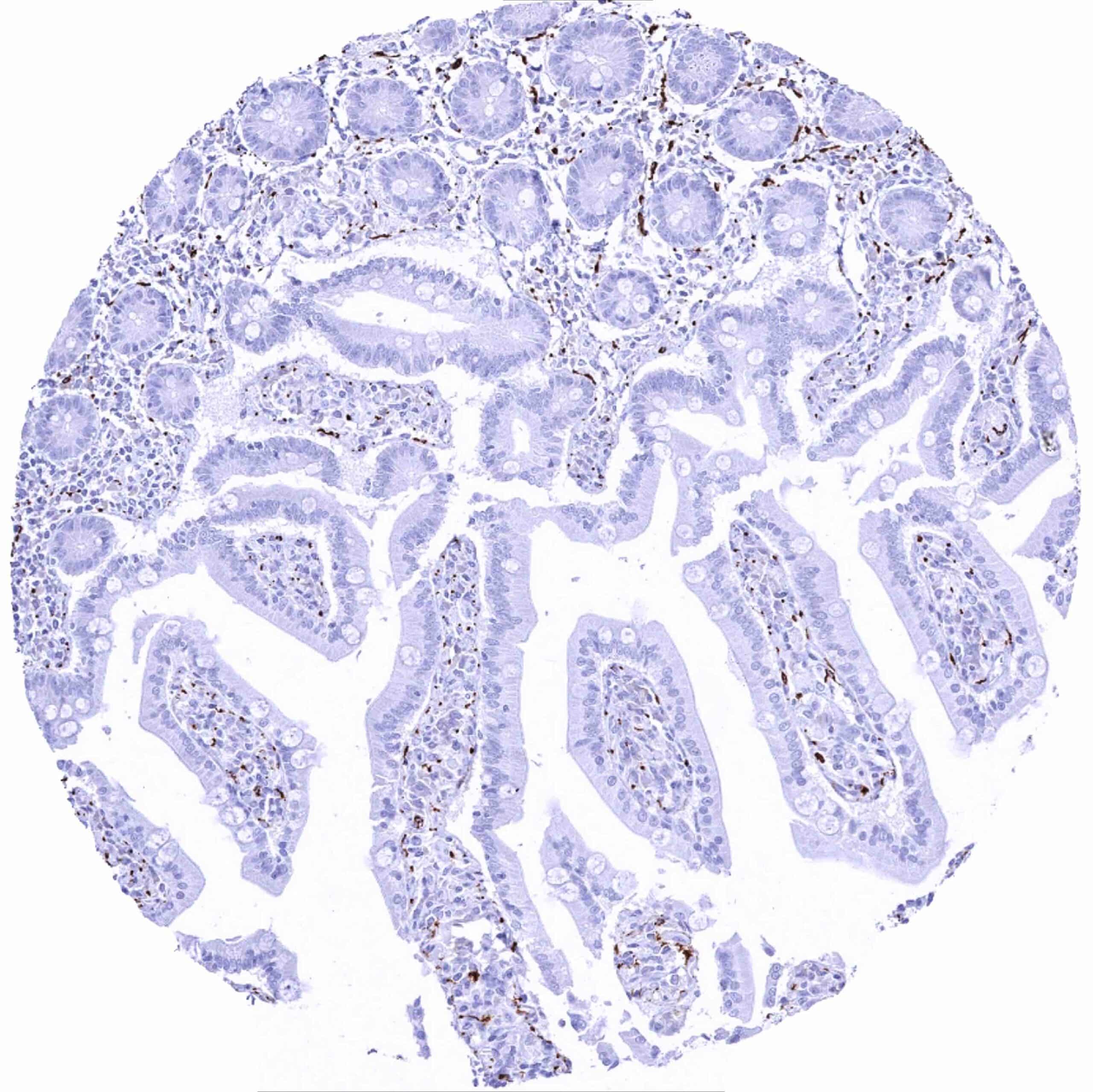
Duodenum, mucosa – Distinct L1CAM staining of nerve fibres
Epididymis (Cauda) – Strong L1CAM staining of nerve fibres
Epididymis (Corpus)
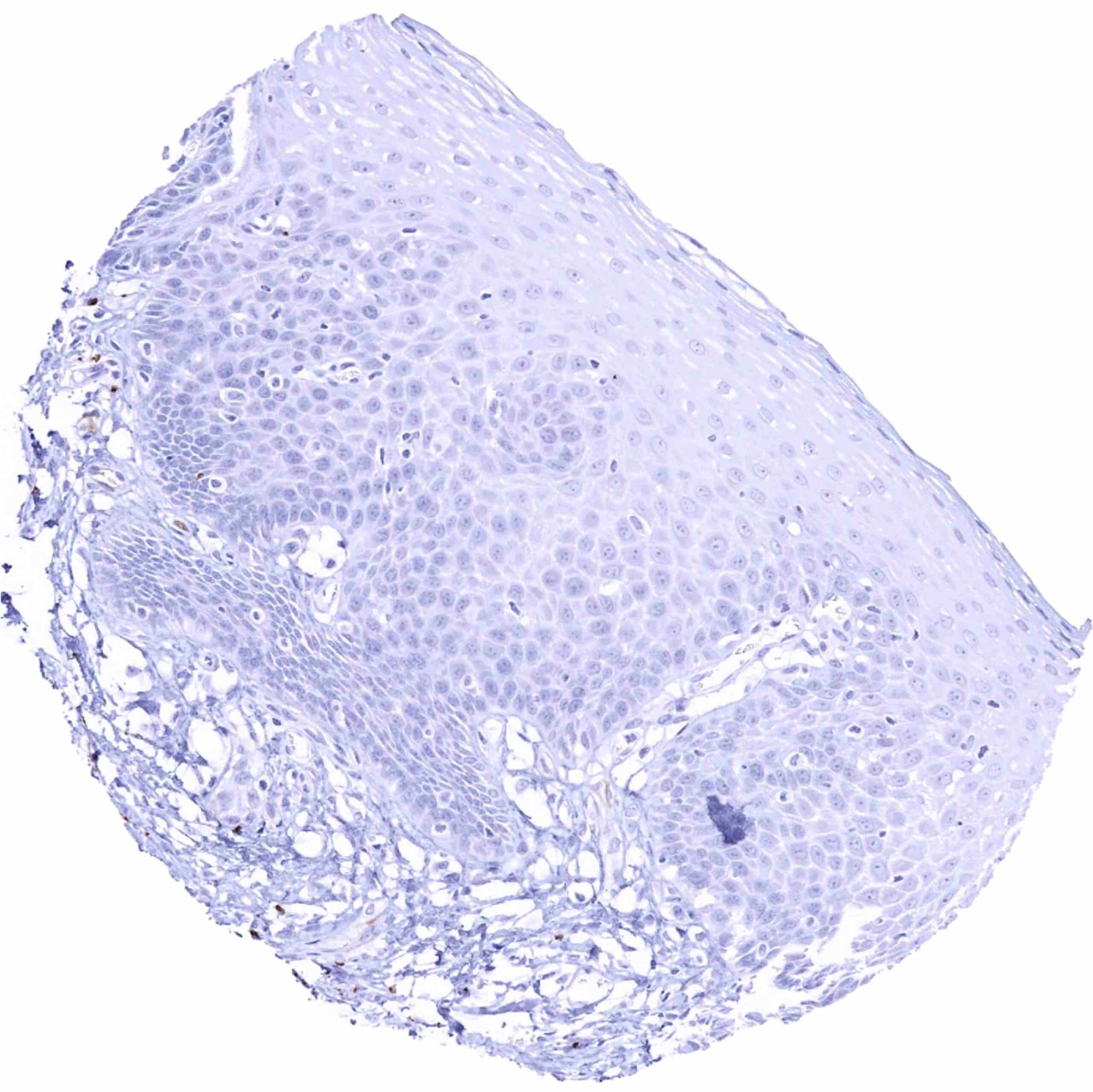
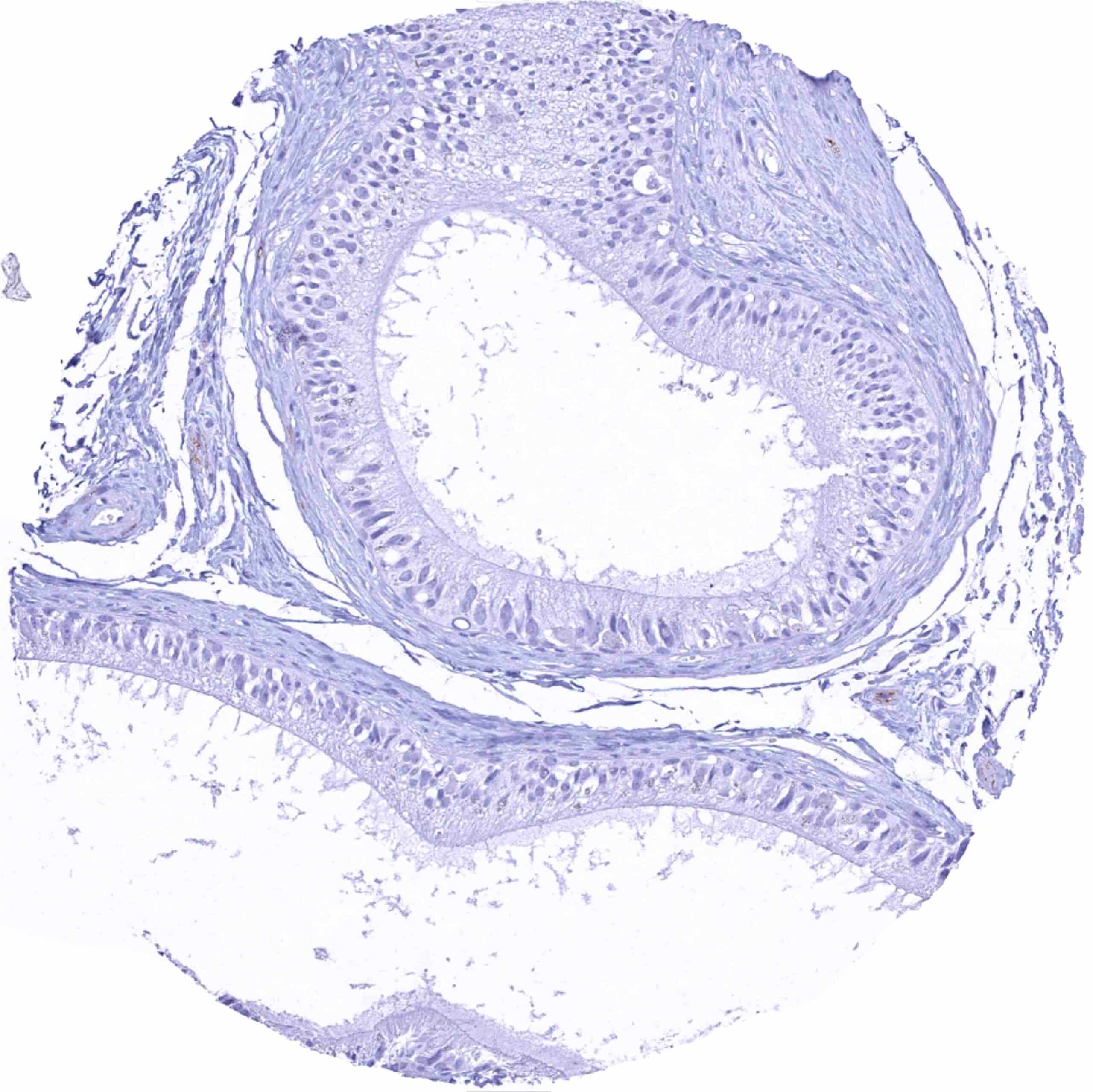
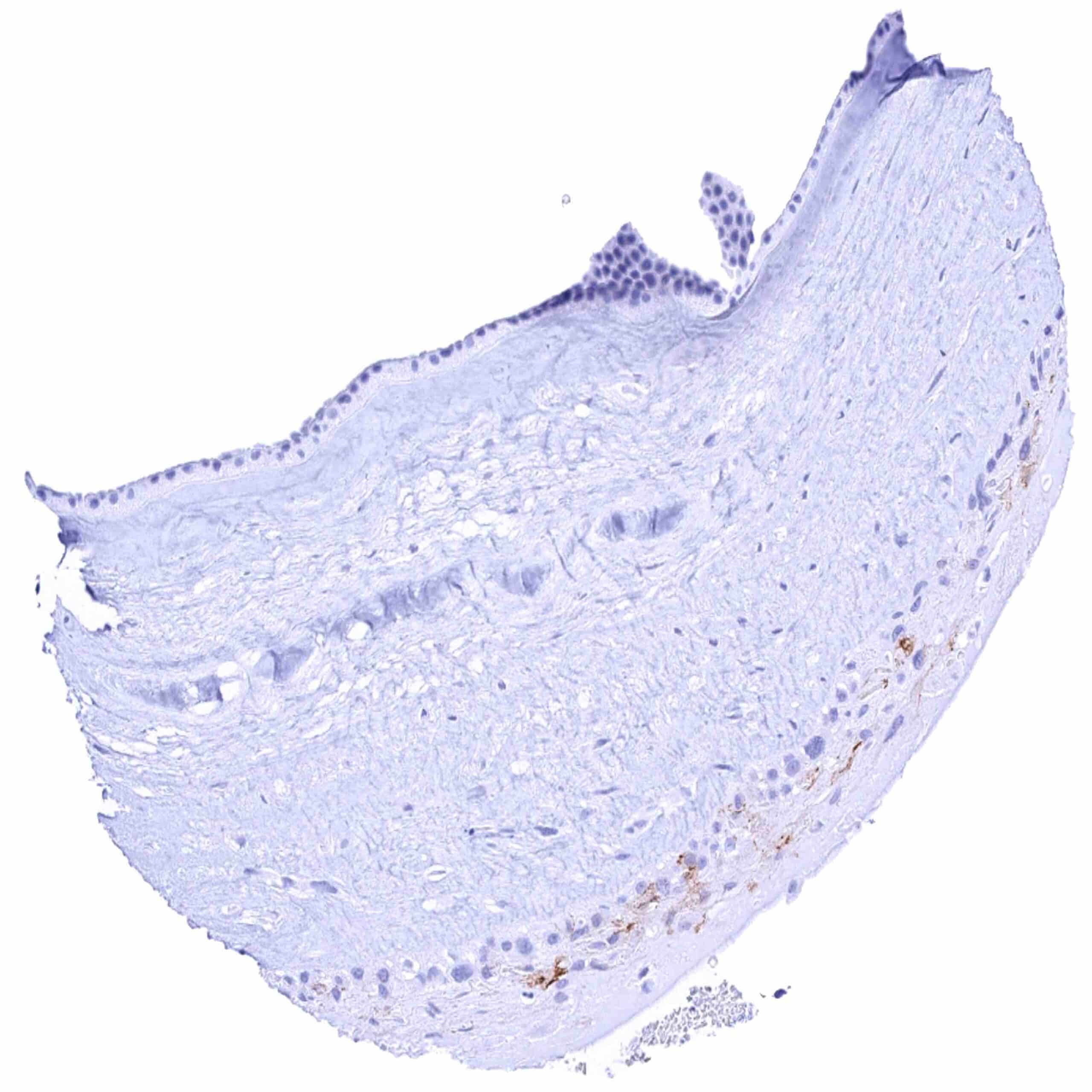
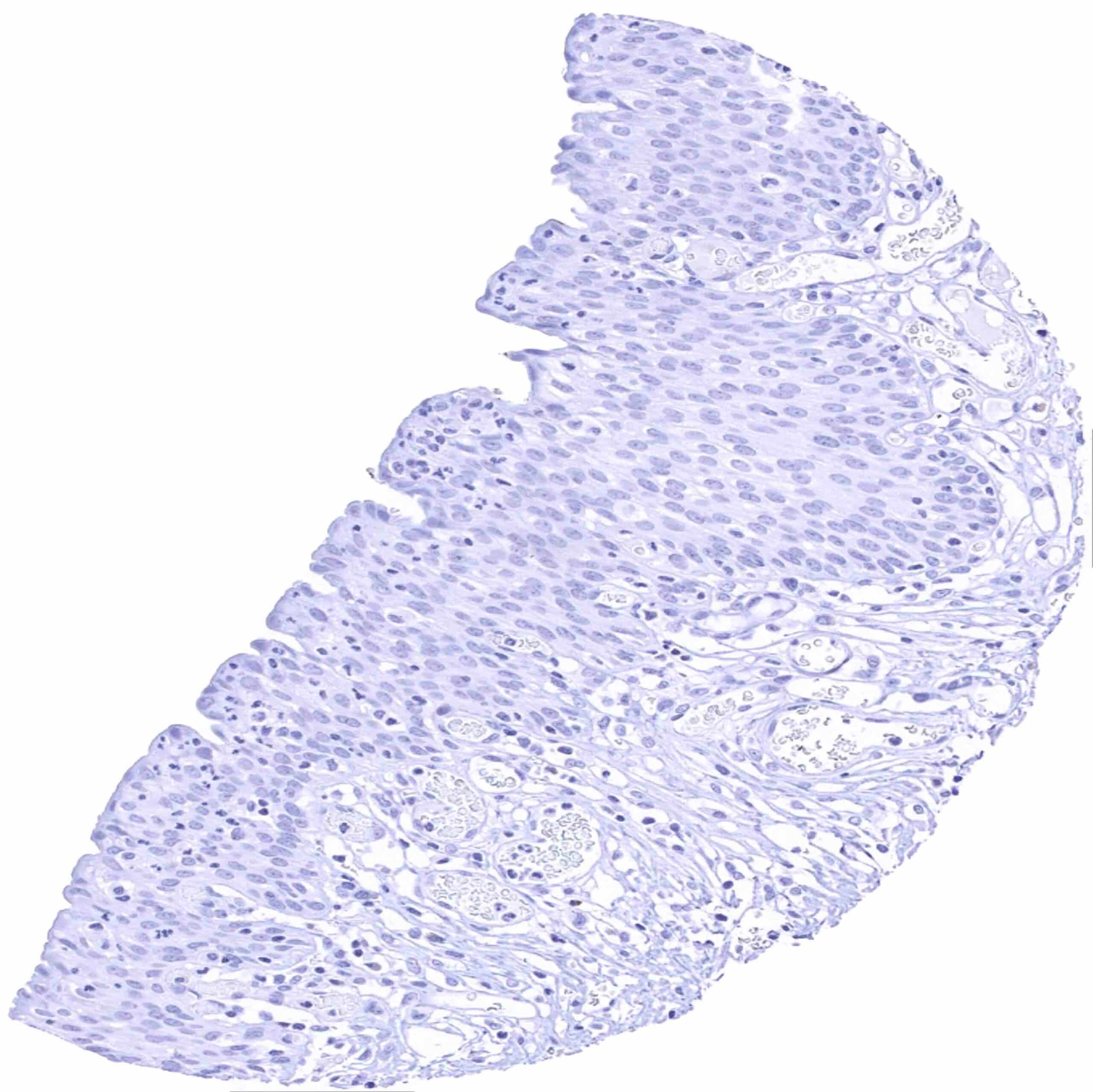
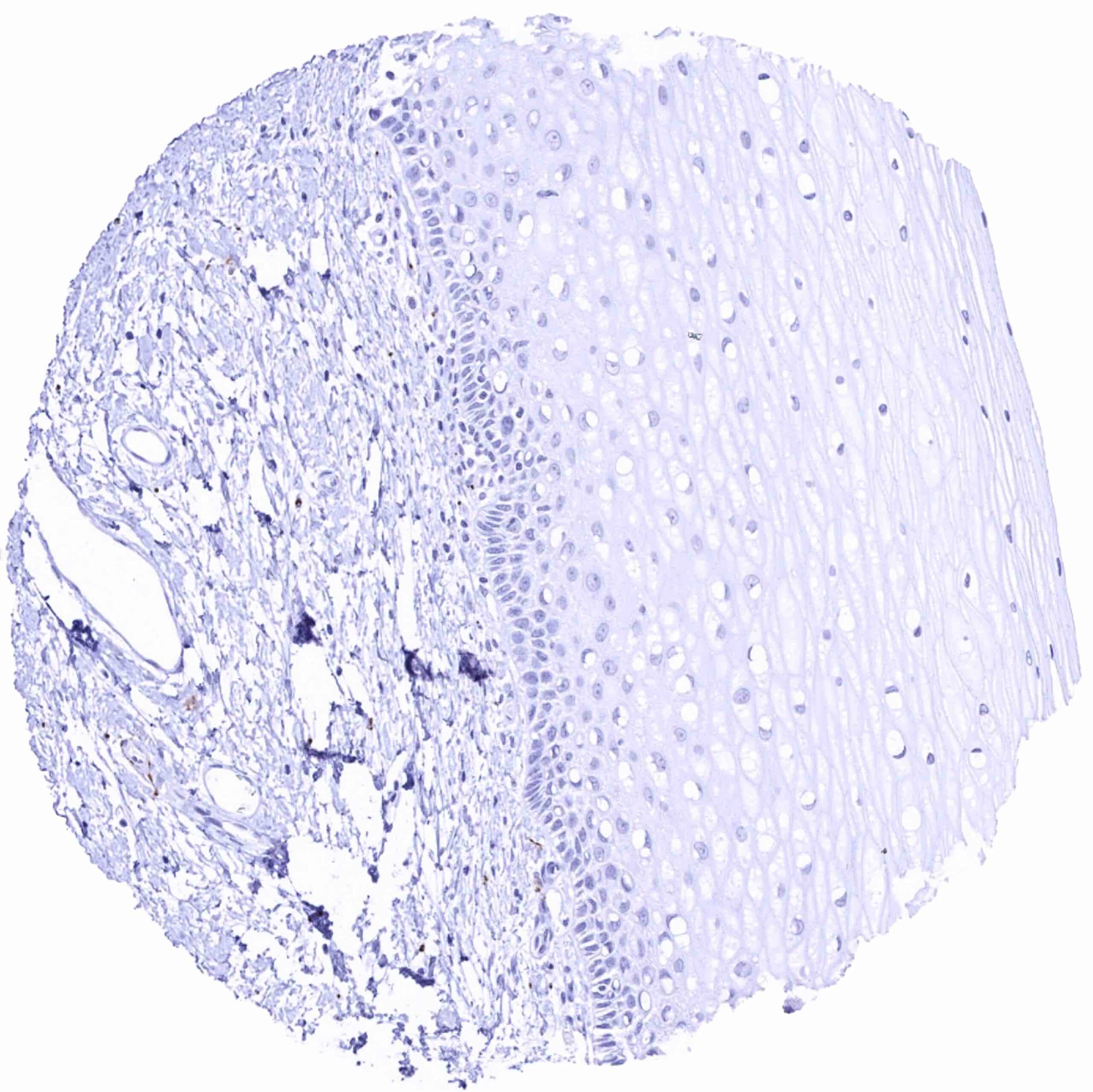
Esophagus, squamous epithelium
Fallopian tube, mucosa – Strong L1CAM staining of a larger subset of epithelial cells in this sample
Fallopian tube, mucosa – Strong L1CAM staining of a small subset of epithelial cells
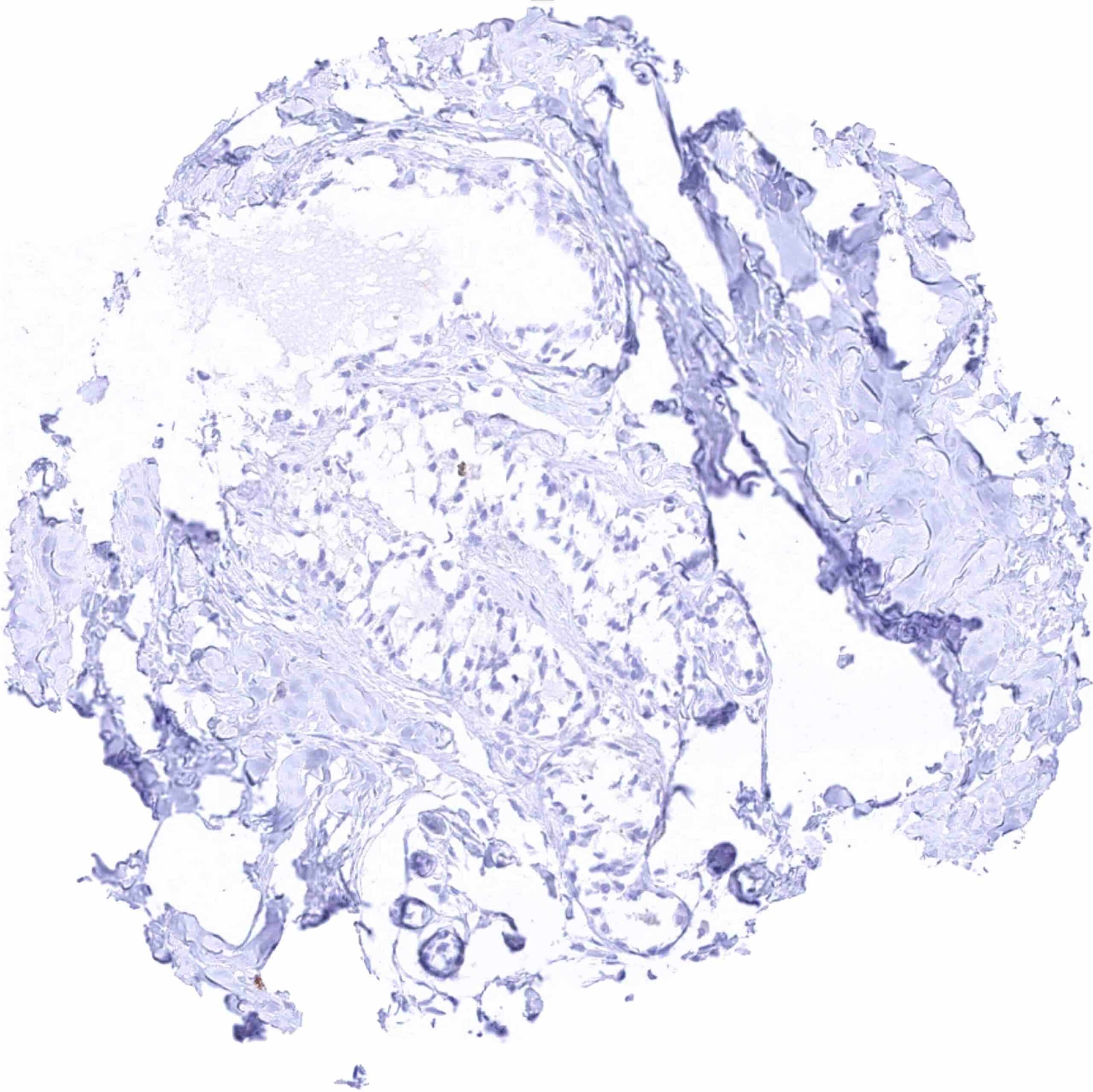
Fat
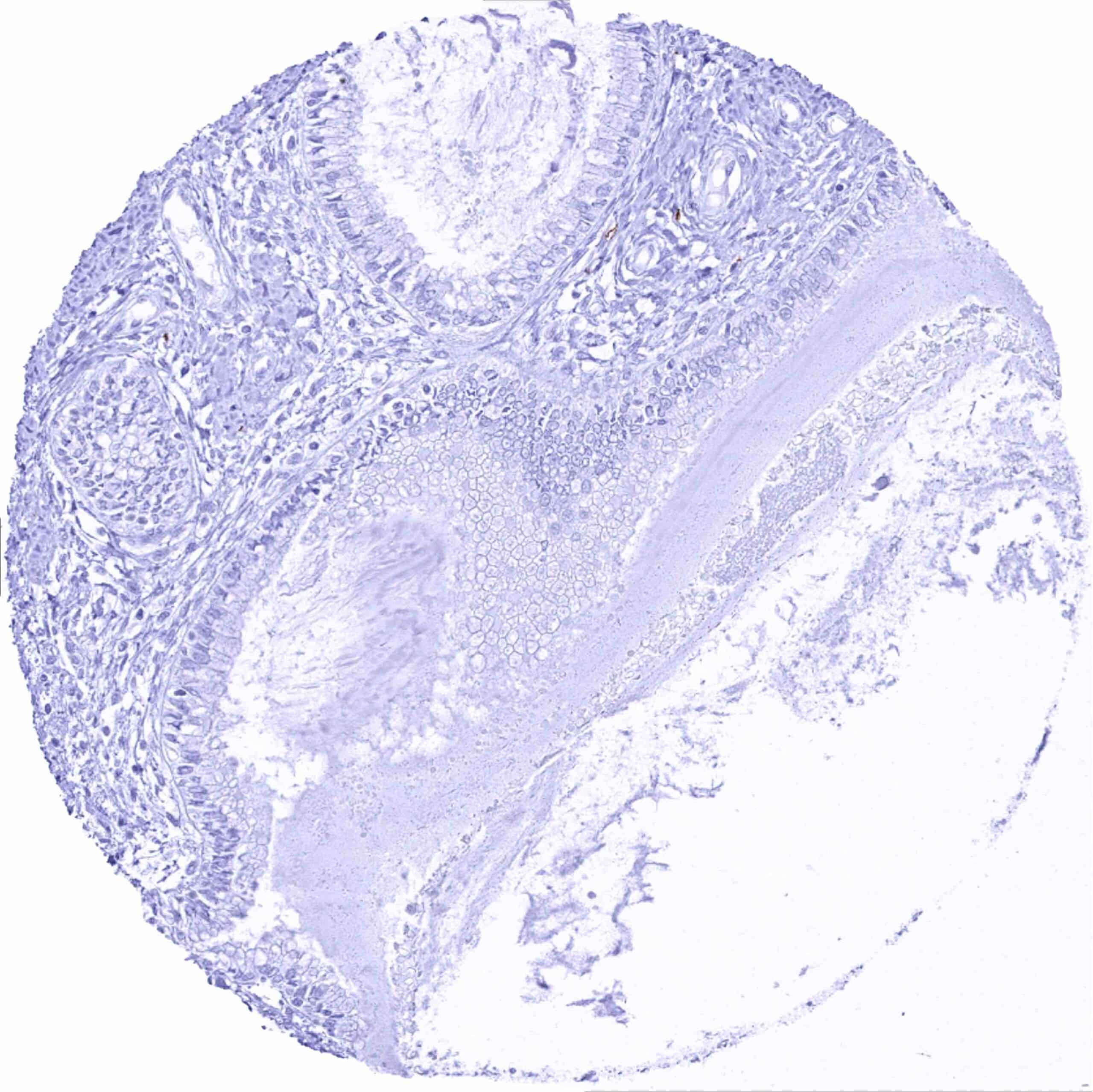
Gallbladder, epithelium
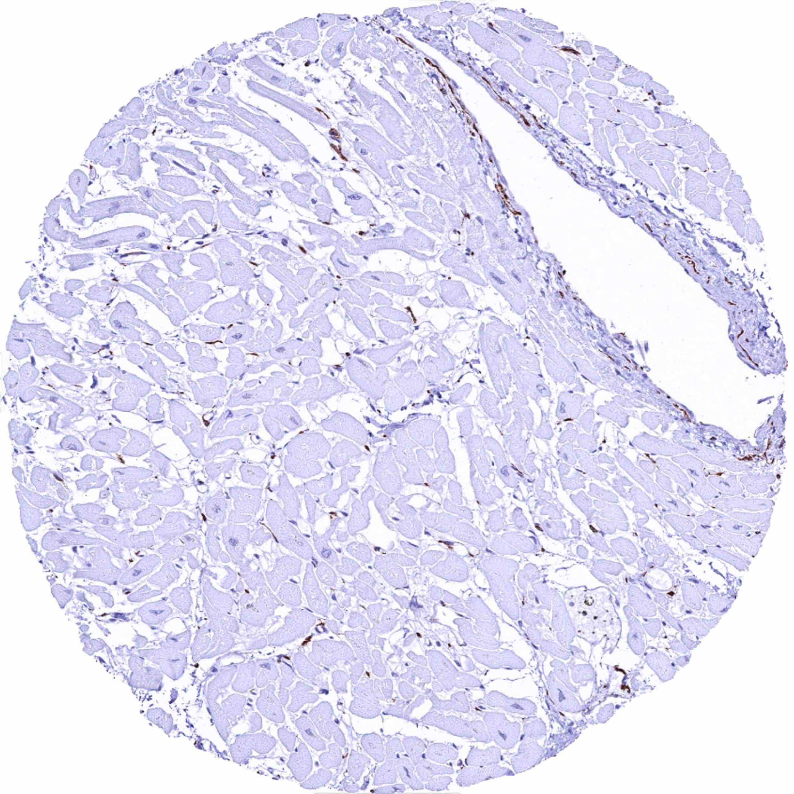
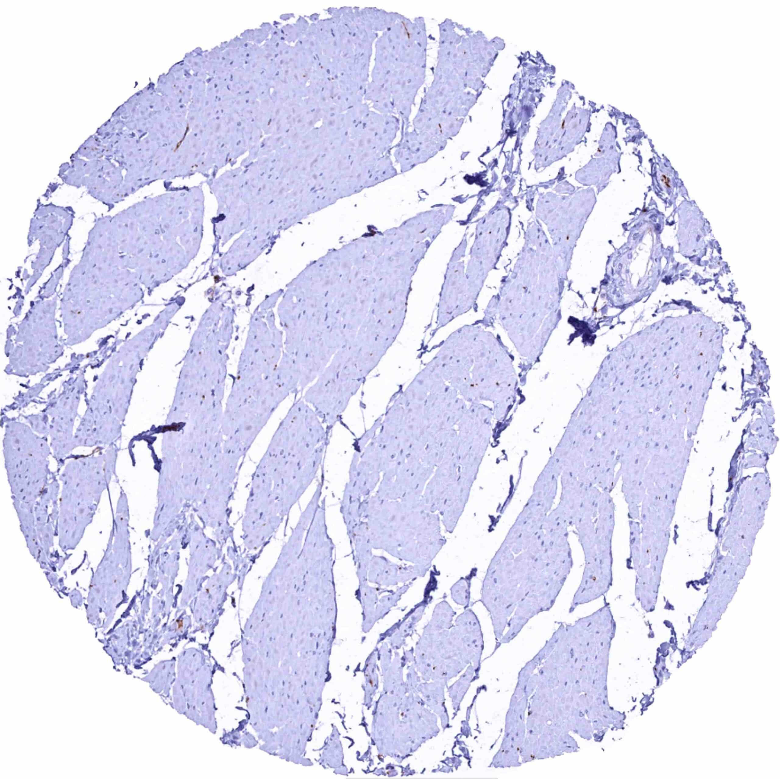
Heart muscle – Strong L1CAM staining of small nerve fibres
Ileum, mucosa – Distinct L1CAM staining of nerve fibres
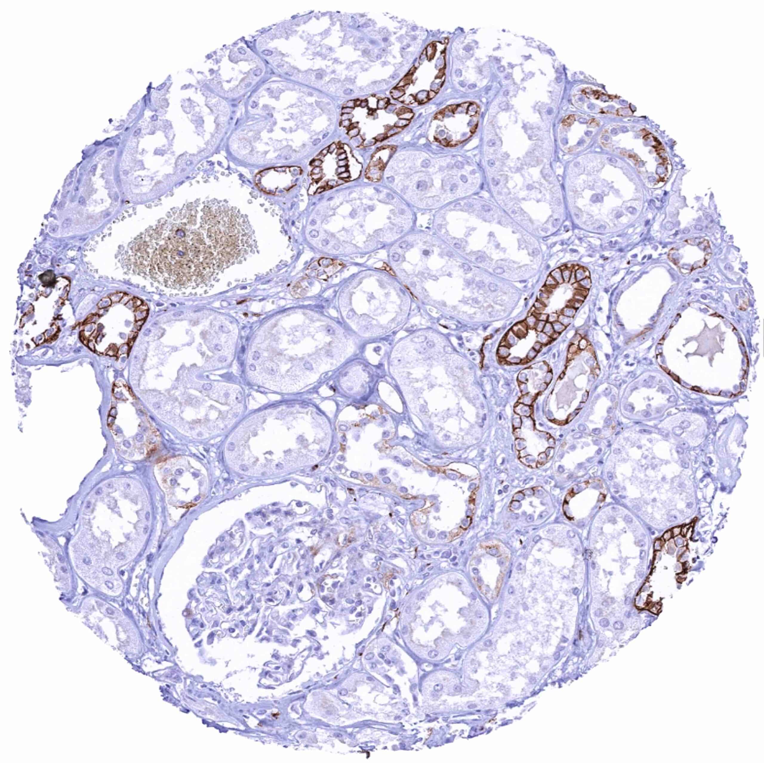
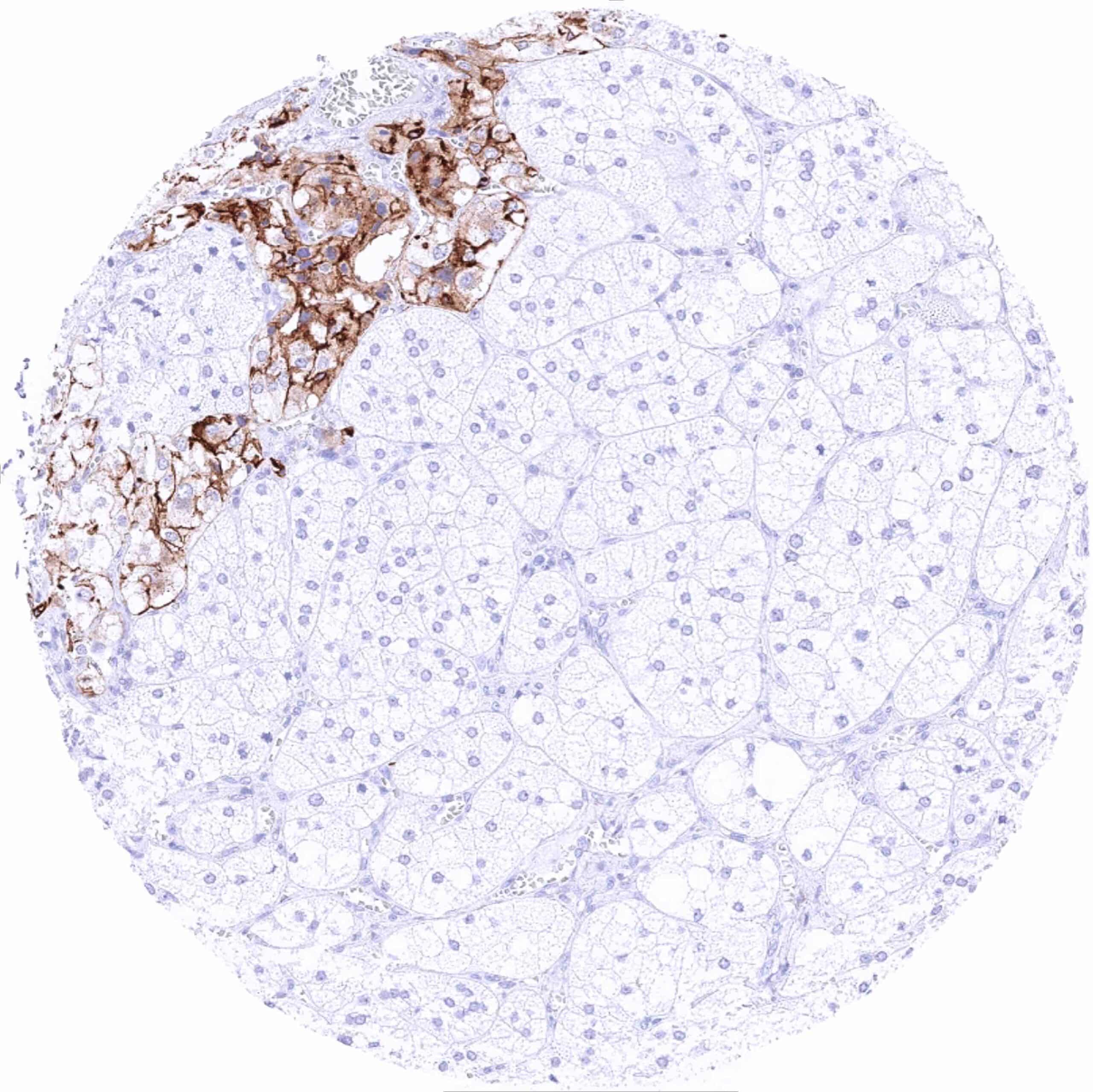
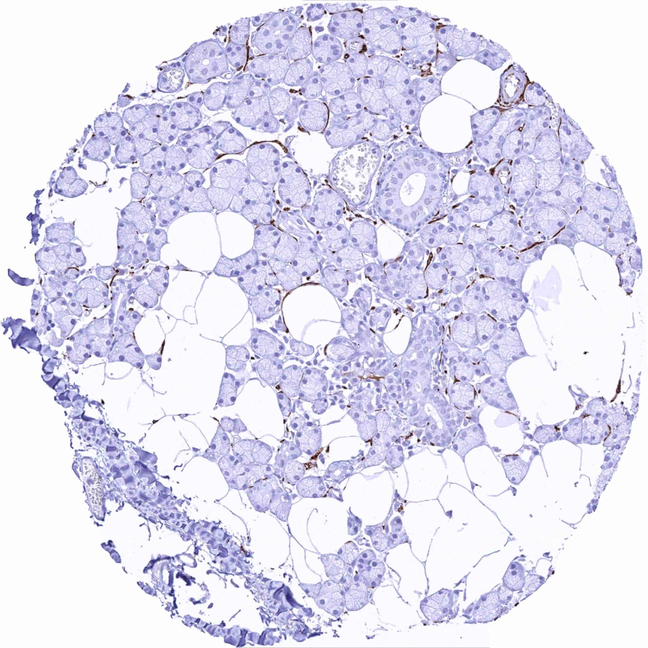
Kidney, cortex – Distinct L1CAM staining of collecting duct (and perhaps distal tubulus) cells
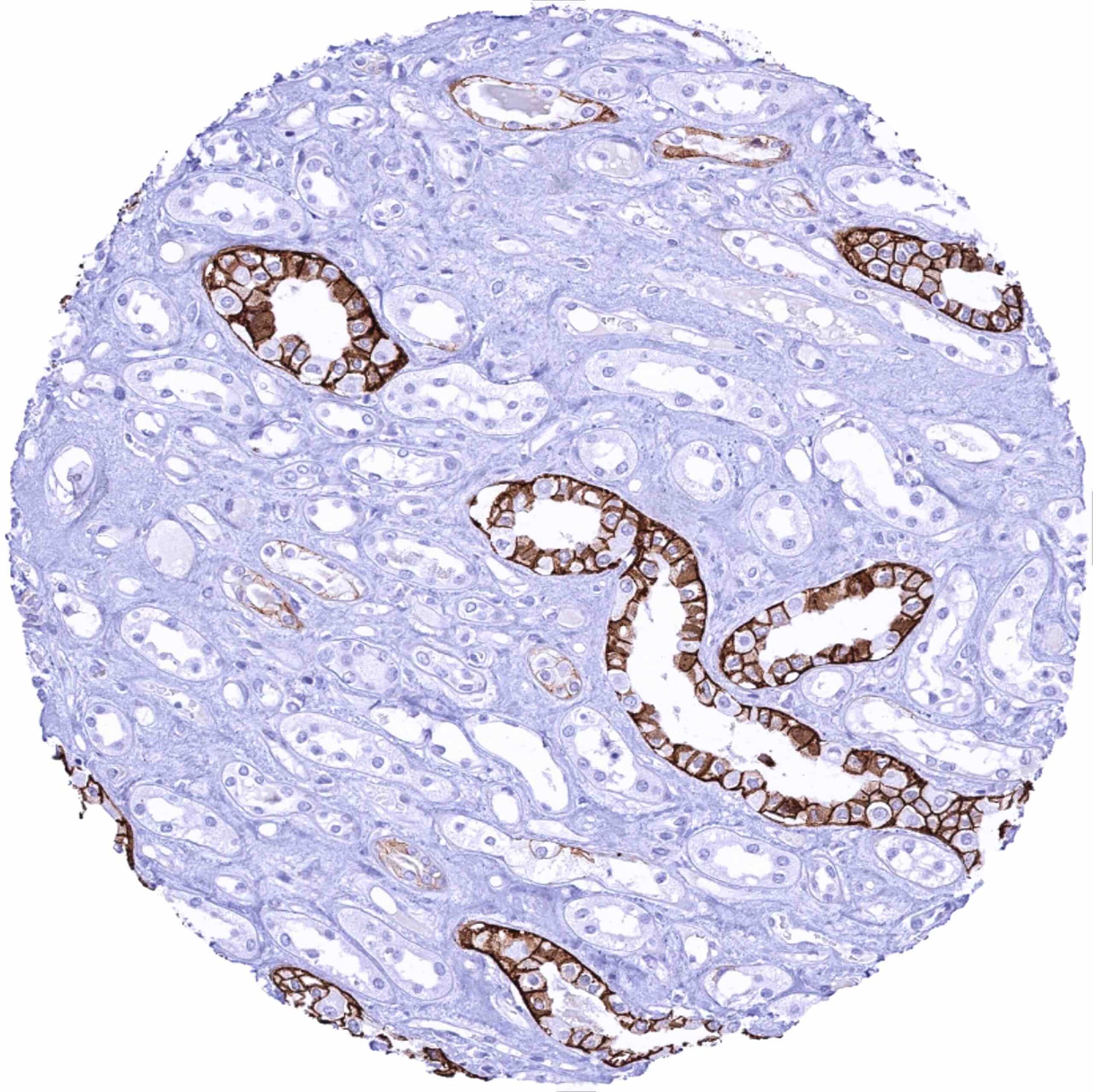
Kidney, medulla – Distinct L1CAM staining of a subset of collecting duct cells
Kidney, pelvis, urothelium
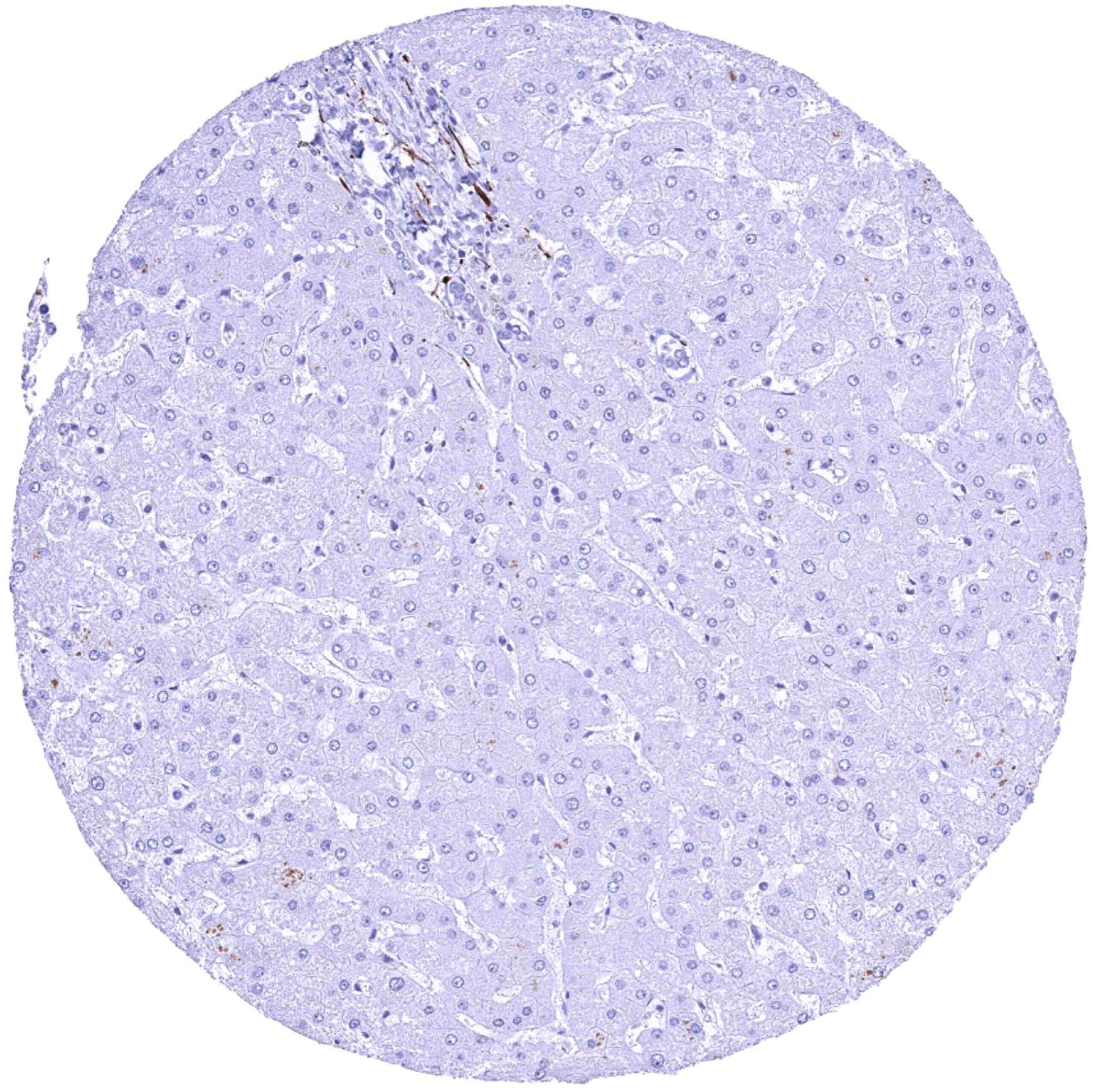
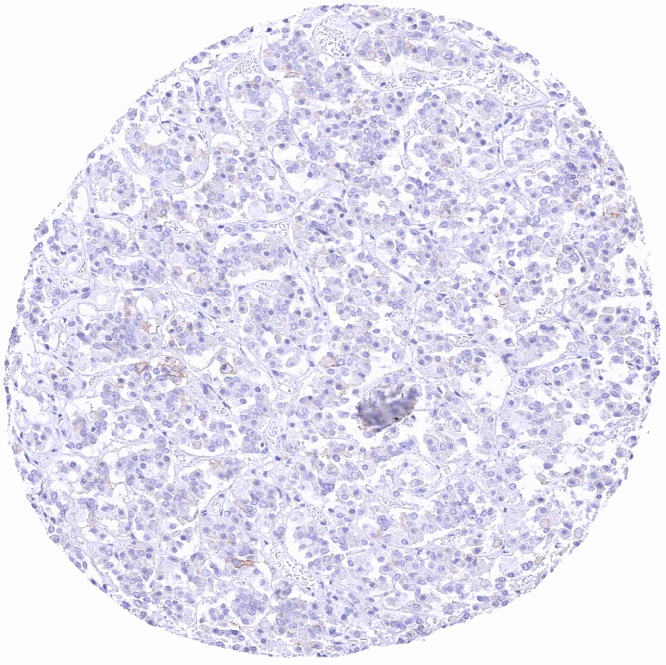
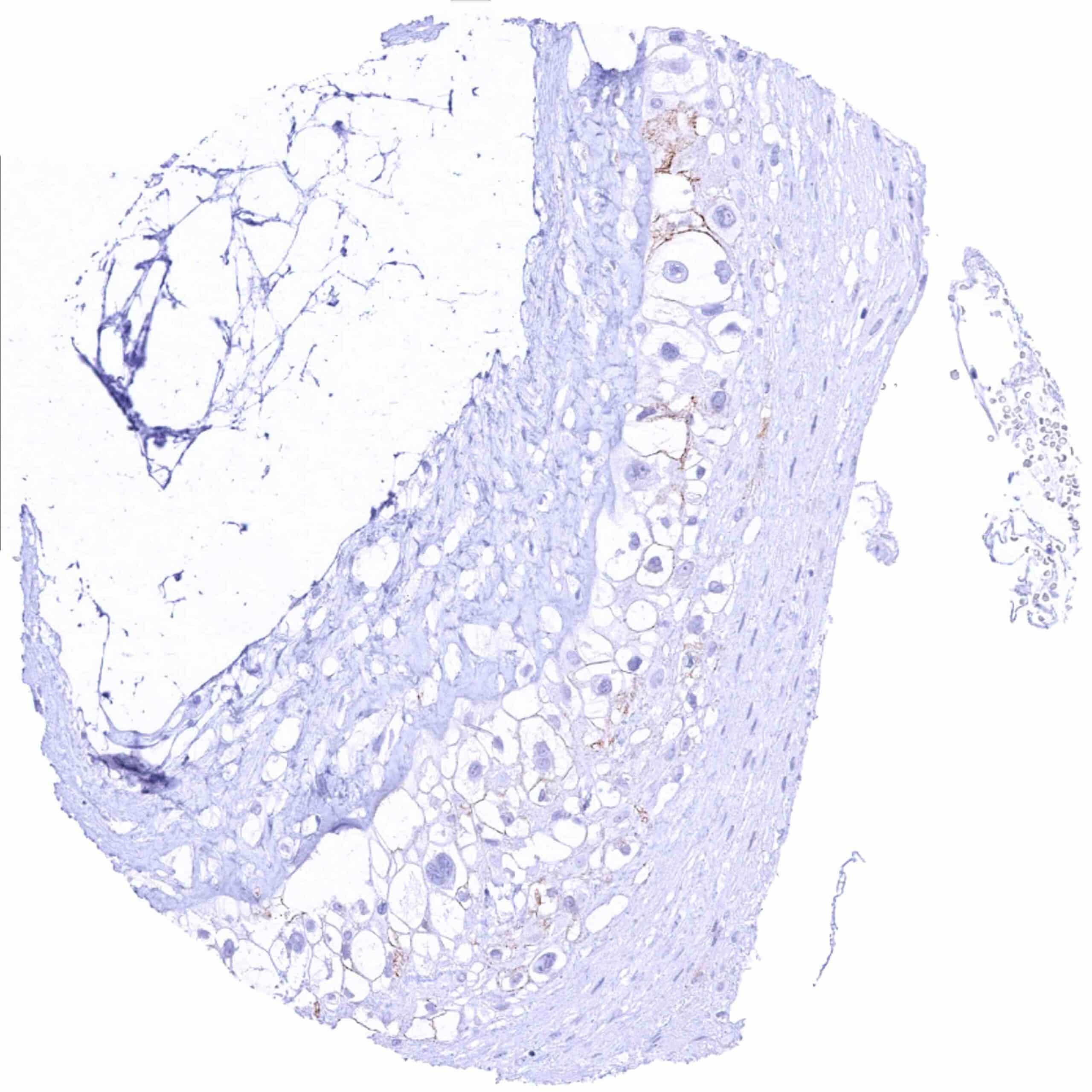
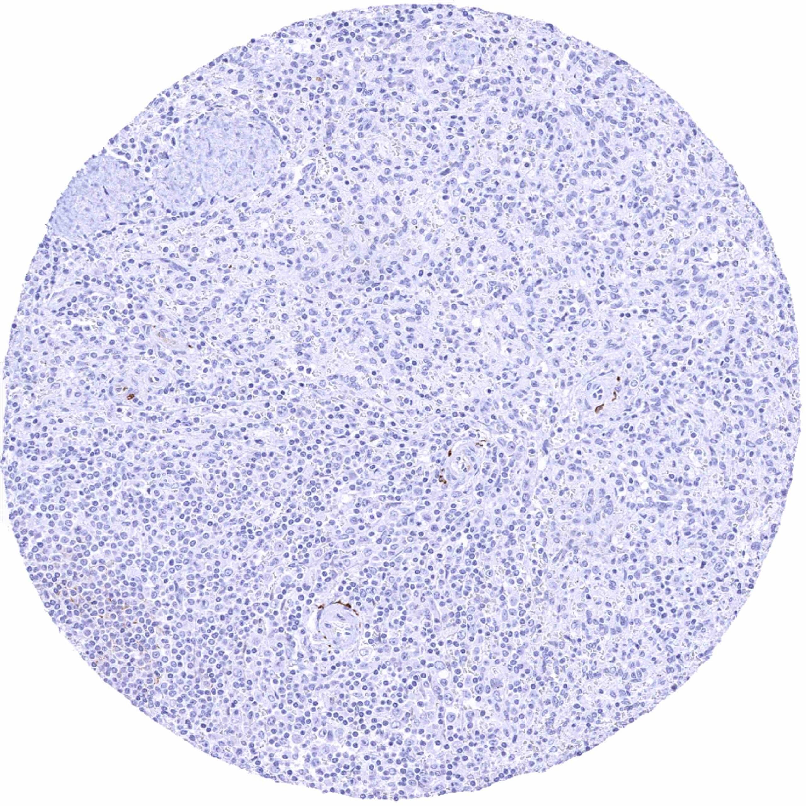
Liver – Distinct L1CAM staining of few nerve fibres
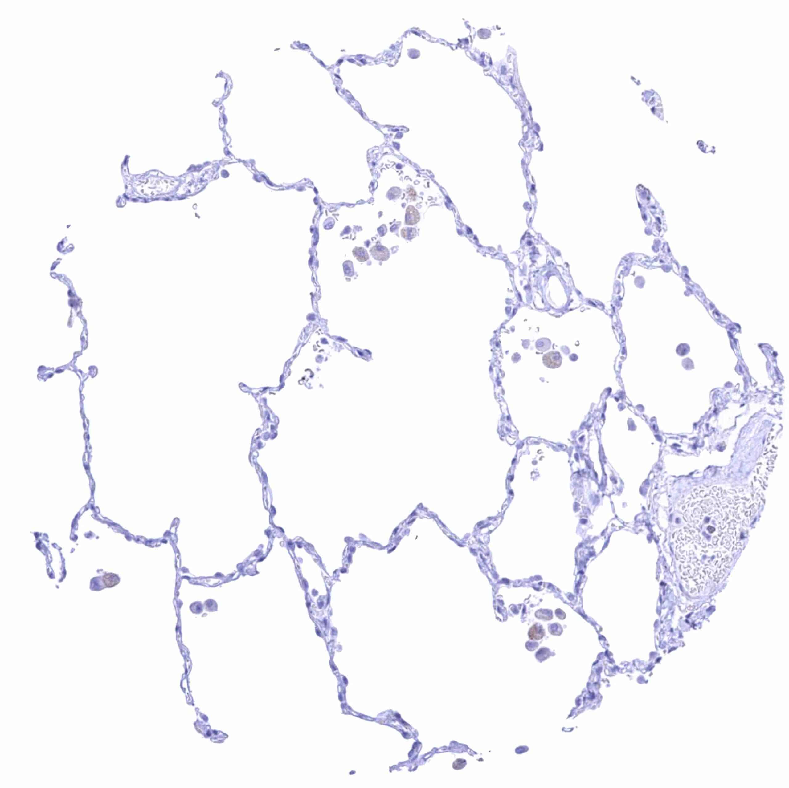
Lung
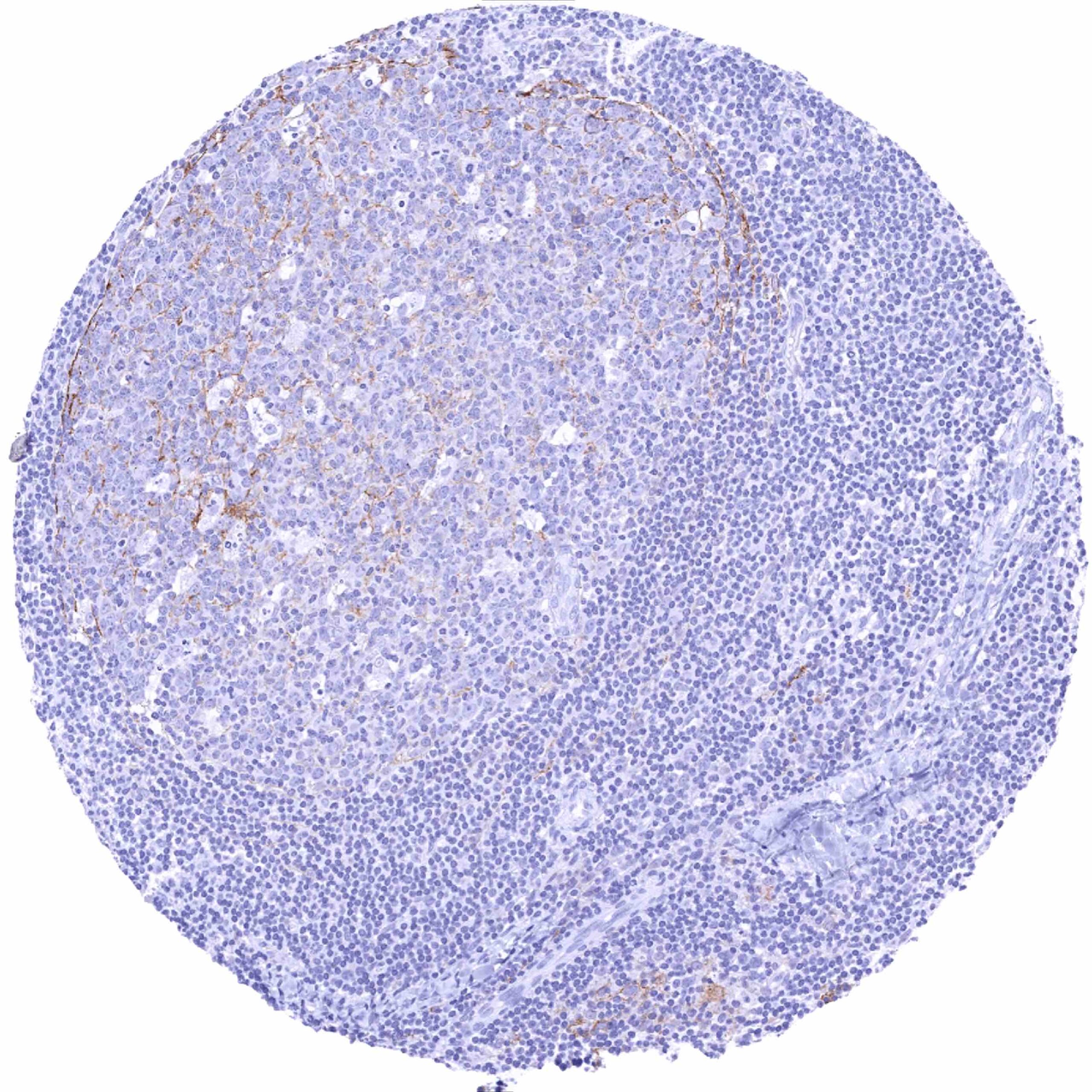
Lymph node – Weak to moderate L1CAM staining of subsets of B-cells and monocytic cells, mainly of the germinal centre
Ovary, stroma
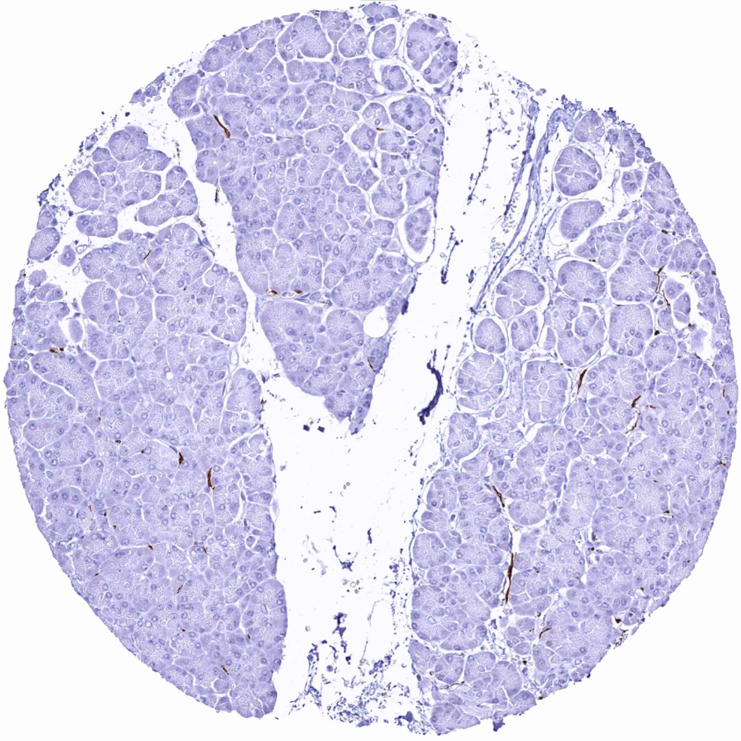
Pancreas – Distinct L1CAM staining of nerve fibres
Parathyroid
Parotid gland – Distinct L1CAM staining of nerve fibres
Pituitary gland, anterior lobe – Moderate intensity L1CAM staining of a subset of epithelial cells
Pituitary gland, posterior lobe – Intense L1CAM staining, especially of nerve fibres
Placenta (amnion and chorion) – Moderate L1CAM staining of chorion cells while amnion cells remain negative
Placenta (chorion) – Weak L1CAM staining of chorion cells
Placenta early, decidua (pregnancy)
Placenta, early
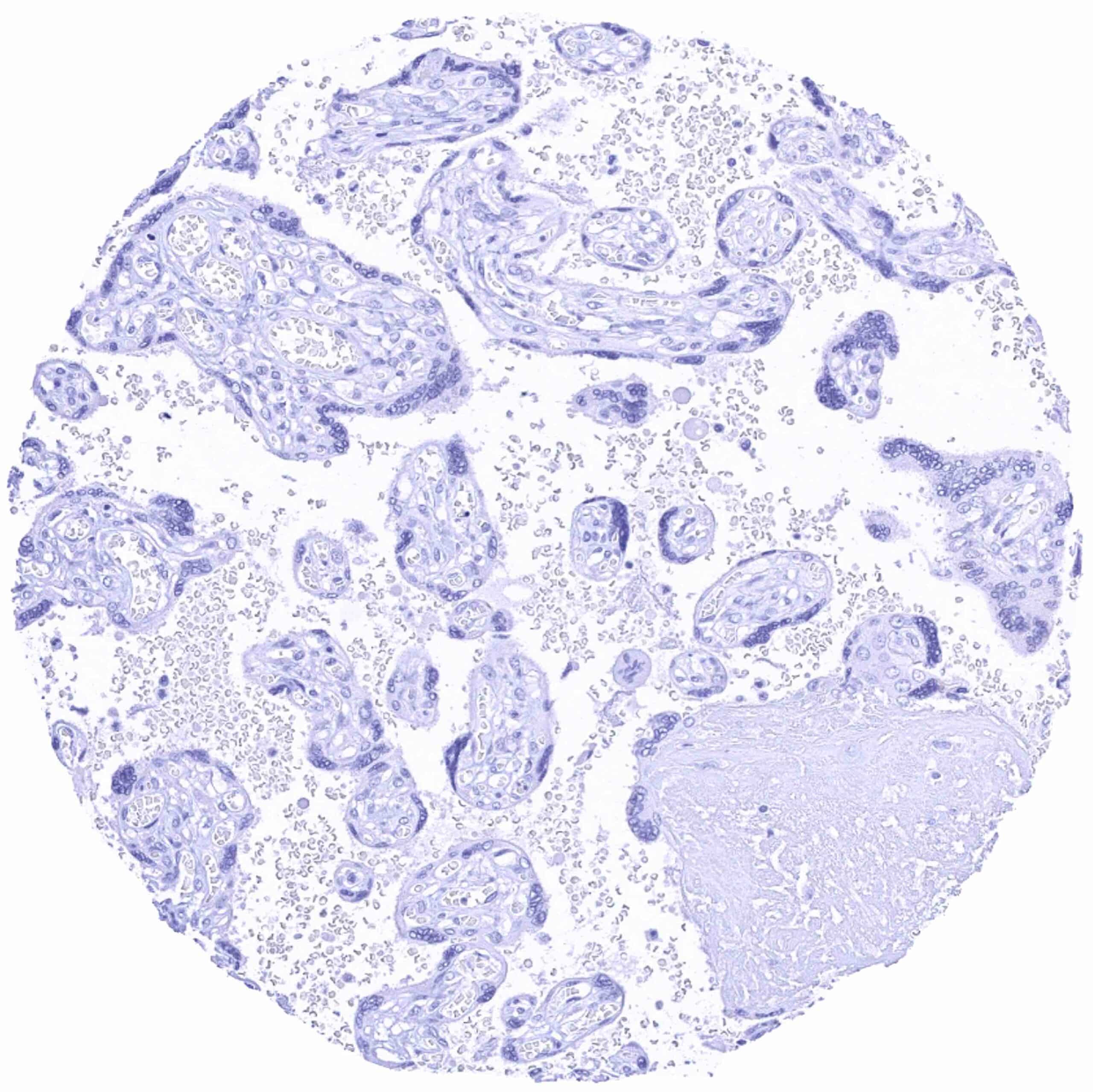
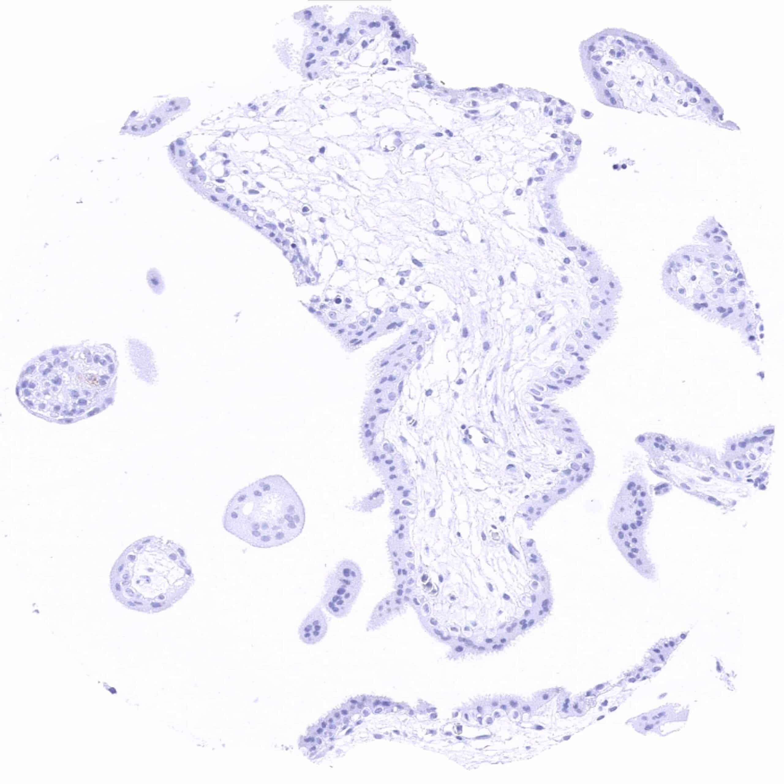
Placenta, mature
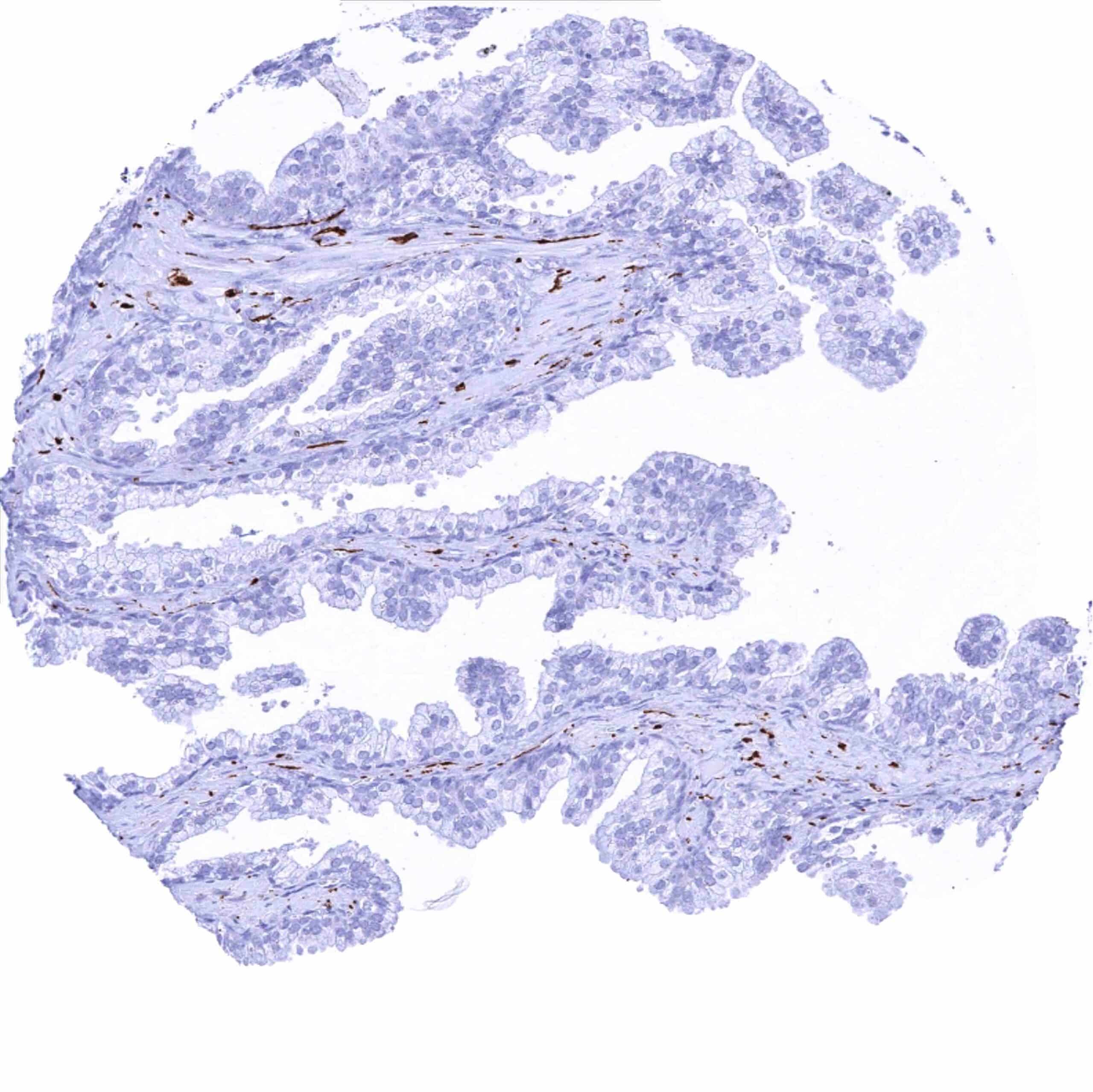
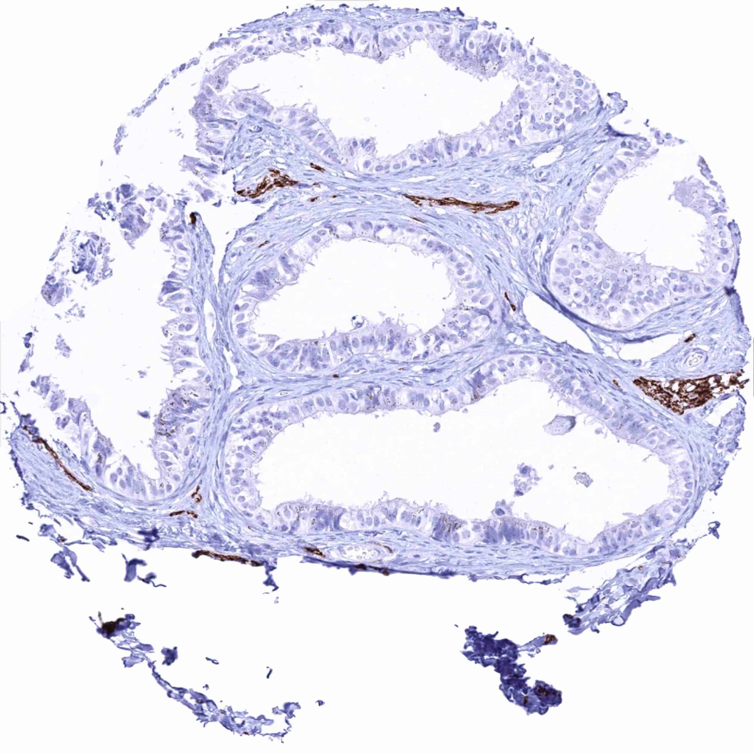
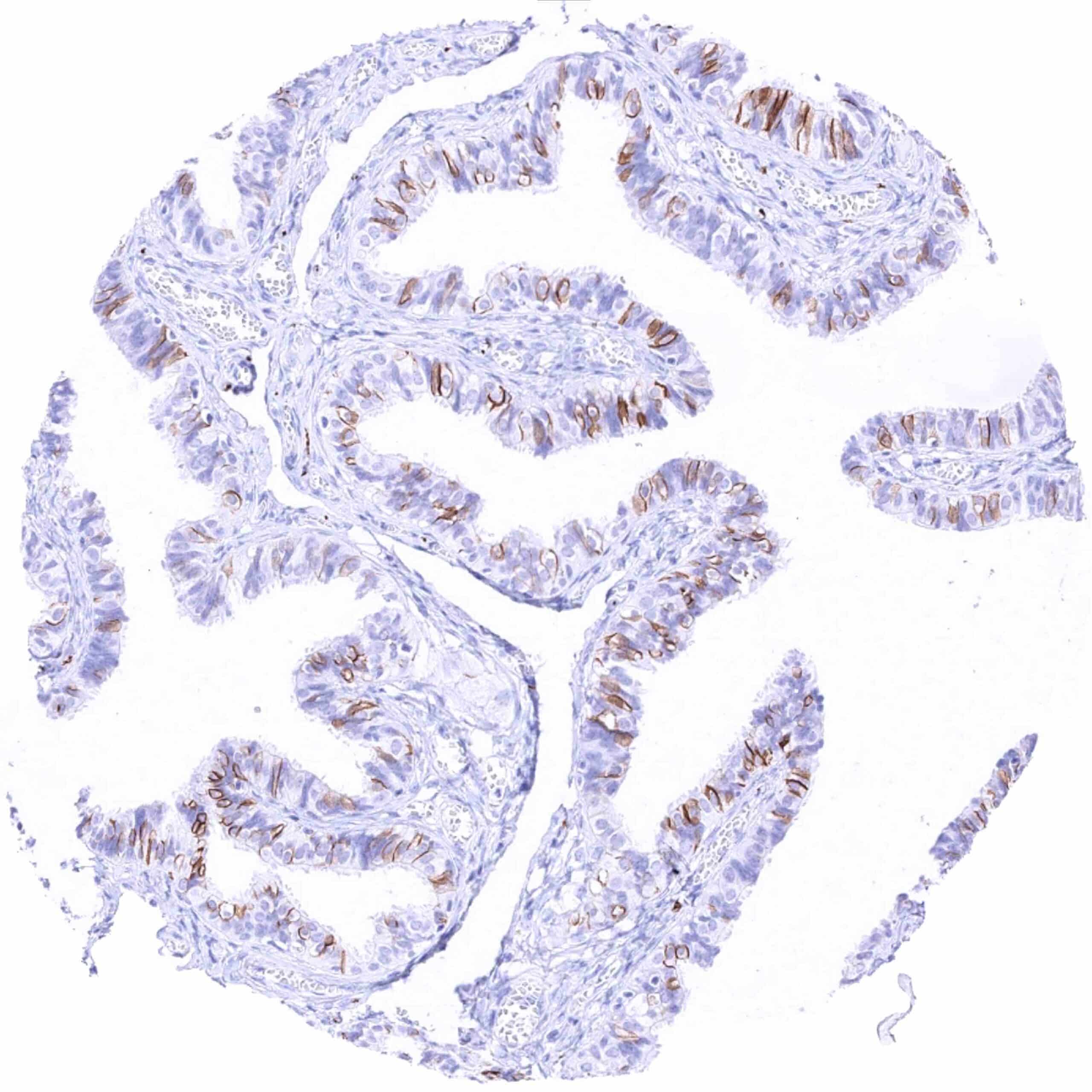
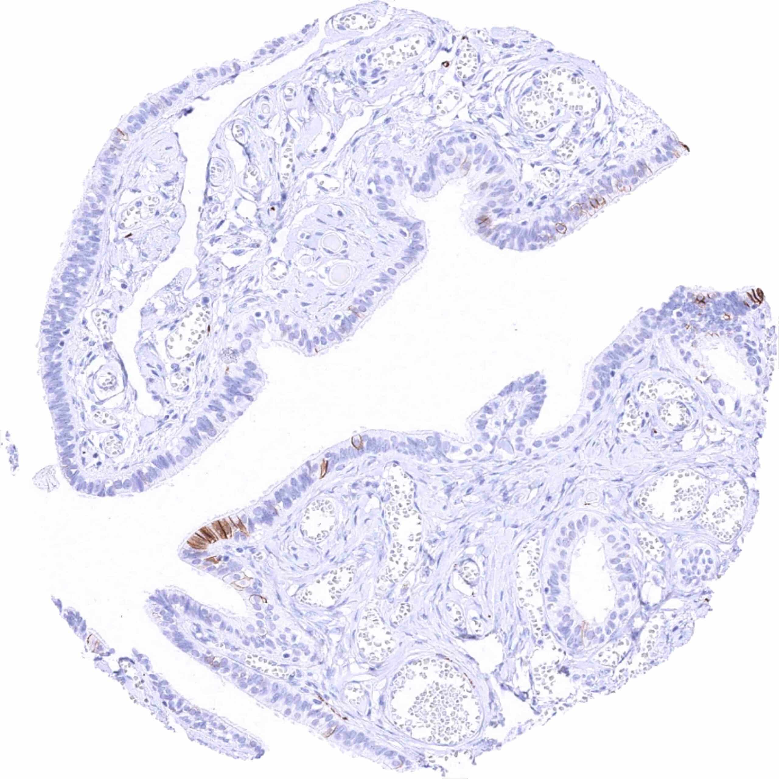
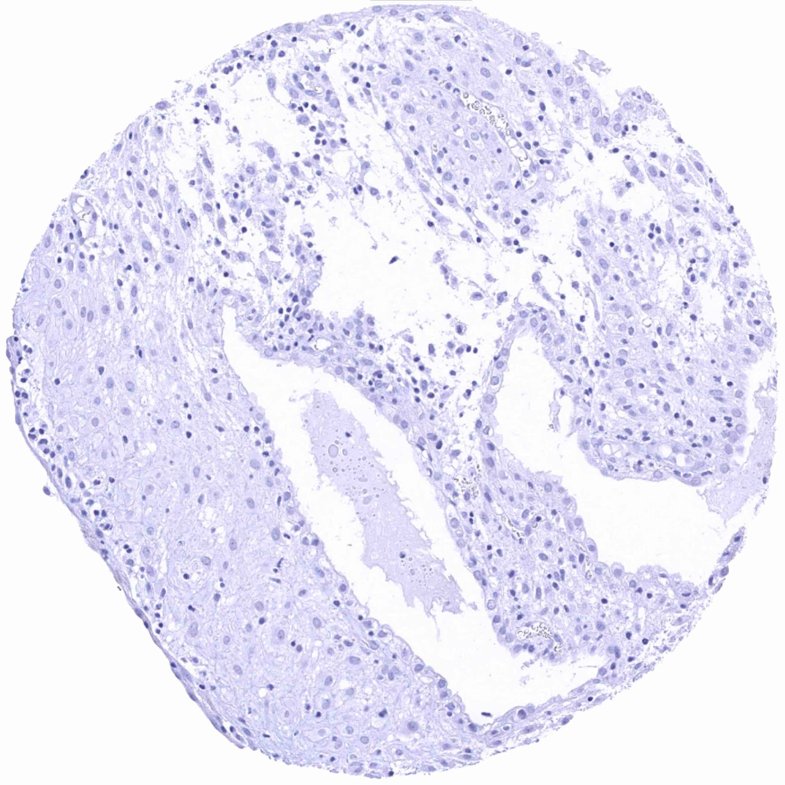
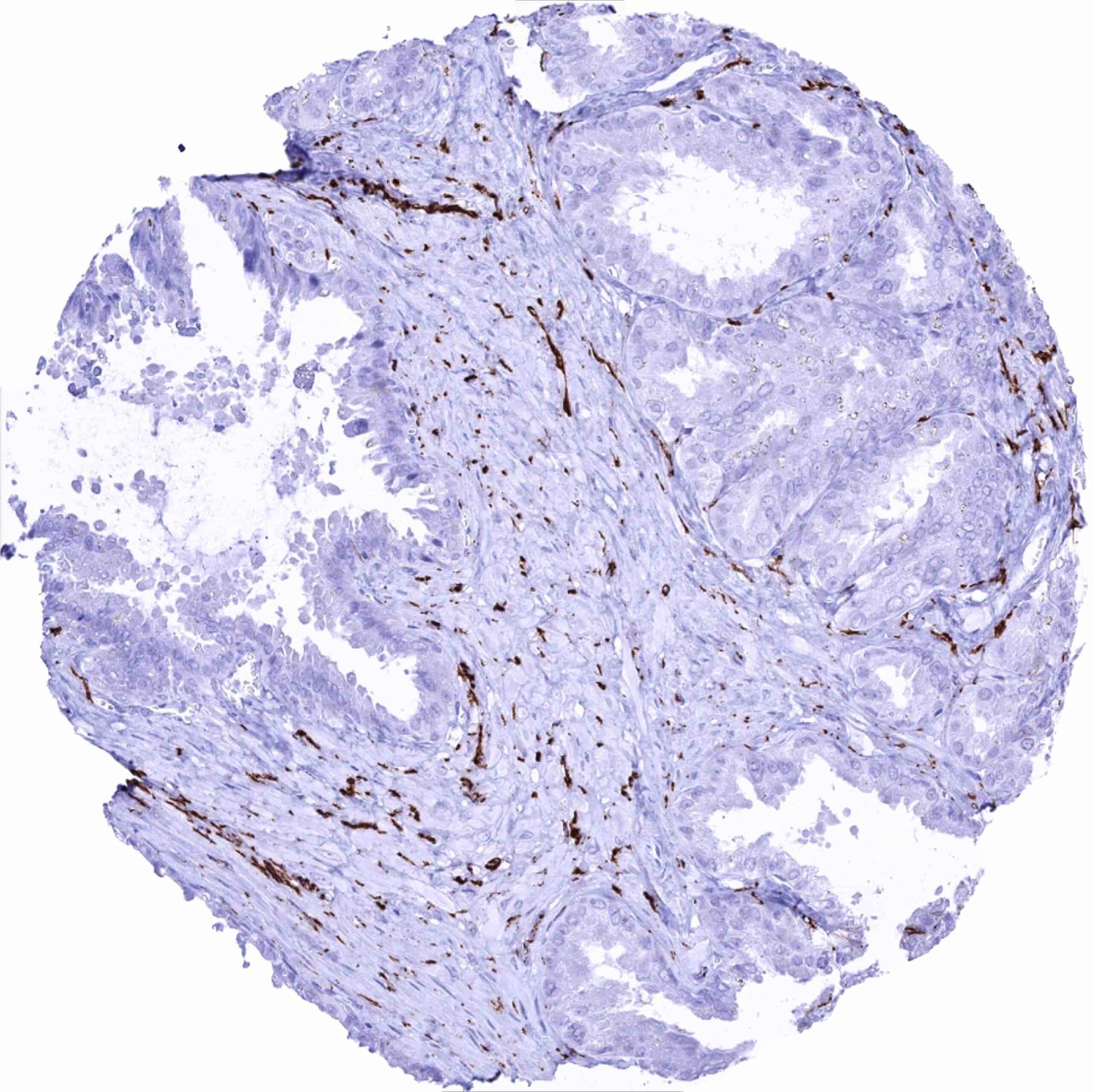
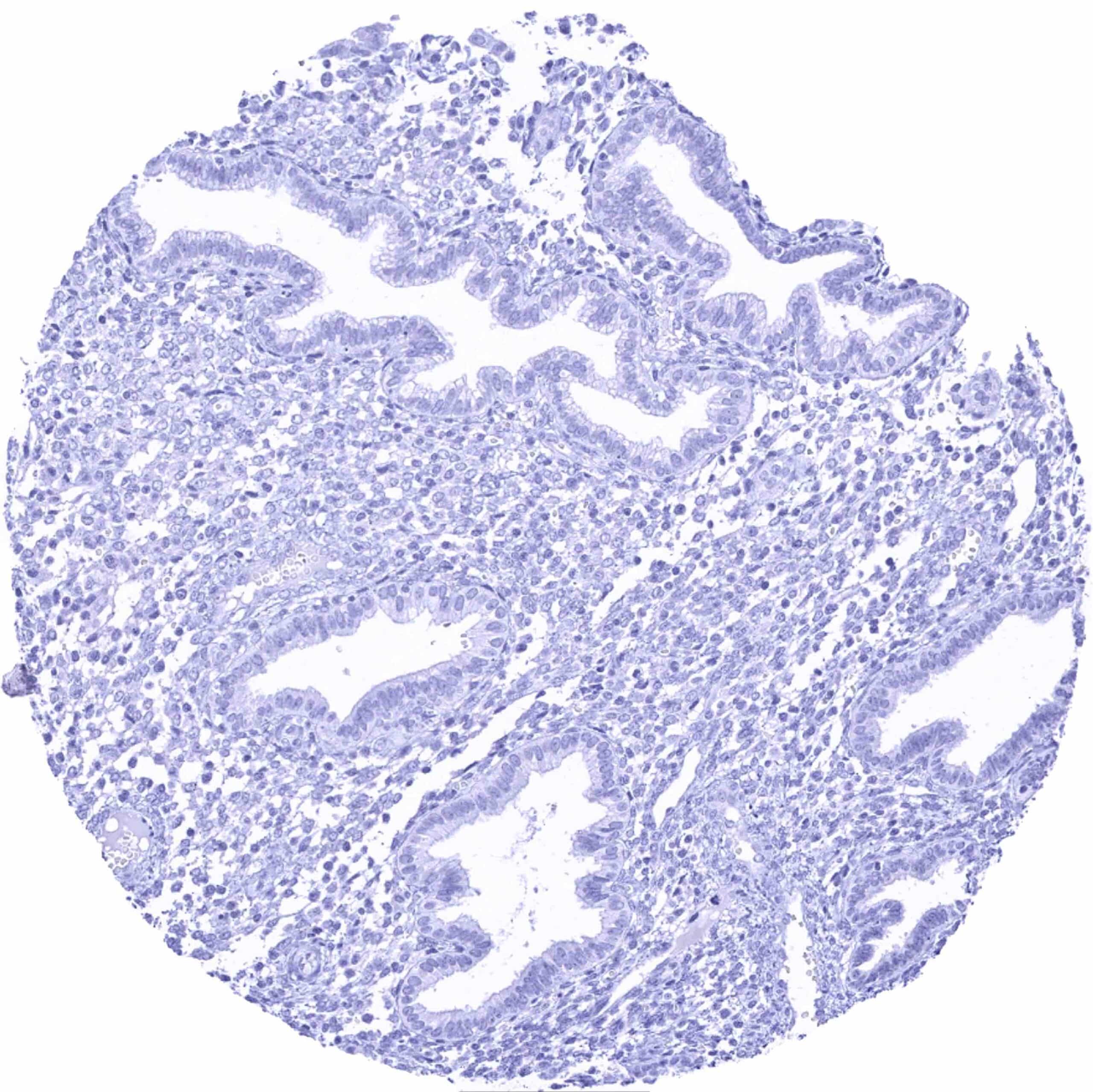
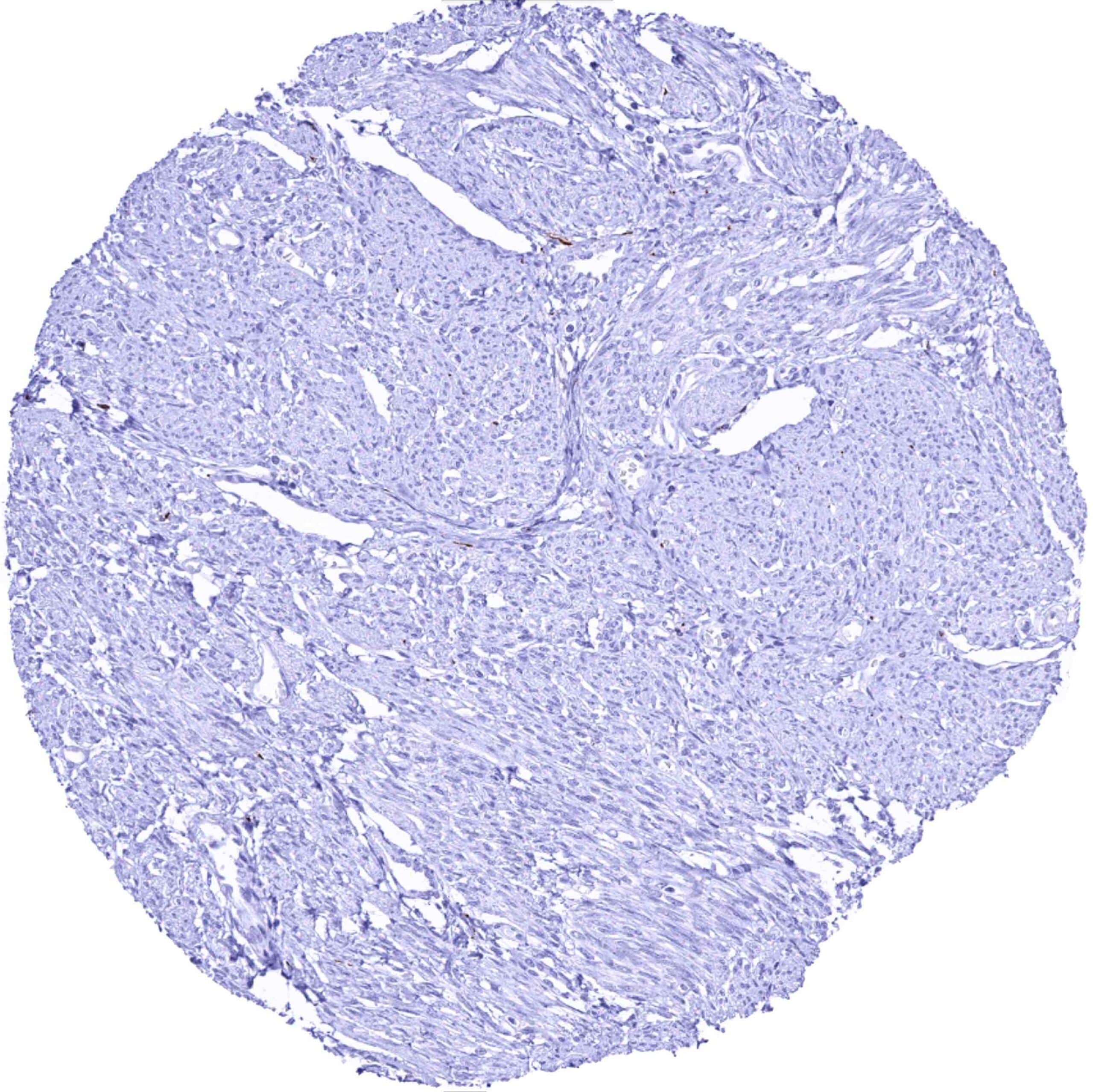
Prostate
Rectum, mucosa
Seminal vesicle – Distinct L1CAM staining of nerve fibres
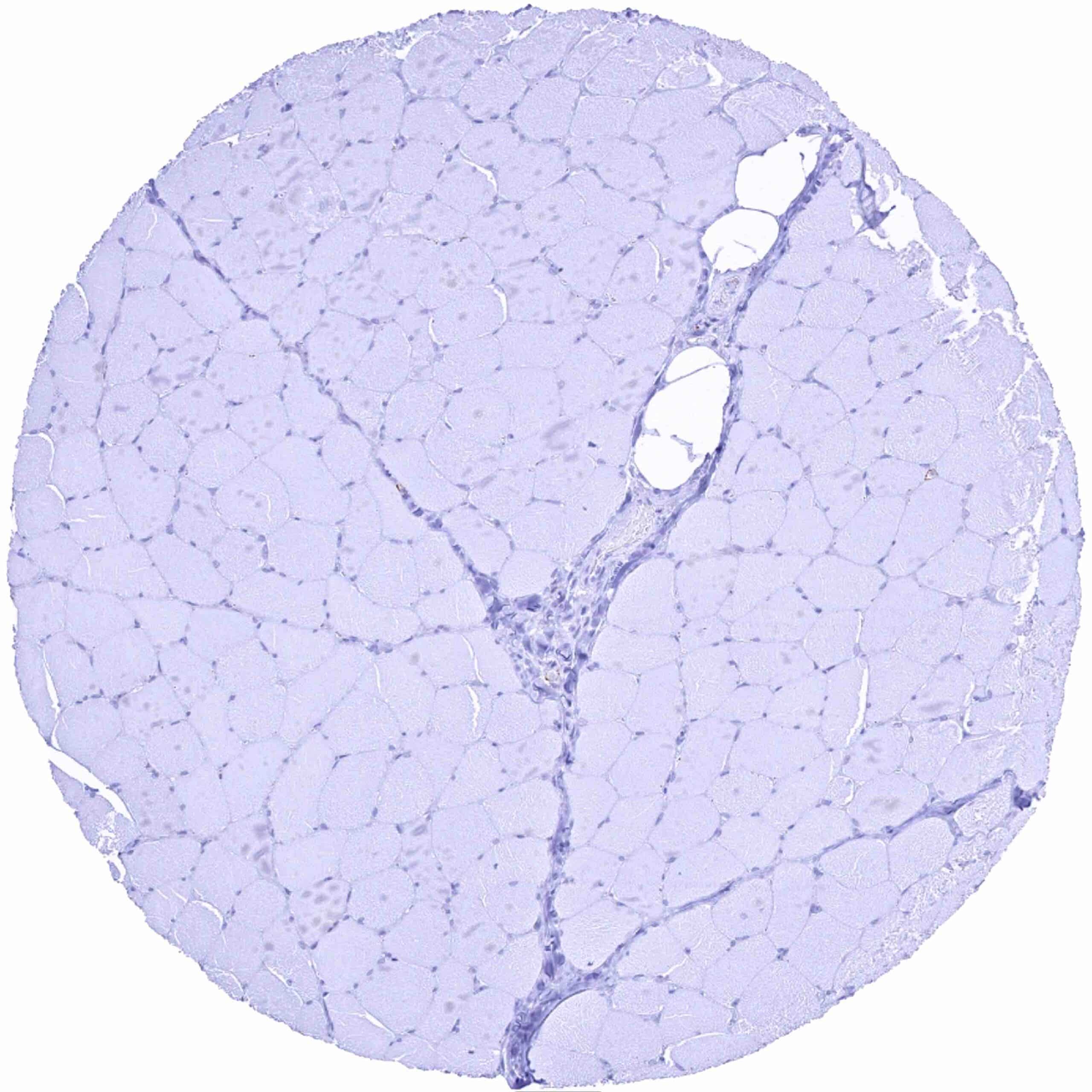
Skeletal muscle
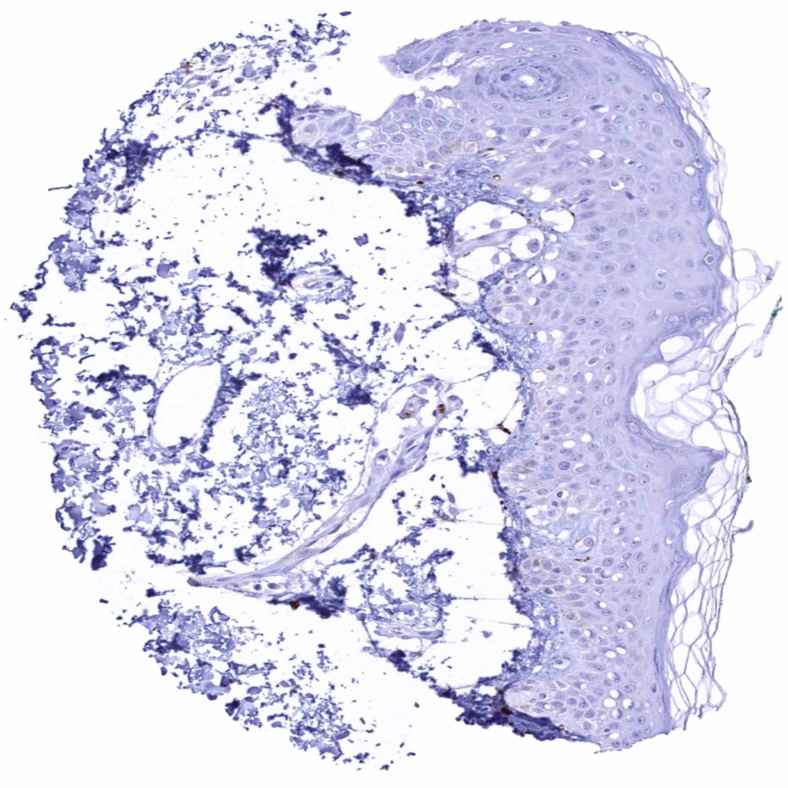
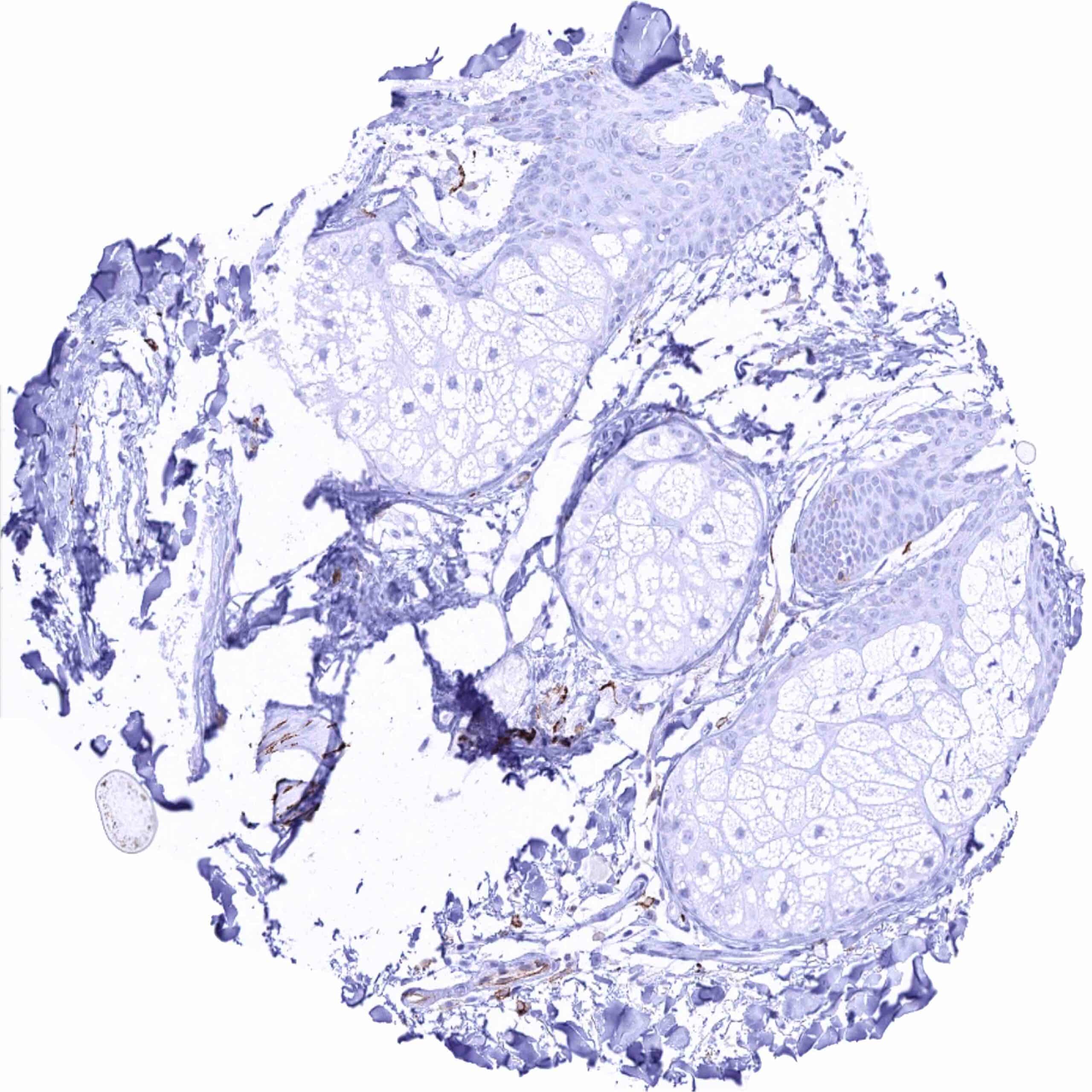
Skin
Skin, hairfollicel and sebaceous glands – Strong L1CAM staining of nerve fibres
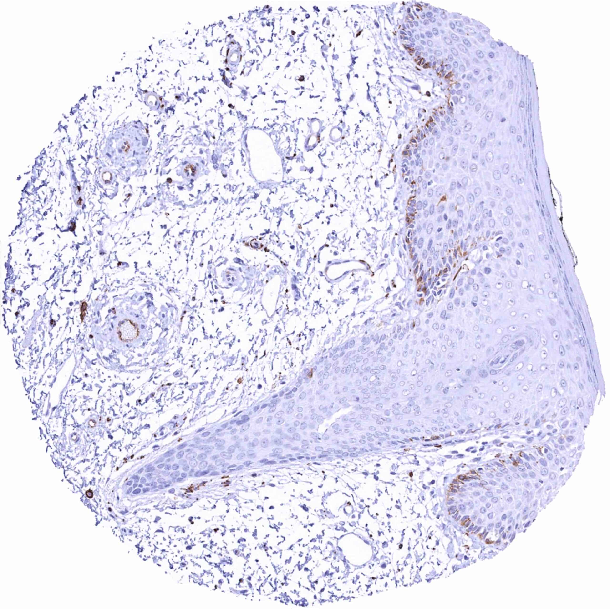
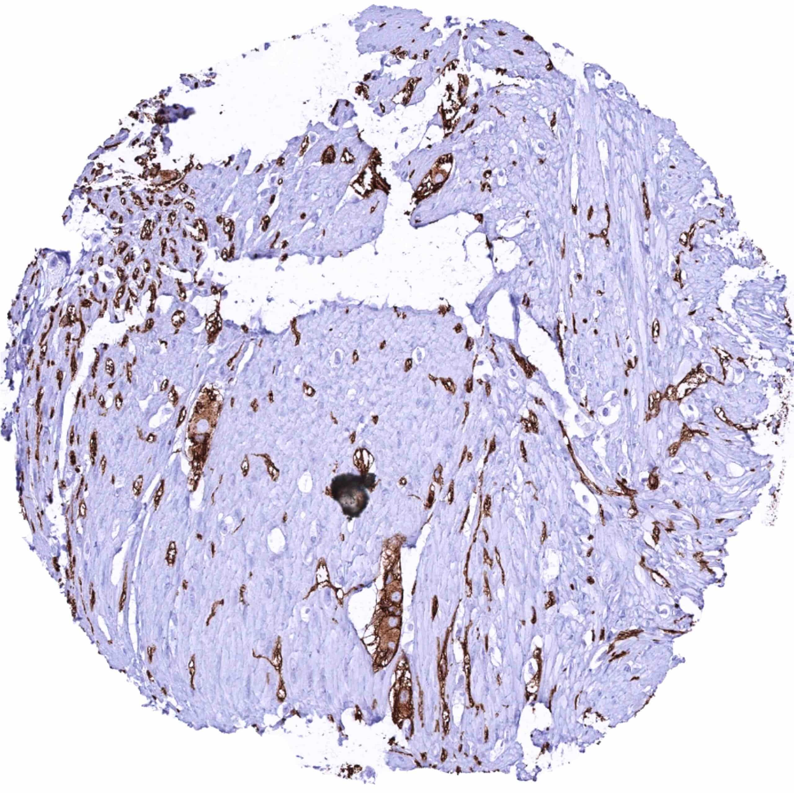
Skin, perianal – Significant L1CAM staining of nerve fibres and endothelial cells in small arteries
Spleen
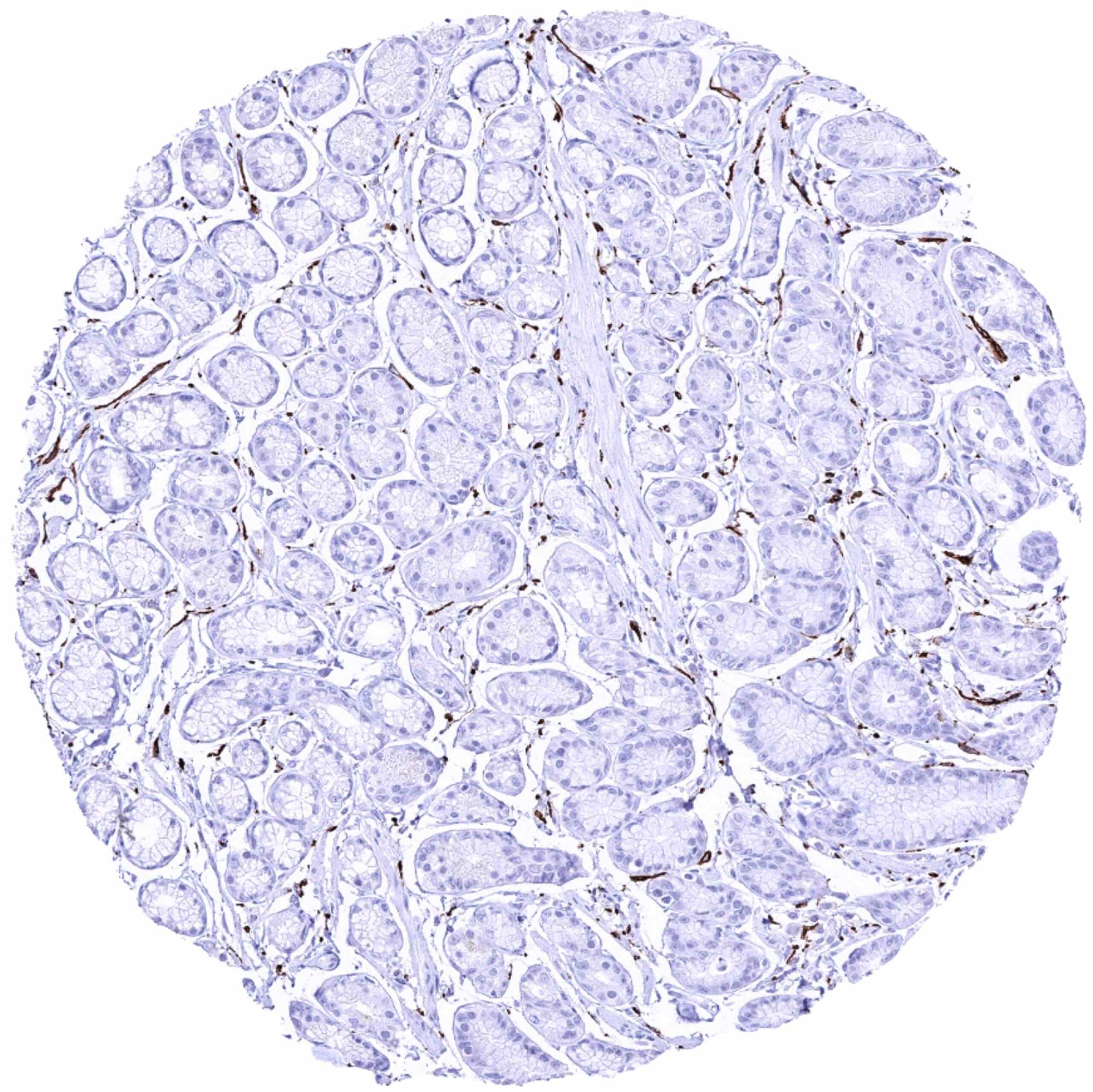
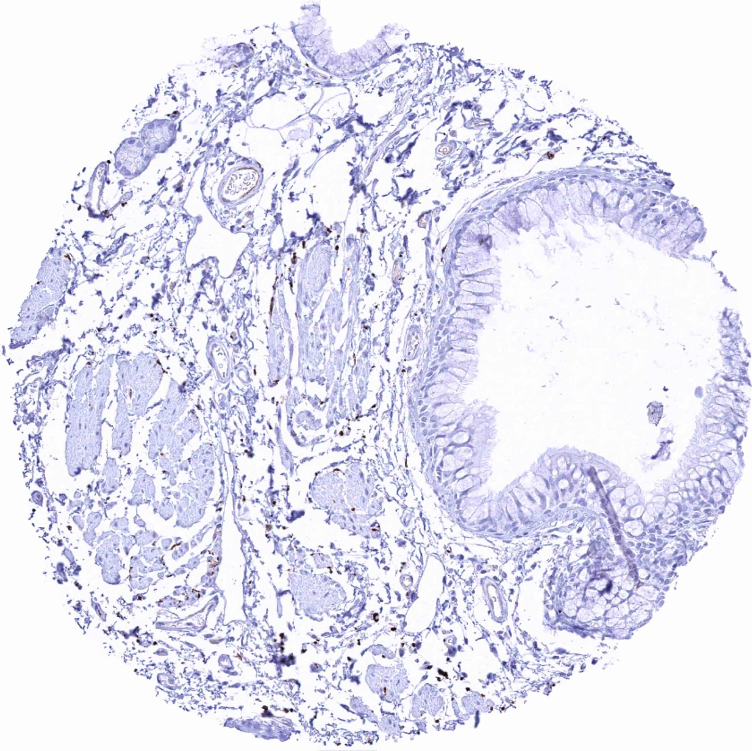
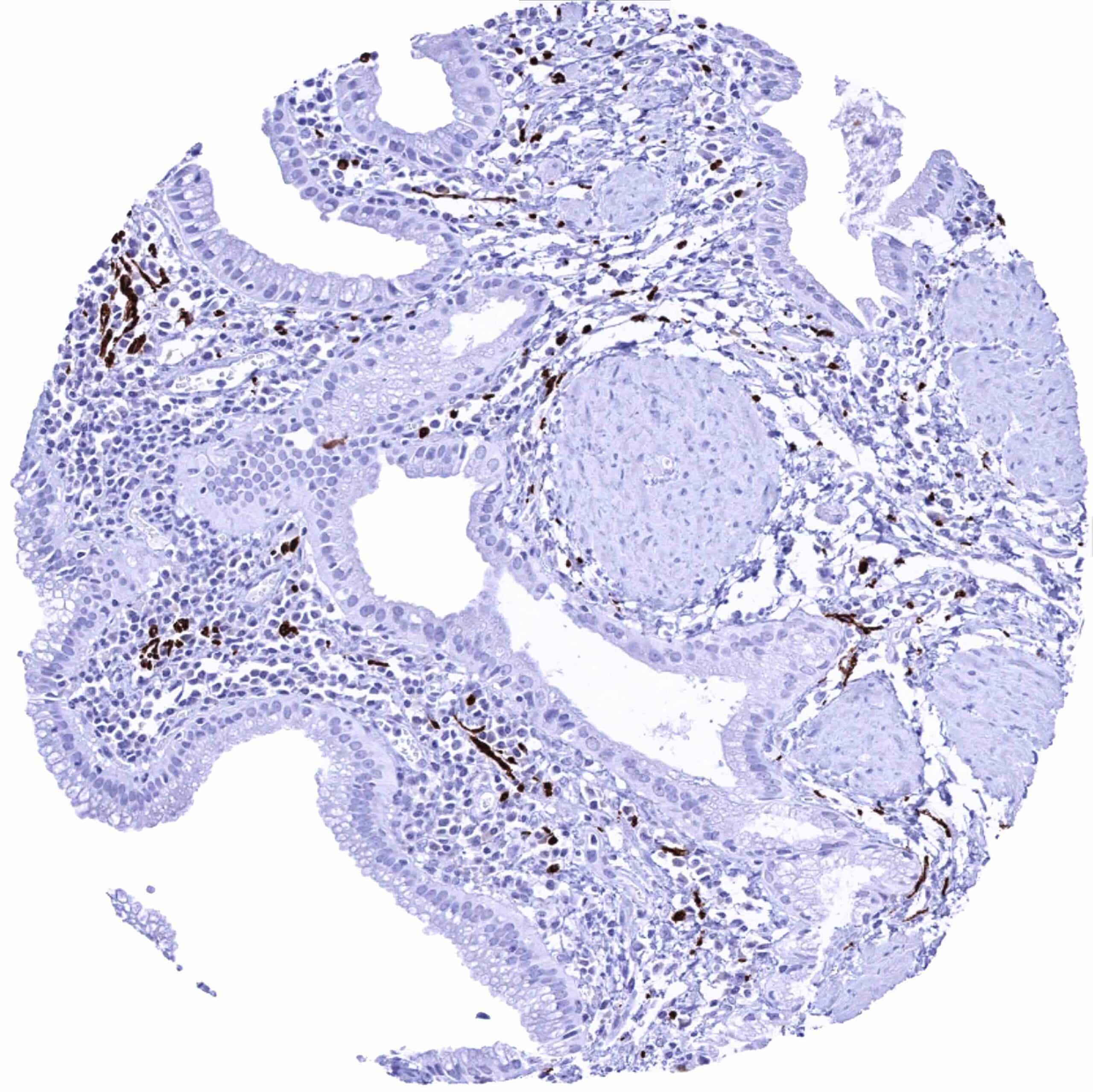
Stomach, antrum – Distinct L1CAM staining of nerve fibres
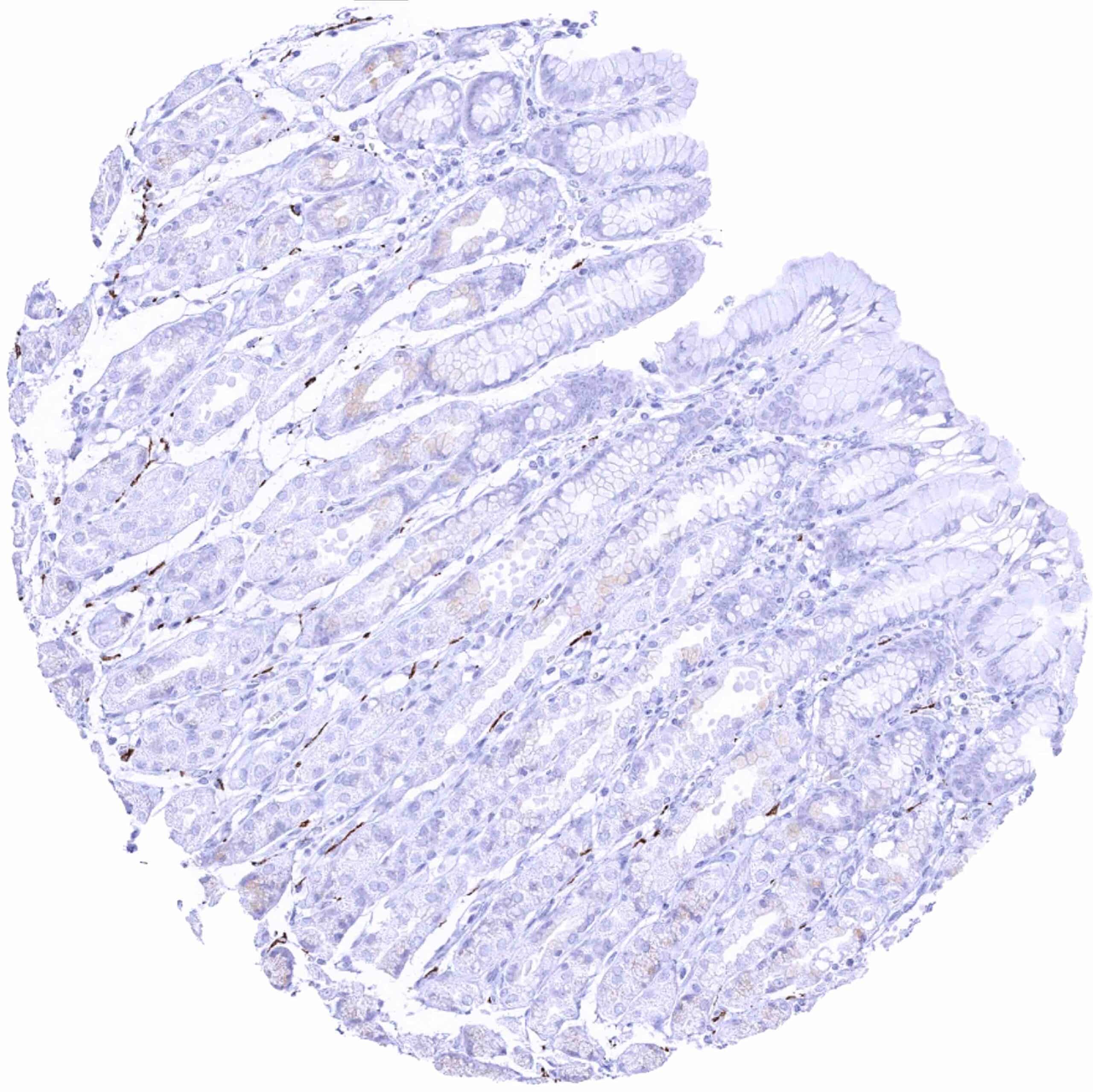
Stomach, corpus – Distinct L1CAM staining of nerve fibres
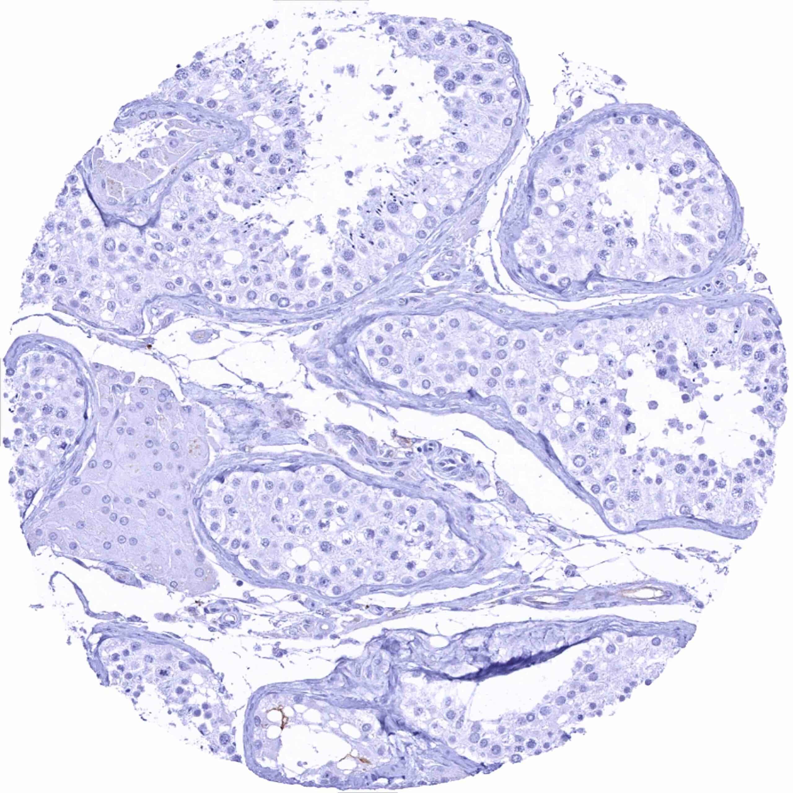
Testis
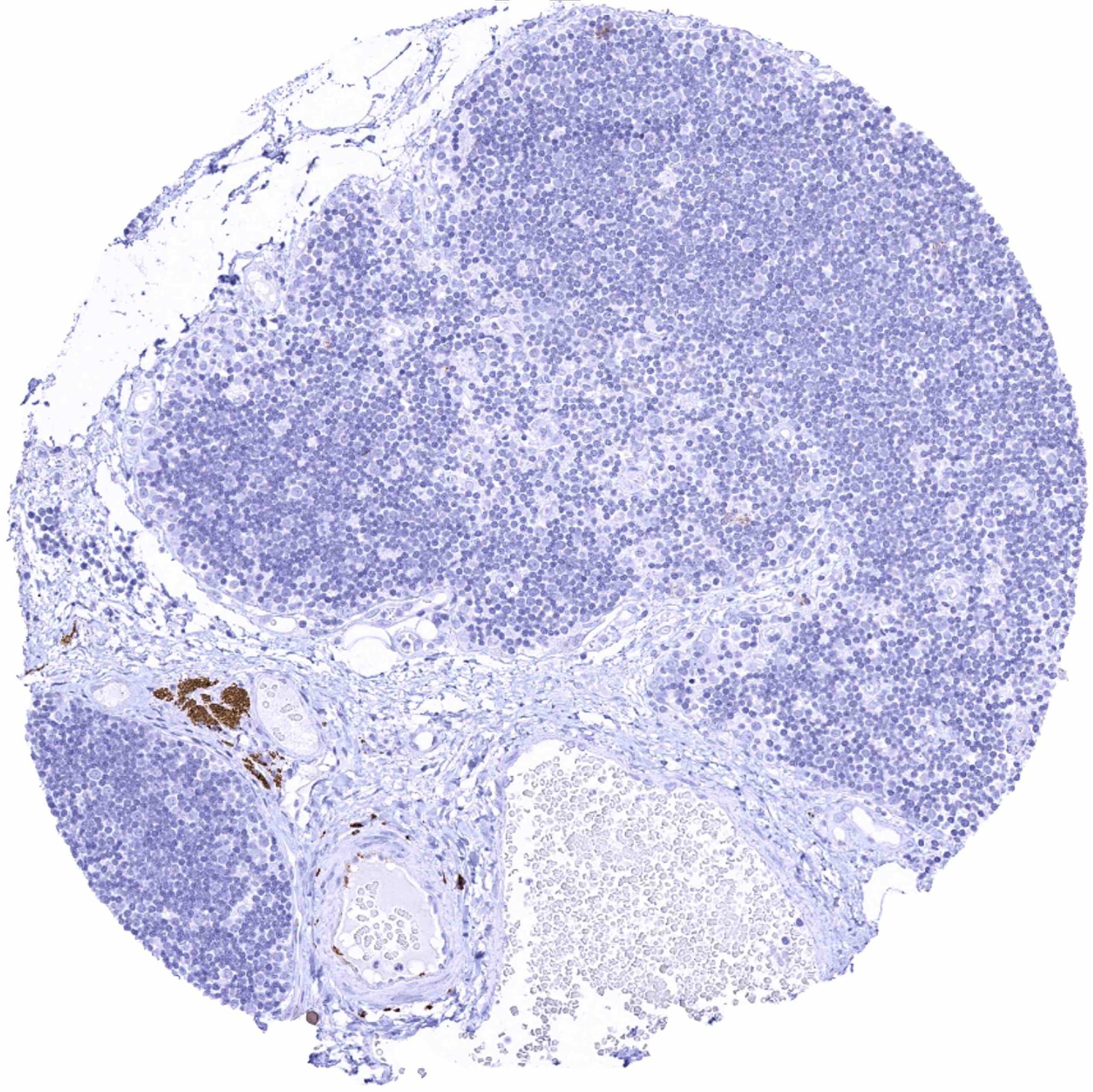
Thymus – Strong L1CAM staining of a nerve
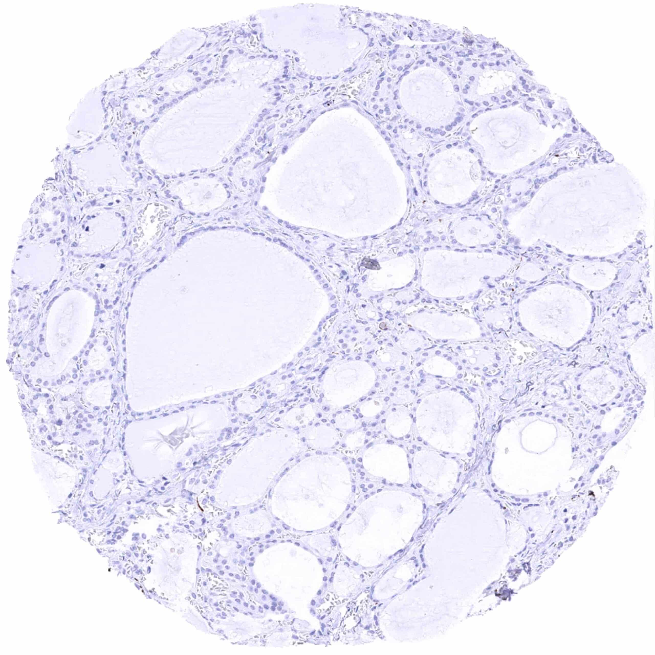
Thyroid gland
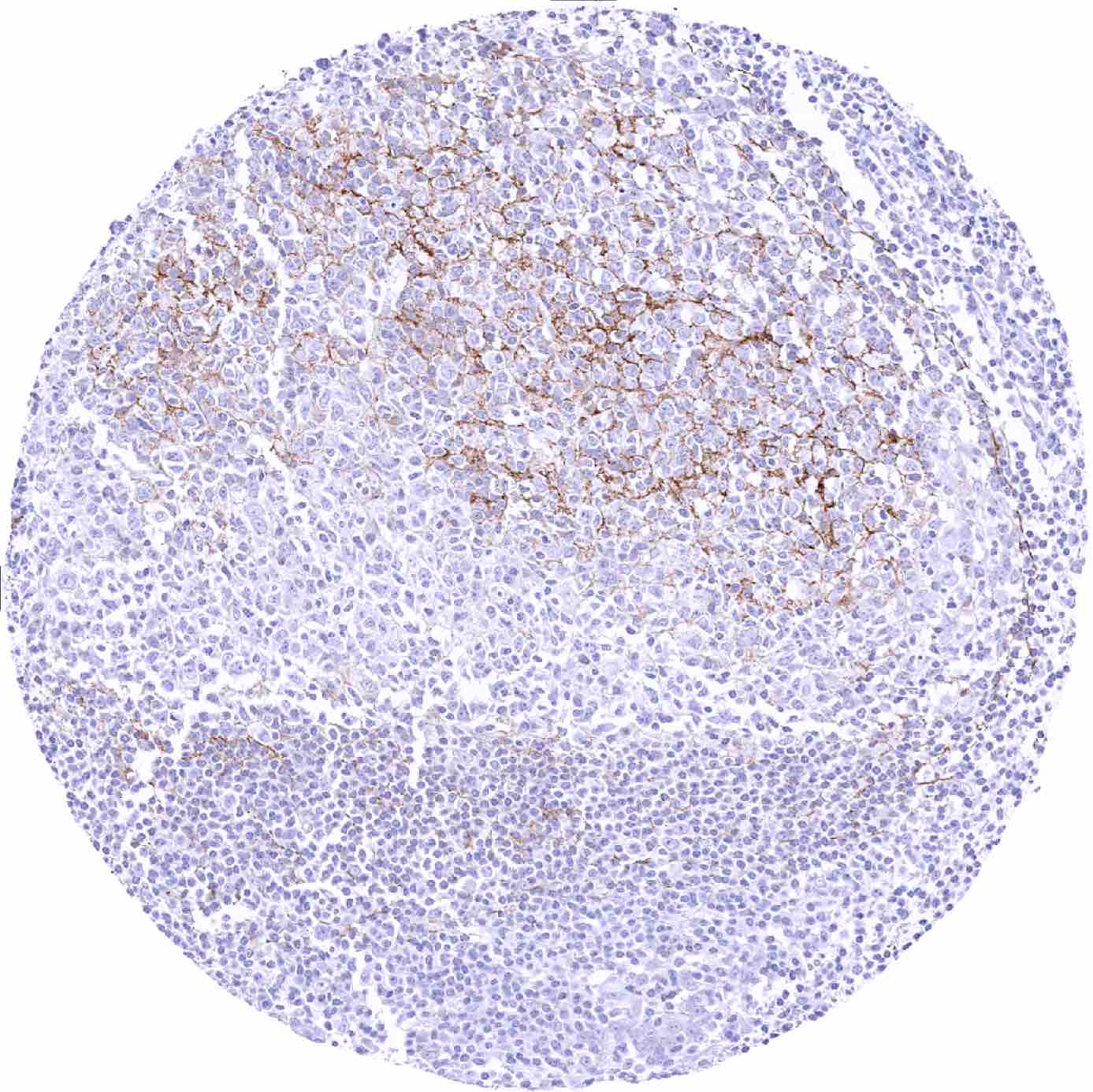
Tonsil – Weak to moderate L1CAM staining of subsets of B-cells and monocytic cells, mainly of the germinal centre (1)
Tonsil – Weak to moderate L1CAM staining of subsets of B-cells and monocytic cells, mainly of the germinal centre (2)
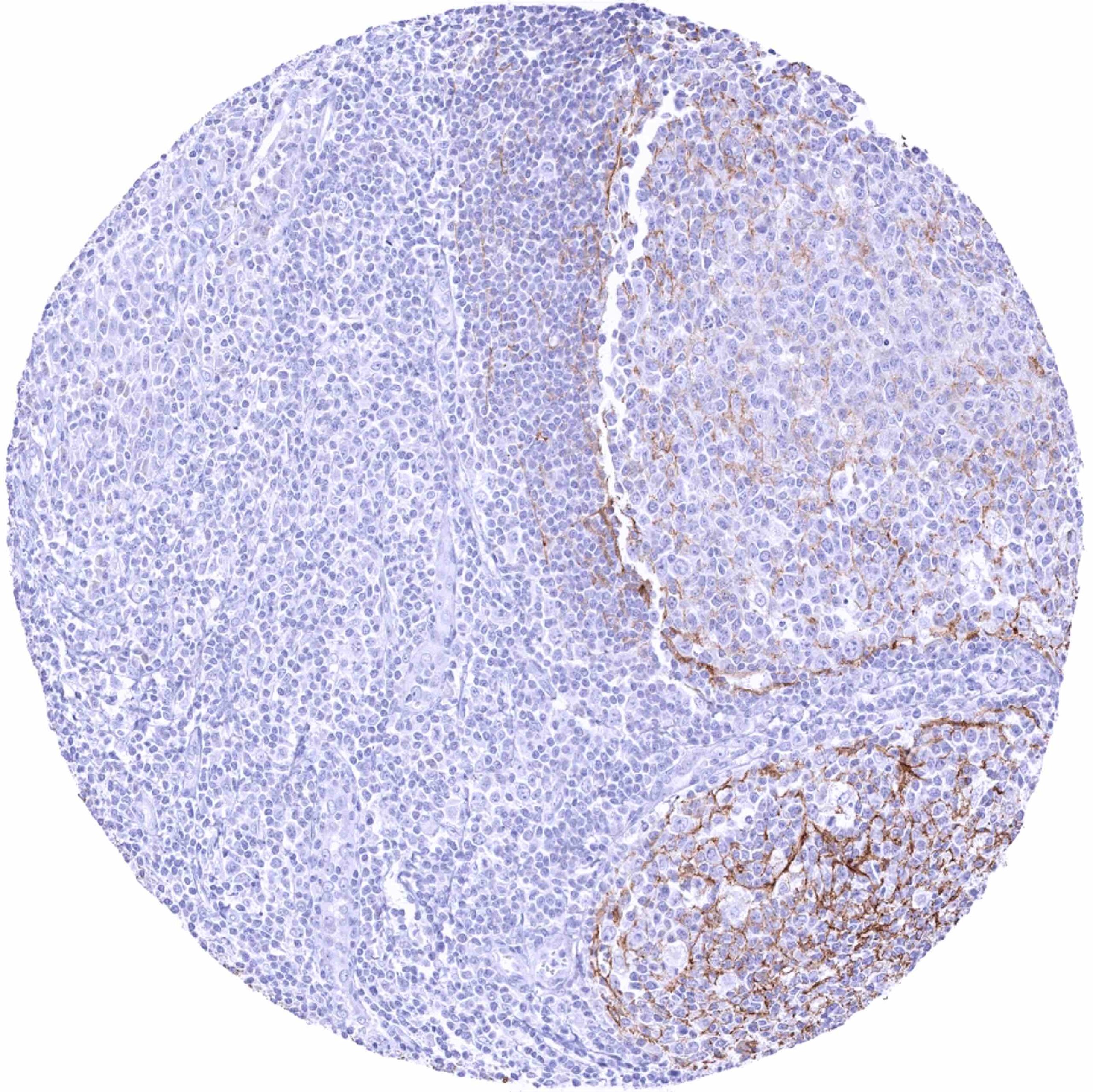
Tonsil – Weak to moderate L1CAM staining of subsets of B-cells and monocytic cells, mainly of the germinal centre (3)
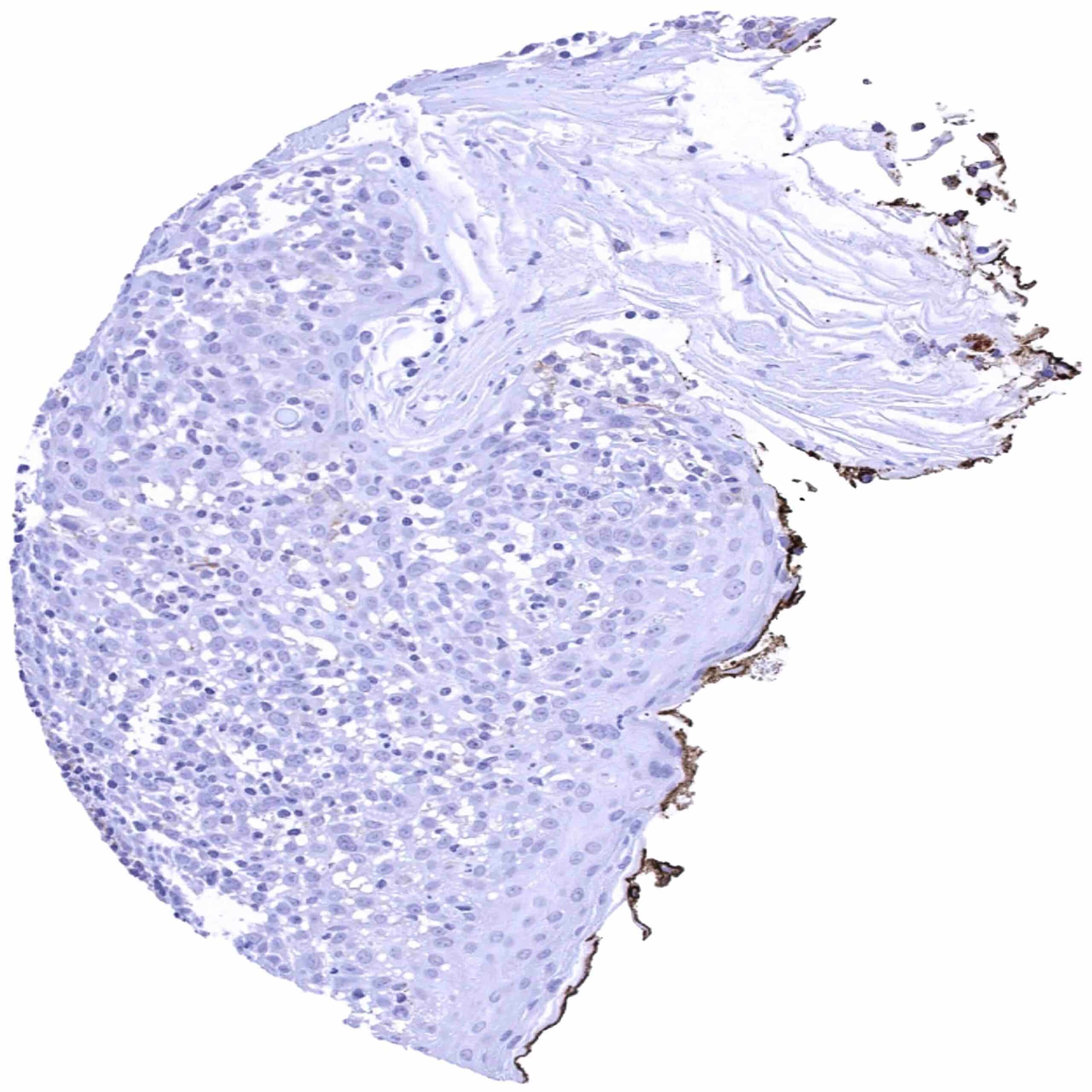
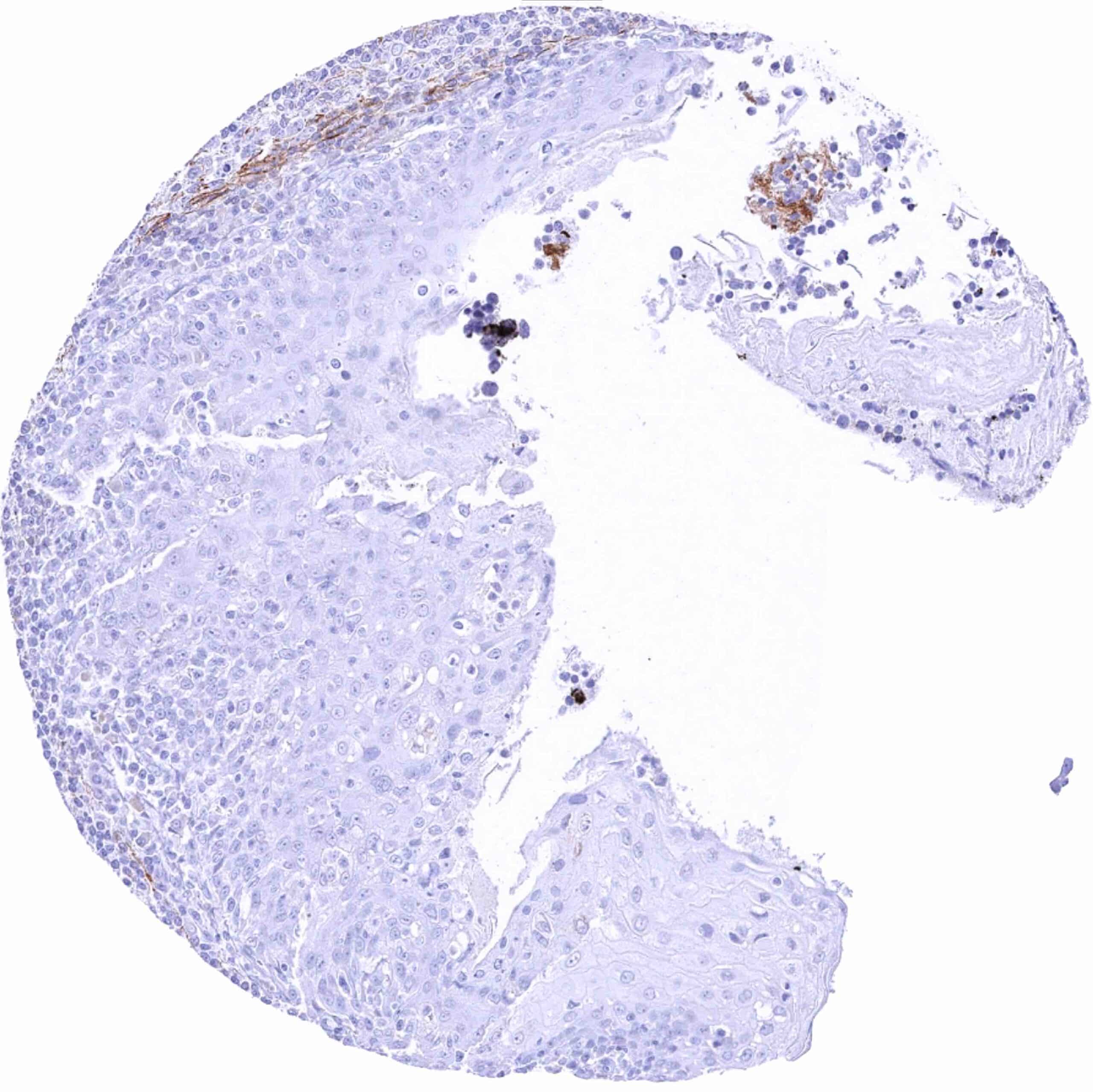
Tonsil, surface
Urinary bladder, muscular wall
Urinary bladder, urothelium
Uterus, ectocervix
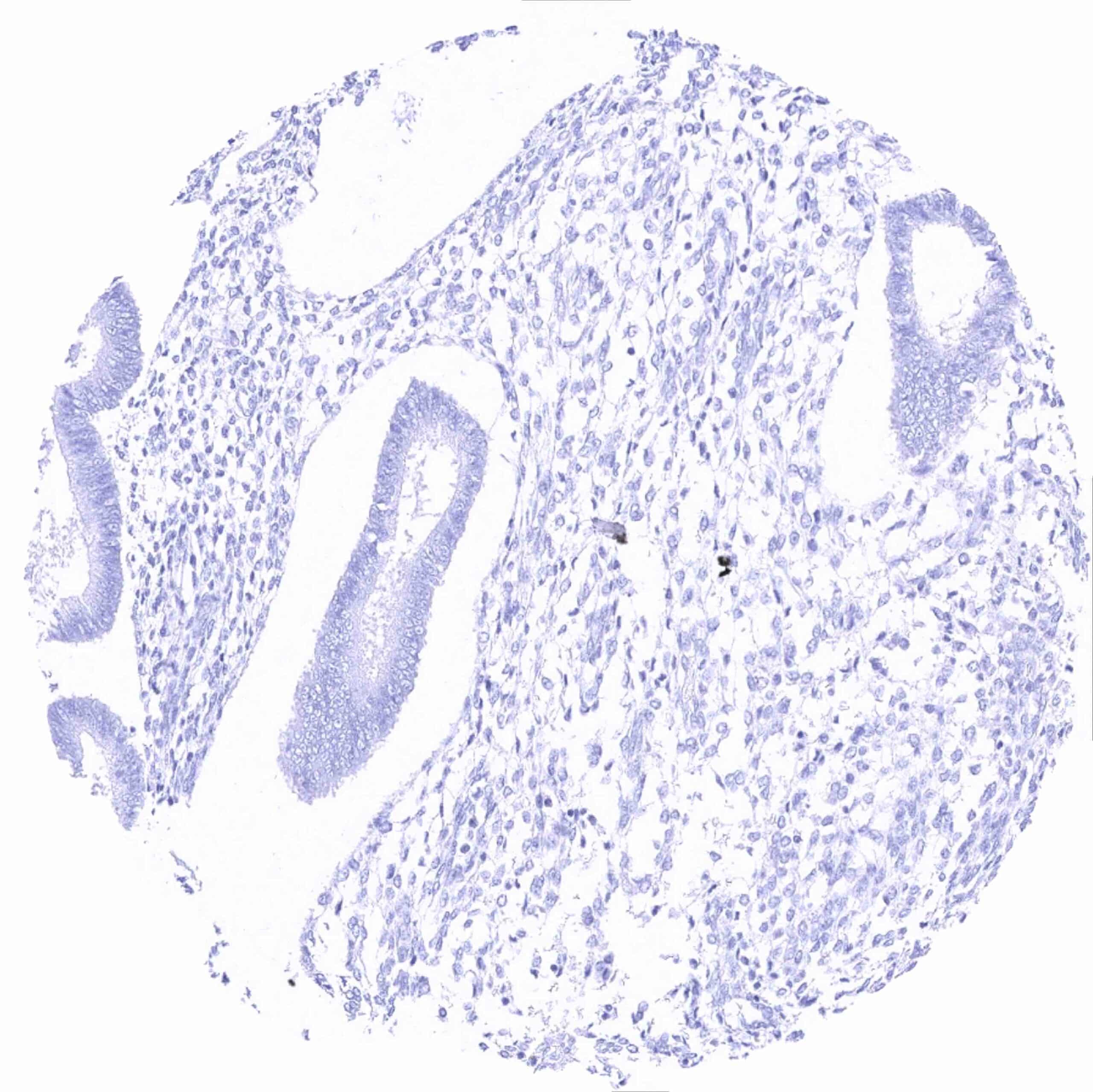
Uterus, endocervix
Uterus, endometrium (secretion) (2)
Uterus, endometrium (secretion)
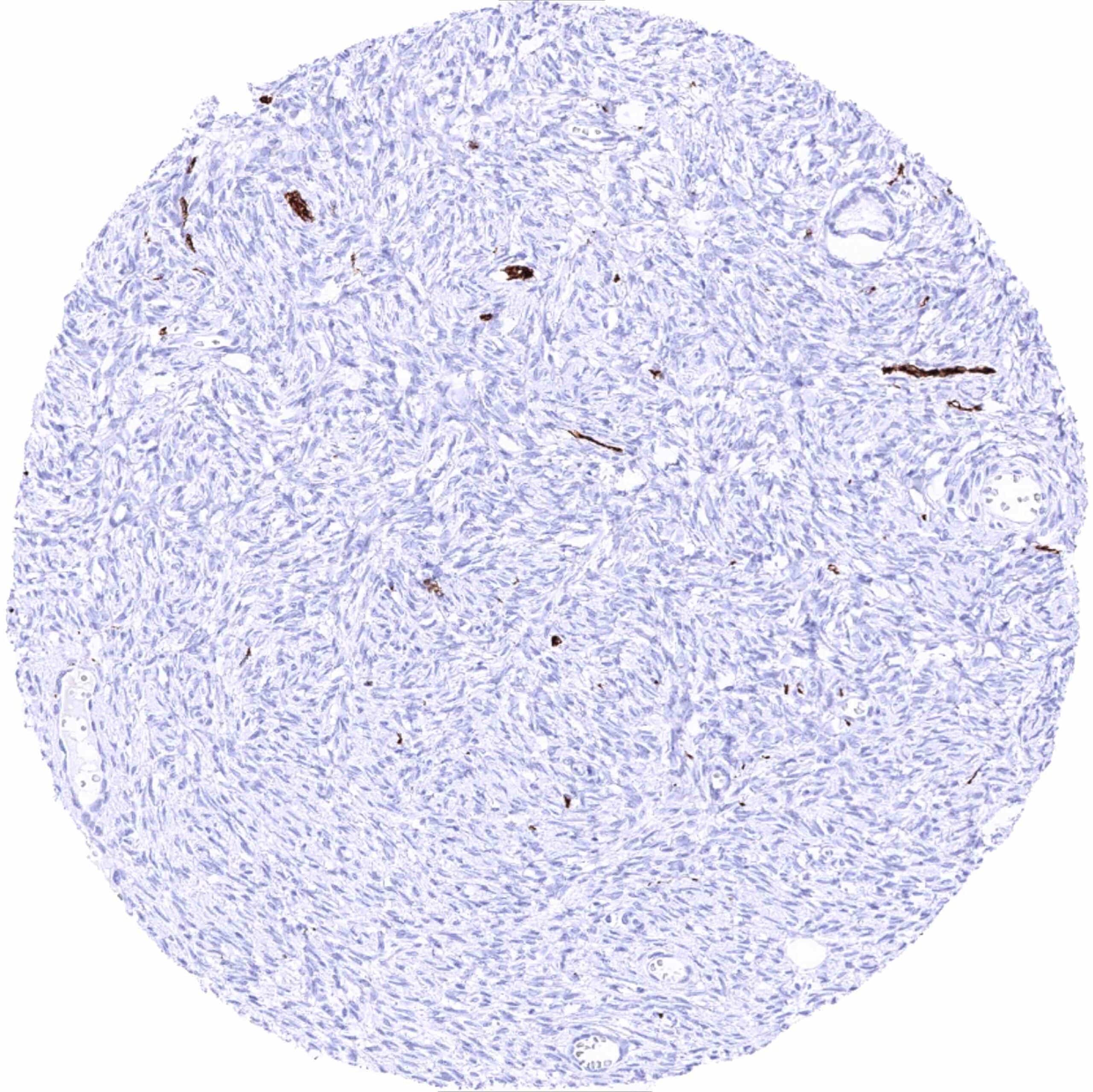
Uterus, myometrium