Adrenal gland
Aorta, media - CD141 staining of endothelial cells (CD141 immunohistochemistry).

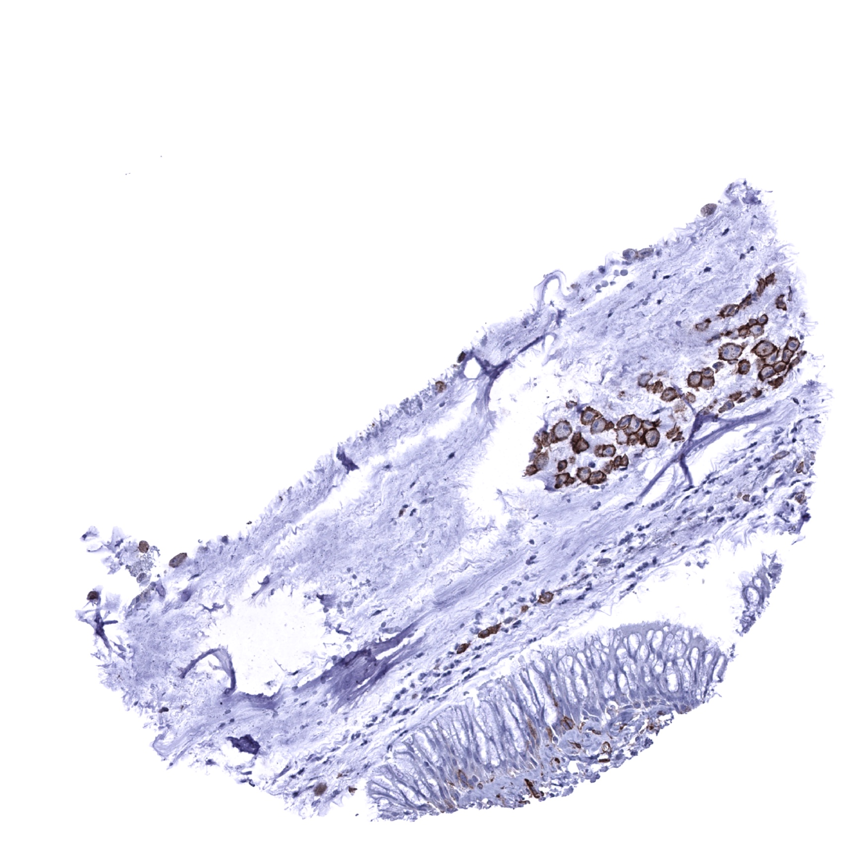
Appendix, mucosa

Appendix, muscular wall
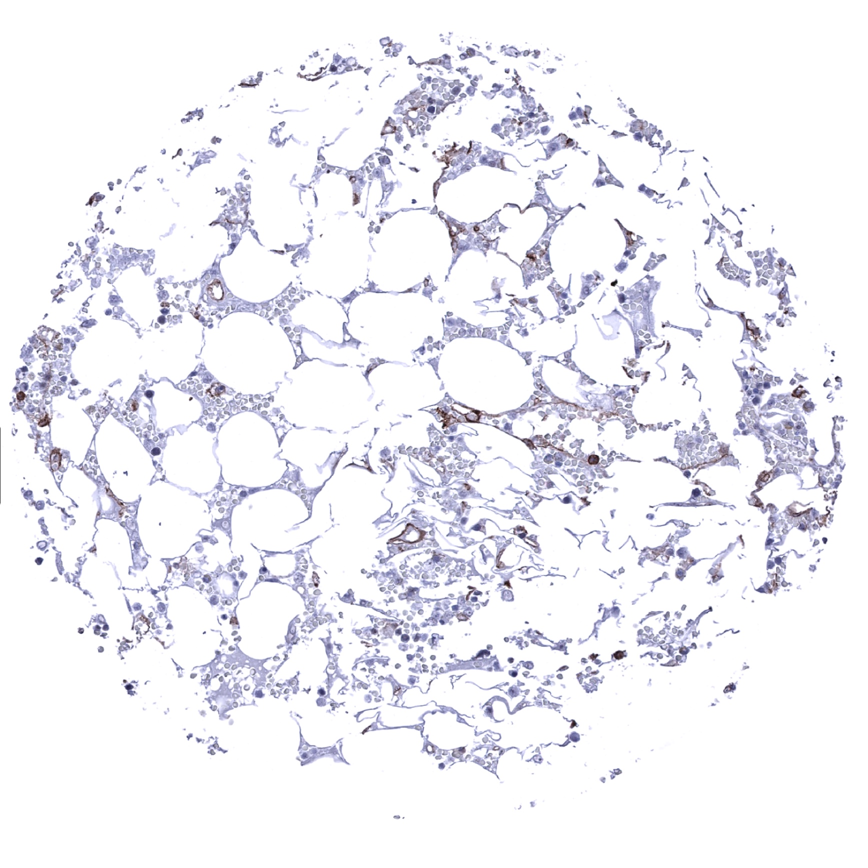
Bone marrow
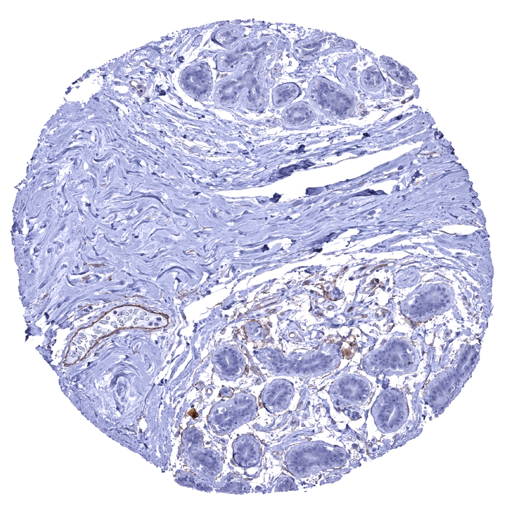
Breast
Bronchus, glands - Few basal cells of respiratory epithelium show a distinct CD141 staining, In addition, strong membranous CD141 positivity is seen in a group of alveolar macrophages.(CD141 immunohistochemistry)
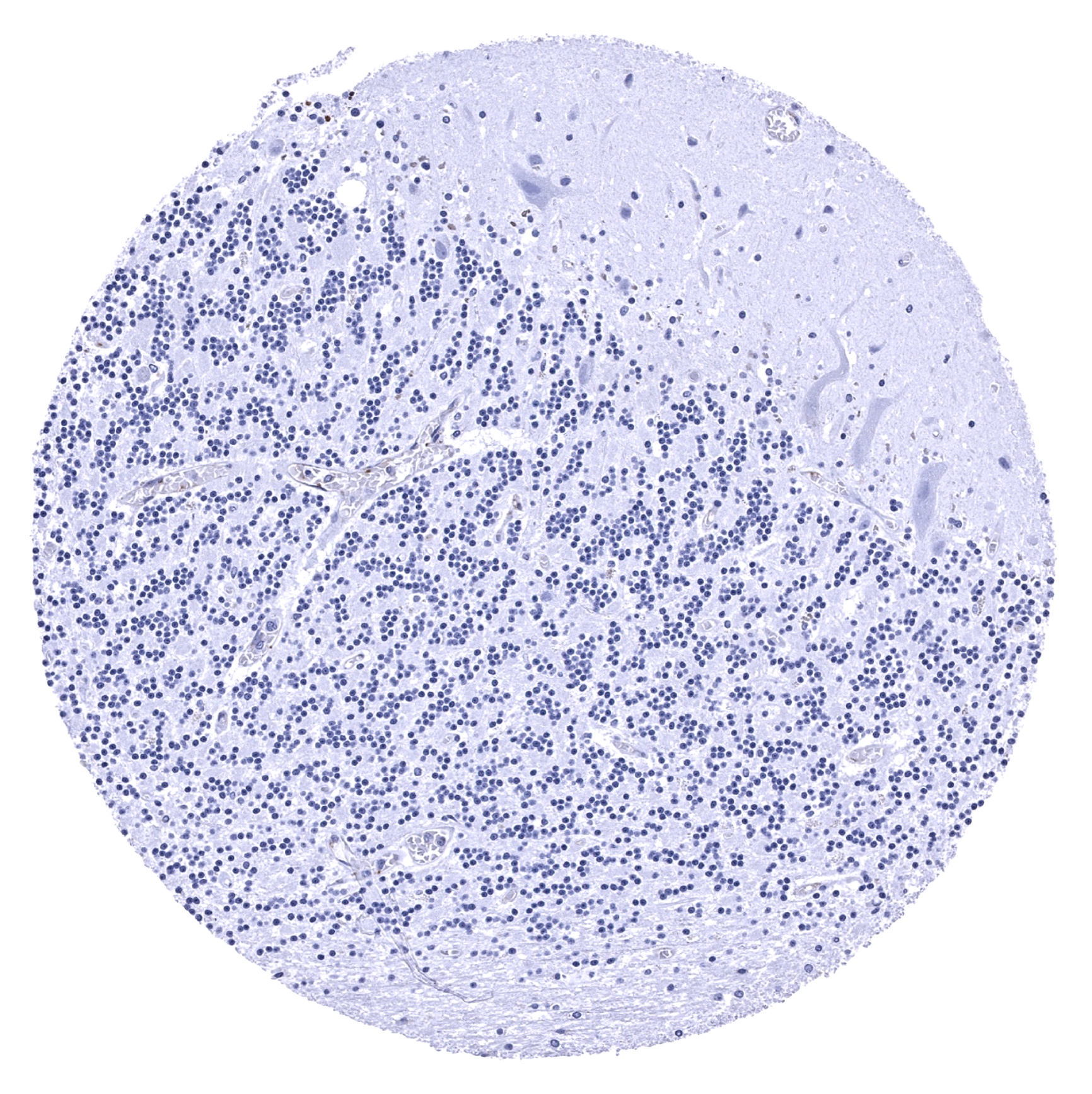
Cerebellum (molecular layer, Purkinje cell layer, granule cell layer)
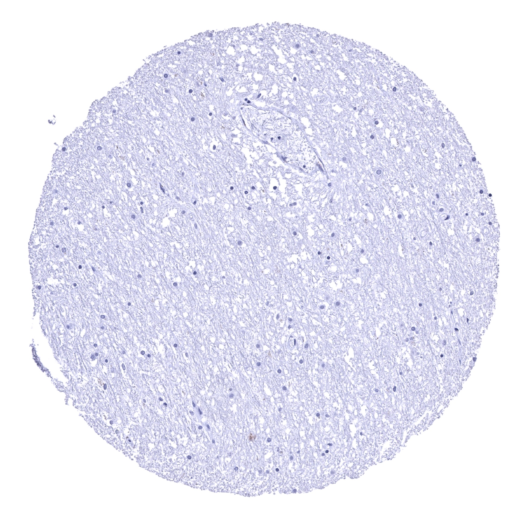
Cerebellum (white matter)

Cerebrum, grey matter

Cerebrum, white matter
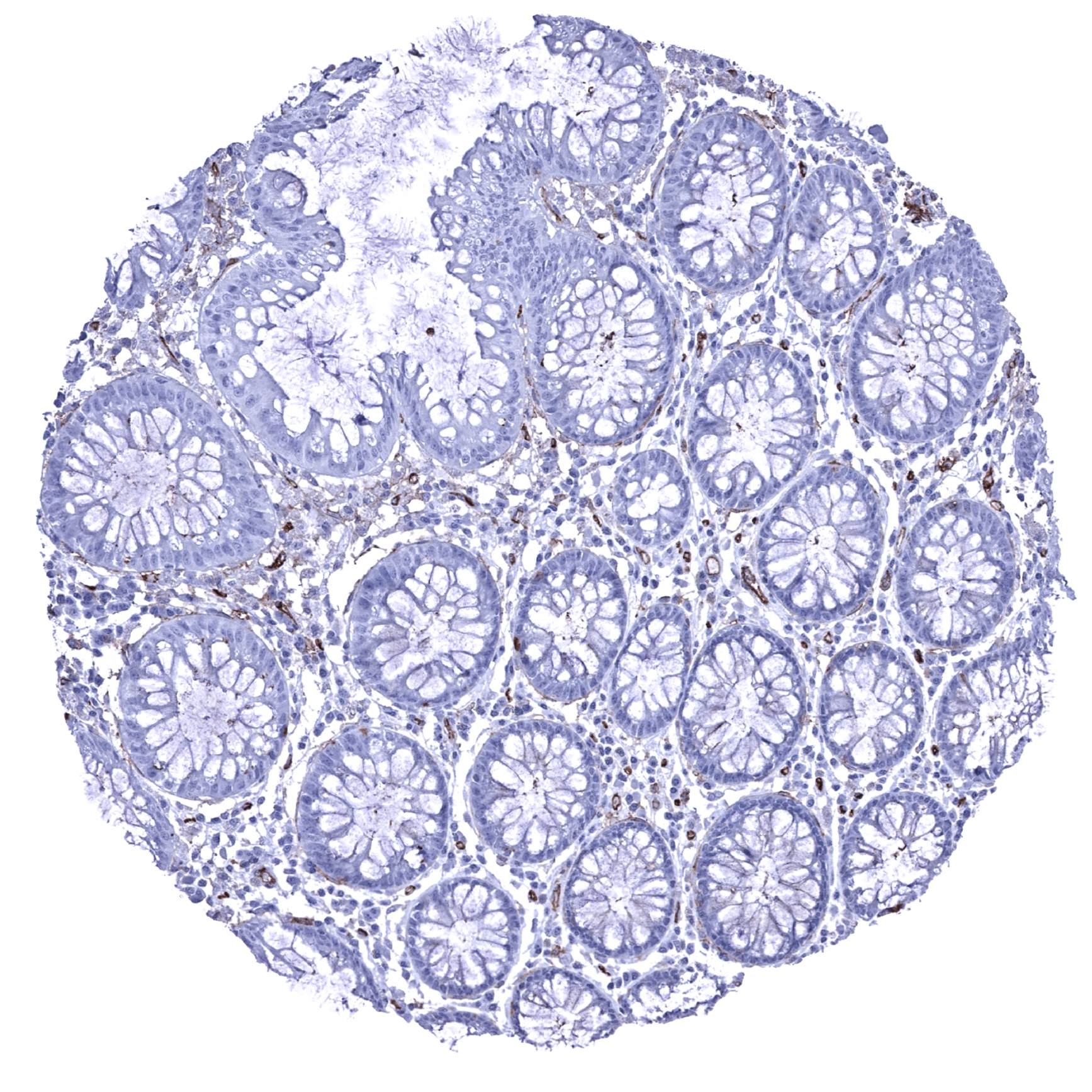
Colon descendens, mucosa
Colon descendens, muscular wall
Duodenum, Brunner gland

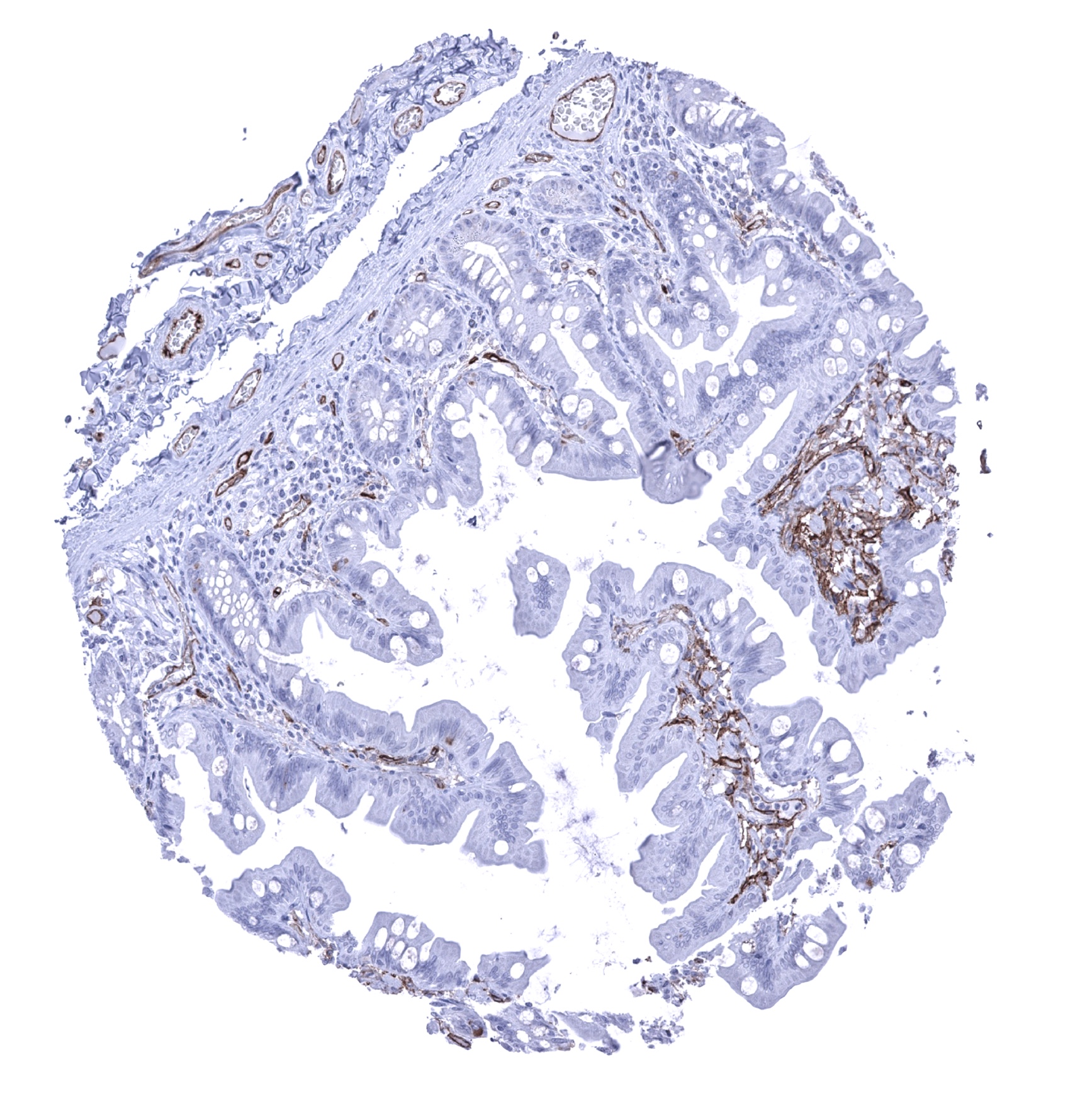
Duodenum, mucosa - Strong CD141 staining of endothelial cells (CD141 immunohistochemistry).
Epididymis
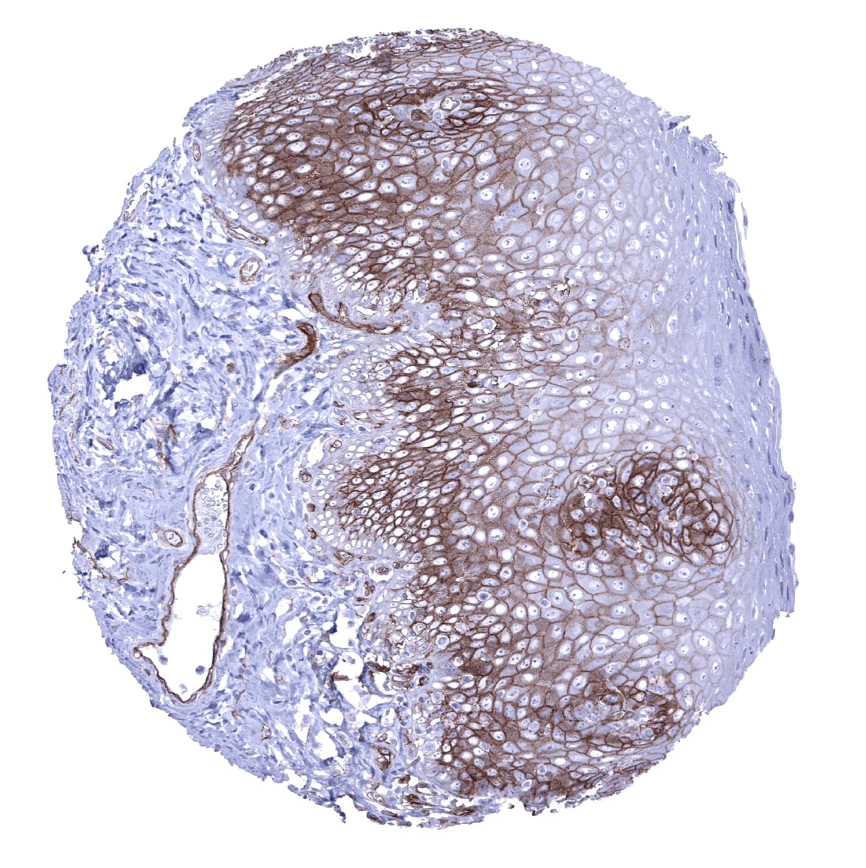
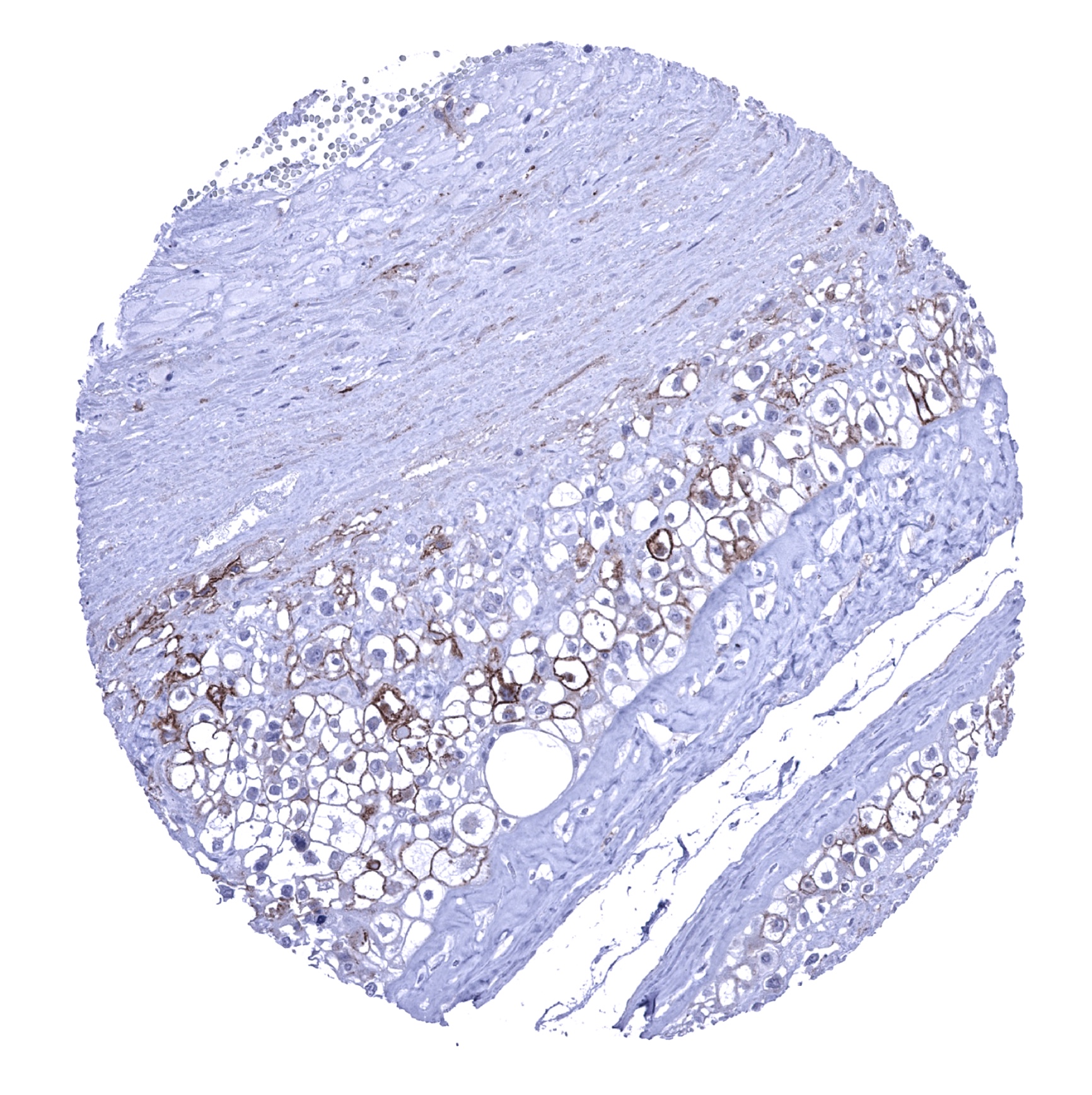
Esophagus, squamous epithelium - Strong membranous CD141 staining of suprabasal epithelial cells (CD141 immunohistochemistry).
Fallopian tube, mucosa

Fat

Gallbladder, epithelium
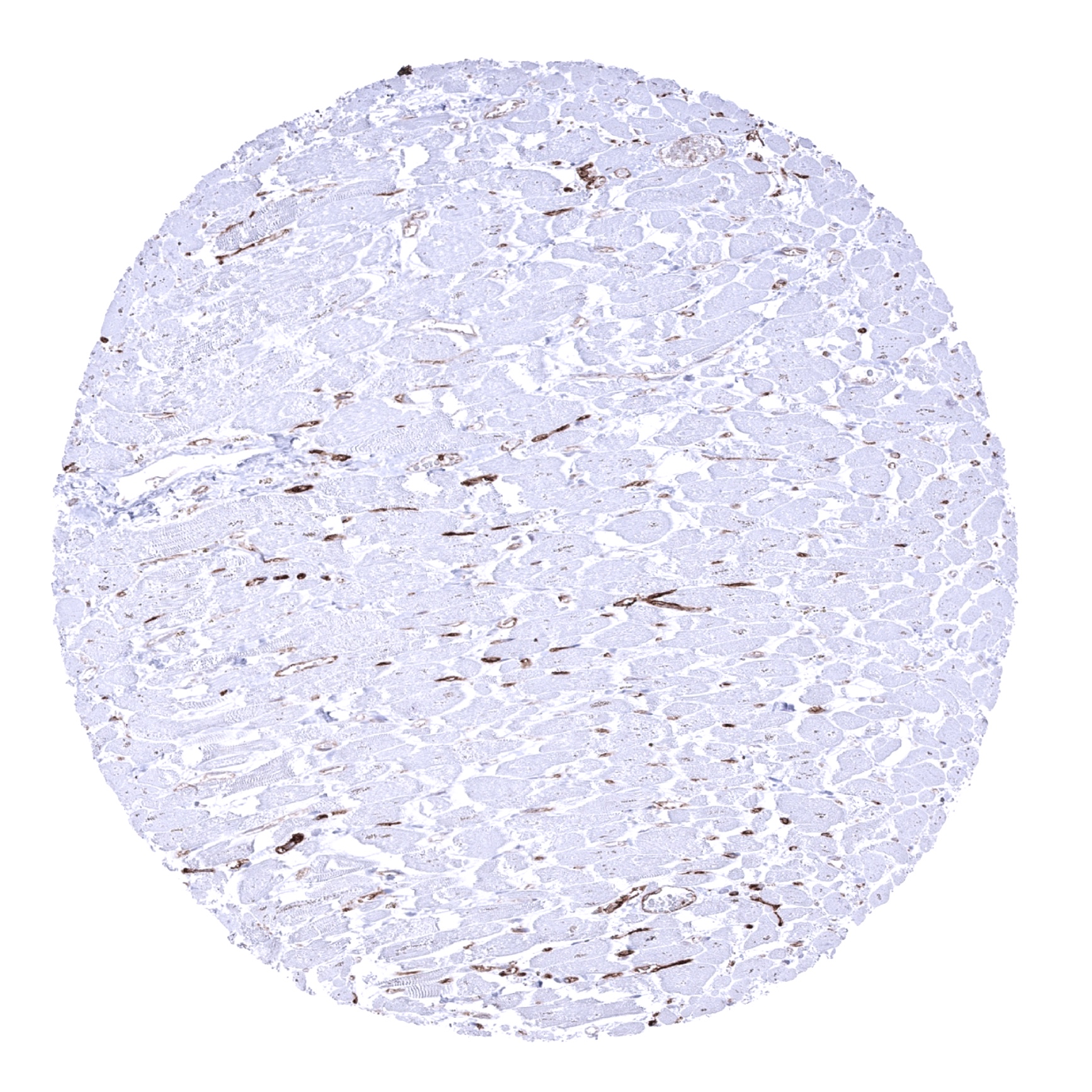
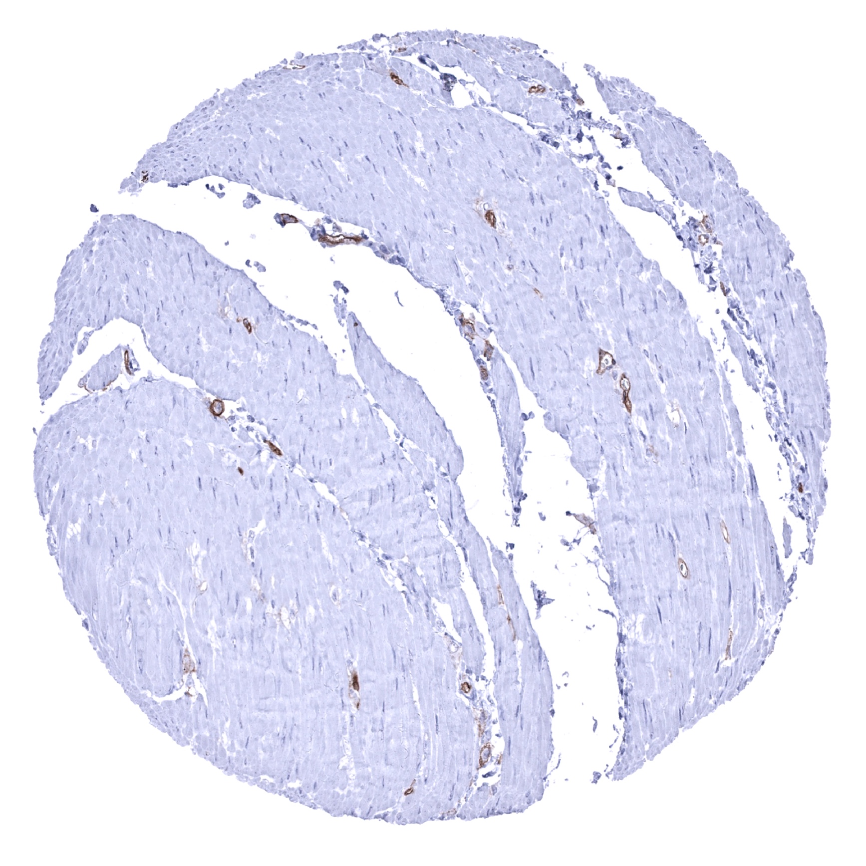
Heart muscle - CD141 staining of endothelial cells (CD141 immunohistochemistry).
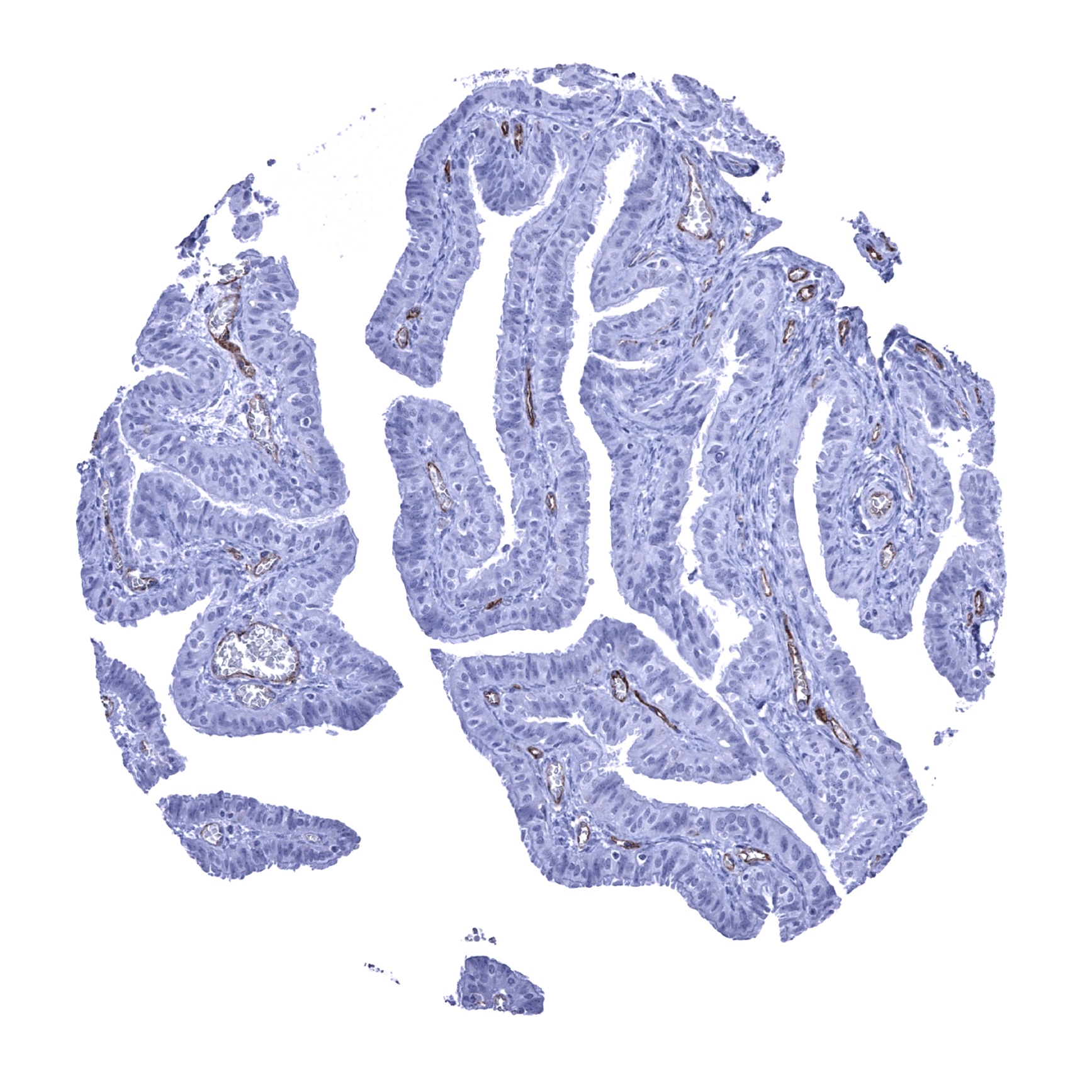
Ileum, mucosa - Strong CD141 staining of endothelial cells (CD141 immunohistochemistry).
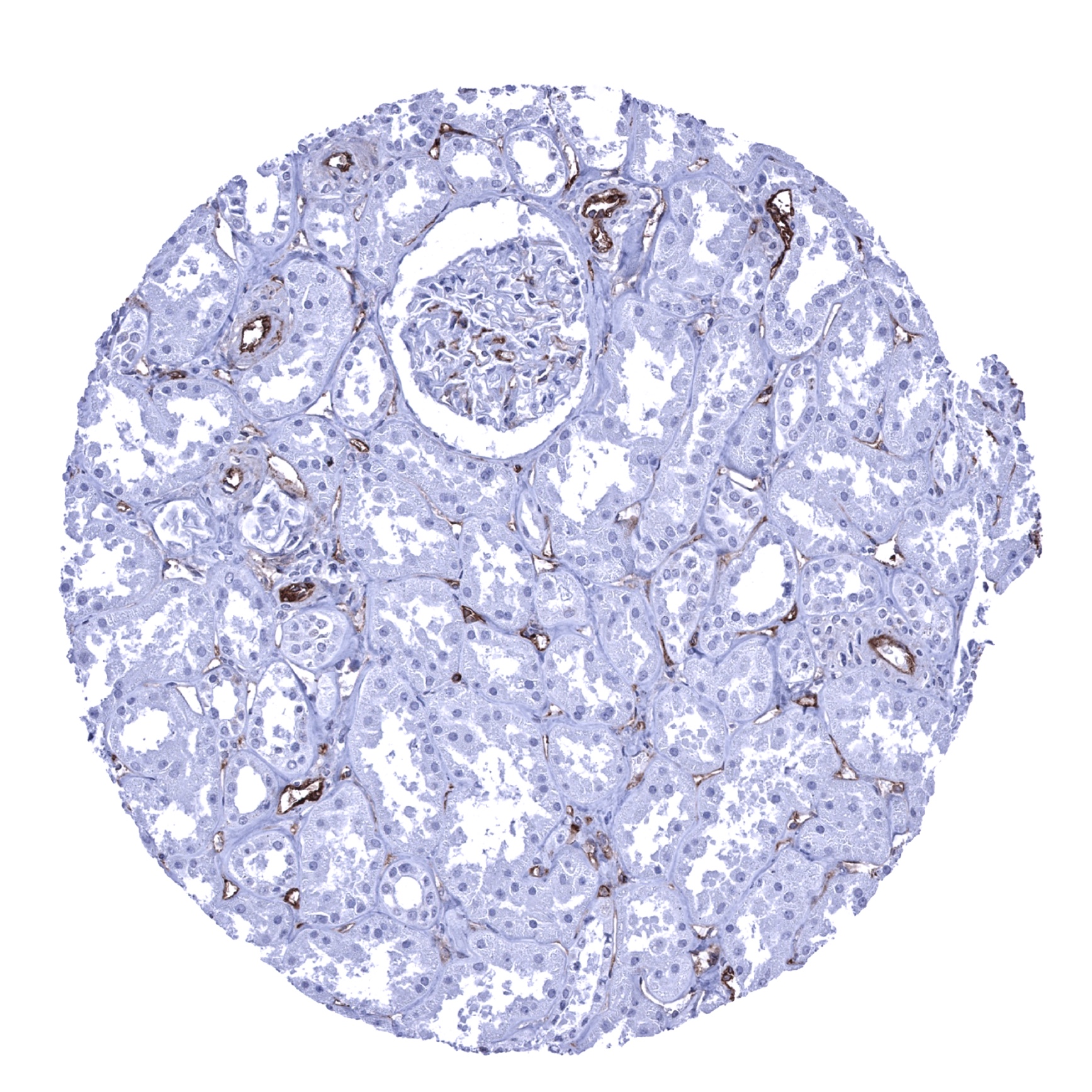
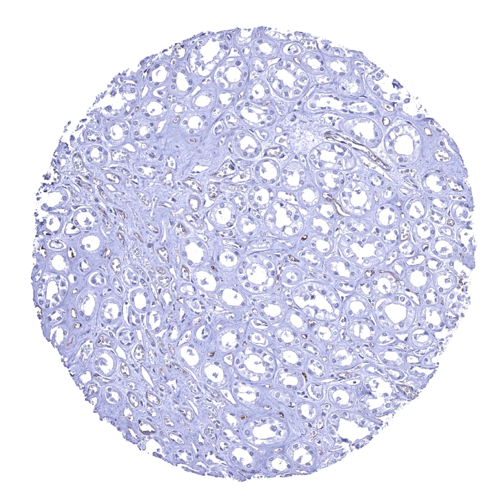
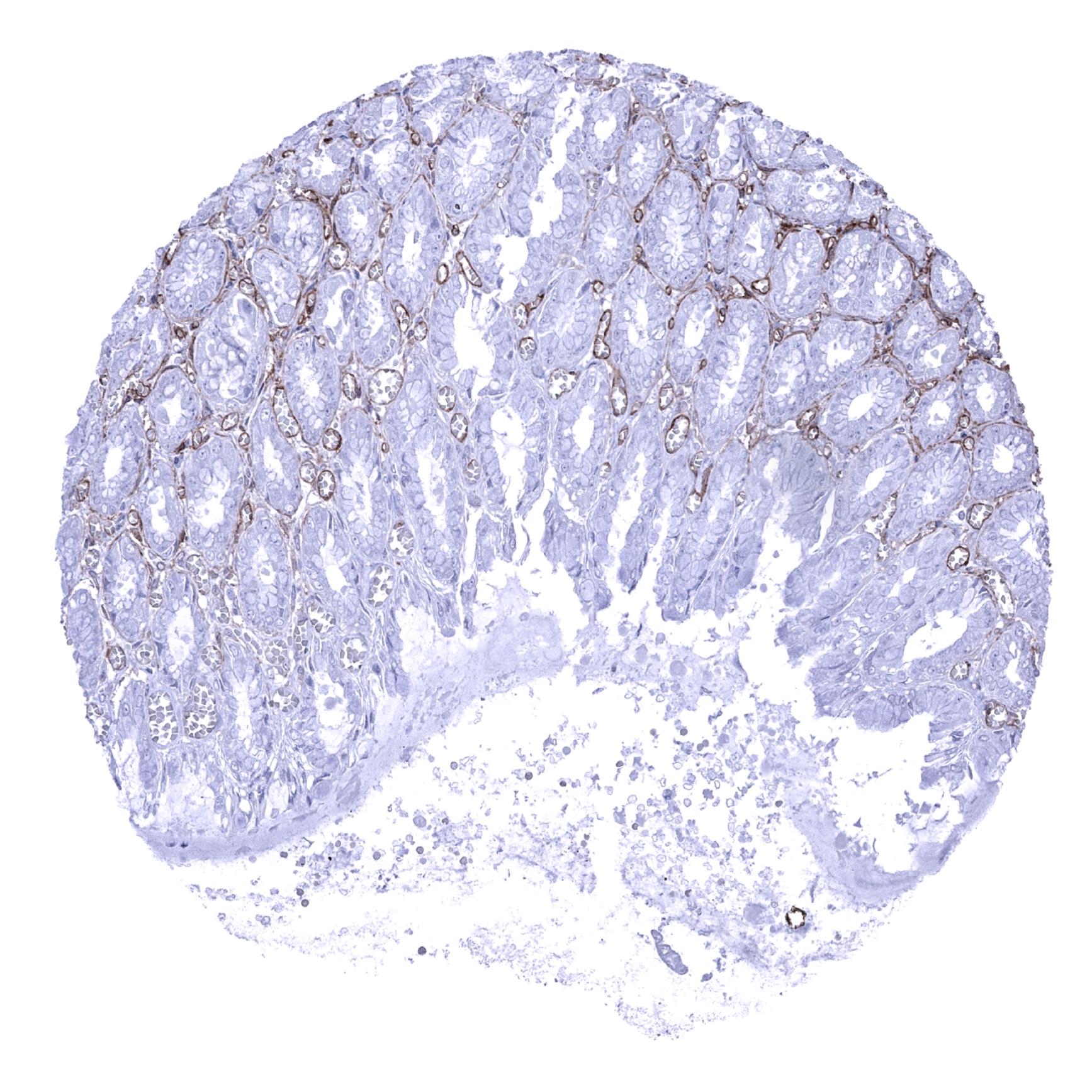
Kidney, cortex - Strong CD141 staining of endothelial cells (CD141 immunohistochemistry).
Kidney, medulla

Kidney, pelvis, urothelium - Faint membranous CD141 staining of urothelial cells (CD141 immunohistochemistry).
Kidney, pelvis, urothelium - Weak membranous CD141 staining of urothelial cells (CD141 immunohistochemistry)
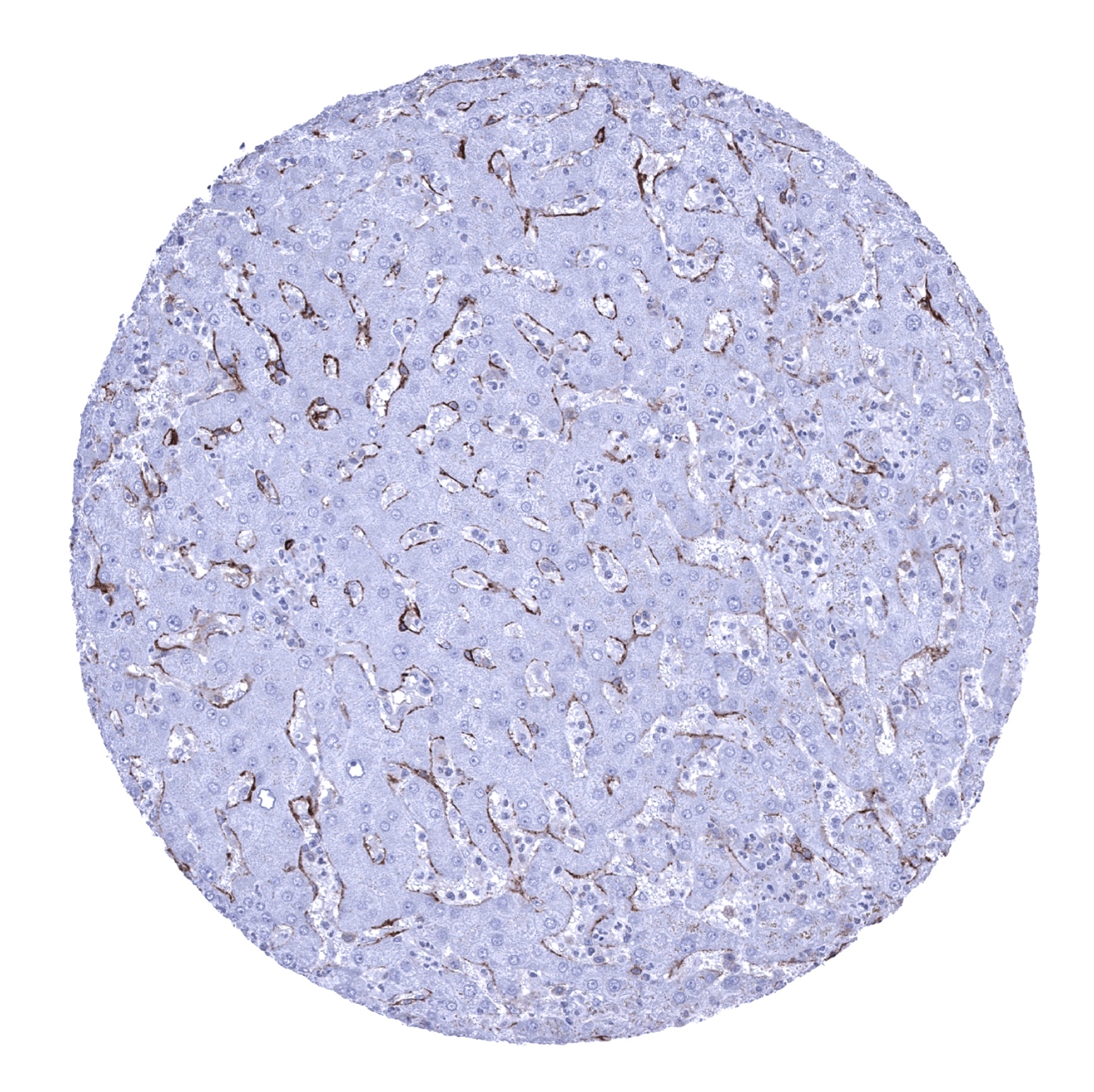
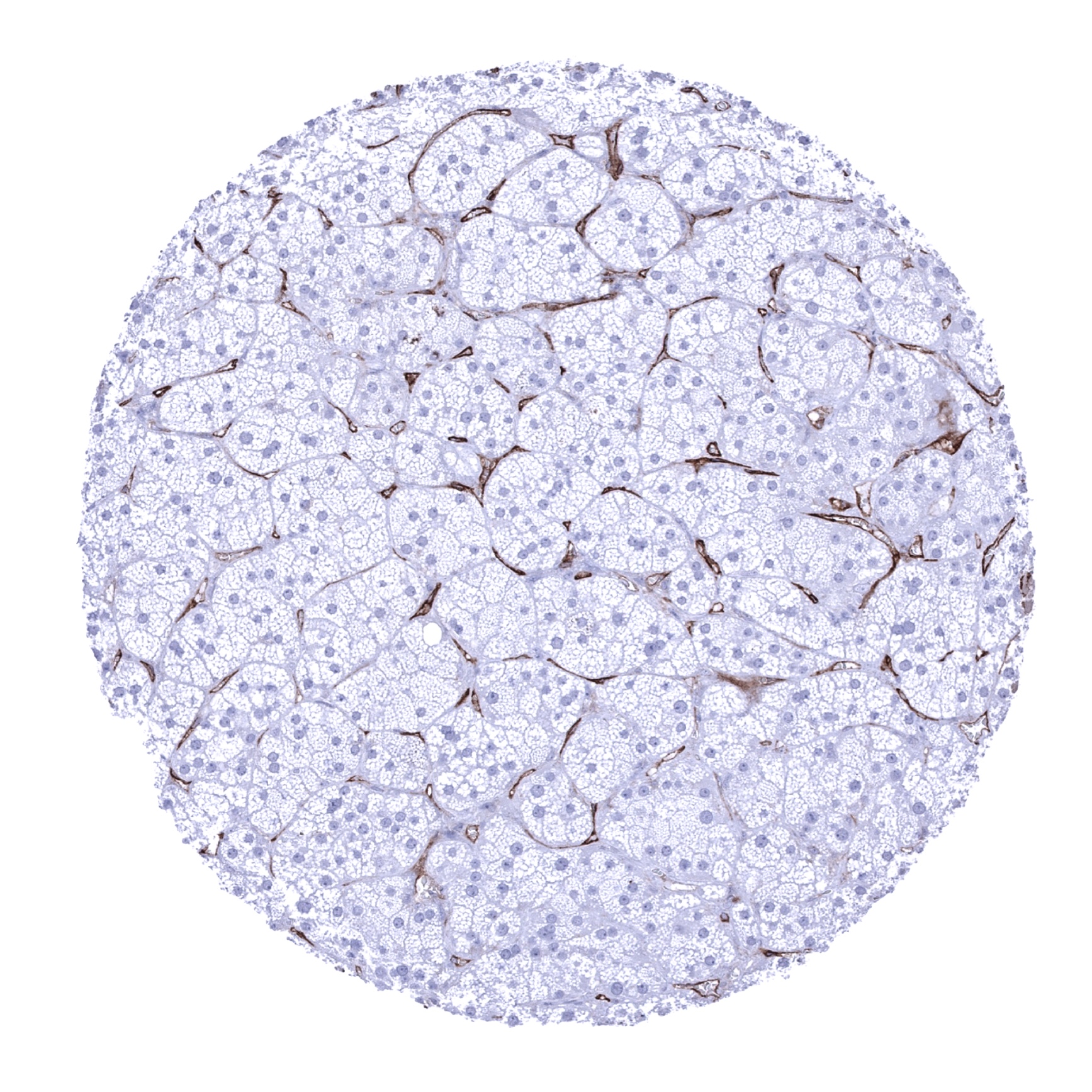
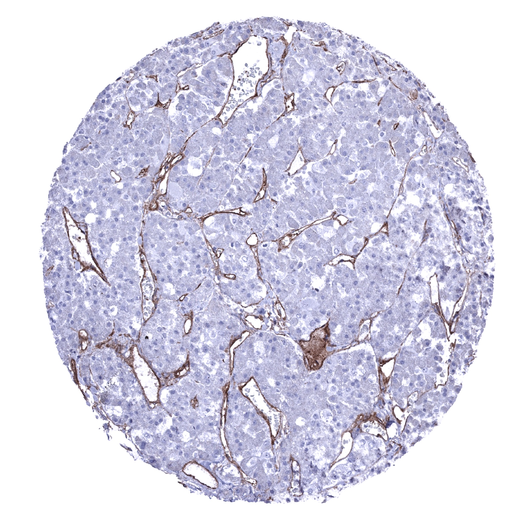
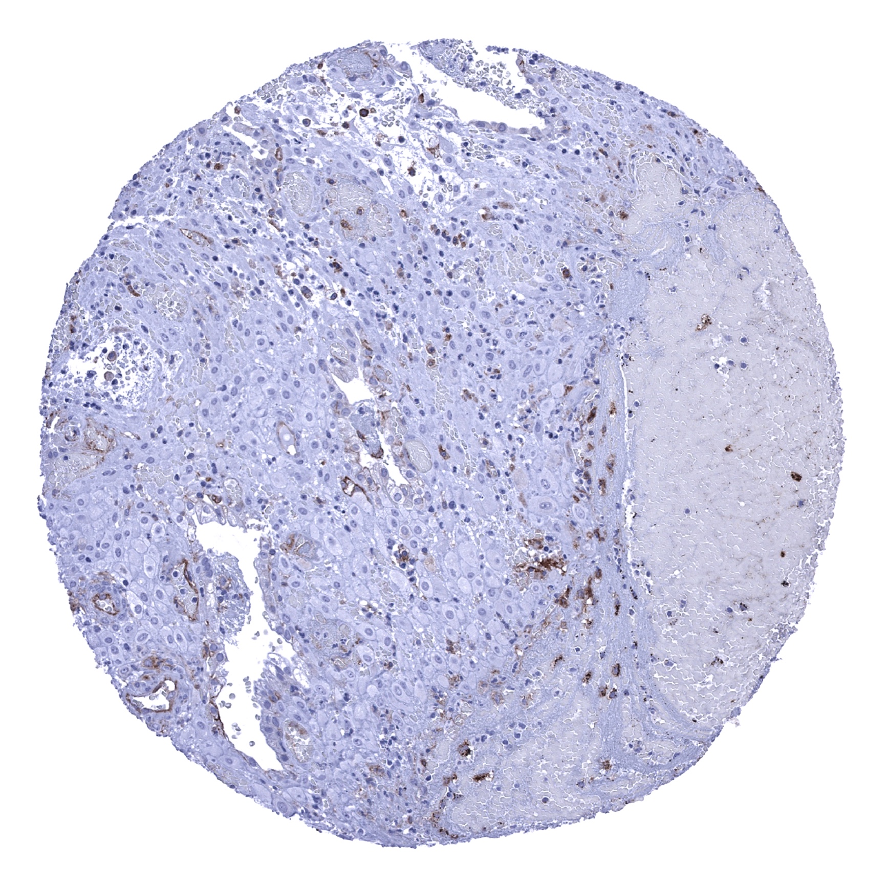
Liver - Moderate CD141 staining of sinusoideal cells (CD141 immunohistochemistry).
Lung - Strong CD141 staining of endothelial cells and of alveolar macrophages (CD141 immunohistochemistry).
Lymph node - CD141 staining primarily occurs in endothelial cells (CD141 immunohistochemistry).
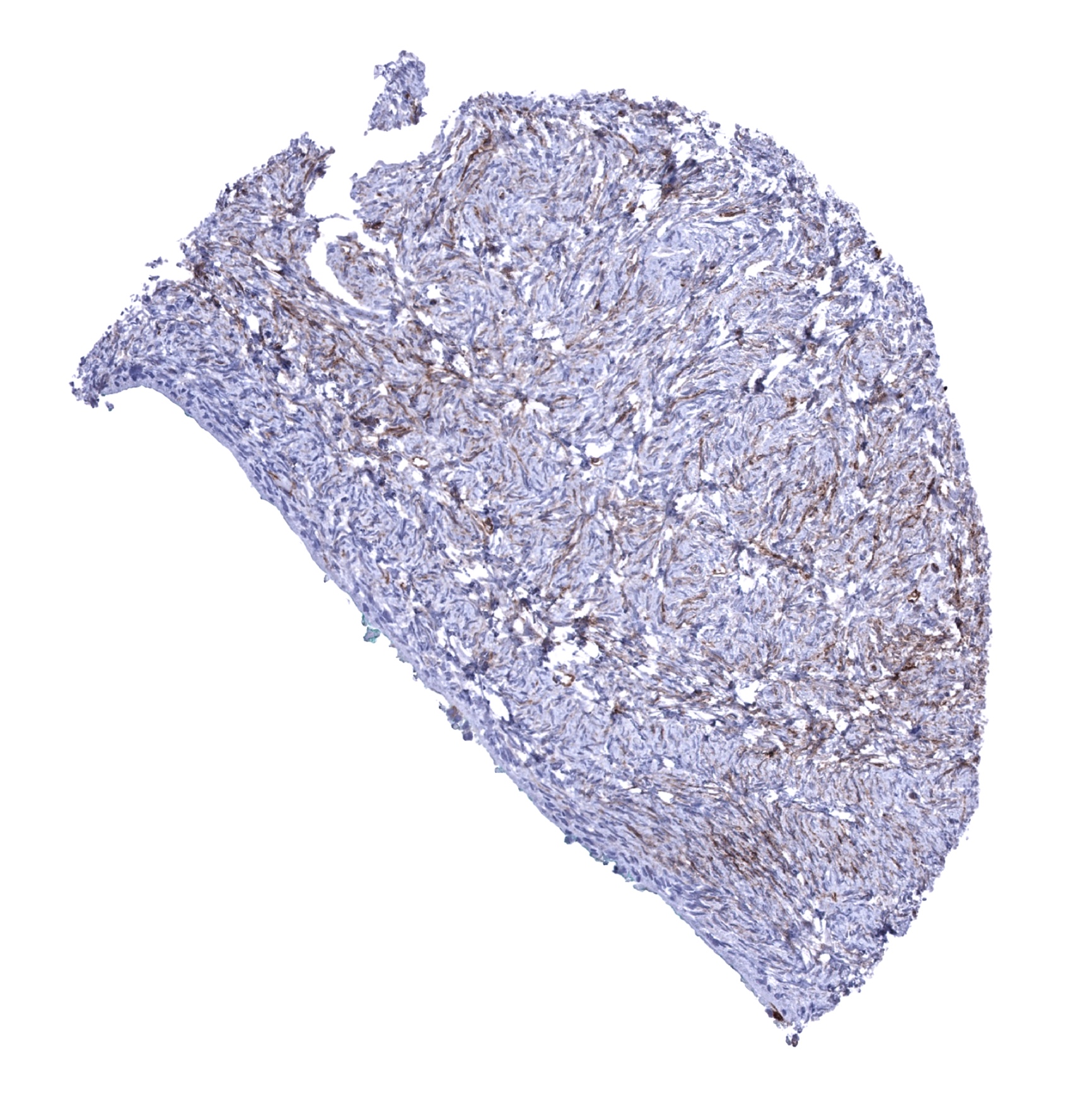
Ovary, stroma - Weak CD141 staining of stromal cells (CD141 immunohistochemistry).
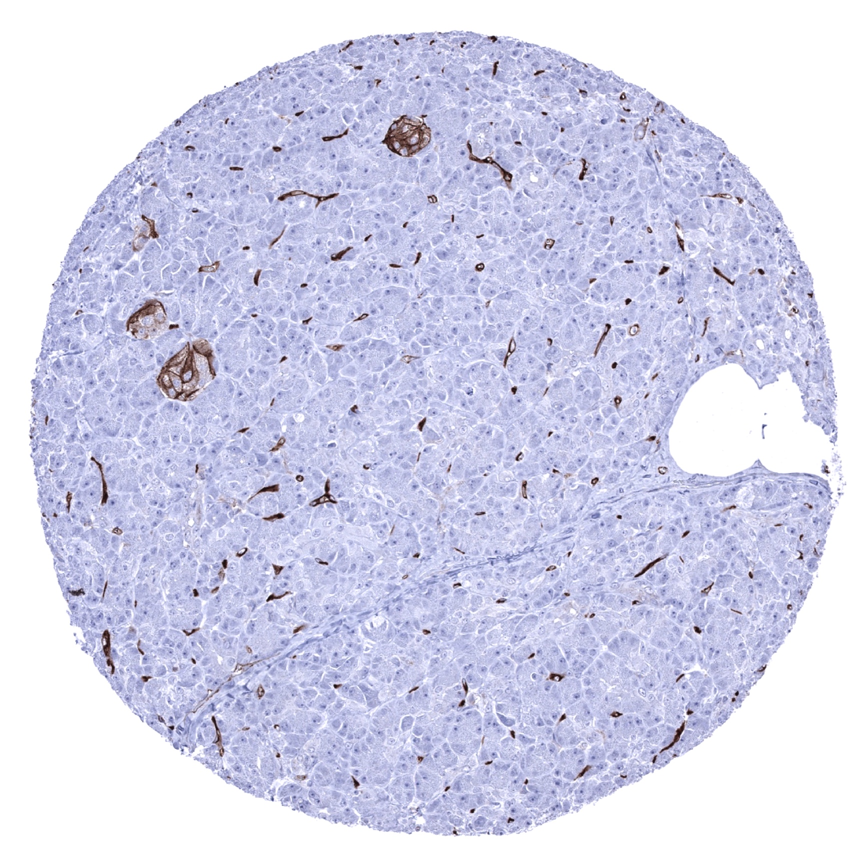
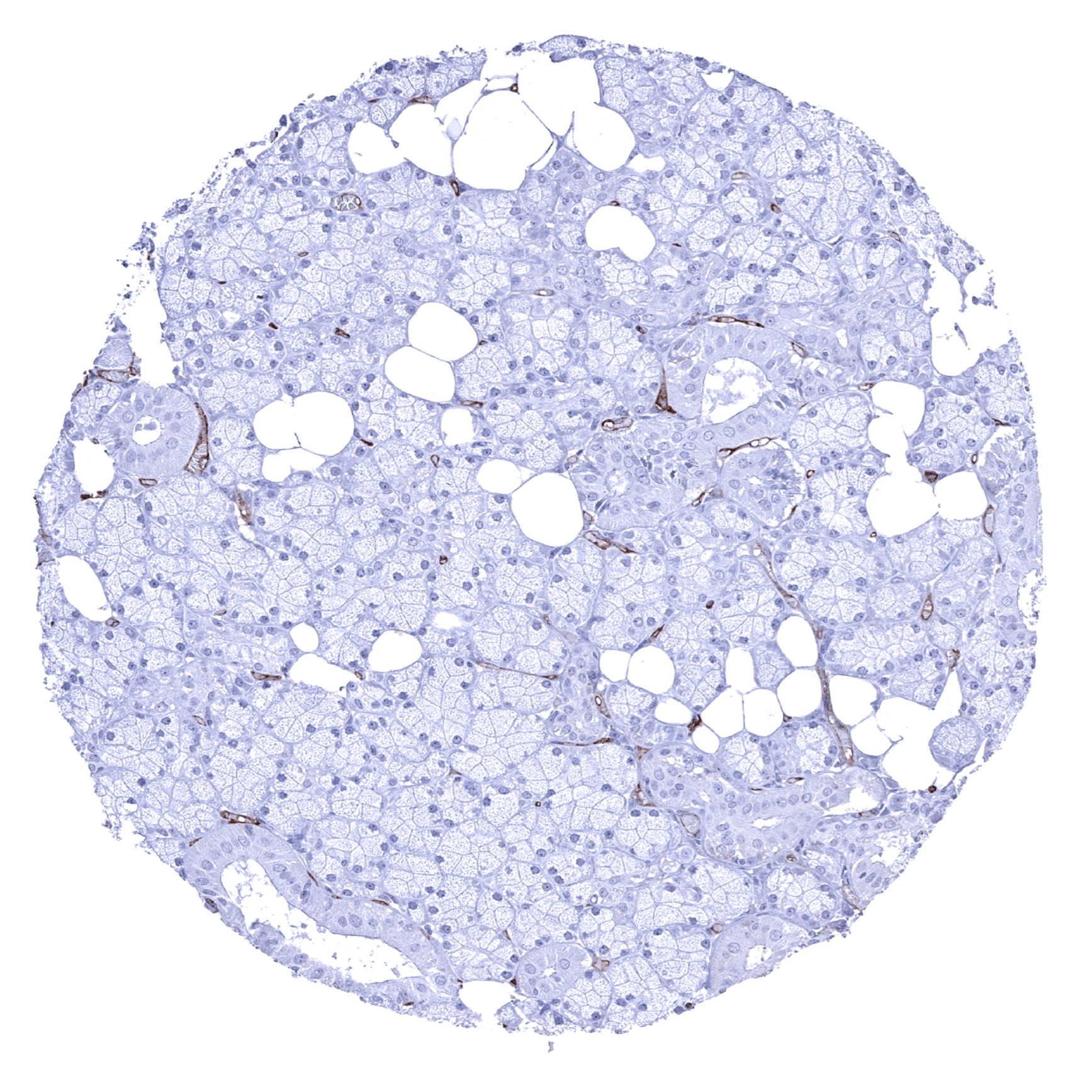
Pancreas - Most cells of islets and endothelial cells show CD141 staining (CD141 immunohistochemistry).
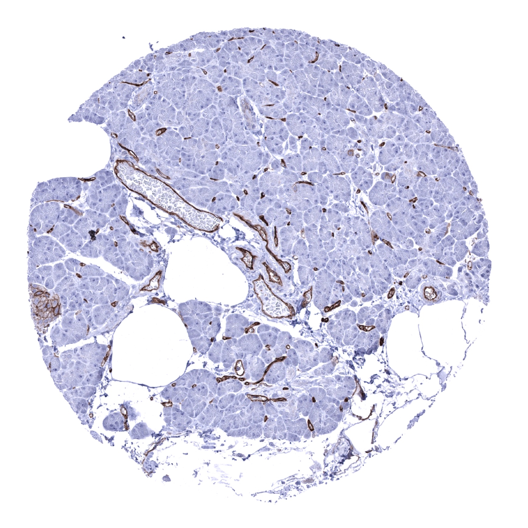
Pancreas - Most cells of pancreatic islets and endothelial cells show CD141 staining (CD141 immunohistochemistry).

Parathyroid gland
Parotid gland
Pituitary gland, anterior lobe - CD141 staining is limited to endothelial cells in this sample (CD141 immunohistochemistry).
Pituitary gland, posterior lobe - In this sample, a strong membranous CD141 staining occurs in a large fraction of epithelial cells (CD141 immunohistochemistry).
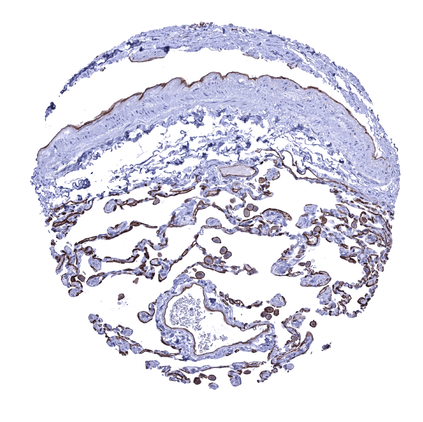
Placenta (amnion and chorion) - Moderate to strong membranous CD141 positivity of amnion cells (CD141 immunohistochemistry).
Placenta (amnion and chorion) - Moderate to strong membranous CD141 staining of chorion cells (CD141 immunohistochemistry).
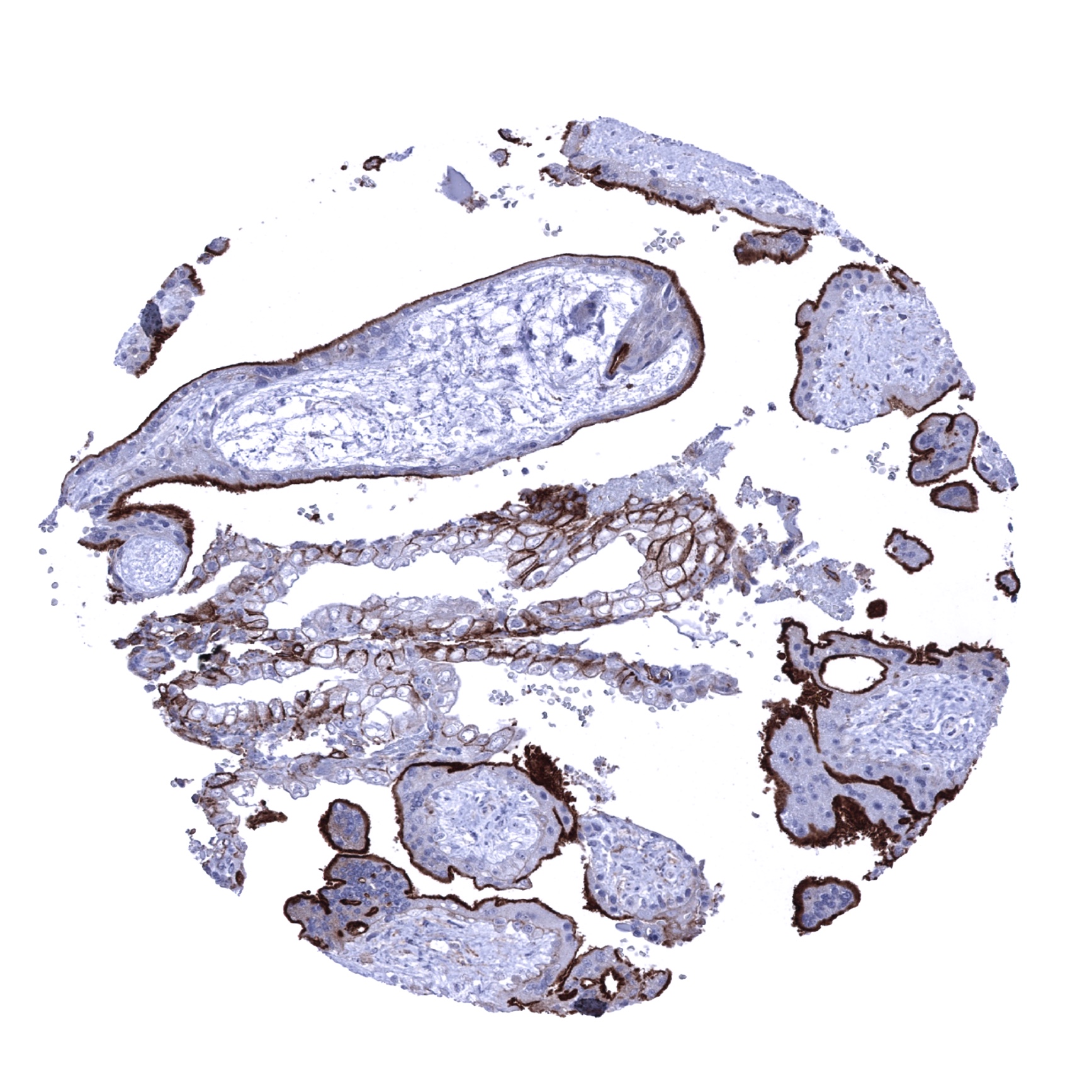
Placenta, early - Strong CD141 staining of the surface cell layer of the cytotrophoblast. In addition, a moderate staining occurs in endometrial cells exhibiting an „Arias-Stella-phenomenon“
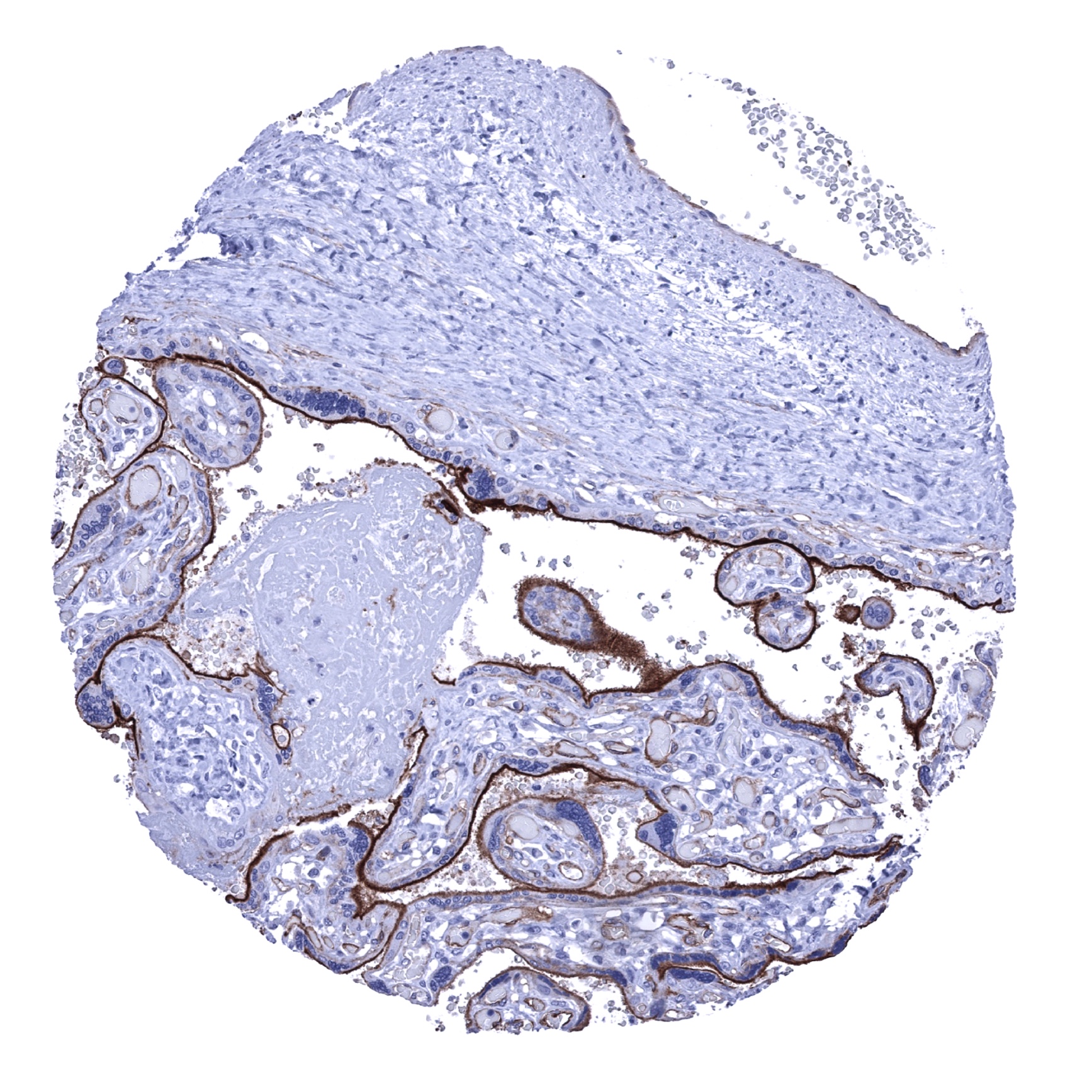
Placenta, mature - Strong CD141 staining of the surface cell layer of the cytotrophoblast (CD141 immunohistochemistry).
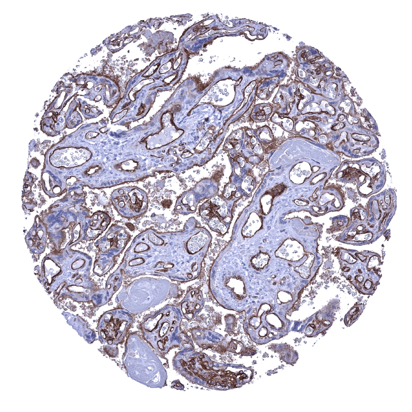
Placenta, mature - Strong CD141 staining of the surface cell layer of the cytotrophoblast and of endothelial cells (CD141 immunohistochemistry).
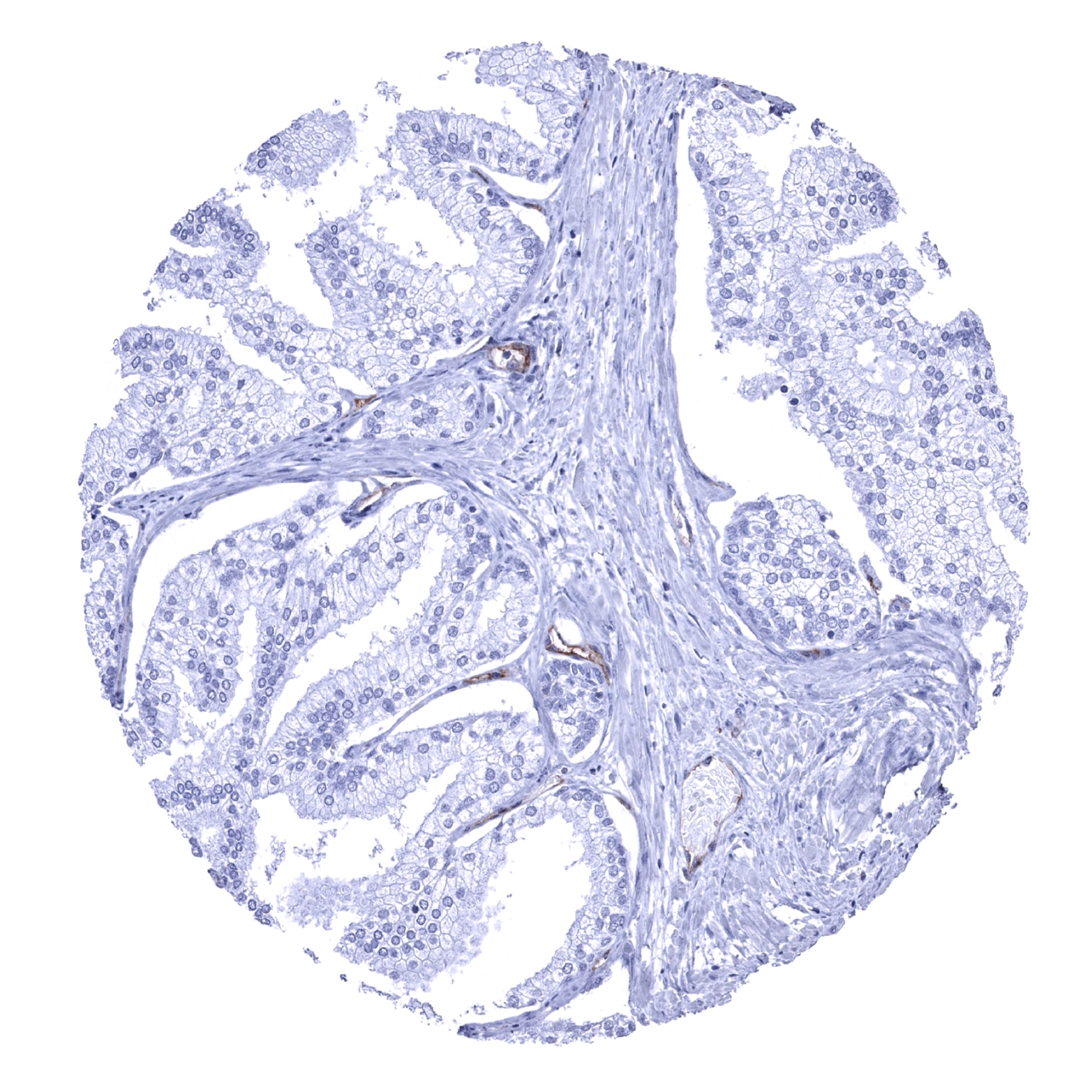
Prostate - Faint CD141 staining of some basal cells (CD141 immunohistochemistry)
Seminal vesicle
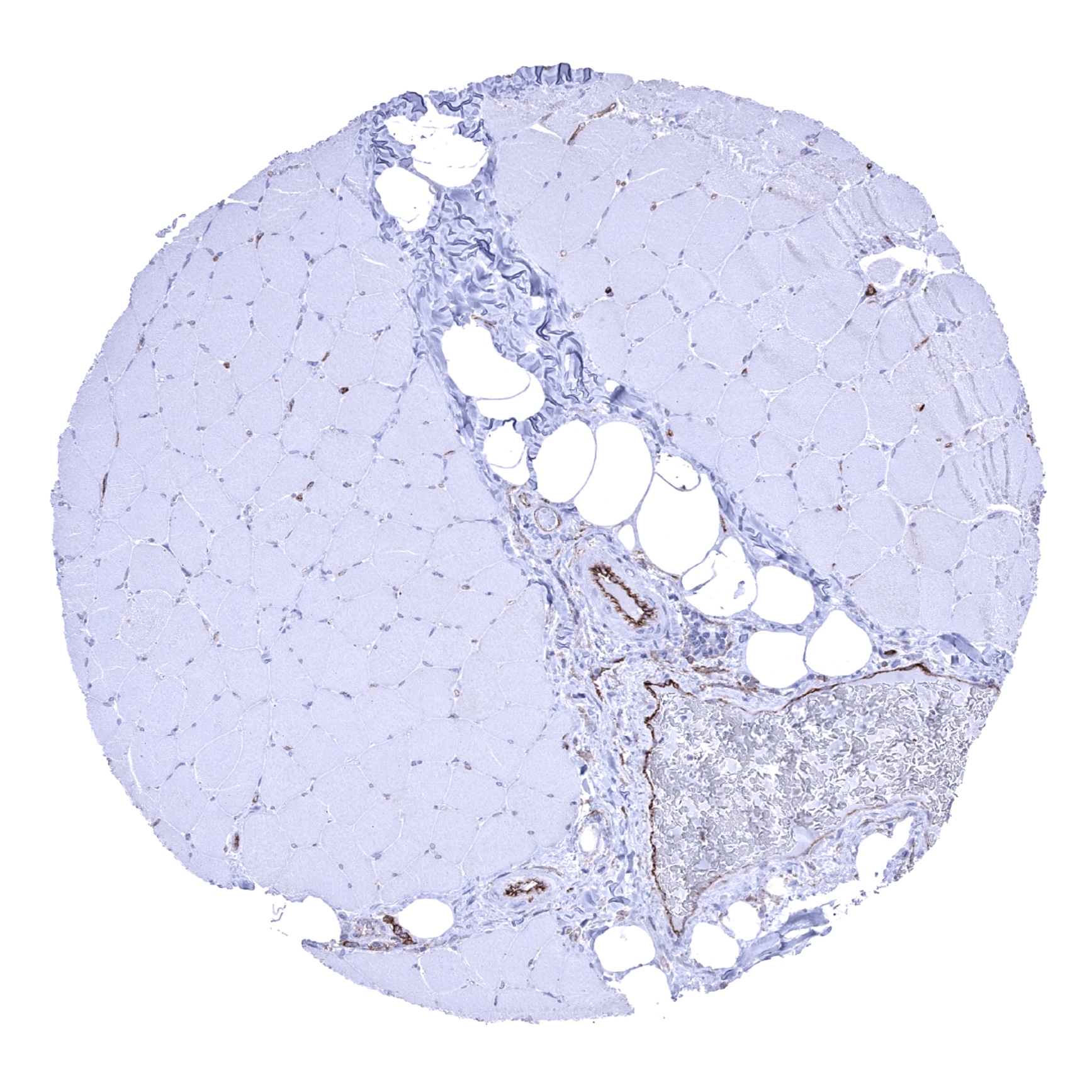
Skeletal muscle - CD141 staining of endothelial cells (CD141 immunohistochemistry).
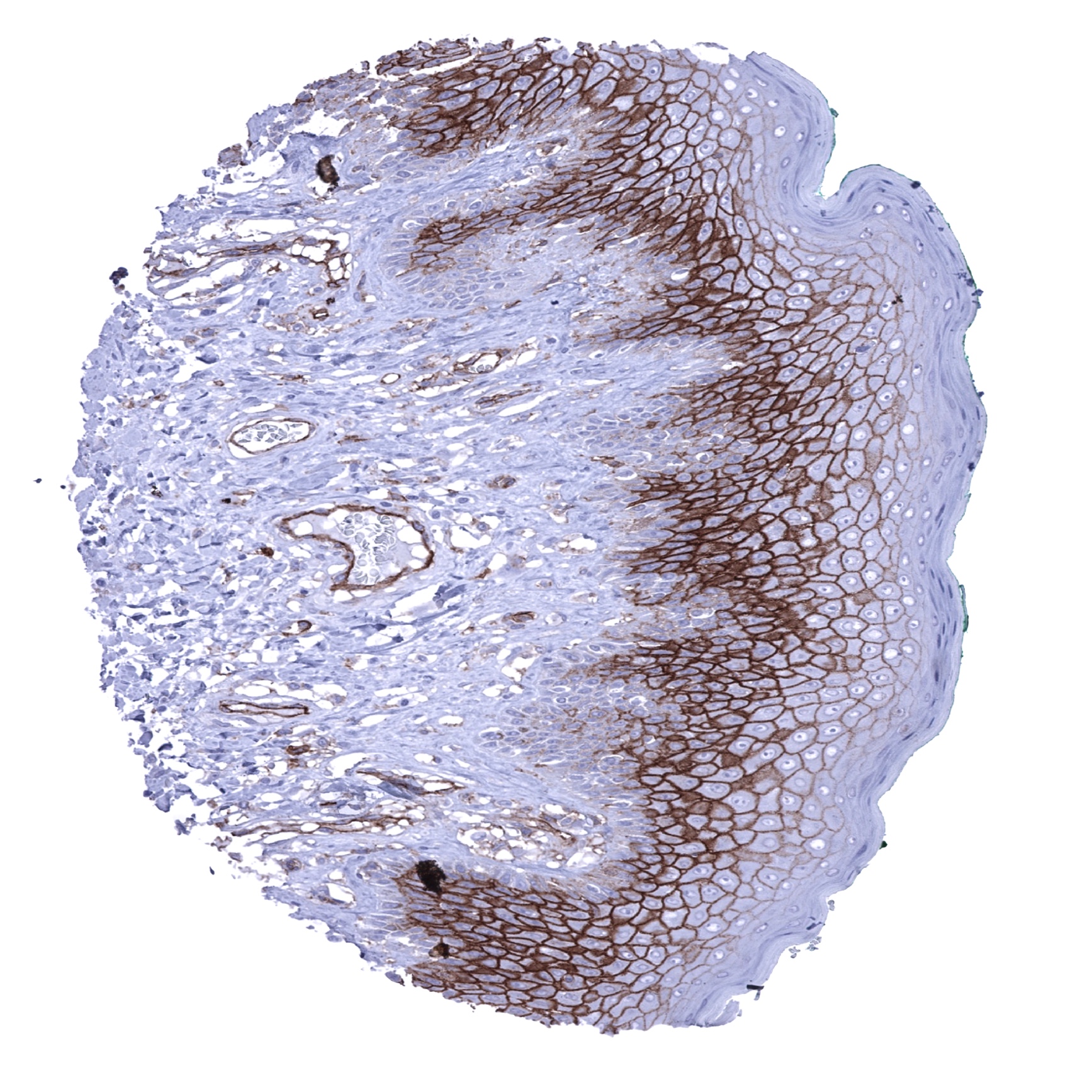
Skin - Strong membranous CD141 staining of suprabasal epithelial cells (CD141 immunohistochemistry).

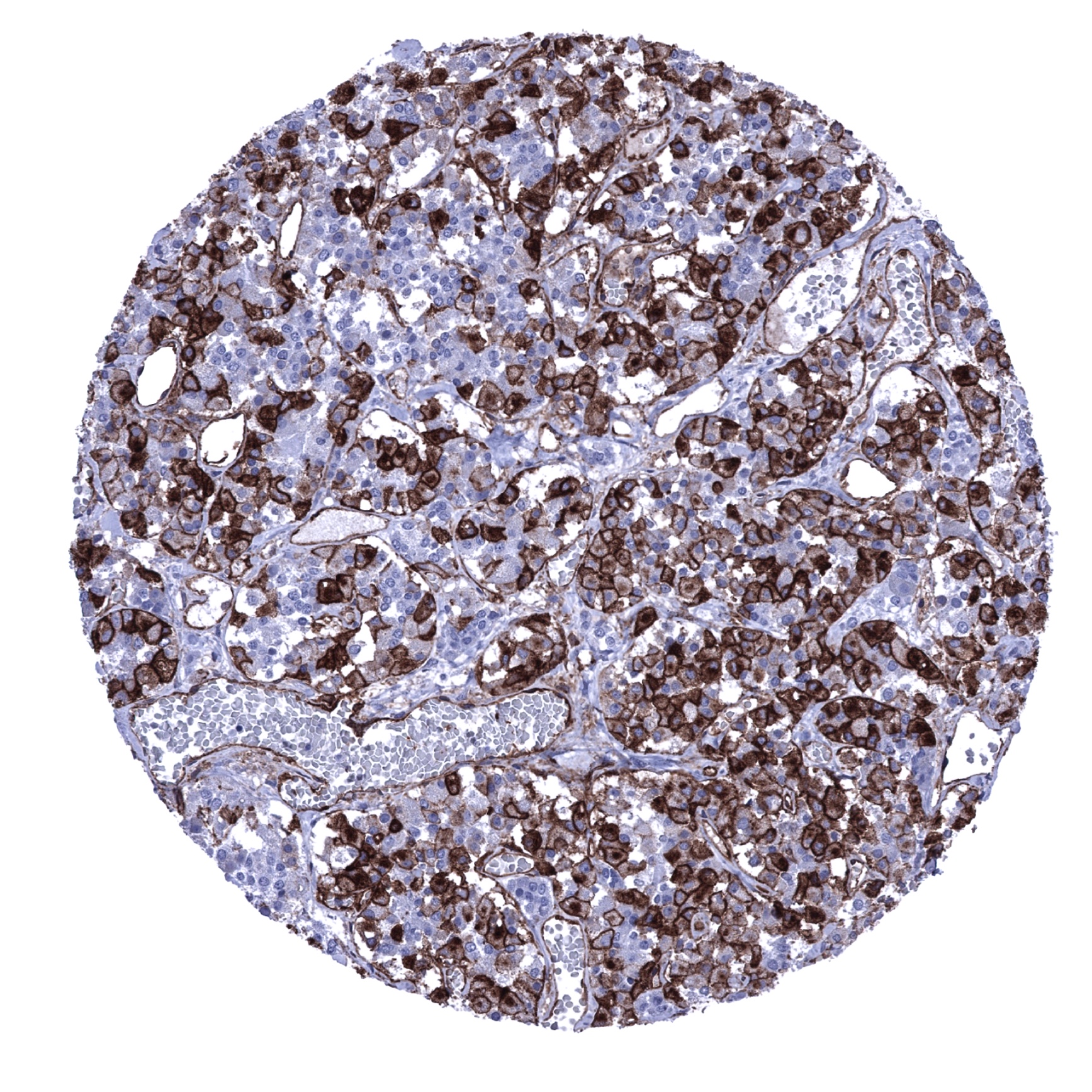
Spleen - Strong CD141 positivity of littoral cells in the spleen (CD141 immunohistochemistry).
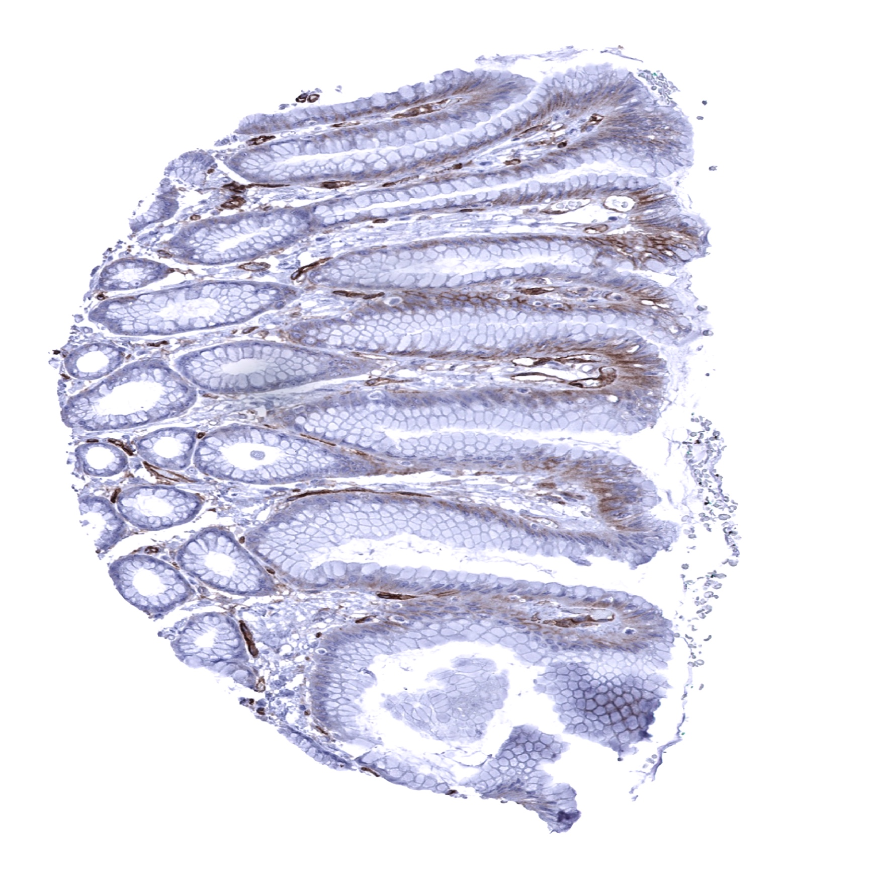
Stomach, antrum - A membranous CD141 staining is seen in basolateral membranes of the surface epithelium (CD141 immunohistochemistry).
Stomach, corpus - CD141 staining ofendothelial cells (CD141 immunohistochemistry).

Testis - Distinct CD141 staining along muscular sheaths of testicular tubules (CD141 immunohistochemistry).

Thymus - Scattered CD141 positive cells occur in the thymus, preferentially in the medulla (CD141 immunohistochemistry).
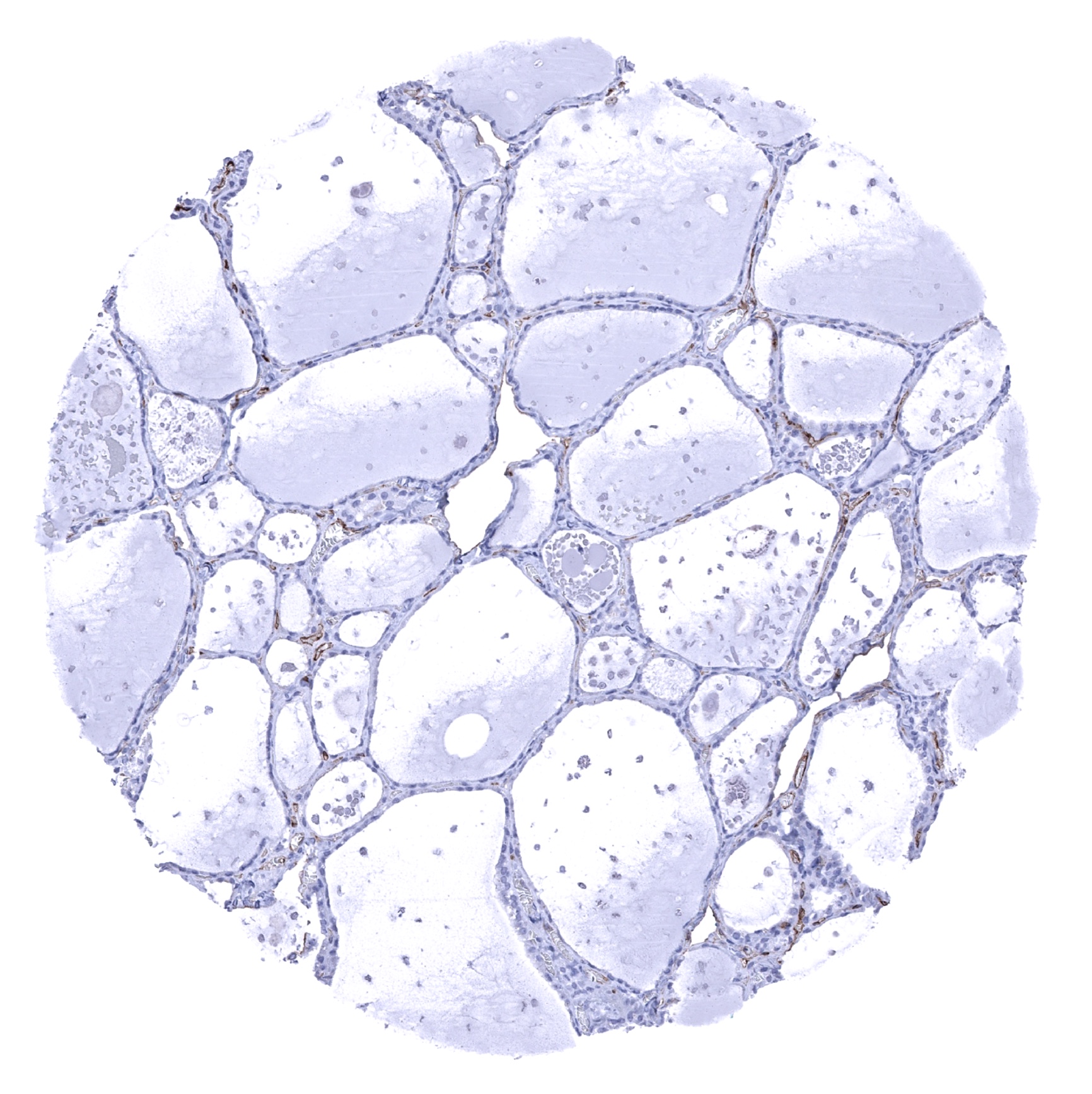
Thyroid gland
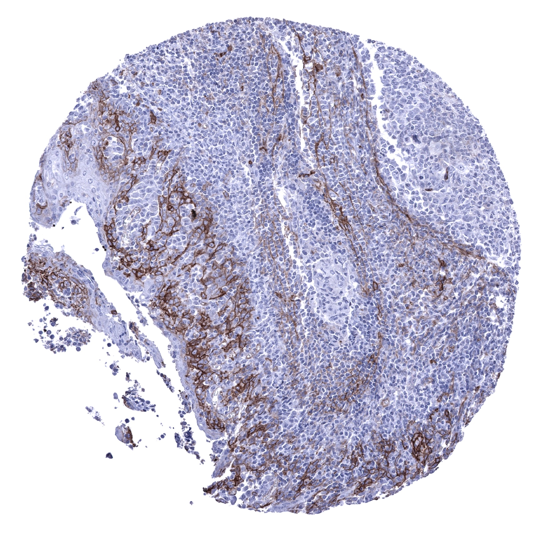
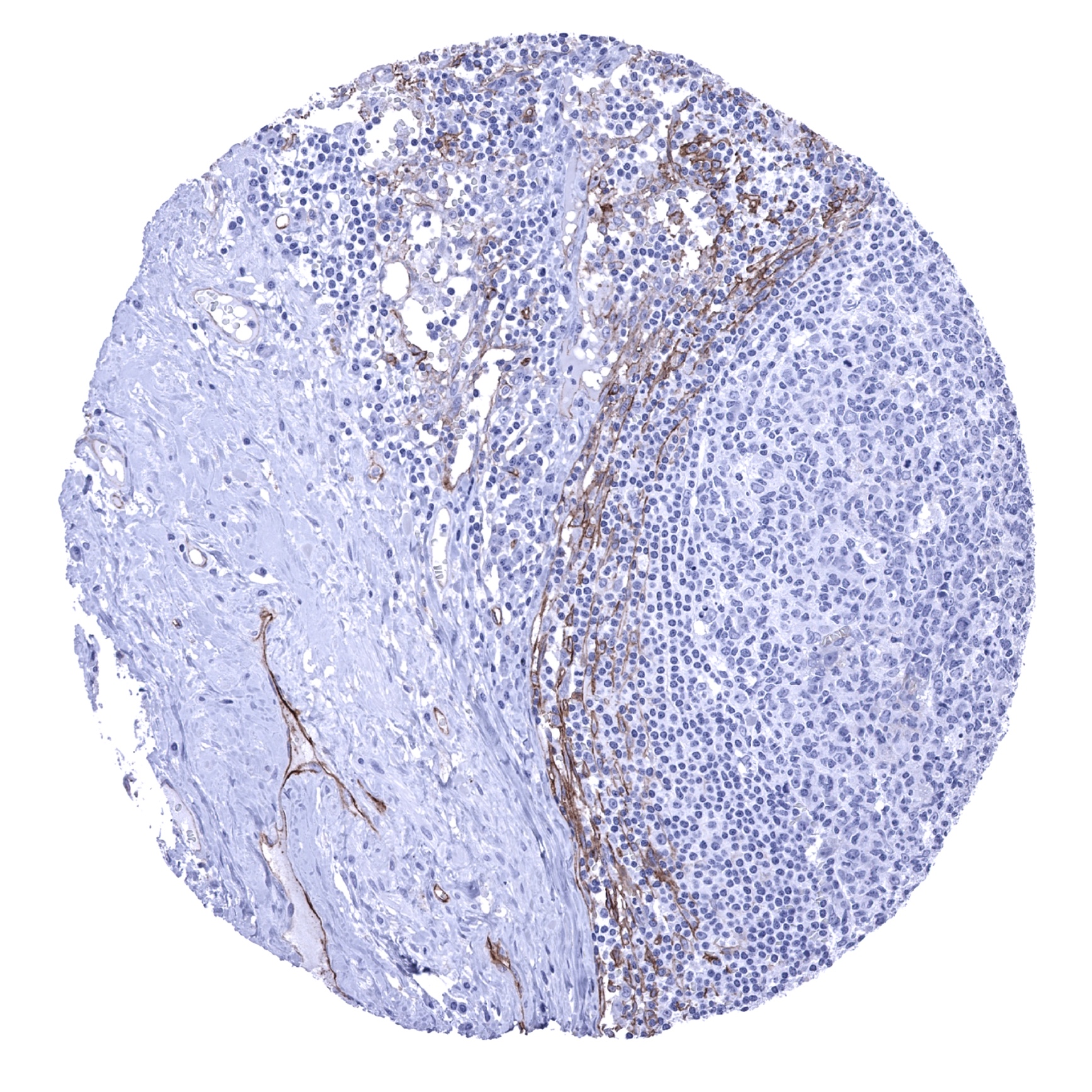
Tonsil - CD141 positivity occurs in squamous epithelial cells and in endothelial cells (CD141 immunohistochemistry).
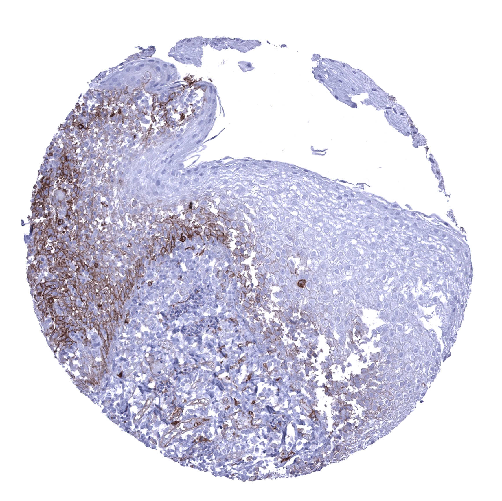
Tonsil, surface epithelium - Moderate to strong membranous CD141 staining of suprabasal epithelial cells (CD141 immunohistochemistry).
Urinary bladder, muscular wall
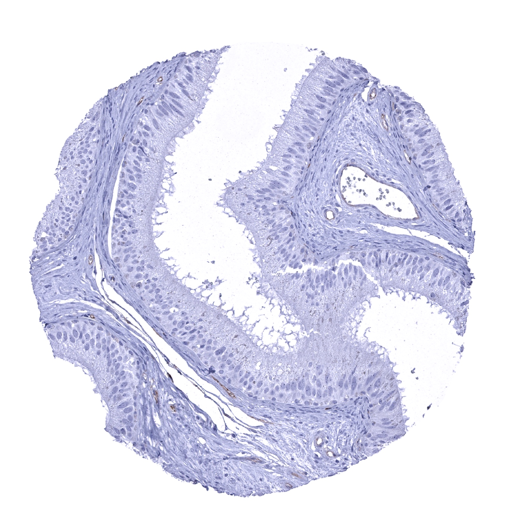
Urinary bladder, urothelium - Faint membranous CD141 staining of urothelial cells in the lower half of the urothelium (CD141 immunohistochemistry).

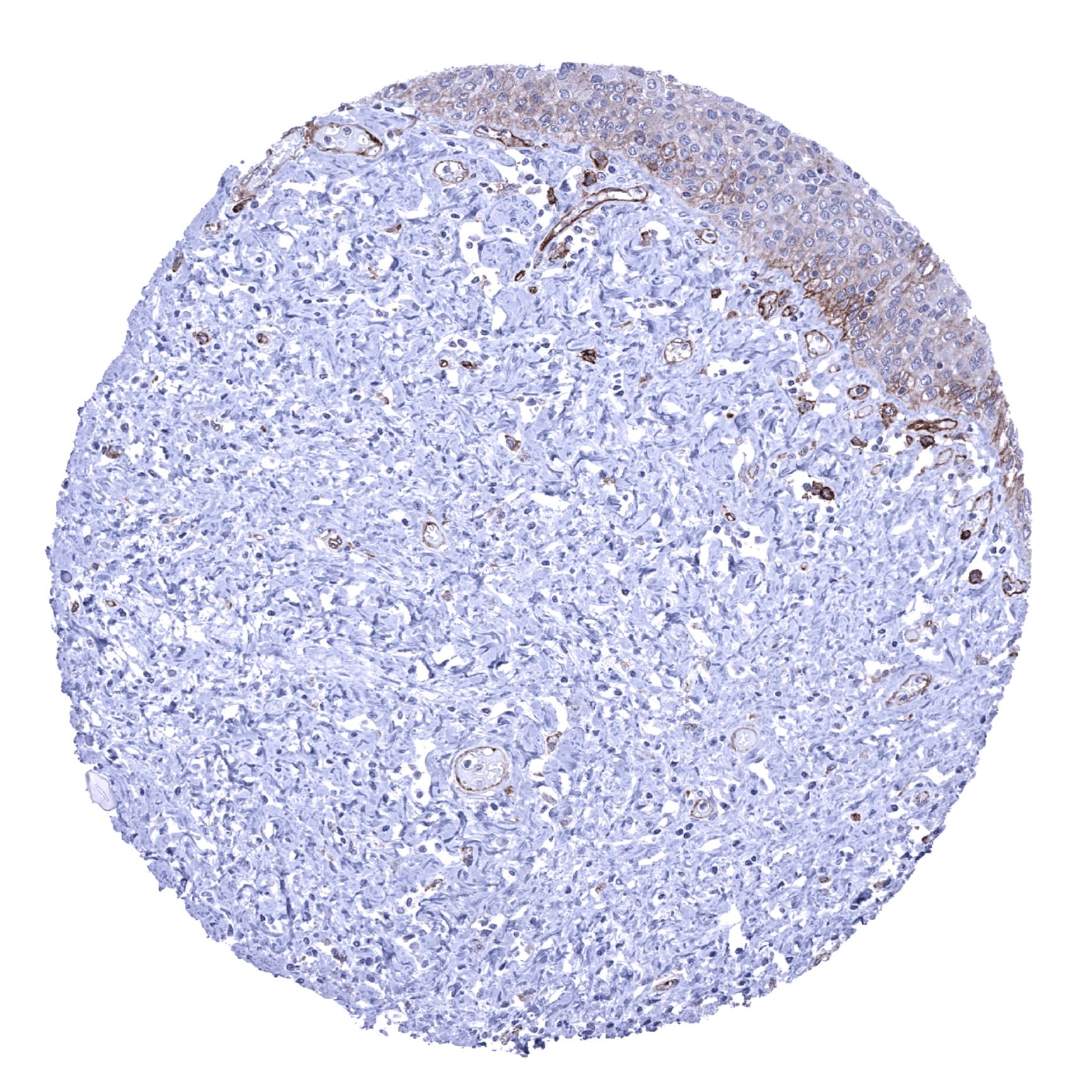
Uterus, ectocervix
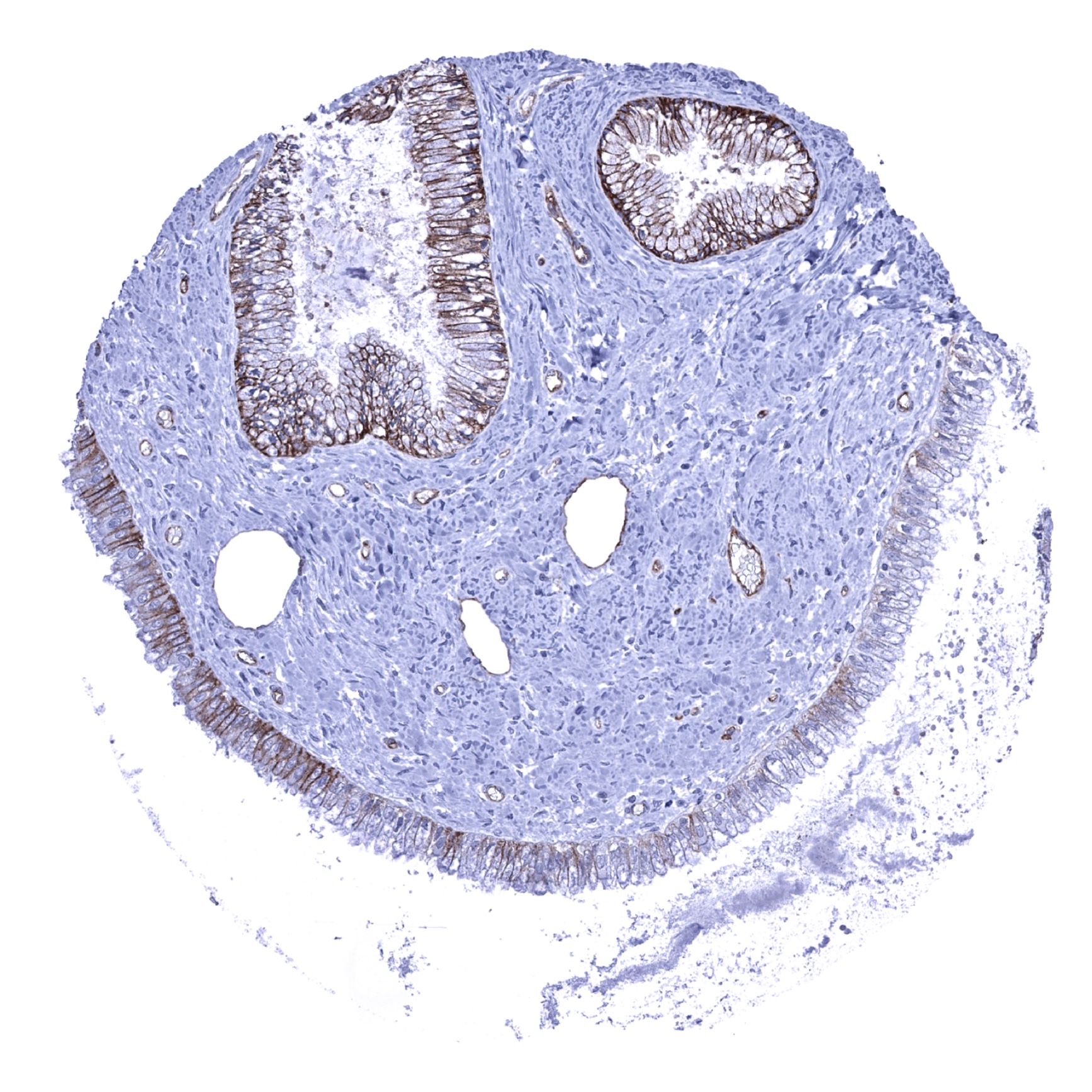
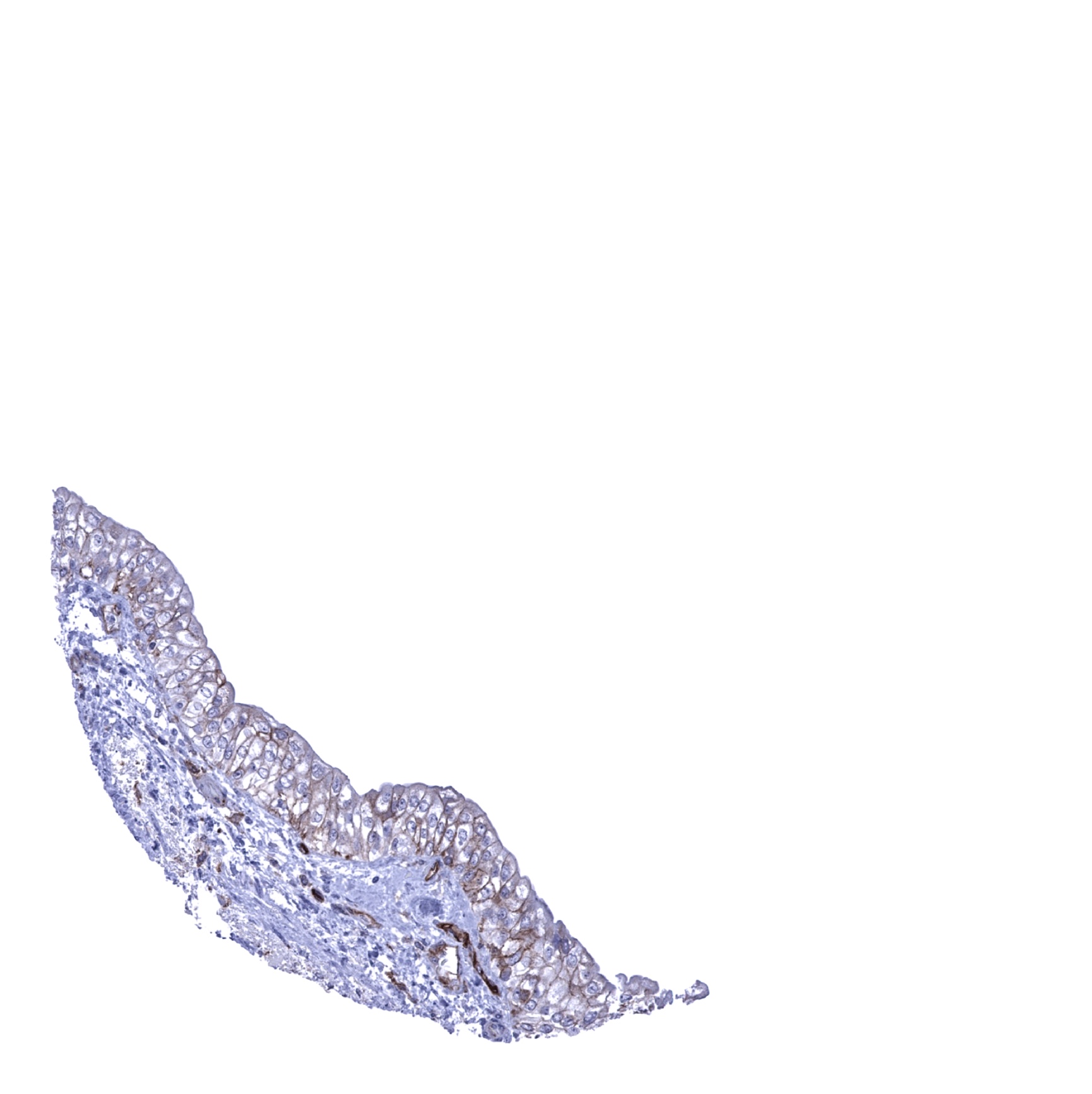
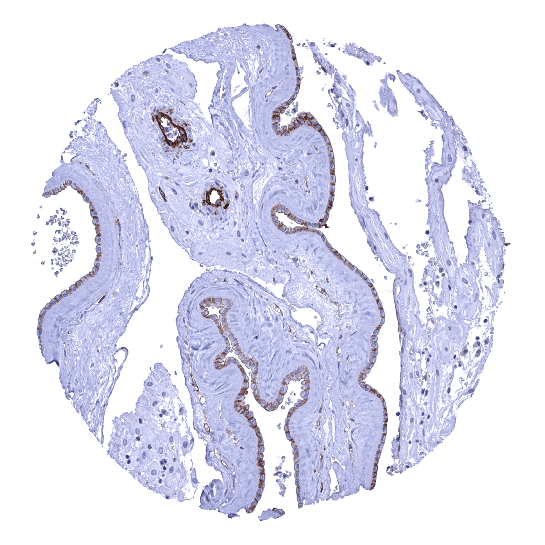
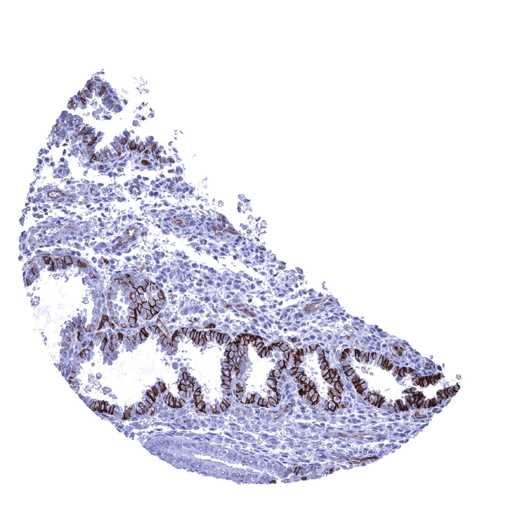
Uterus, endocervix - Membranous CD141 staining predominates in basolateral cell compartments of endocervical glands (CD141 immunohistochemistry).
Uterus, endometrium (pregnancy)
Uterus, endometrium (proliferation)
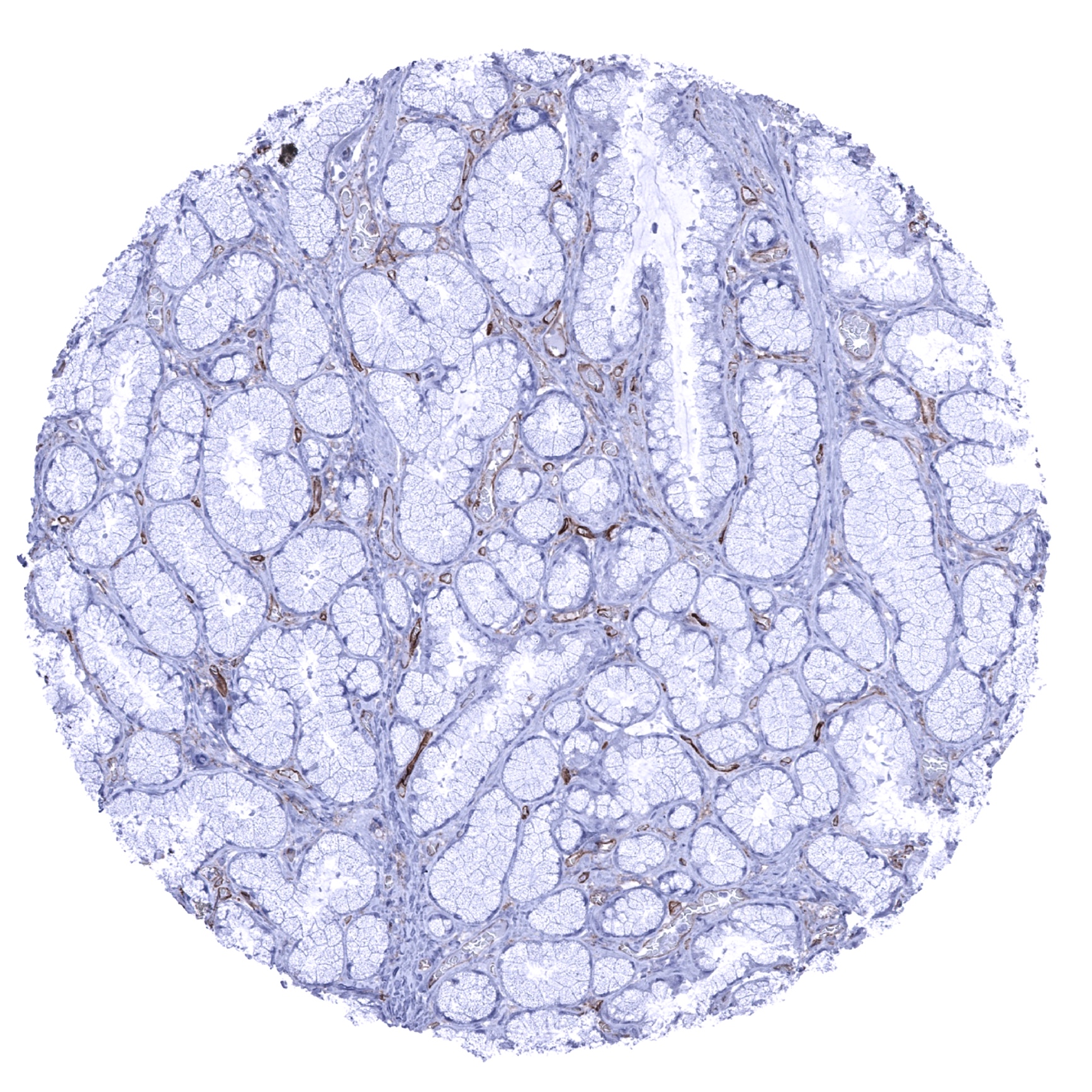
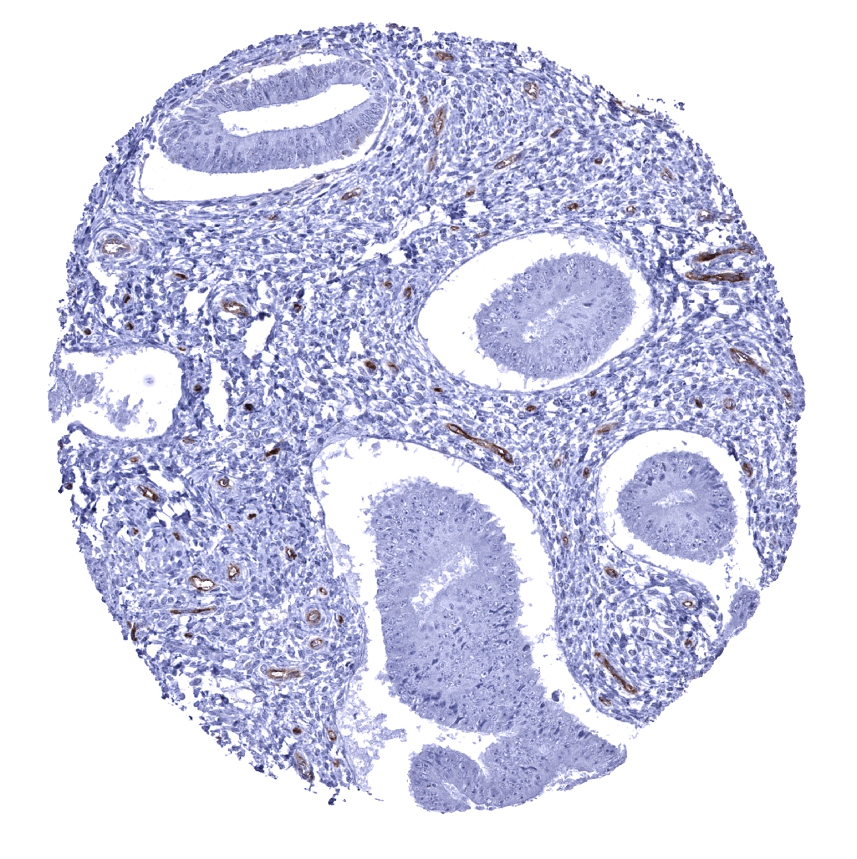
Uterus, endometrium (secretion) - Predominantly basolateral membranous CD141 staining of endometrial glands (CD141 immunohistochemistry).
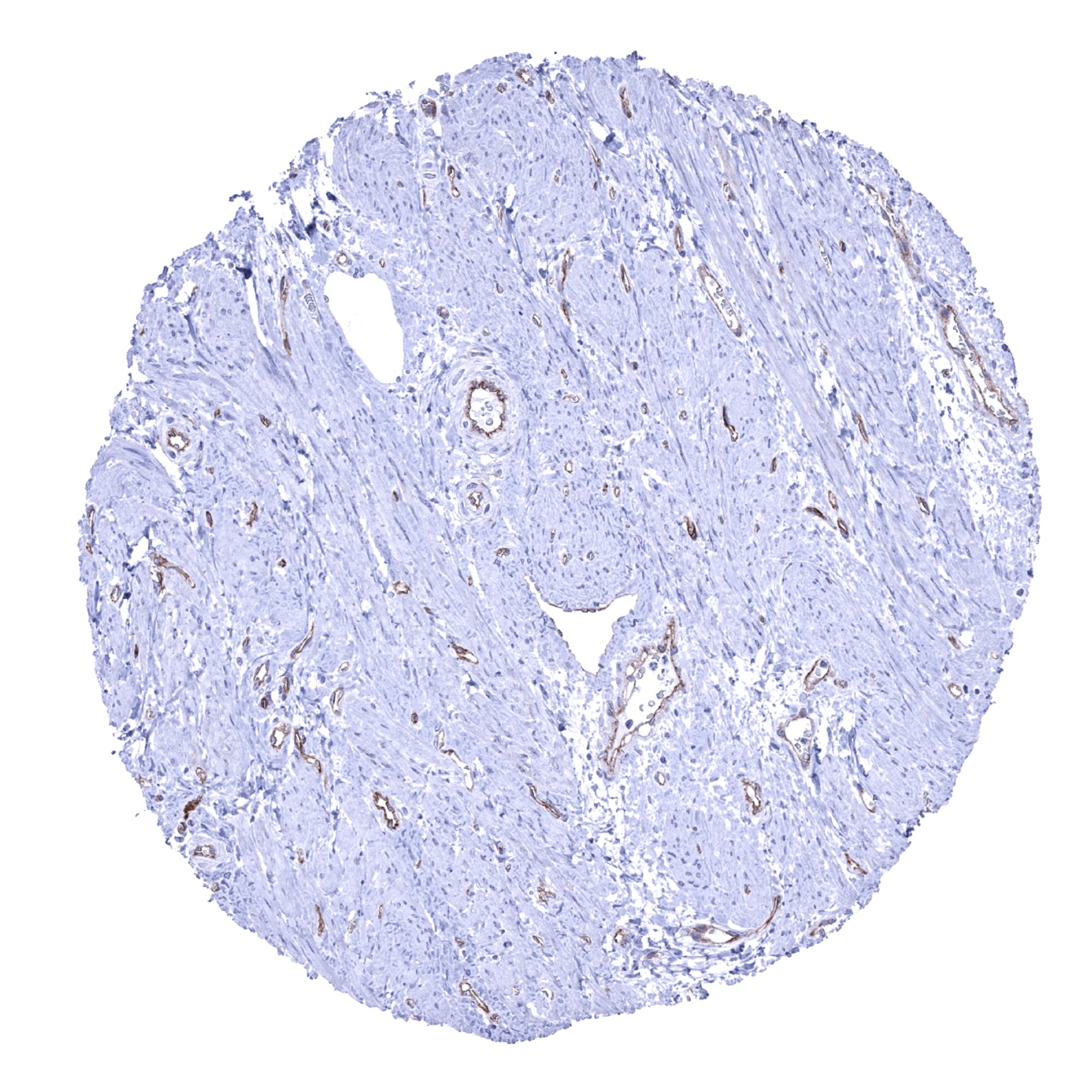
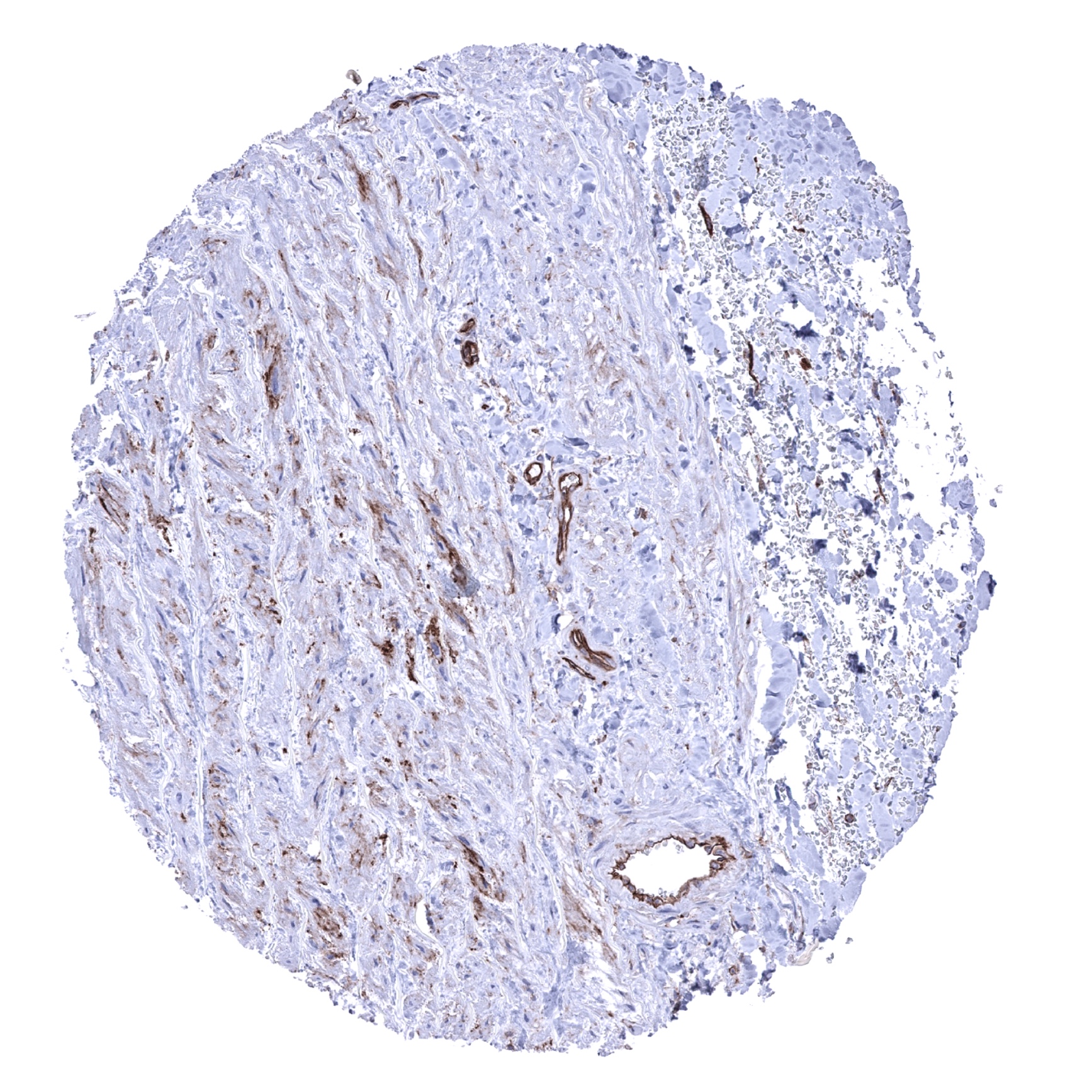
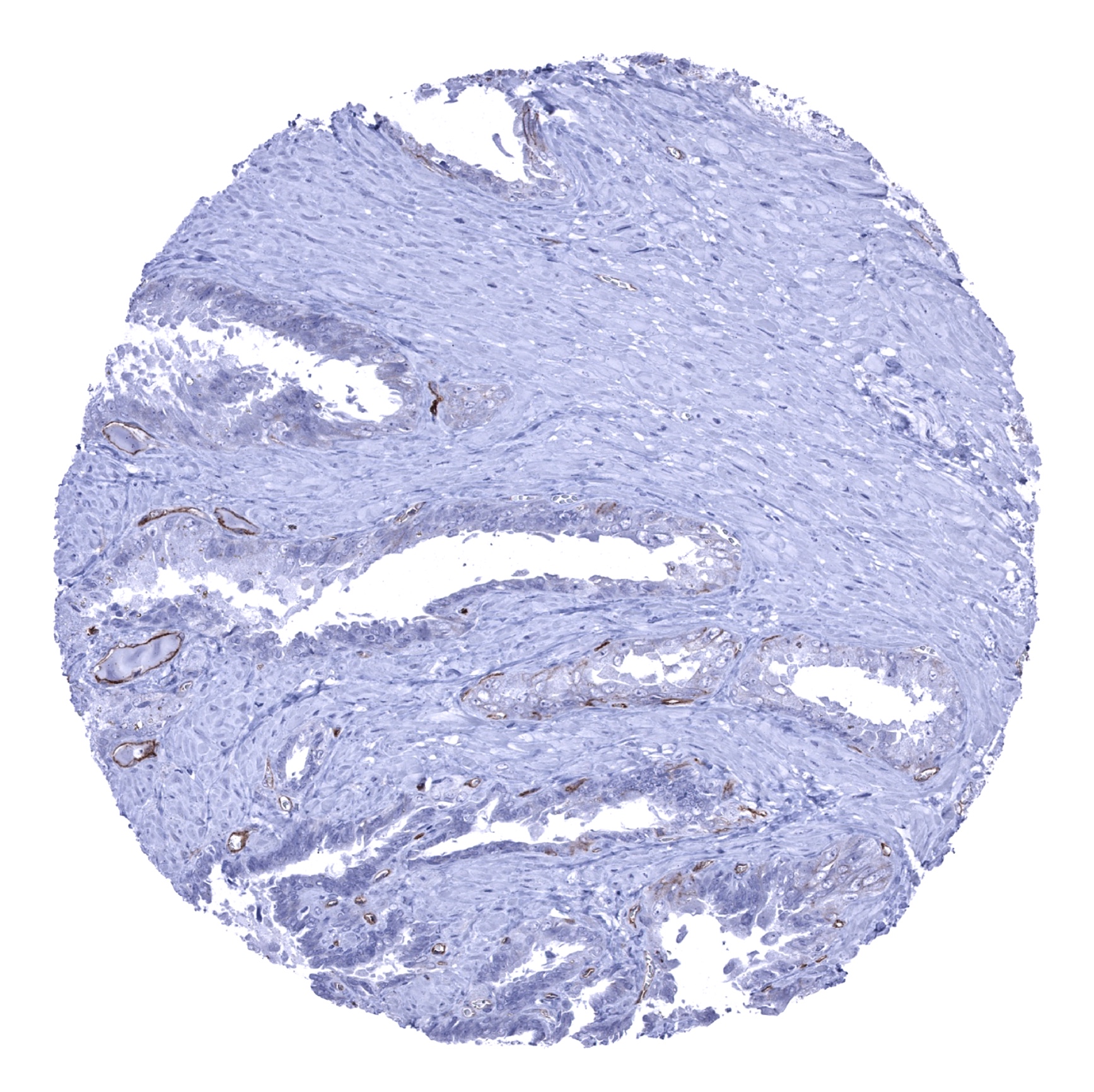
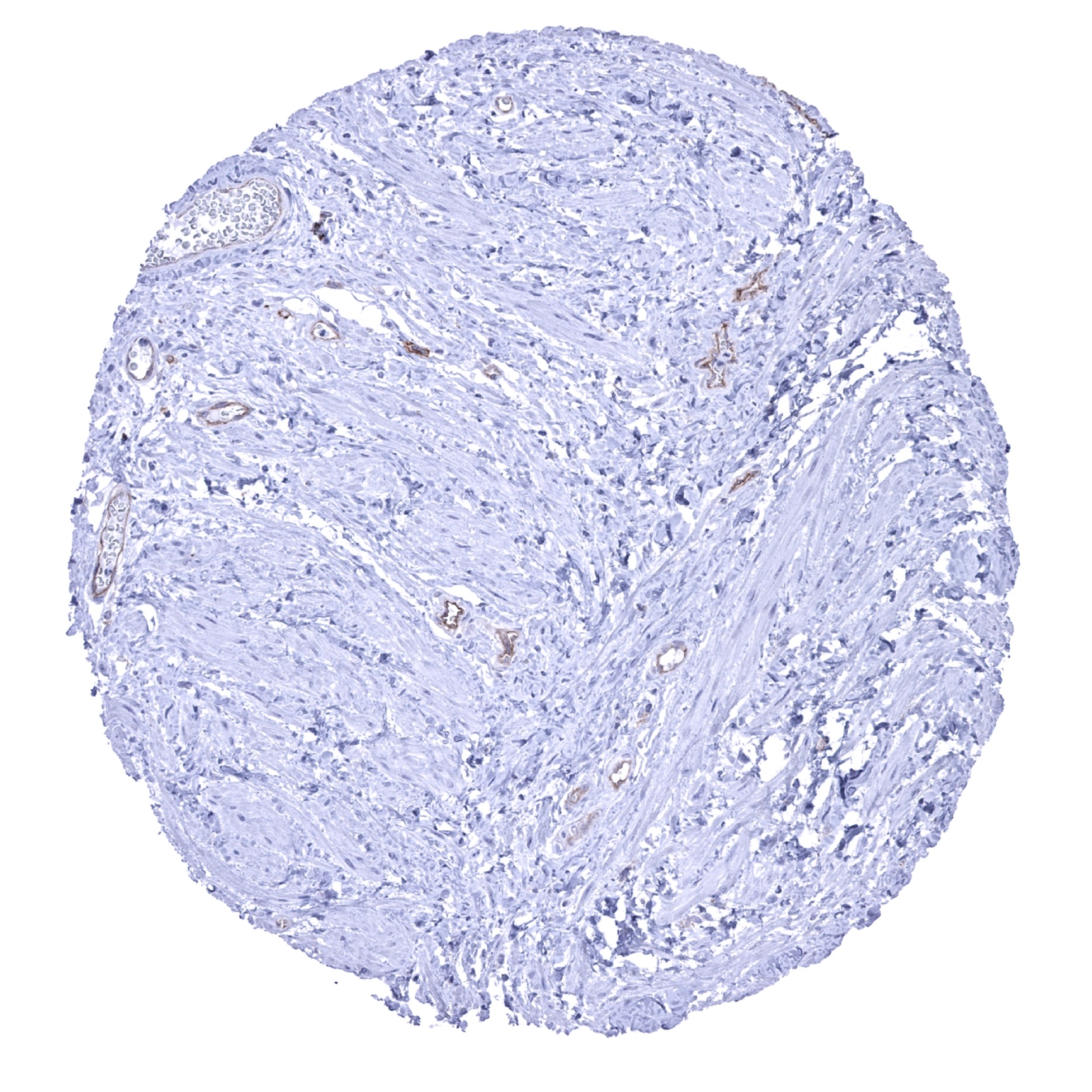
Uterus, myometrium - CD141 staining of endothelial cells (CD141 immunohistochemistry).