Adrenal gland – Moderate cytoplasmic bcl-2 staining of medullary cells while cortical cells are negative.
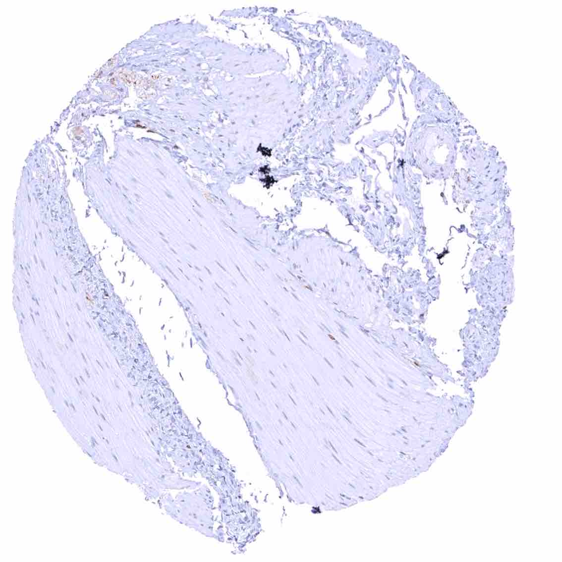
Aorta, media
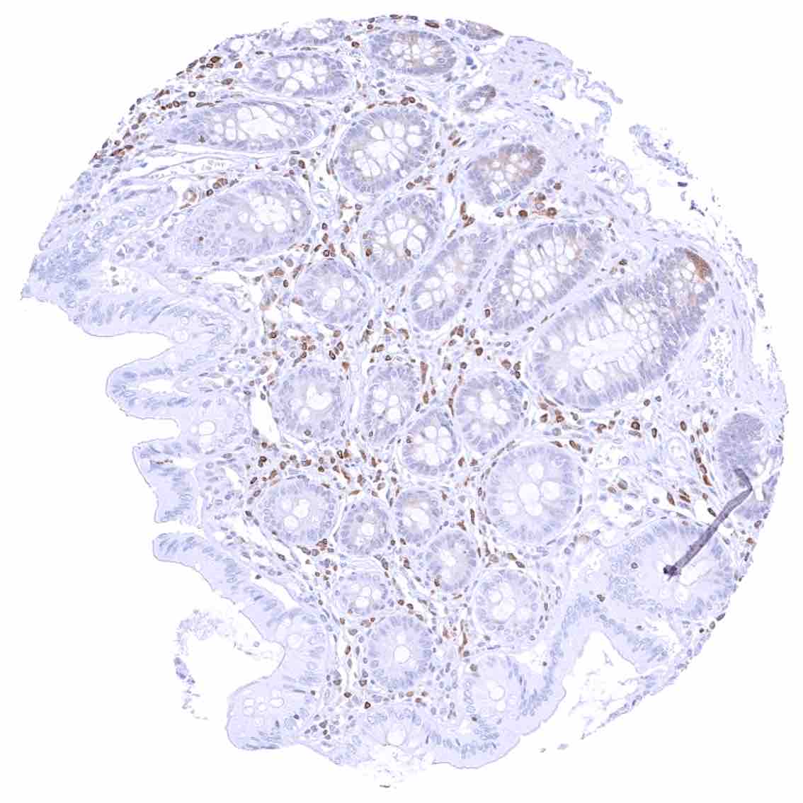
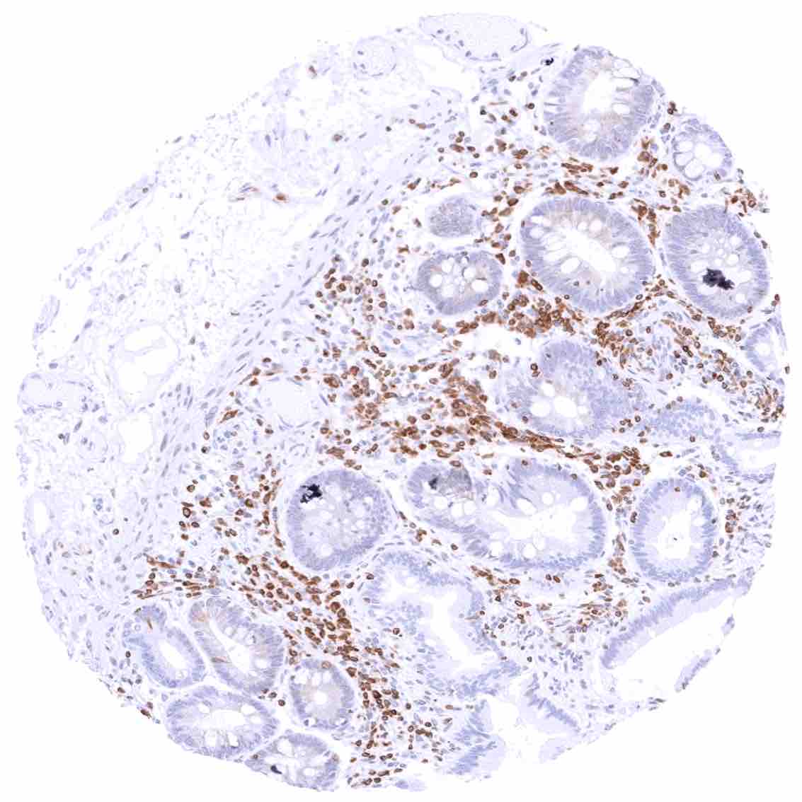
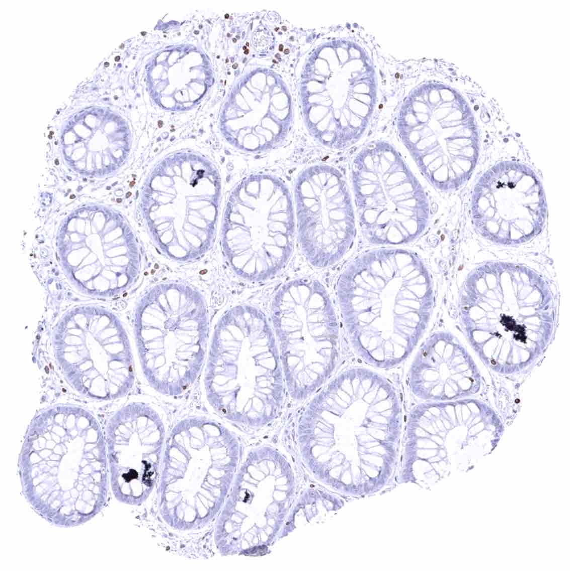
Appendix, mucosa – Cytoplasmic bcl-2 staining is largely limited to (non-germinal centre) lymphocytes. Weak bcl-2 staining of crypt cells.
Appendix, muscular wall
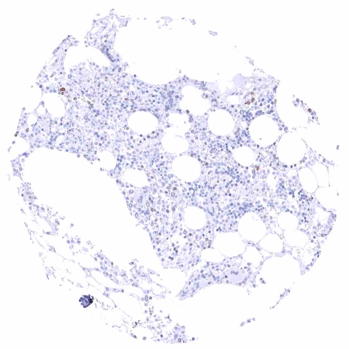
Bone marrow – Only few cells are bcl-2 positive.
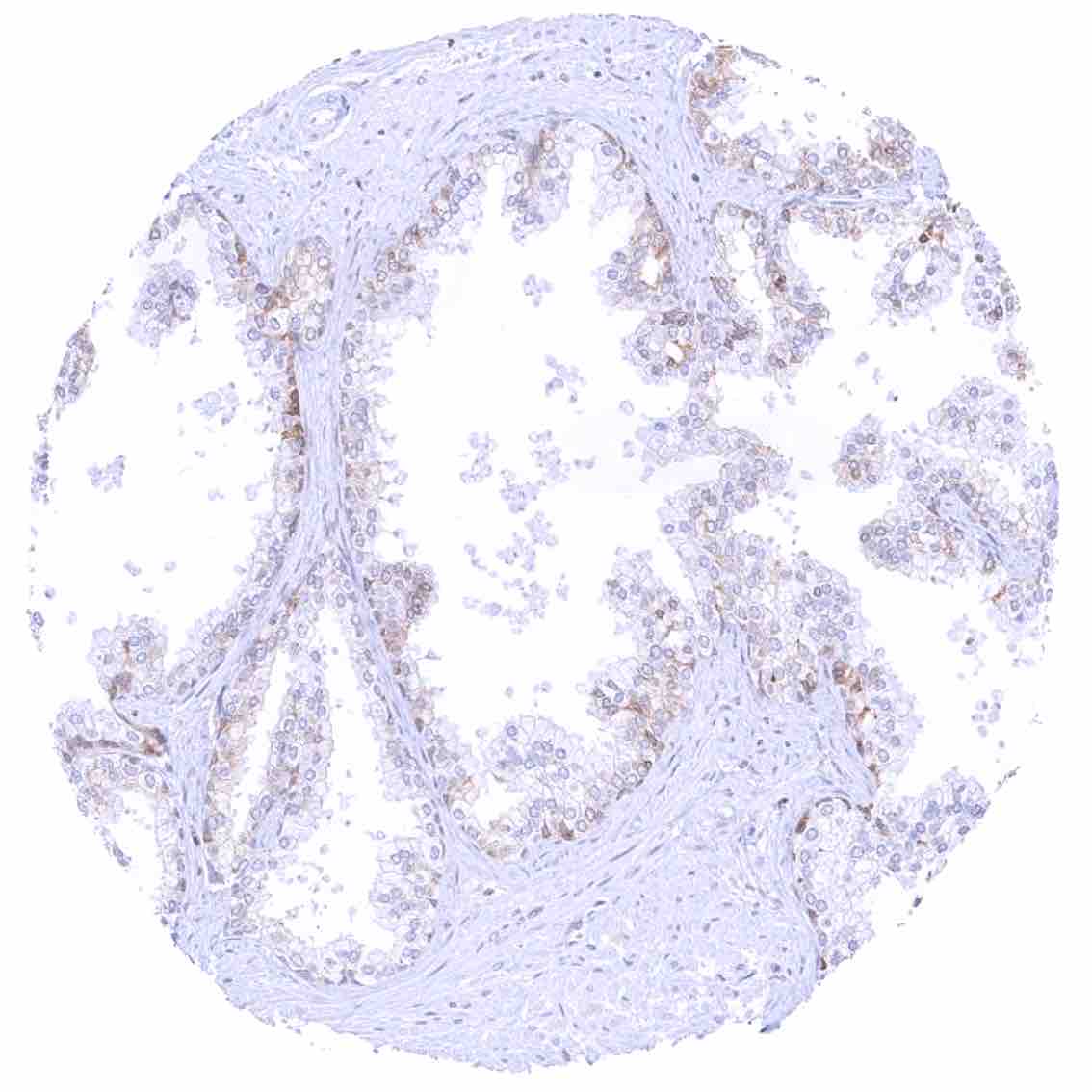
Breast – Distinct cytoplasmic bcl-2 staining of epithelial cells with higher intensity in acinar than in myoepithelial cells.
Bronchus, mucosa – Moderate cytoplasmic bcl-2 staining of basal cells of the respiratory epithelium.
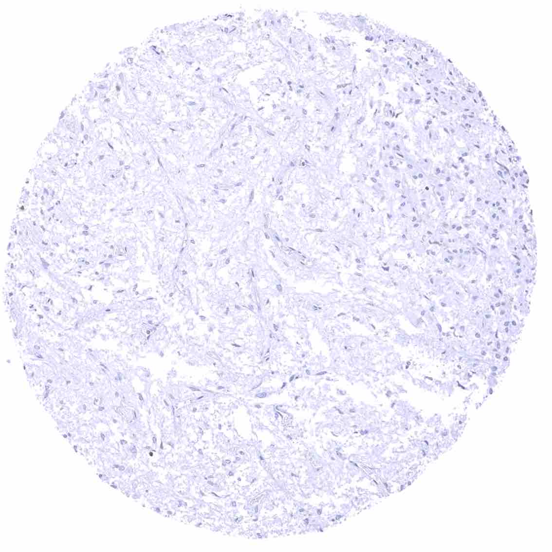
Cerebellum (white matter)
Cerebellum, cortex (molecular layer, Purkinje cell layer, granule cell layer, white matter)
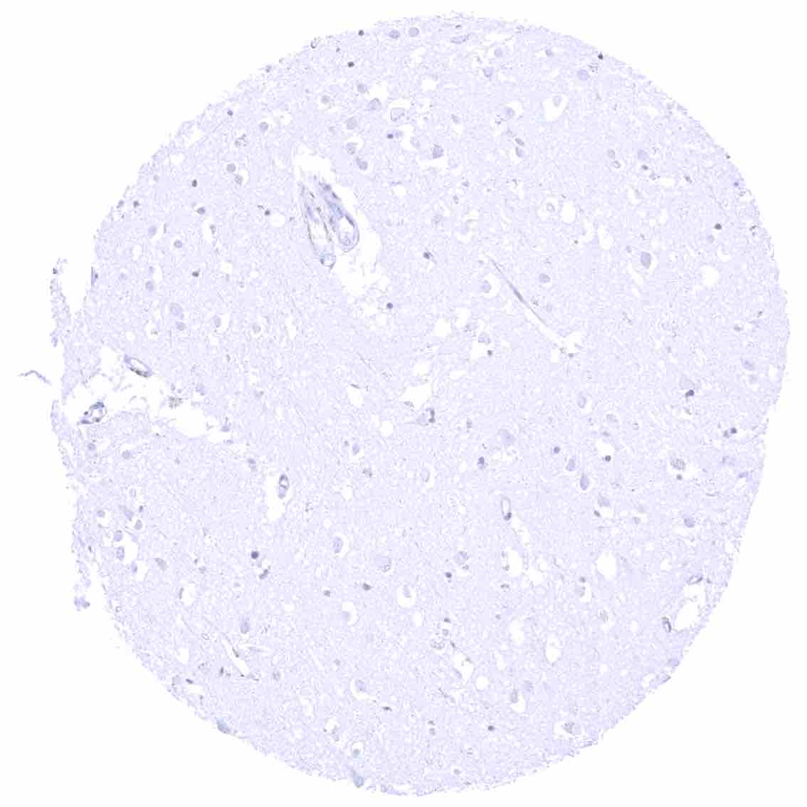
Cerebrum (grey matter)
Cerebrum (white matter)
Colon descendens, mucosa
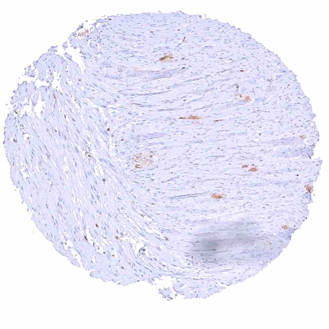
Colon descendens, muscular wall
Duodenum, mucosa – Cytoplasmic bcl-2 staining is largely limited to lymphocytes. The epithelium is mostly negative, but a weak staining occurs in some crypt cells.
Epididymis (Caput) – Weak bcl-2 staining of epithelial cells.
Epididymis (Cauda) – Weak bcl-2 staining of epithelial cells.
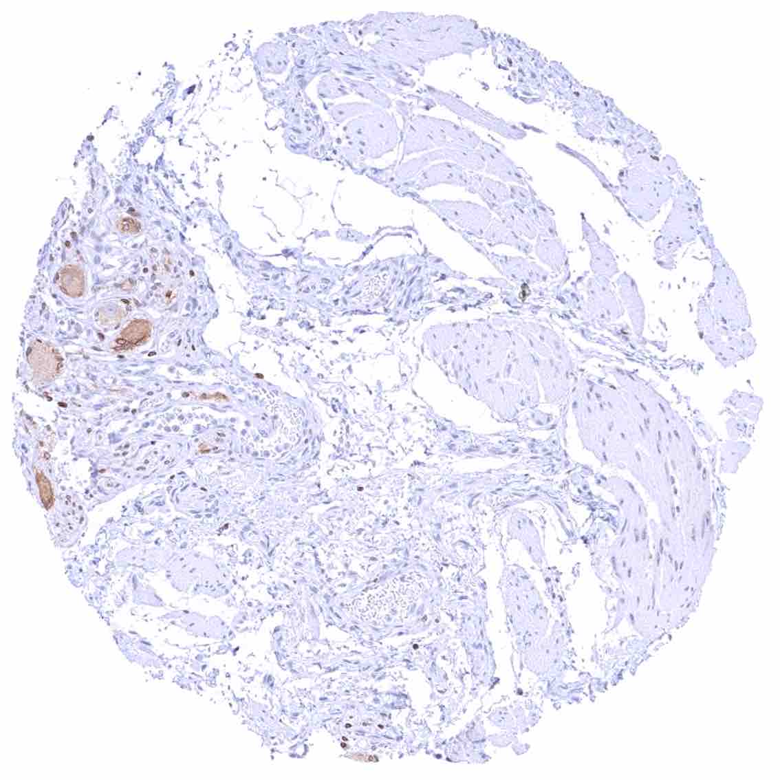
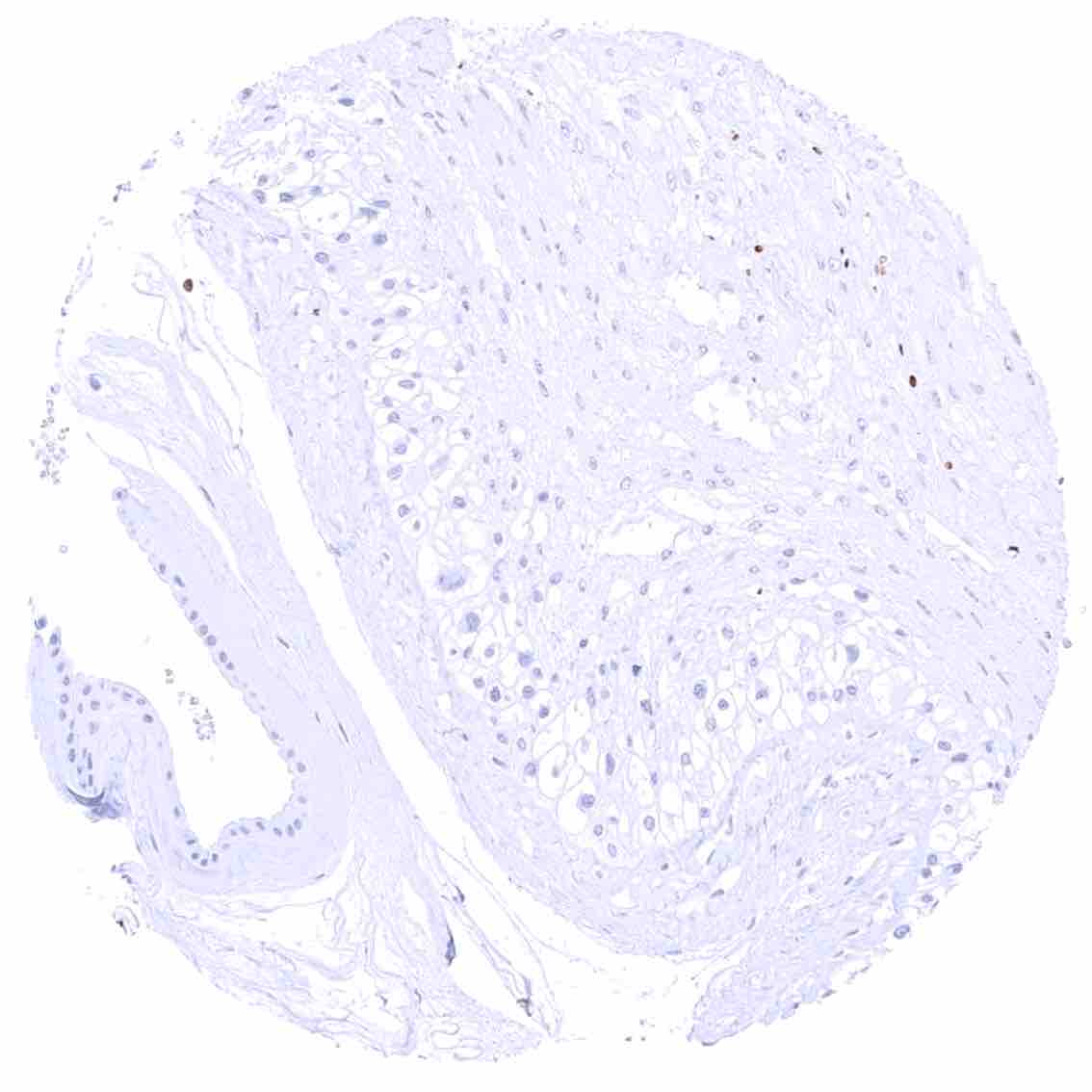
Esophagus, muscular wall – Moderate cytoplasmic bcl-2 staining of ganglion cells of the muscular wall. .jpeg
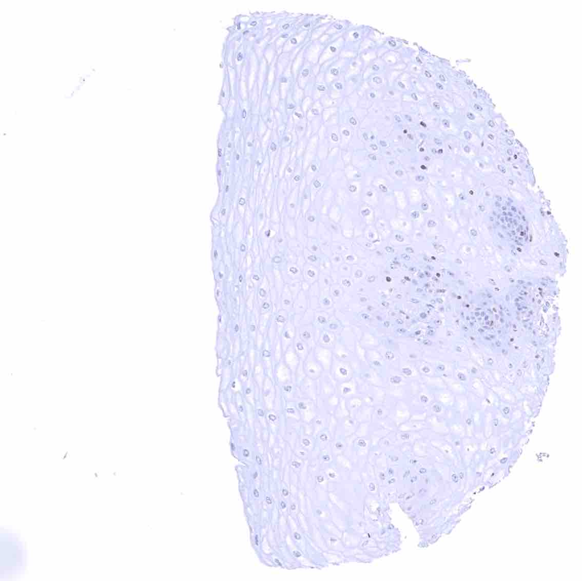
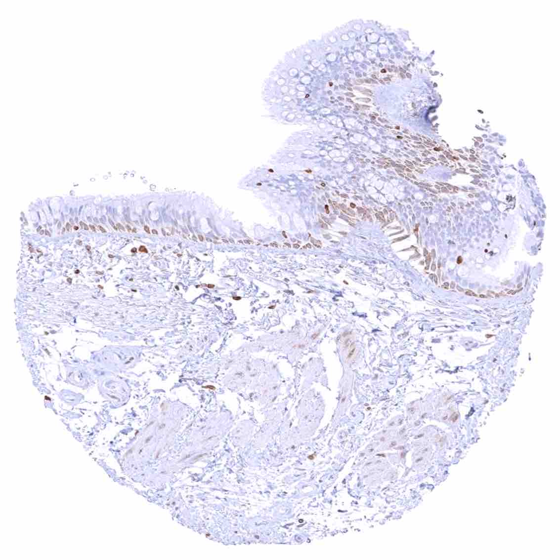
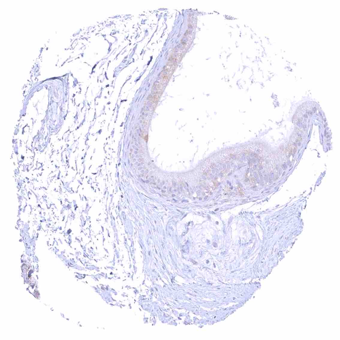
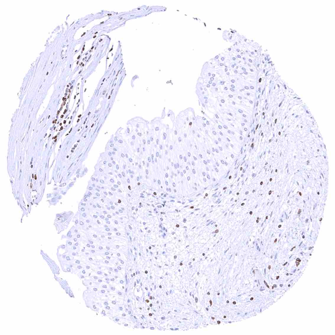
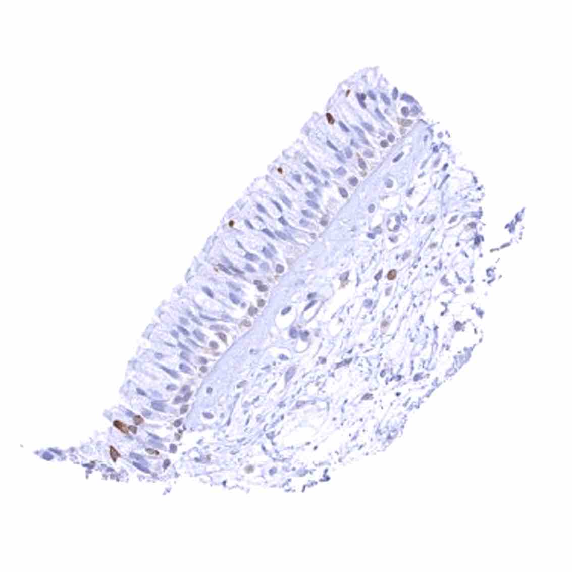
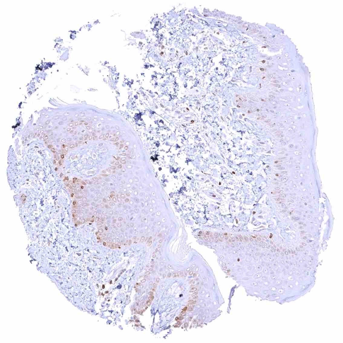
Esophagus, squamous epithelium
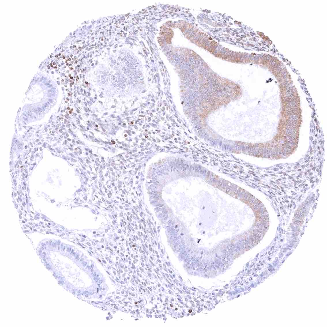
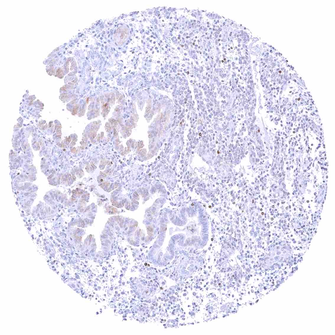
Fallopian tube, mucosa – Strong cytoplasmic bcl-2 staining of a subset of epithelial cells.
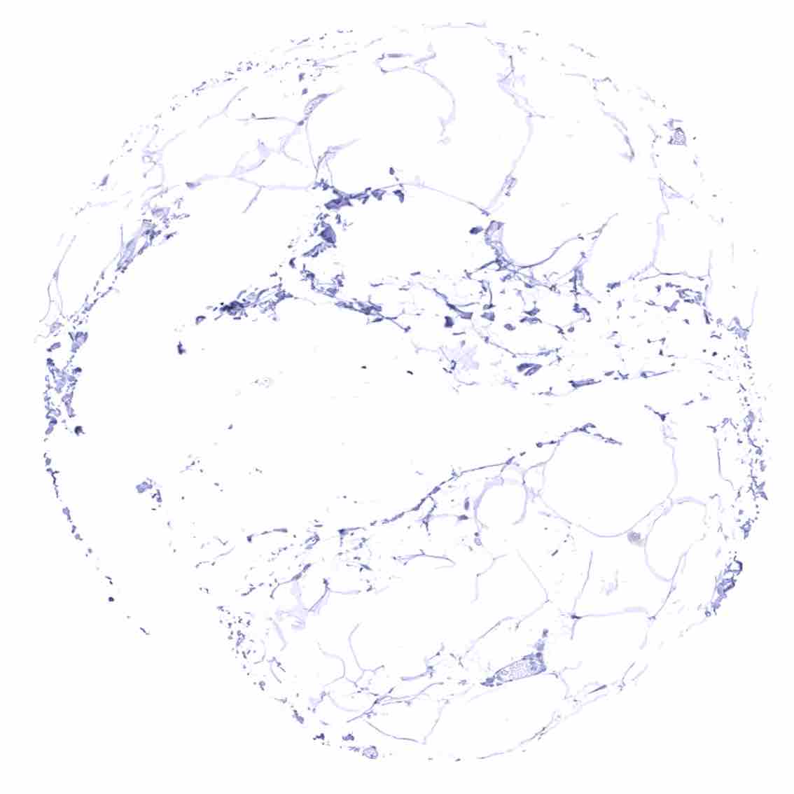
Fat
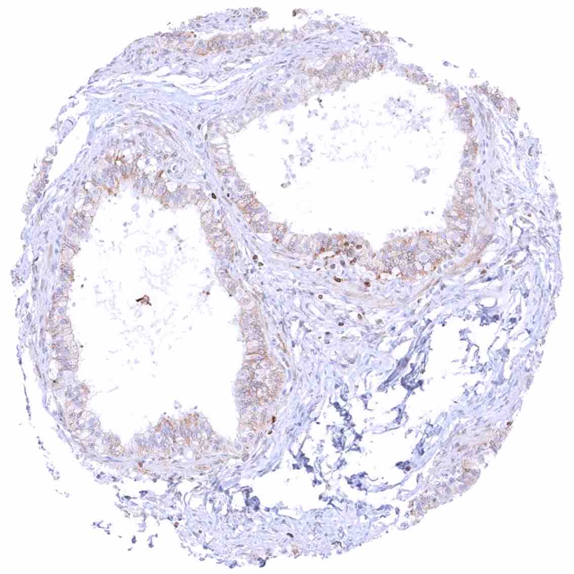
Gallbladder, epithelium
Heart muscle
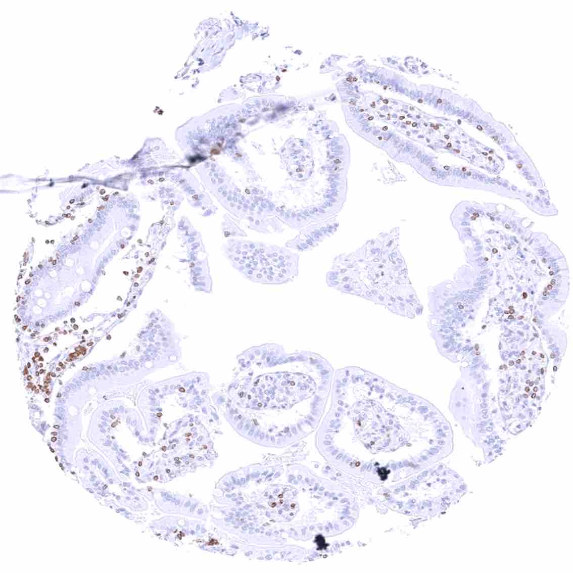
Ileum, mucosa – Cytoplasmic bcl-2 staining is limited to lymphocytes.
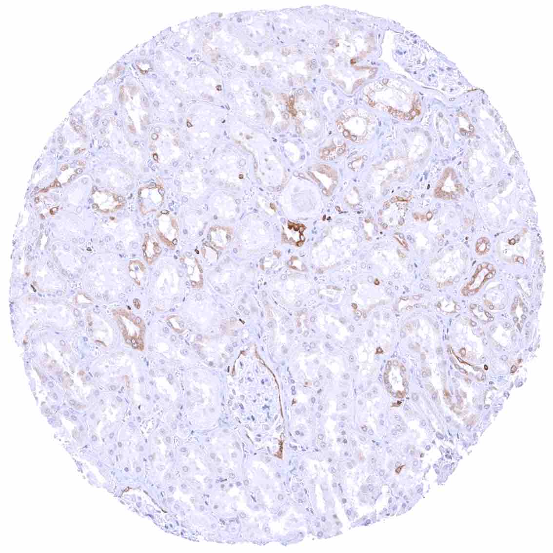
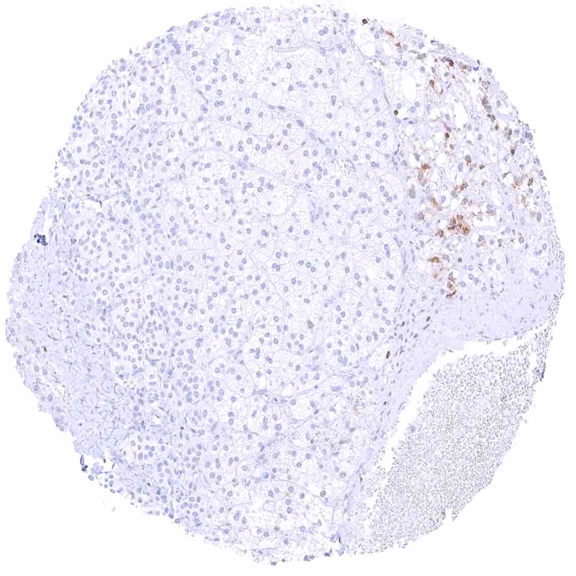
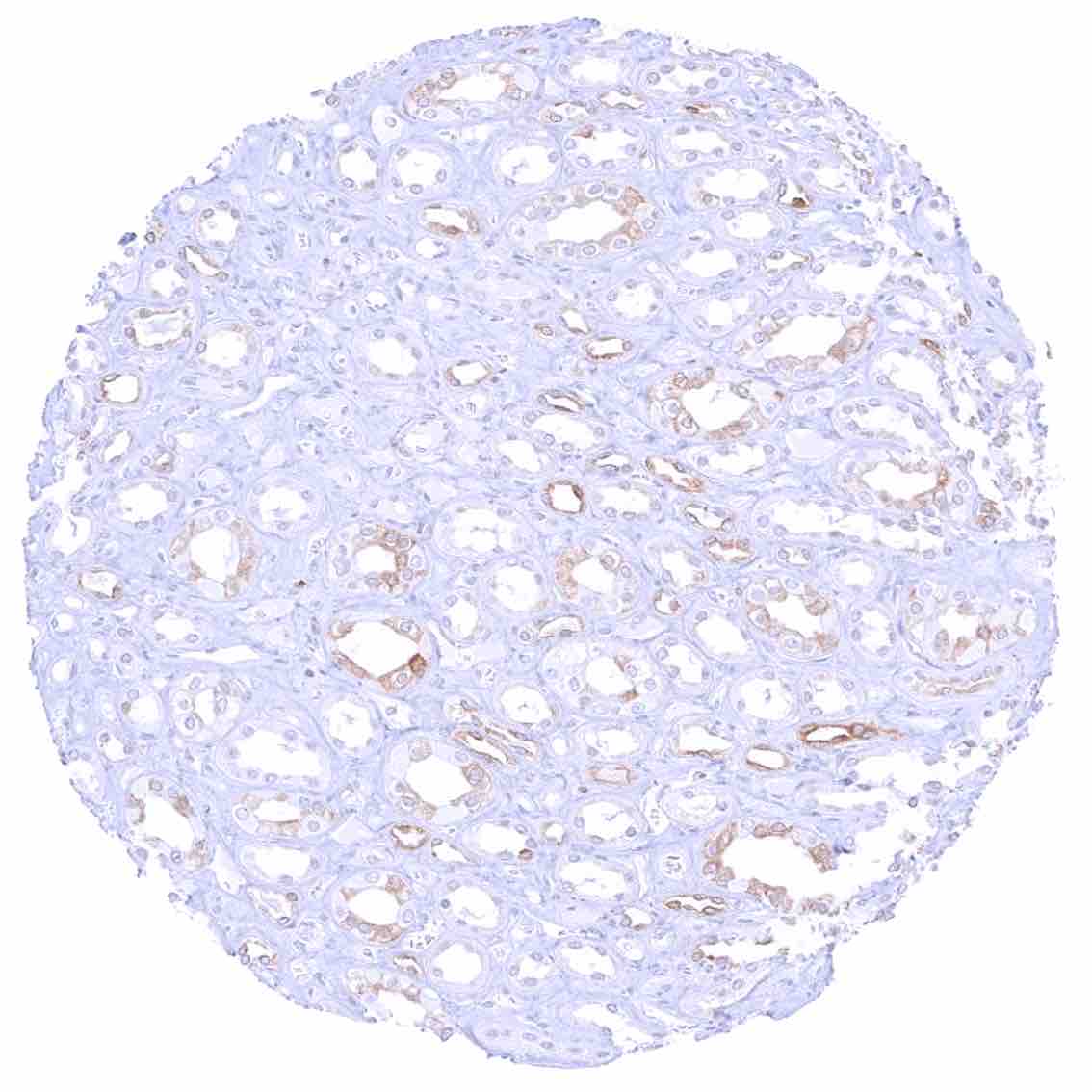
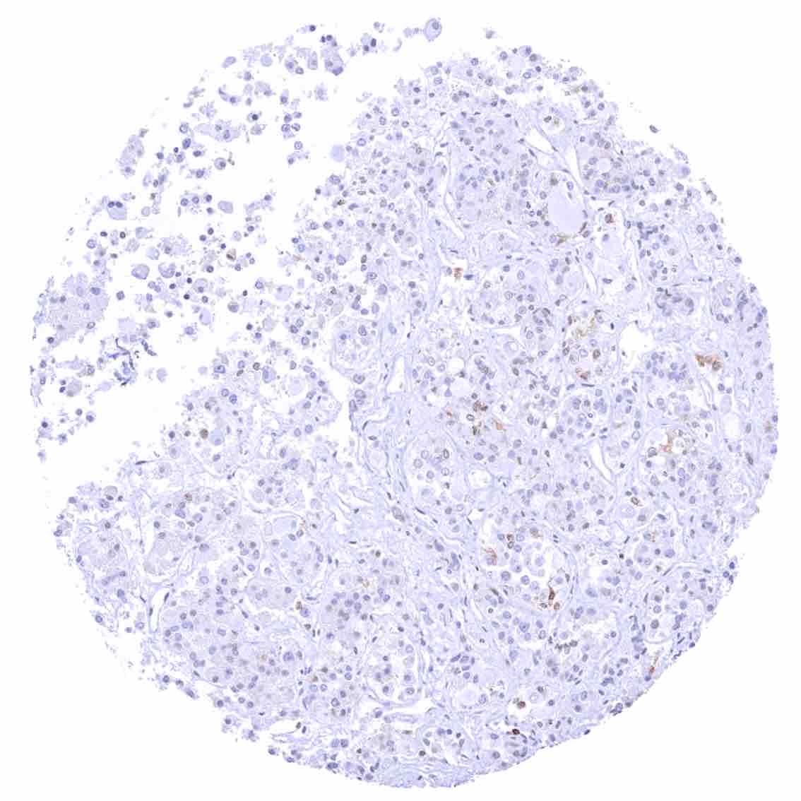
Kidney, cortex – Weak to moderate bcl-2 staining in a fraction of (mostly distal) tubuli and collecting ducts.
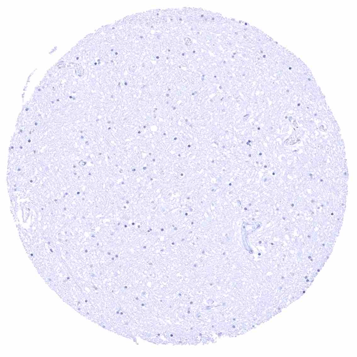
Kidney, medulla
Kidney, pelvis, urothelium .jpeg
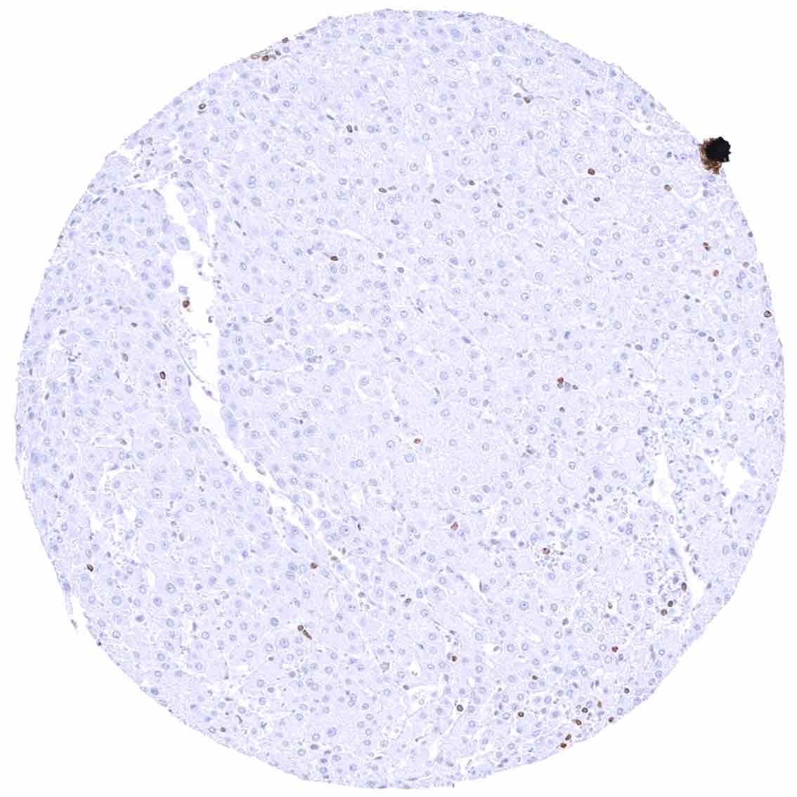
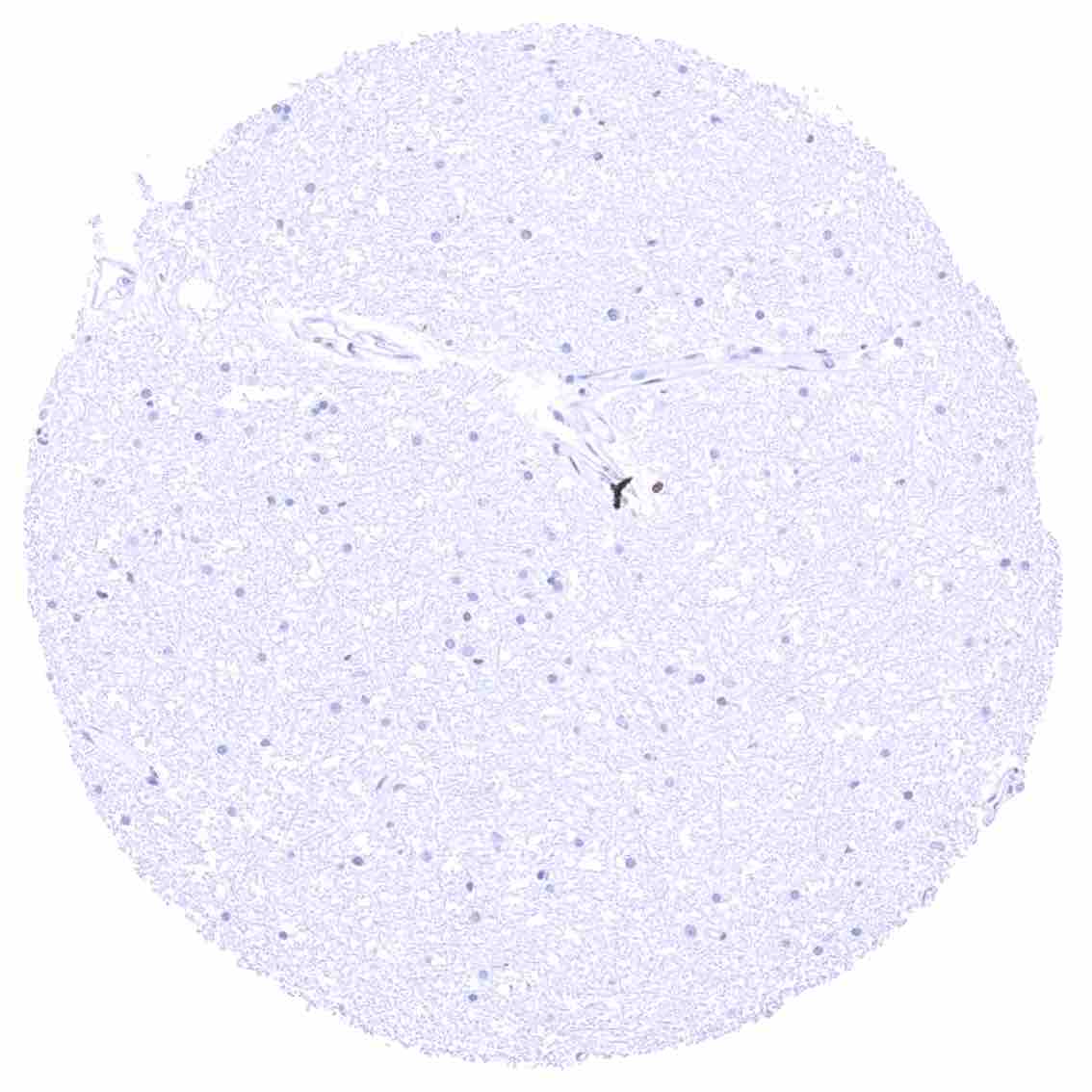
Liver
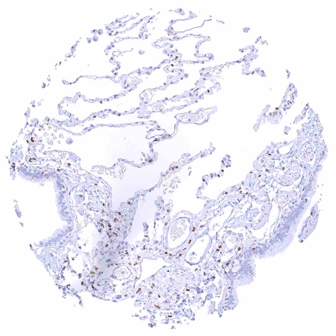
Lung – Pneumocytes are largely negative while a weak cytoplasmic bcl-2 staining can appear in some bronchiolar cells.
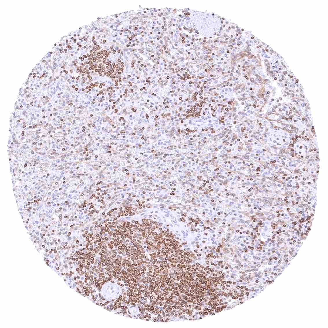
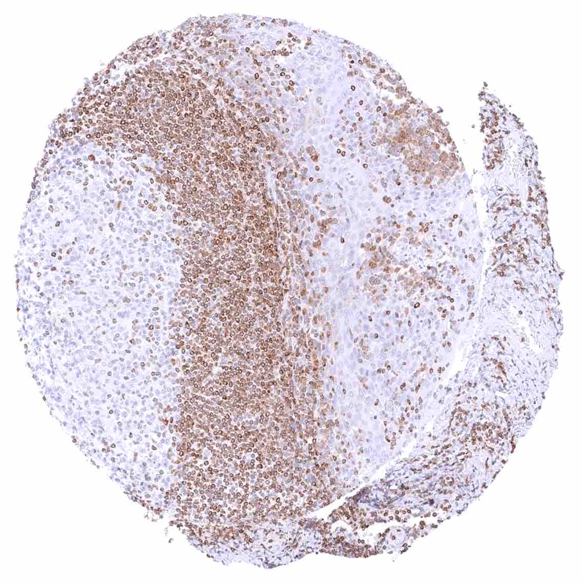
Lymph node – Strong bcl-2 positivity of a large fraction of lymphocytic cells in the interfollicular area and around germinal centres while almost all cells in germinal centres are bcl-2 negative.
Ovary, corpus luteum
Ovary, stroma – Strong cytoplasmic bcl-2 staining of stroma cells.
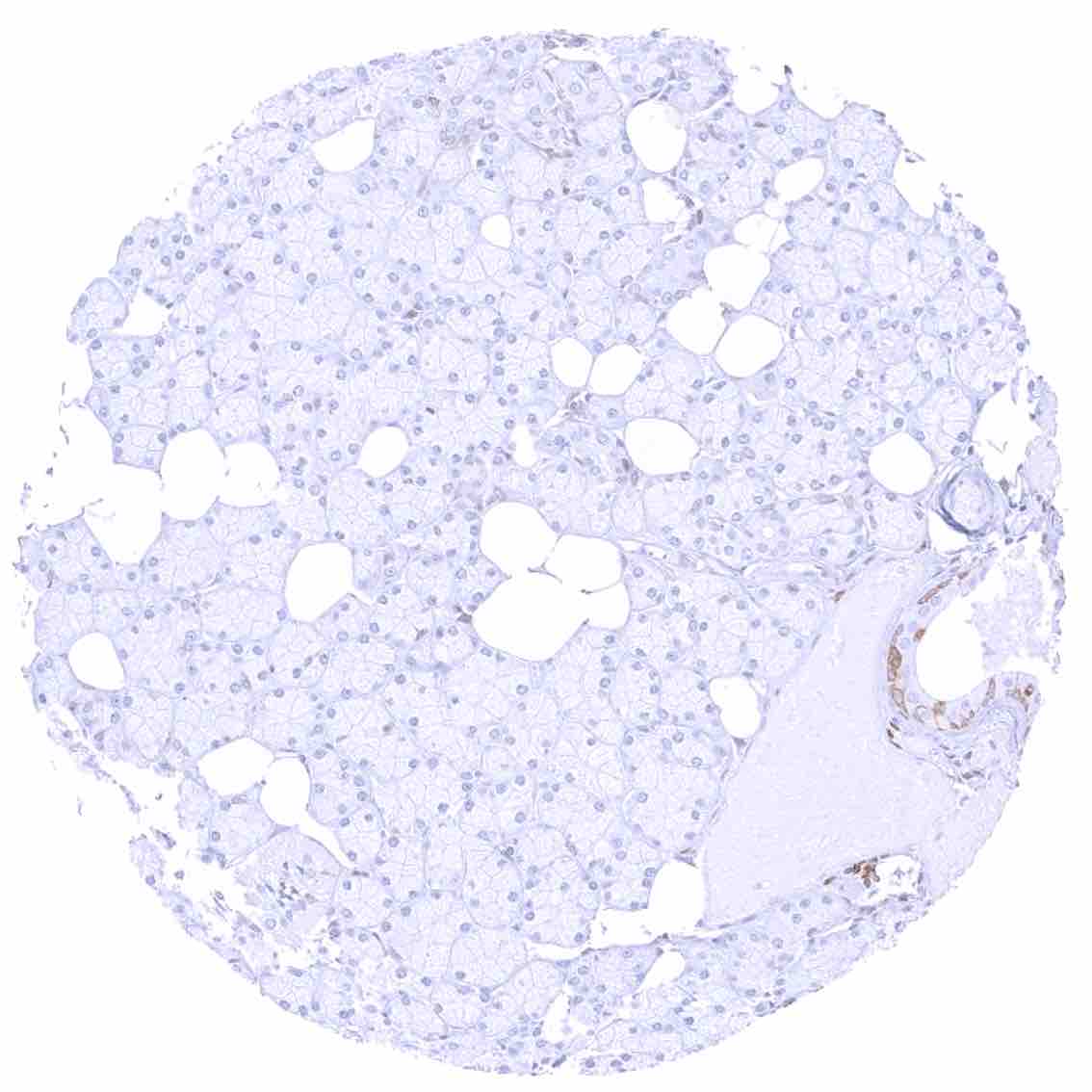
Pancreas – Weak bcl-2 staining of excretory duct cells.
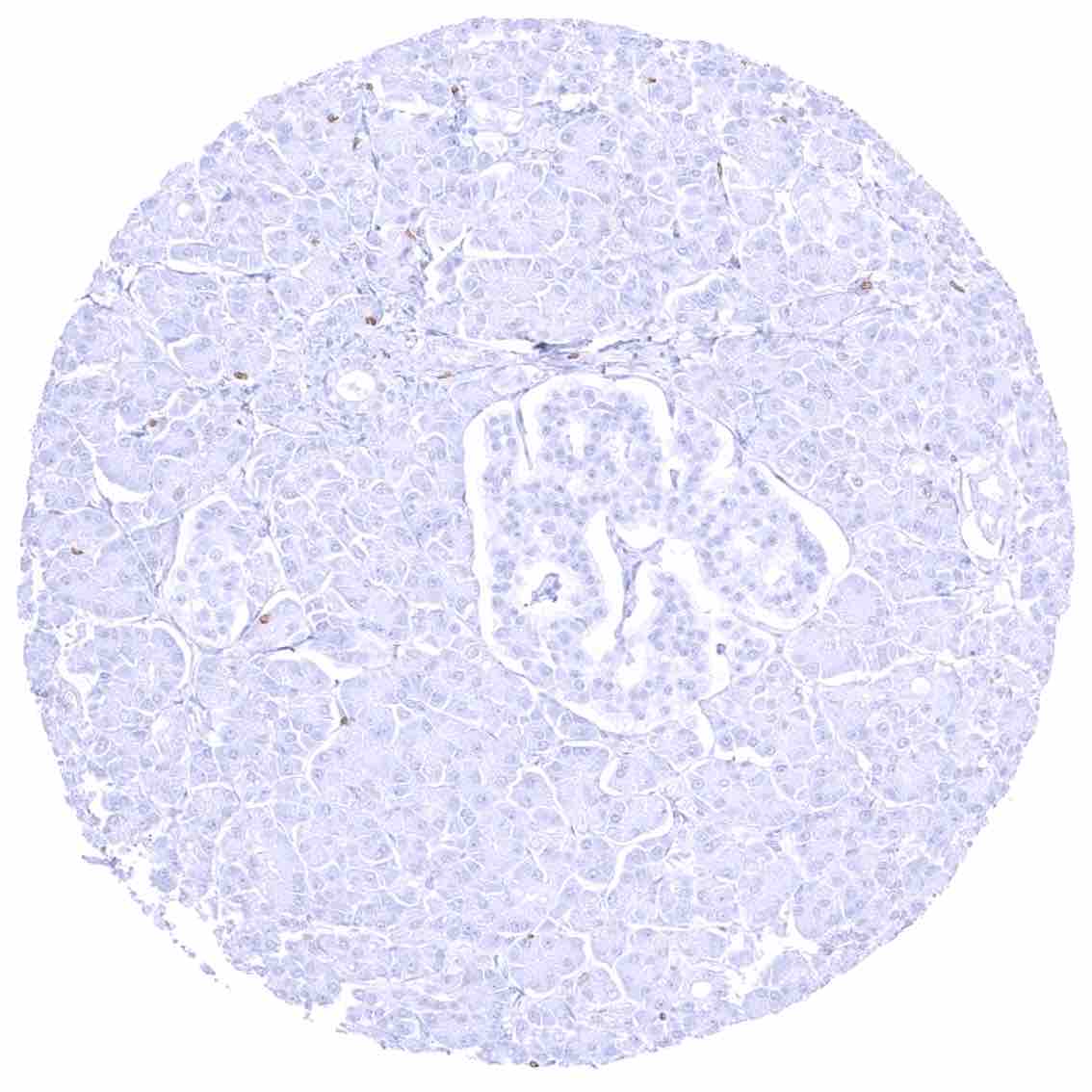
Pancreas
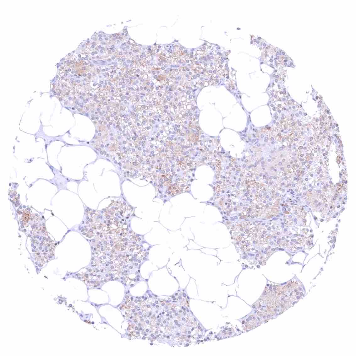
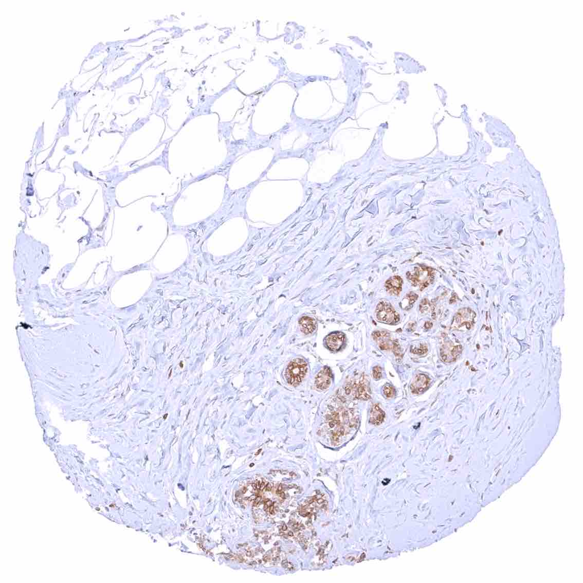
Parathyroid gland – Moderate cytoplasmic bcl-2 staining of epithelial cells.
Parotid gland – Moderate cytoplasmic bcl-2 staining of some myoepithelial cells of excretory ducts. .jpeg
Pituitary gland, anterior lobe – Few bcl-2 positive epithelial cells.
Pituitary gland, posterior lobe
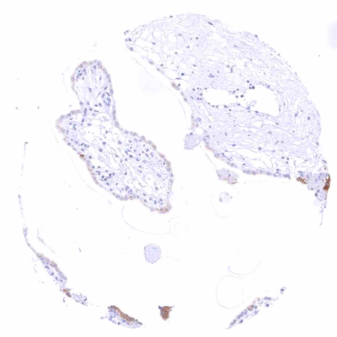
Placenta (amnion and chorion)
Placenta, early – Moderate to strong cytoplasmic bcl-2 staining of syncytiotrophoblast cells while the cytotrophoblast and other cells remain negative.
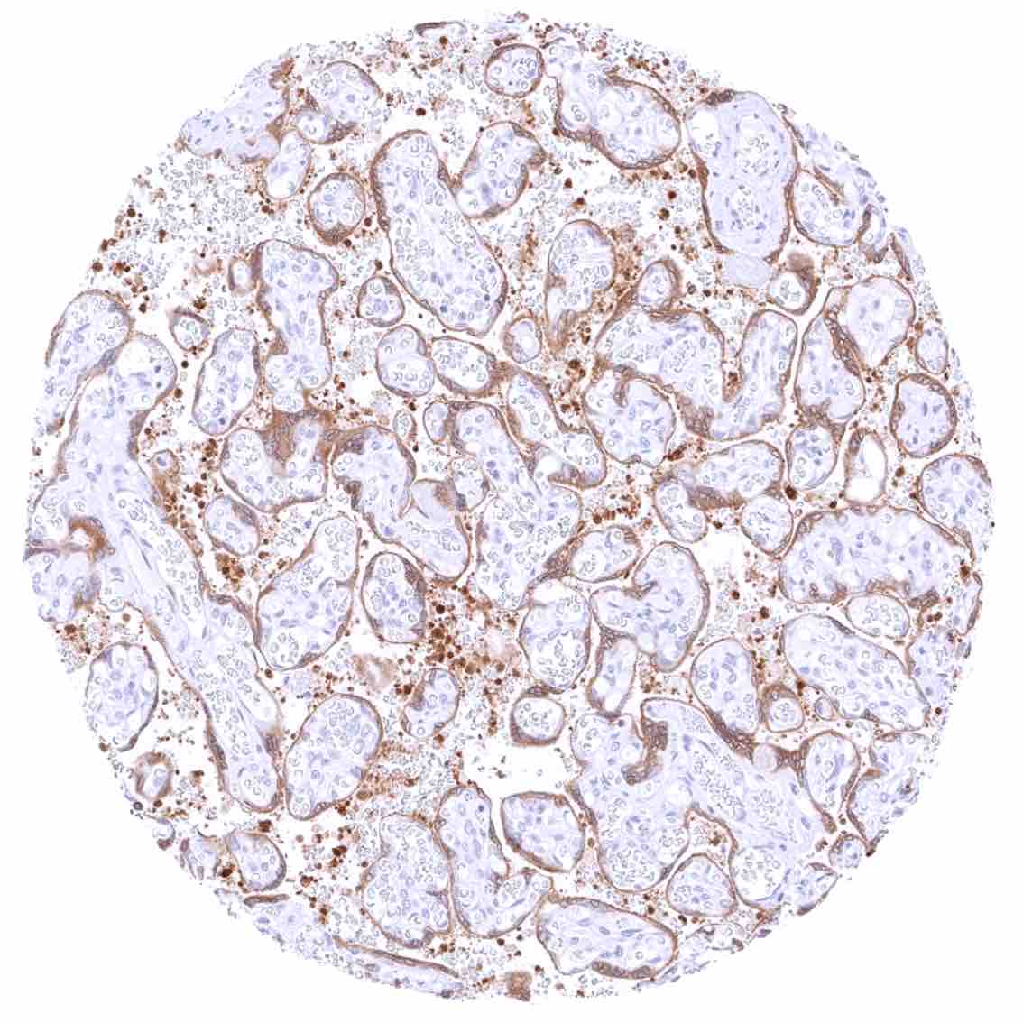
Placenta, mature – Moderate to strong cytoplasmic bcl-2 staining of trophoblast cells.
Prostate – Weak to moderate bcl-2 staining of a fraction of basal cells.
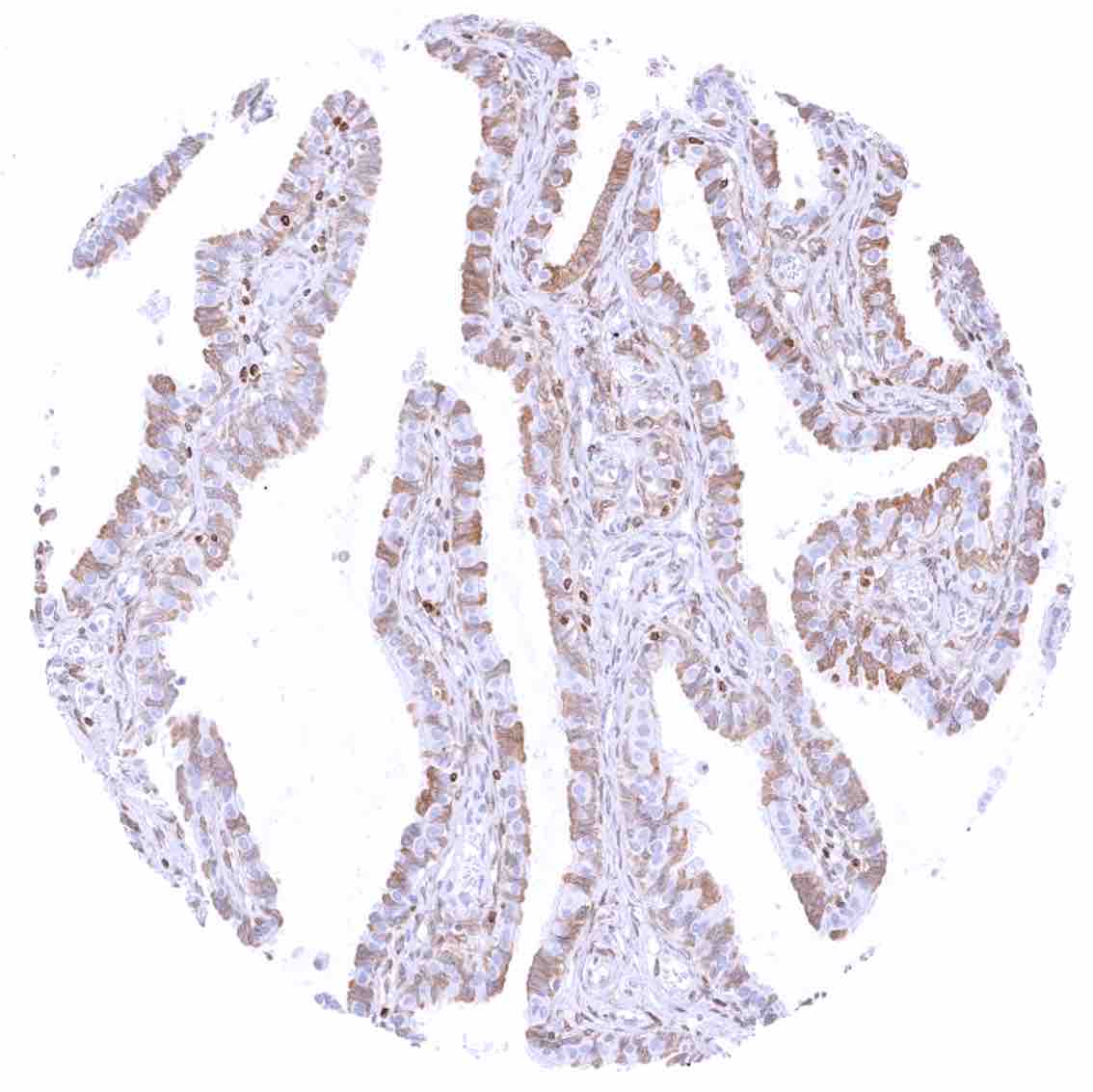
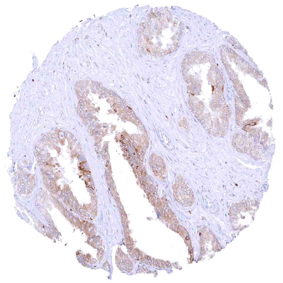
Rectum, mucosa .jpeg
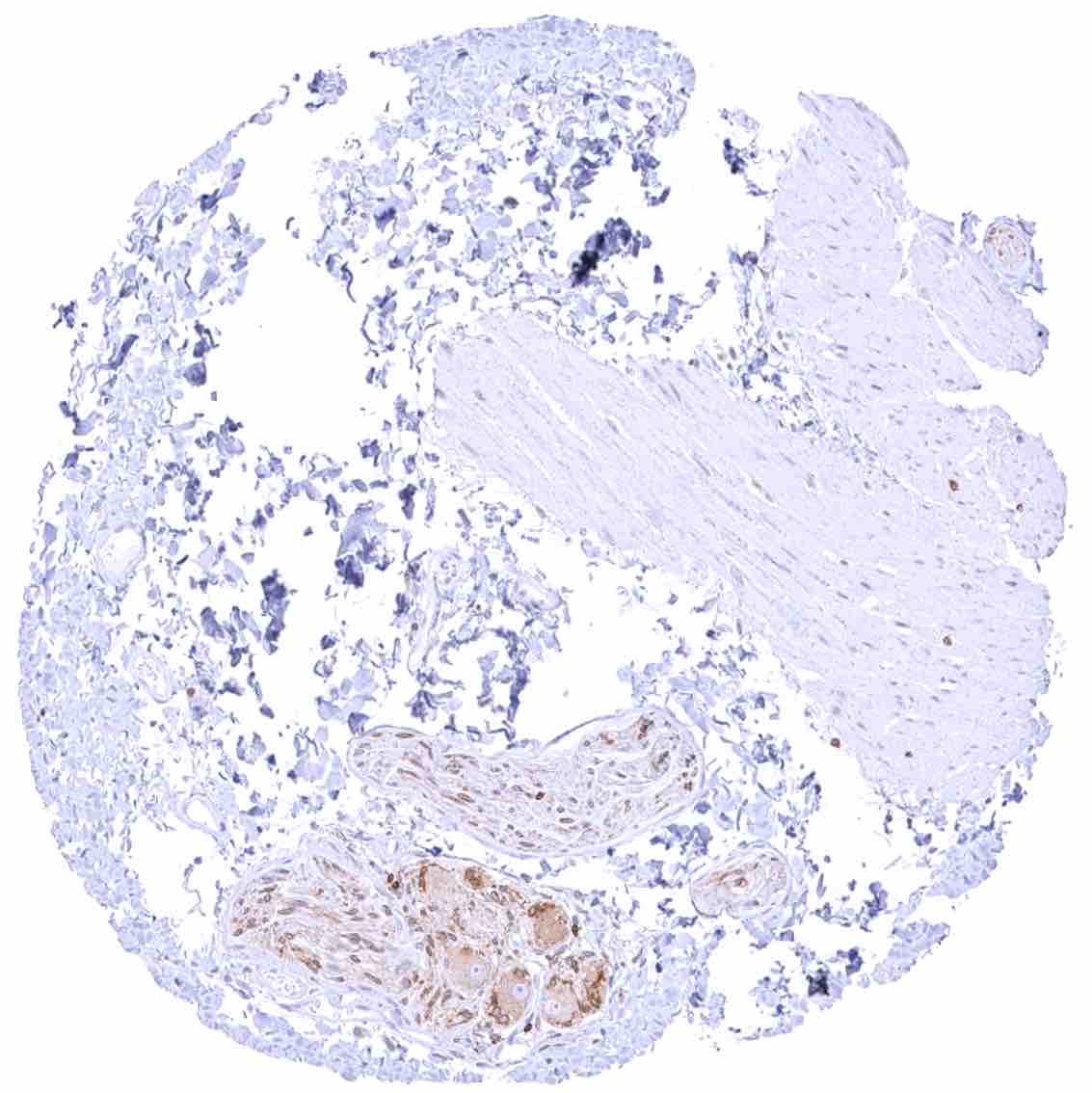
Seminal vesicle – Moderate to strong bcl-2 staining of epithelial cells.
Sinus paranasales – Weak cytoplasmic bcl-2 staining of basal cells of the respiratory epithelium. .jpeg
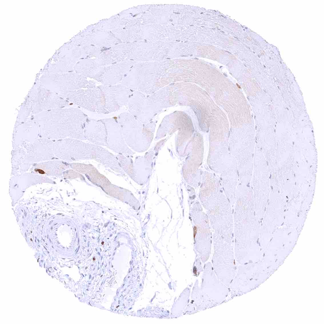
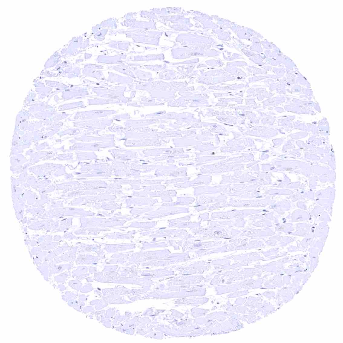
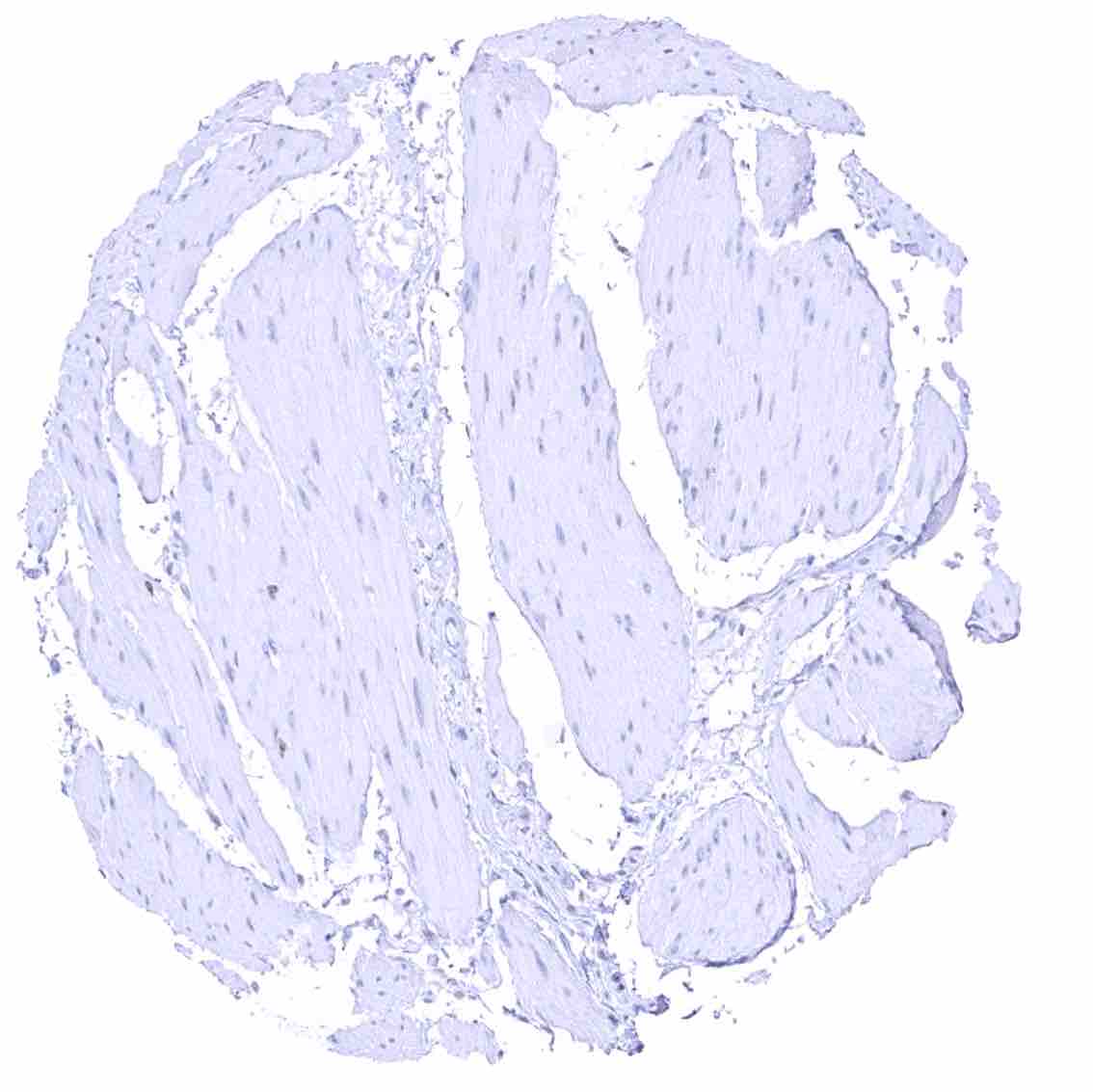
Skeletal muscle – Faint cytoplasmic bcl-2 staining of muscle fibers.
Skin
Skin, hairfollicel and sebaceous glands
Spleen – Strong bcl-2 positivity of the lymphocytes of the white pulpa and of a small fraction of cells in the red pulpa. .jpeg

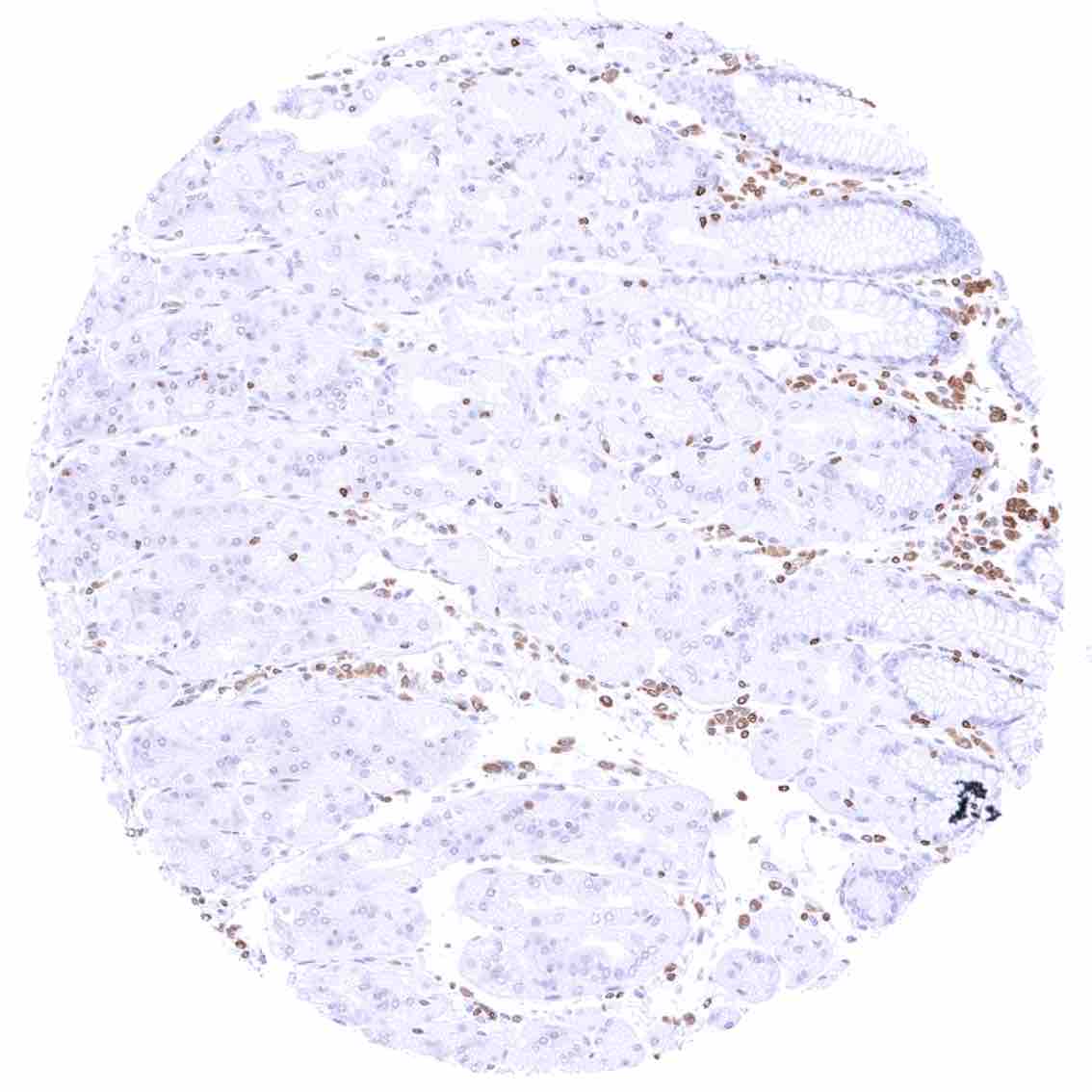
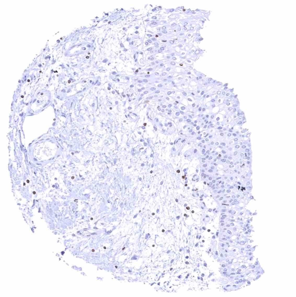
Stomach, antrum – The epithelium is bcl-2 negative. Bcl-2 staining is limited to lymphocytes. .jpeg
Stomach, corpus
Stomach, muscular wall – Moderate cytoplasmic bcl-2 staining of nerves and ganglion cells in the muscular wall.
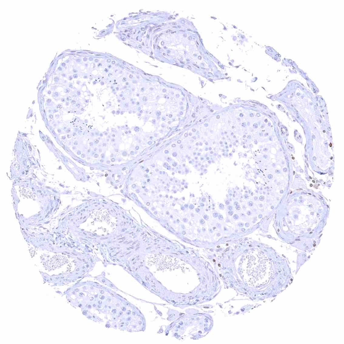
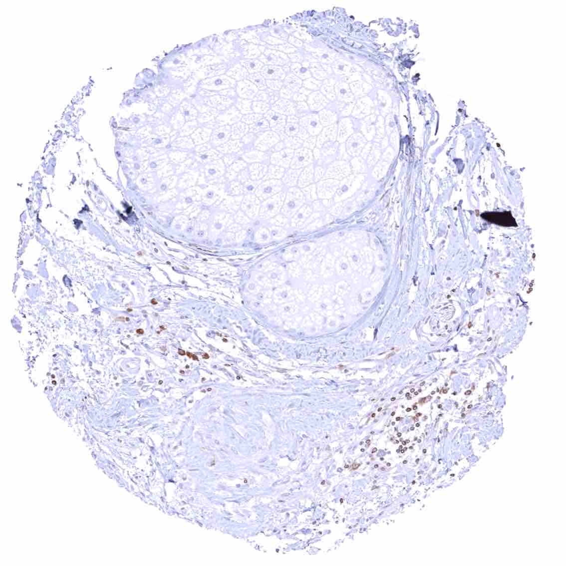
Testis
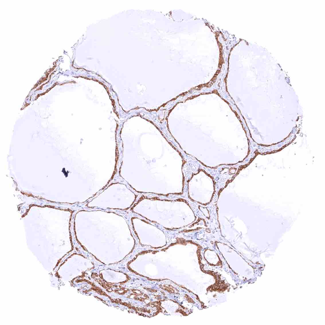
Thyroid gland – Strong cytoplasmic bcl-2 staining of follicular cells.
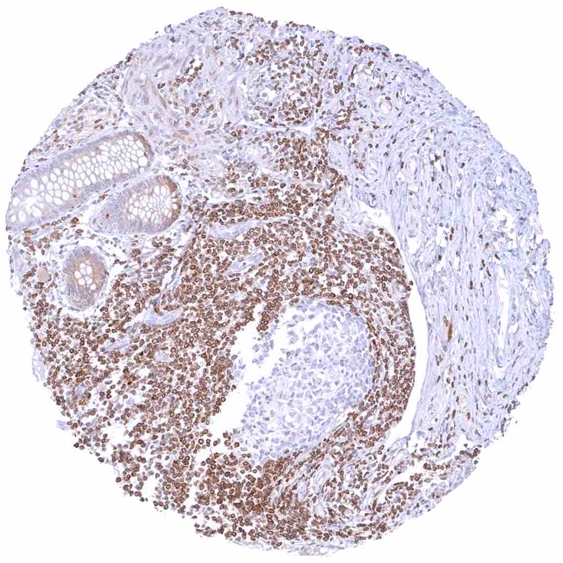
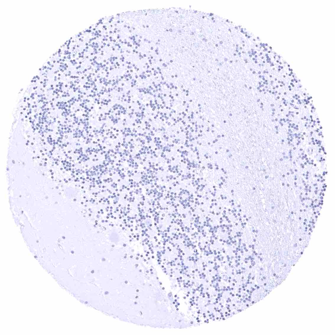
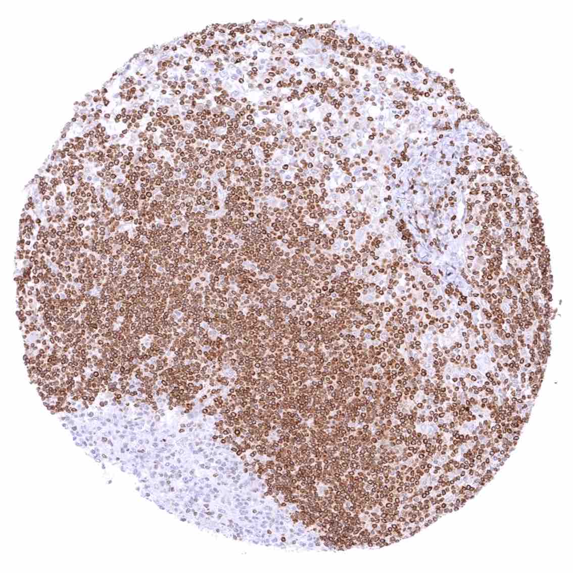
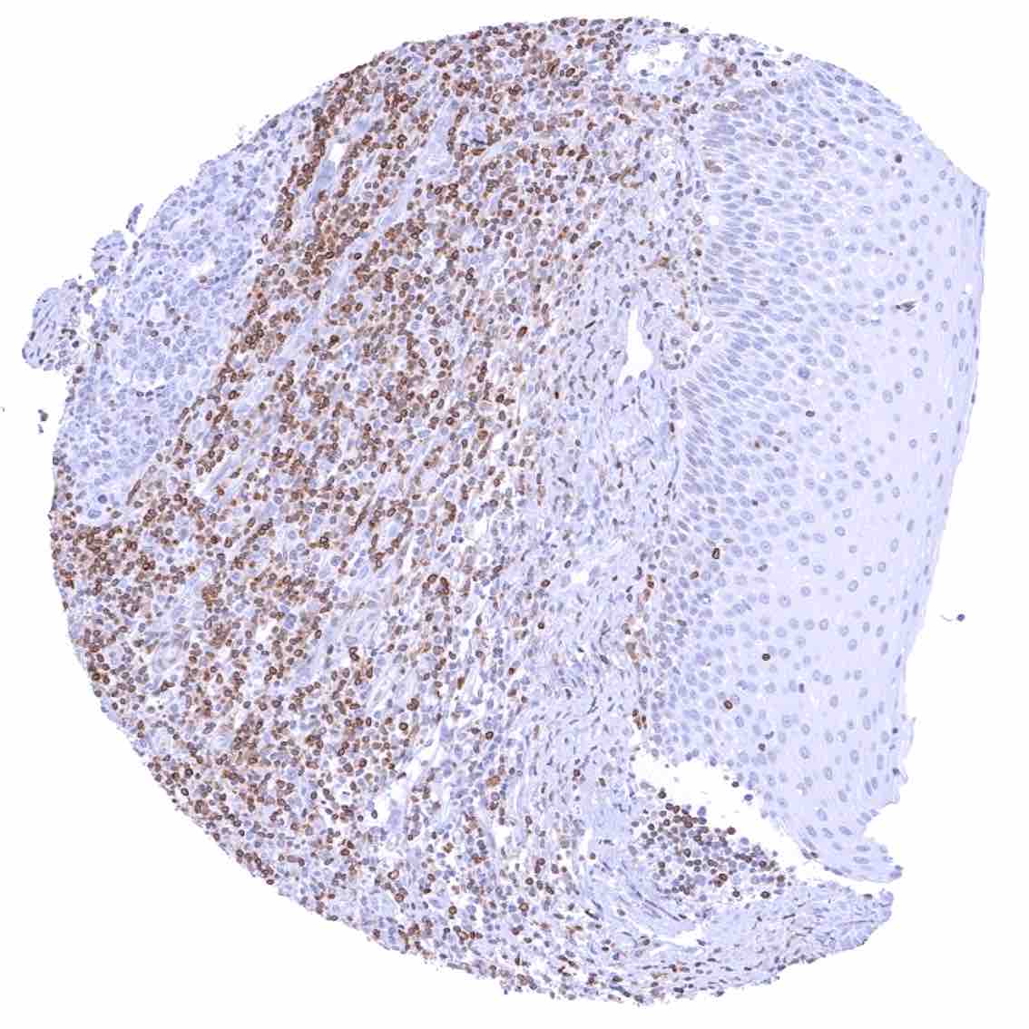
Tonsil –Strong bcl-2 positivity of large fraction of lymphocytic cells in the interfollicular area & around germinal centres. Cells in germinal centres are largely bcl-2 negative. Squamous epithelium with weak bcl-2 staining of the basal cell layer
Tonsil, surface epithelium
Urinary bladder, muscular wall
Urinary bladder, urothelium
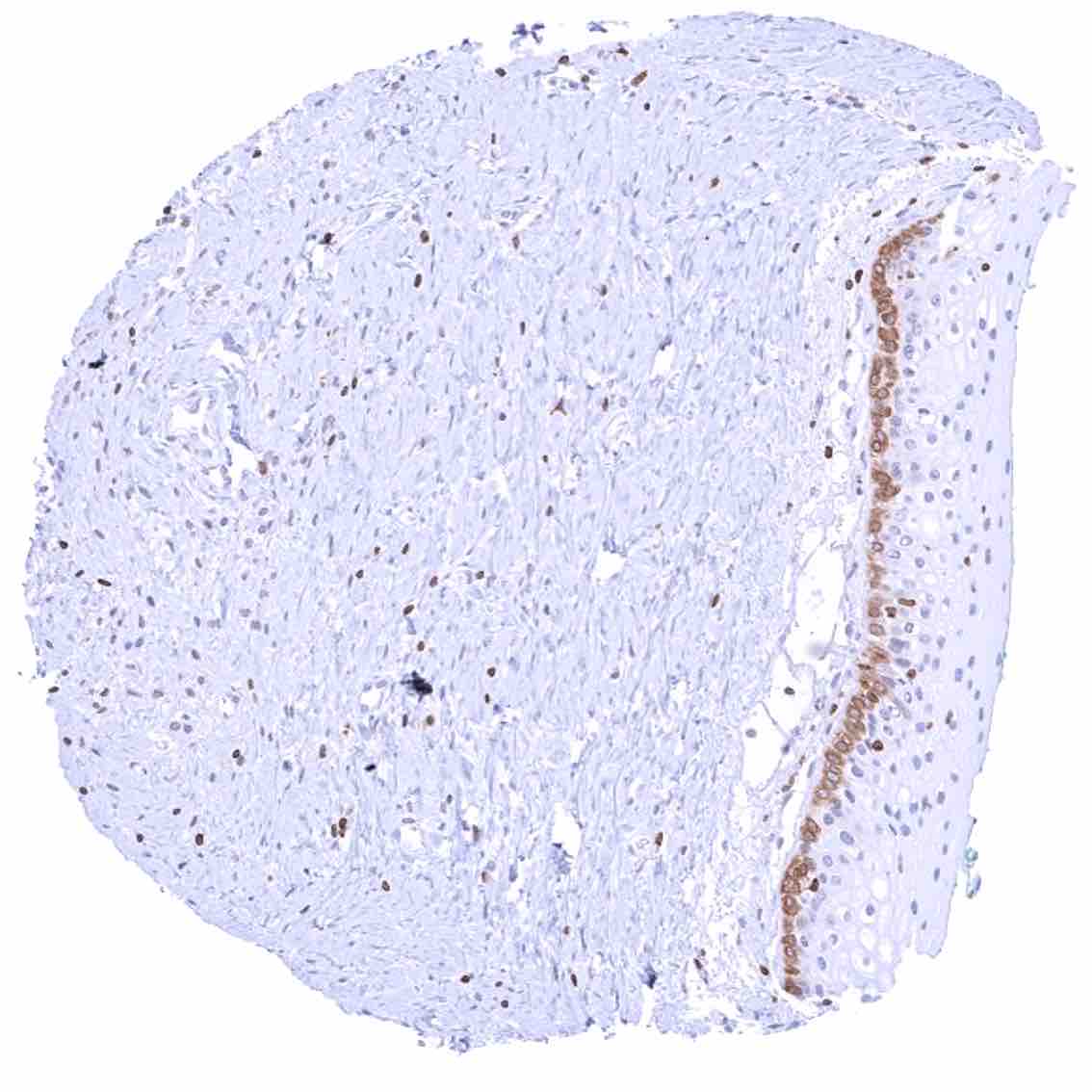
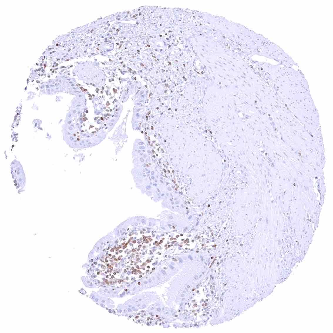
Uterus, ectocervix – Strong cytoplasmic bcl-2 staining of basal cells of the squamous epithelium in this sample.
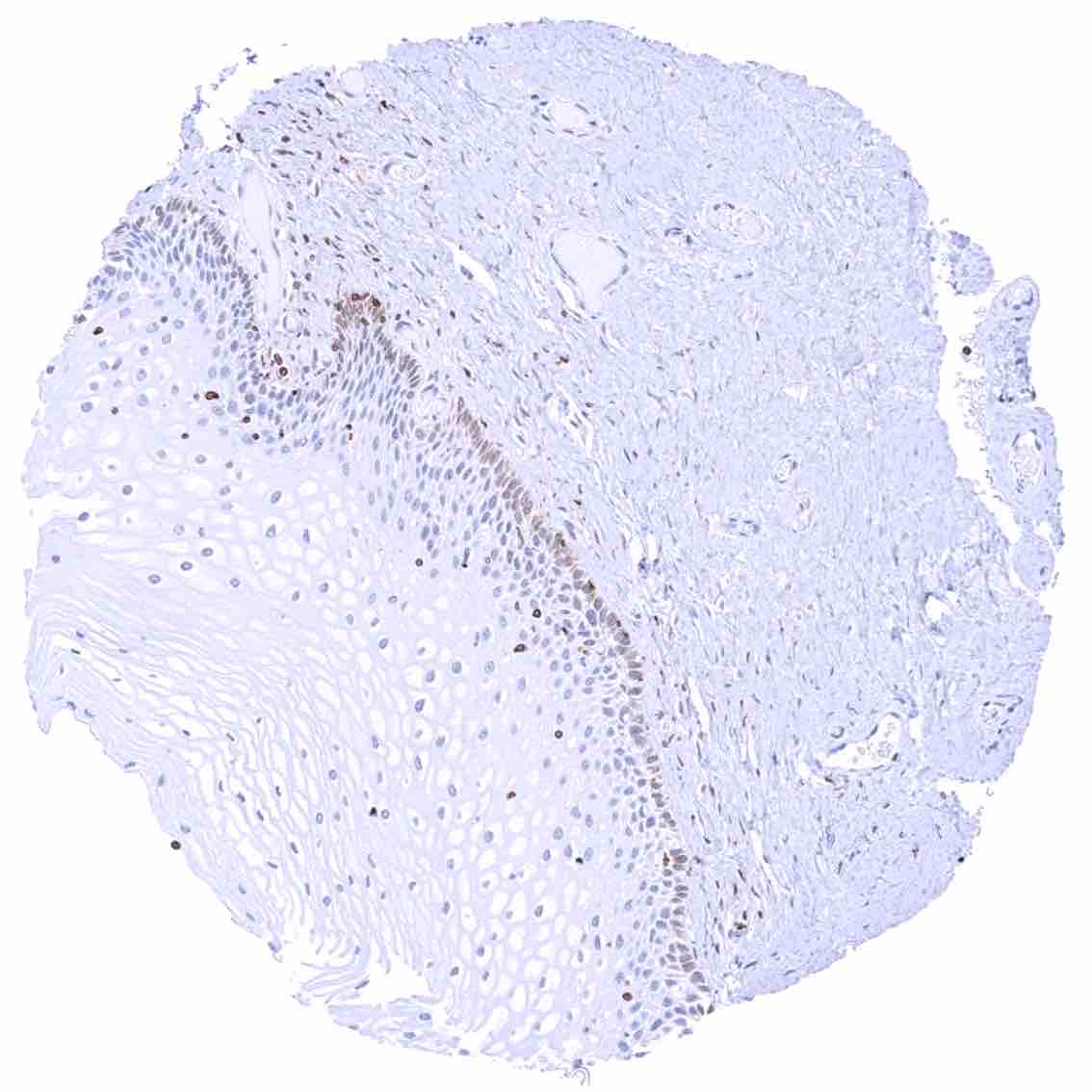
Uterus, ectocervix – Weak cytoplasmic bcl-2 staining of basal cells of the squamous epithelium.
Uterus, endocervix
Uterus, endometrium (proliferation) – Cytoplasmic bcl-2 staining of variable intensity in epithelial cells while staining is faint in stromal cells.
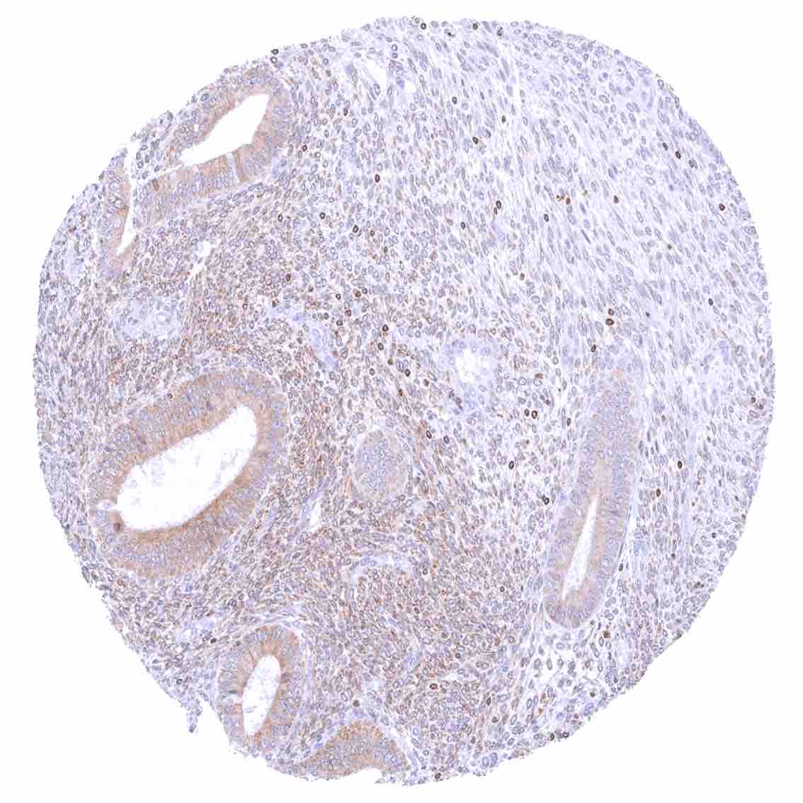
Uterus, endometrium (proliferation) – Weak to moderate cytoplasmic bcl-2 staining of epithelial and stromal cells.
Uterus, endometrium (secretion) – Variable (negative to moderate) cytoplasmic bcl-2 staining of epithelial cells.
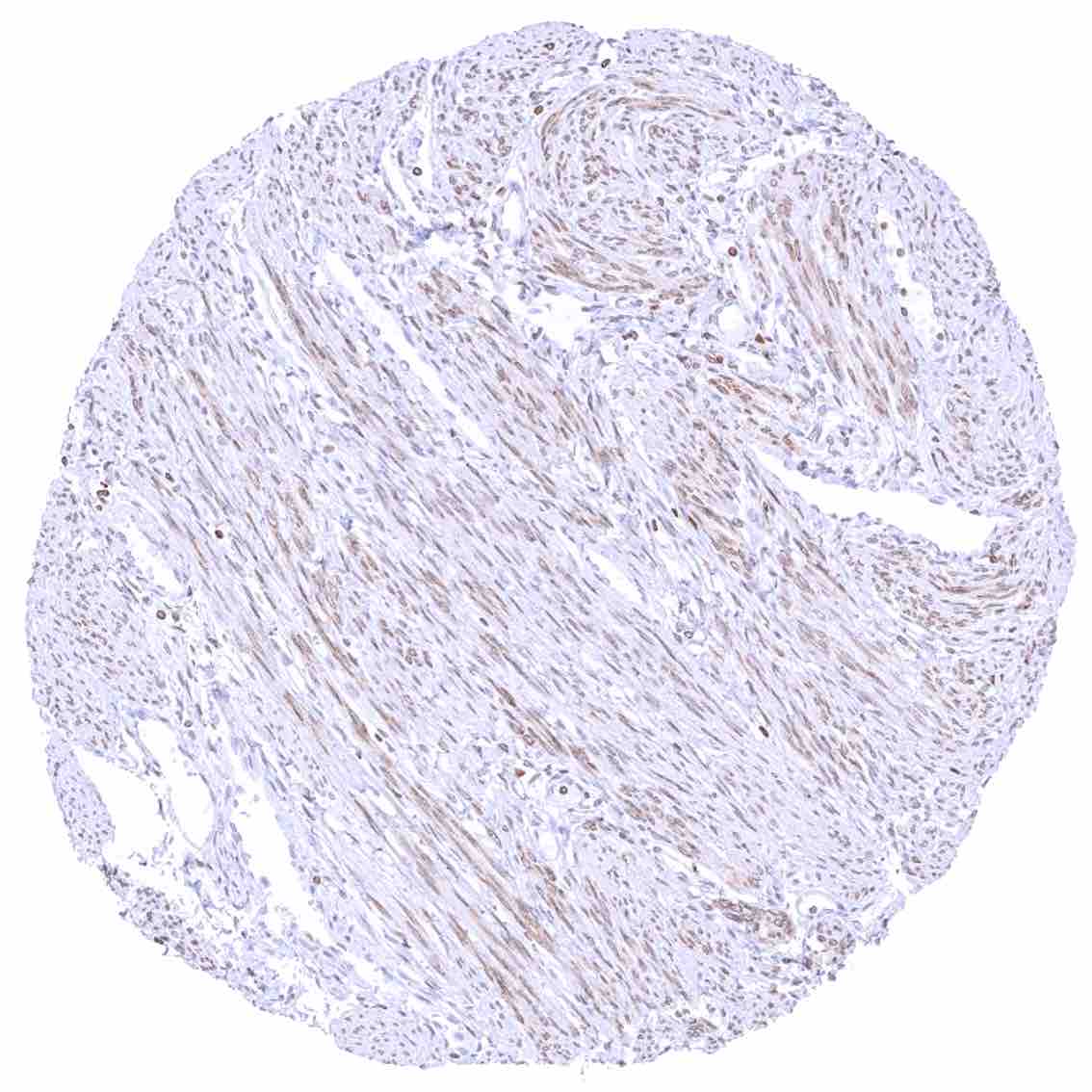
Uterus, myometrium – Weak to moderate cytoplasmic bcl-2 staining of smooth muscle fibers.