Login / Register
MS Validated Antibodies - Logo
  • About us
  • Products
    • Primary antibodies
    • Gallery
  • Distributors
  • Validation
    • Tissue Microarrays
  • Publications
    • Large Scale Studies
    • Further Publications
    • Events
    • Quiz
  • Contact
  • About us
  • Products
    • Primary antibodies
    • Gallery
  • Distributors
  • Validation
    • Tissue Microarrays
  • Publications
    • Large Scale Studies
    • Further Publications
    • Events
    • Quiz
  • Contact
×

Cancer Tissue Gallery MX1

Path Created with Sketch.
Go to MX1 Normal Tissue GalleryGo back to Product Page
Adrenal gland – MX1 negative adrenocortical adenoma
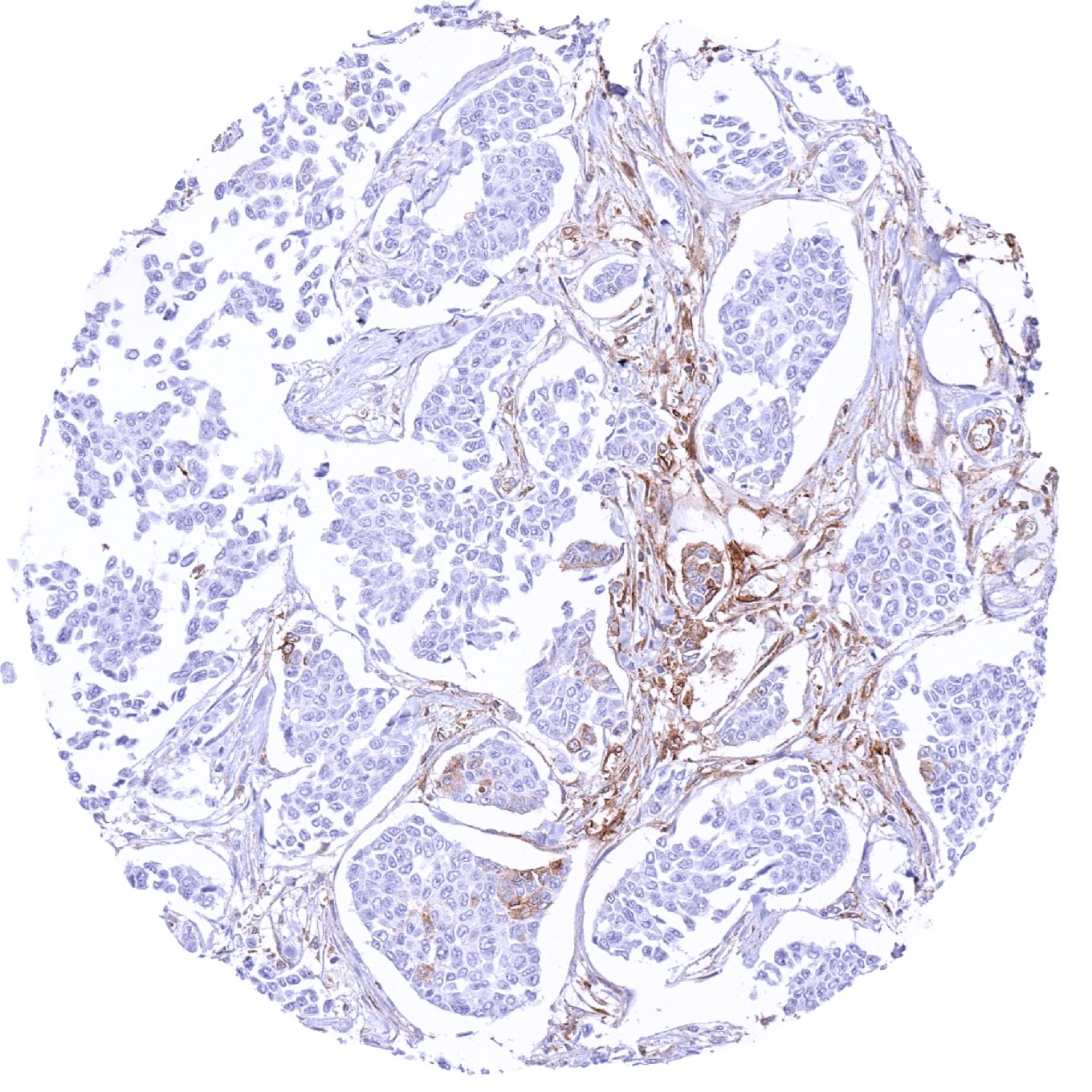
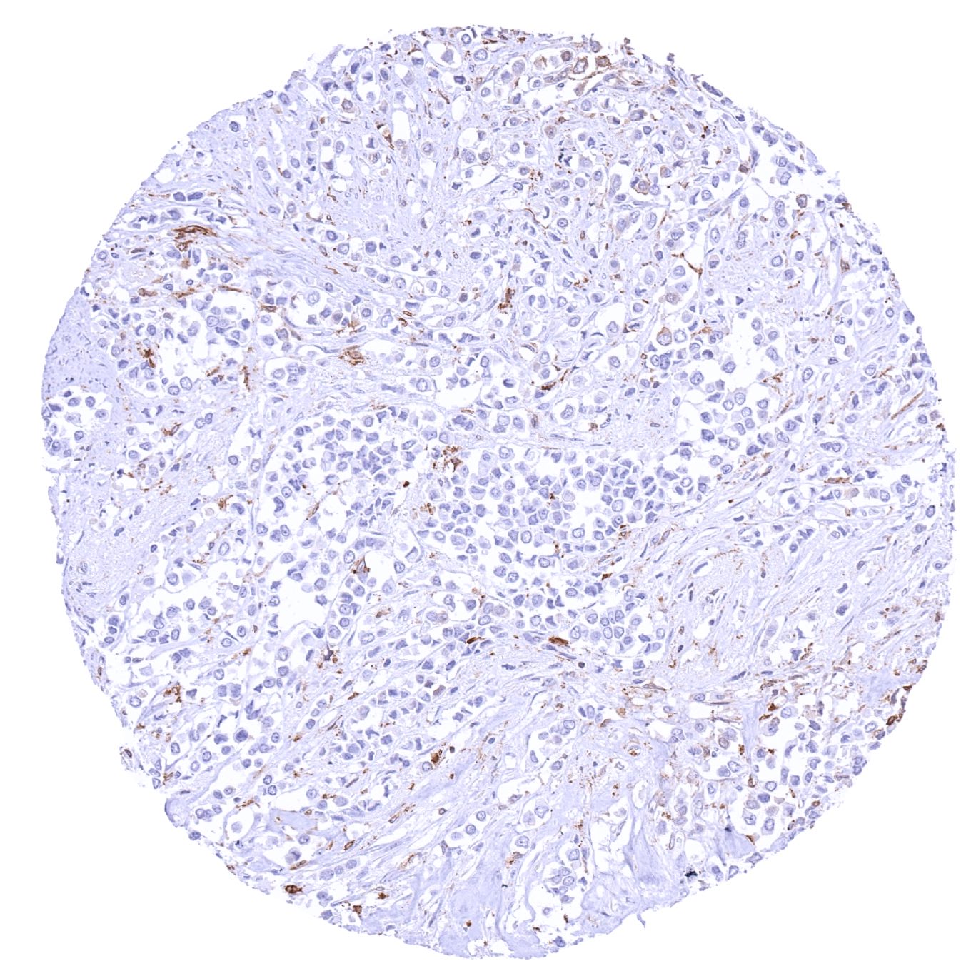
Breast – Invasive breast cancer of no special type (NST) with faint MX1 staining of few tumor cells
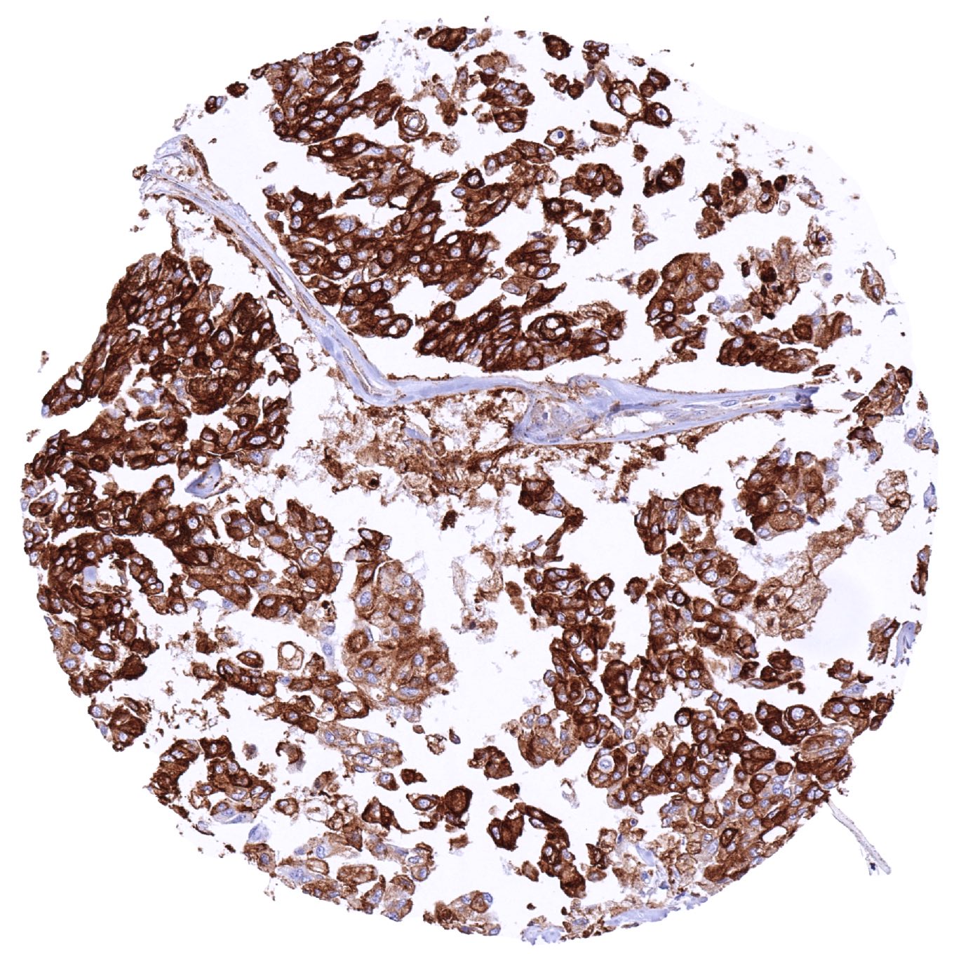
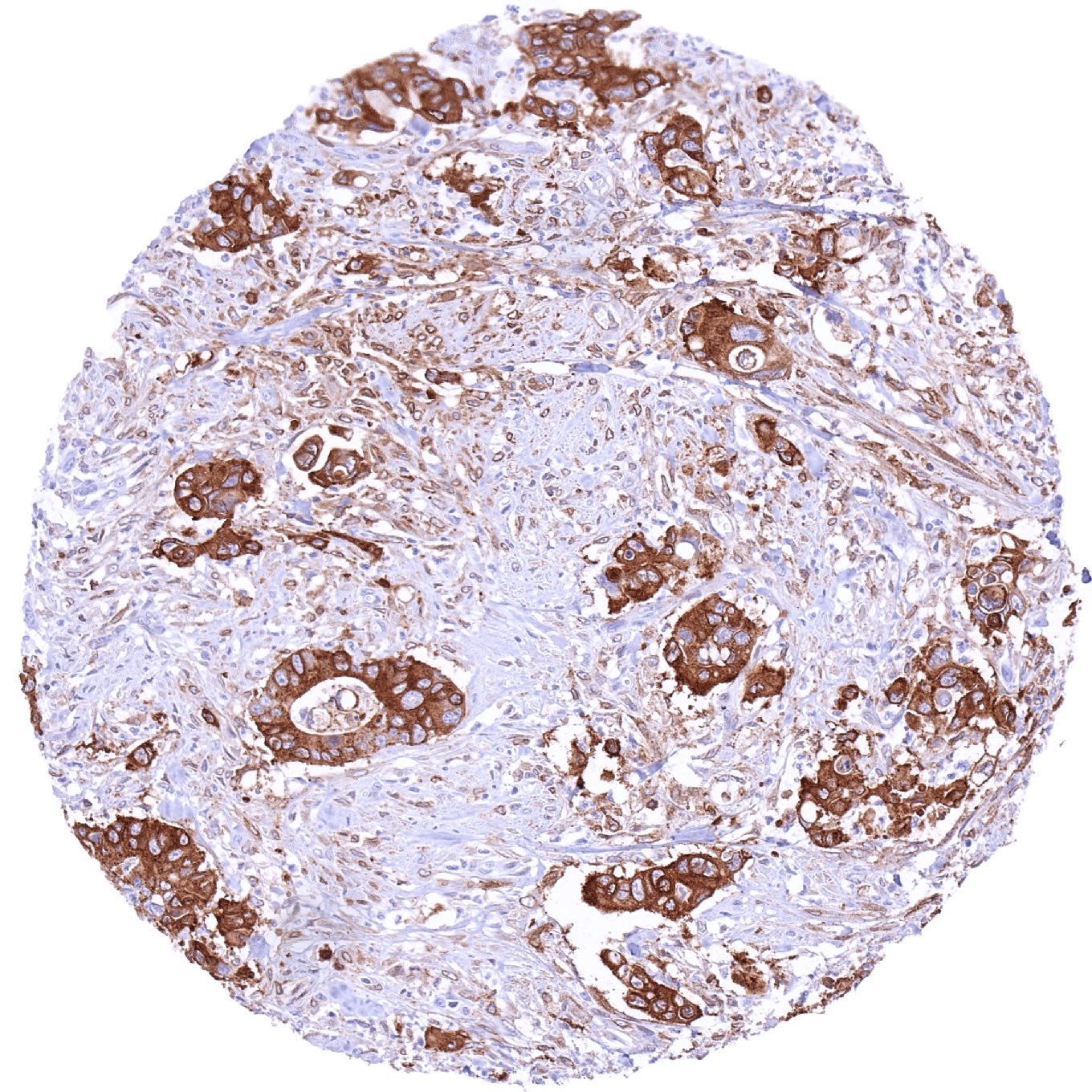
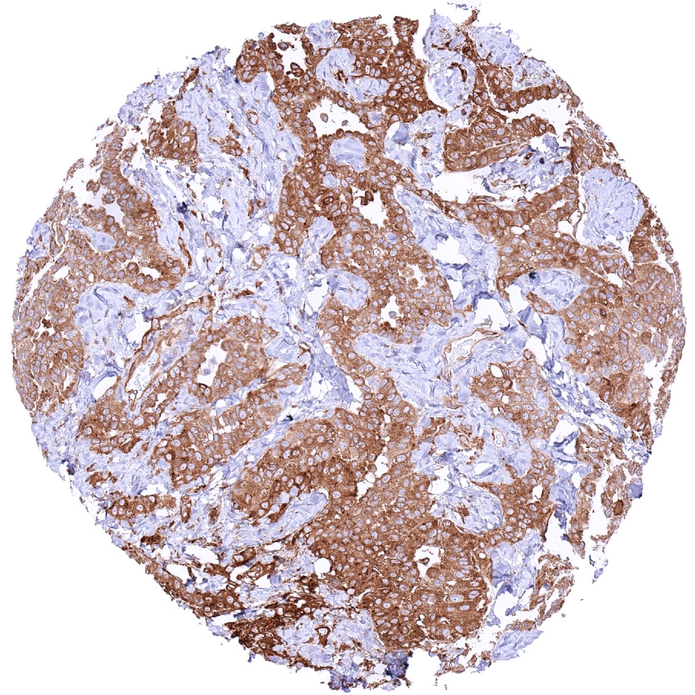
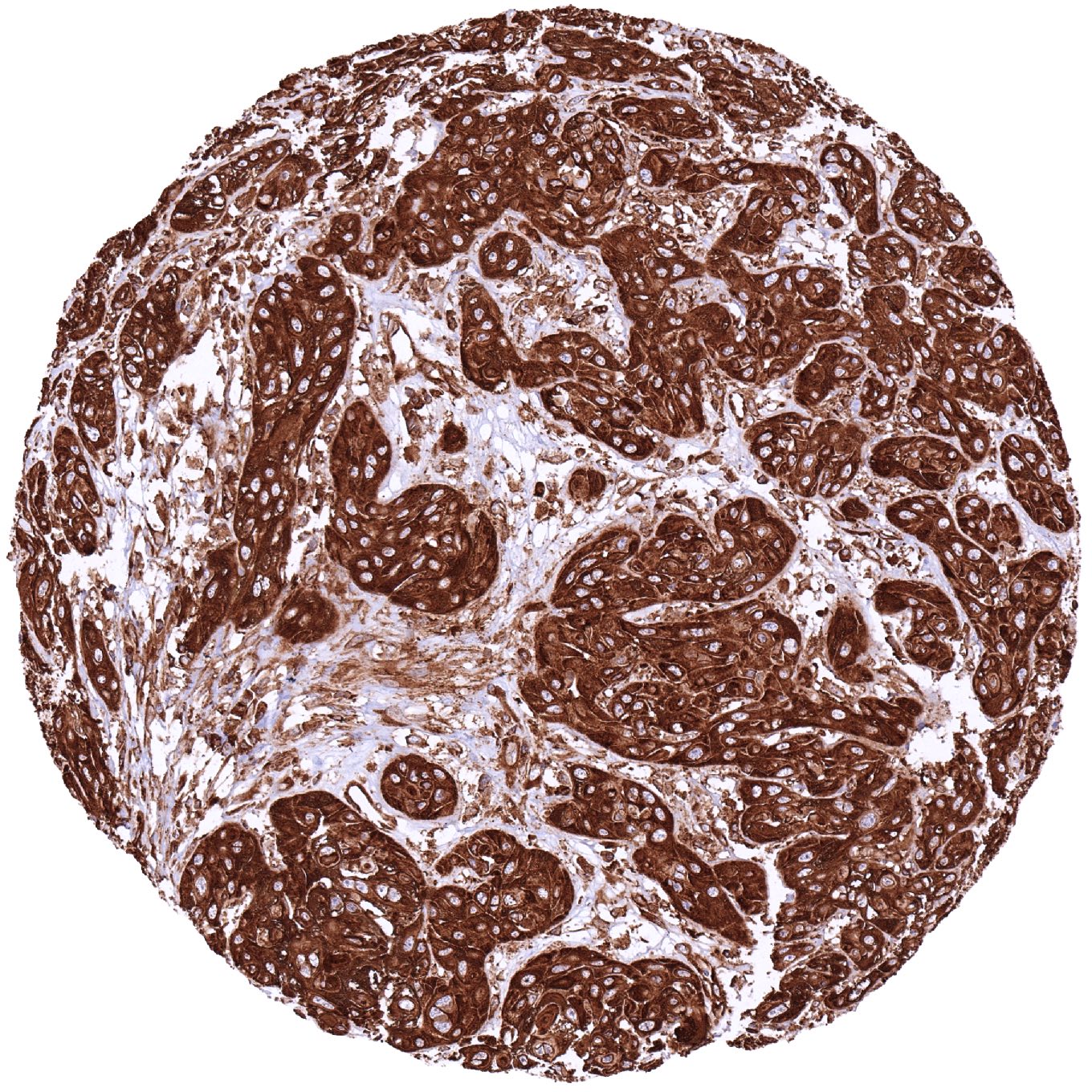
Breast – Invasive breast cancer of no special type (NST) with strong MX1 positivity of tumor cells.jpeg
Breast – Invasive breast cancer of no special type (NST) with strong MX1 positivity of tumor cells
Breast – Invasive lobular breast cancer with moderate to strong MX1 positivity of tumor cells
Breast – MX1 negative invasive lobular breast cancer
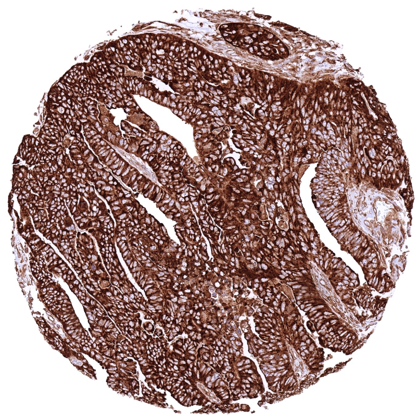
Colon – Colorectal adenocarcinoma with strong MX1 immunostaining of tumor cells
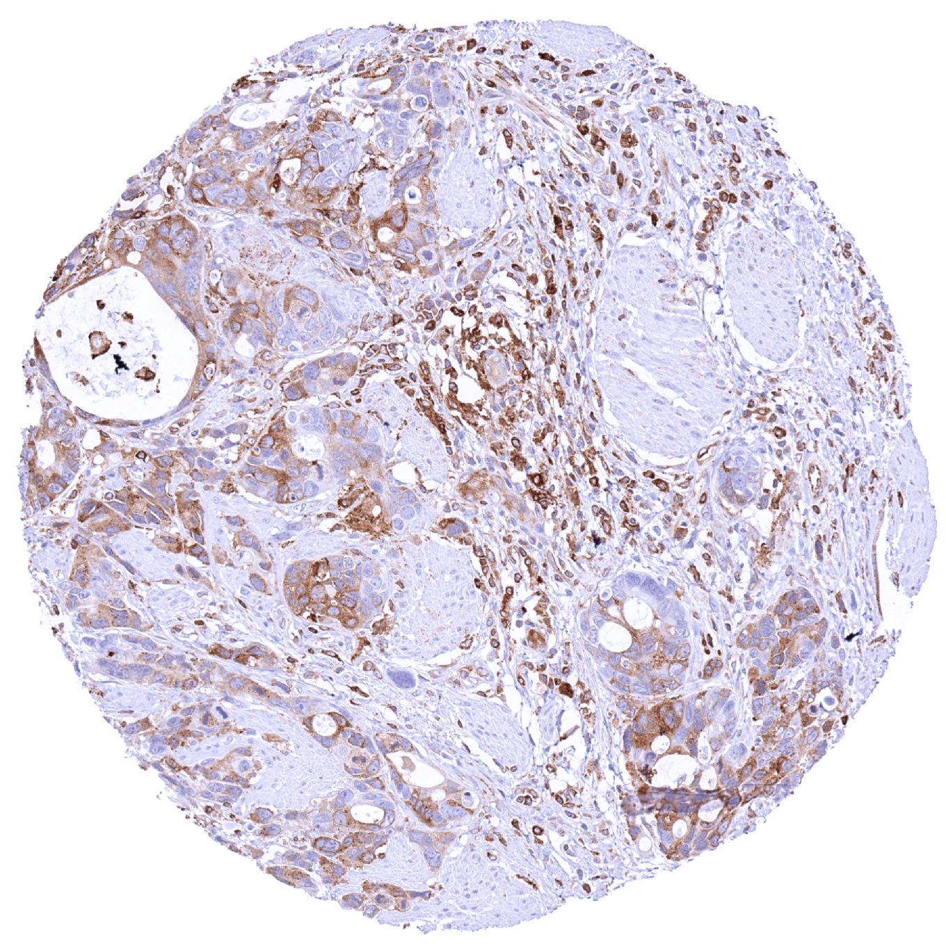
Esophagus – Adenocarcinoma with moderate to strong MX1 immunostaining of tumor cells
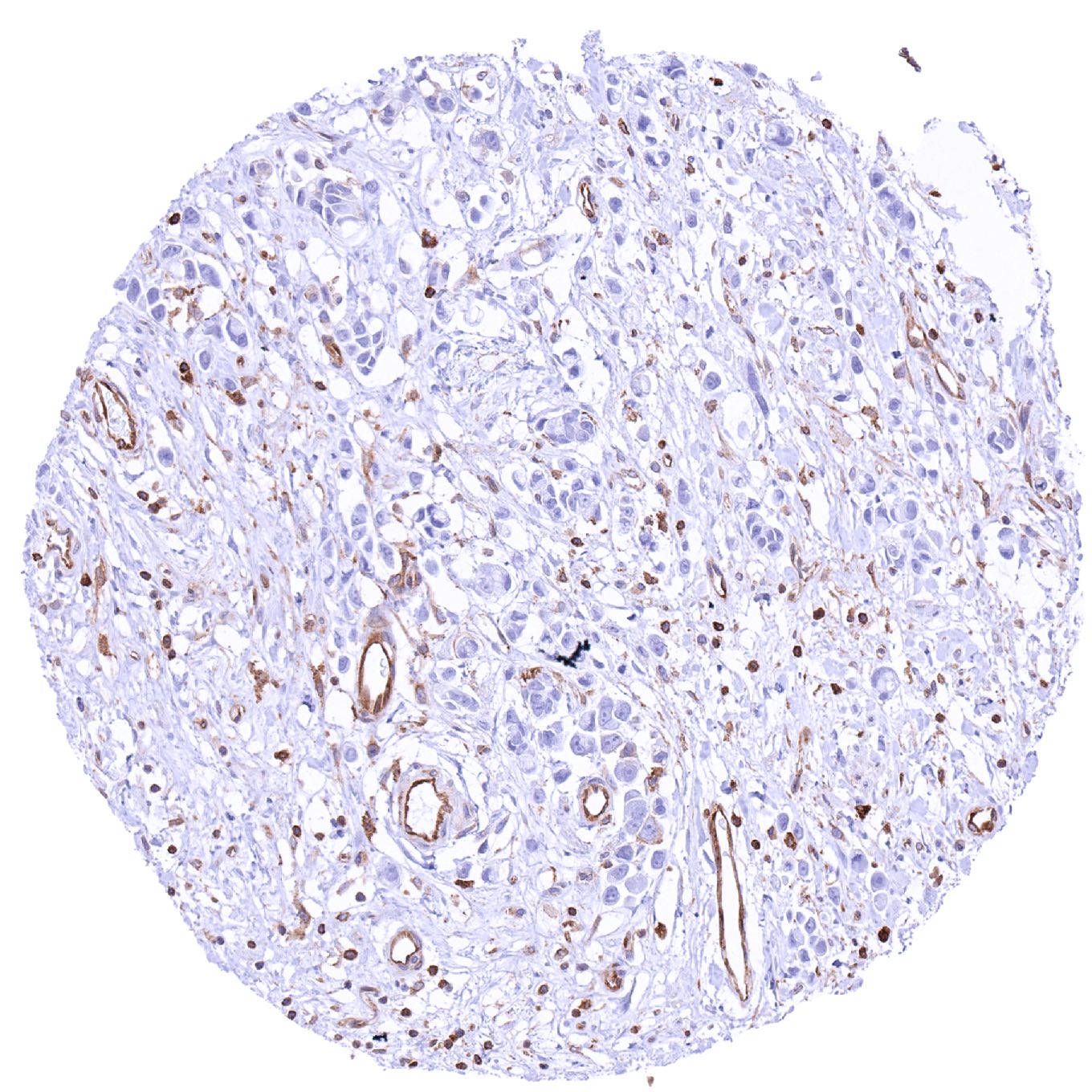
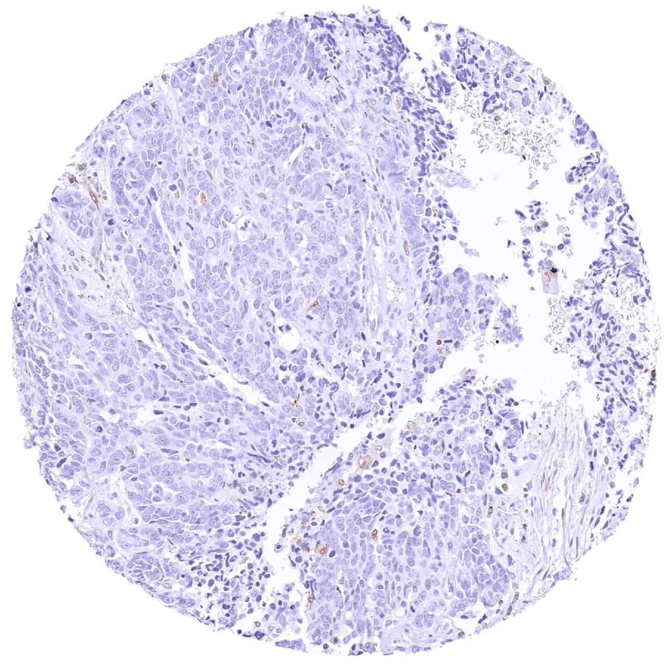
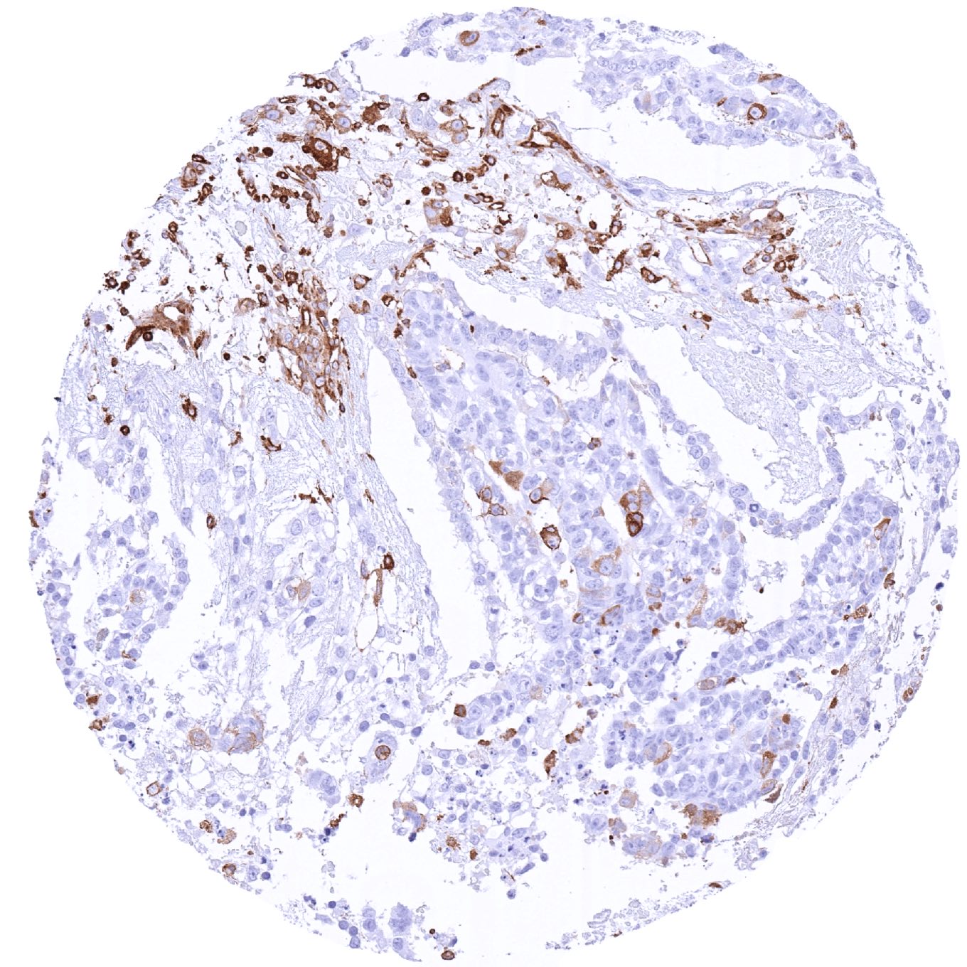
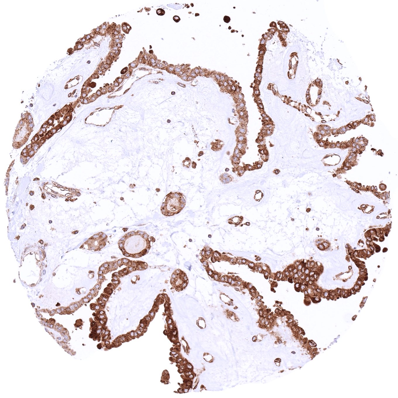
Esophagus – MX1 negative adenocarcinoma. Strong MX1 staining of inflammatory and endothelial cells
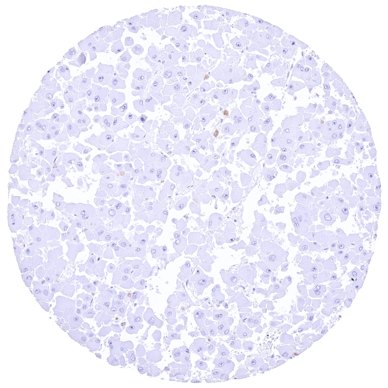
Kidney – MX1 negative clear cell carcinoma
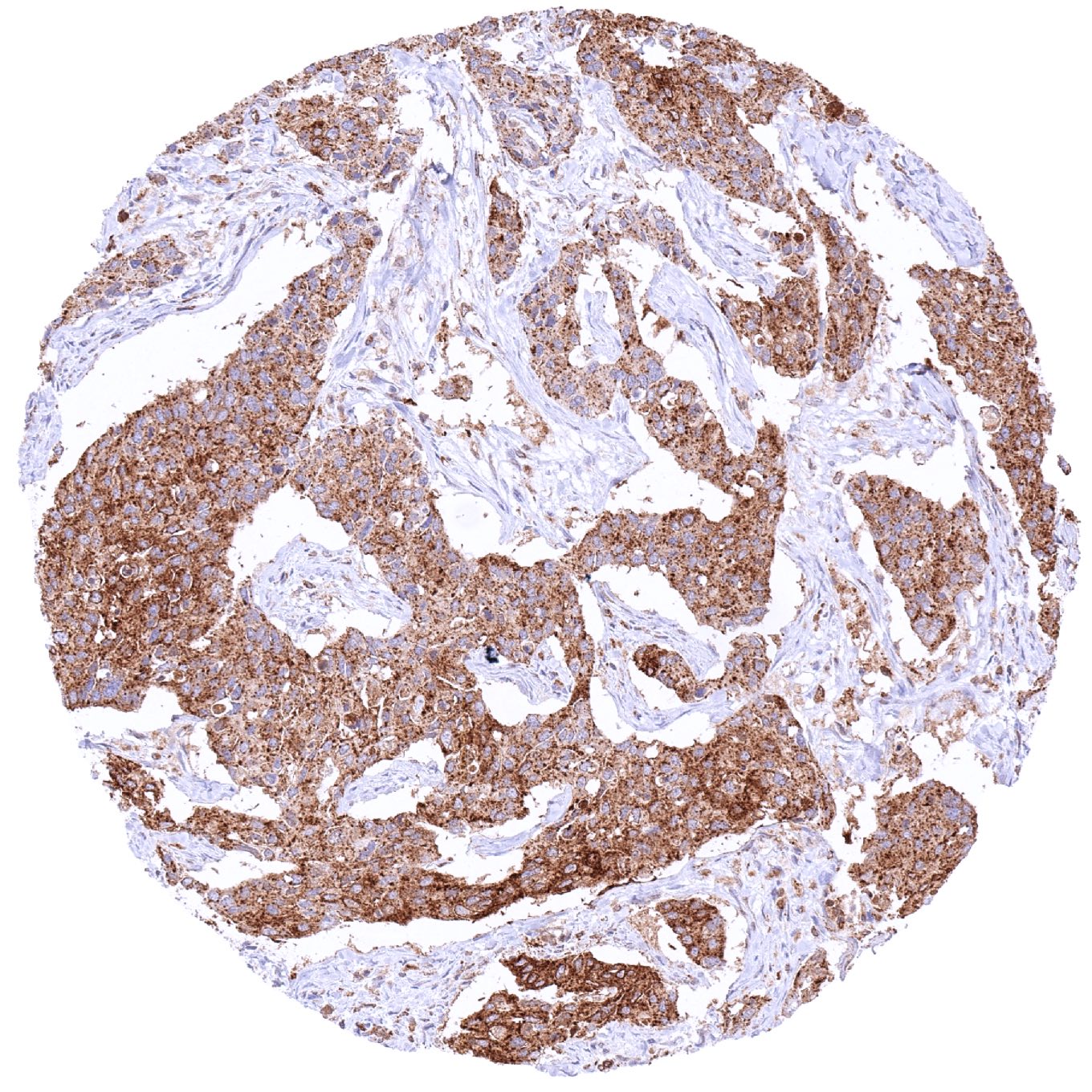
Larynx – Squamous cell carcinoma with strong MX1 immunostaining of tumor cells
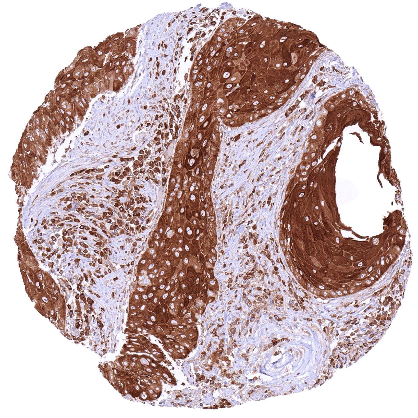
Larynx – Squamous cell carcinoma with strong MX1 staining of tumor cells. Stromal and inflammatory cells show a less intense MX1 staining
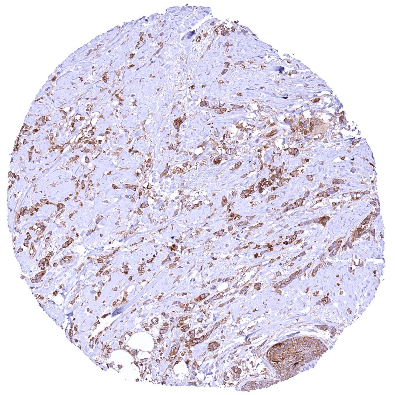
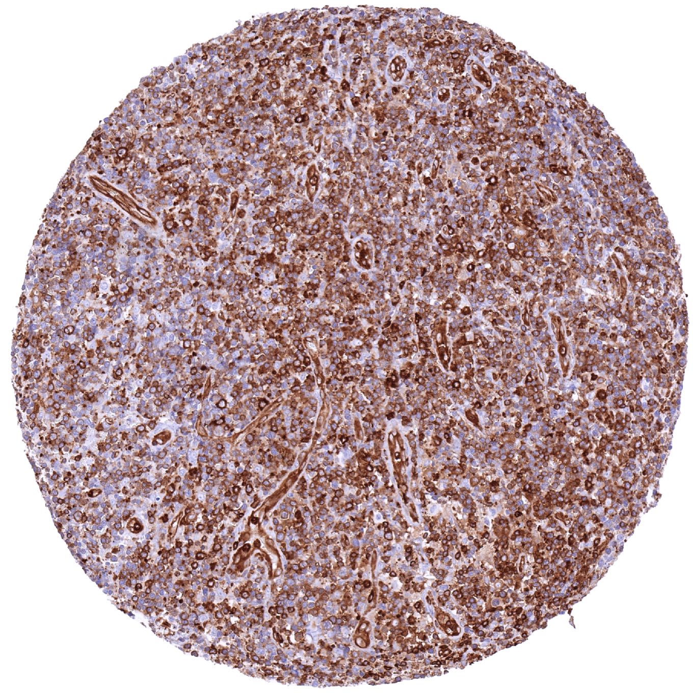
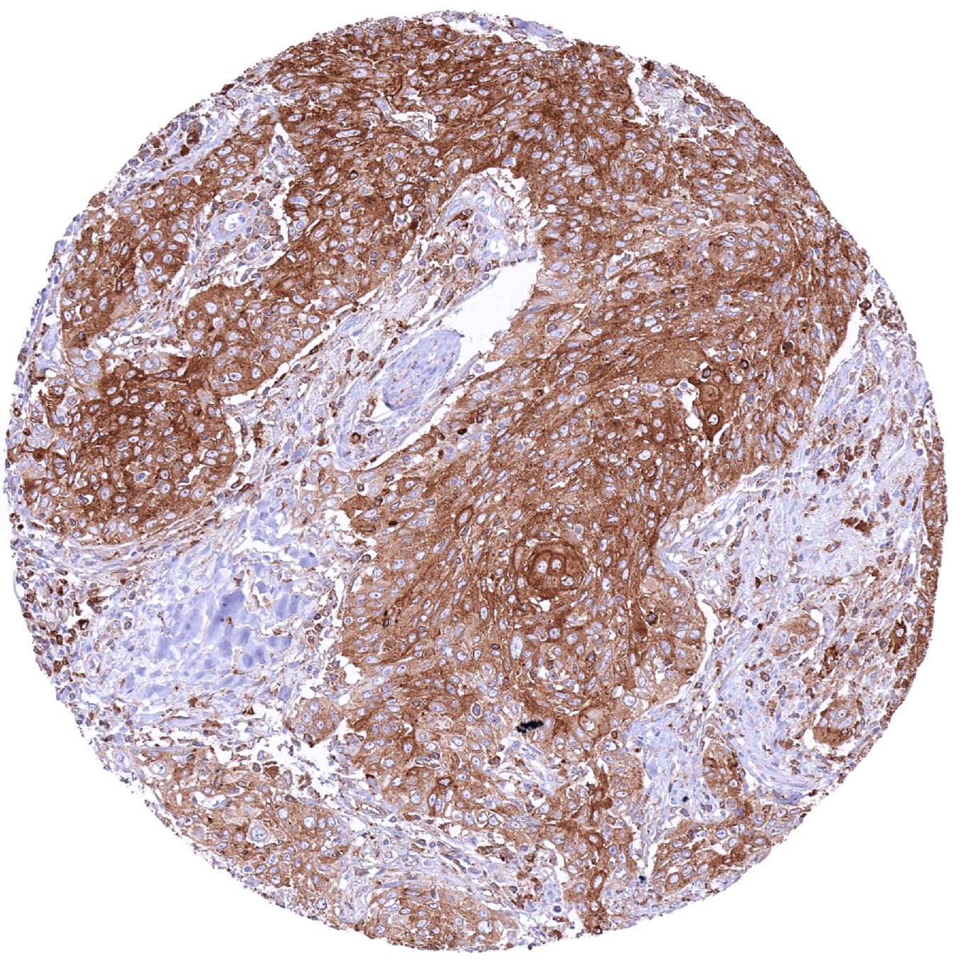
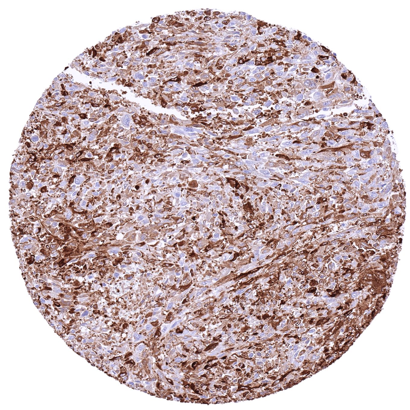
Liver – Cholangiocellular carcinoma with strong MX1 staining of tumor and stroma cells
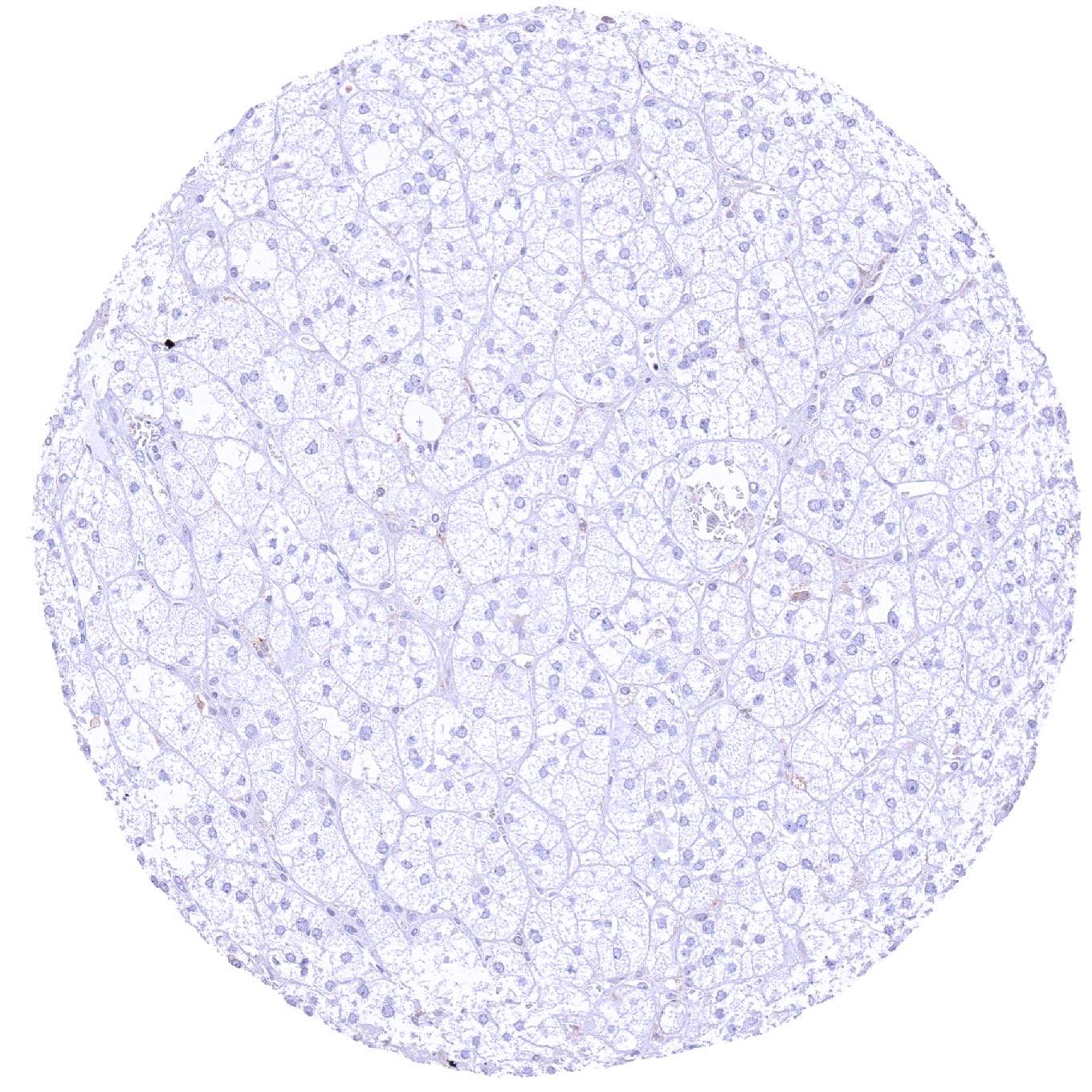
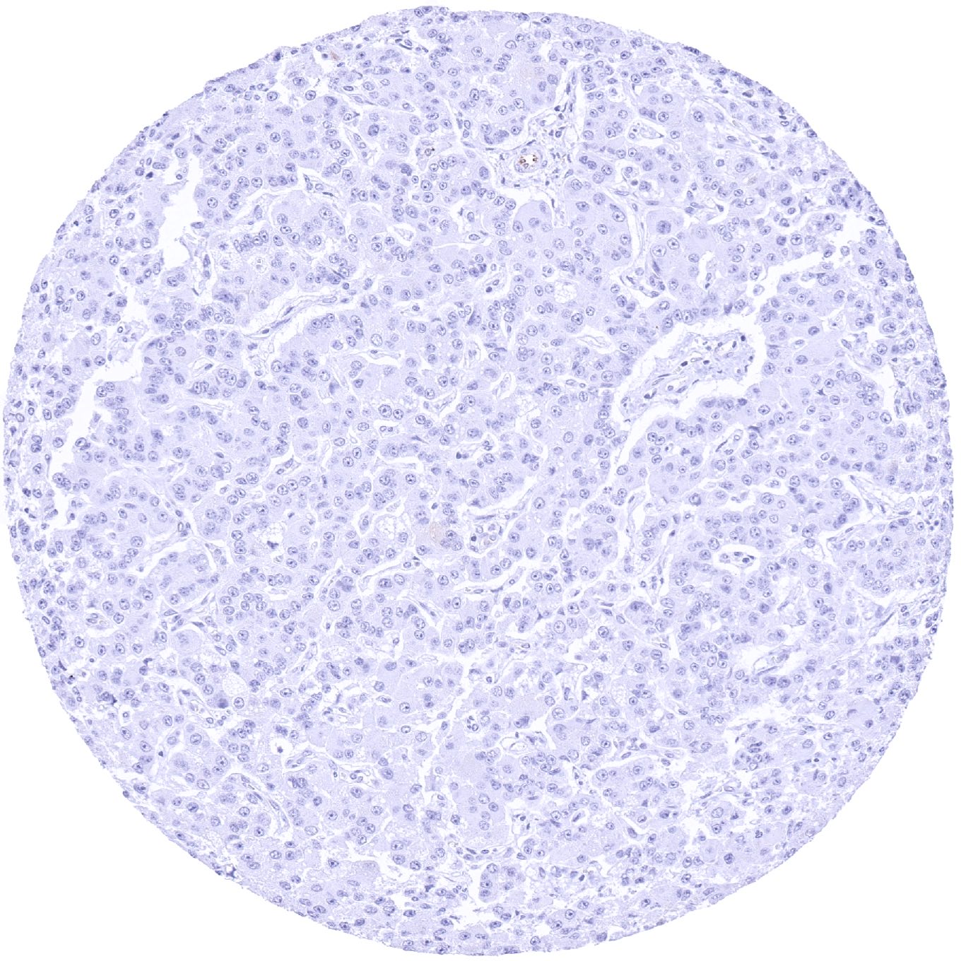
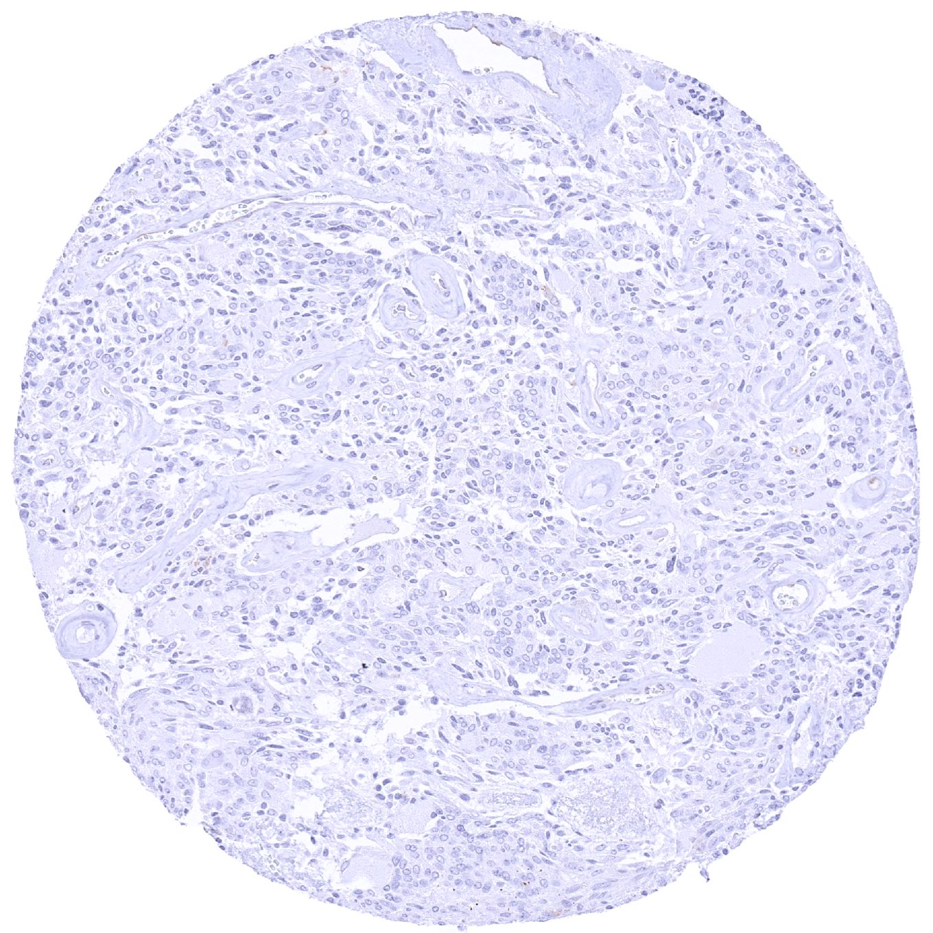
Liver – MX1 negative hepatocellular carcinoma
Lung – Adenocarcinoma with strong MX1 staining of tumor cells. Inflammatory and stroma cells are also MX1 positive
Lung – Malignant mesothelioma with strong MX1 positivity of tumor cells
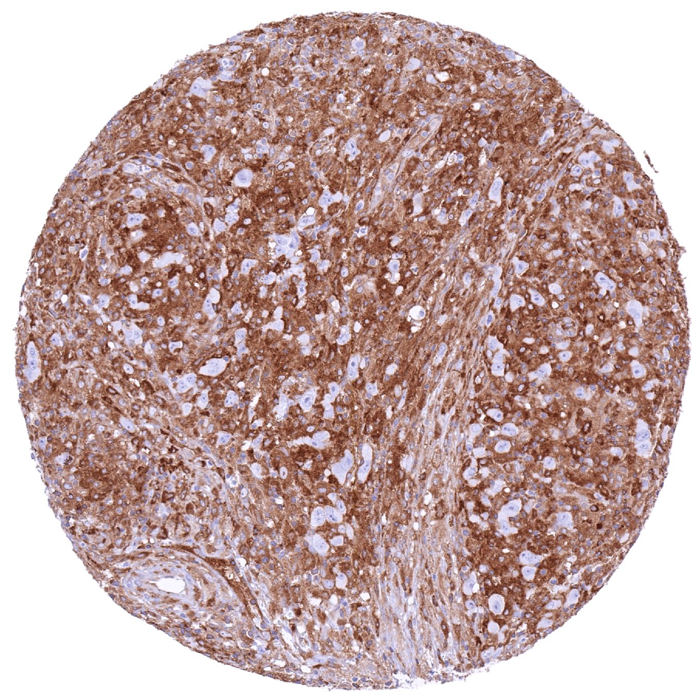
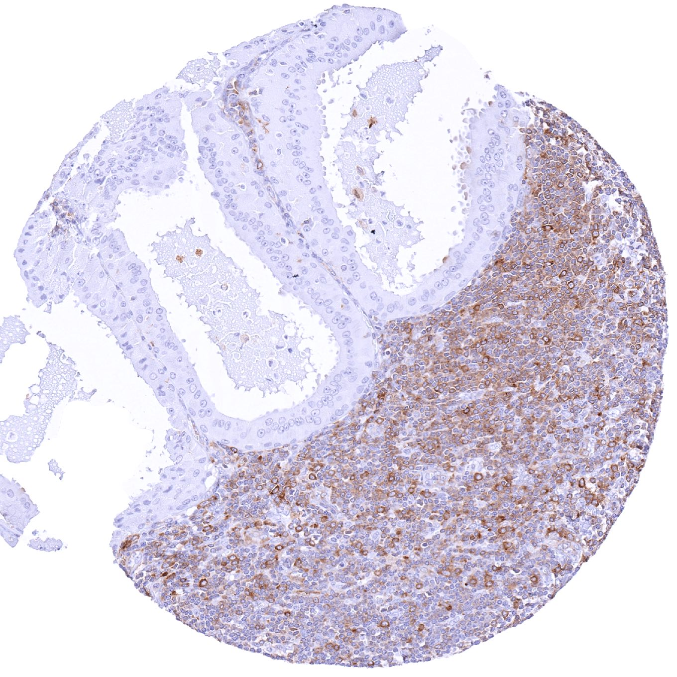
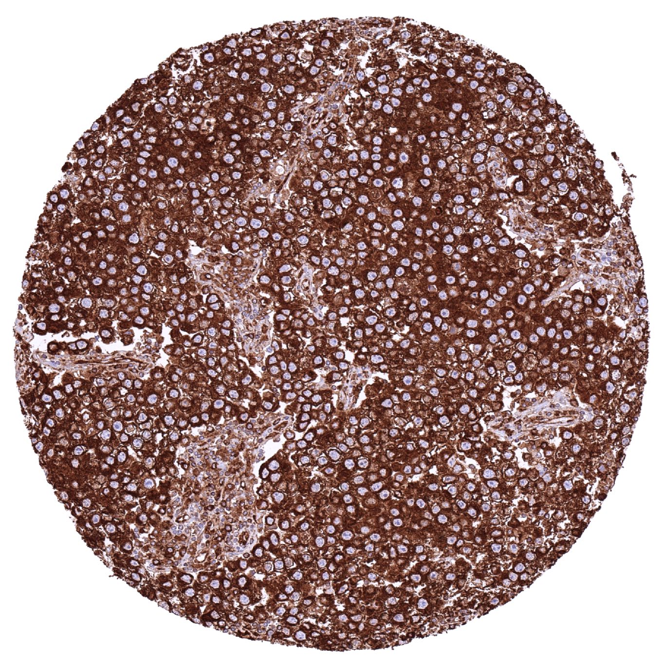
Lymph node – B-CLL with strong MX1 positivity of virtually all tumor cells
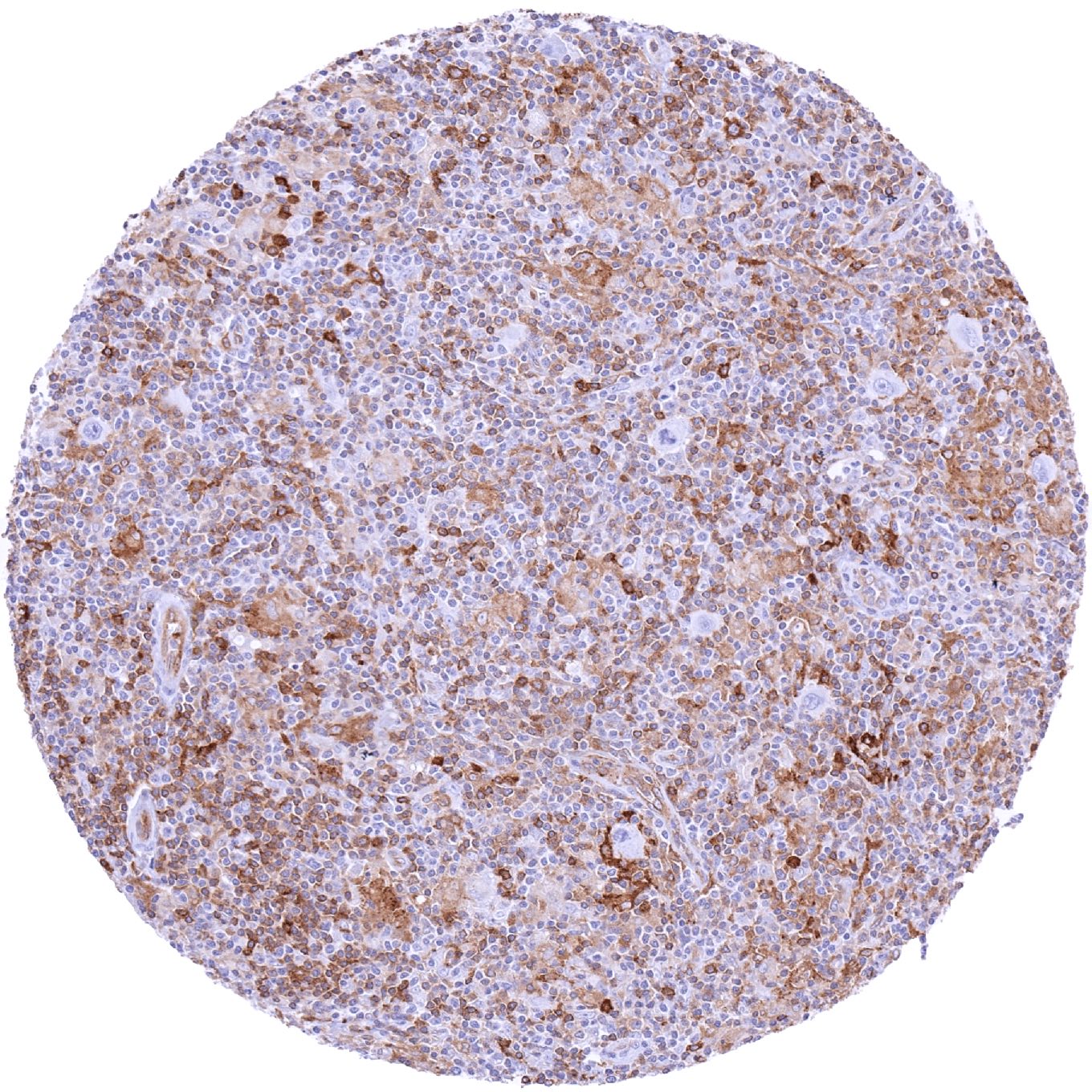
Lymph node – Diffuse large B-cell lymphoma with strong MX1 staining of all tumor cells
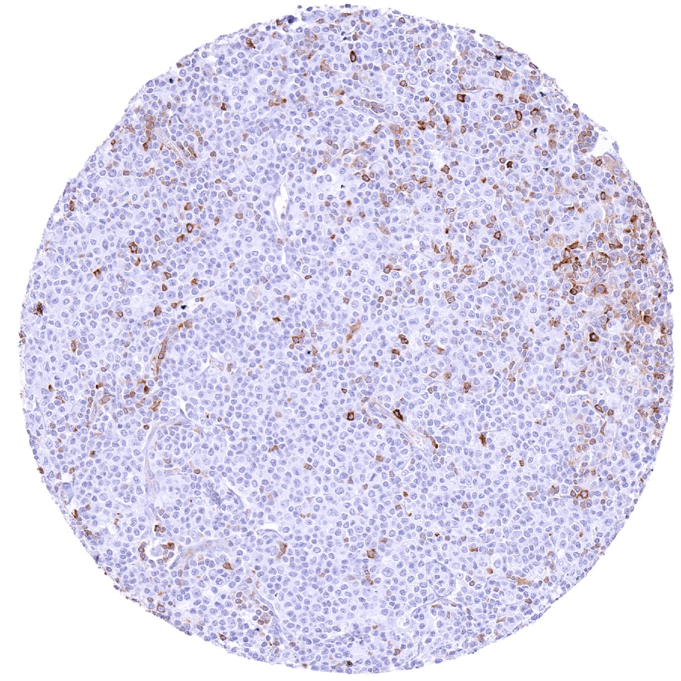
Lymph node – MX1 negative B-CLL
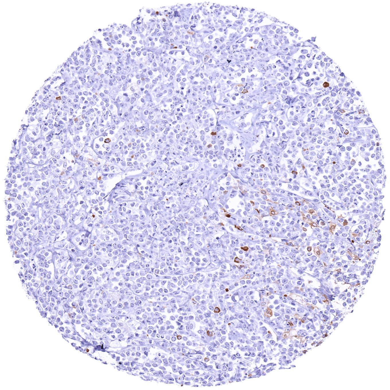
Lymph node – MX1 negative diffuse large B-cell lymphoma
Lymph node – MX1 negative Hodgkin’s lymphoma. Distinct MX1 staining of non-neoplastic cell types
Lymph node – MX1 negative Hodgkin’s lymphoma. Strong MX1 staining of non-neoplastic cells
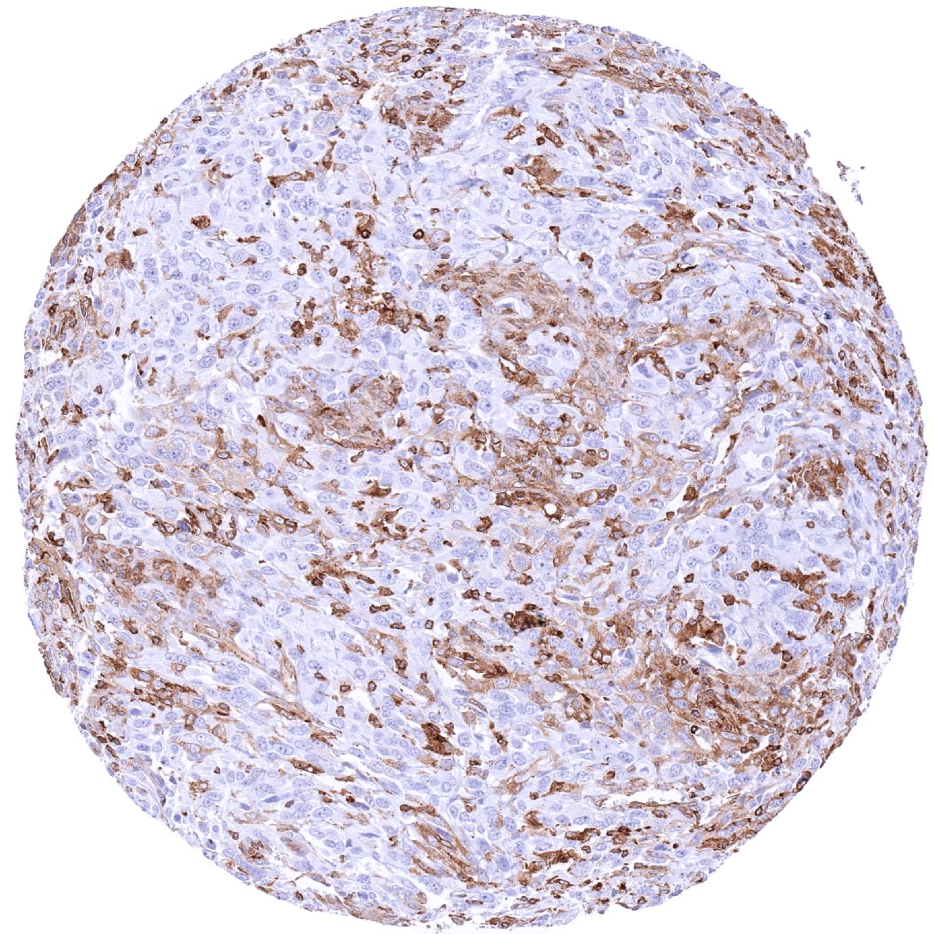
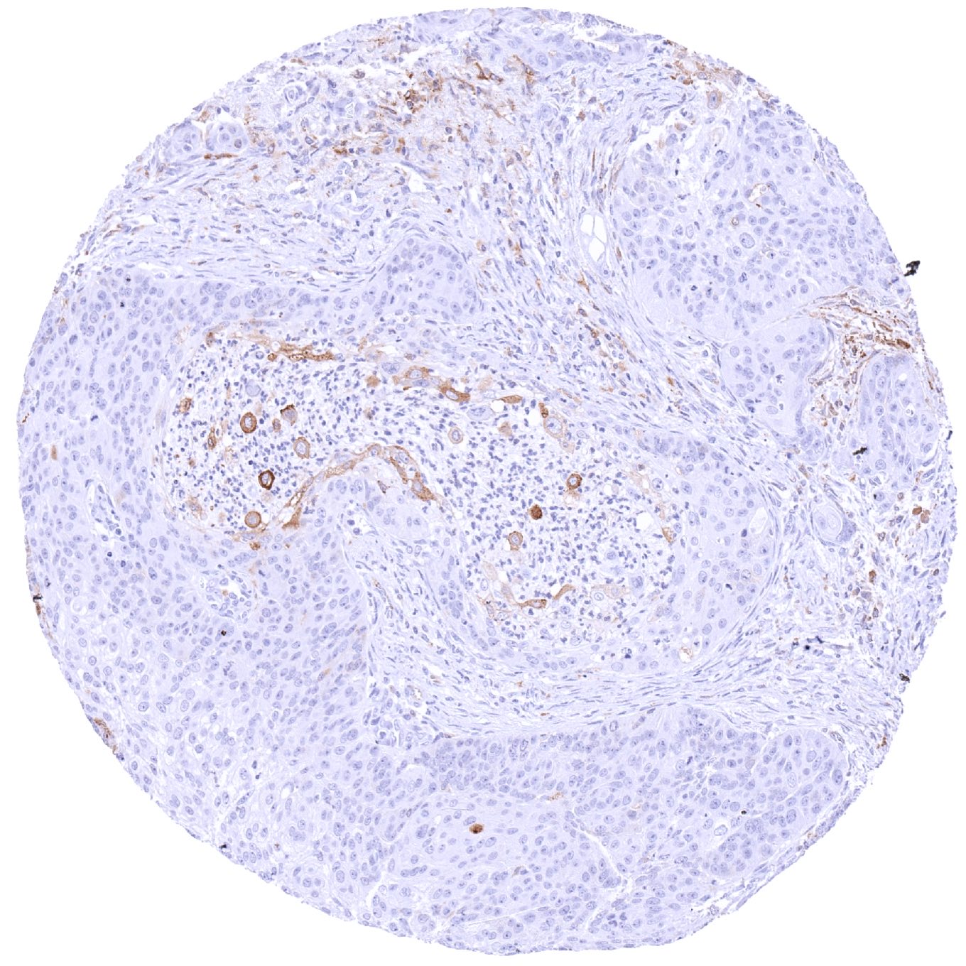
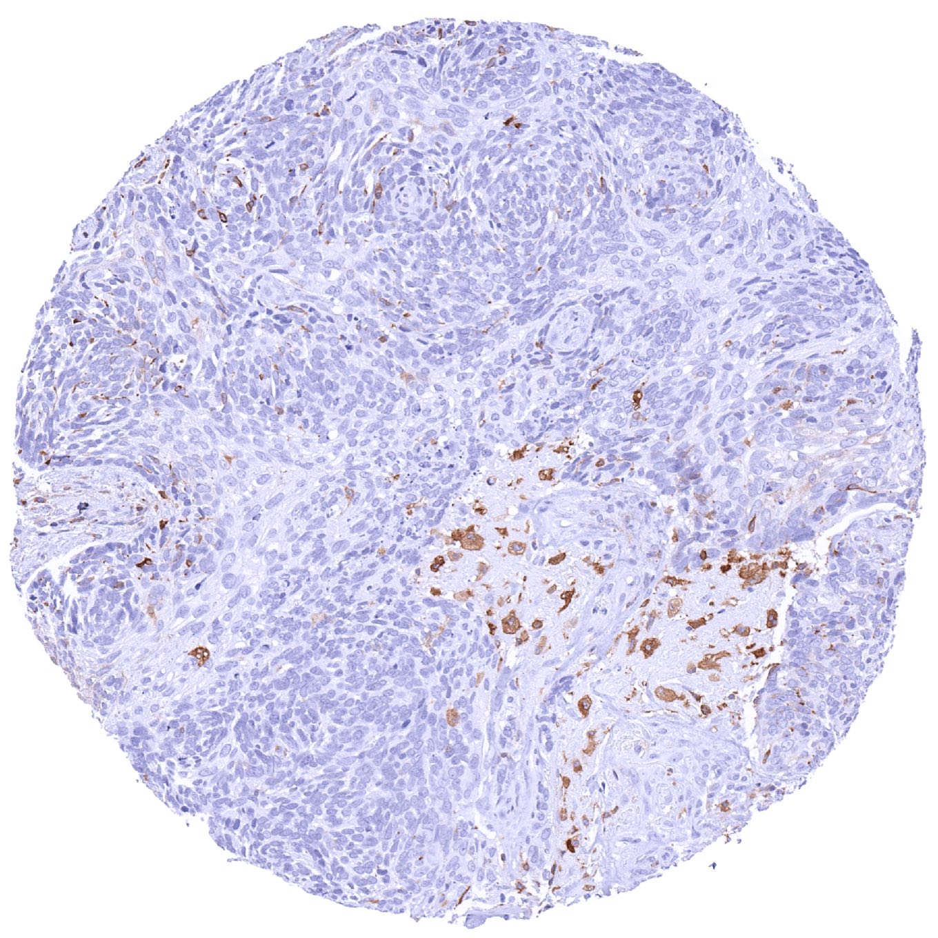
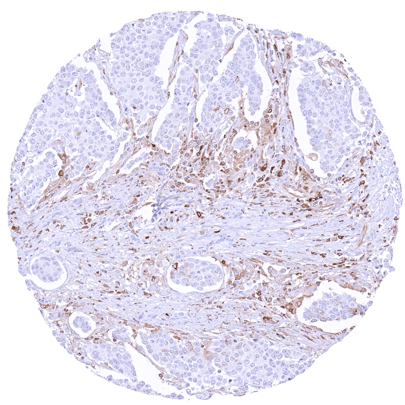
Oral cavity – Squamous cell carcinoma with faint MX1 positivity of only few tumor cells. Stromal and inflammatory cells exhibit a markedly more intense MX1 staining
Ovary – MX1 negative serous high-grade carcinoma
Ovary – Serous high-grade carcinoma with strong MX1 staining of all tumor cells
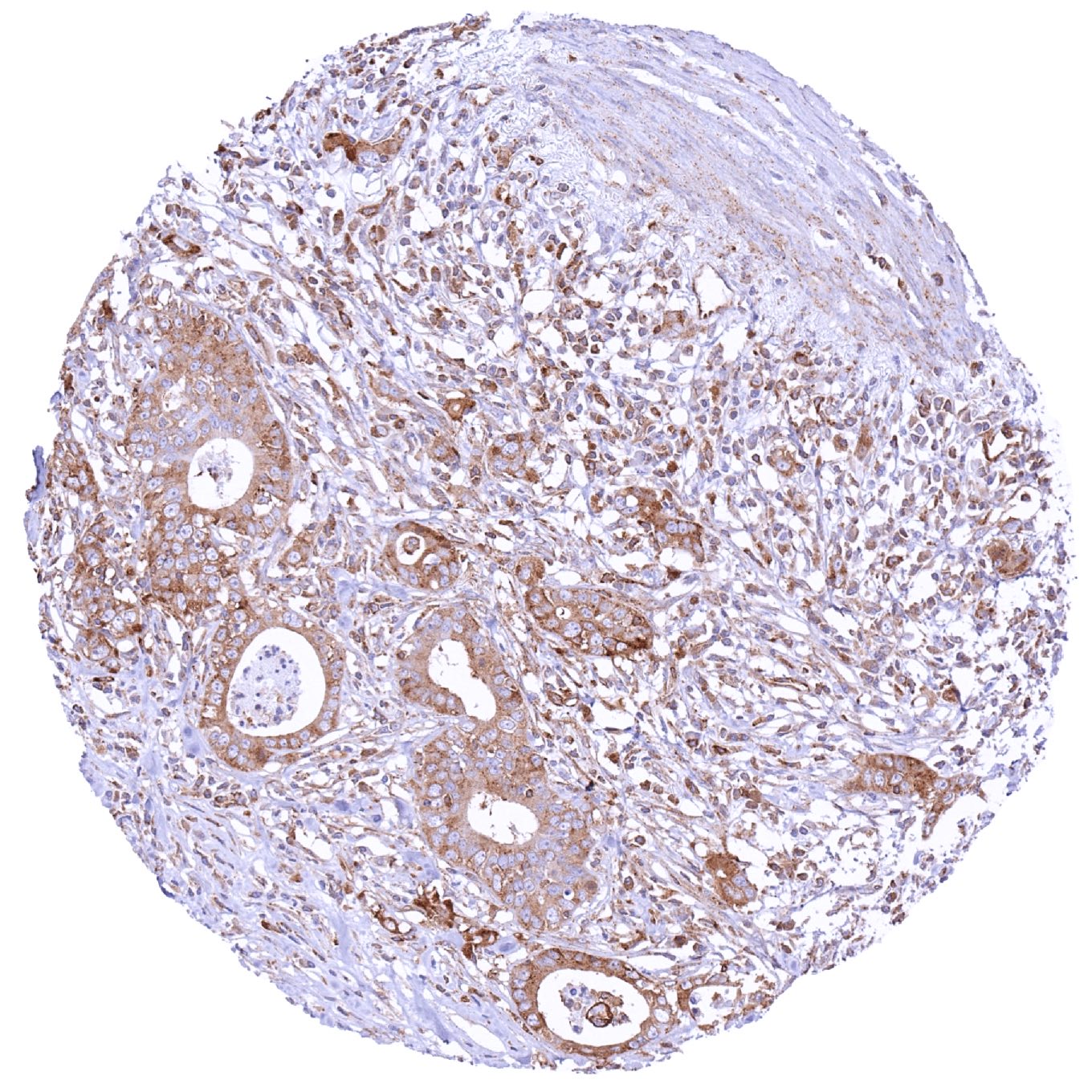
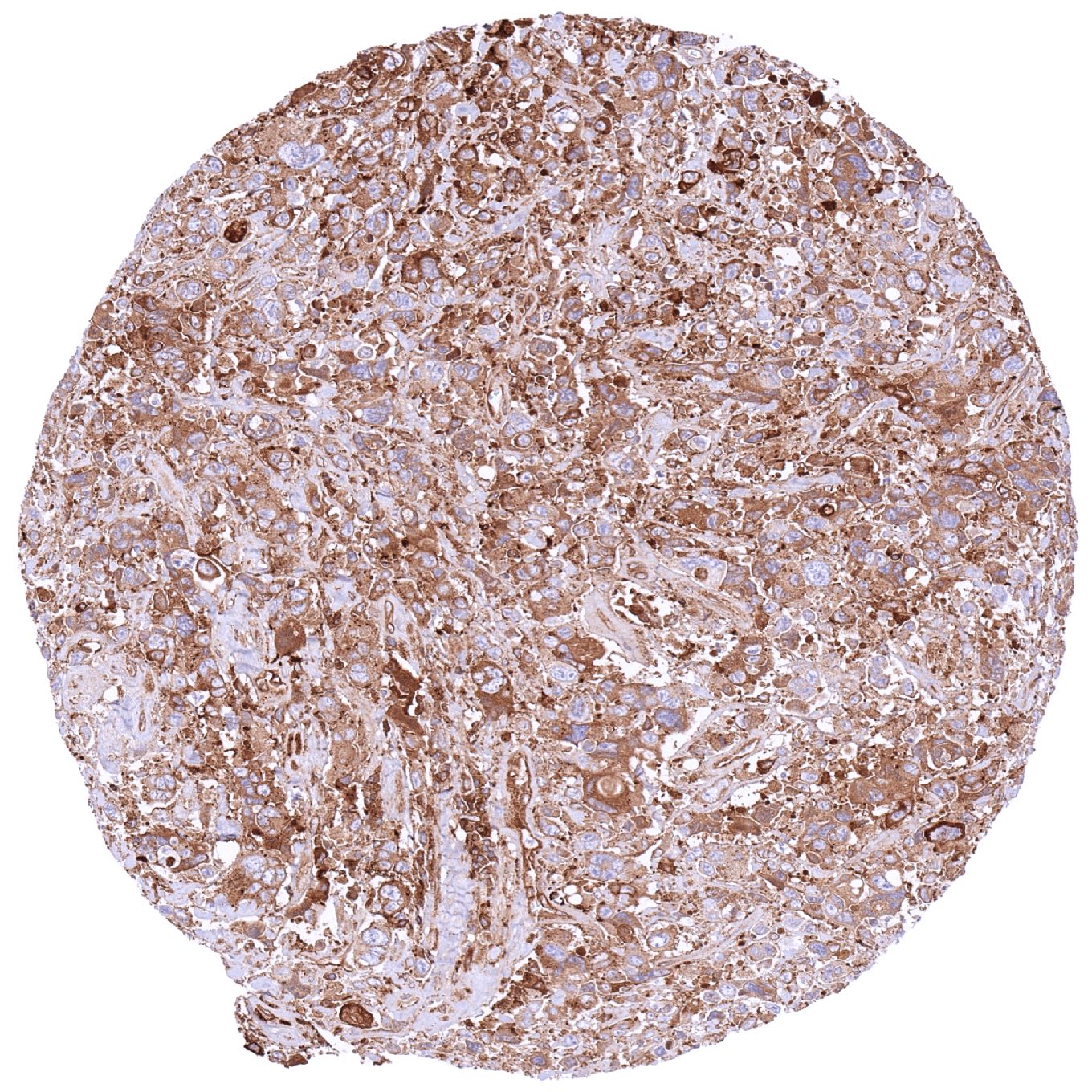
Pancreas – Ductal adenocarcinoma showing strong MX1 staining of tumor cells
Pancreas – MX1 negative neuroendocrine tumor
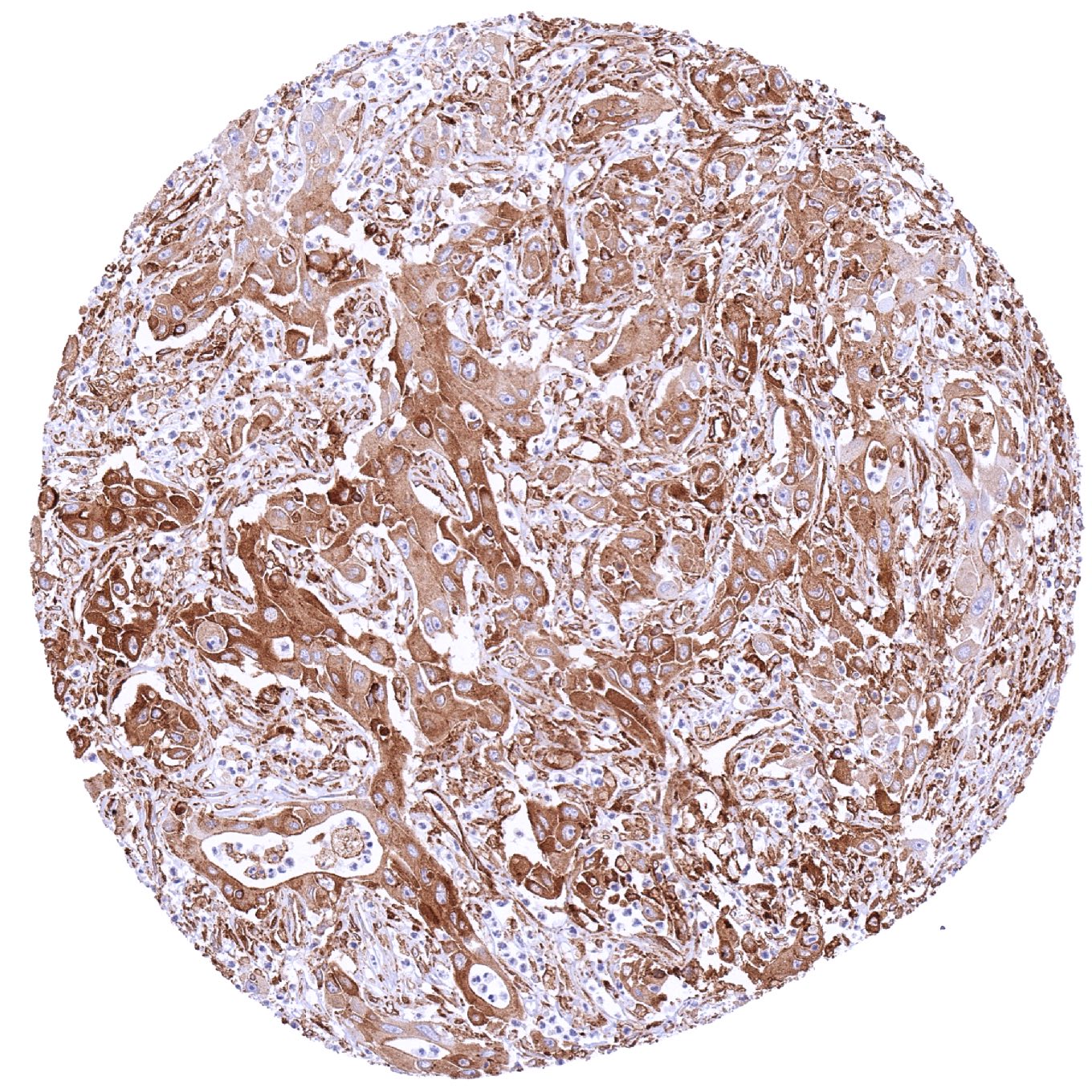
Penis – Squamous cell carcinoma with strong MX1 staining of tumor cells
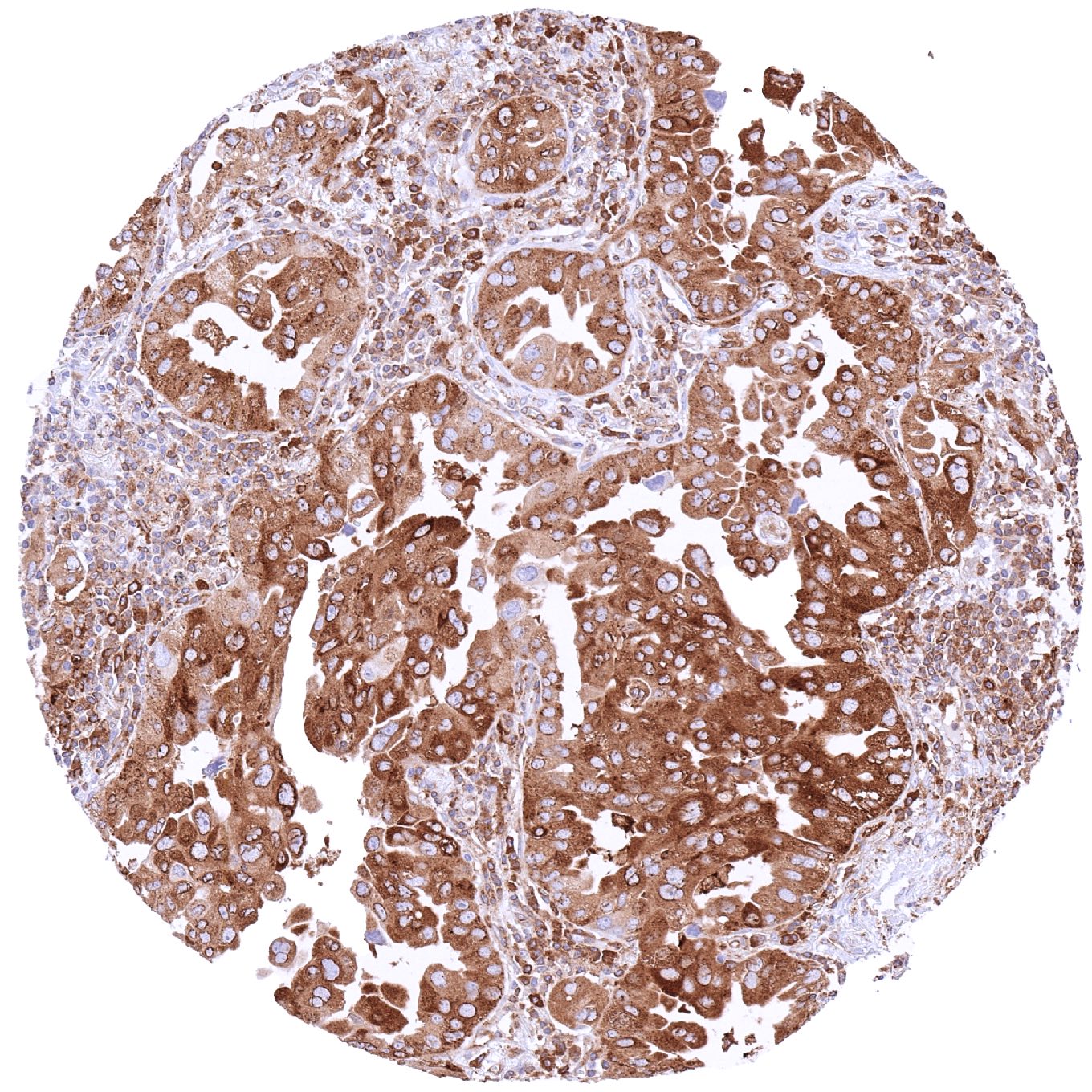
Prostate – Adenocarcinoma (Gleason 5+5=10) with strong MX1 immunostaining of tumor cells.jpeg
Prostate – Adenocarcinoma (Gleason 5+5=10) with strong MX1 immunostaining of tumor cells
Prostate – MX1 negative adenocarcinoma (Gleason 3+3=6)
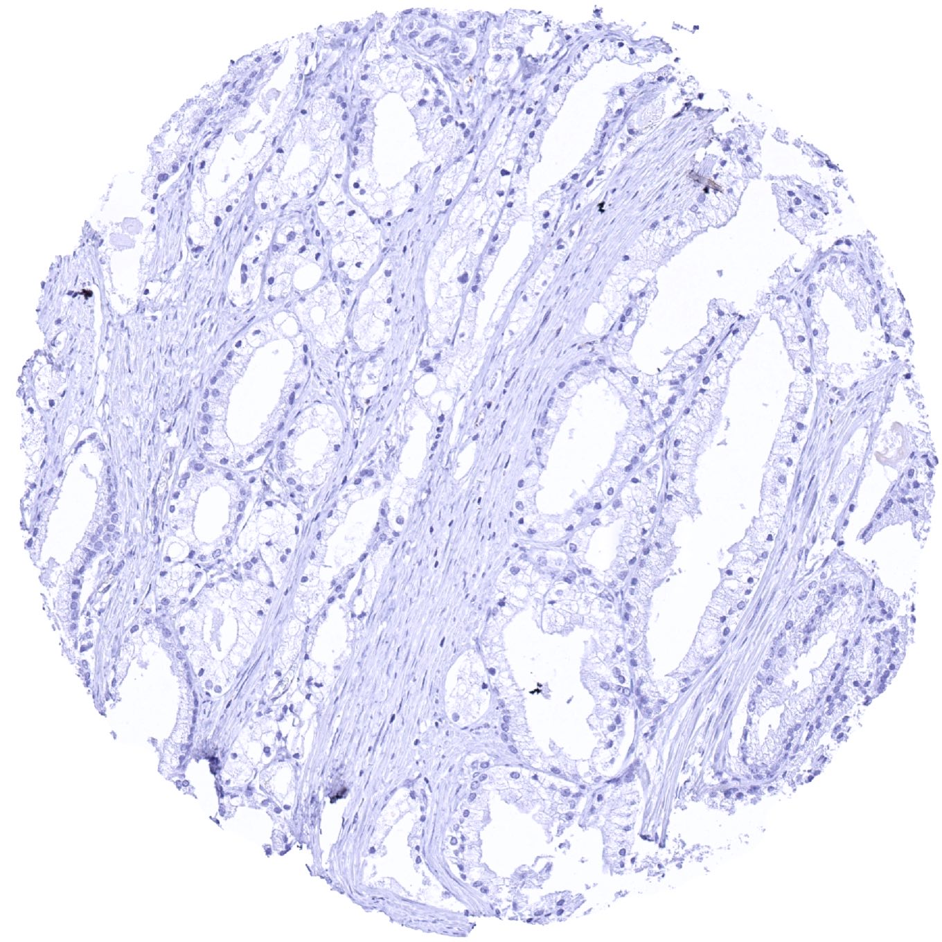
Salivary gland – MX1 negative Warthin tumor. Tumor-associated lymphocytes are MX1 positive
Skin – MX1 negative basal cell carcinoma. Distinct MX1 staining of inflammatory cells
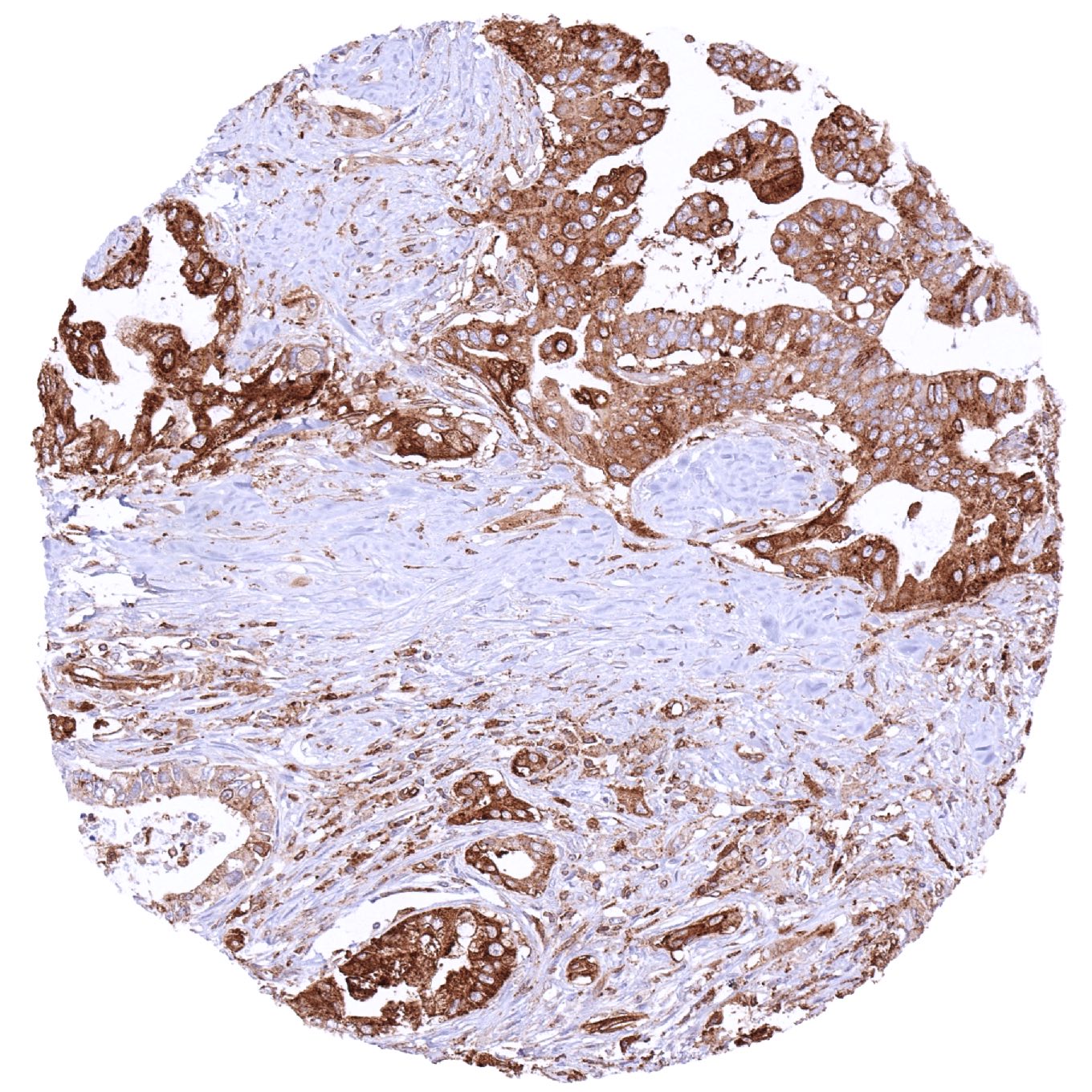
Stomach – Gastric adenocarcinoma (intestinal type) with moderate MX1 immunostaining of tumor cells. Inflammatory and stroma cells are also MX1 positive
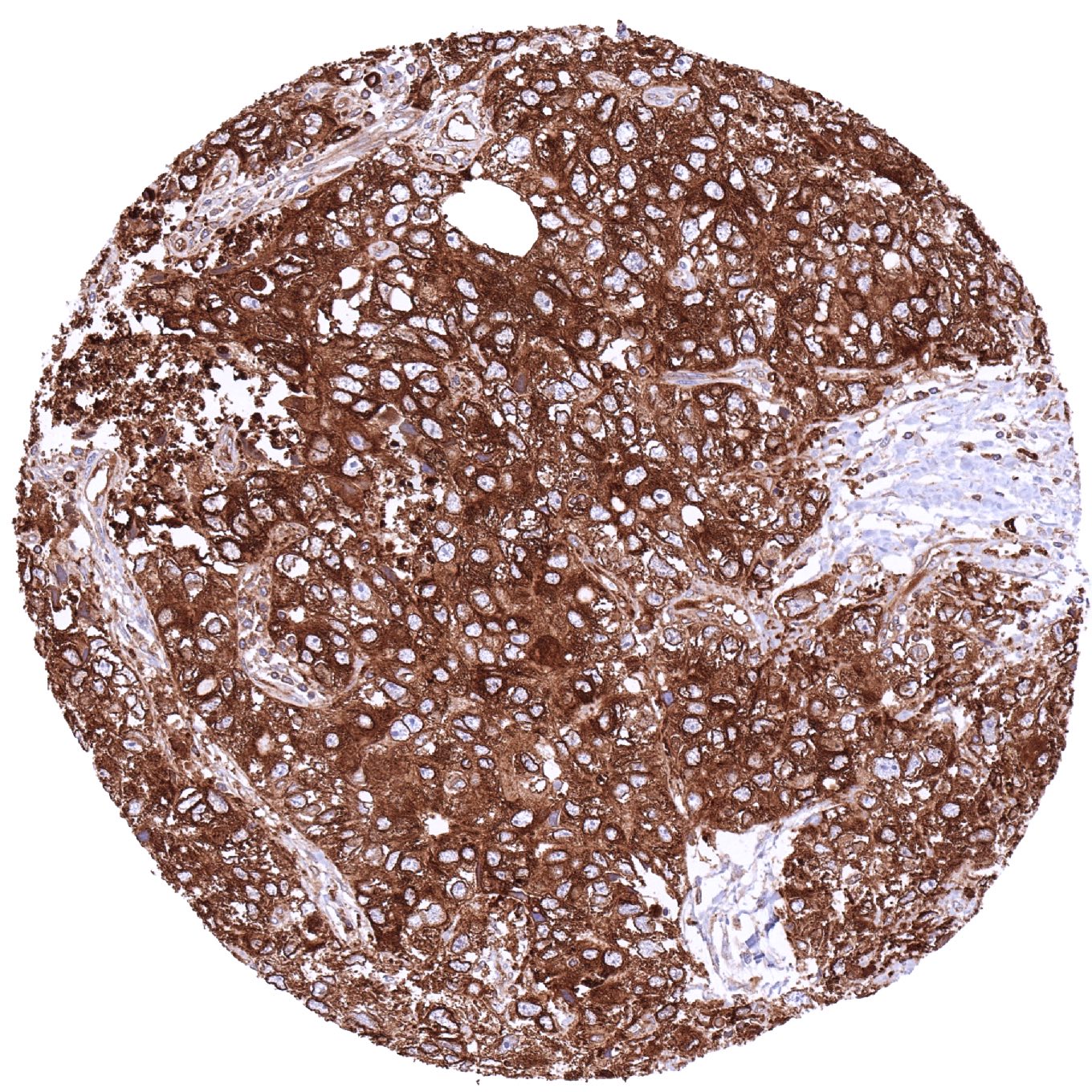
Stomach – Gastrointestinal stromal tumor (GIST) with intense MX1 staining of tumor cells
Testis – MX1 negative Leydig cell tumor
Testis – Seminoma with strong MX1 immunostaining of tumor cells. Tumor-infiltrating lymphocytes are also MX1 positive
Testis – Yolk sac tumor with weak MX1 immunostaining of few tumor cells. Strong MX1 staining of inflammatory cells.jpeg
Testis – Yolk sac tumor with weak MX1 immunostaining of few tumor cells. Strong MX1 staining of inflammatory cells
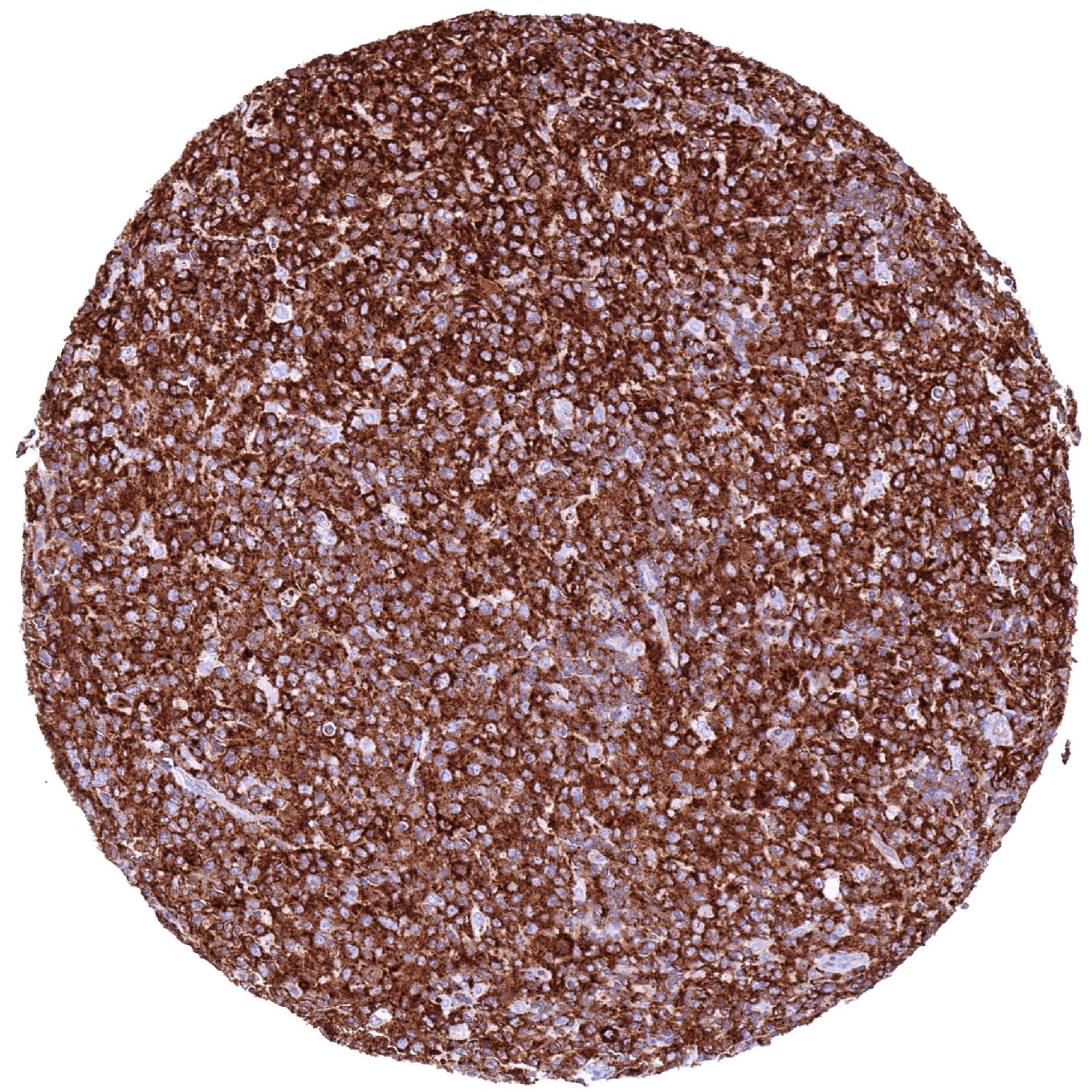
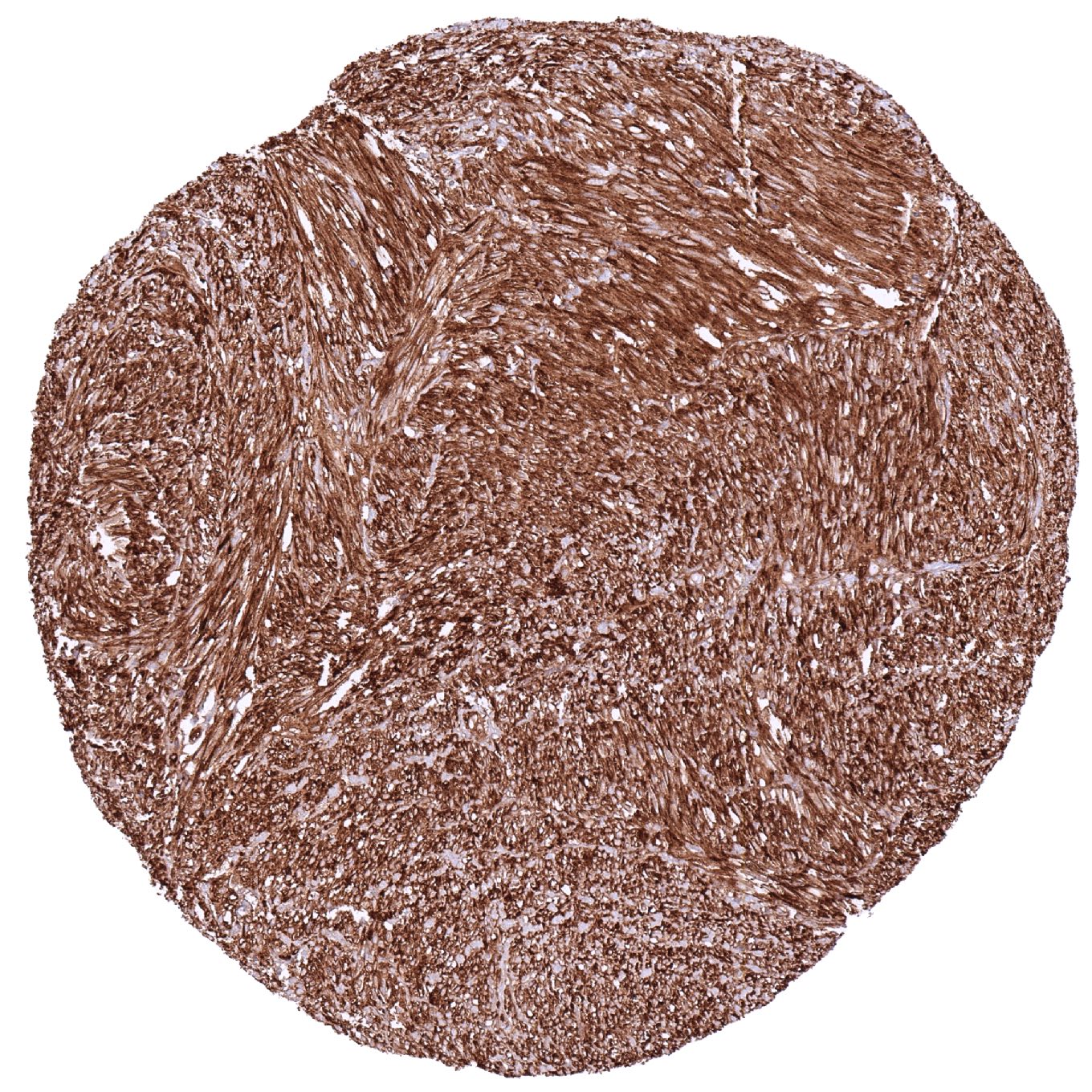
Thyroid – Anaplastic carcinoma with strong MX1 positivity of tumor and stroma cells
Thyroid – MX1 negative medullary carcinoma
Thyroid – Papillary cancer with strong MX1 immunostaining of tumor cells
Urinary bladder – Muscle-invasive urothelial carcinoma with strong MX1 staining of tumor and stroma cells
Urinary bladder – MX1 negative muscle-invasive urothelial carcinoma
Uterus, cervix – Adenocarcinoma with intense MX1 immunostaining of tumor cells
Vulva – Squamous cell carcinoma with strong MX1 staining of tumor cells. Stromal and inflammatory cells show a less intense MX1 staining
  • Linkedin
  • My Account
  • Search
    ×
  • Cart 0
© 2025 MS Validated Antibodies
  • Imprint
  • Privacy Policy
  • Distributors