Login / Register
MS Validated Antibodies - Logo
  • About us
  • Products
    • Primary antibodies
    • Gallery
  • Distributors
  • Validation
    • Tissue Microarrays
  • Publications
    • Large Scale Studies
    • Further Publications
    • Events
    • Quiz
  • Contact
  • About us
  • Products
    • Primary antibodies
    • Gallery
  • Distributors
  • Validation
    • Tissue Microarrays
  • Publications
    • Large Scale Studies
    • Further Publications
    • Events
    • Quiz
  • Contact
×

Cancer Tissue Gallery INI1

Path Created with Sketch.
Go to INI1 Normal Tissue Gallery
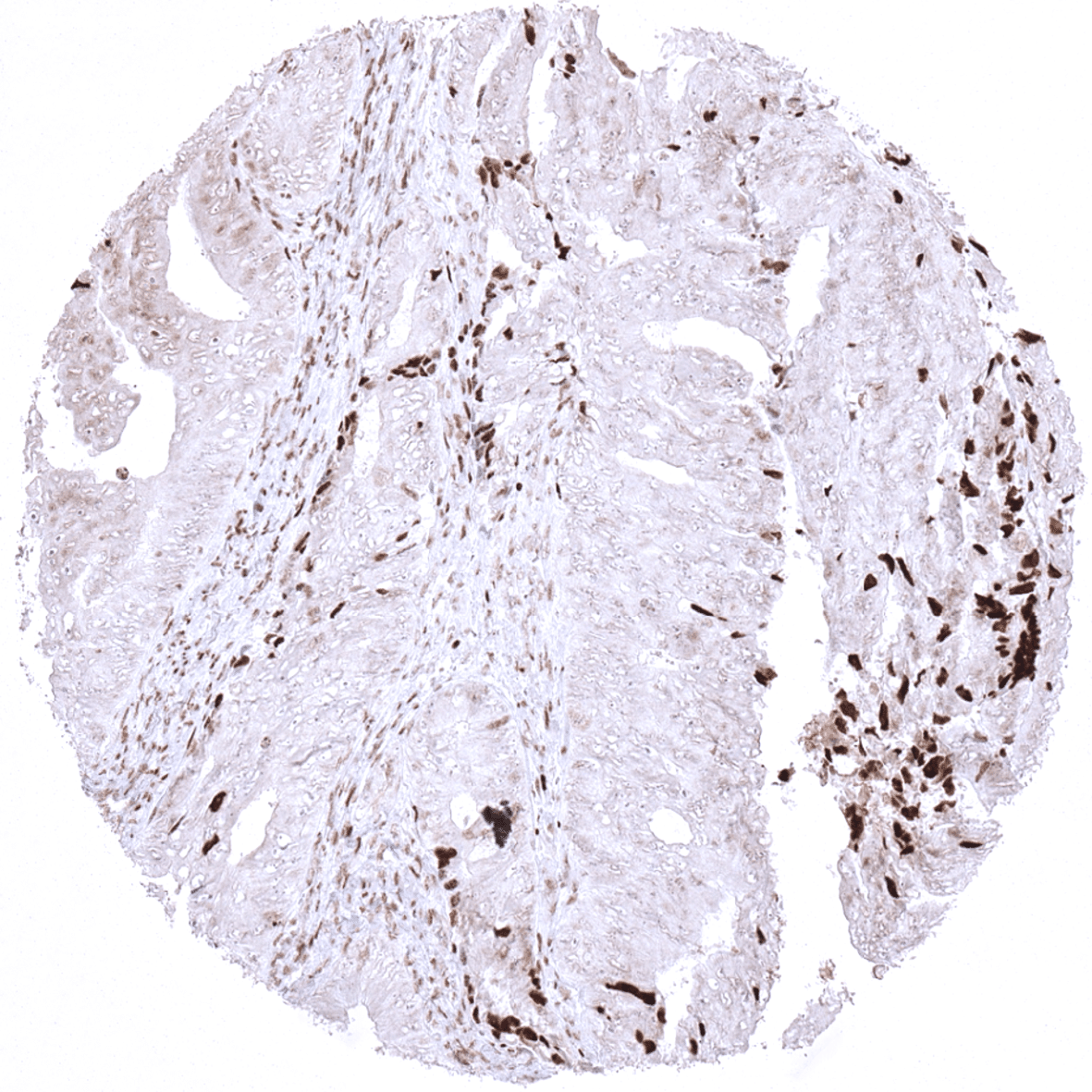
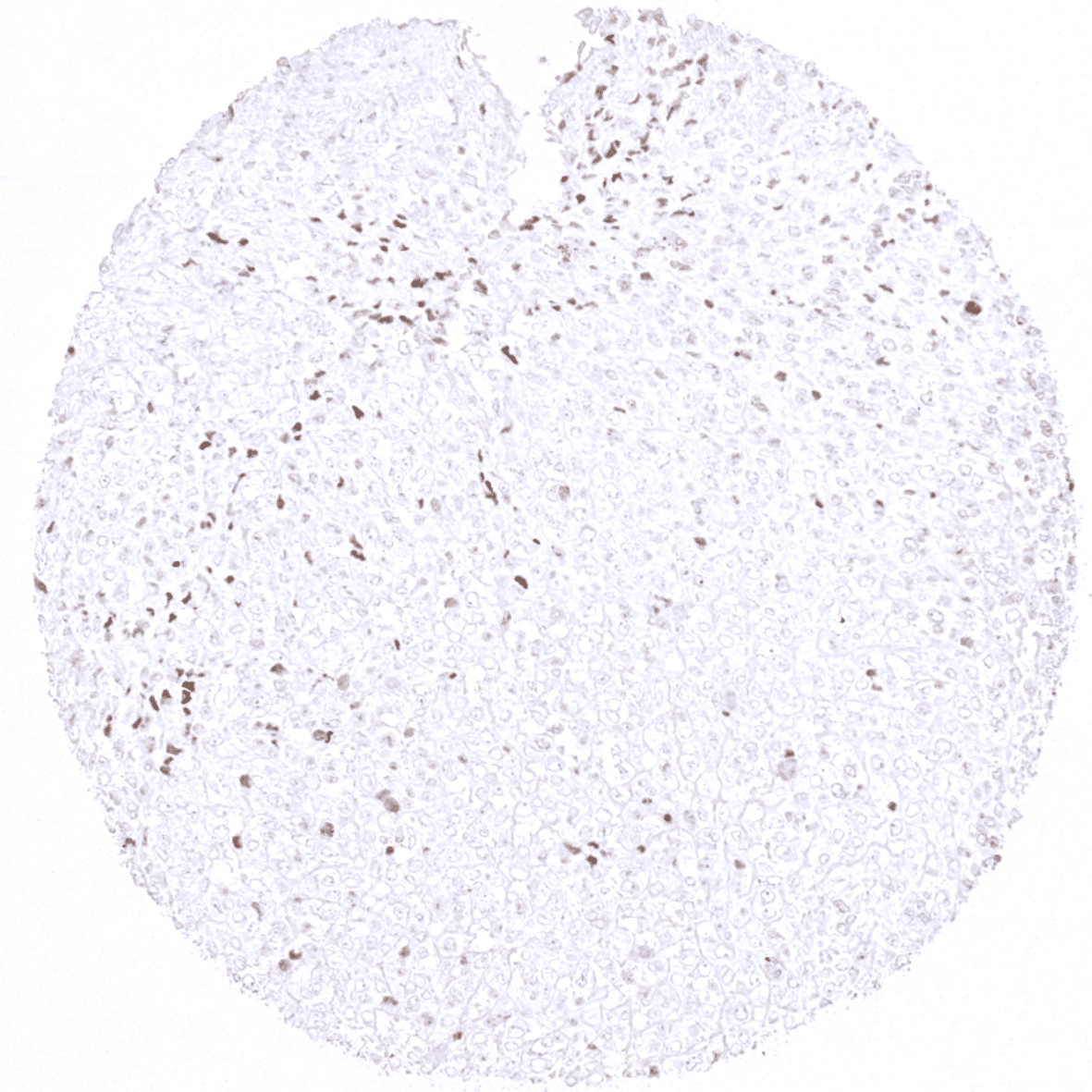
Colon - Absence of INI1 immunostaining in tumor cells of a colorectal adenocarcinoma while stroma cells and tumor infiltrating lymphocytes show strong positivity.
Colon – Absence of INI1 immunostaining in tumor cells of a colorectal adenocarcinoma while stroma cells and tumor infiltrating lymphocytes show strong positivity.
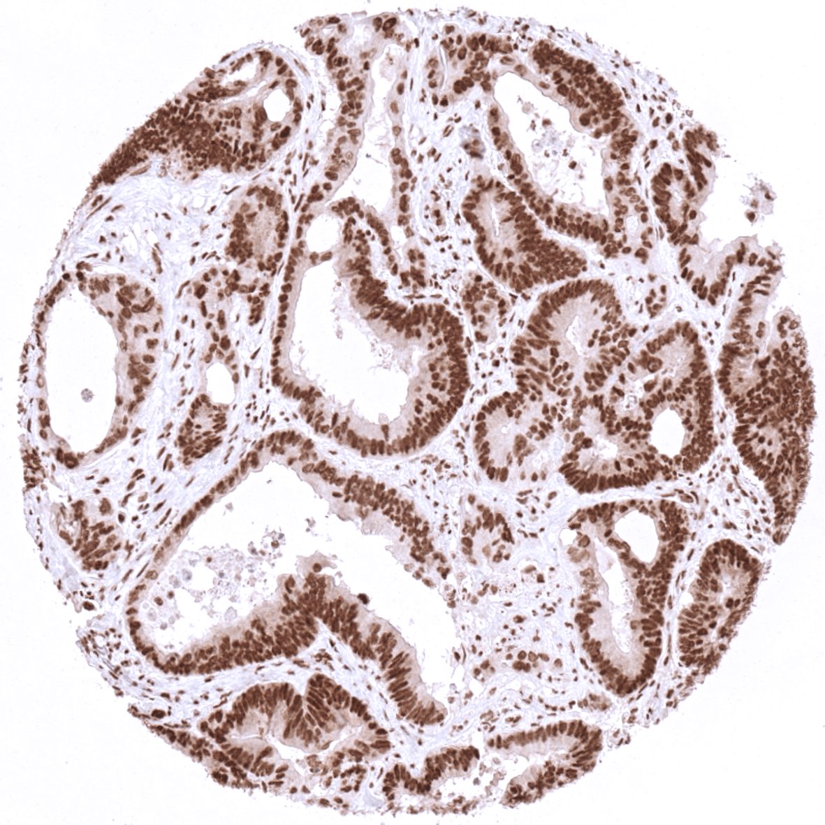
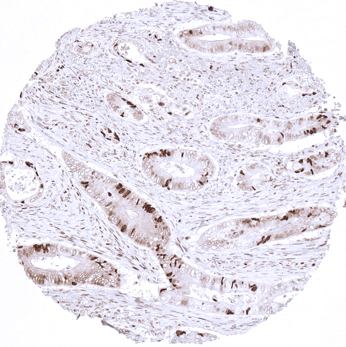
Colon - Adenocarcinoma of the colon showing strong INI1 immunostaining in tumor and stroma cells.
Colon – Adenocarcinoma of the colon showing strong INI1 immunostaining in tumor and stroma cells.
Colon - Almost complete loss of INI1 immunostaining in tumor cells of a colorectal adenocarcinoma. Tumor infiltrating lymphocytes are INI1 positive.
Colon – Almost complete loss of INI1 immunostaining in tumor cells of a colorectal adenocarcinoma. Tumor infiltrating lymphocytes are INI1 positive.
Soft tissues - Angiosarcoma showing complete lack of INI1 immunostaining in tumor cells. Normal blood vessels and lymphocytes show strong INI1 positivity.
Soft tissues – Angiosarcoma showing complete lack of INI1 immunostaining in tumor cells. Normal blood vessels and lymphocytes show strong INI1 positivity.
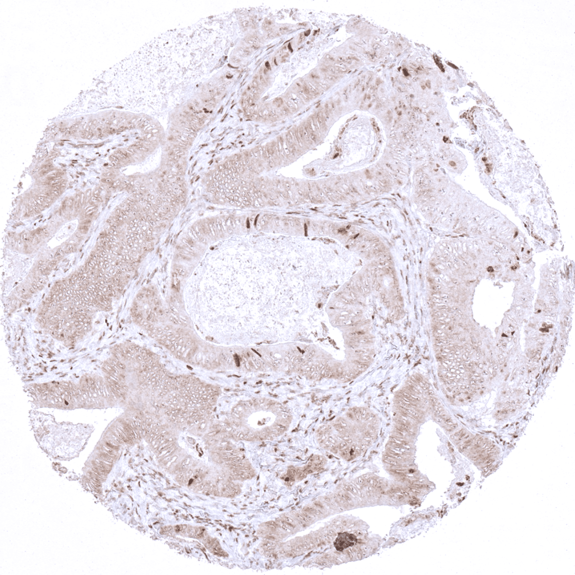
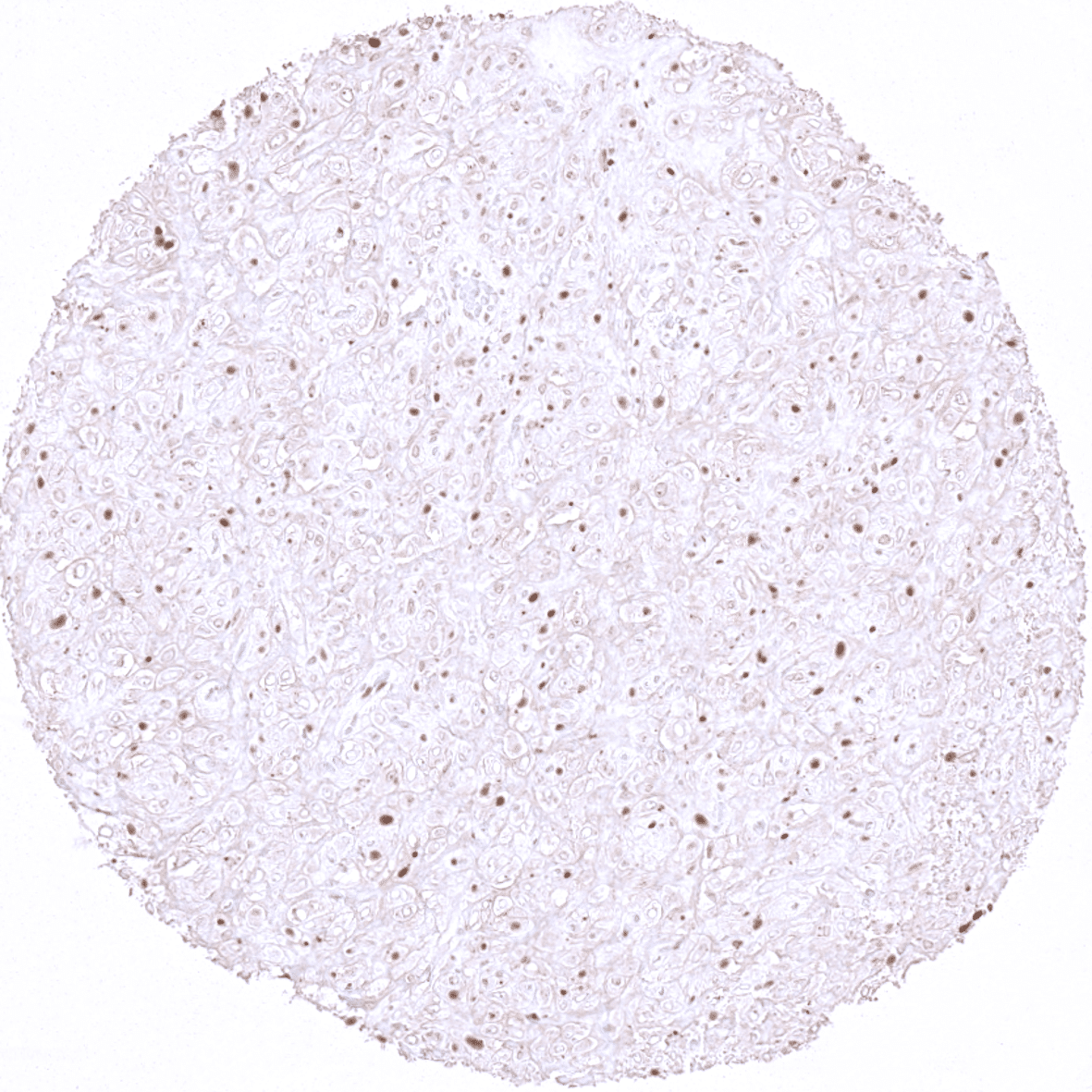
Colon - Colorectal adenocarcinoma showing markedly reduced INI1 immunostaining in a distinct fraction of tumor cell nuclei (mosaic pattern). Stroma cells and lymphocytes show strong INI1 positivity.
Colon – Colorectal adenocarcinoma showing markedly reduced INI1 immunostaining in a distinct fraction of tumor cell nuclei (mosaic pattern). Stroma cells and lymphocytes show strong INI1 positivity.
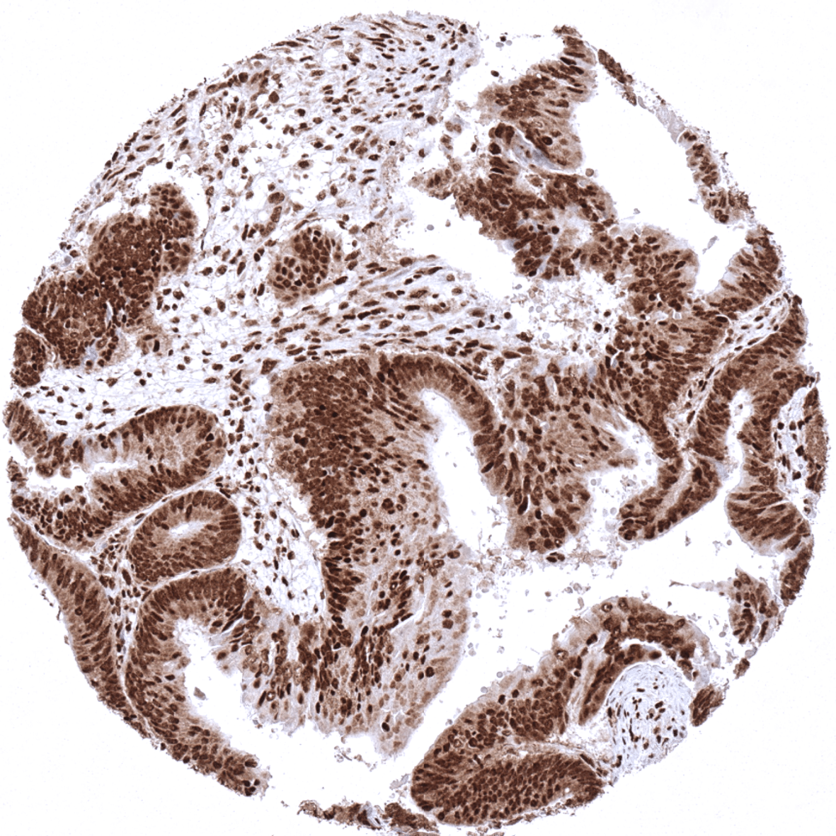
Colon - Colorectal adenocarcinoma with strong INI1 immunostaining in tumor cells and tumor associated stroma.
Colon – Colorectal adenocarcinoma with strong INI1 immunostaining in tumor cells and tumor associated stroma.
Colon - Complete loss of INI1 immunostaining in tumor cells of a colorectal adenocarcinoma. Tumor infiltrating lymphocytes and stroma cells exhibit strong INI1 positivity.
Colon – Complete loss of INI1 immunostaining in tumor cells of a colorectal adenocarcinoma. Tumor infiltrating lymphocytes and stroma cells exhibit strong INI1 positivity.
Bone - Complete loss of INI1 immunostaining in tumor cells of a peripheral neuroectodermal tumor (PNET). Tumor infiltrating lymphocytes show strong INI1 positivity.
Bone – Complete loss of INI1 immunostaining in tumor cells of a peripheral neuroectodermal tumor (PNET). Tumor infiltrating lymphocytes show strong INI1 positivity.
Stomach - Gastrointestinal stromal tumor (GIST) with markedly reduced INI1 immunostaining in tumor cells.
Stomach – Gastrointestinal stromal tumor (GIST) with markedly reduced INI1 immunostaining in tumor cells.
Colon - Markedly reduced INI1 immunostaining in a fraction of tumor cells of a colorectal adenocarcinoma (mosaic pattern).
Colon – Markedly reduced INI1 immunostaining in a fraction of tumor cells of a colorectal adenocarcinoma (mosaic pattern).
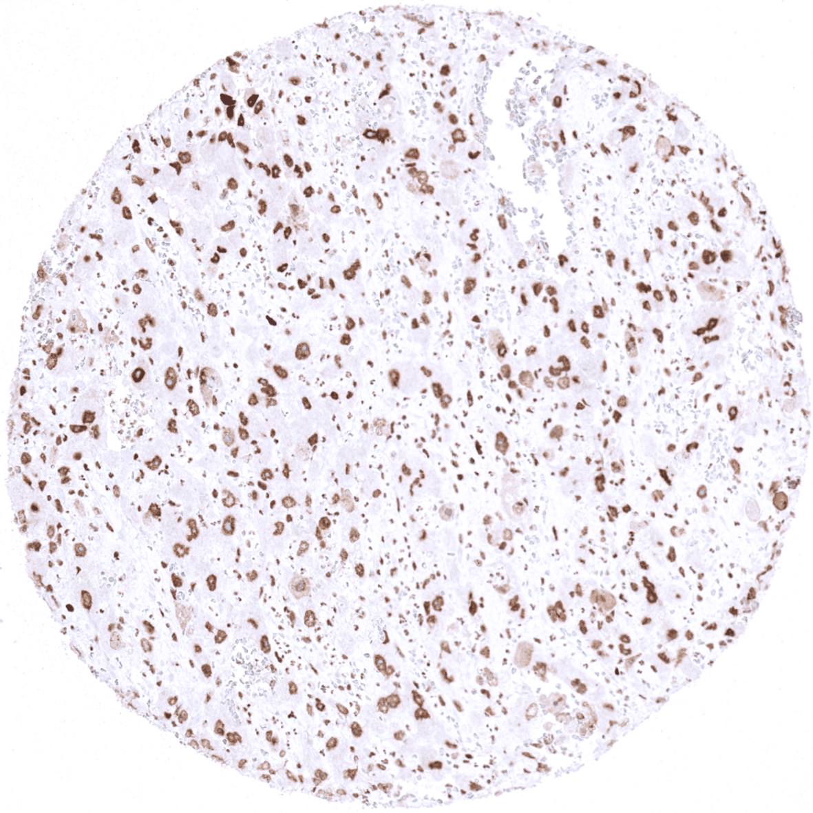
Bone - Peripheral neuroectodermal tumor (PNET) with retained INI1 immunostaining in tumor cells.
Bone – Peripheral neuroectodermal tumor (PNET) with retained INI1 immunostaining in tumor cells.
Soft tissues - Strong INI1 immunostaining in an angiosarcoma.
Soft tissues – Strong INI1 immunostaining in an angiosarcoma.
  • Linkedin
  • My Account
  • Search
    ×
  • Cart 0
© 2025 MS Validated Antibodies
  • Imprint
  • Privacy Policy
  • Distributors