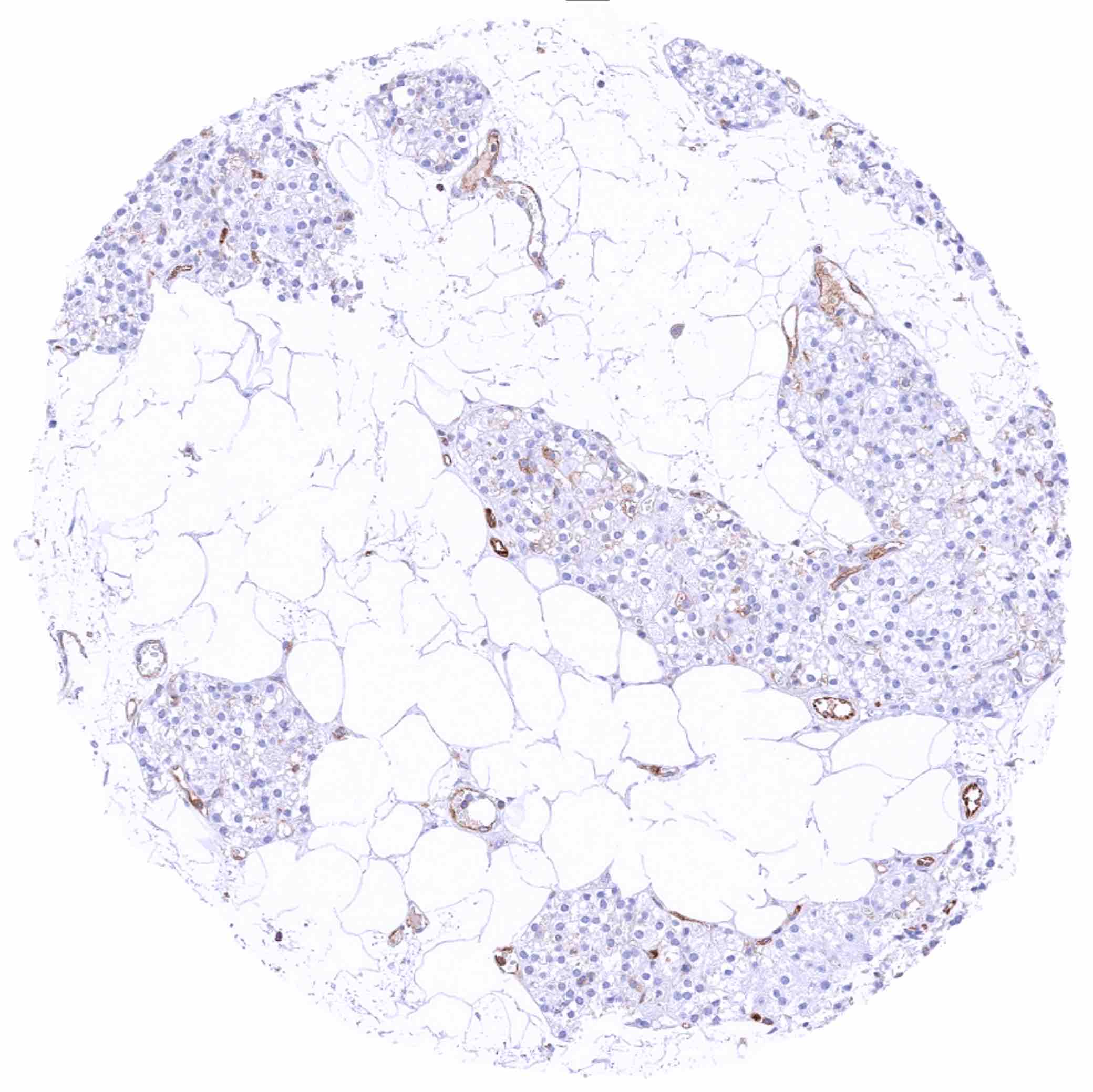
Adrenal gland – Moderate MX1 staining of medullary cells. Adrenocortical cells are MX1 negative
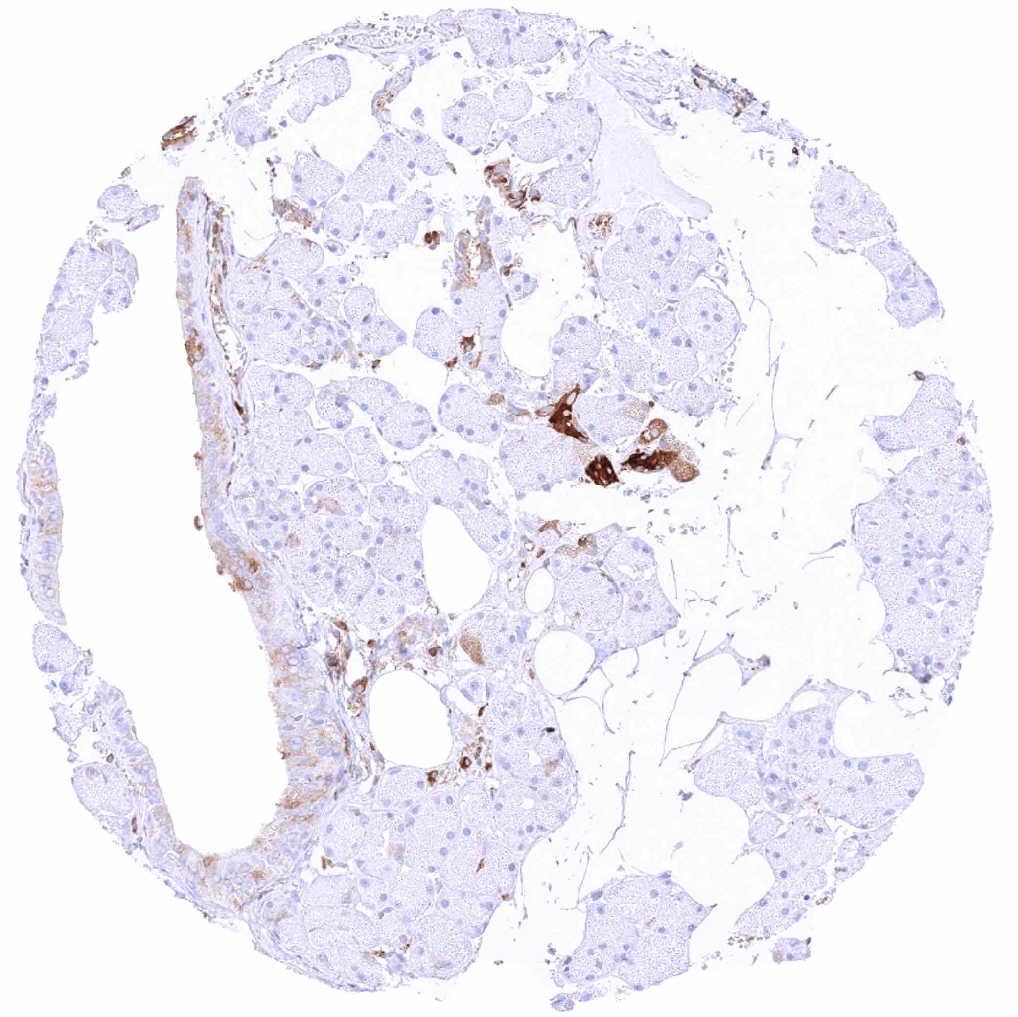
Adrenal gland – Very weak cytoplasmic MX1 staining of some adrenocortical cells. Moderate staining of endothelial cells
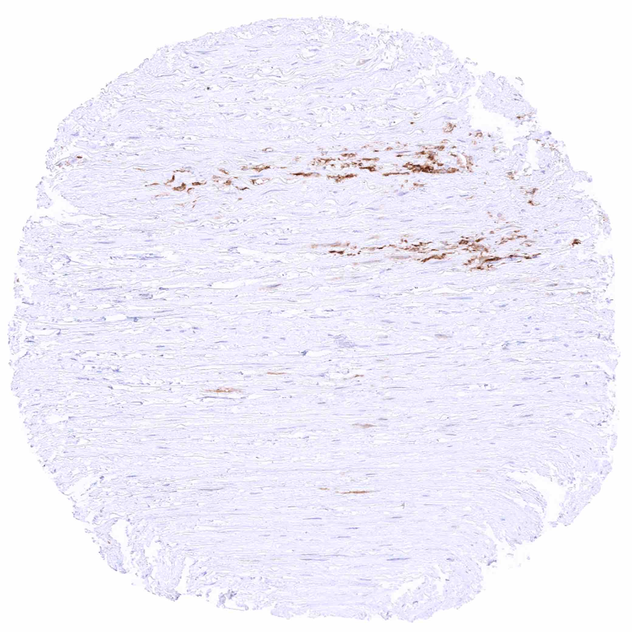
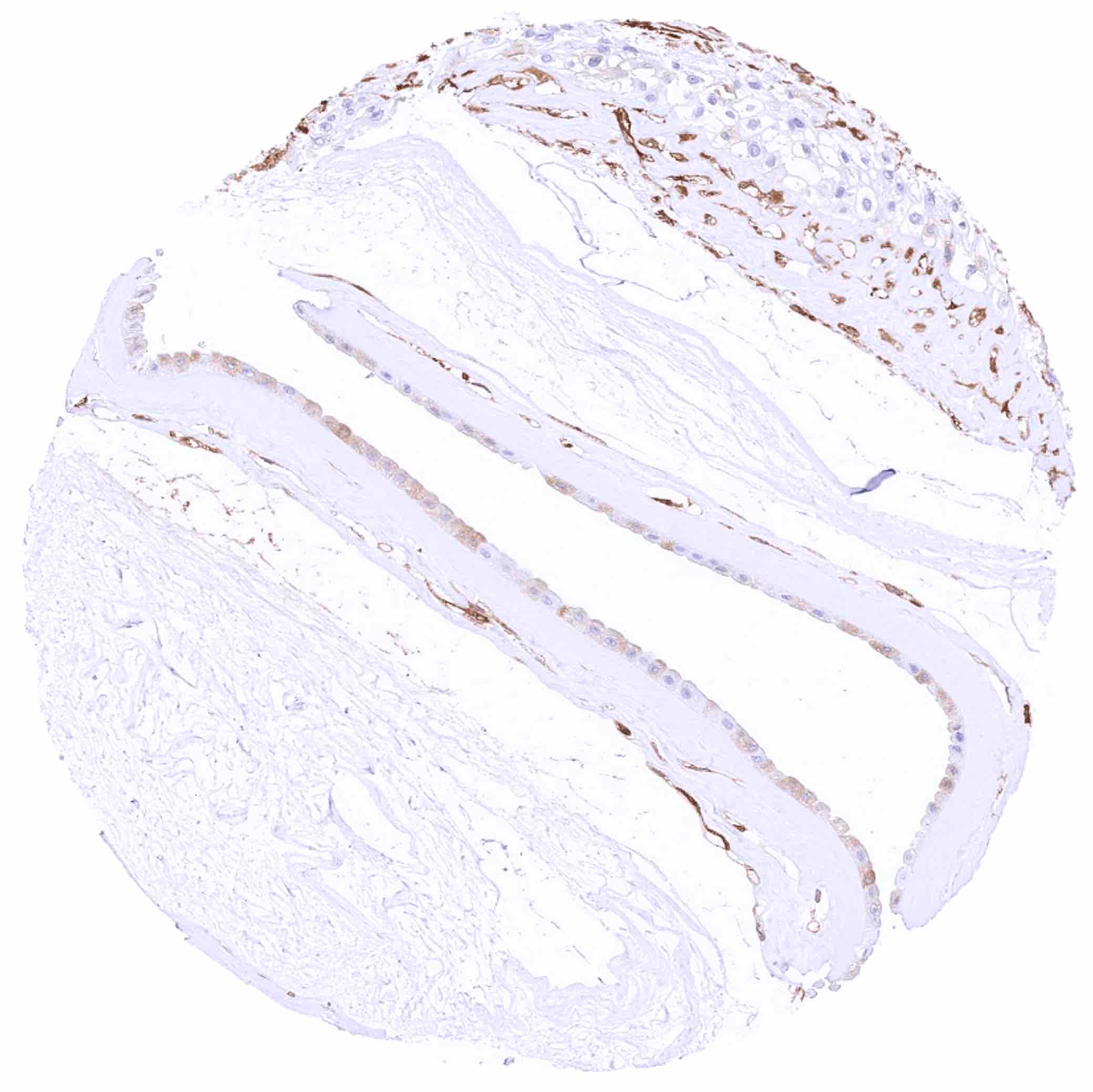
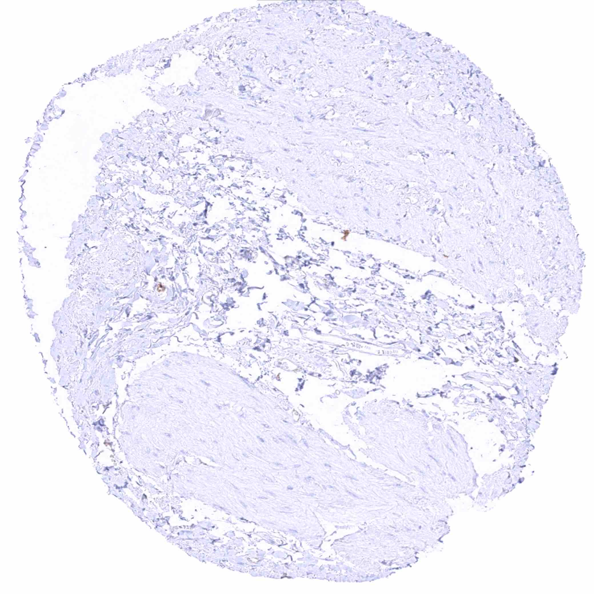
Aorta, media – MX1 positive material can be seen in this sample
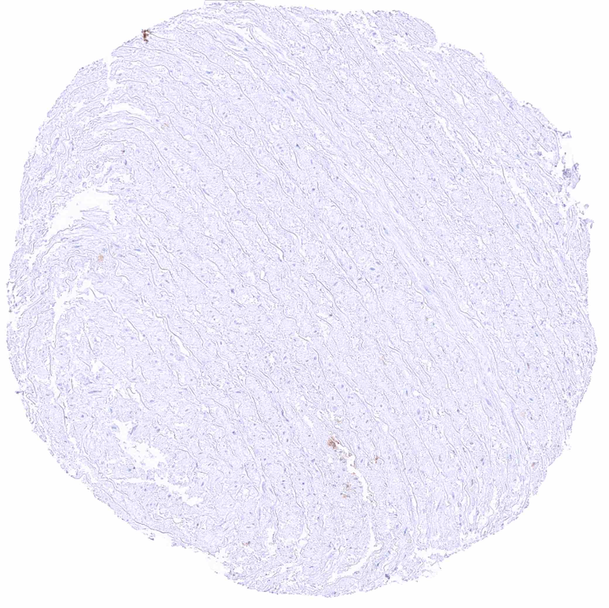
Aorta, media
Appendix, mucosa – Abundant cytoplasmic MX1 staining of lymphocytic cells. Crypt epithelial cells are MX1 negative
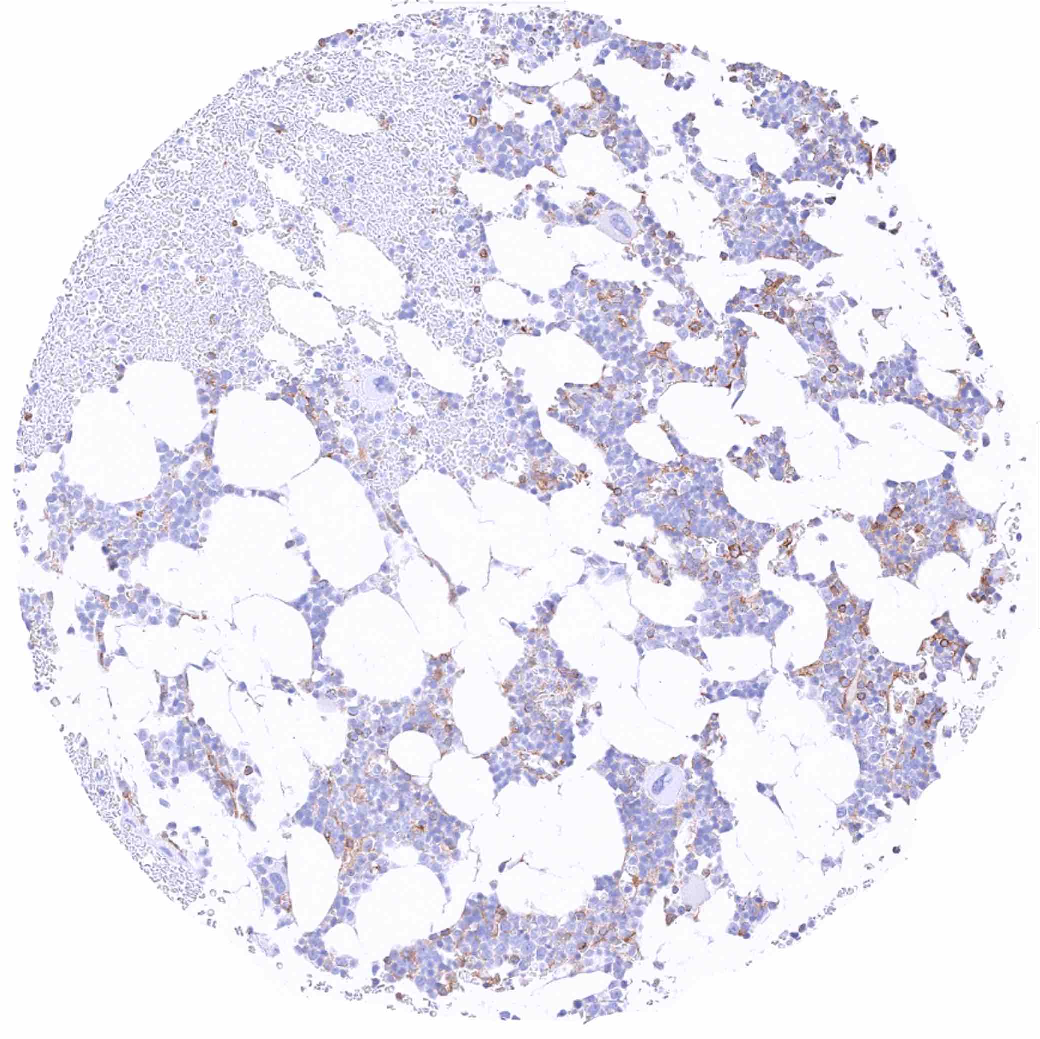
Bone marrow – Moderate MX1 staining of several hematopoetic cell types
Breast – MX1 staining is limited to inflammatory and endothelial cells
Bronchus, glands – Moderate to strong cytoplasmic MX1 staining in subsets of epithelial cells
Bronchus, glands – Weak to moderate cytoplasmic MX1 staining in subsets of epithelial cells
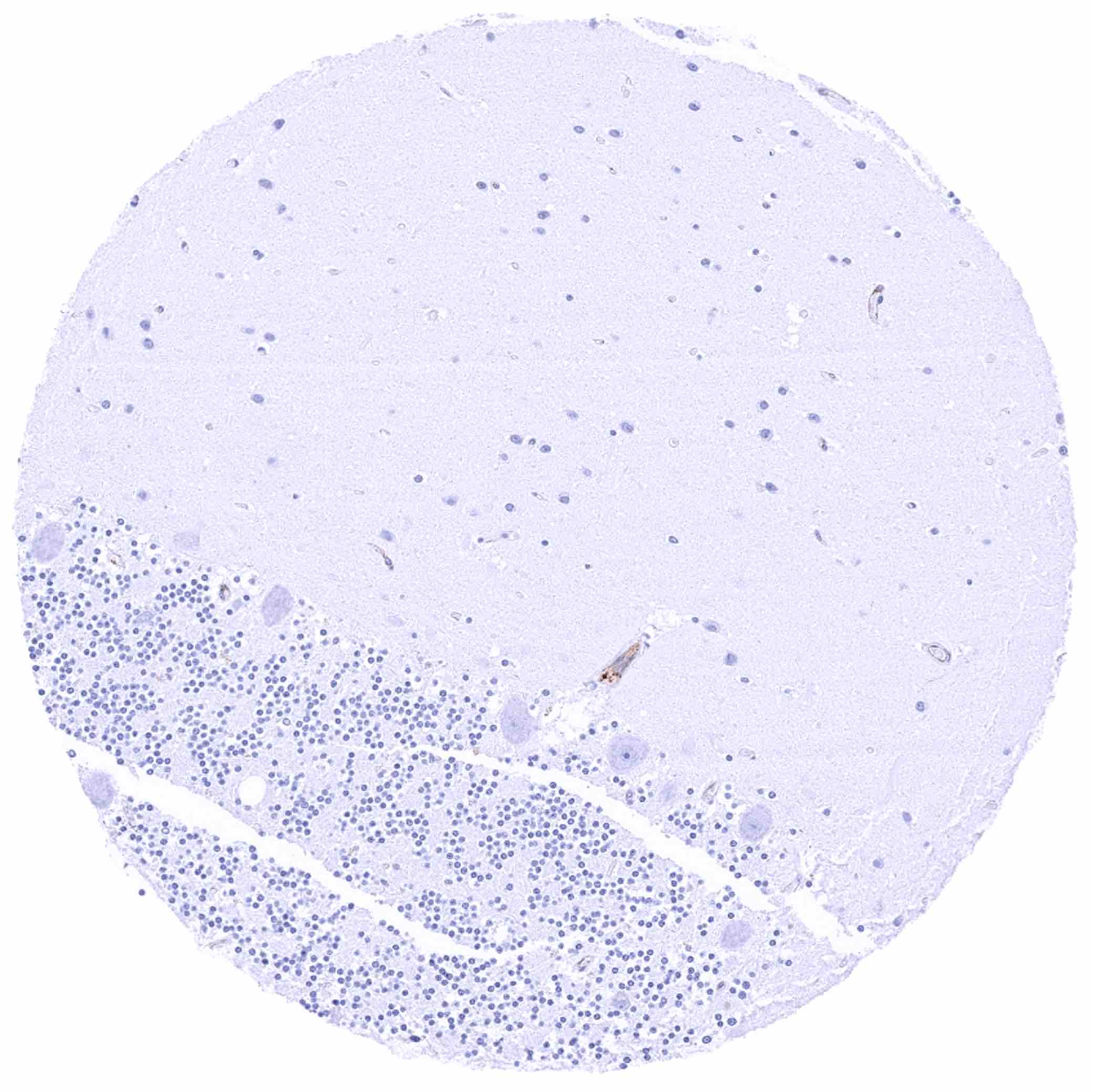
Cerebellum (molecular layer, purkinje cell layer, granule cell layer)
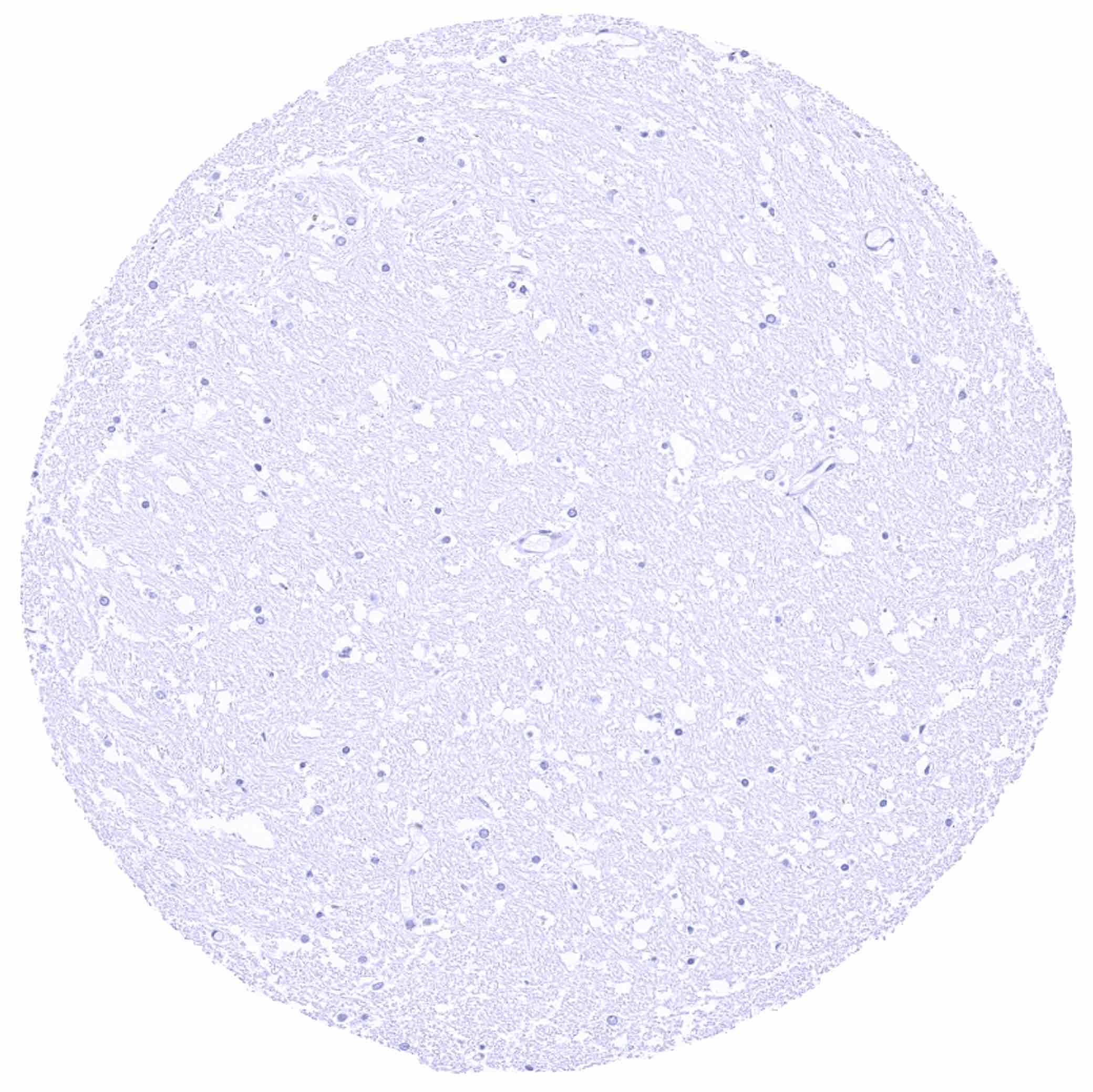
Cerebellum (white matter)
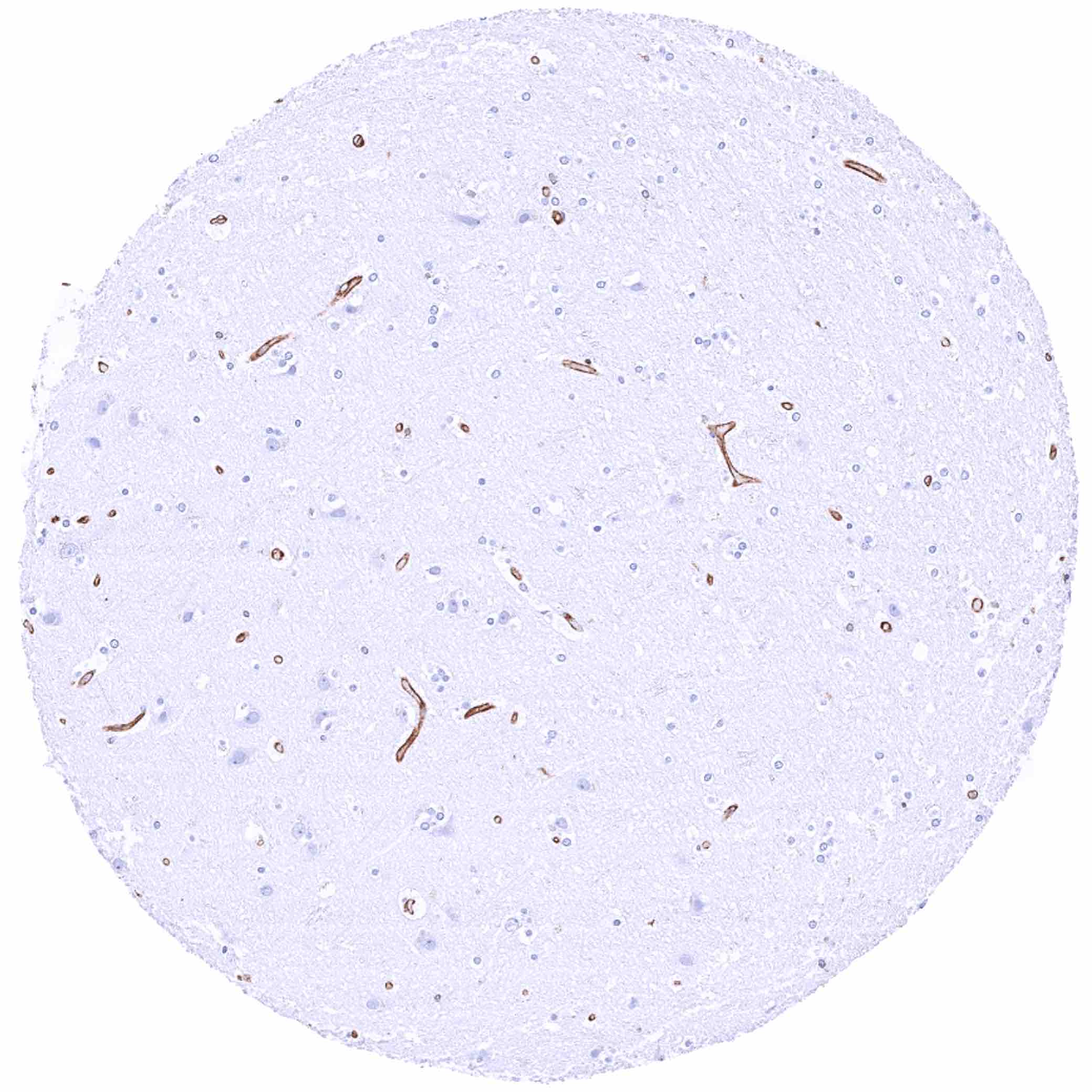
Cerebrum, grey matter
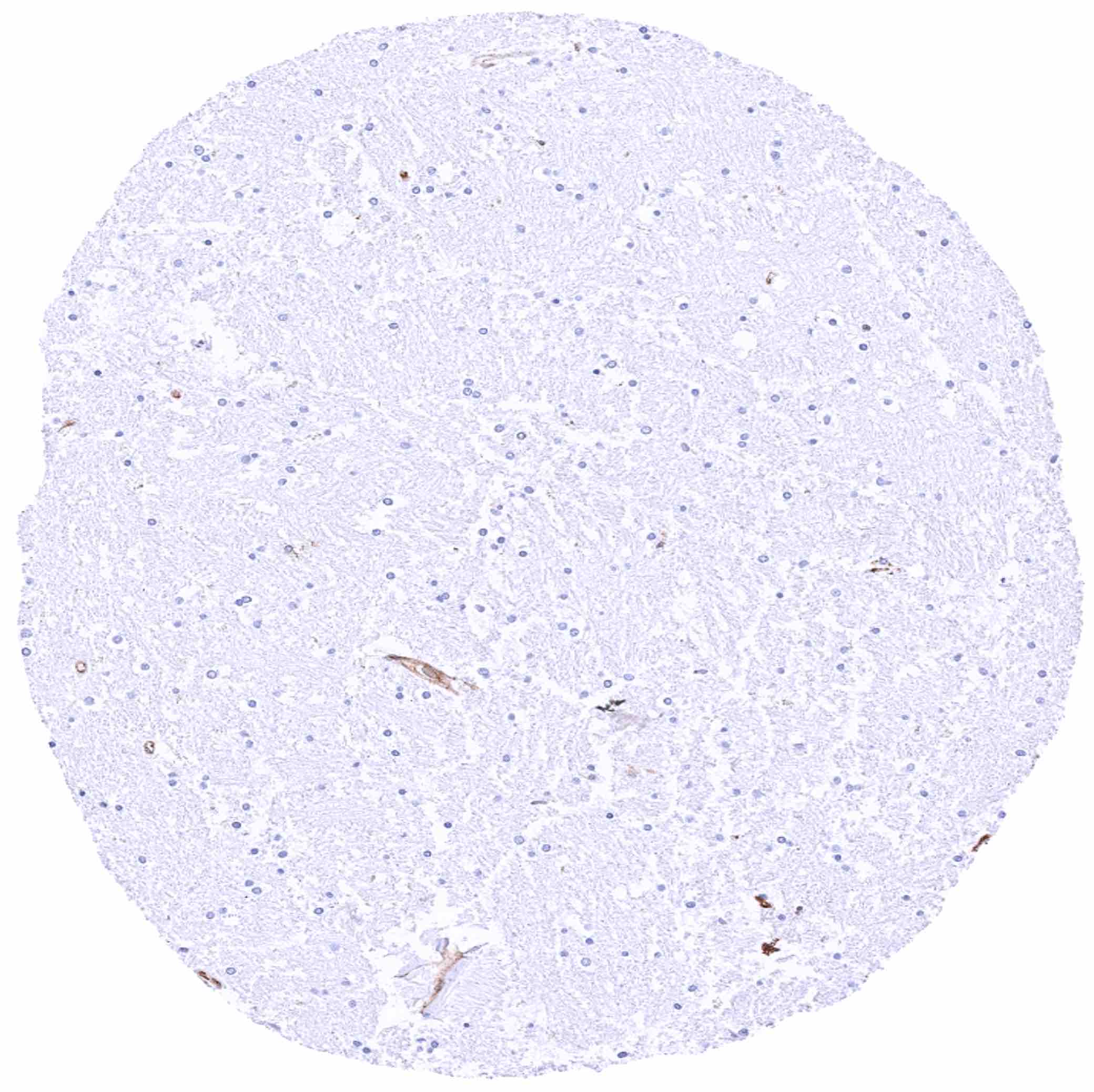
Cerebrum, white matter
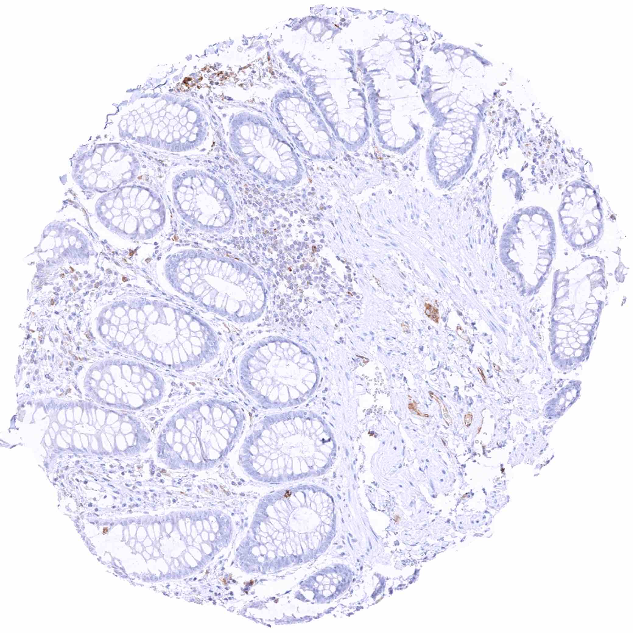
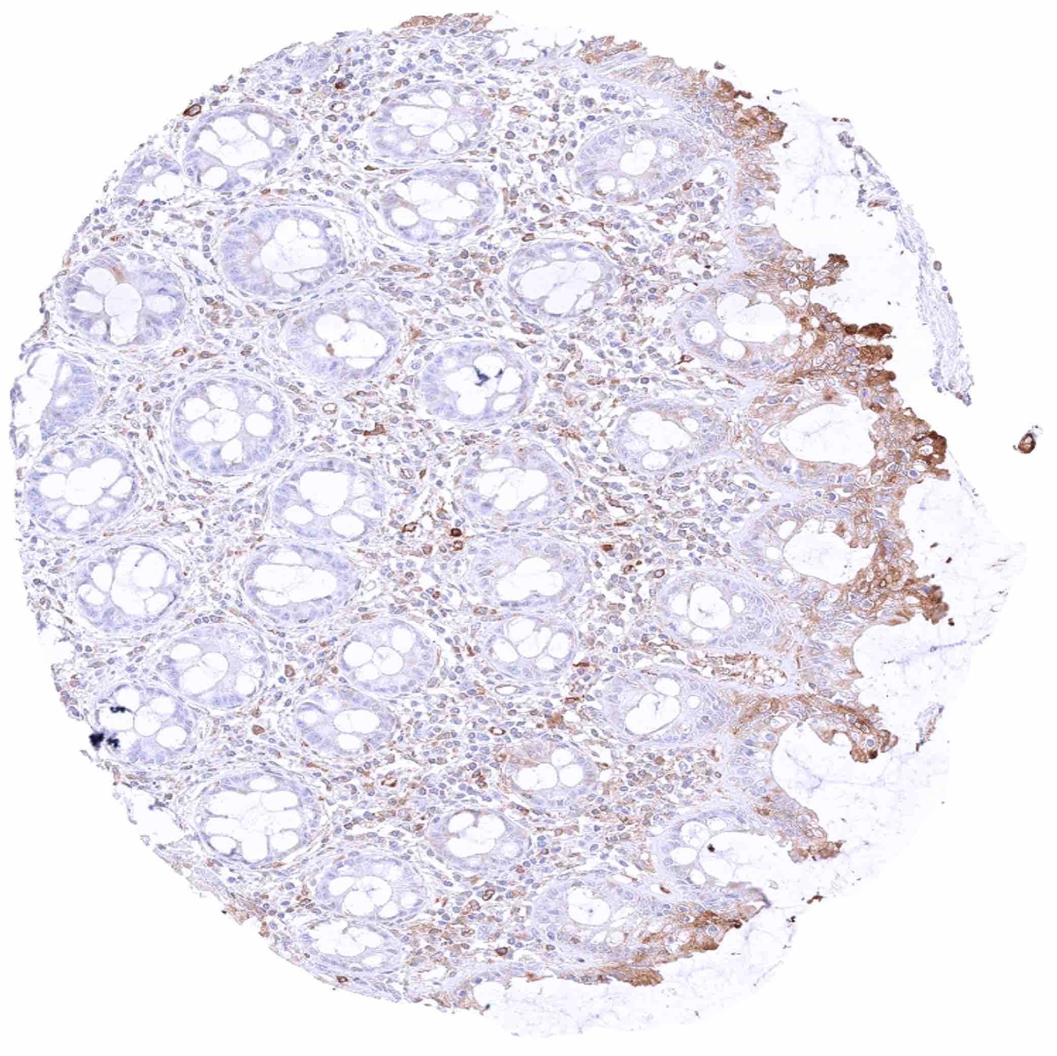
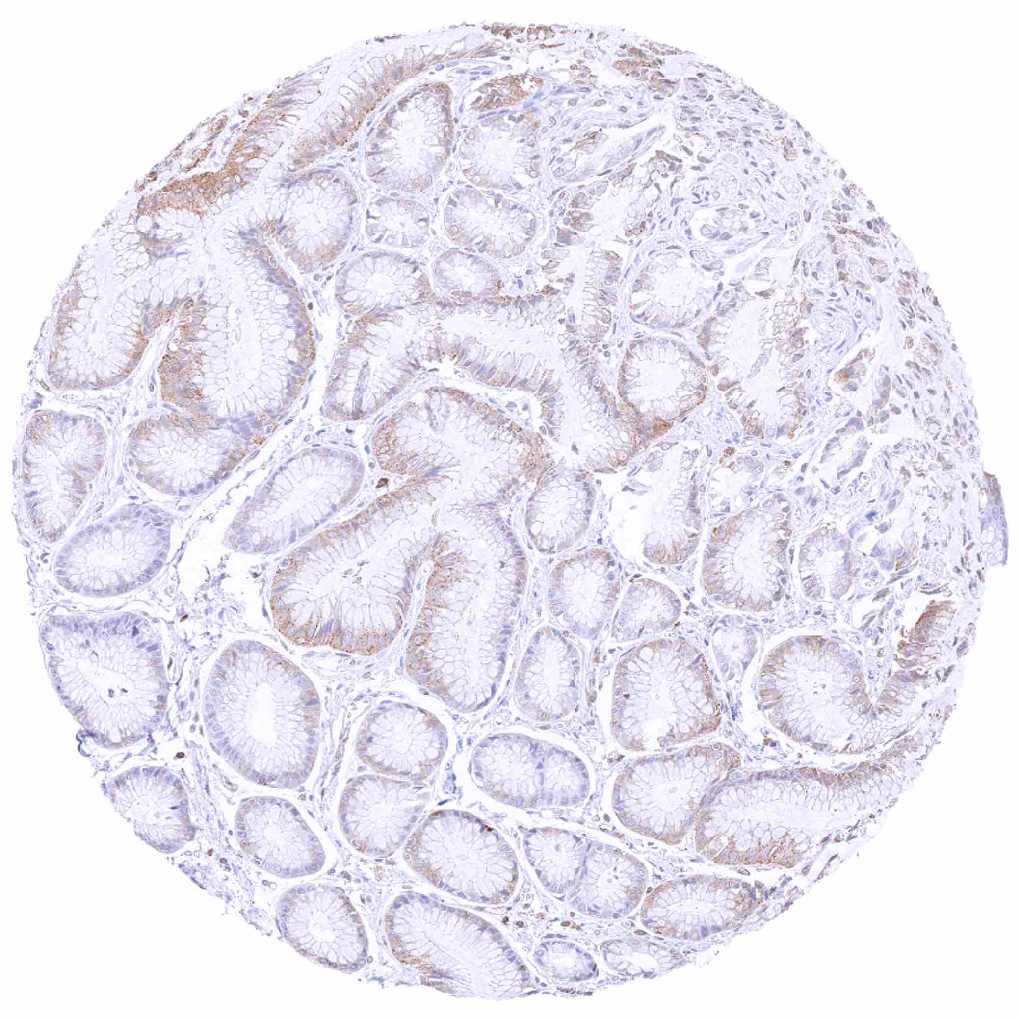
Colon descendens, mucosa – Crypt epithelial cells are largely MX1 negative
Duodenum, brunner gland – Brunner gland cells are MX1 negative
Epididymis – In the corpus, a cytoplasmic MX1 staining mainly occurs in basal cells and in subset of chief cells
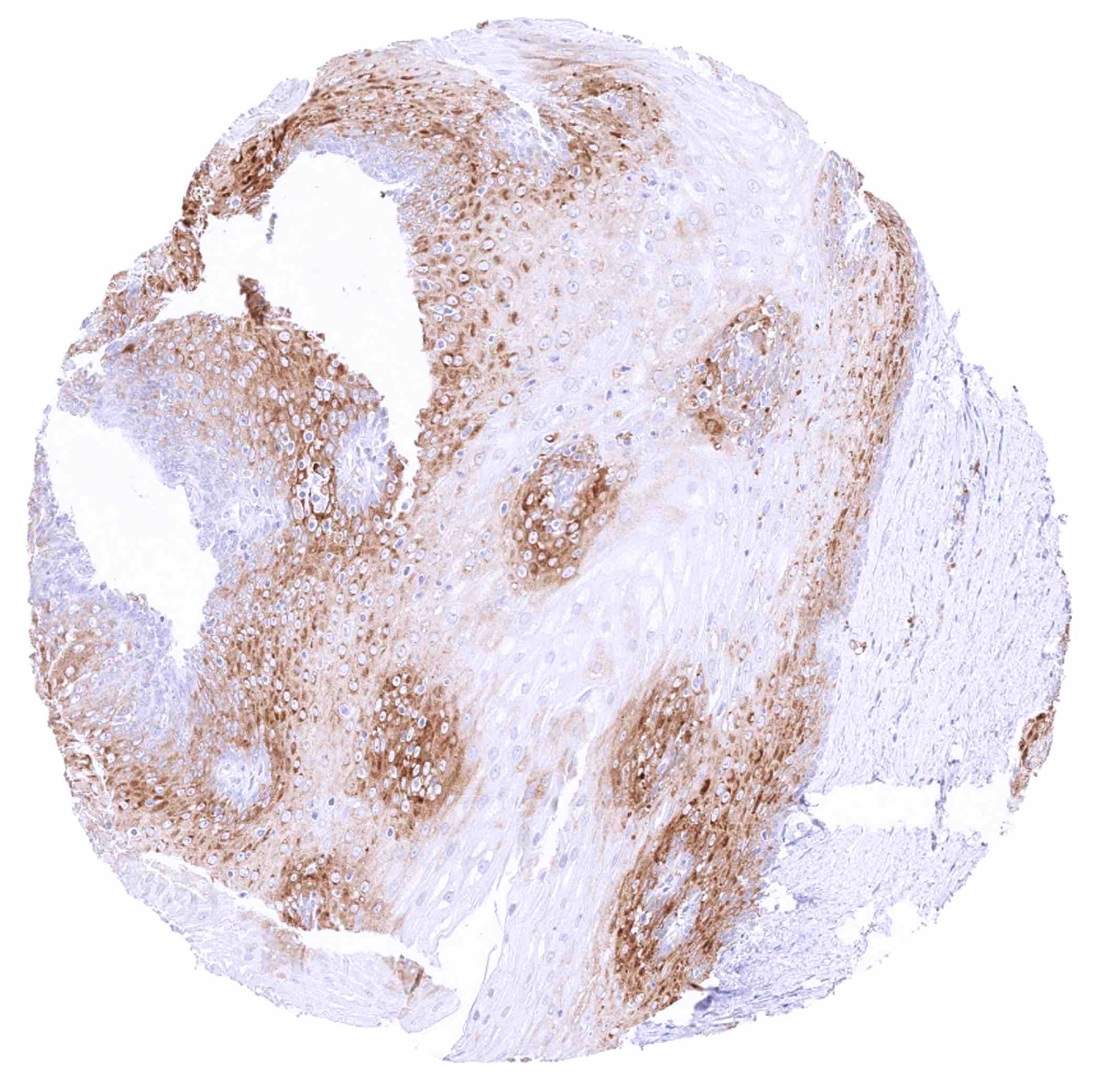
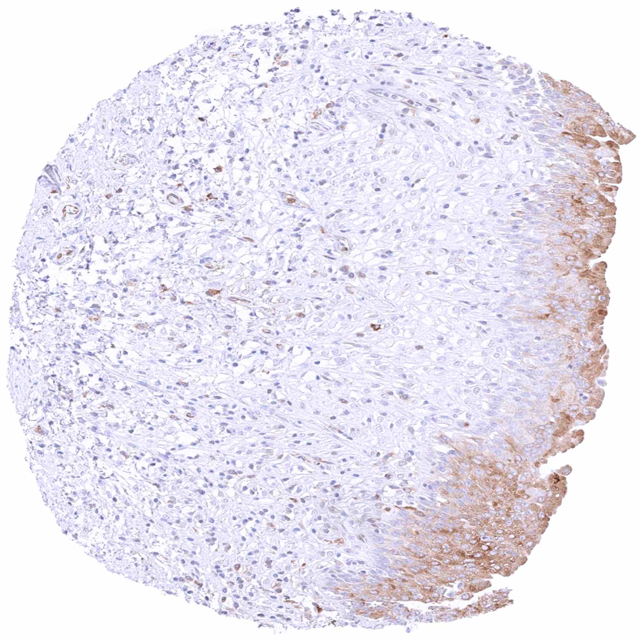
Esophagus, squamous epithelium – Moderate cytoplasmic MX1 staining of suprabasal cell layers
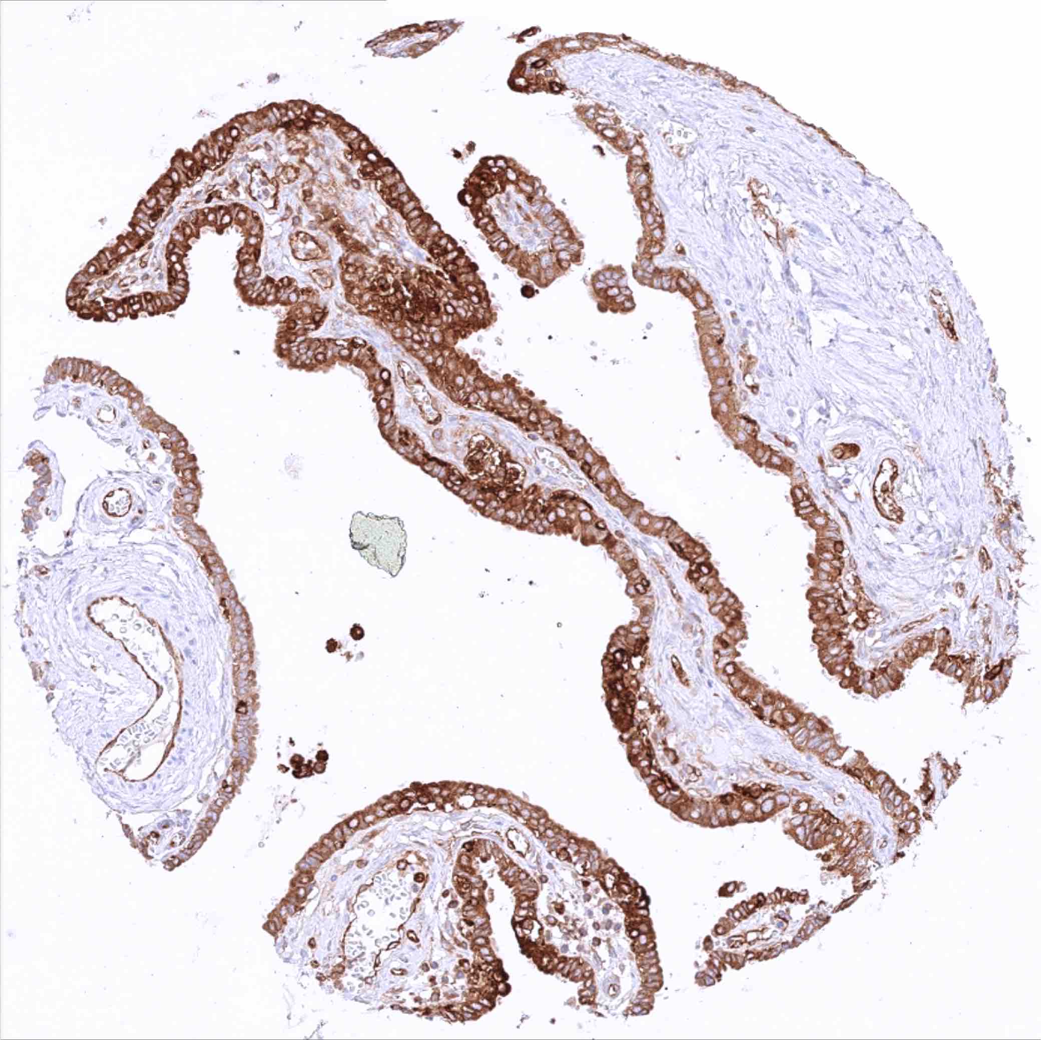
Fallopian tube, mucosa – Strong cytoplasmic MX1 staining of epithelial cells in this sample
Fallopian tube, mucosa – Weak to moderate cytoplasmic MX1 staining of epithelial cells
Fat
Gallbladder, epithelium – Strong focal MX1 staining of epithelial cells while other regions remain MX1 negative
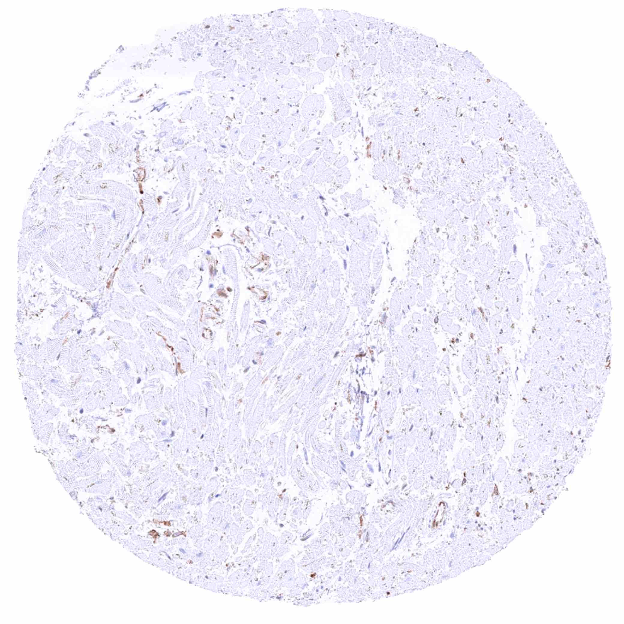
Heart muscle – MX1 staining prdominates in the endothelium of small vessels
Ileum, mucosa – Weak to moderate cytoplasmic MX1 staining of epithelial cells
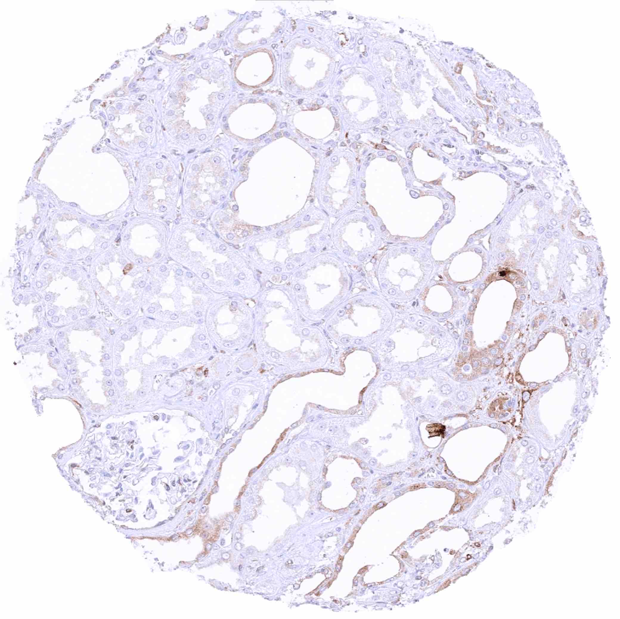
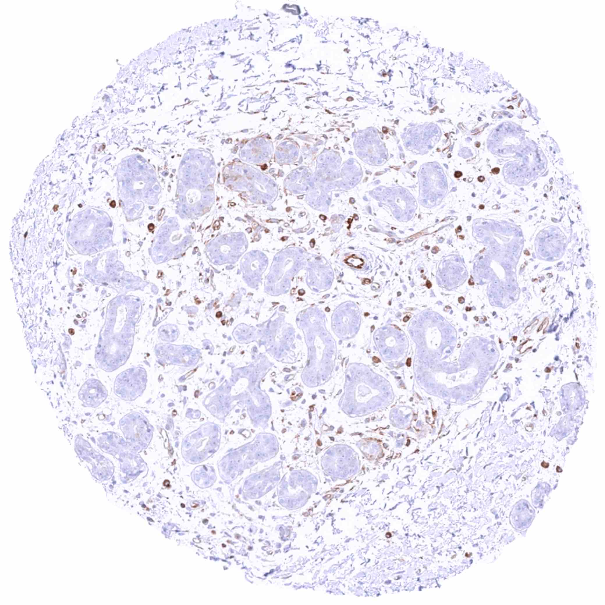
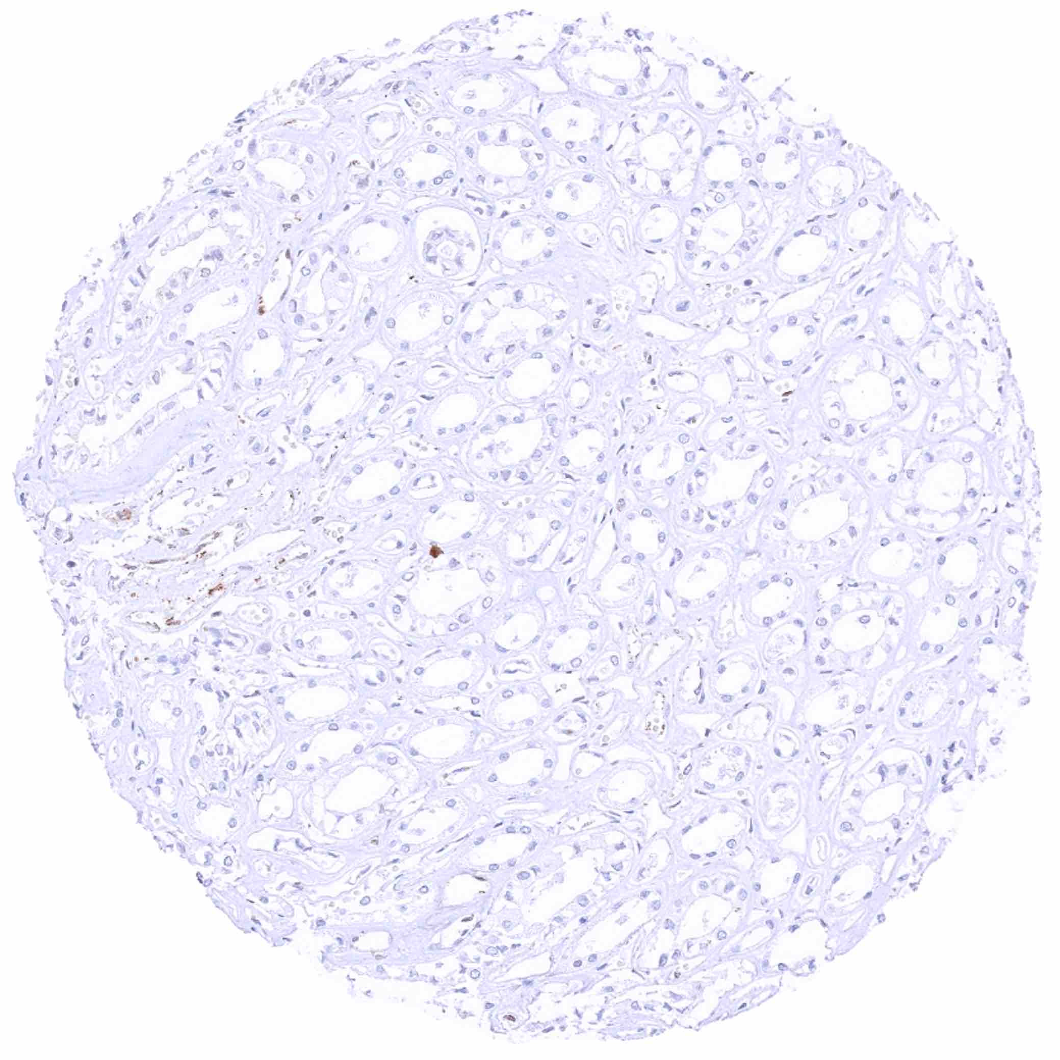
Kidney, cortex – Cytoplasmic MX1 staining mainly occurs in atrophic tubuli
Kidney, medulla – Some cytoplasmic MX1 staining in most ceollecting ducts of this sample
Kidney, medulla
Kidney, pelvis, urothelium
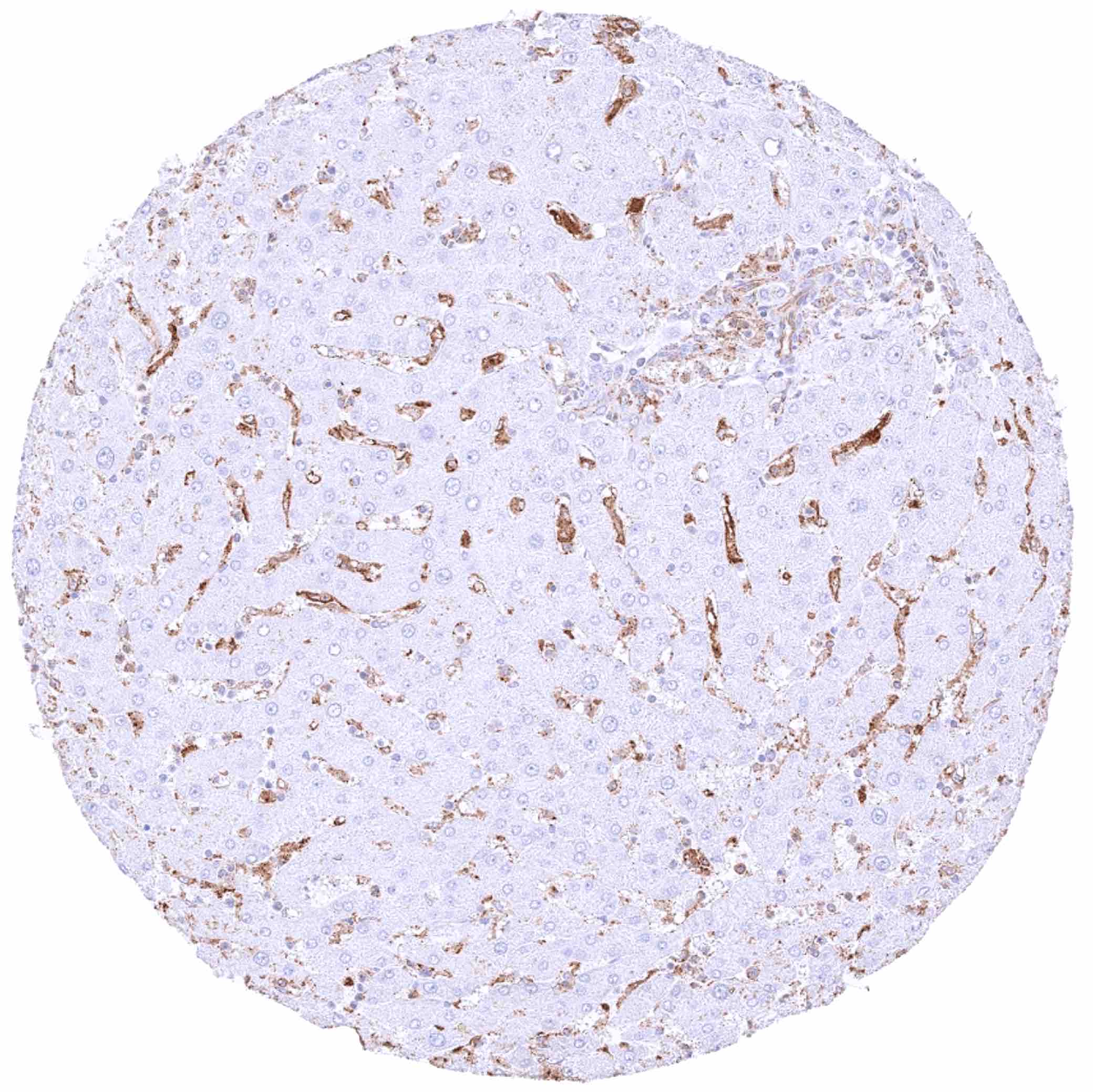
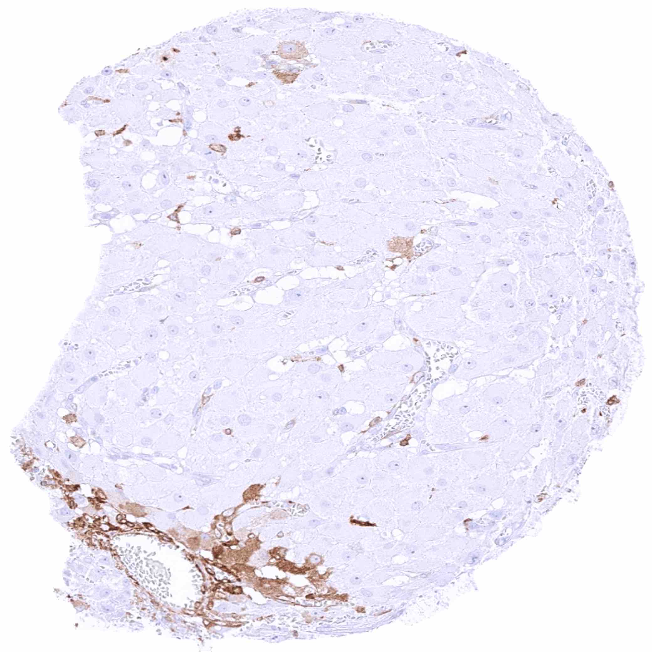
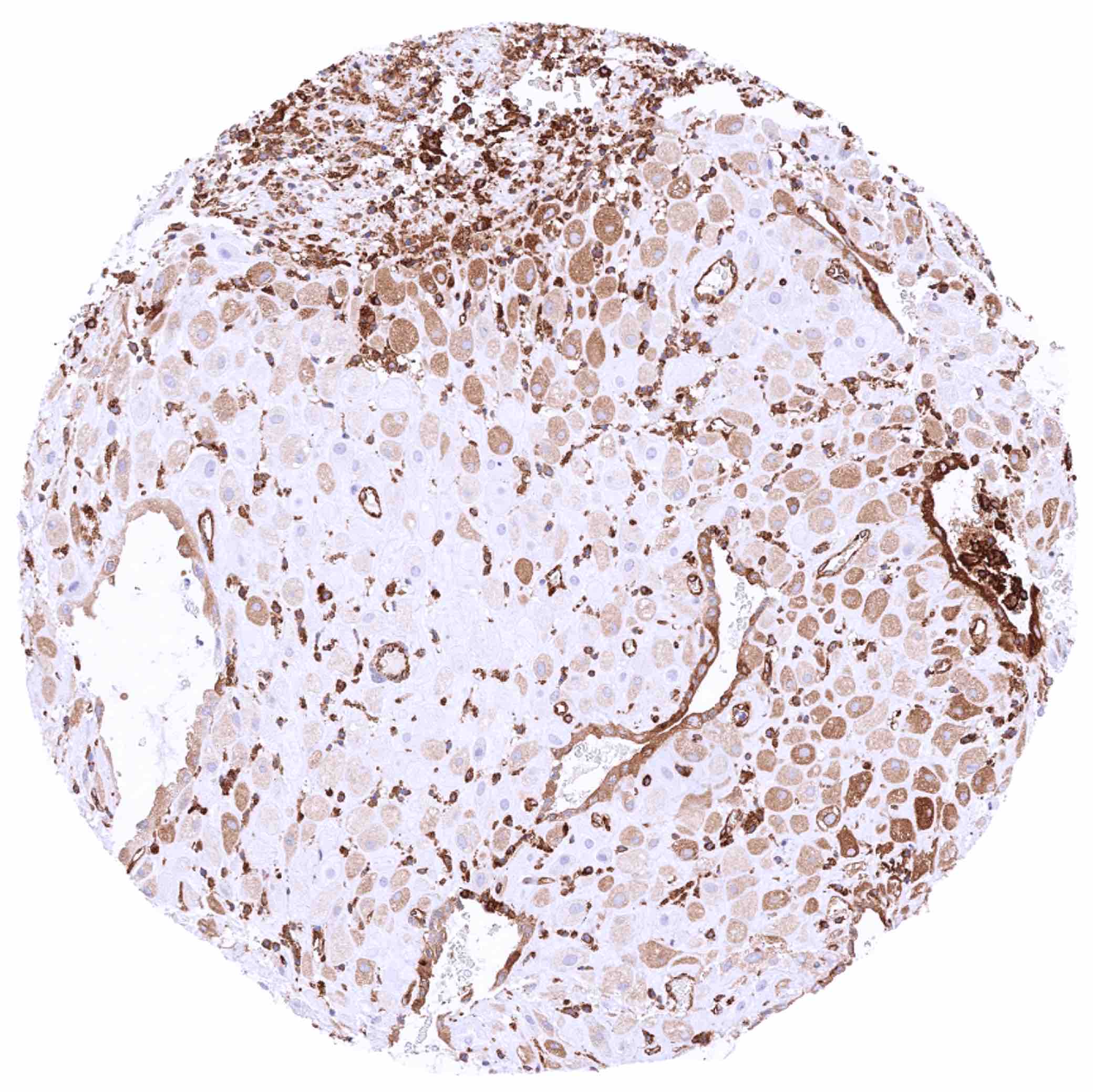
Liver – Strong MX1 staining of sinus cells while hepatocytes are MX1 negative
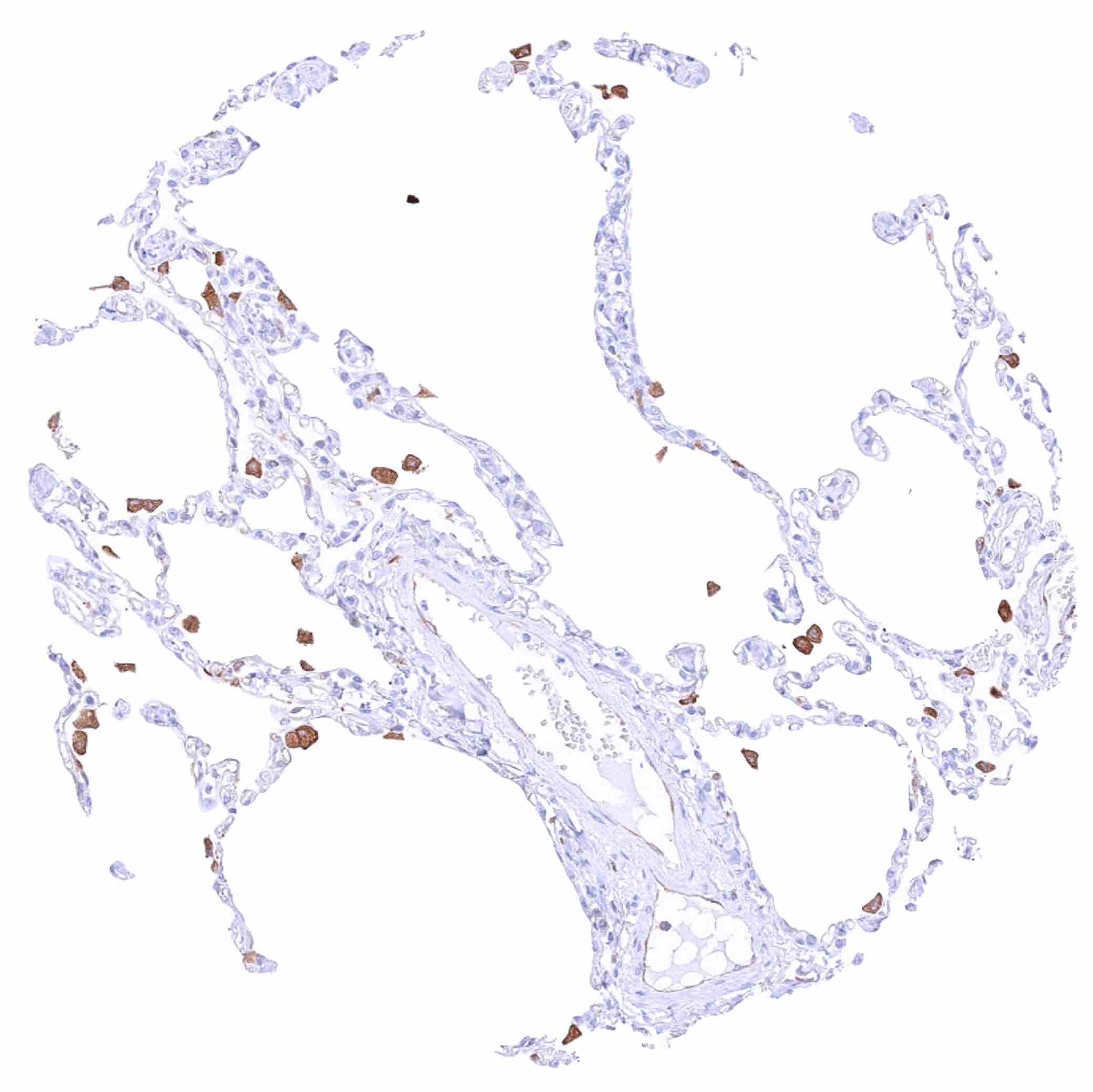
Lung – Moderate to strong cytoplasmic MX1 staining of macrophages
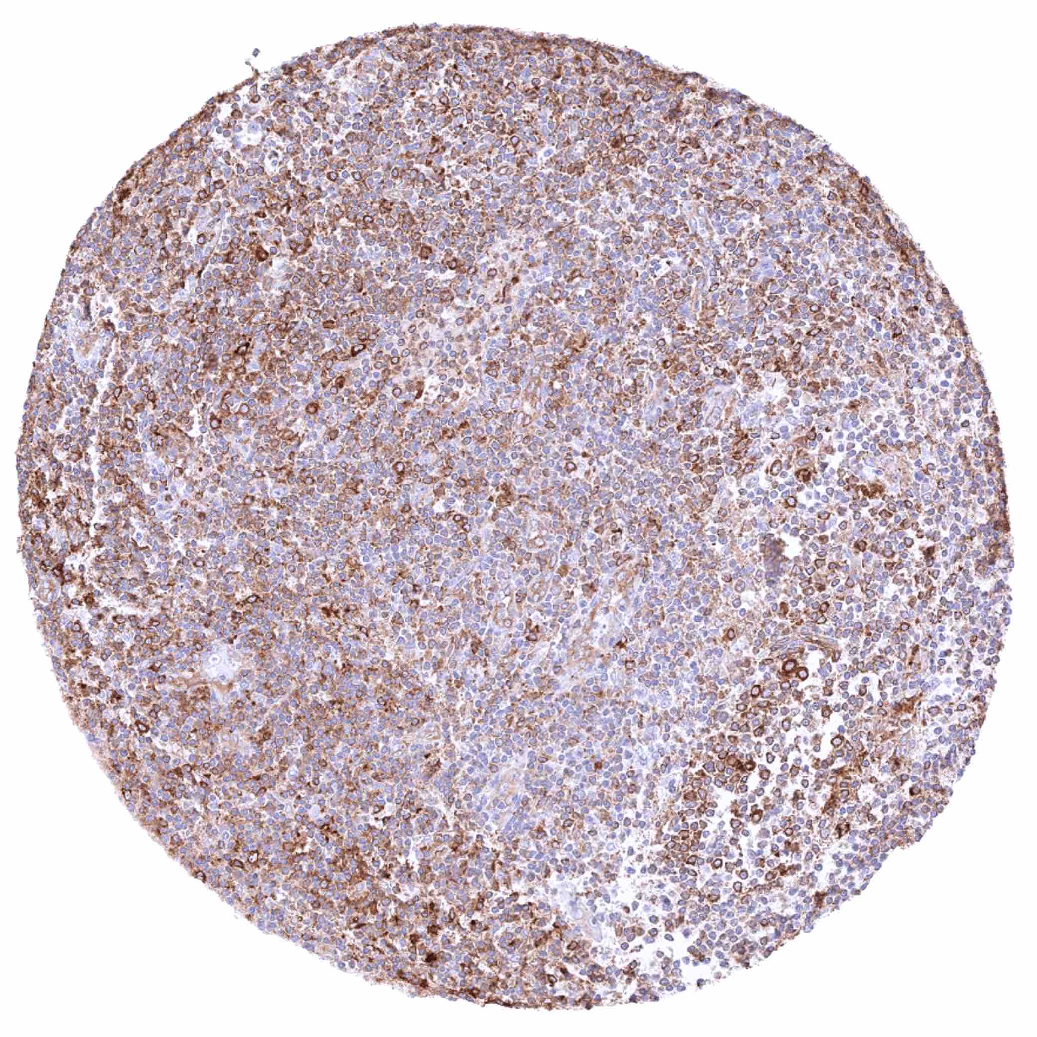
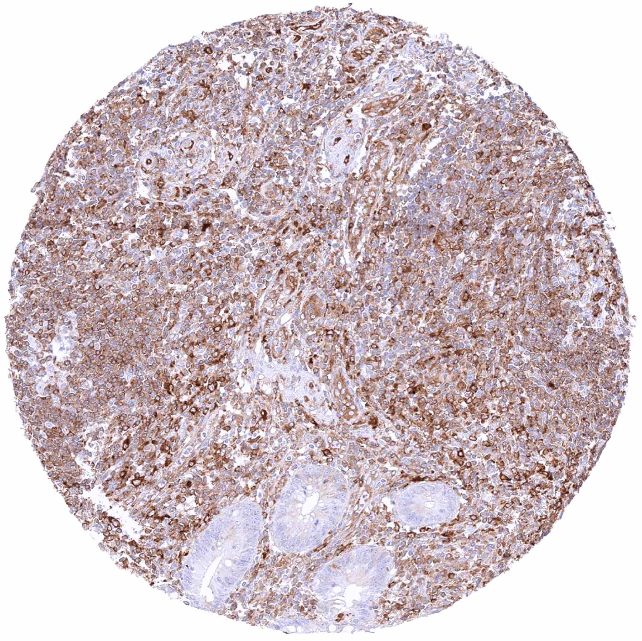
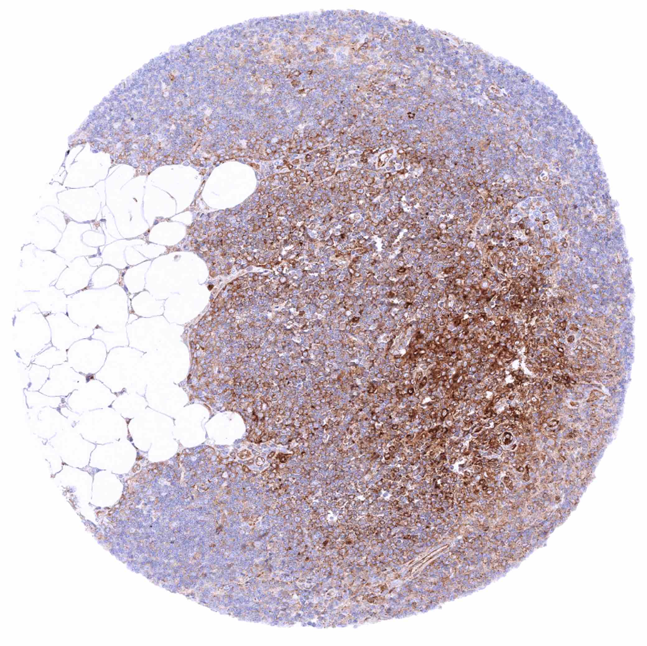
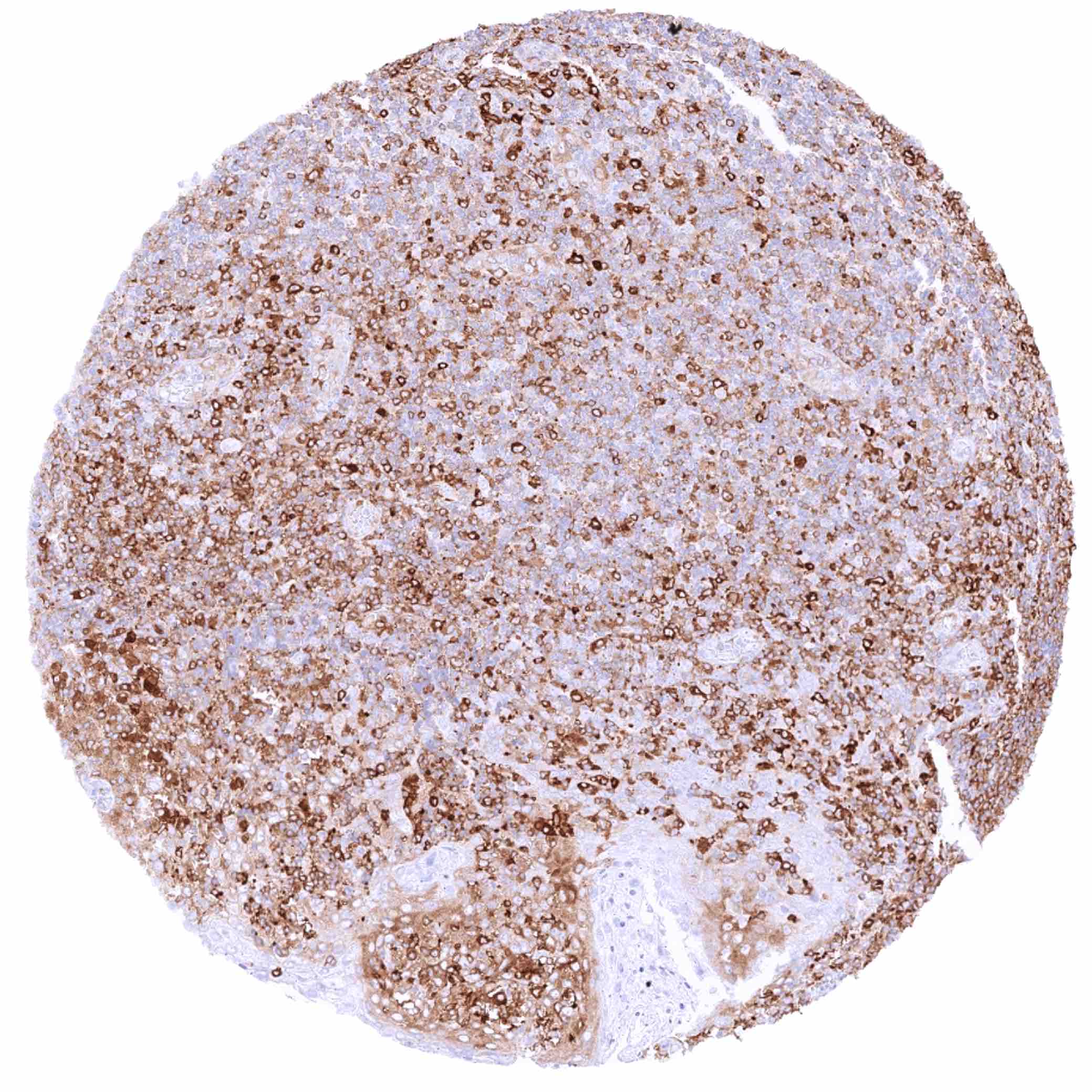
Lymph node – Cytoplasmic MX1 staining at variable intensity of a large fraction of cells
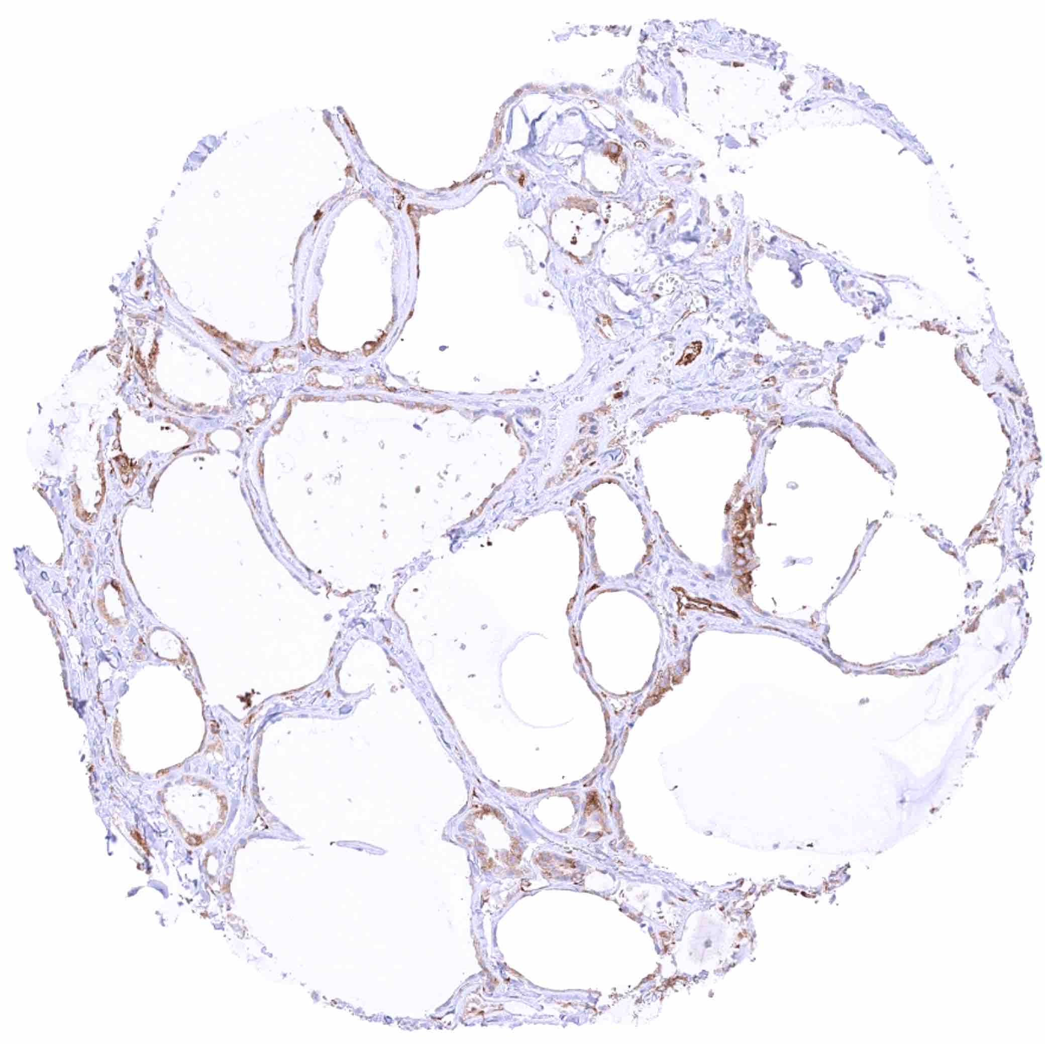
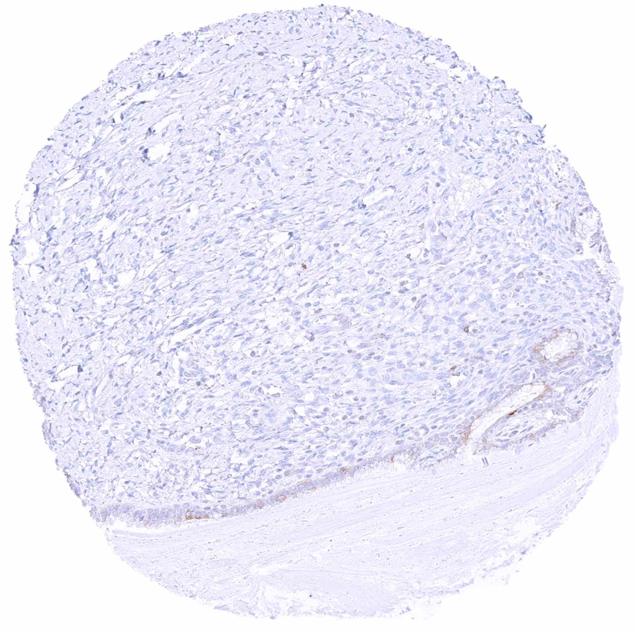
Ovary, corpus luteum – Most corpus luteum cells are MX1 negative
Ovary, stroma

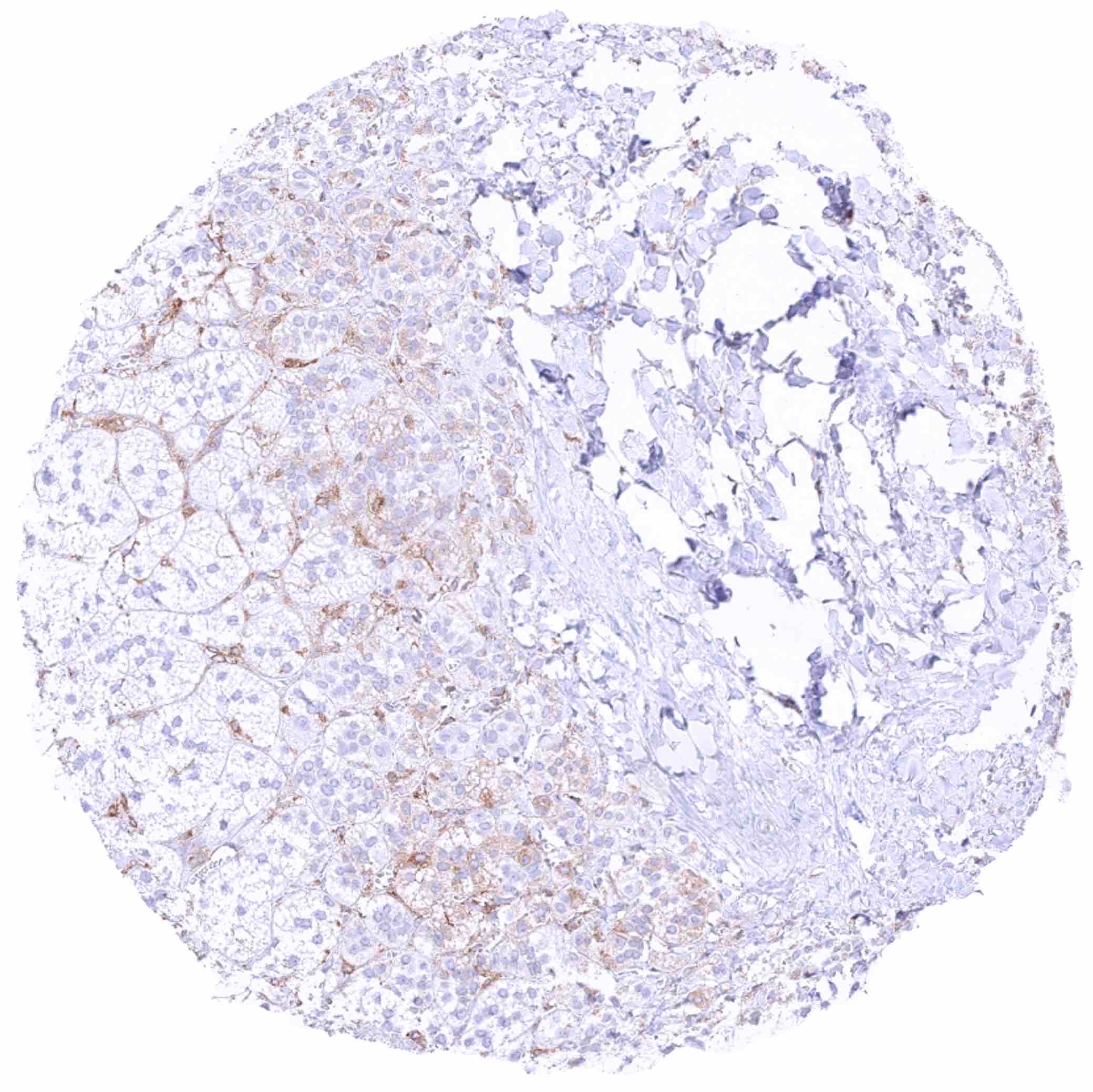
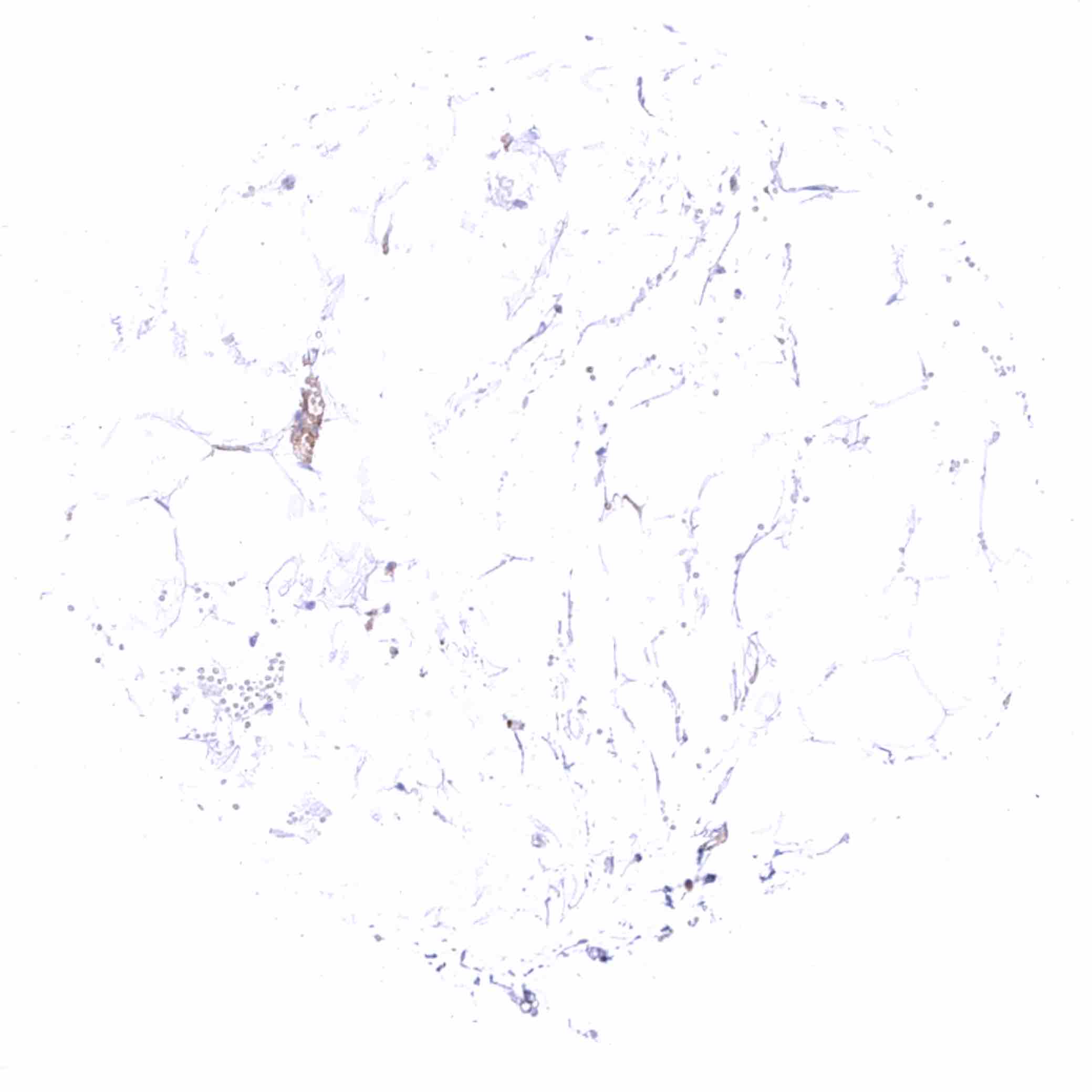
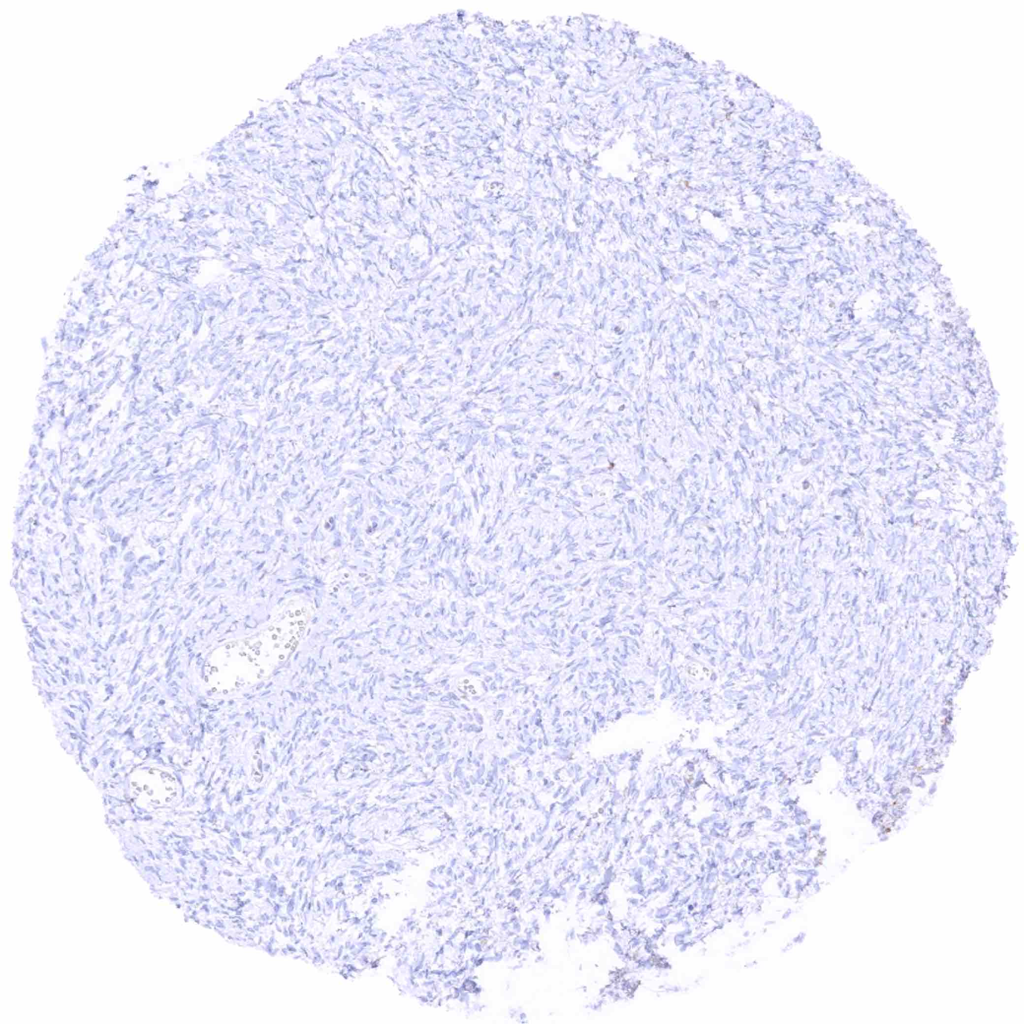
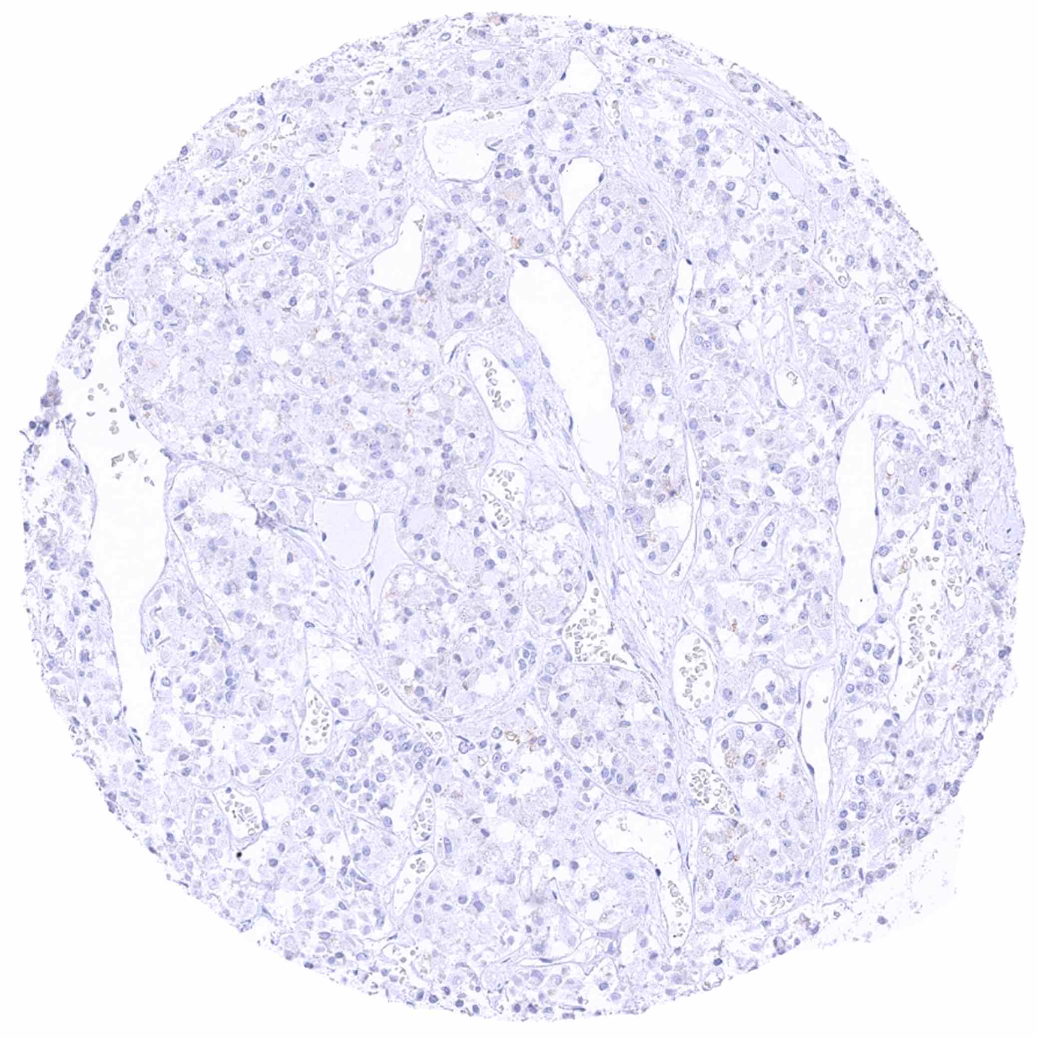
Pancreas – MX1 staining is completely absent in this sample
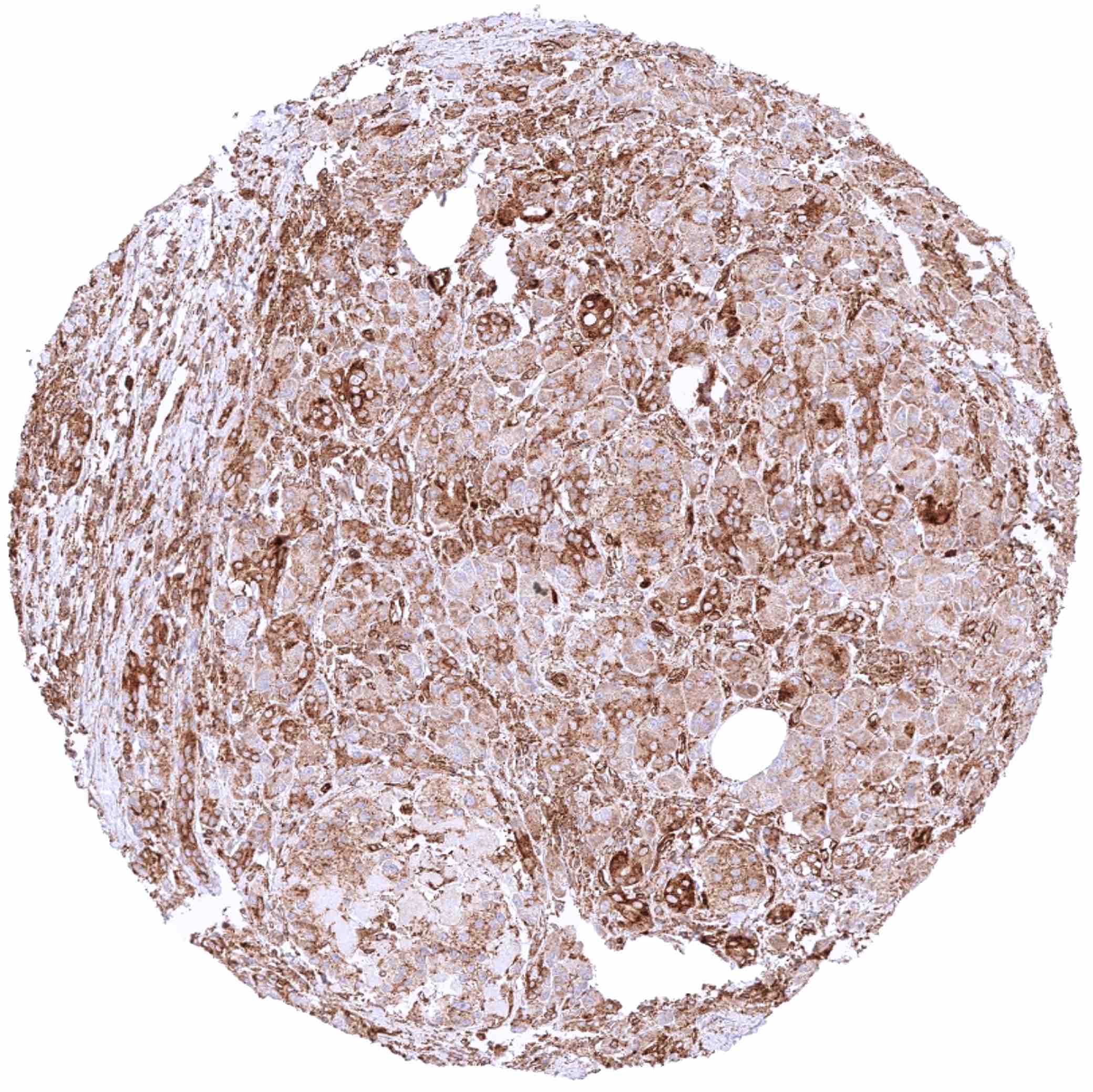
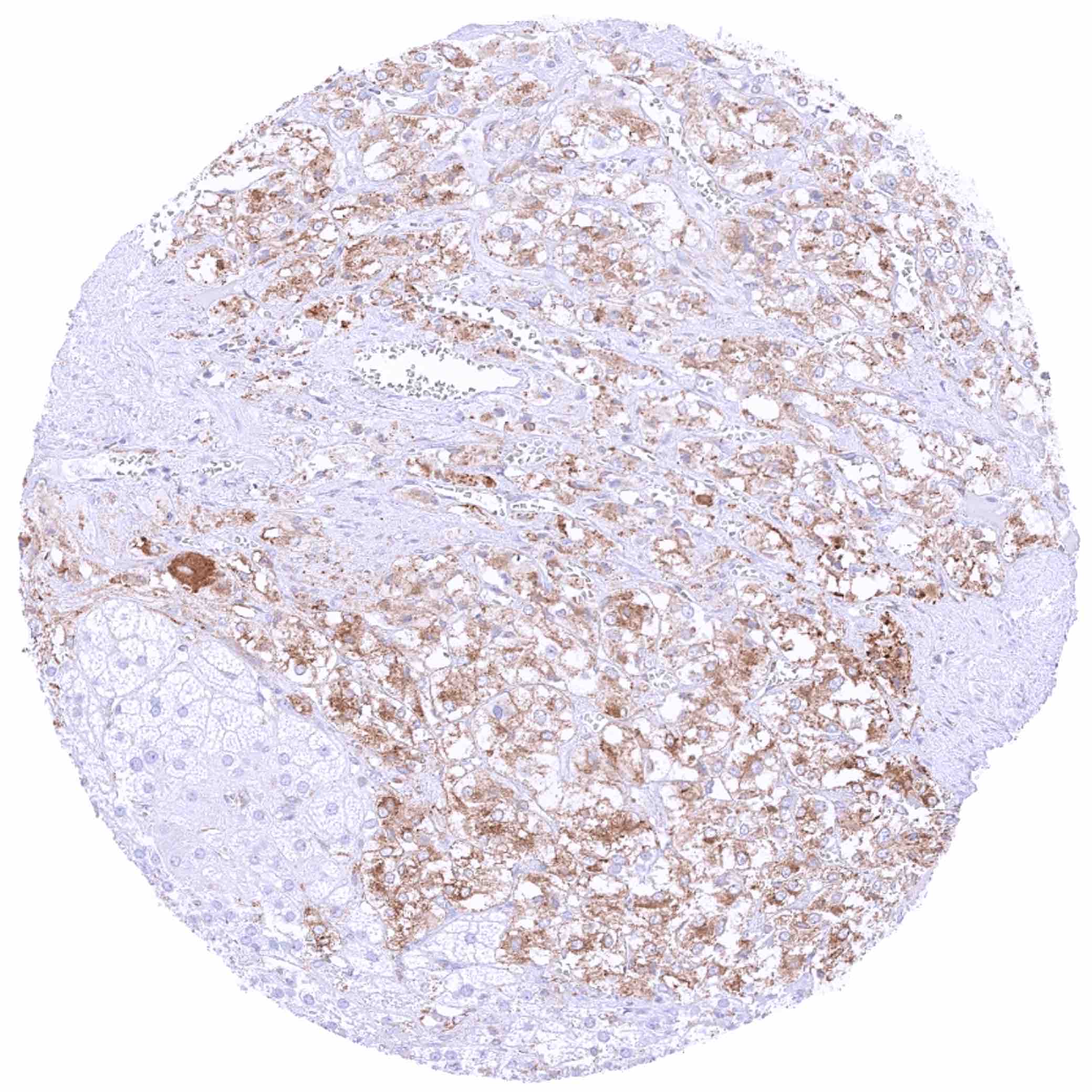
Pancreas – Strong cytoplasmic MX1 staining of all epithelial cell types in a sample with some inflammation and scar formation
Parathyroid gland – MX1 staining is limited to endothelial cells
Parotid gland – Moderate to strong cytoplasmic MX1 staining can be seen in some epithelial cell groups
Pituitary gland, posterior lobe
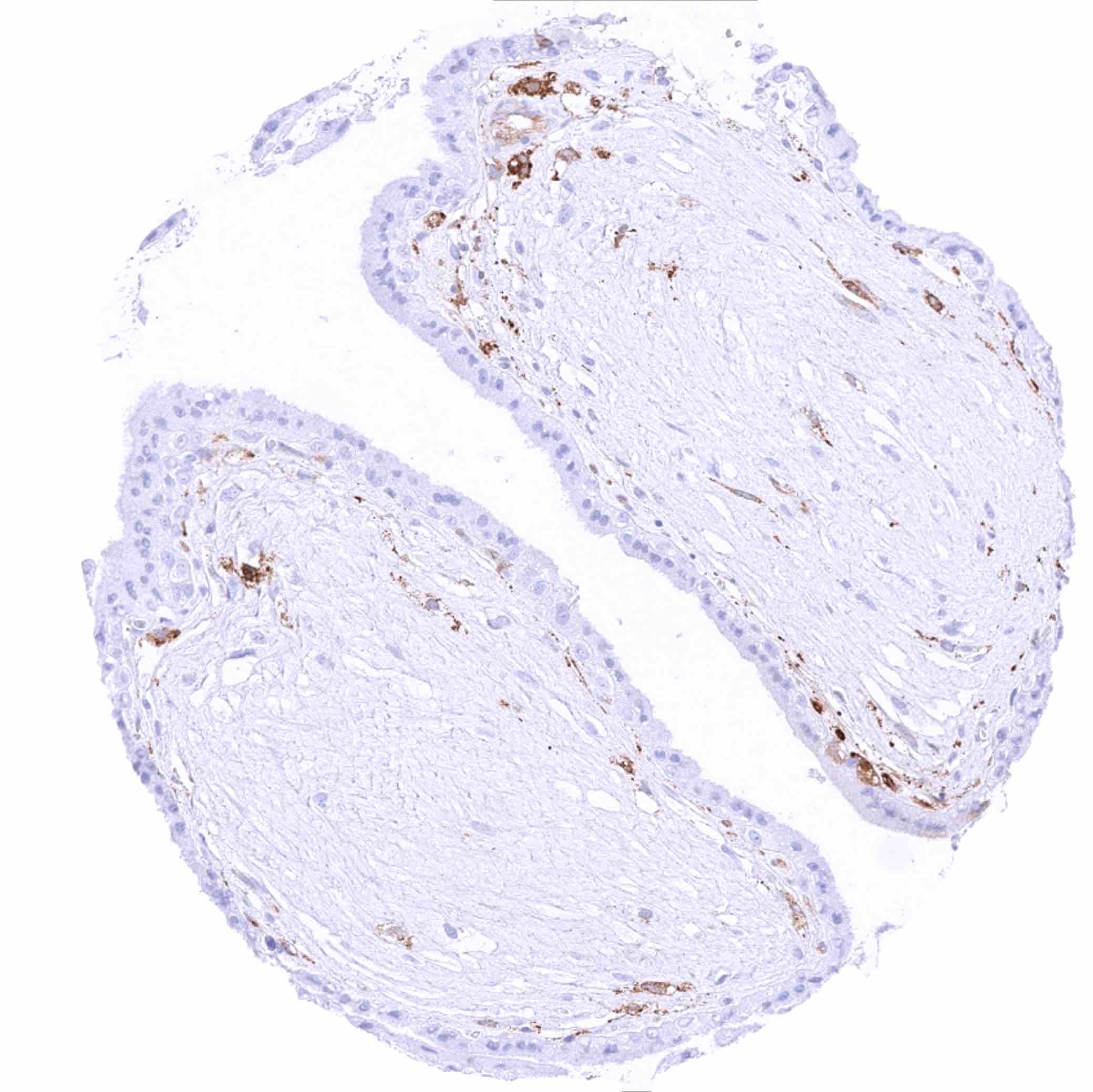
Placenta (amnion and chorion) – Weak cytoplasmic MX1 staining of some amnion cells while chorion cells are negative. Strong staining of small vessels
Placenta, early – Trophoblast cells are MX1 negative. Strong staining is only seen in endothelial cells
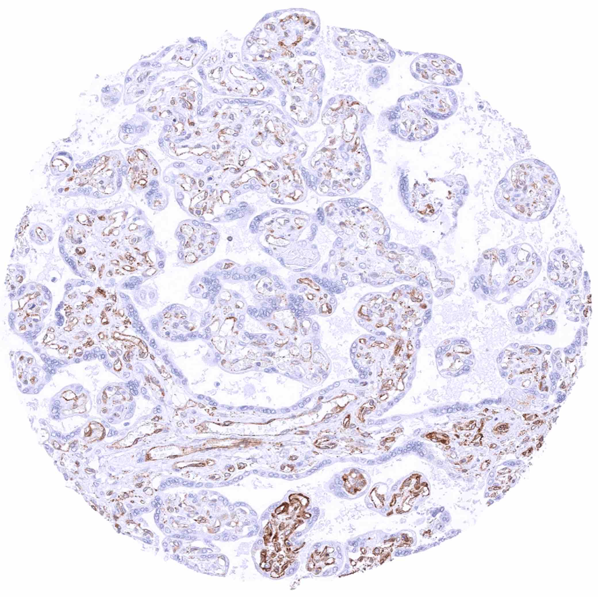
Placenta, mature – Trophoblast cells are MX1 negative. Strong staining is only seen in endothelial cells
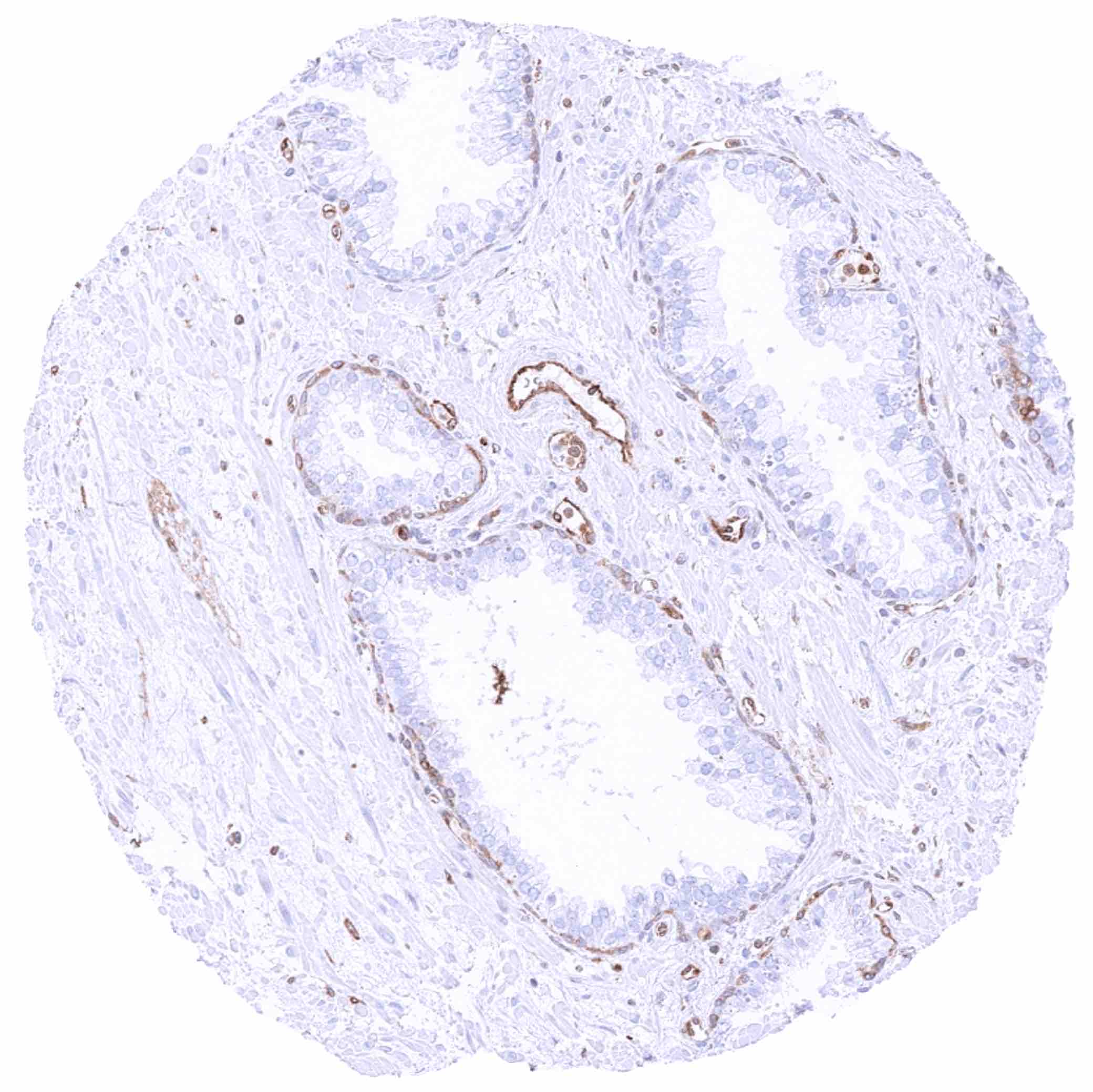
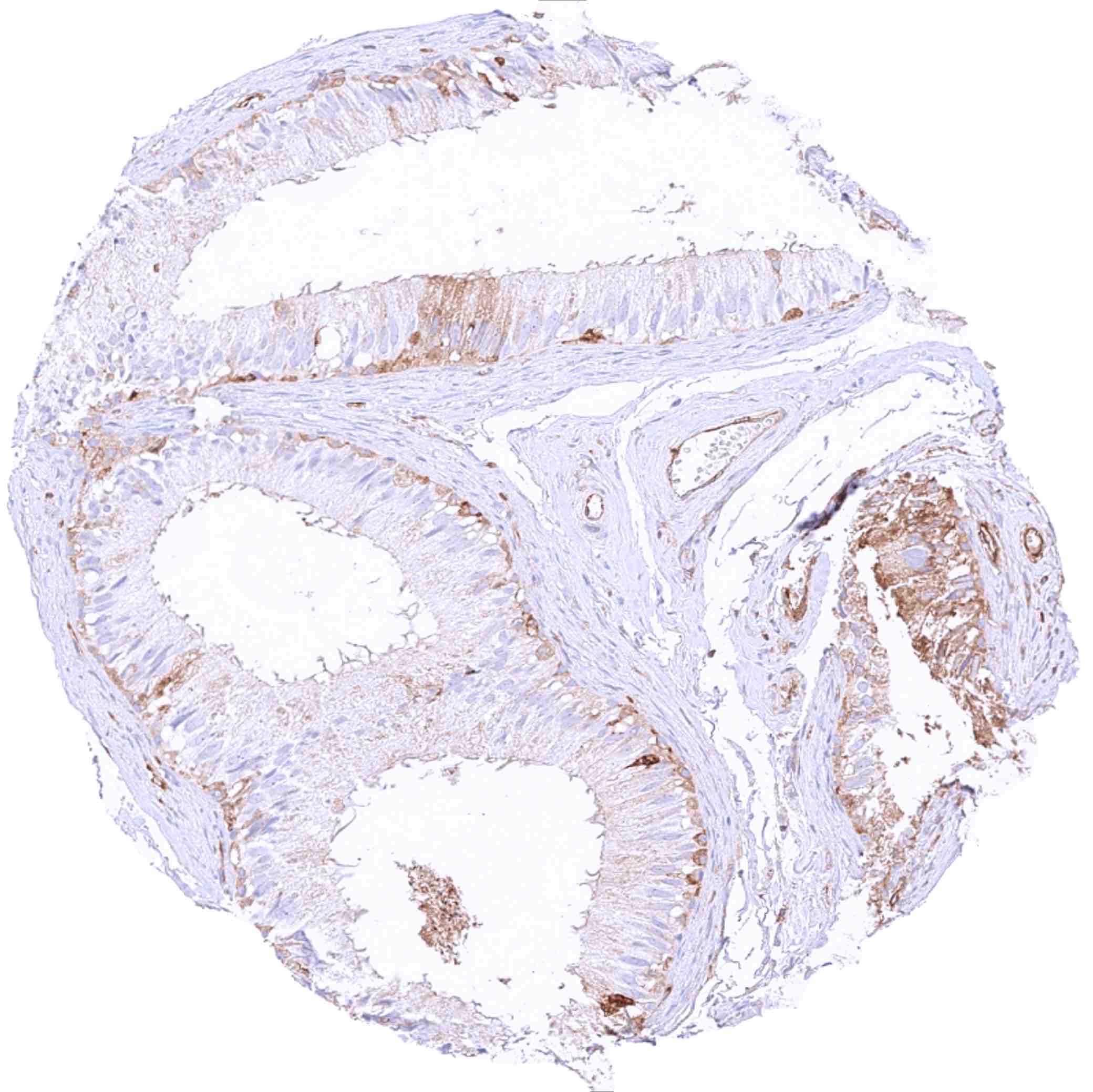
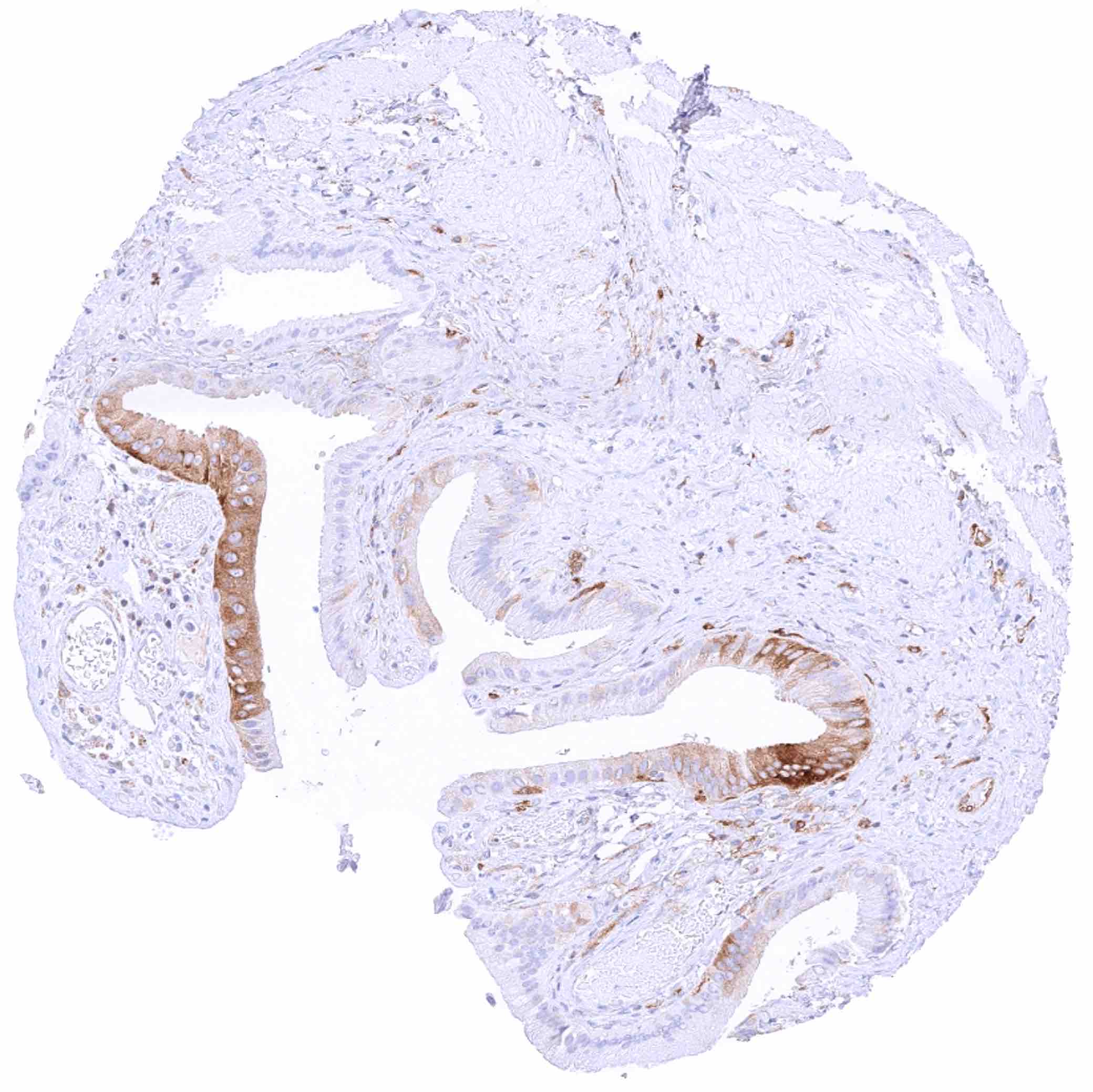
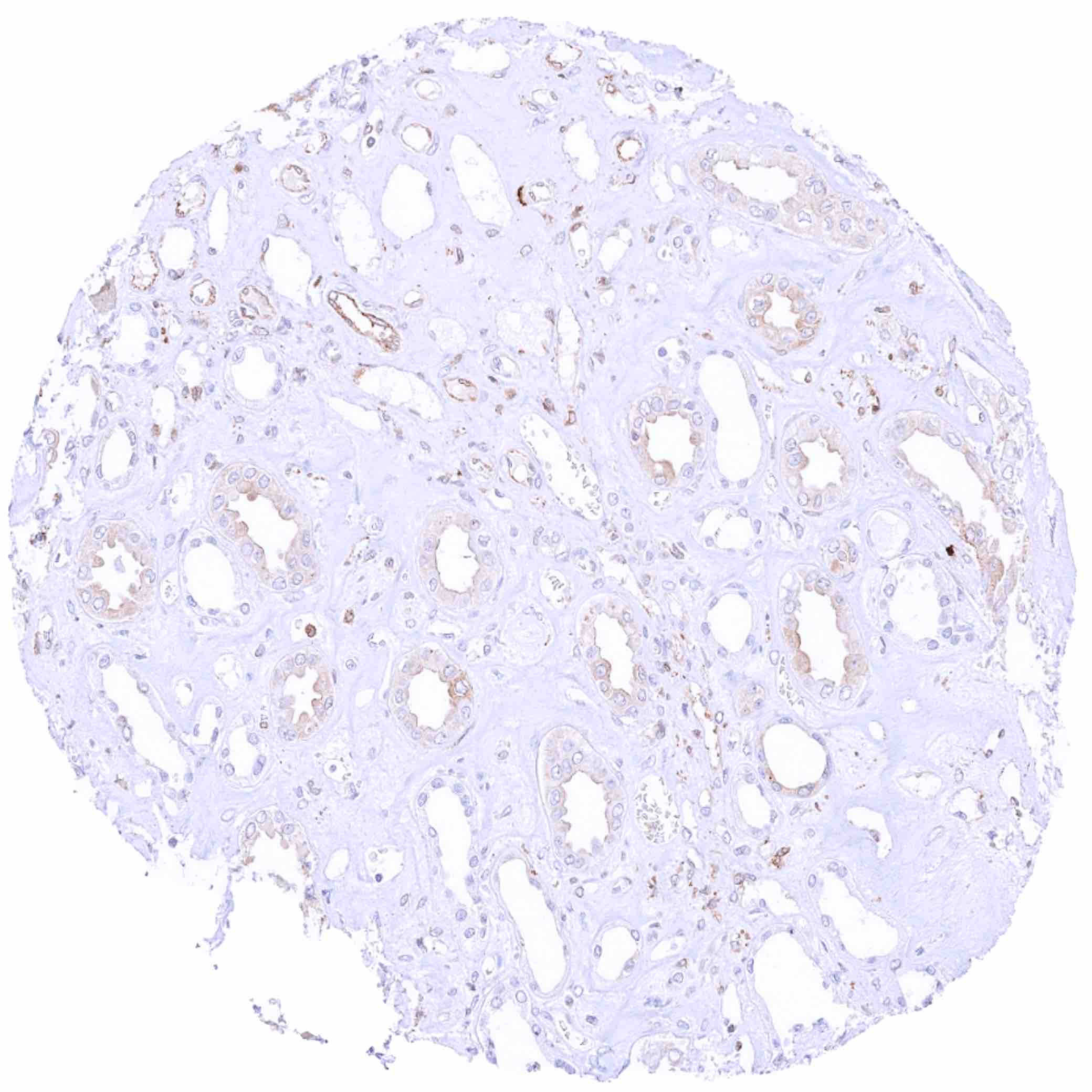
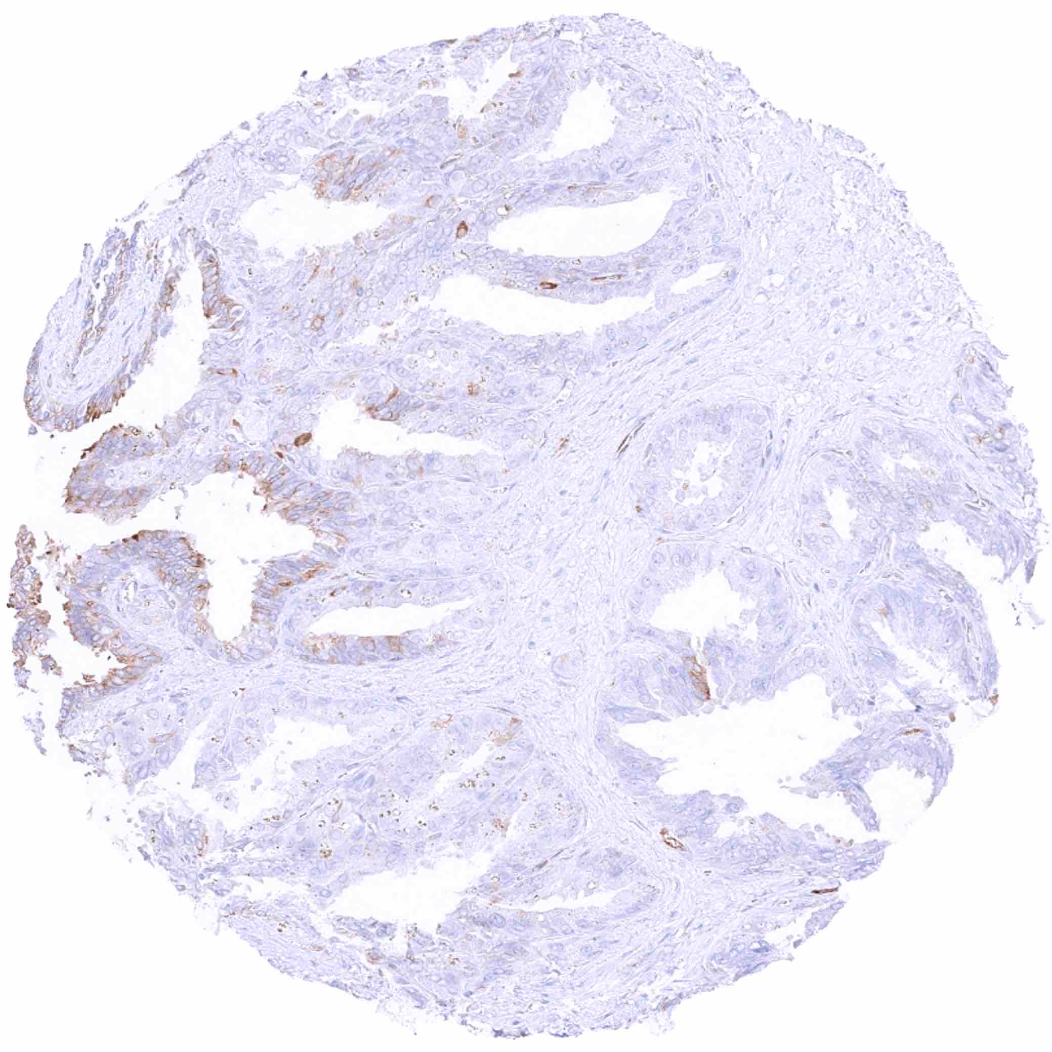
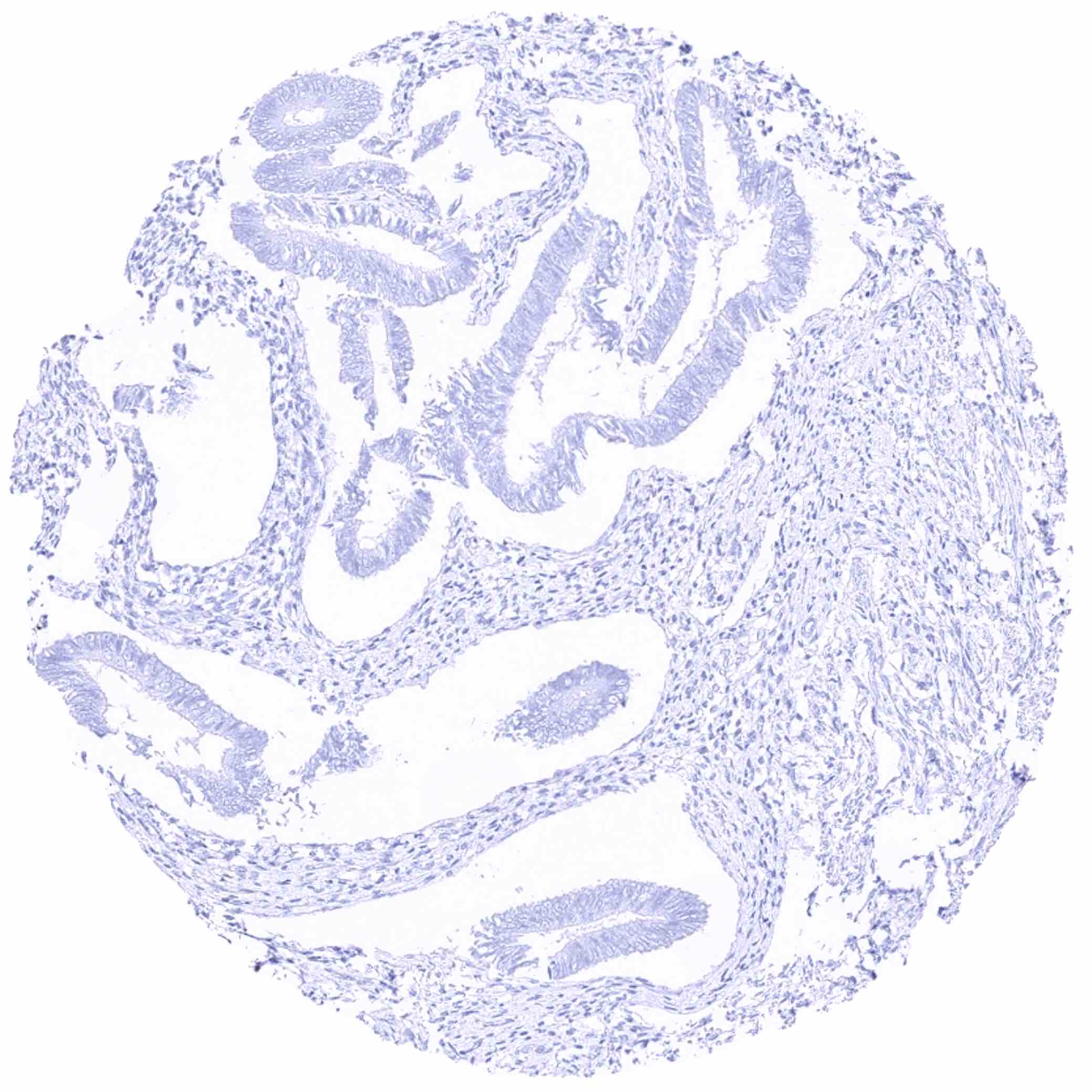
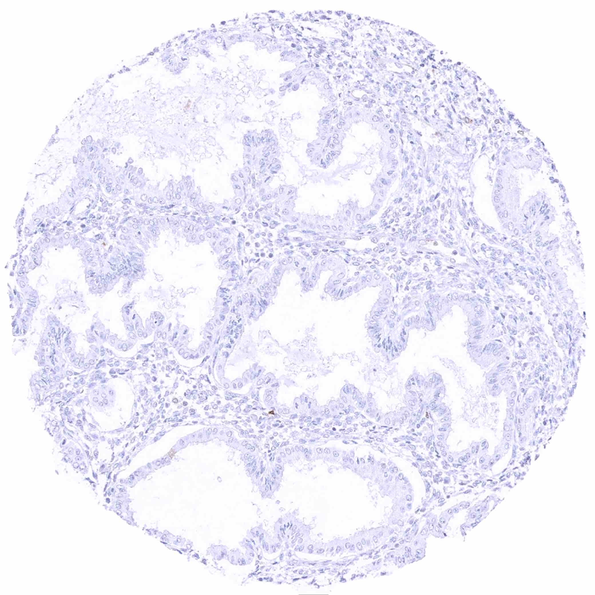
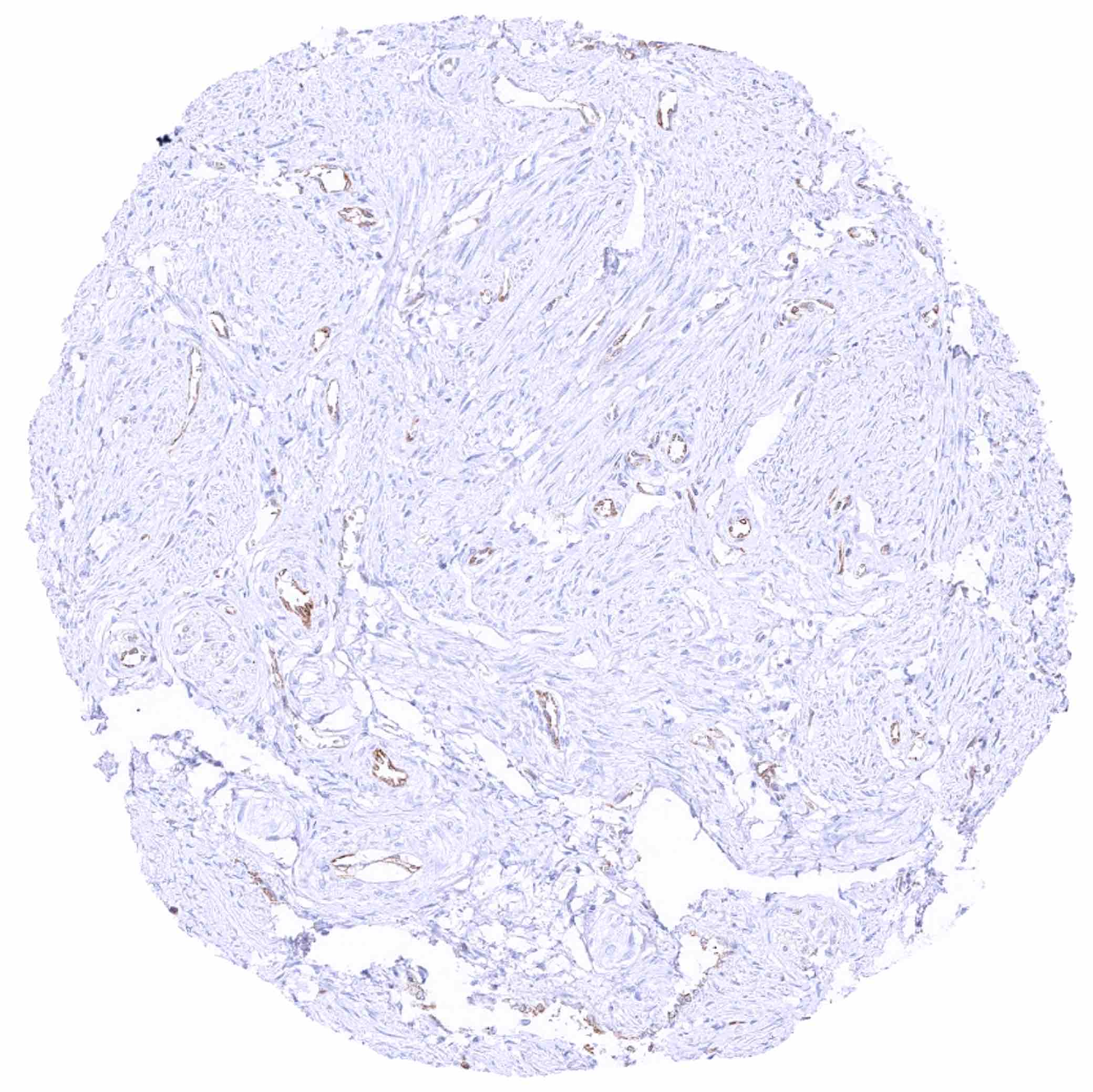
Prostate – Cytoplasmic MX1 staining of basal cells and of the endothelium of small vessels.
Rectum, mucosa – MX1 staining of epithelial cells is largely restricted to superficial cell layers
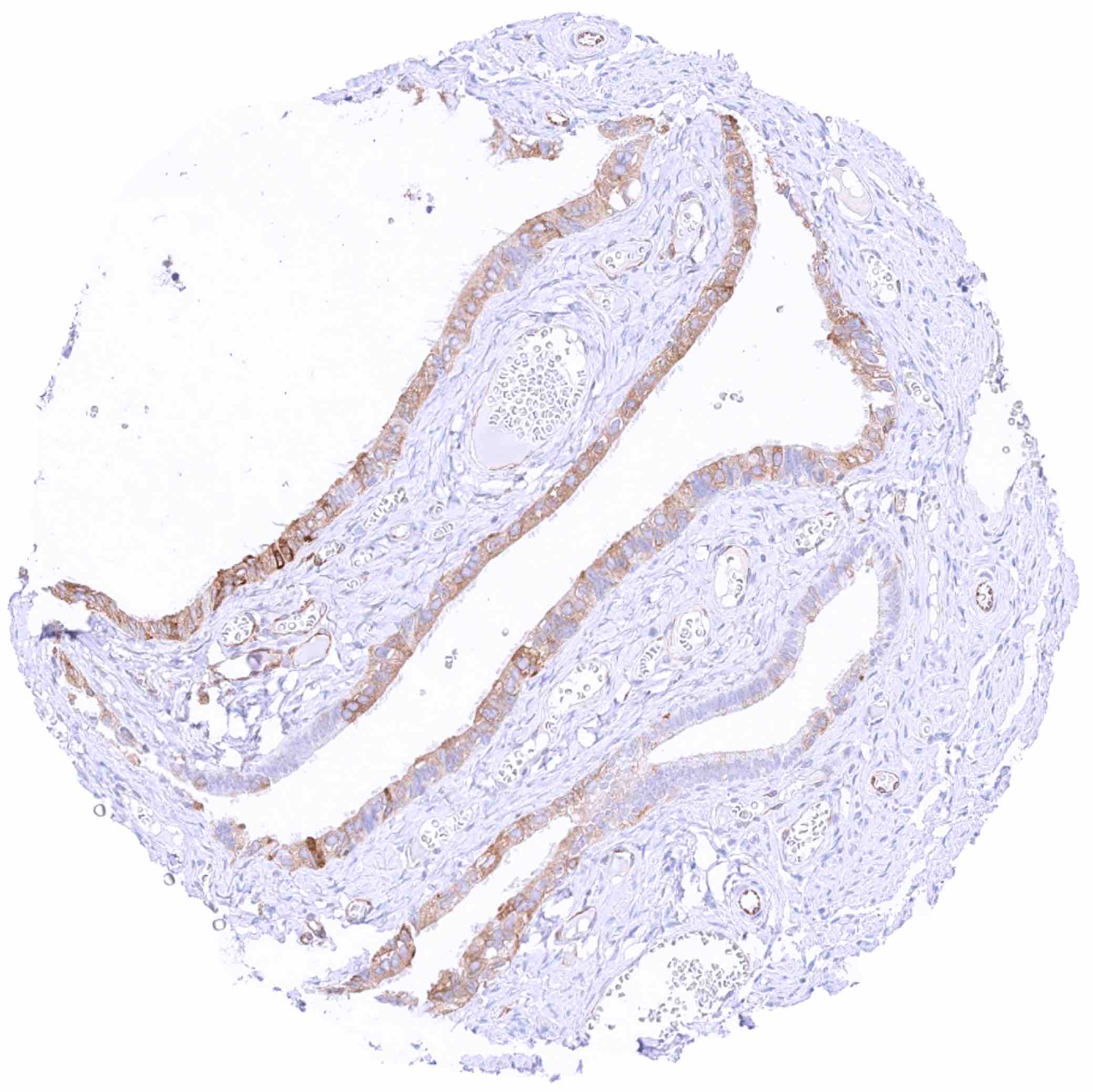
Seminal vesicle – Cytoplasmic MX1 staining of a subset of epithelial cells
Sinus paranasales – Focal, moderate to strong cytoplasmic MX1 staining in respiratory epithelial cells
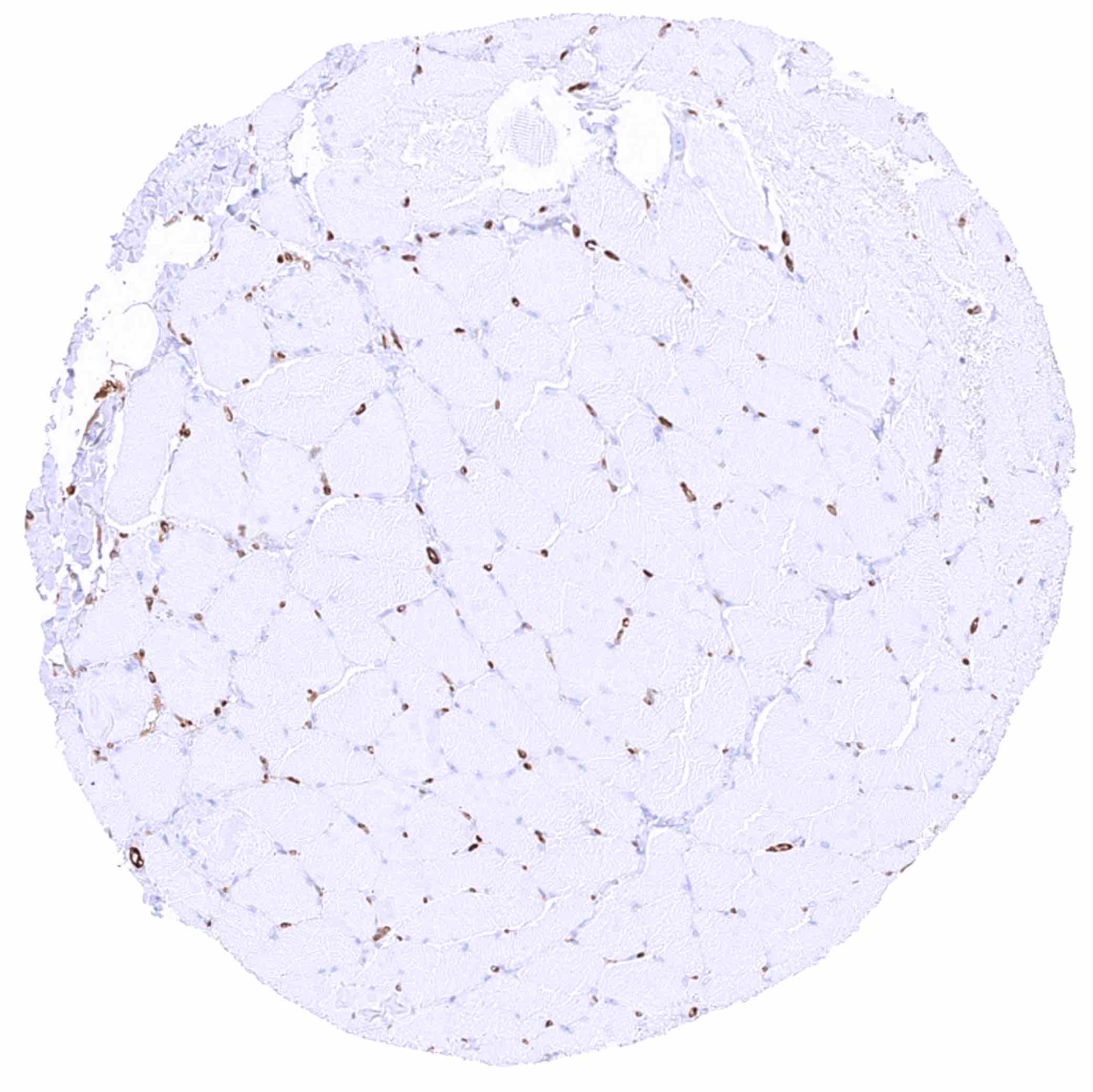
Skeletal muscle – MX1 staining prdominates in the endothelium of small vessels
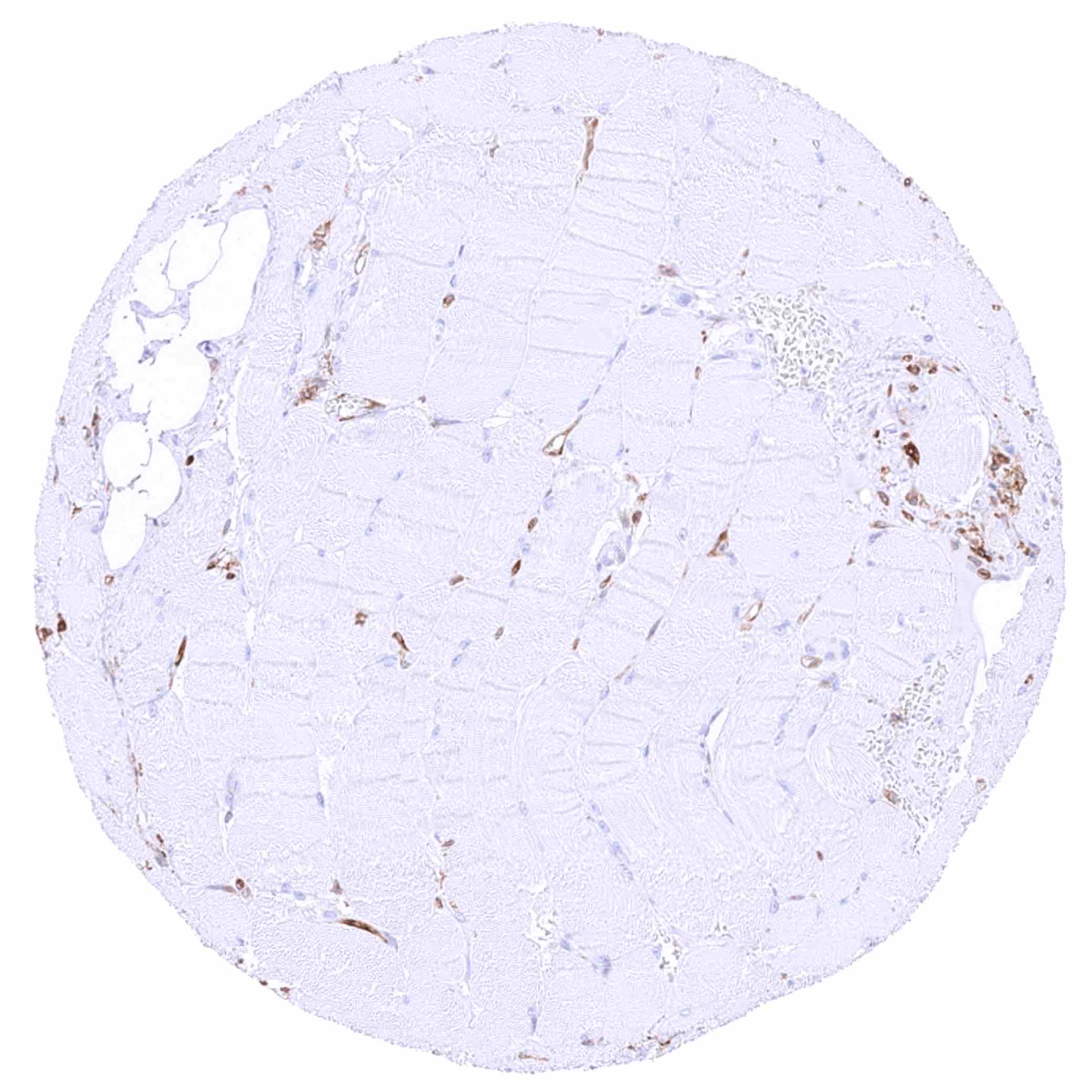
Skeletal muscle.jpeg
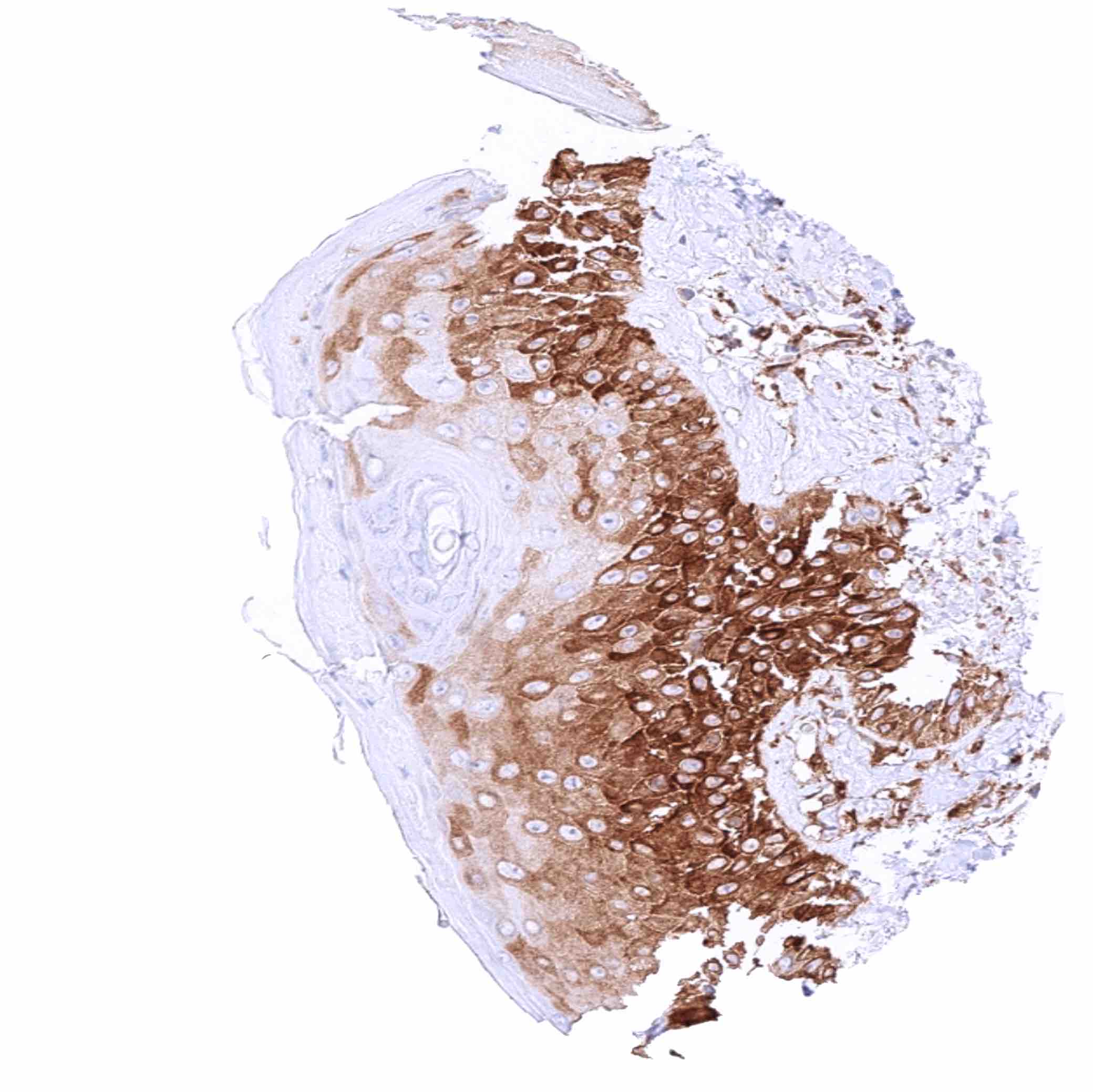
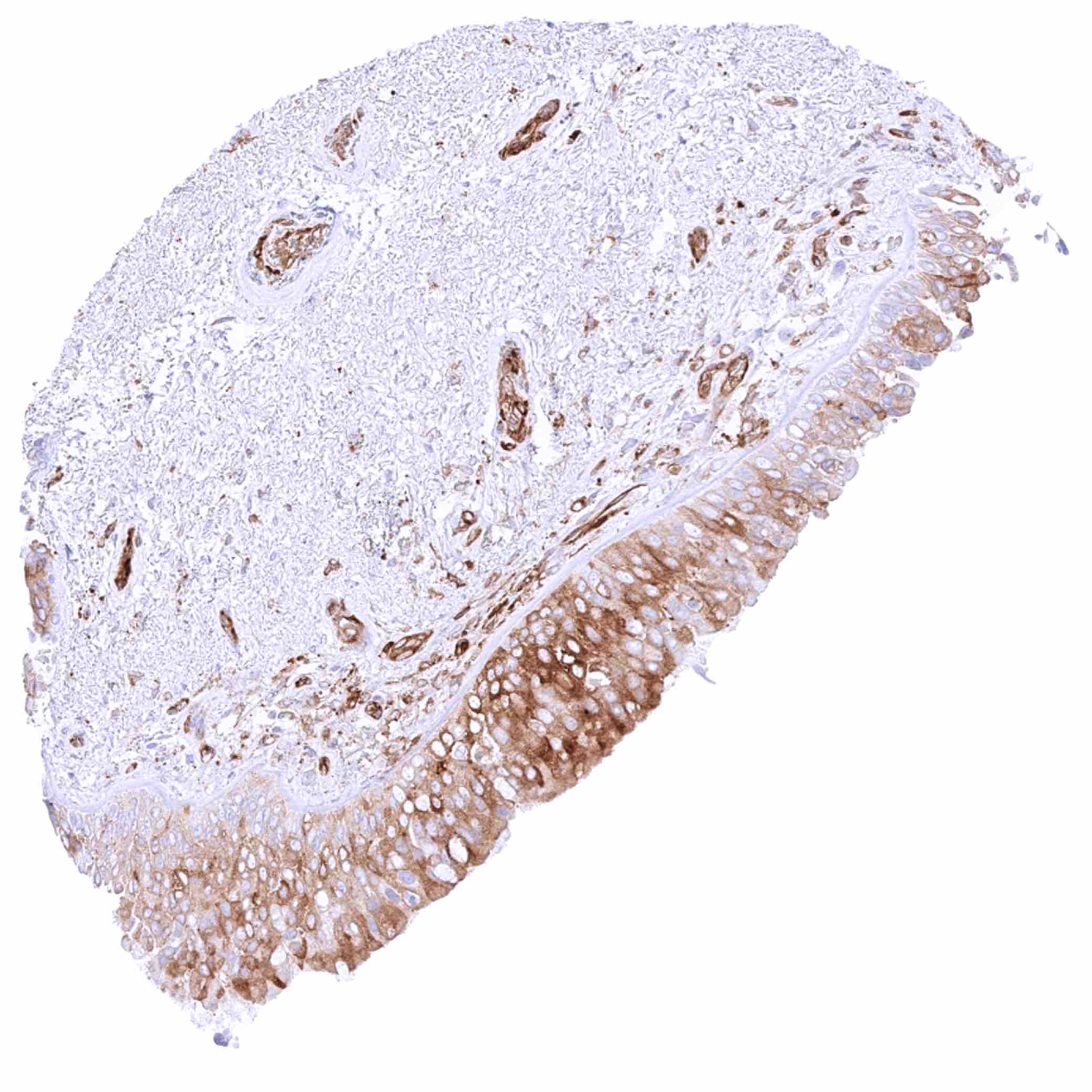
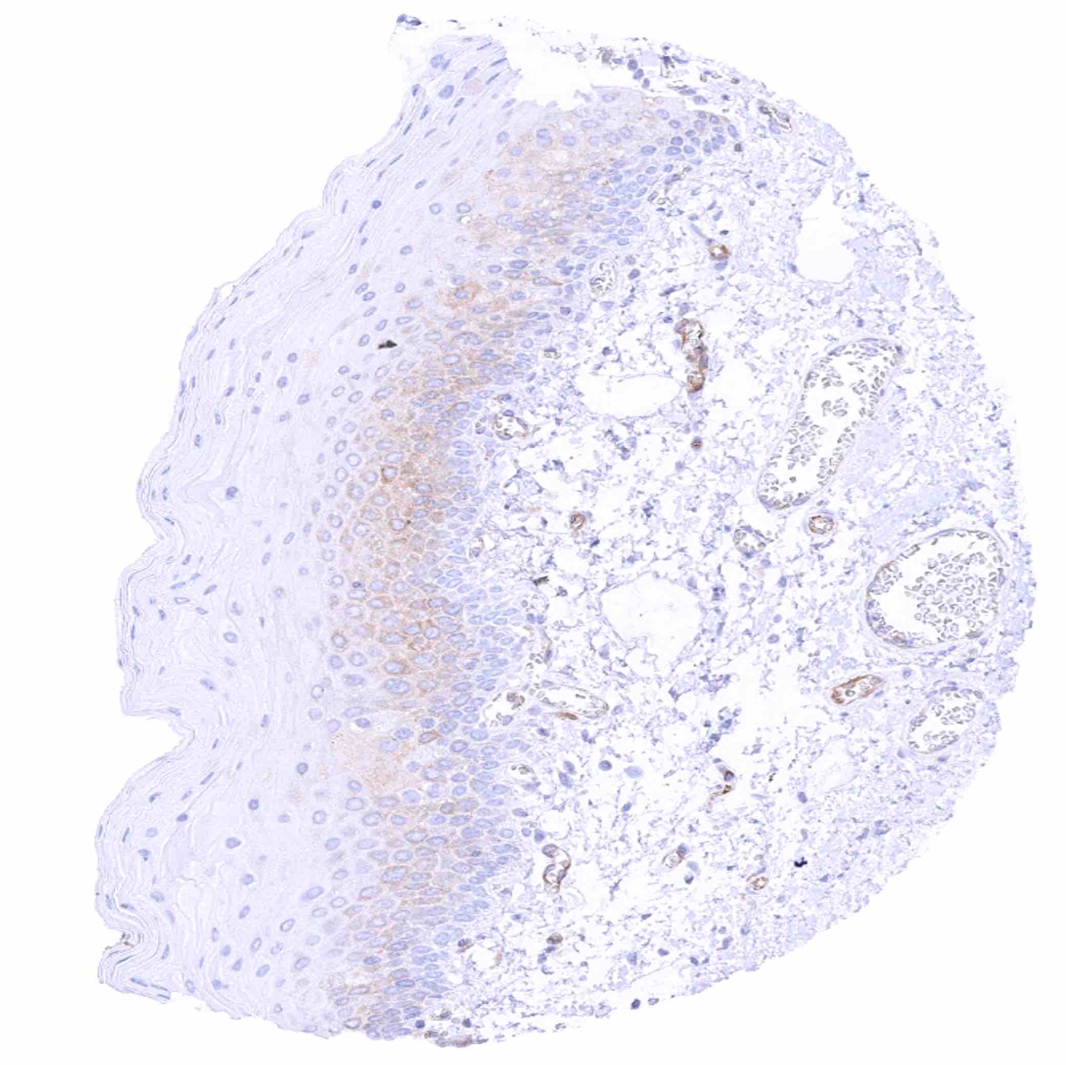
Skin – Strong cytoplasmic MX1 positivity of bottom half of epithelial cells in this sample
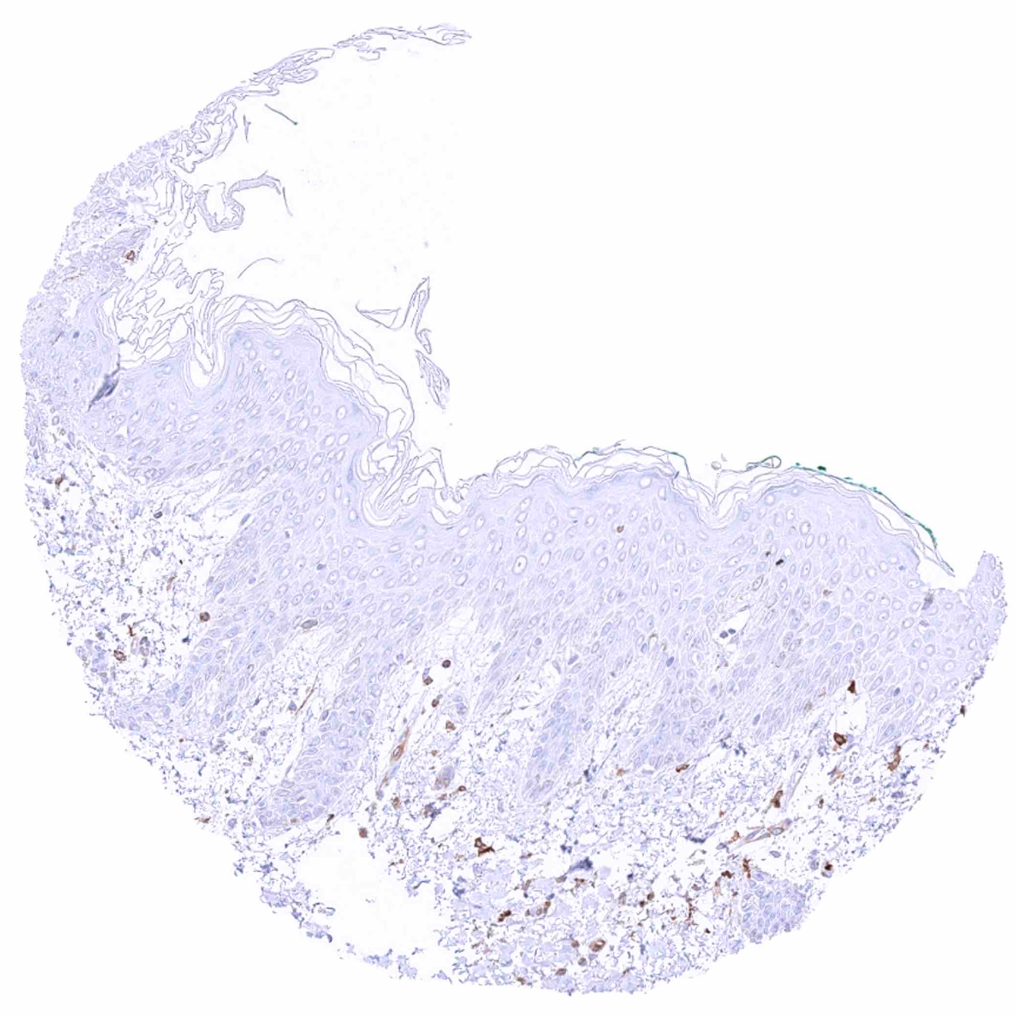
Skin
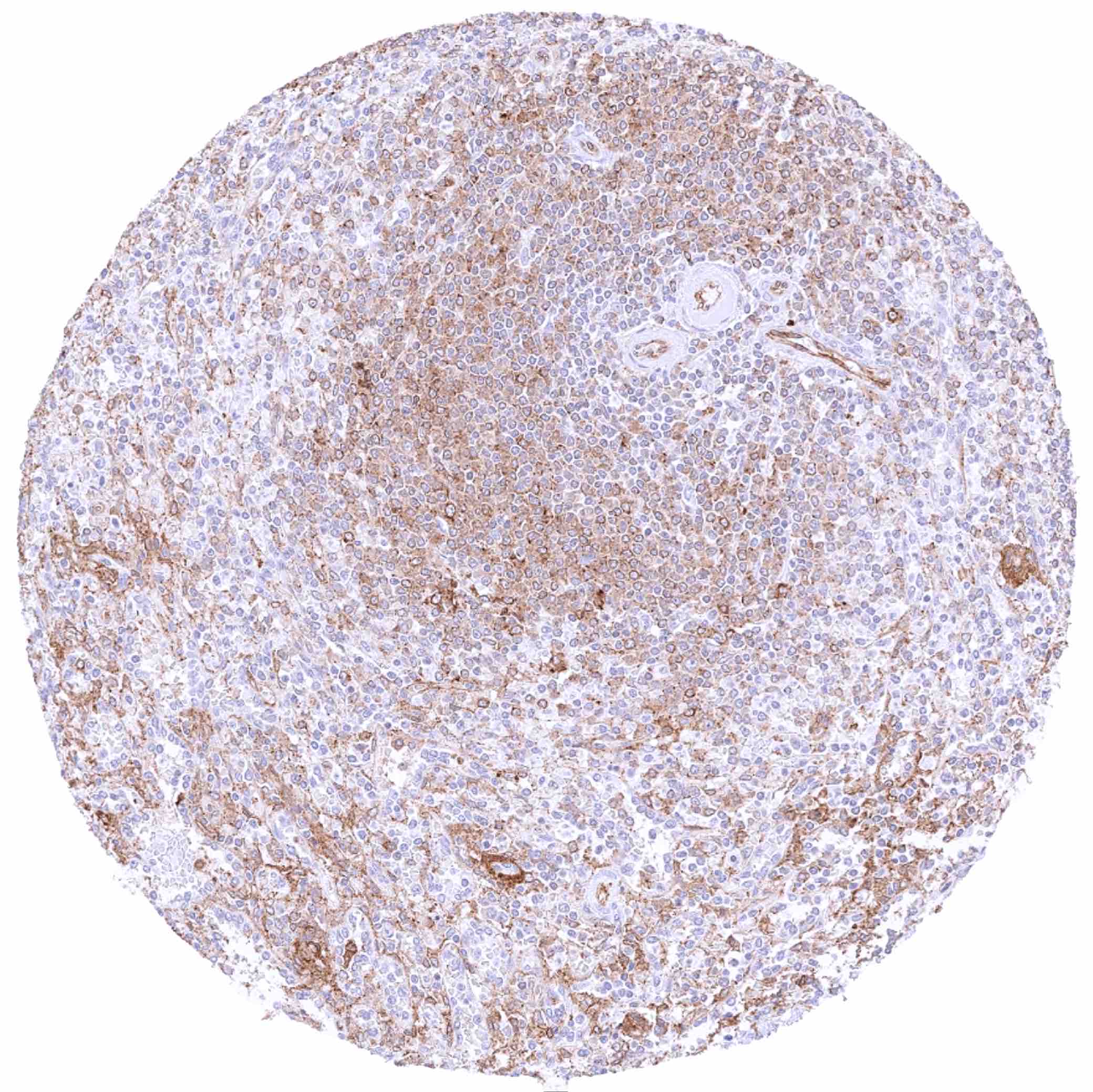
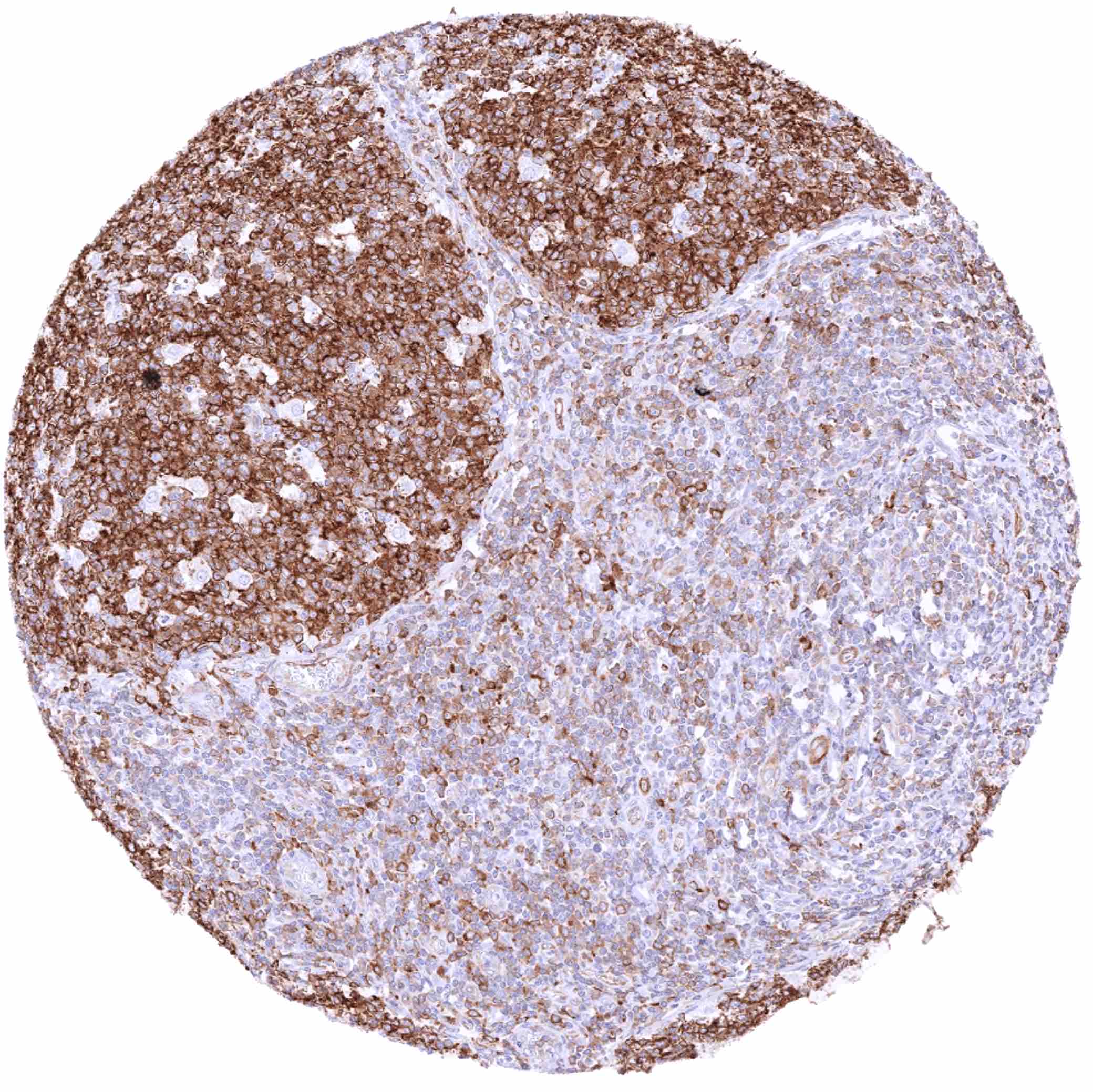
Spleen – Cytoplasmic MX1 staining at variable intensity of a large fraction of cells
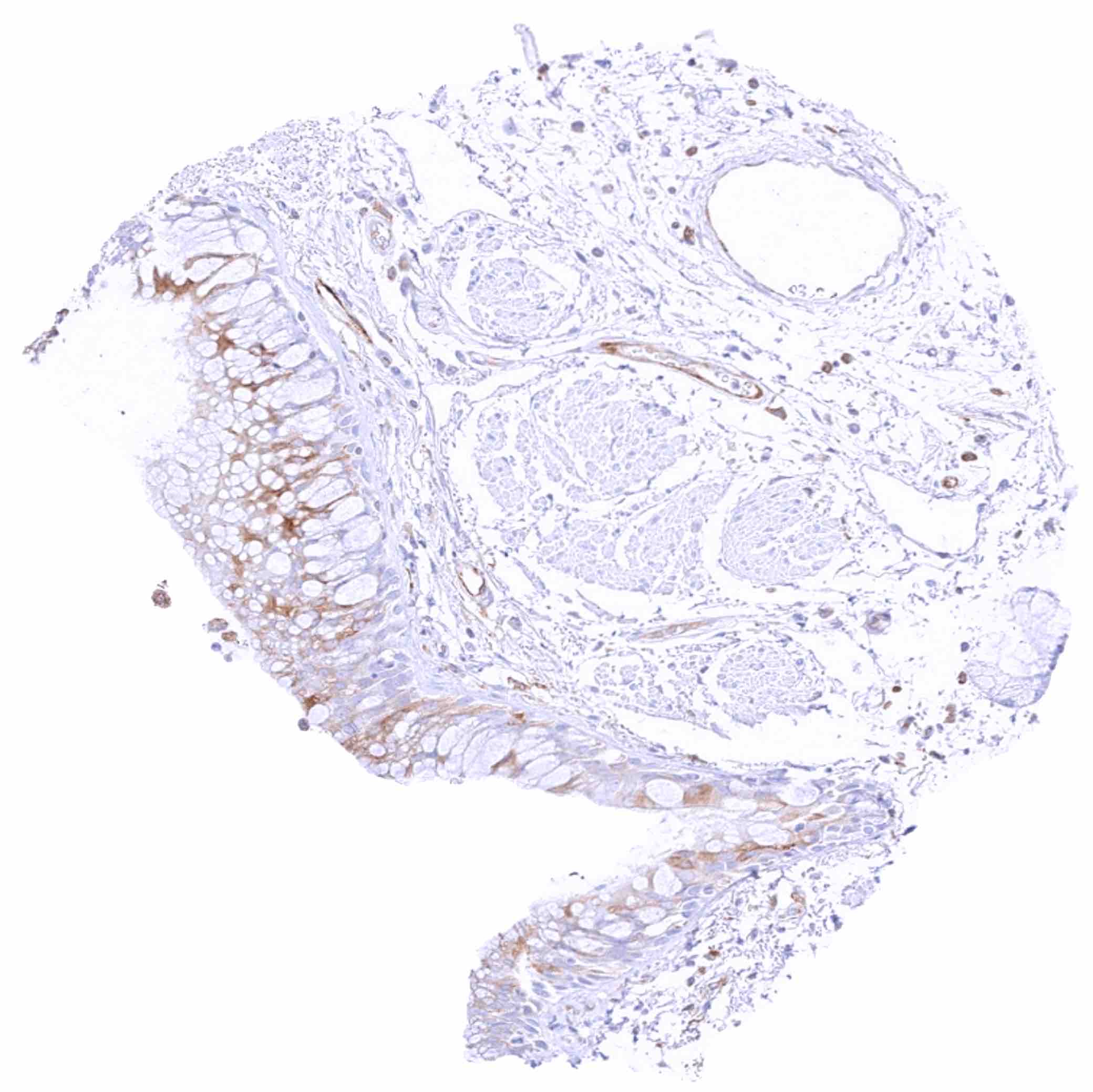
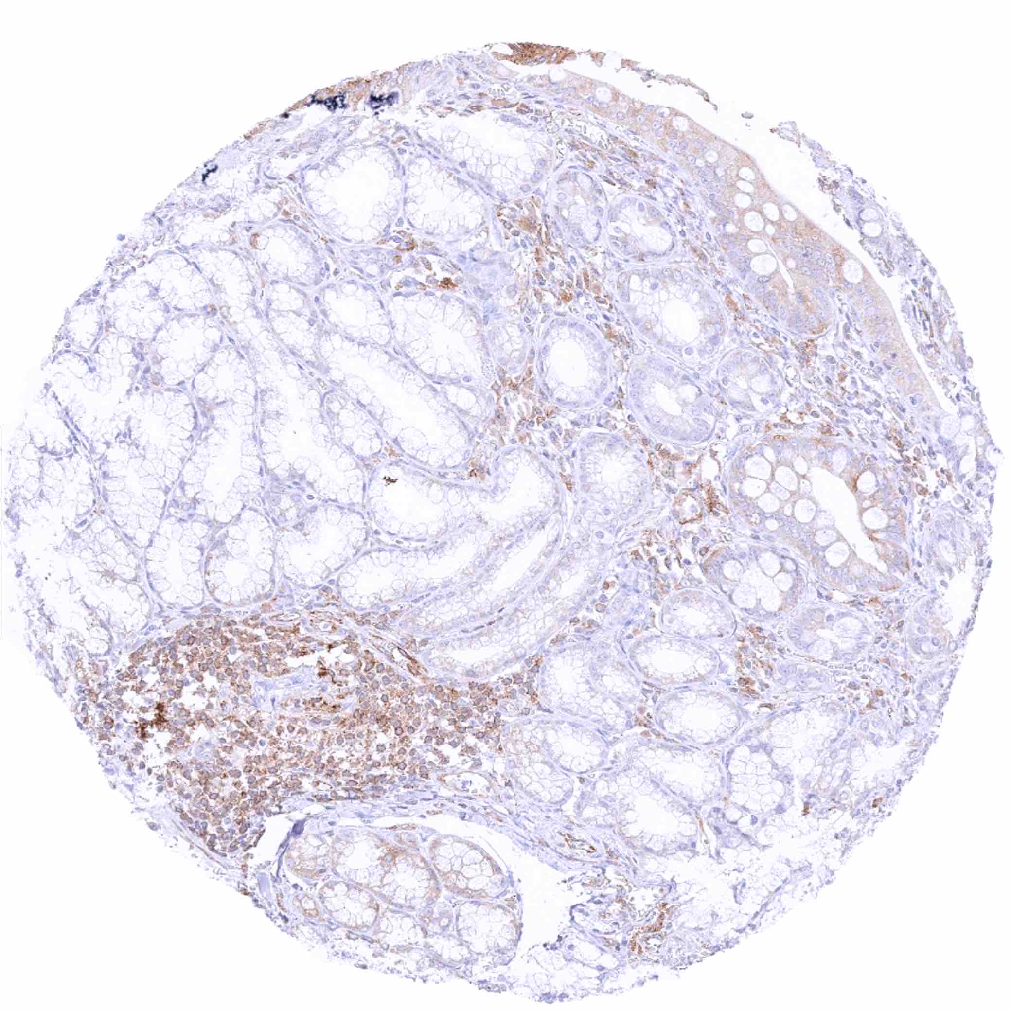
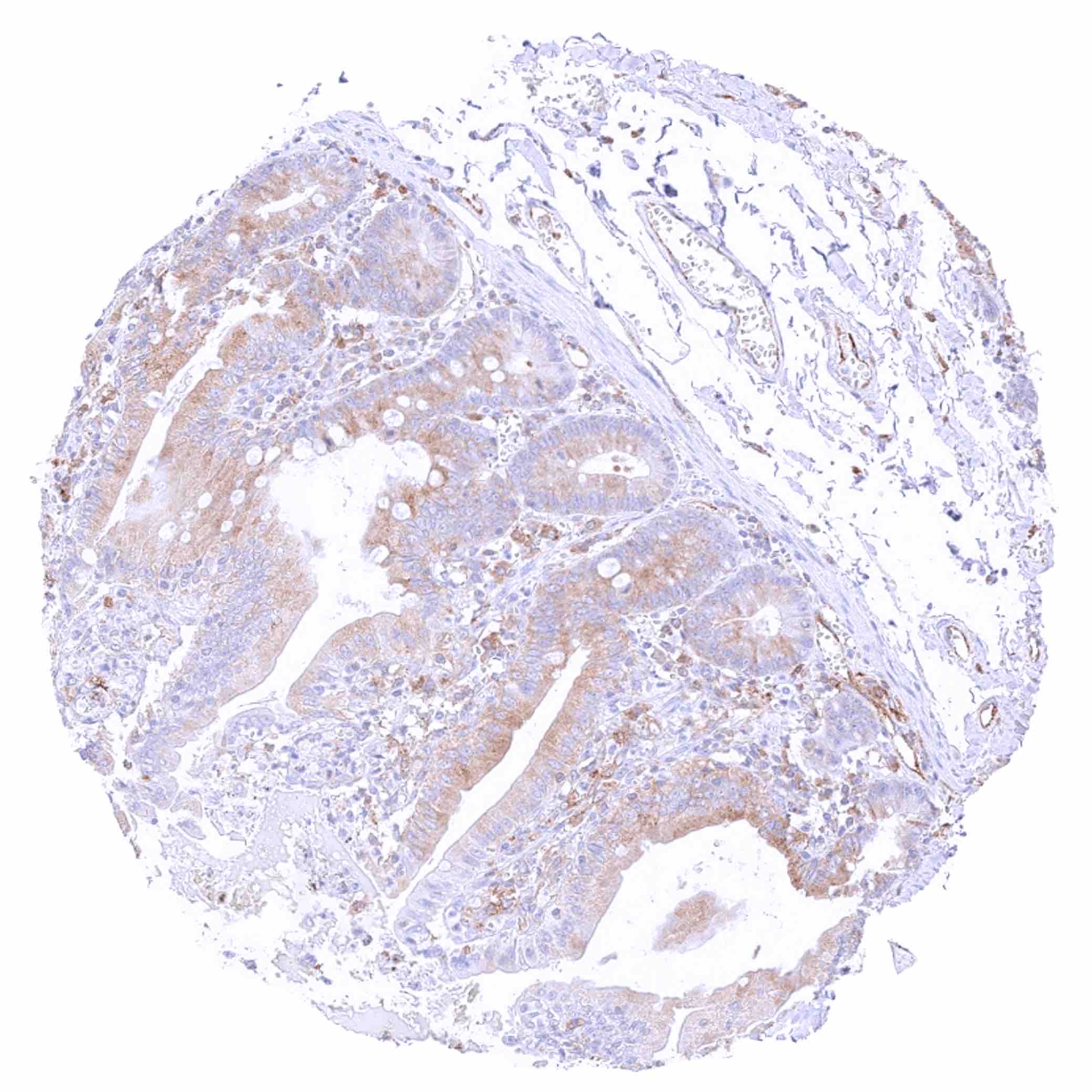
Stomach, antrum – Some cytoplasmic MX1 staining of epithelial cells, mainly of the surface epithelium
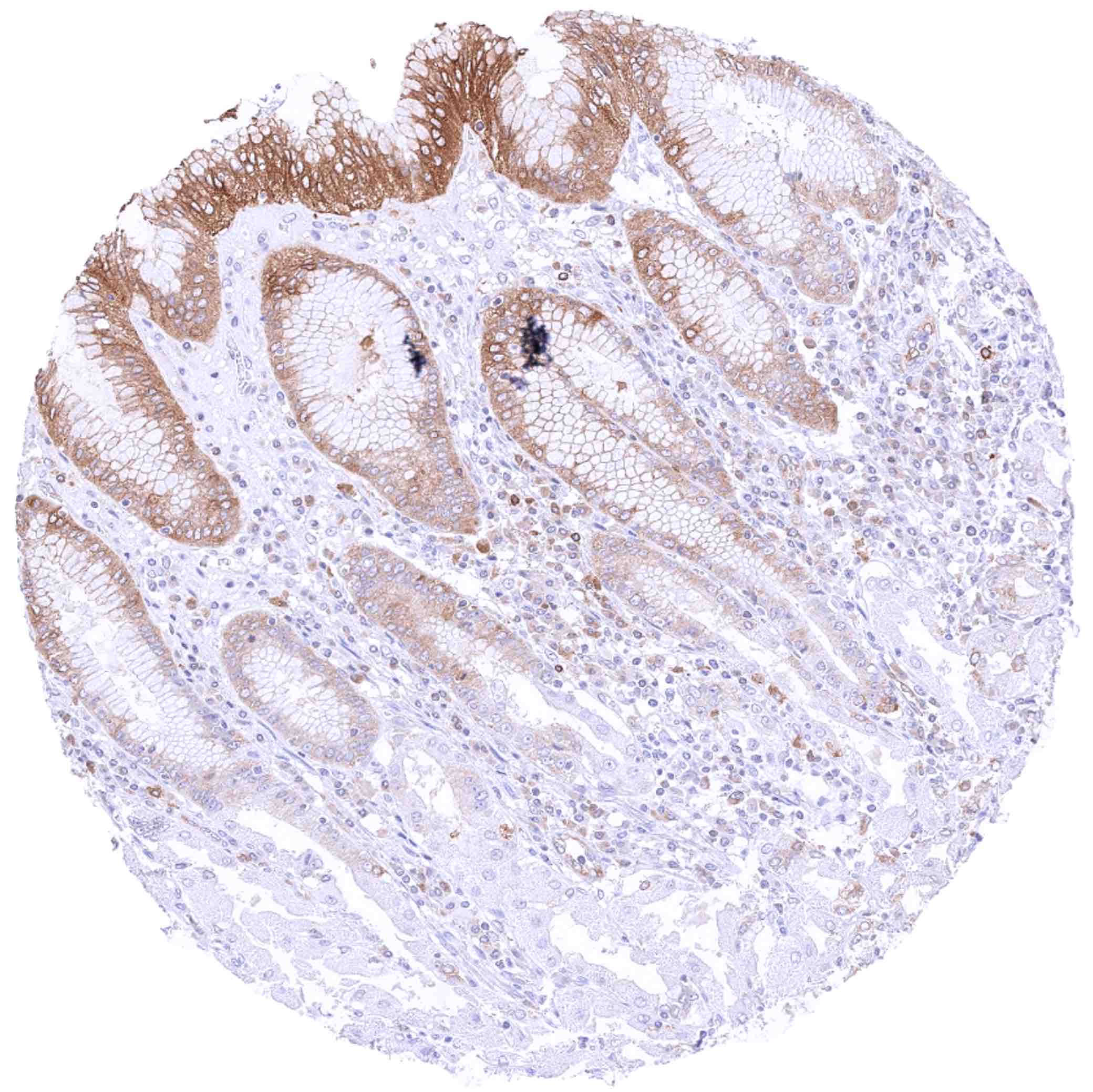
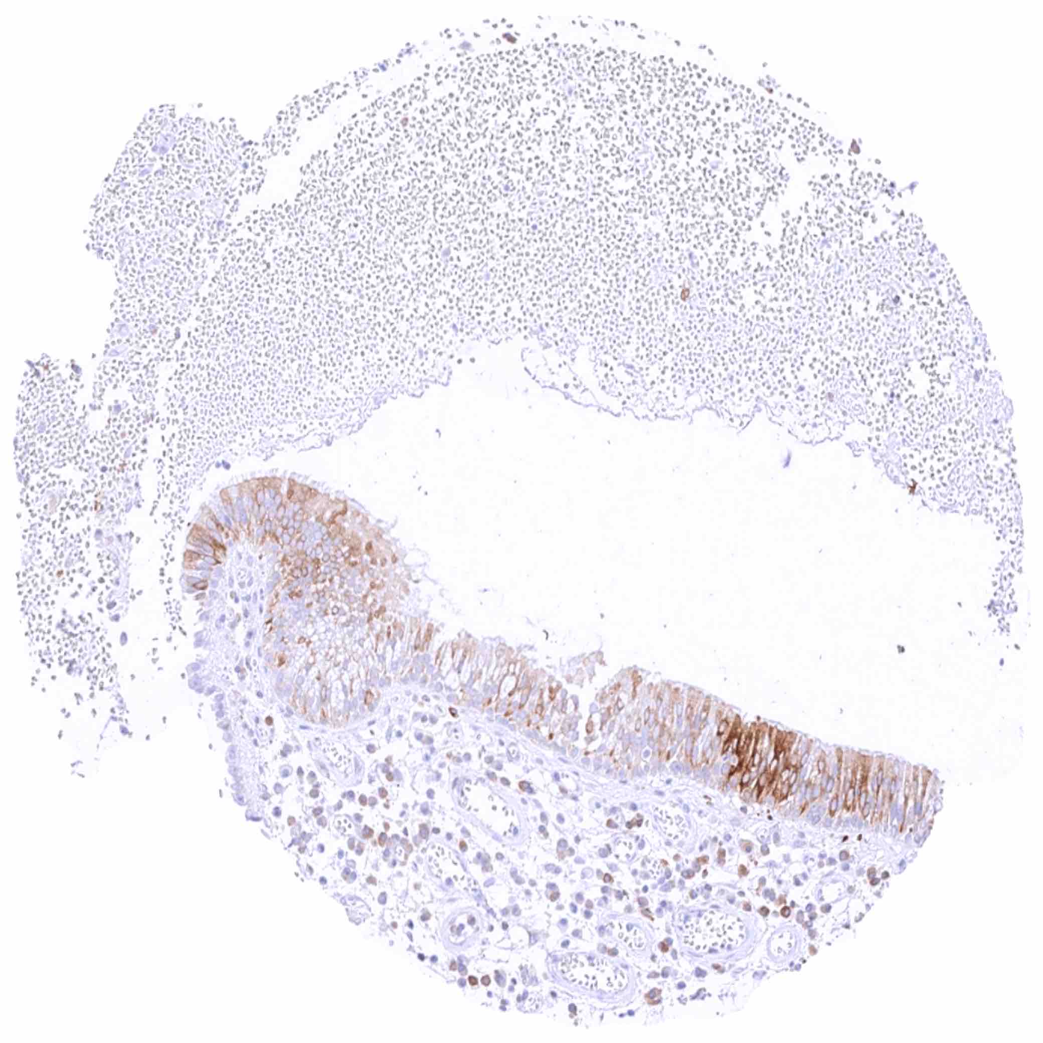
Stomach, corpus – Moderate to strong cytoplasmic MX1 staining of surface epithelial cells in a case with chronic inflammation
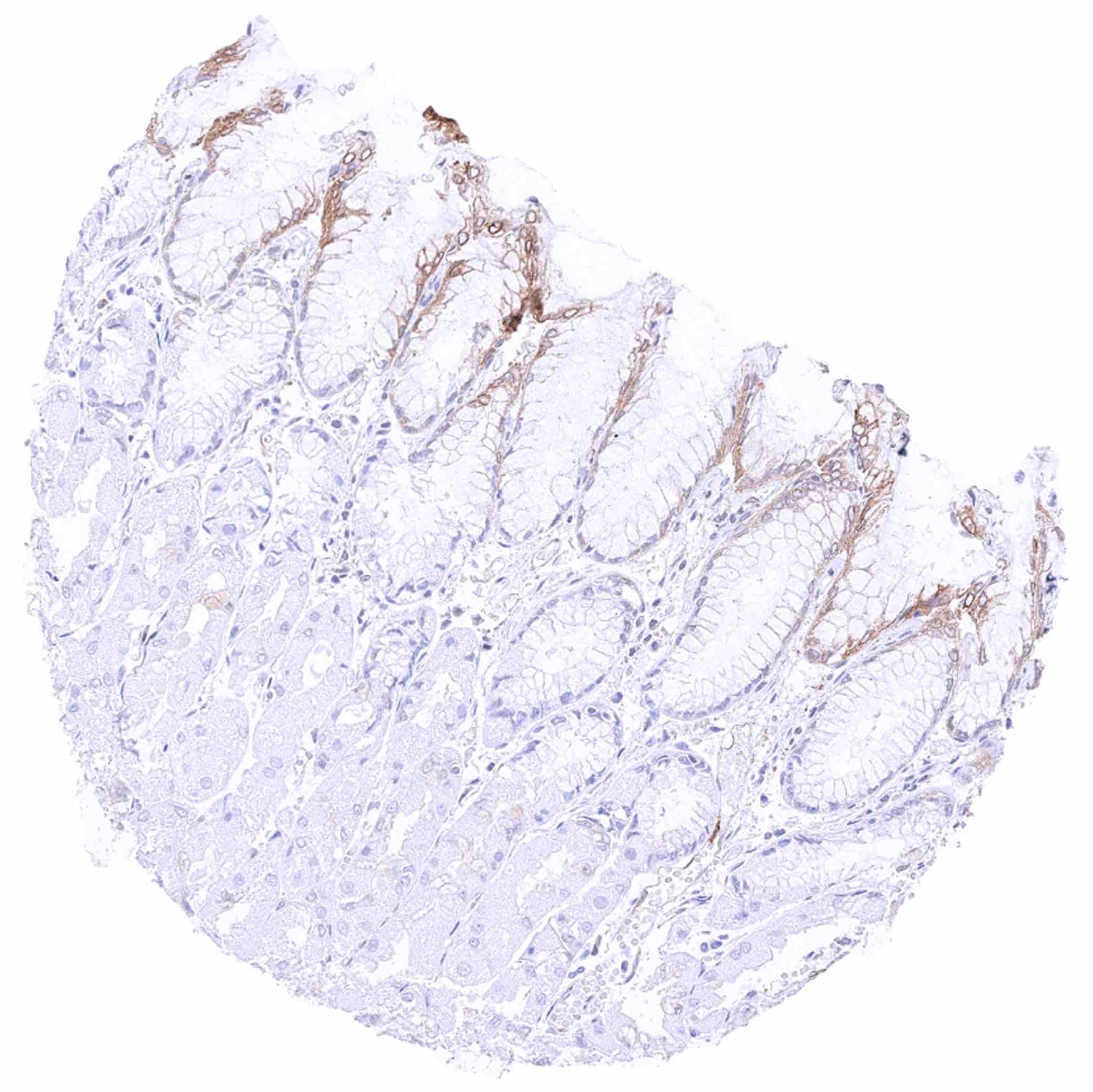
Stomach, corpus – Weak to moderate cytoplasmic MX1 staining of surface epithelial cells
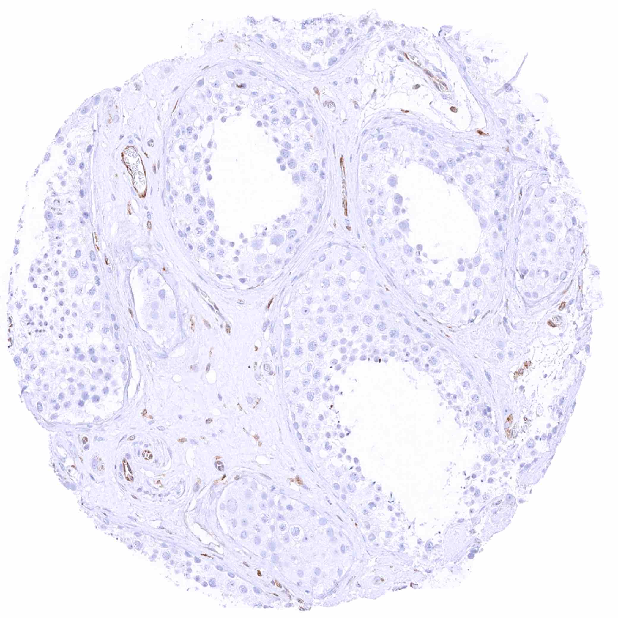
Testis – Cytoplasmic MX1 staining is restricted to endothelial cells.jpeg
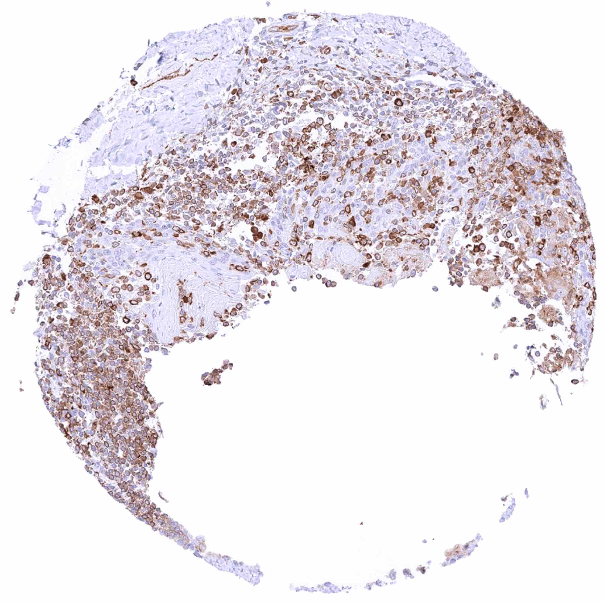
Thymus – Cytoplasmic MX1 staining at variable intensity of a large fraction of cells
Thyroid gland – Weak to moderate cytoplasmic MX1 staining of follicular cells
Tonsil – Cytoplasmic MX1 staining of lymphocytic cells is most intensive in germinal centres
Tonsil – Cytoplasmic MX1 staining of most lymphocytic cells at variable intensity while crypt epithelial cells are largely negative
Tonsil, surface epithelium – Abundant cytoplasmic MX1 staining of lymphocytic cells while epithelial cells remain negative
Tonsil, surface epithelium – Weak cytoplasmic MX1 staining of lowest third of epithelial cells in this sample
Urinary bladder, muscular wall – MX1 staining prdominates in the endothelium of small vessels
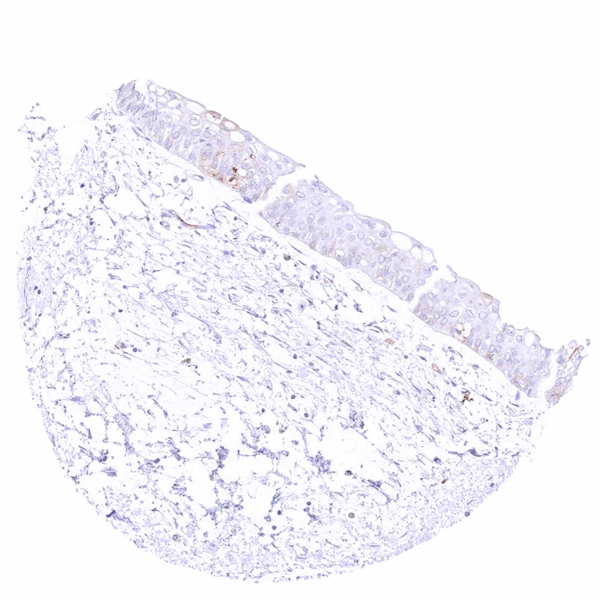
Urinary bladder, urothelium – Weak cytoplasmic MX1 staining of upper-half of urothelial cell layers
Urinary bladder, urothelium – Weak to moderate cytoplasmic MX1 staining of upper-half of urothelial cell layers
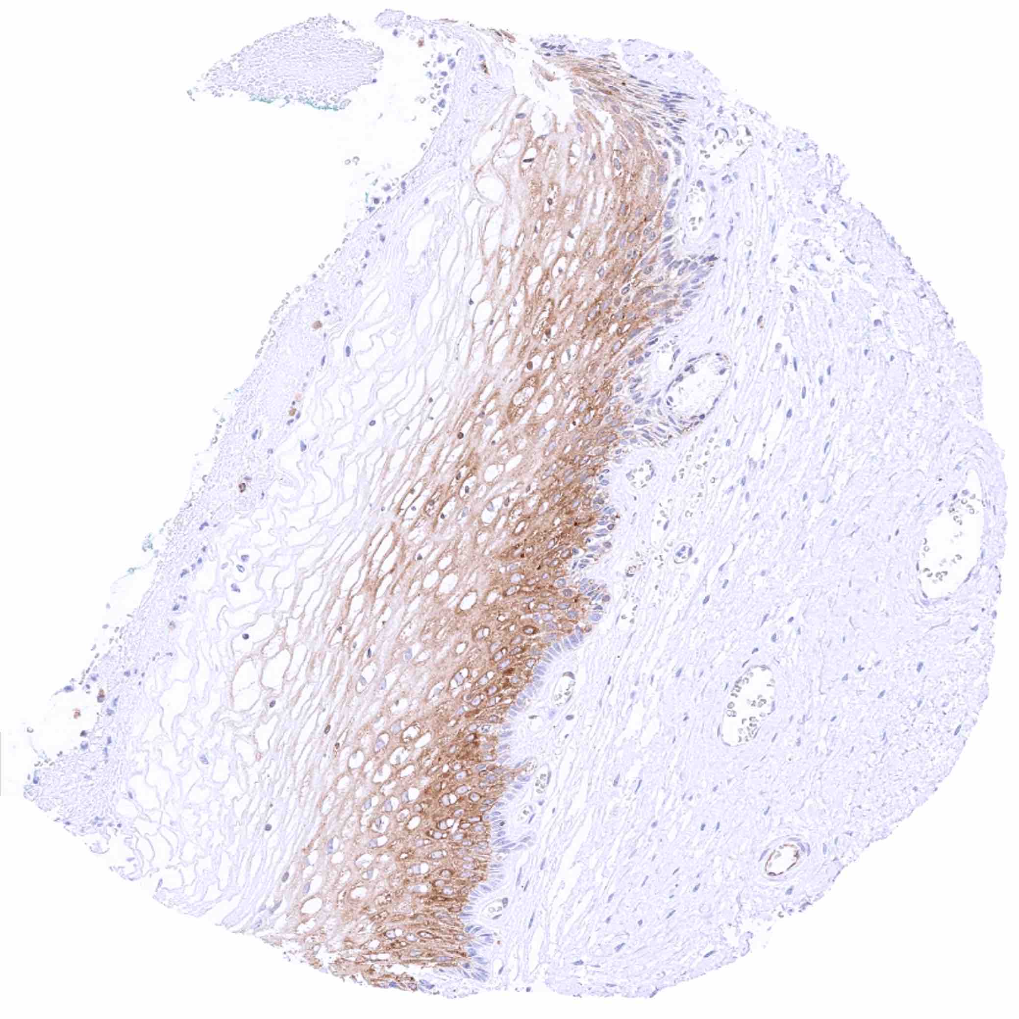
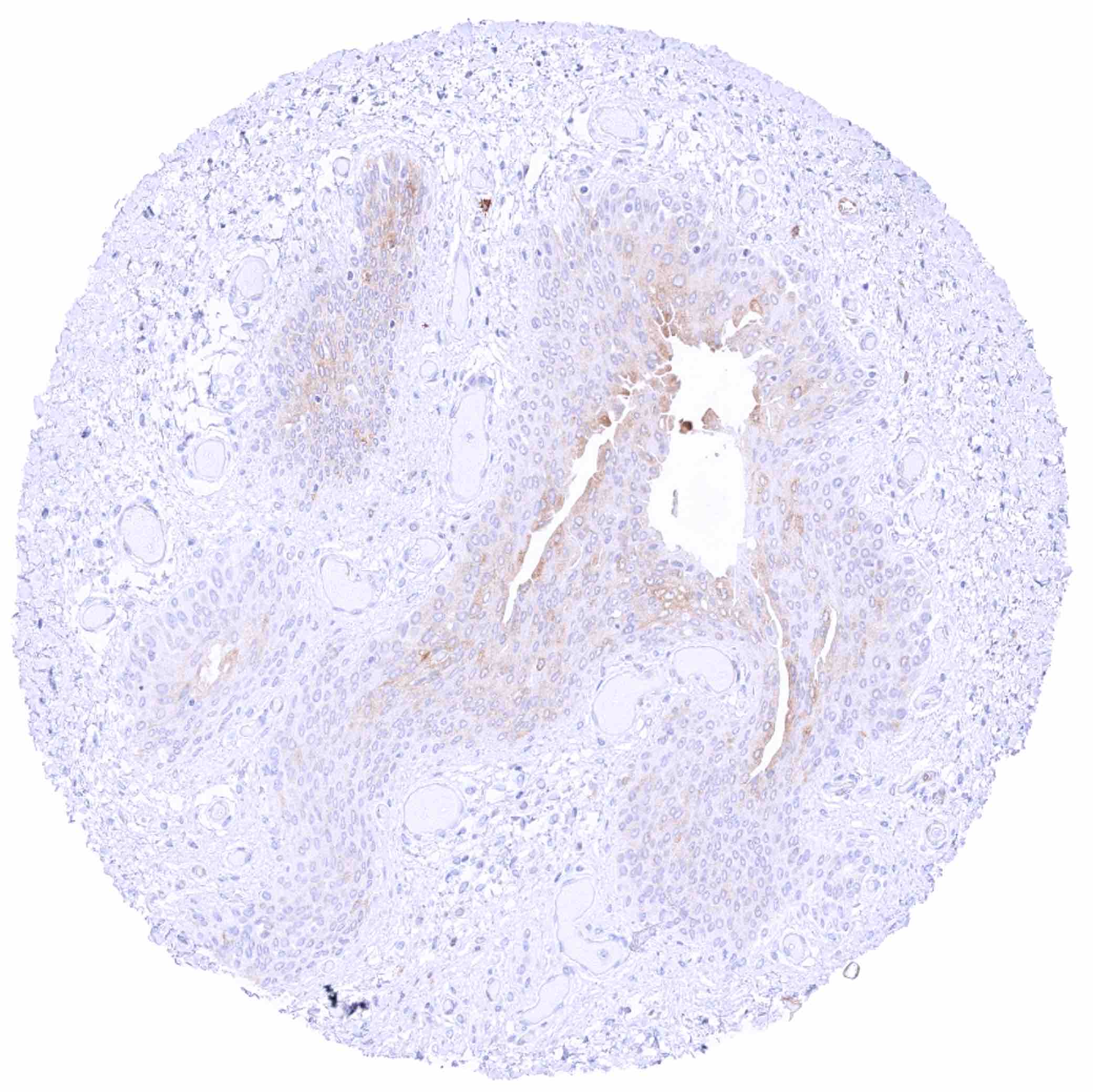
Uterus, ectocervix – Moderate cytoplasmic MX1 staining of suprabasal cell layers
Uterus, endocervix – Faint MX1 staining of some epithelial cells.jpeg
Uterus, endometrium (pregnancy) – Strong MX1 staining of endometrium cells. Moderate intensity MX1 staining of decidua cells
Uterus, endometrium (proliferation)
Uterus, endometrium (secretion)
Uterus, myometrium – MX1 staining prdominates in the endothelium of small vessels