Adrenal gland
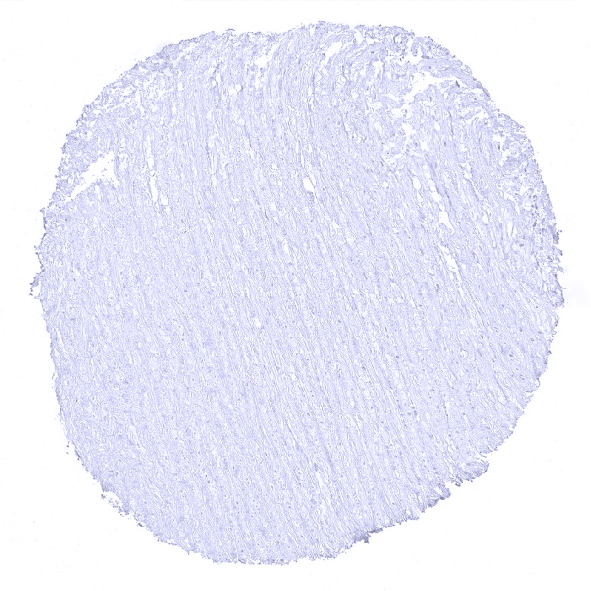
Aorta, media
Appendix, mucosa
Appendix, muscular wall
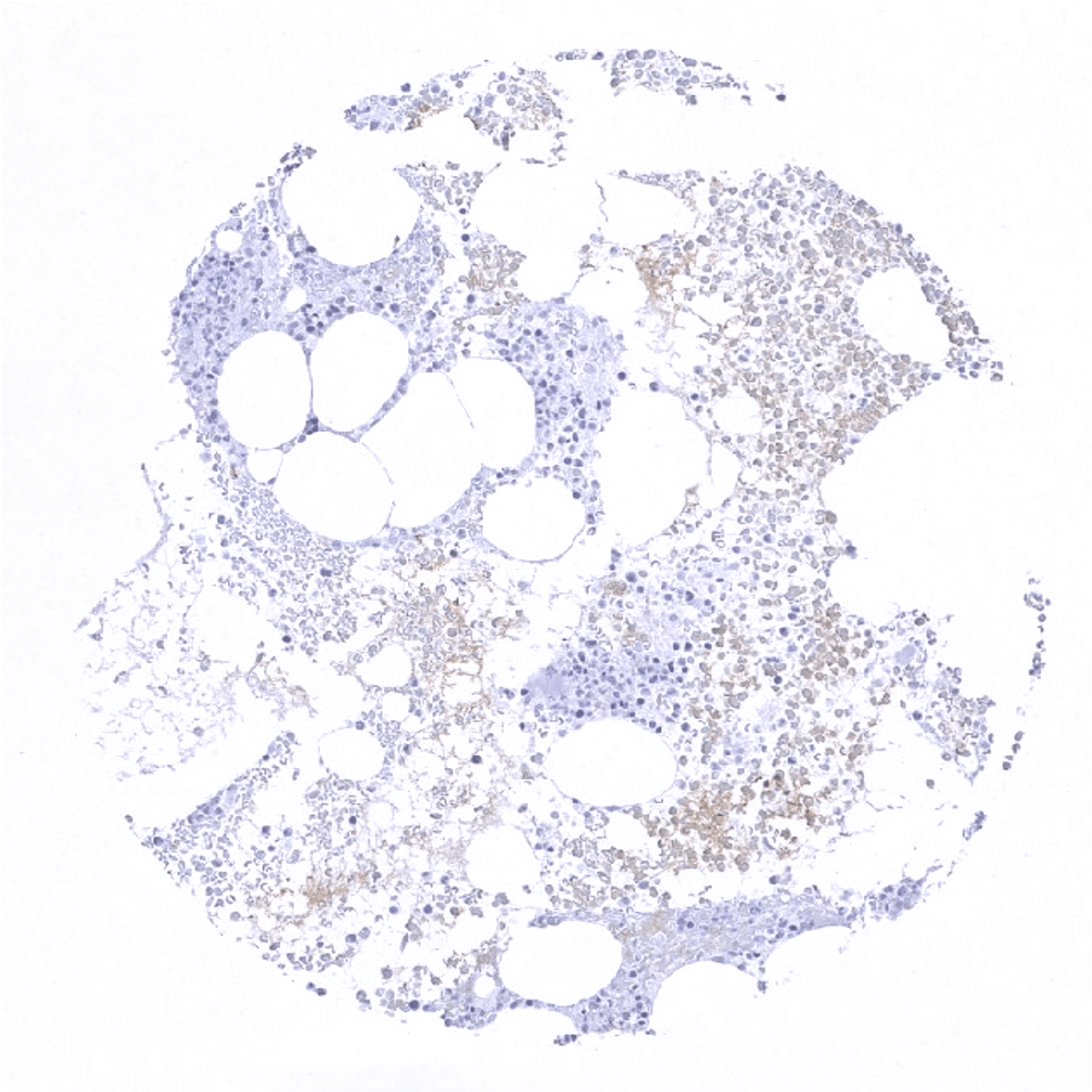
Bone marrow
Breast - Luminal cells show markedly stronger CK7 staining than basal cells.
Bronchus, mucosa - Strong CK7 immunostaining of all cell types except basal cells.
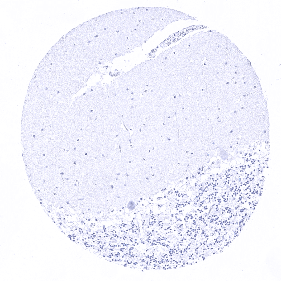
Cerebellum (molecular layer, Purkinje cell layer, granule cell layer)
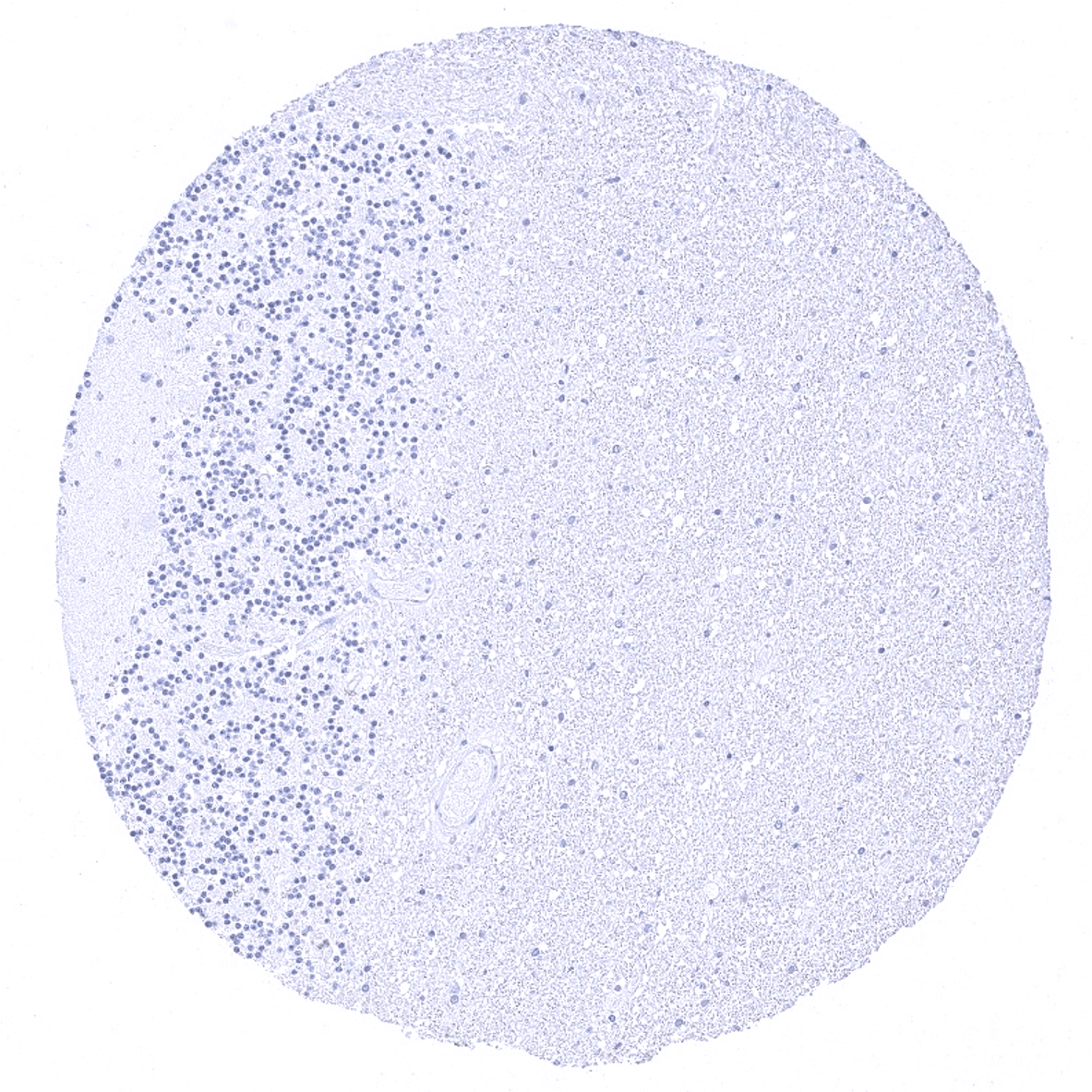
Cerebellum (granule cell layer, white matter)
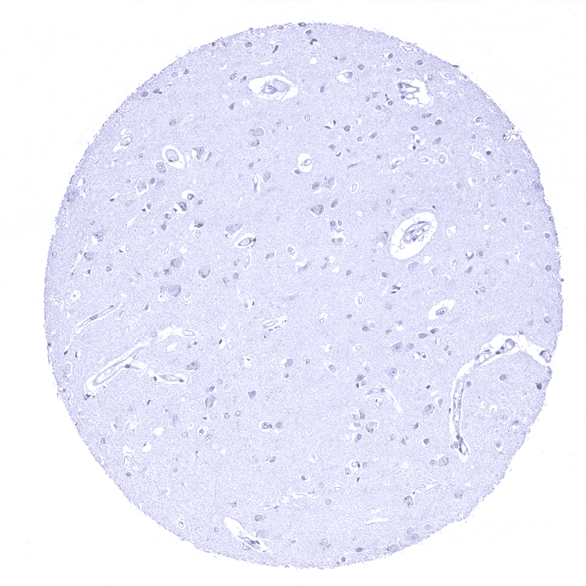
Cerebrum, grey matter

Cerebrum, grey matter - Some pigmented cells are seen in this sample. Absence of true CK7 immunostaining.

Cerebrum, white matter
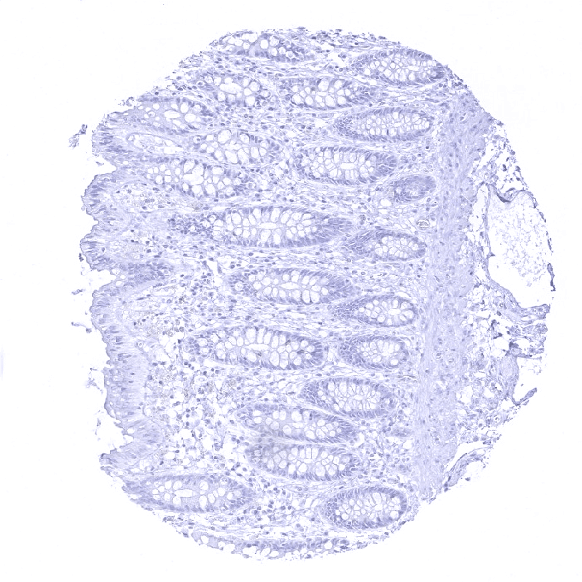
Colon descendens, mucosa
Colon descendens, muscular wall
Duodenum, Brunner gland
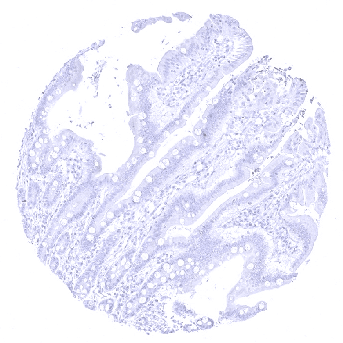
Duodenum, mucosa
Ectocervix
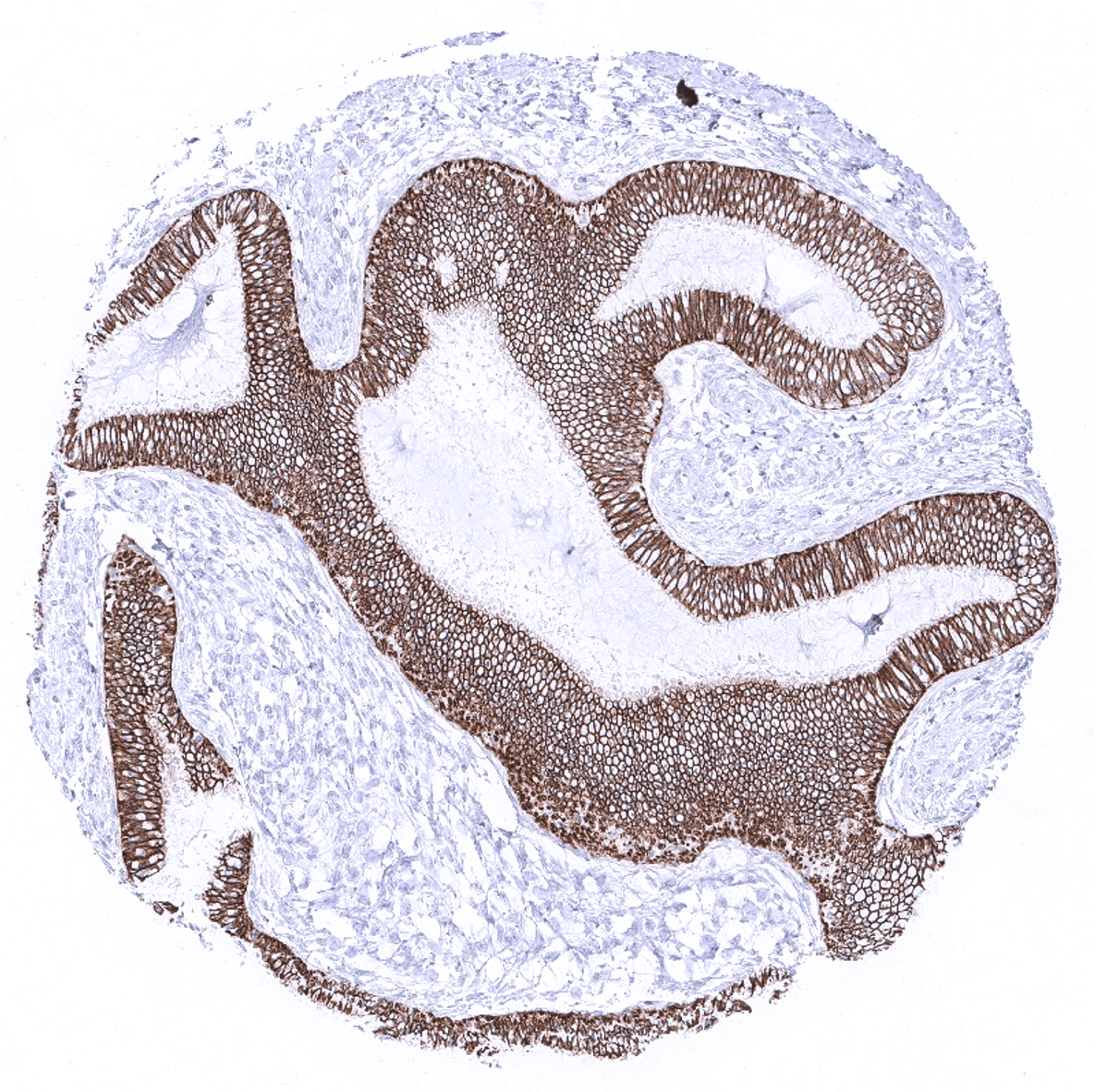
Endocervix
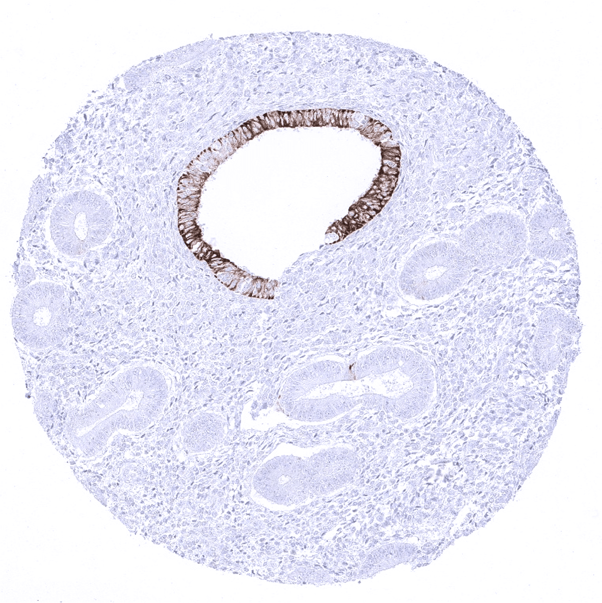
Endometrium, proliferation - The CK7 findings are variable in the endometrium: Not all glands show CK7 positivity.

Endometrium, secretion - The CK7 findings are variable in the endometrium. In this sample, all epithelial cells show a strong CK7 positivity.
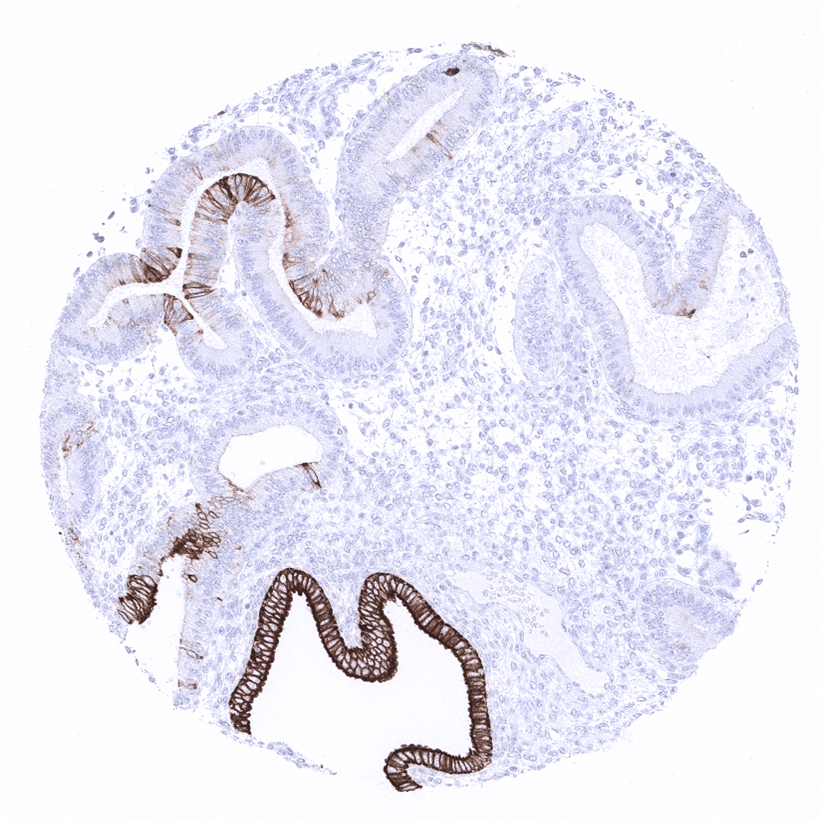
Endometrium, secretion - The CK7 findings are variable in the endometrium. In this sample, a fraction epithelial cells shows a strong CK7 positivity while others are completely negative.
Epididymis
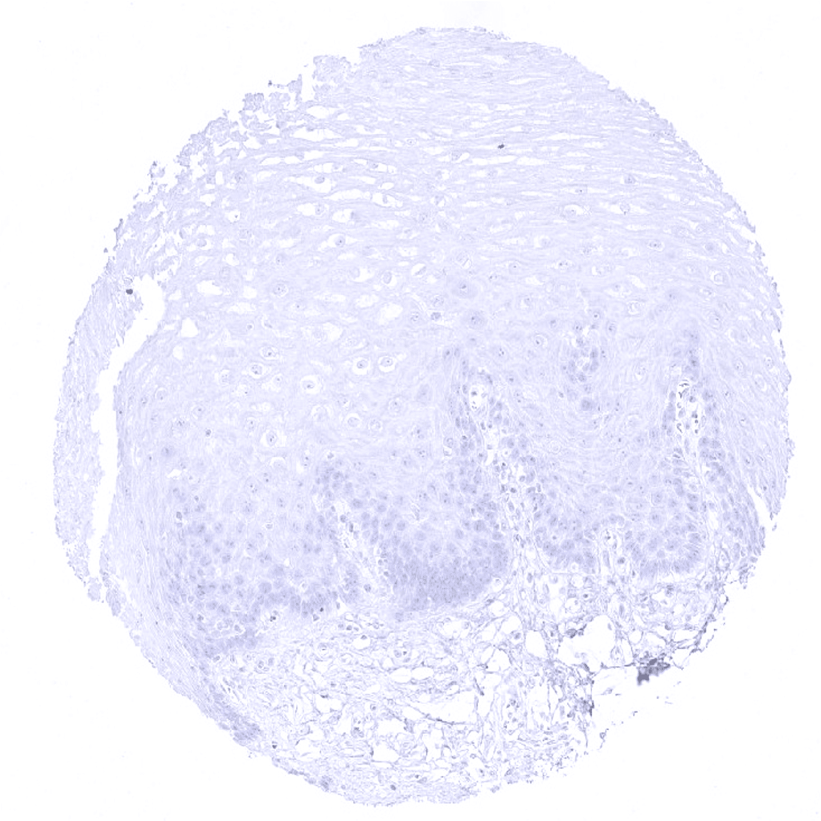
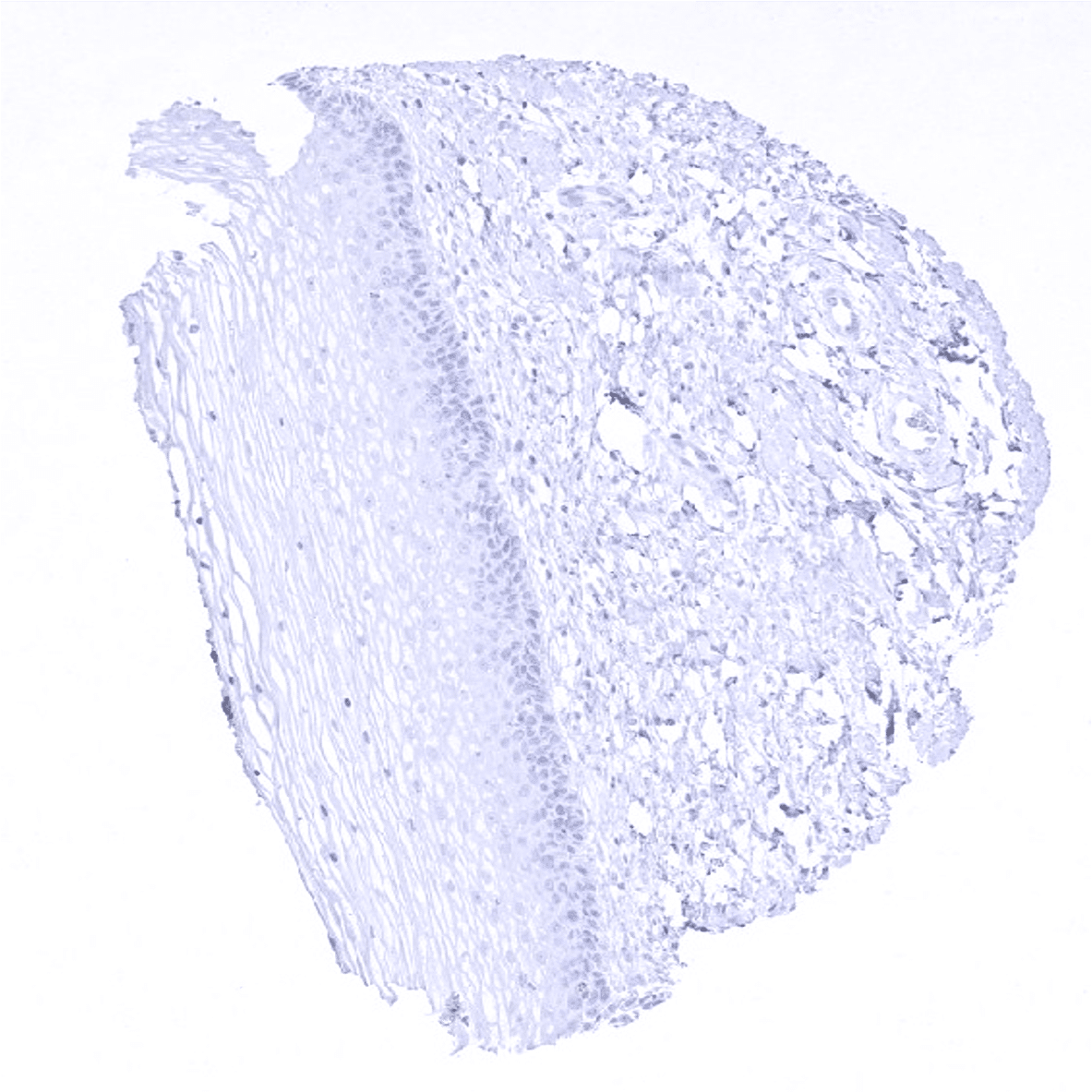
Esophagus, squamous epithelium

Esophagus, squamous epithelium
Fallopian tube, mucosa - All epithelial cells but ciliated cells stain CK7 positive in the fallopian tube.
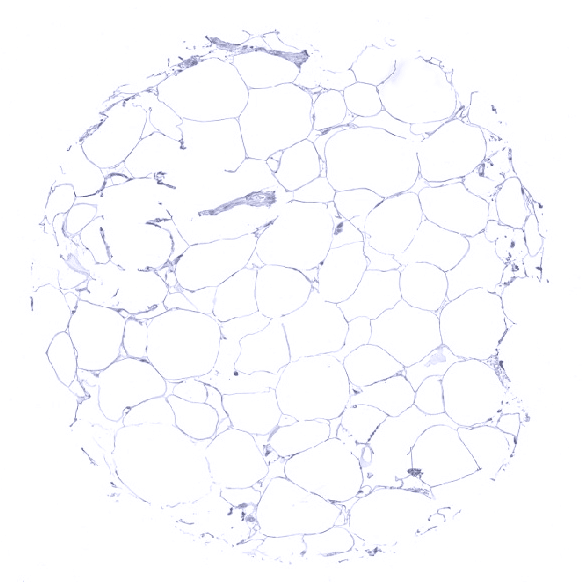
Fat
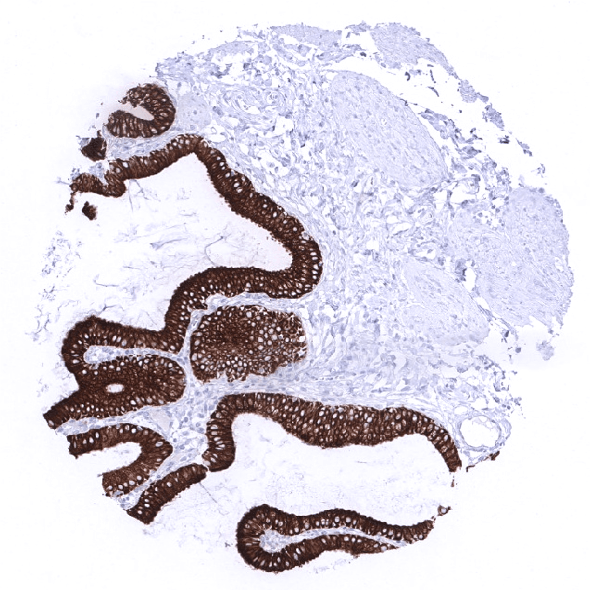
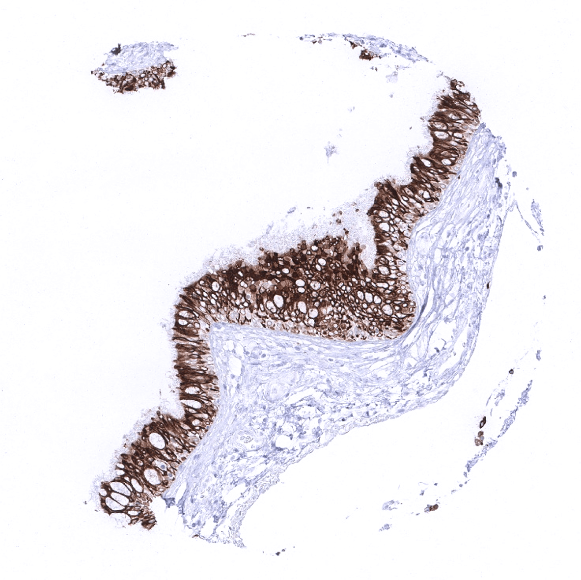
Gallbladder, epithelium

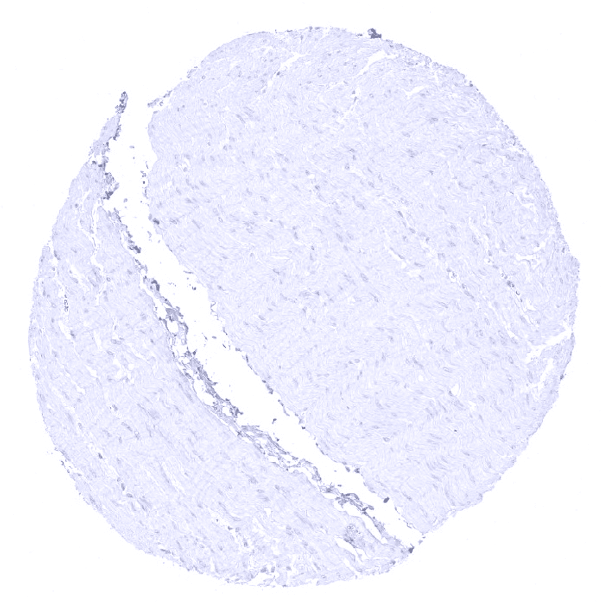
Heart
Ileum, mucosa
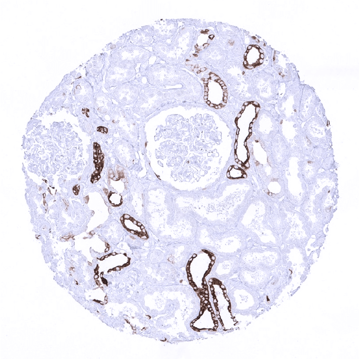
Kidney, cortex - CK 7 staining is seen in collecting ducts and in scattered epithelial cells of the visceral layer of the Bowman capsule.
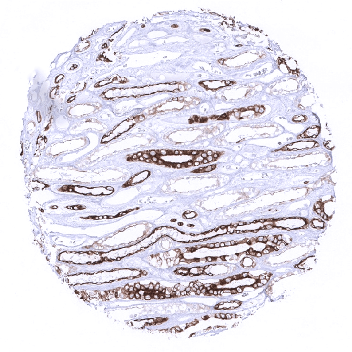
Kidney, medulla - Strong CK 7 staining is seen in collecting ducts.
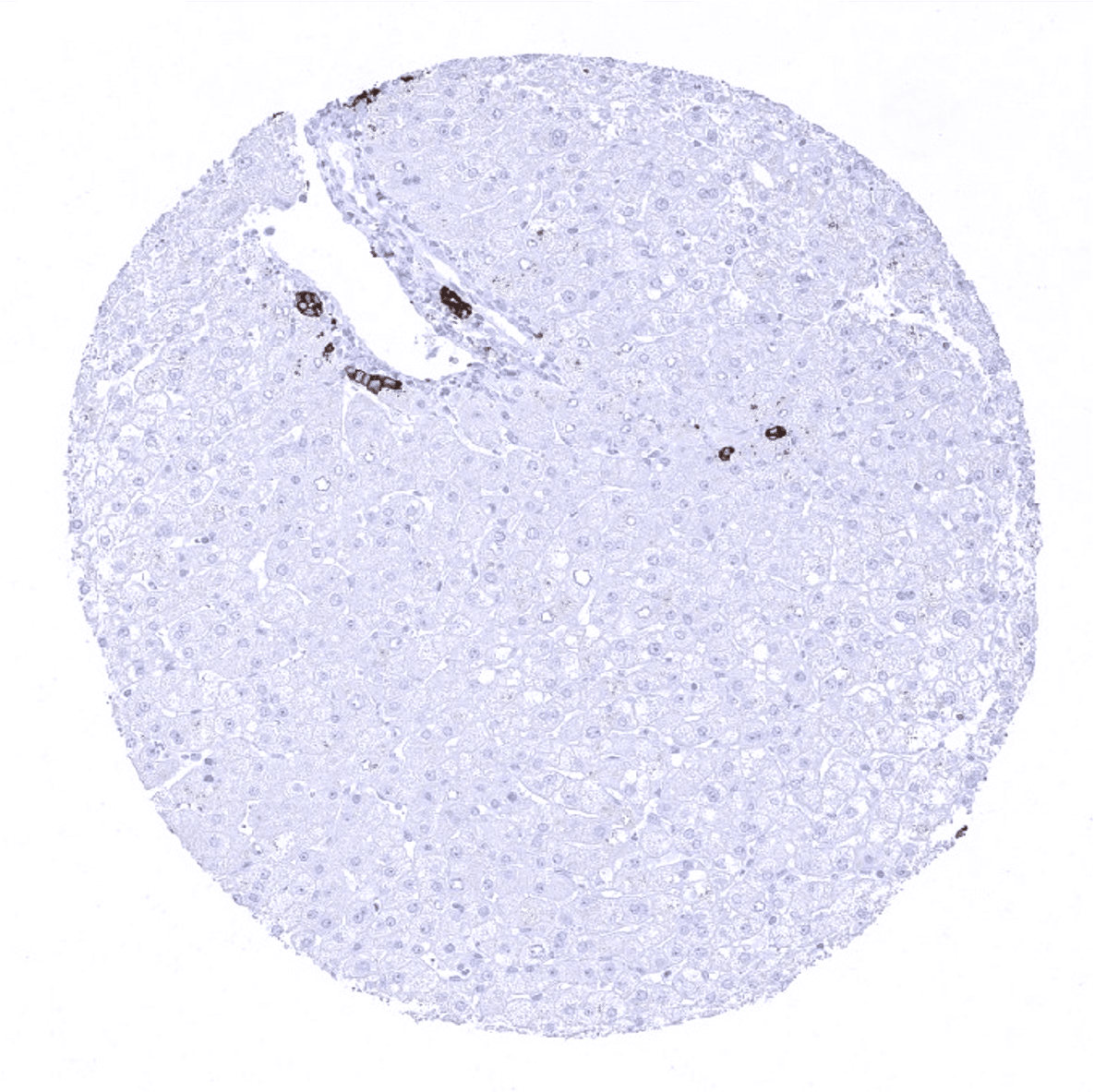
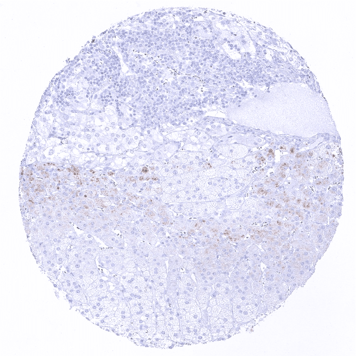
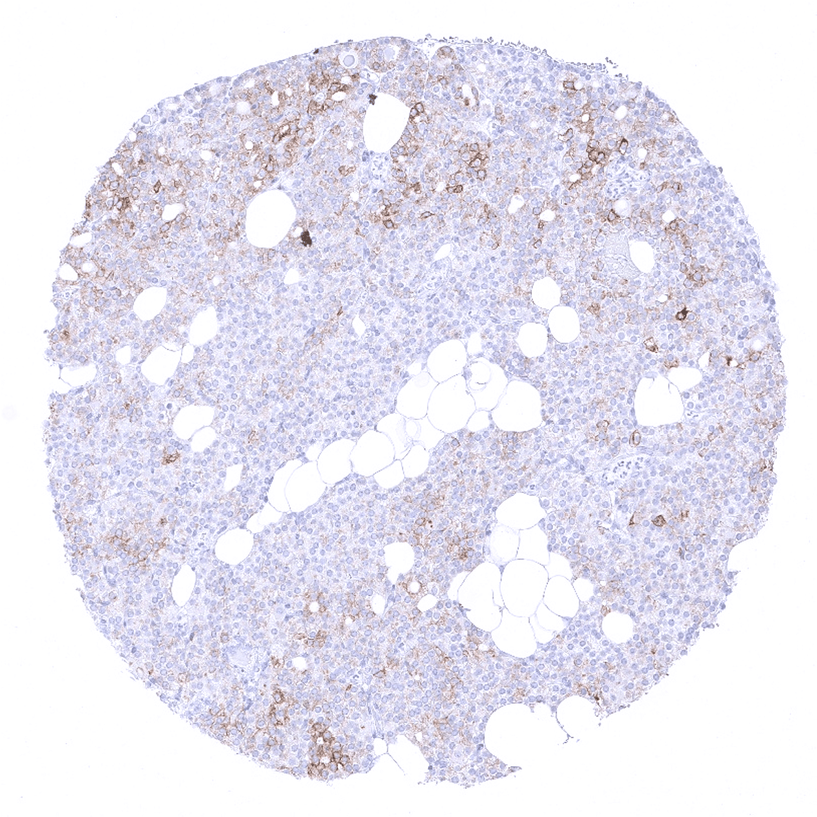
Liver
Lung - Moderate CK7 positivity of pneumocytes.
Lymph node
Ovary, stroma
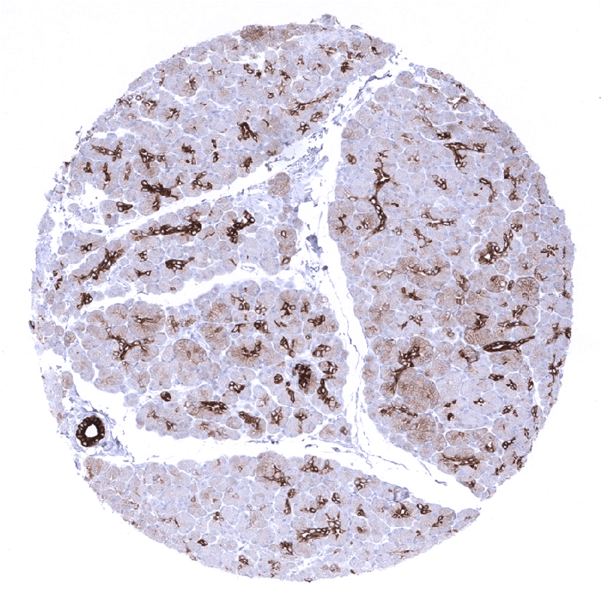
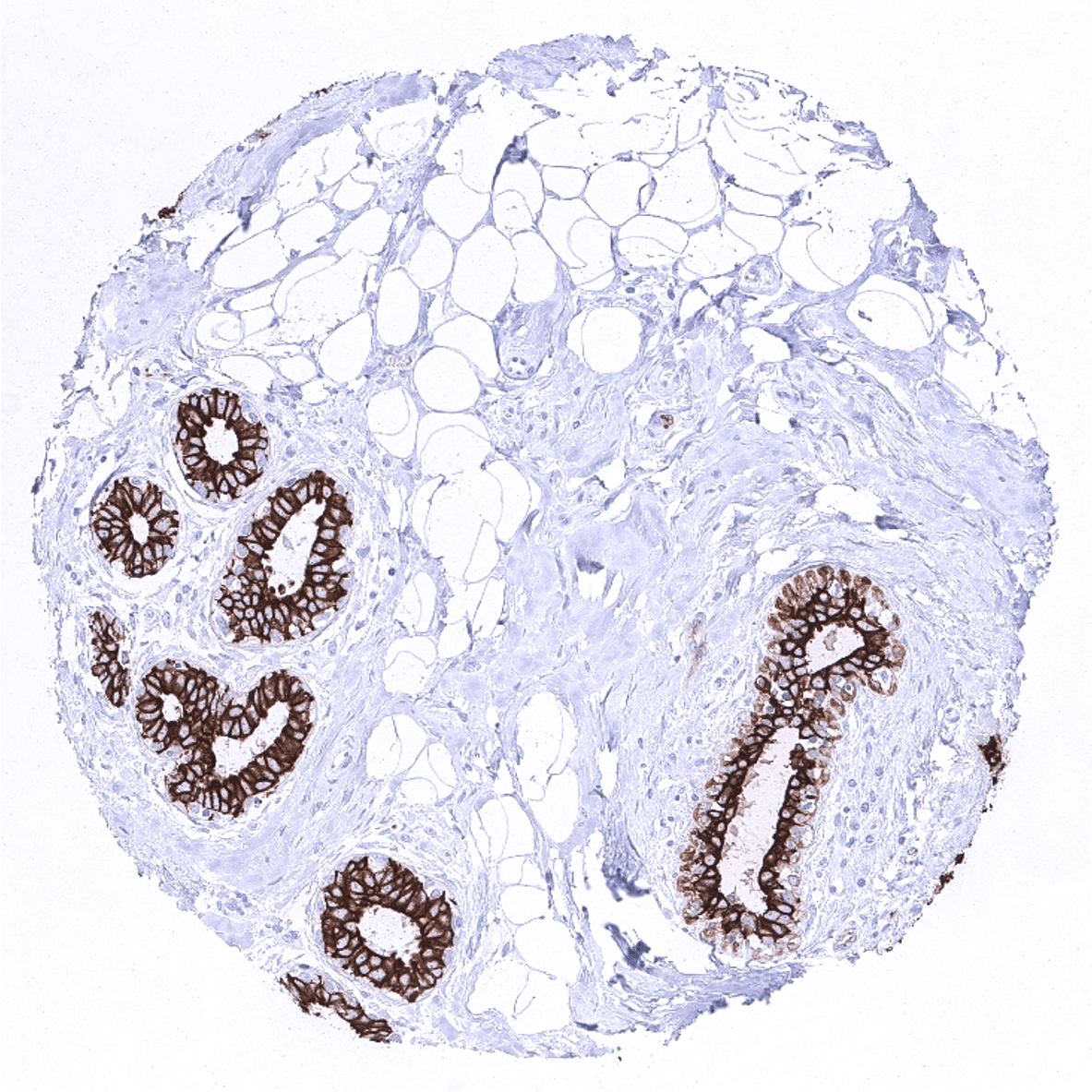
Pancreas - Strong CK7 immunostaining of centroacinar cells of small intercalated ducts in the pancreas. A much weaker memranous and cytoplasmic staining of acinar cells can regionally be seen in some cases.
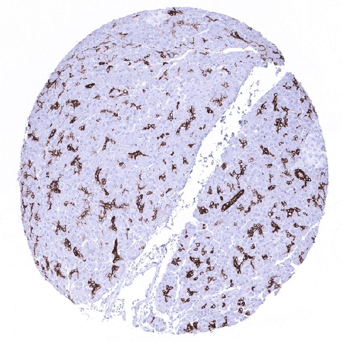
Pancreas - Strong CK7 immunostaining of centroacinar cells of small intercalated ducts in the pancreas.
Parathyroid - Few cells in the parathyroid glands show moderate intensity CK7 immunostaining.

Parotid gland
Pituitary gland, anterior lobe - CK7 immunostaining is not seen in this adenohypophysis sample.

Pituitary, anterior lobe - Few CK7 cells can be seen in the adenohypophysis in some cases.
Pituitary gland, posterior lobe
Pregnant uterus (decidua)
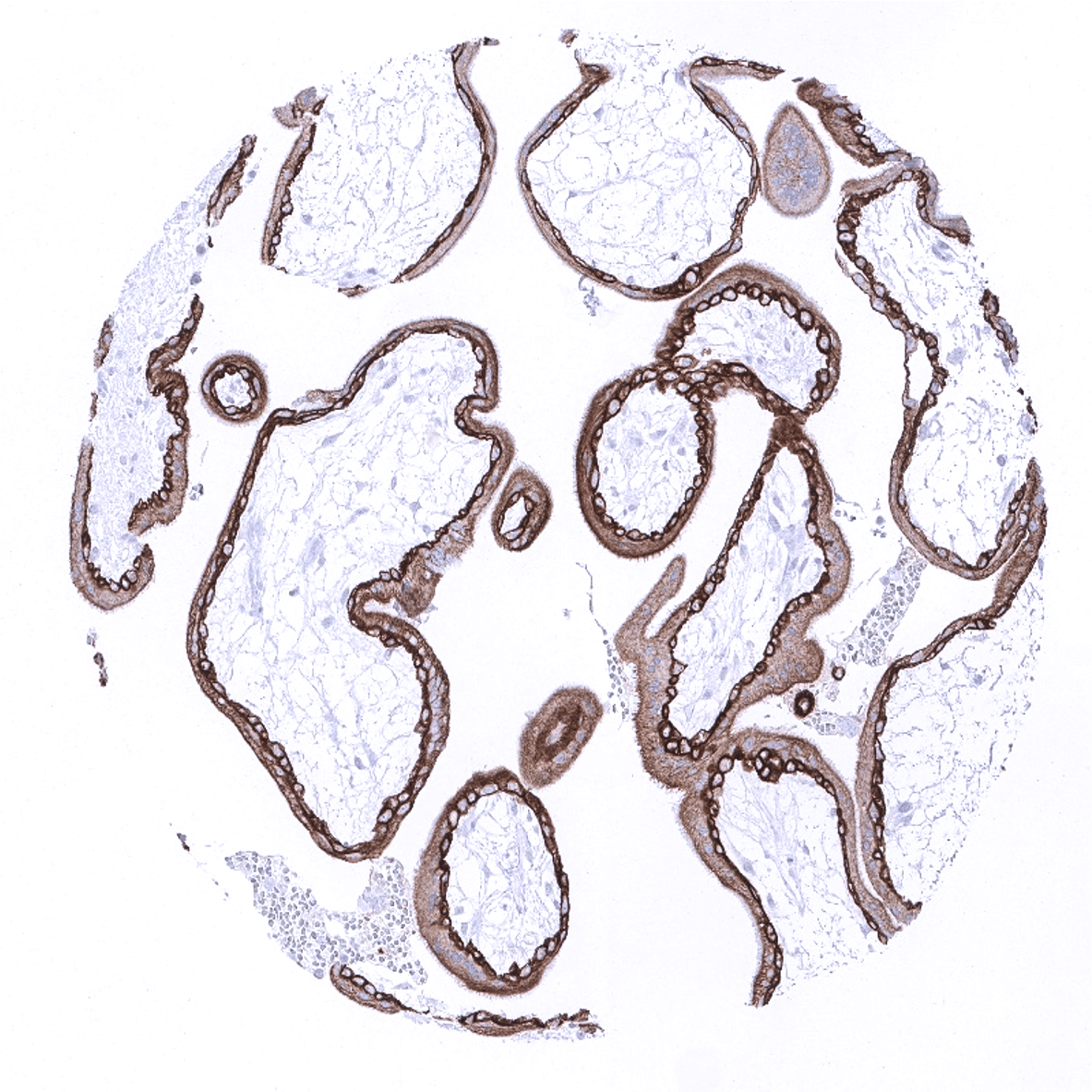
Placenta, early
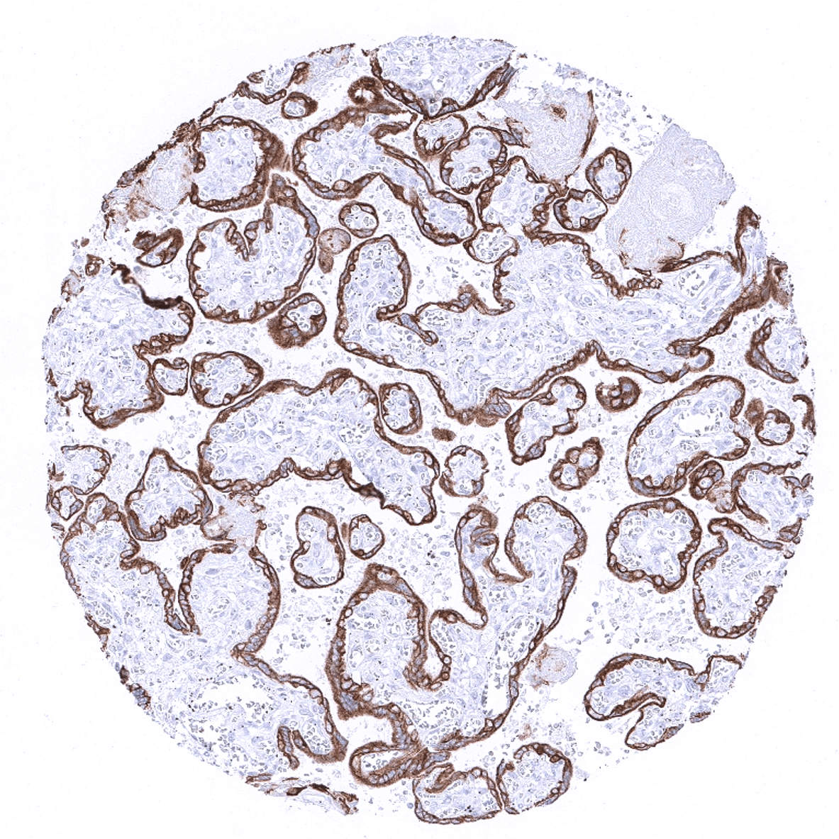
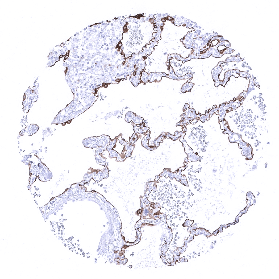
Placenta, mature

Placenta (amnion and chorion)
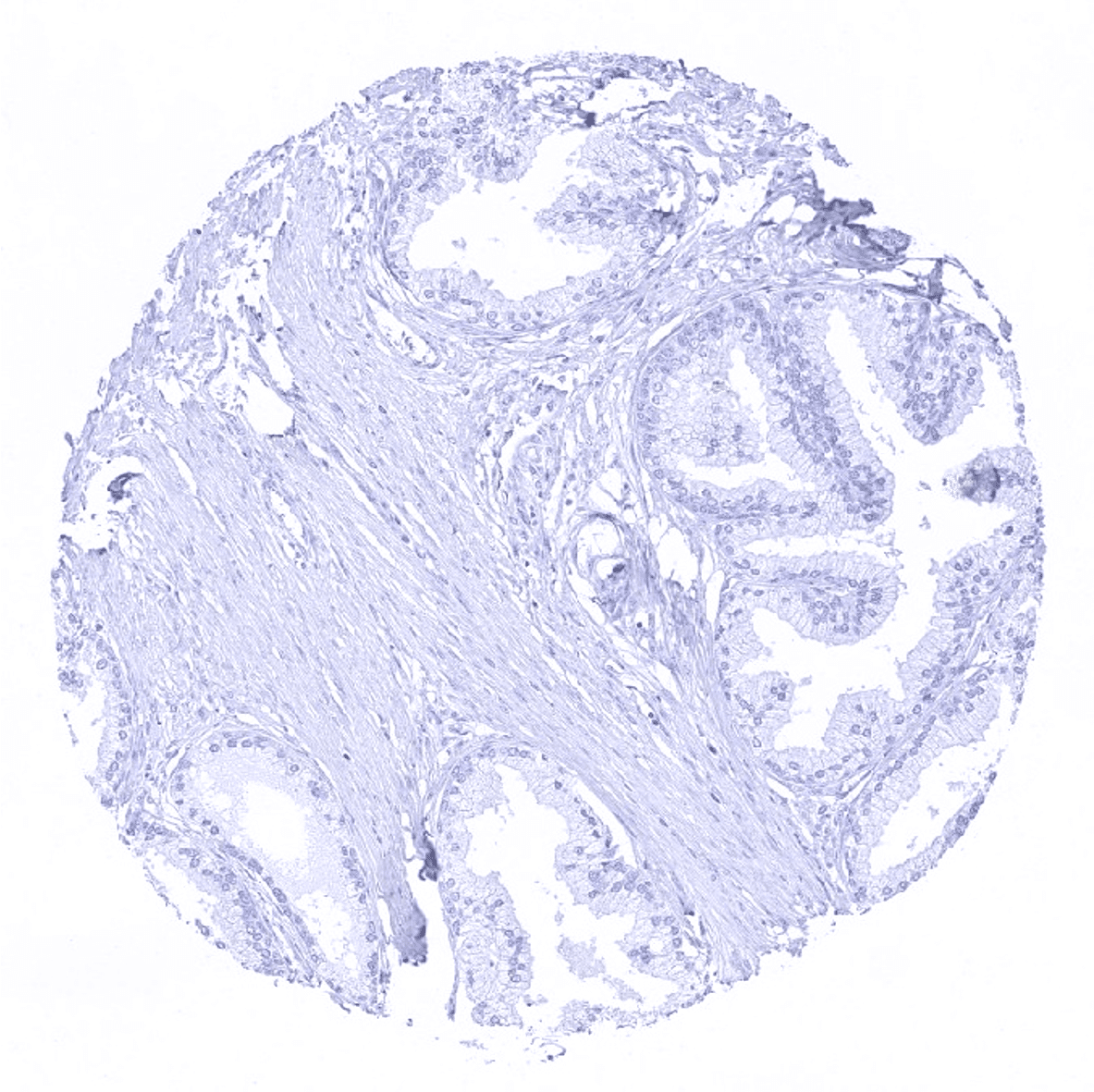
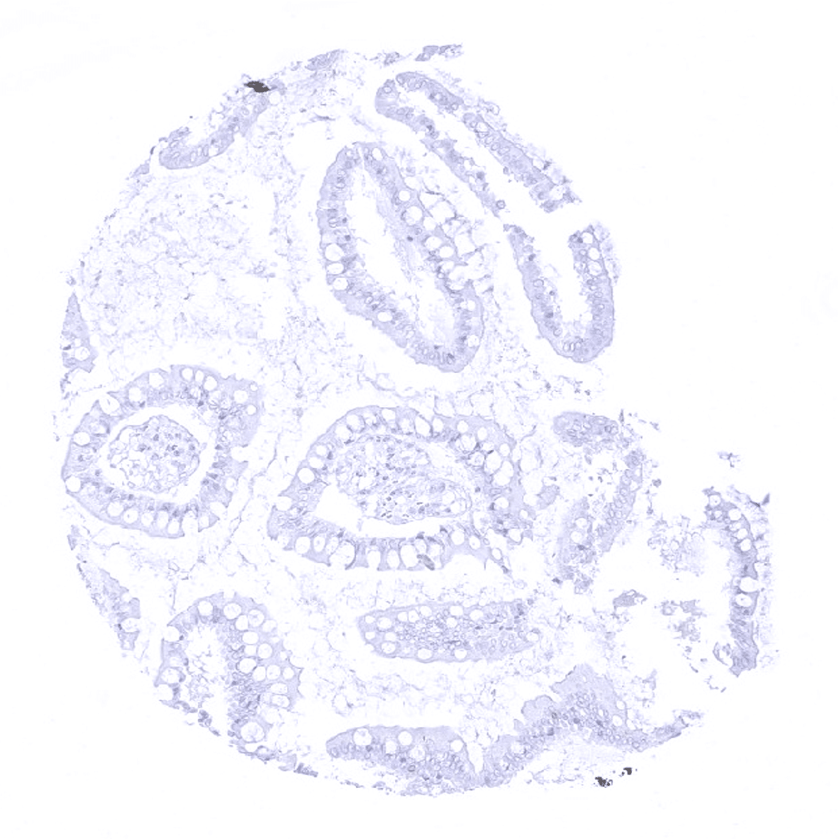
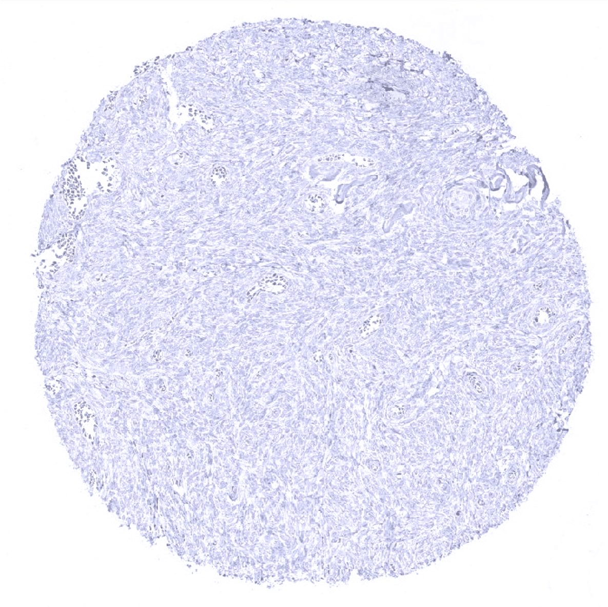
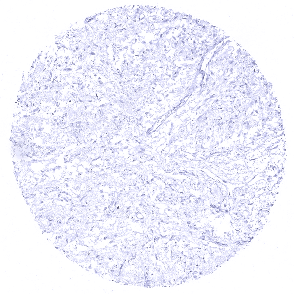
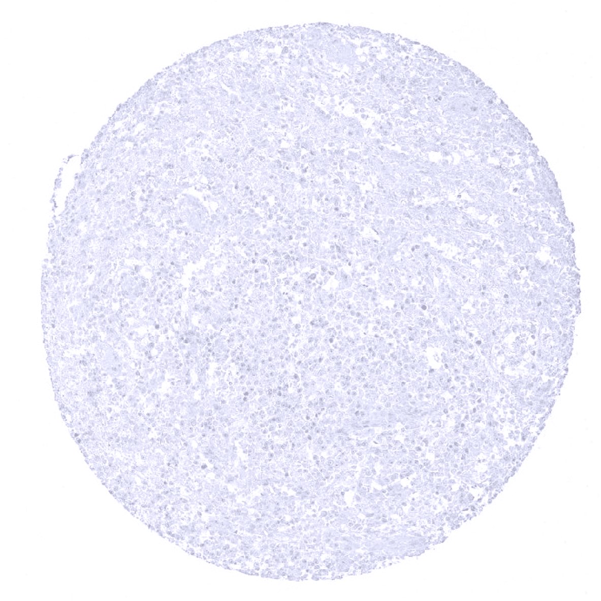
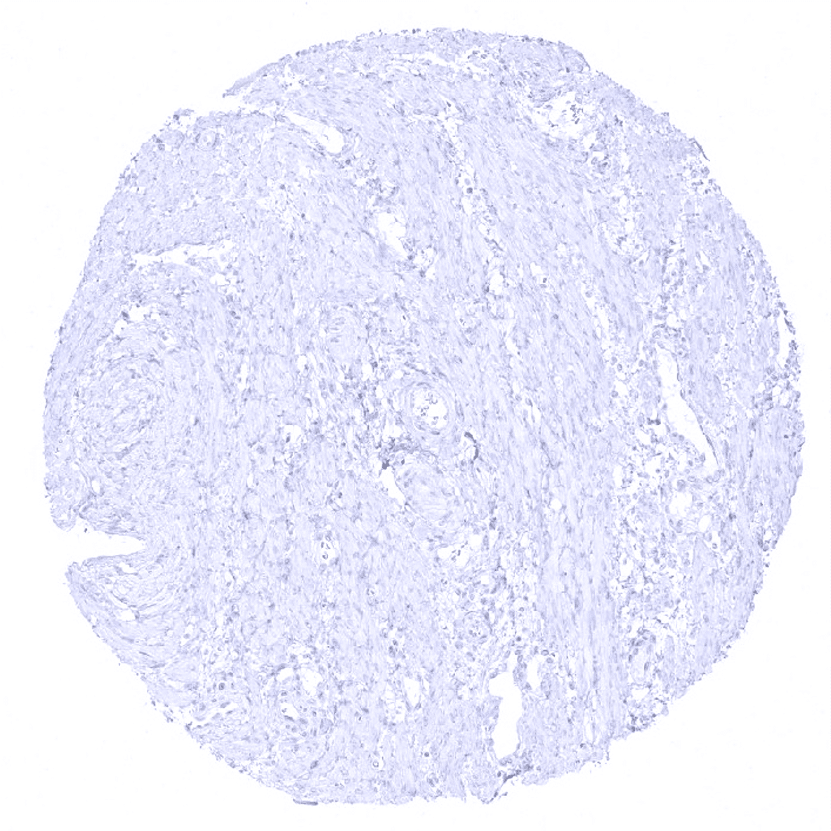
Prostate - CK7 immunostaining is completely absent in this sample.
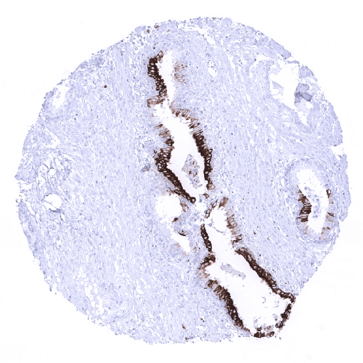
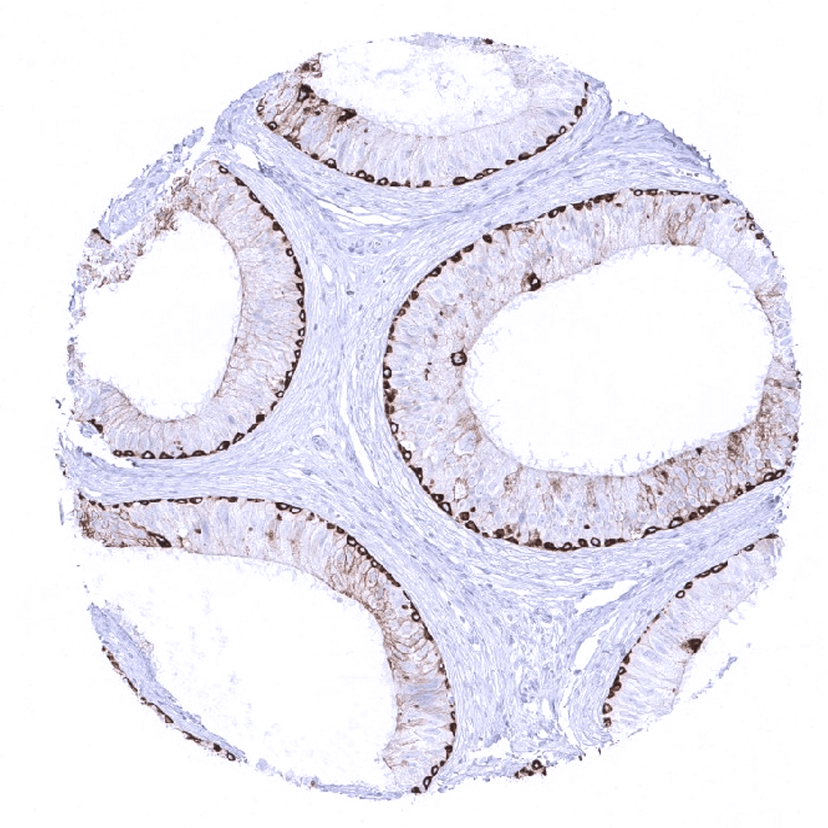
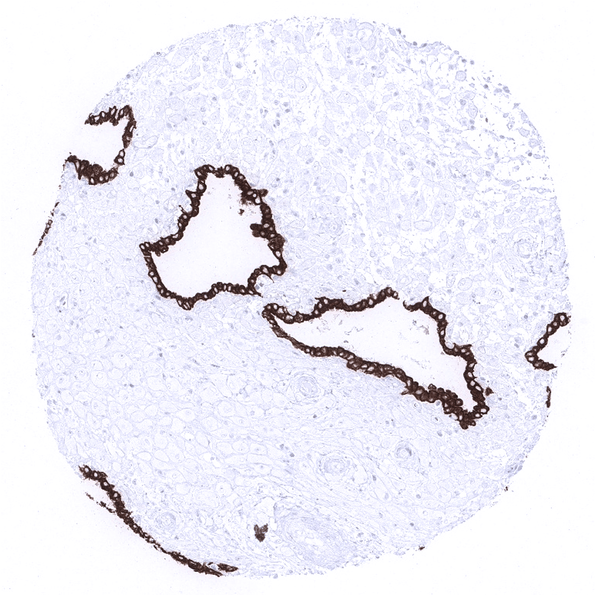
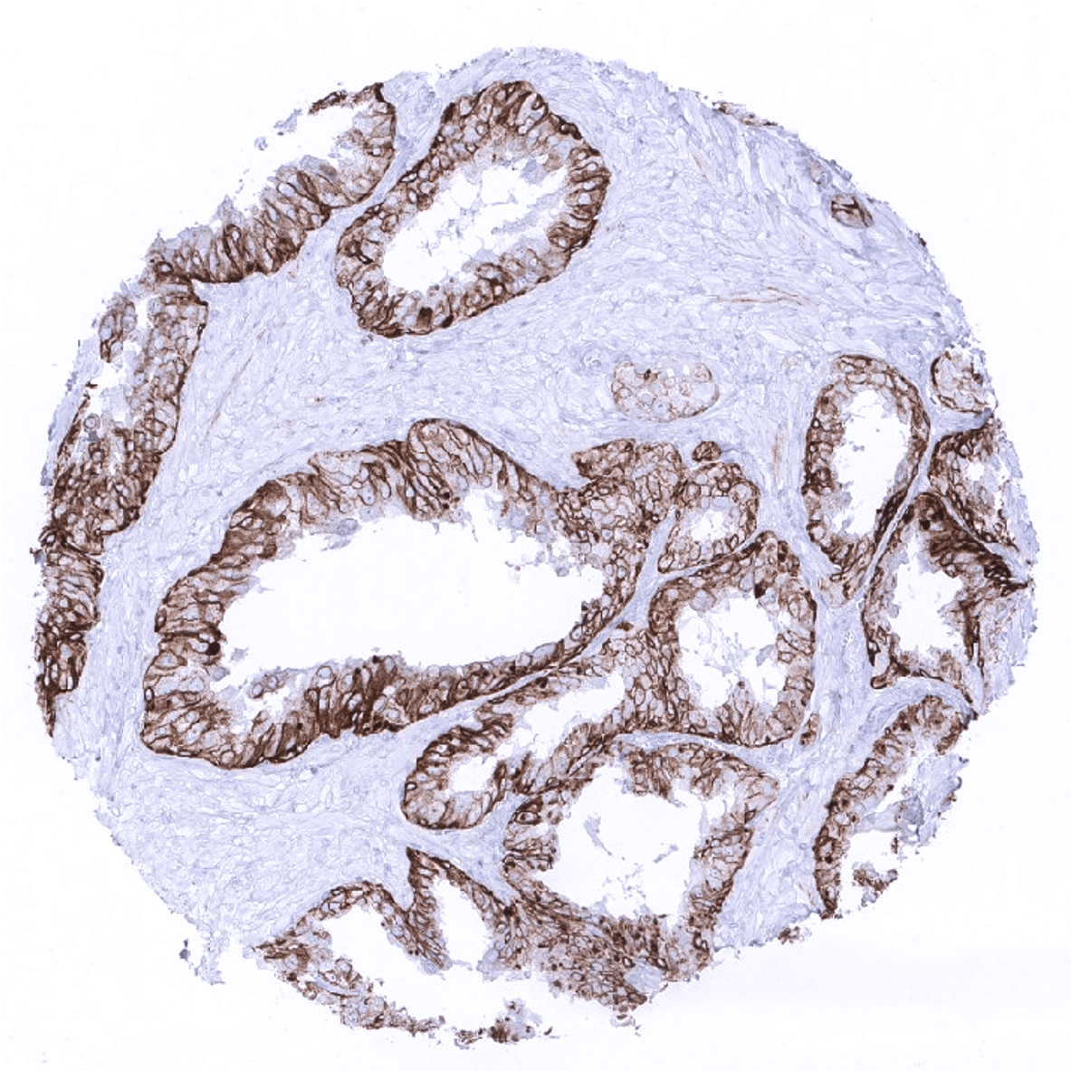
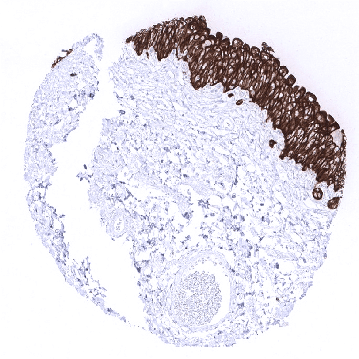
Prostate - A focal CK7 positivity of acinar cells is seen in this sample while staining is absent in basal cells.
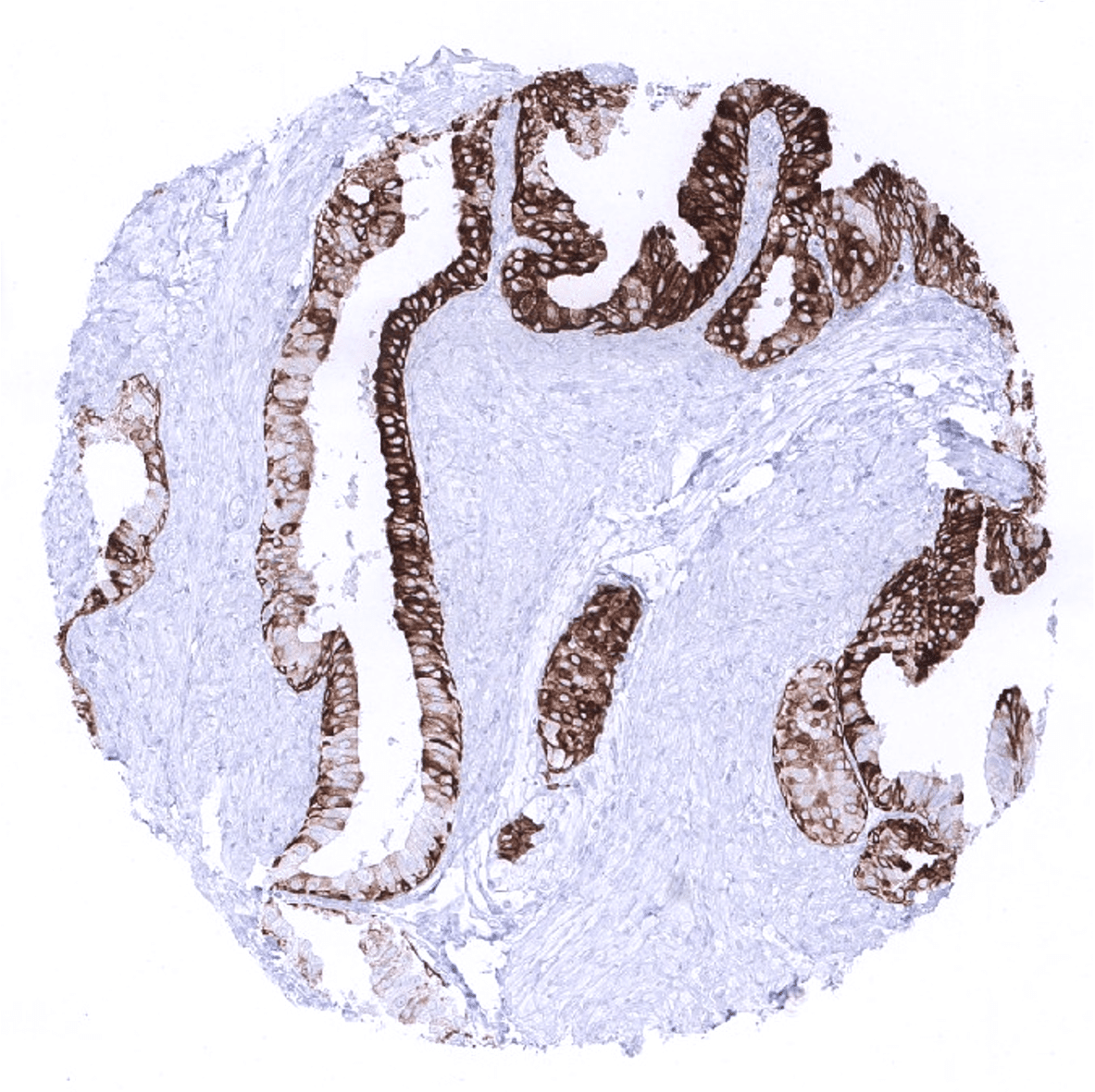
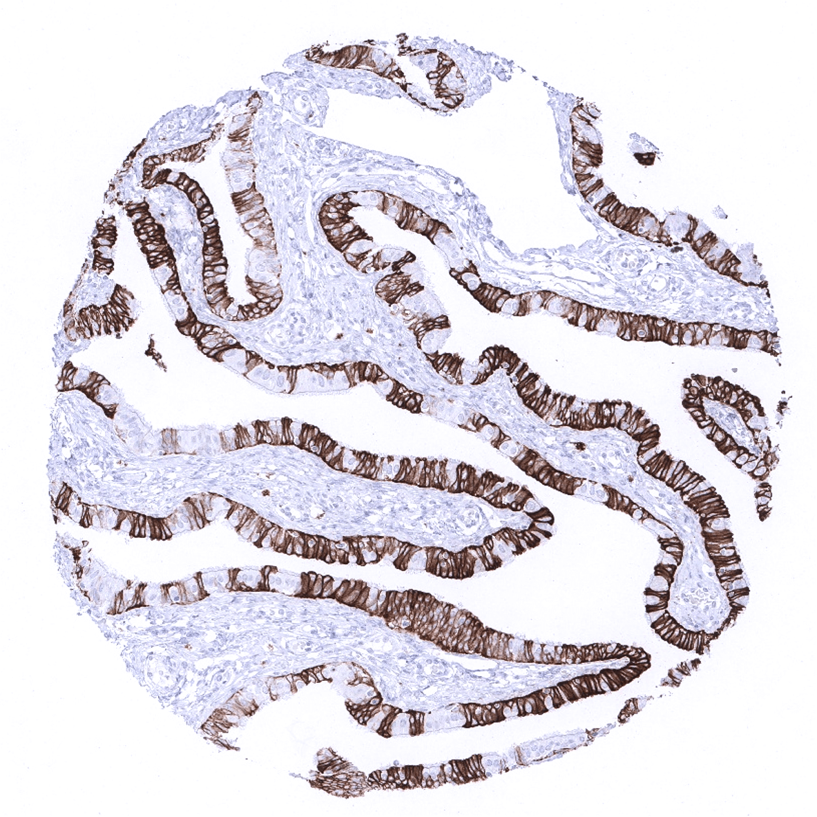
Prostate - A moderate to strong CK7 immunostaining of both acinar and basal cells can be seen in this sample.

Rectum, mucosa
Seminal vesicle
Sinus paranasales - Strong CK7 immunostaining of ciliated and goblet cells. Basal cells exhibit only a weak staining.

Skin
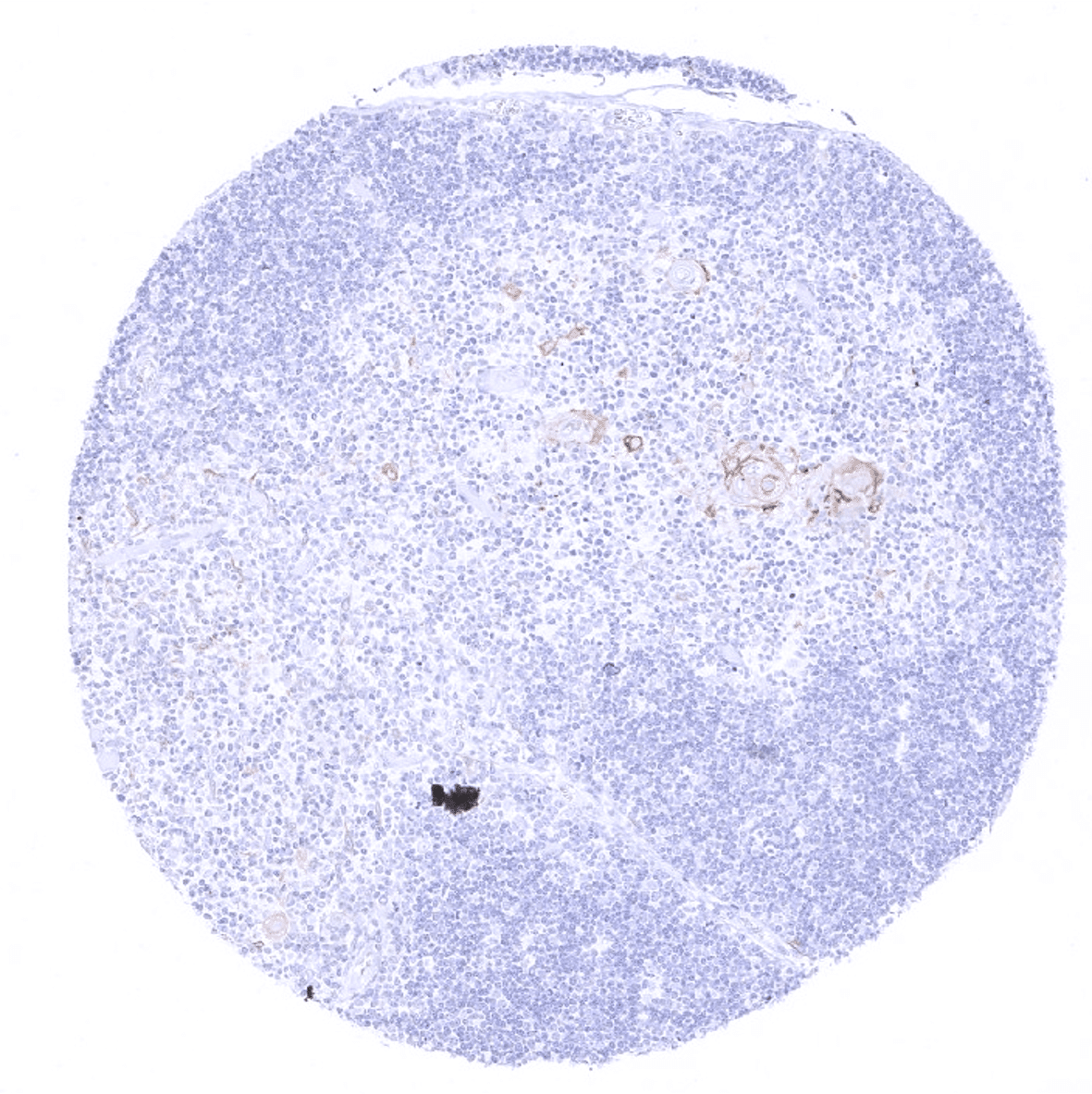
Spleen
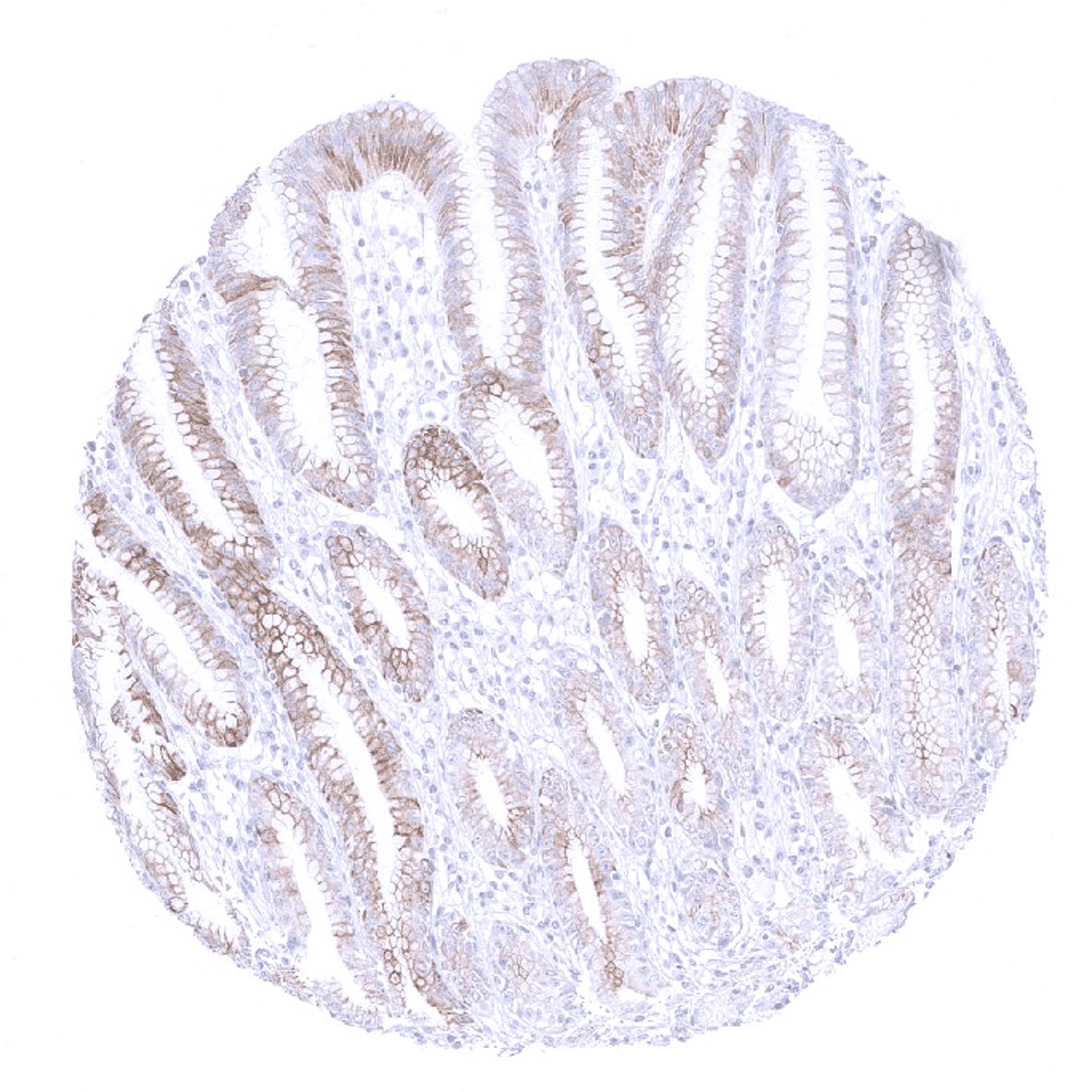
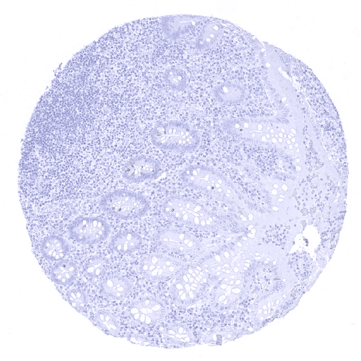
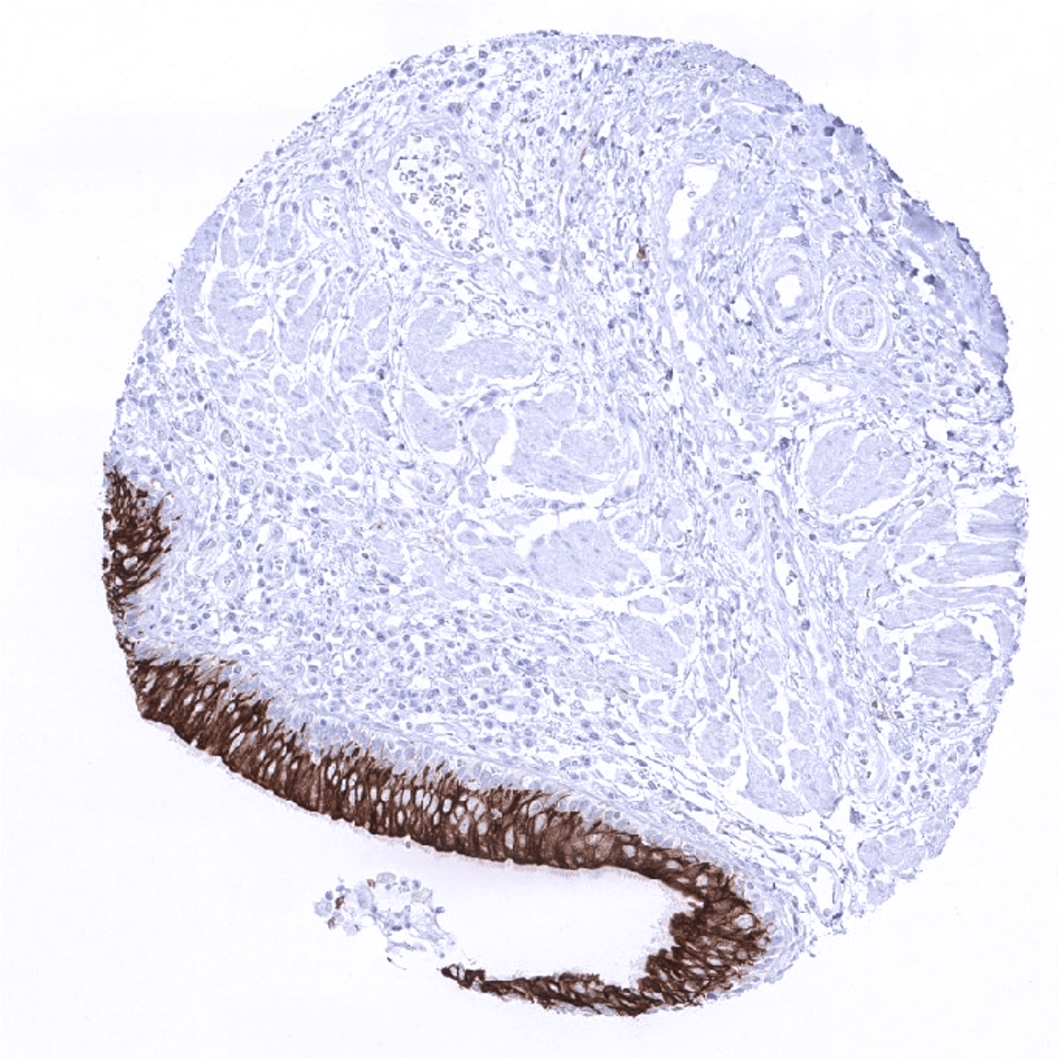
Stomach, antrum
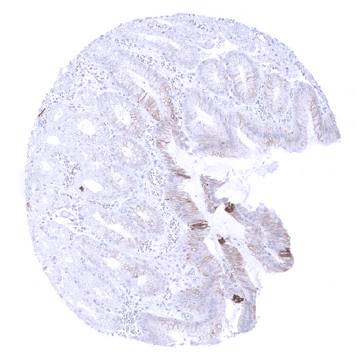
Stomach, corpus
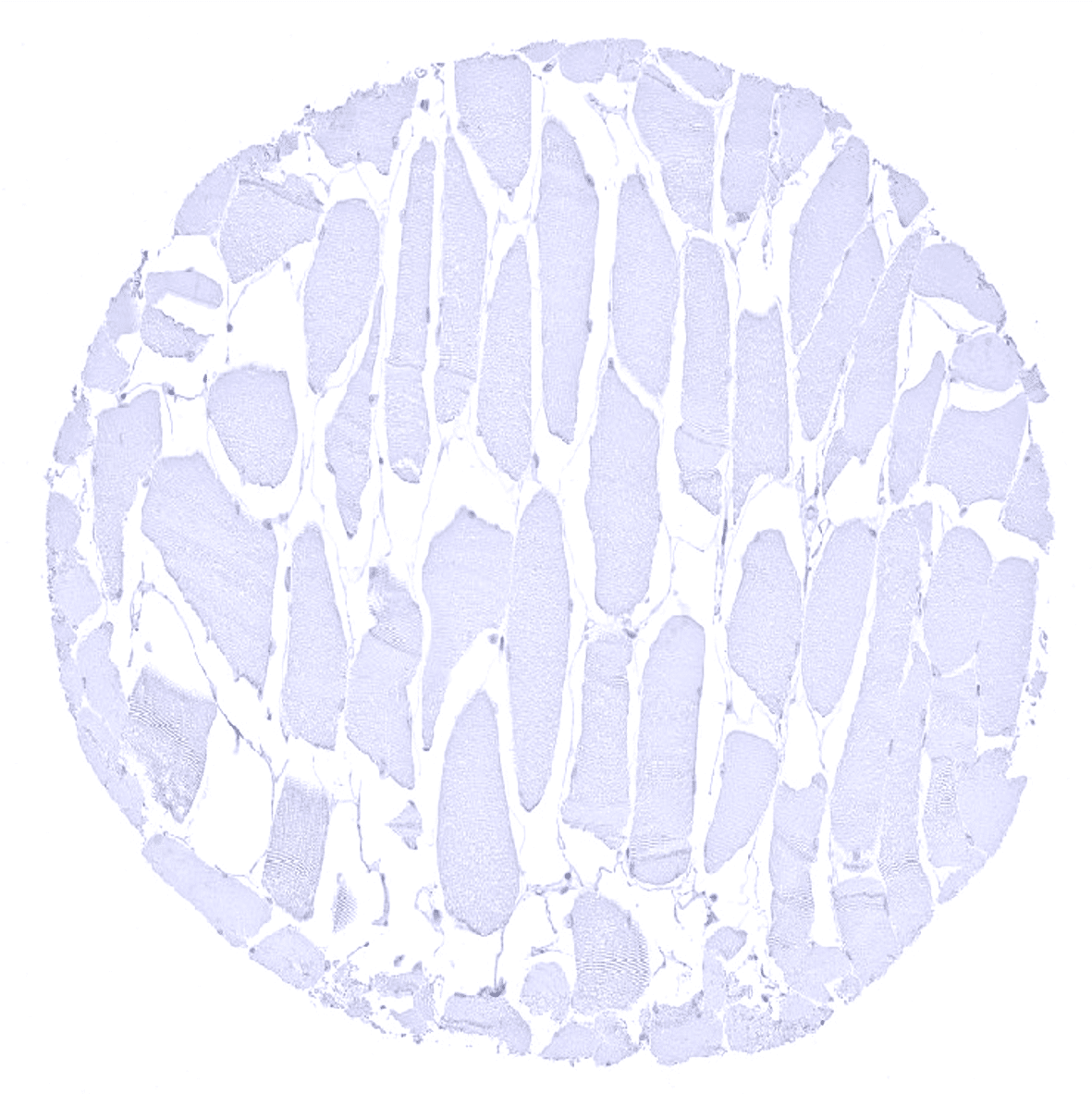
Striated muscle
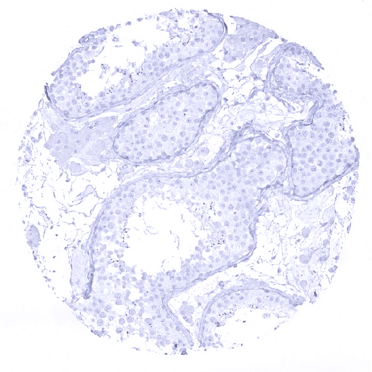
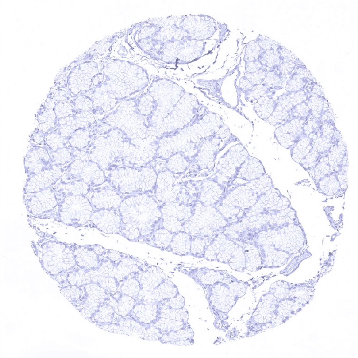
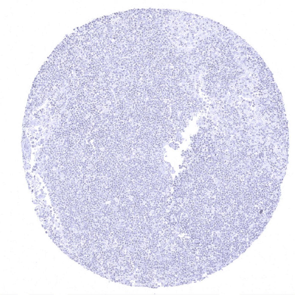
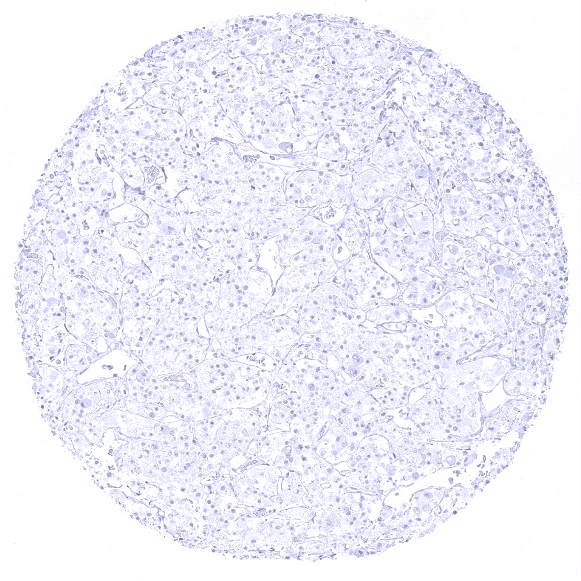
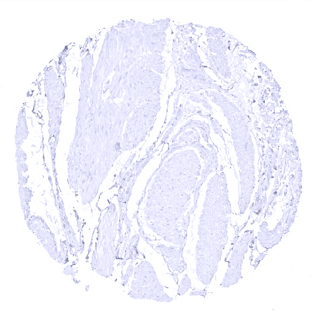
Testis - CK7 immunostaining is negative.
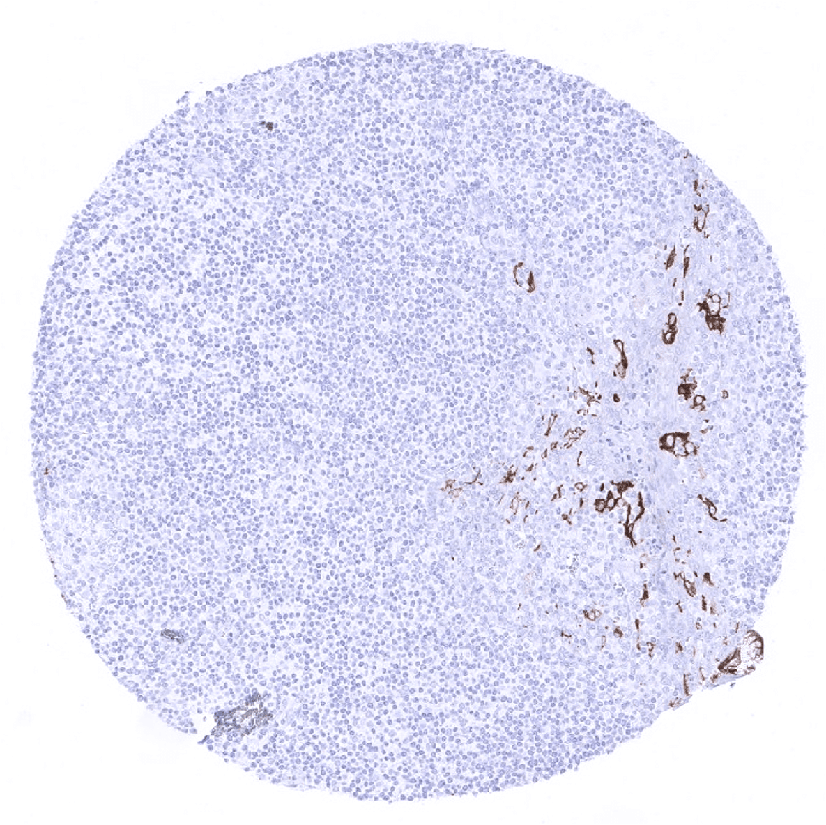
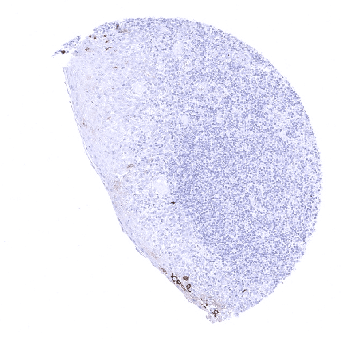
Thymus - Weak CK7 immunostaining of some elements of corpuscles of Hassall's.

Thyroid gland
Tonsil - A small fraction of squamous epithelial cells of the crypts show a moderate to strong CK7 immunostaining.
Tonsil, surface epithelium - Few scattered squamous epithelial cells may stain CK7 positive in the surface epithelium of the tonsil.
Urinary bladder, muscular wall
Urinary bladder, urothelium
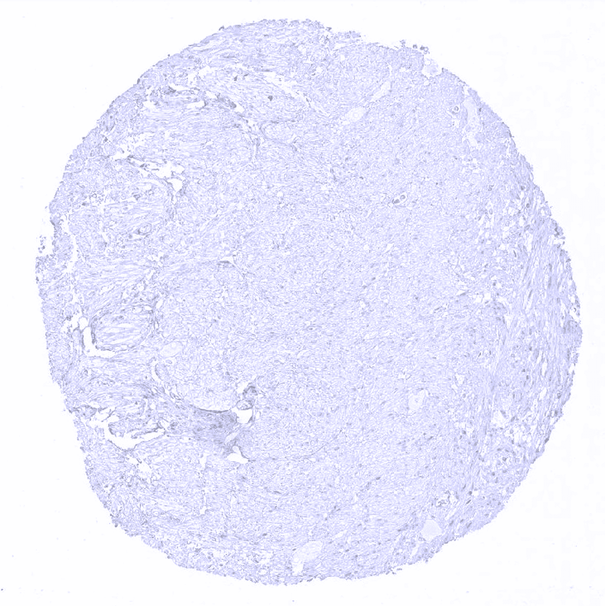
Uterus, myometrium