Product details
Synonyms = FLJ32198, MGC10902, p35, UPIIIb
Antibody type = Mouse monoclonal / IgG
Clone = MSVA-736M
Positive control = Urinary bladder: A moderate to strong membranous Upk3b immunostaining should be seen in umbrella cells of the normal urothelium (the staining is usually limited to apical surface membrane of umbrella cells).
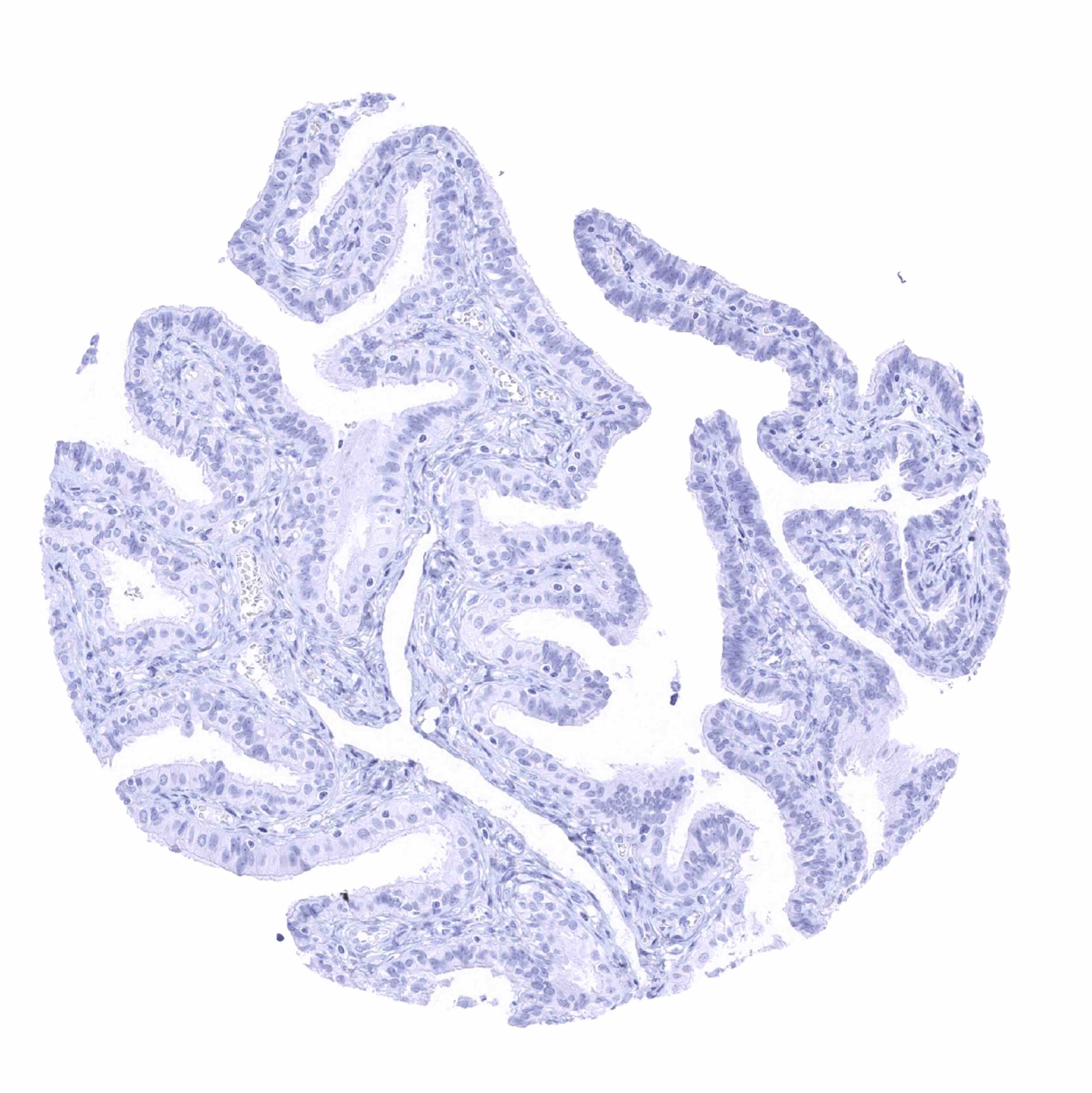
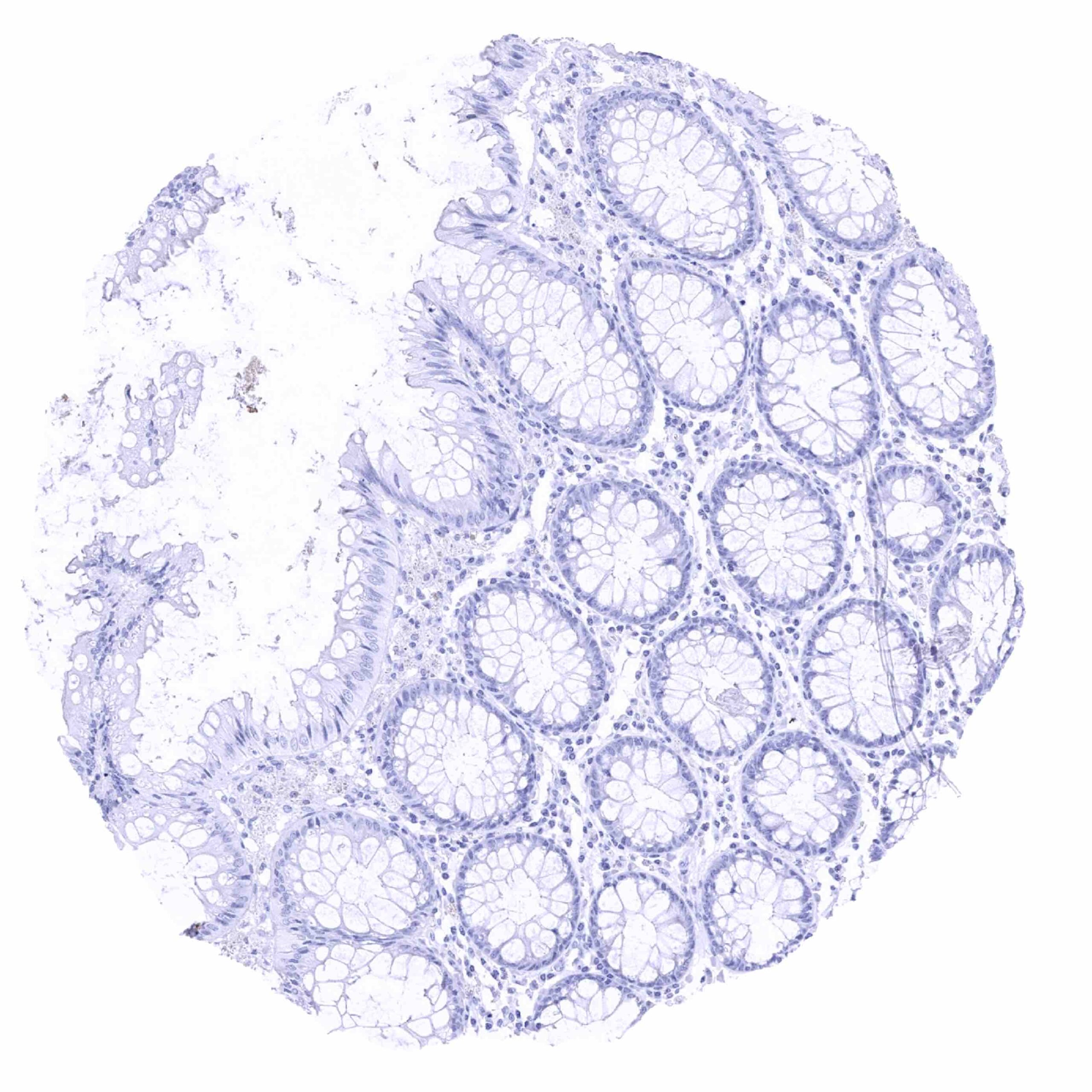
Negative control = Colon: Upk3b immunostaining should be absent in all cells of the colon mucosa.
Cellular localization = Cell Surface and cytoplasm
Reactivity = Human
Application = Immunohistochemistry
Dilution = 1:100 – 1:200
Intended Use = Research Use Only
Relevance of Antibody
Uroplakin 3B is a marker for mesothelial cells and umbrella cells.
Biology Behind
The Uroplakin 3B (Upk3b) protein is coded by the UPK3b gene located at 7q1.23. Upk3b is one out of 5 known uroplakin (Upk) protein particles that cooperatively form apical asymmetrical unit membrane (AUM) plaques which play an important role in the stabilization and strengthening of epithelial cells that line the bladder. These AUM plaques enable the inner bladder membrane to stretch and prevent urothelial cells from rupturing during bladder distension. Upks are assembled in the endoplasmic reticulum (ER), where they heterodimerize prior to escaping the ER. Upk3b heterodimerizes with Upk1b. Upk heterodimers subsequently form tetramers which then combine as concentric hexameric rings that are packaged into vesicles and trafficked to the cell surface. AUMs and Upk proteins may have a role in mediating membrane permeability and signal transduction events that are involved in the regulation of cell development, activation, growth, and motility.
Staining Pattern in Normal Tissues
Uroplakin 3B staining pattern in Normal Tissues with antibody MSVA-736M (Images shown in our “Normal Tissue Gallery”)
| Brain | Cerebrum | Negative. |
| Cerebellum | Negative. | |
| Endocrine Tissues | Thyroid | Negative. |
| Parathyroid | Negative. | |
| Adrenal gland | Negative. | |
| Pituitary gland | Negative. | |
| Respiratory system | Respiratory epithelium | Negative. |
| Lung | Negative. | |
| Gastrointestinal Tract | Salivary glands | Negative. |
| Esophagus | Negative. | |
| Stomach | Negative. | |
| Duodenum | Negative. | |
| Small intestine | Negative. | |
| Appendix | Negative. | |
| Colon | Negative. | |
| Rectum | Negative. | |
| Liver | Negative. | |
| Gallbladder | Negative. | |
| Pancreas | Negative. | |
| Genitourinary | Kidney | Negative. |
| Urothelium | Moderate to strong, membranous Upk3b staining of the apical membrane of umbrella cells. | |
| Male genital | Prostate | Negative. |
| Seminal vesicles | Negative. | |
| Testis | Negative. | |
| Epididymis | Negative. | |
| Female genital | Breast | Negative. |
| Uterus, myometrium | Negative. | |
| Uterus, ectocervix | Negative. | |
| Uterus endocervix | Negative. | |
| Uterus, endometrium | Negative. | |
| Fallopian Tube | Negative. | |
| Ovary | Negative. | |
| Placenta early | Negative. | |
| Placenta mature | Negative. | |
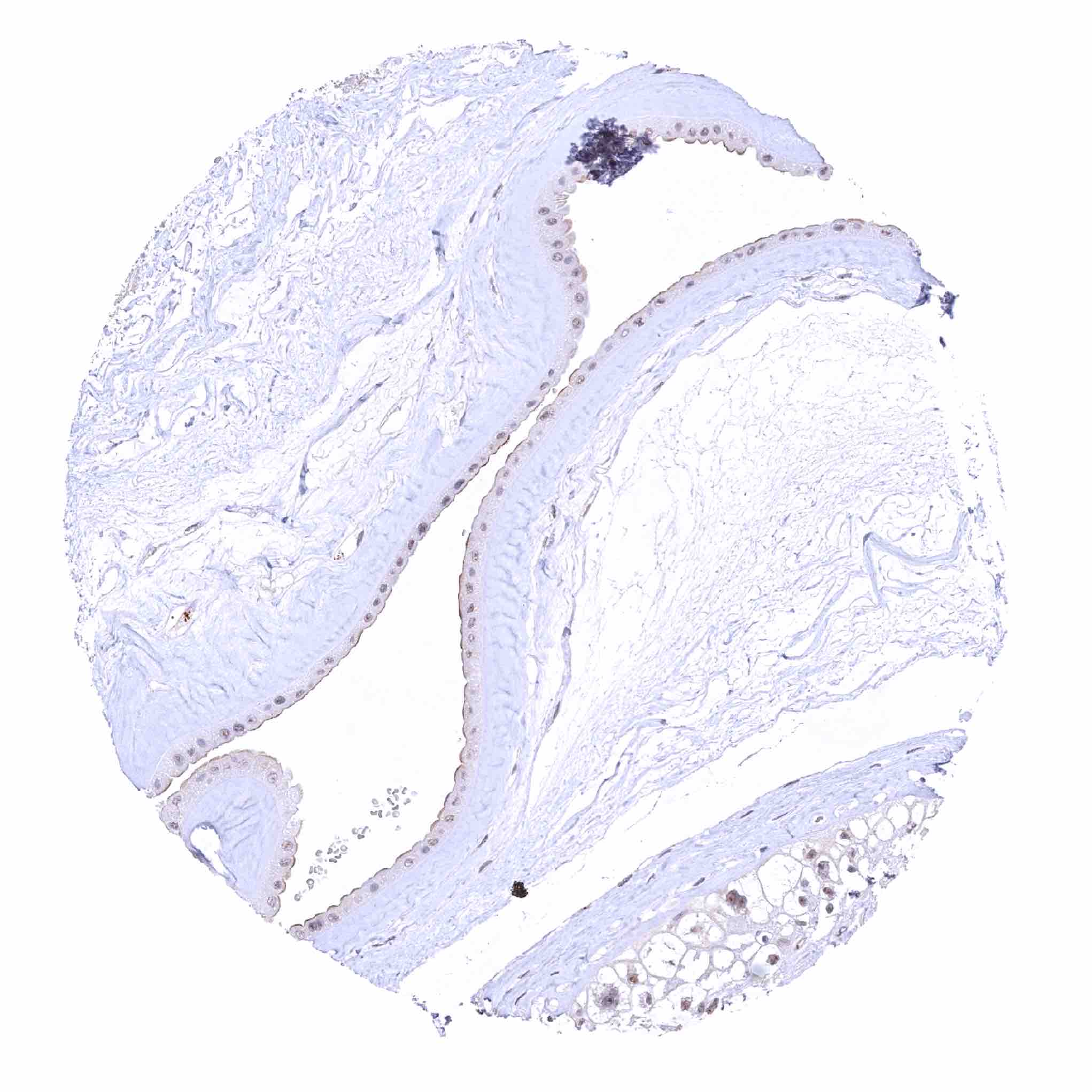
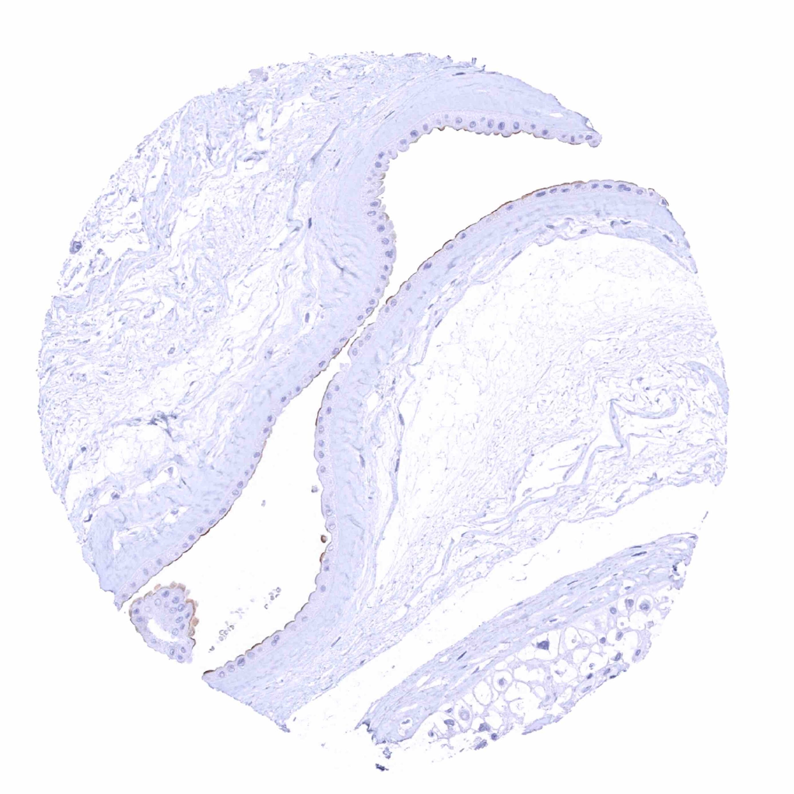
| Amnion | Moderate to strong, membranous Upk3b staining of the apical membrane of amnion cells. | |
| Chorion | Negative. | |
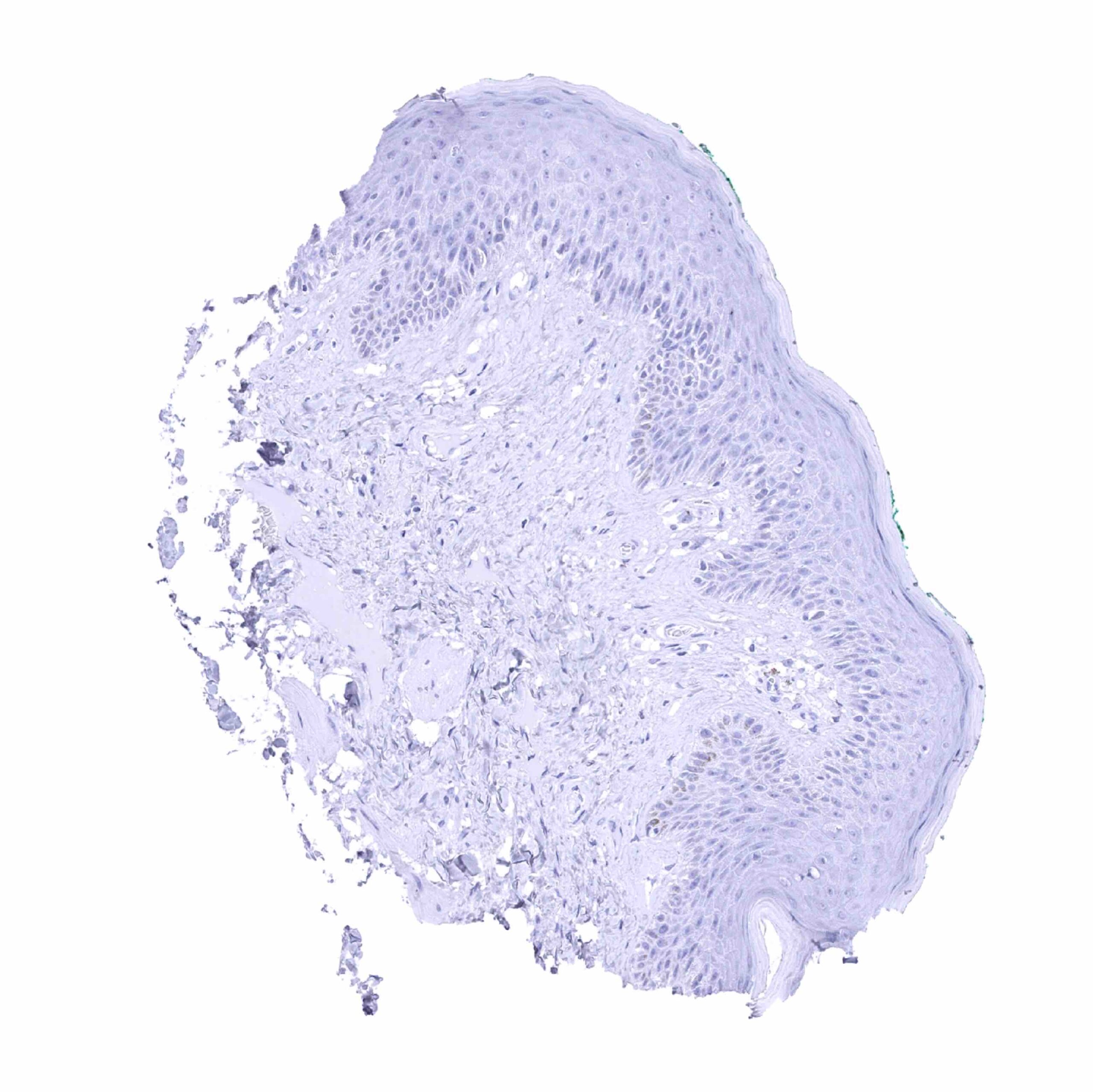
| Skin | Epidermis | Negative. |
| Sebaceous glands | Negative. | |
| Muscle/connective tissue | Heart muscle | Negative. |
| Skeletal muscle | Negative. | |
| Smooth muscle | Negative. | |
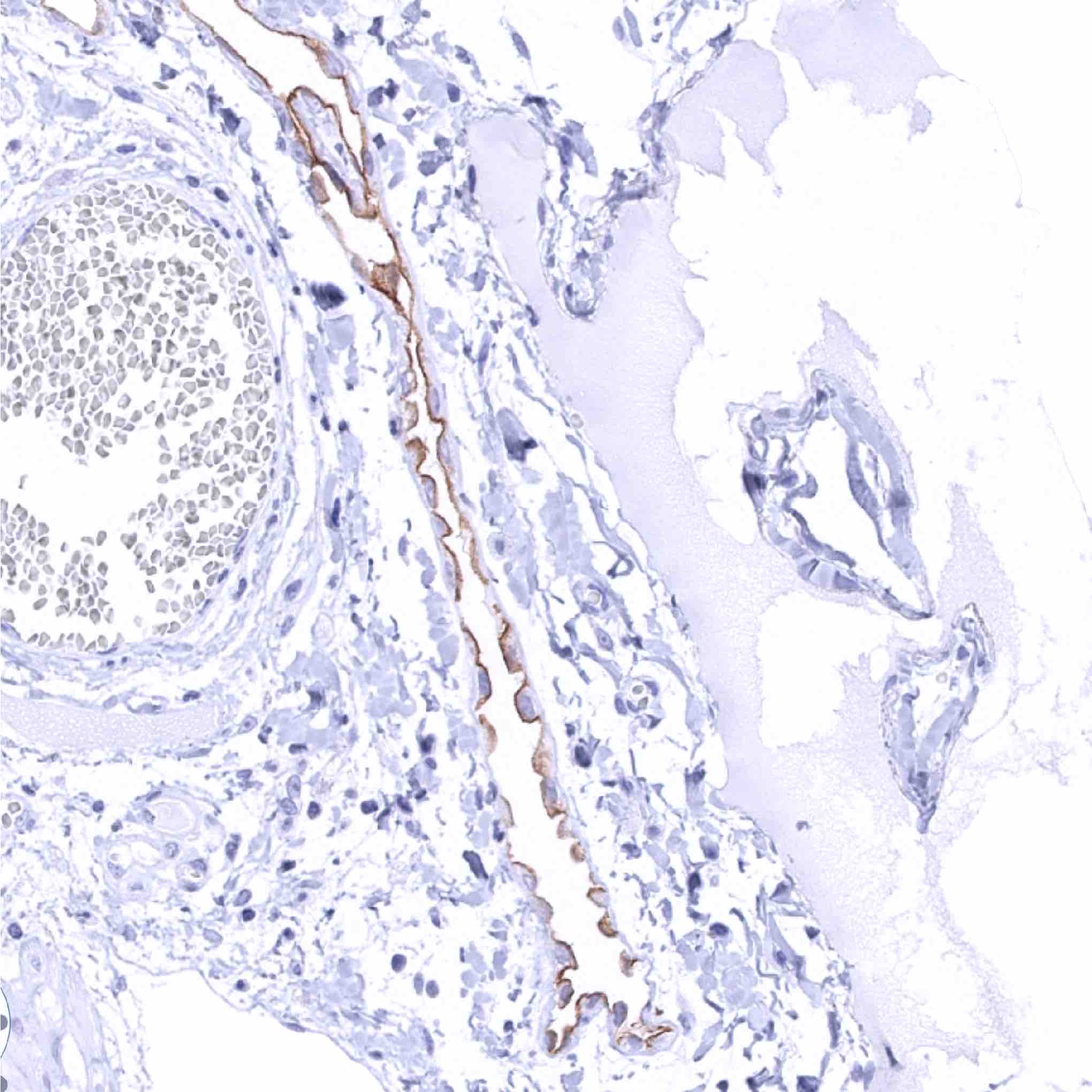
| Vessel walls | Negative (faint staining of the muscular wall or perivascular fibrous structures of small vessels can be seen in case of too sensitive staining protocols). | |
| Fat | Negative. | |
| Stroma | Negative (faint staining of fibrous structures – often perivascular – can be seen in case of too sensitive staining protocols). | |
| Endothelium | Negative. | |
| Bone marrow/lymphoid tissue | Bone marrow | Negative. |
| Lymph node | Negative. | |
| Spleen | Negative. | |
| Thymus | Negative. | |
| Tonsil | Negative. | |
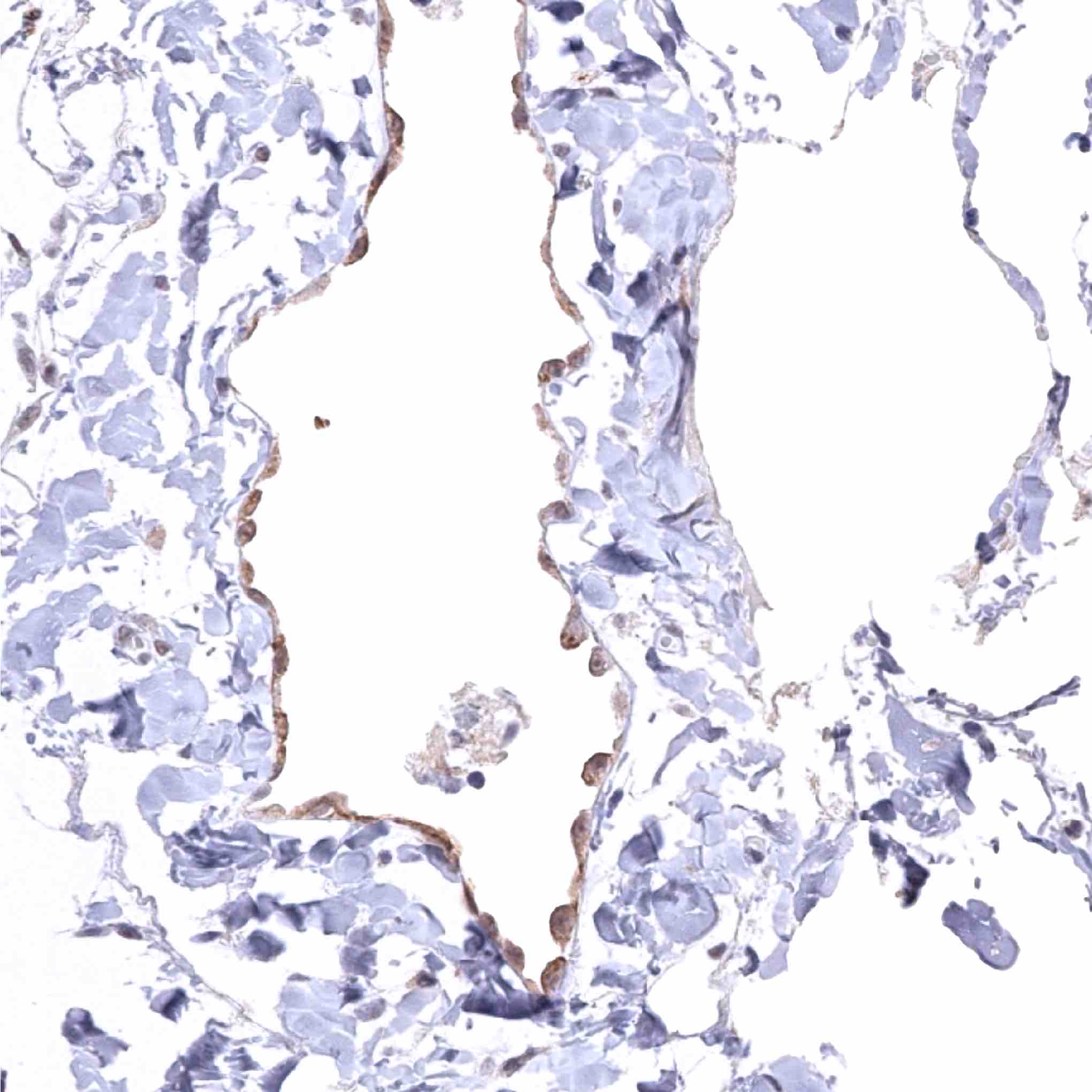
| Remarks | Moderate to strong, membranous Upk3b staining of the apical membrane of mesothelial cells of all organs. |
These findings are largely consistent with the RNA and protein data described in the Human Protein Atlas (Tissue expression Uroplakin 3B) All organs with documented Upk3b RNA expression (urinary bladder, kidney, prostate, gallbladder, stomach, placenta, fallopian tube, uterine cervix, tonsil) with the only exception of smooth muscle are IHC positive for MSV-736M. Given the immediate vicinity of smooth muscle to multiple Upk3b positive tissues, smooth muscle RNA positivity may represent a contamination artifact.
Positive control = Urinary bladder: A moderate to strong membranous Upk3b immunostaining should be seen in umbrella cells of the normal urothelium (the staining is usually limited to apical surface membrane of umbrella cells).
Negative control = Colon: Upk3b immunostaining should be absent in all cells of the colon mucosa.

Staining Pattern in Relevant Tumor Types
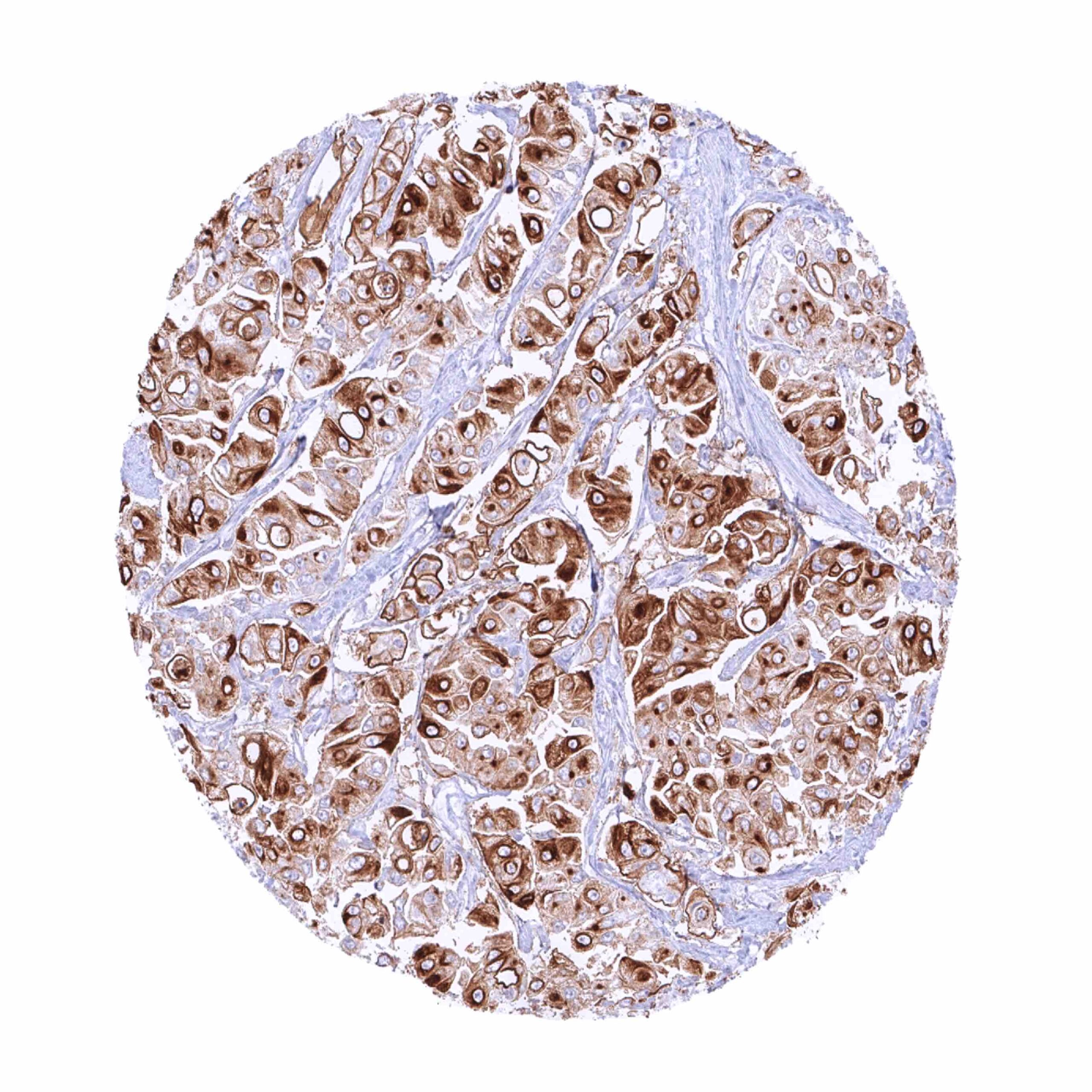
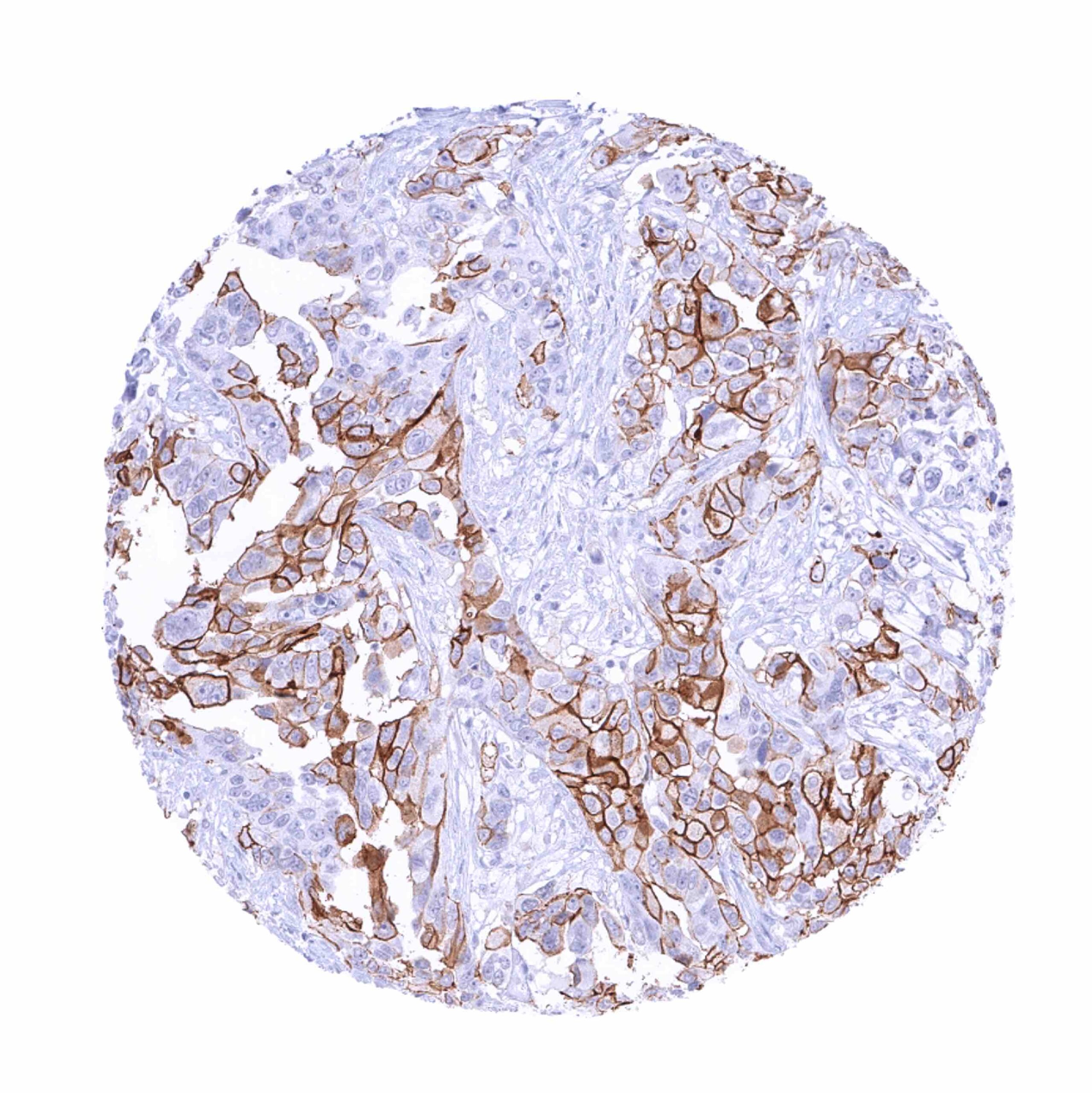
According to the TCGA data, Upk3b expression is most commonly seen in urothelial carcinomas and in ovarian cancers. Rarely, it can also be found in other tumors.
The TCGA findings on Uroplakin 3B RNA expression in different tumor categories have been summarized in the Human Protein Atlas.
Compatibility of Antibodies
Uroplakin 3b (Upk3b) (MSVA-736M) publication summary
Relevant publication: Lennartz et al.: “Analysis of More than 16,000 Human Tumor and Normal Tissues Identifies Uroplakin 3B as a Useful Diagnostic Marker for Mesothelioma and Normal Mesothelial Cells.” Published in Diagnostics (Basel). 2022 Oct 17;12(10):2516. PMID: 36292206
A total of 16’185 tumors were successfully analyzed from 151 different tumor categories by using the following protocol: Heat-induced antigen retrieval for 5 minutes in an autoclave at 121°C in pH7,8 Target Retrieval Solution buffer. MSVA-736M at a dilution of 1:150 at 37°C for 60 minutes. Visualization of bound antibody by the EnVision Kit (Dako, Agilent). This protocol was also used for all stainings depicted in our tumor and normal tissue galleries.
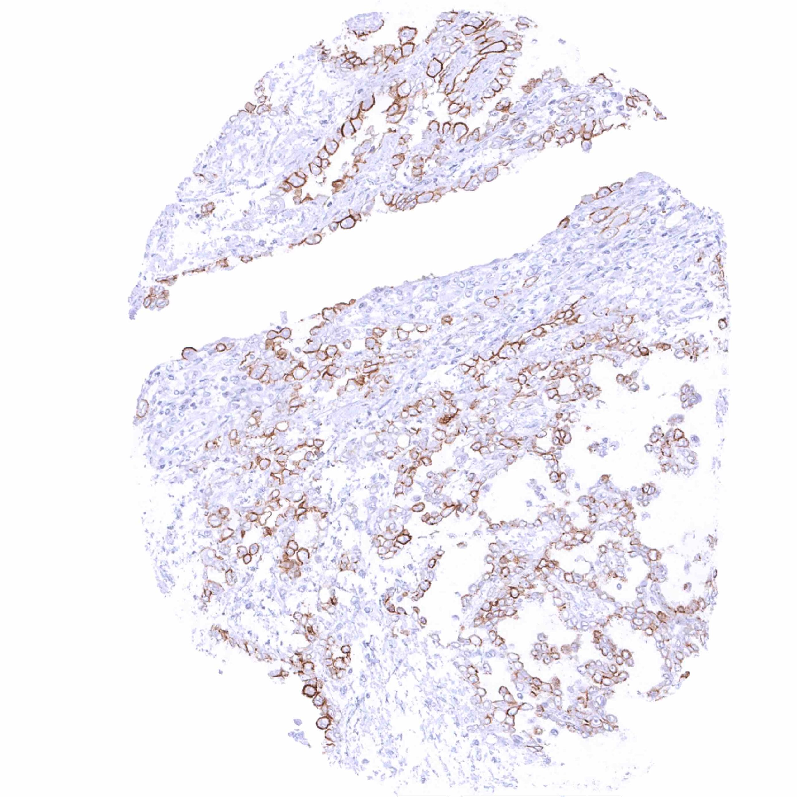
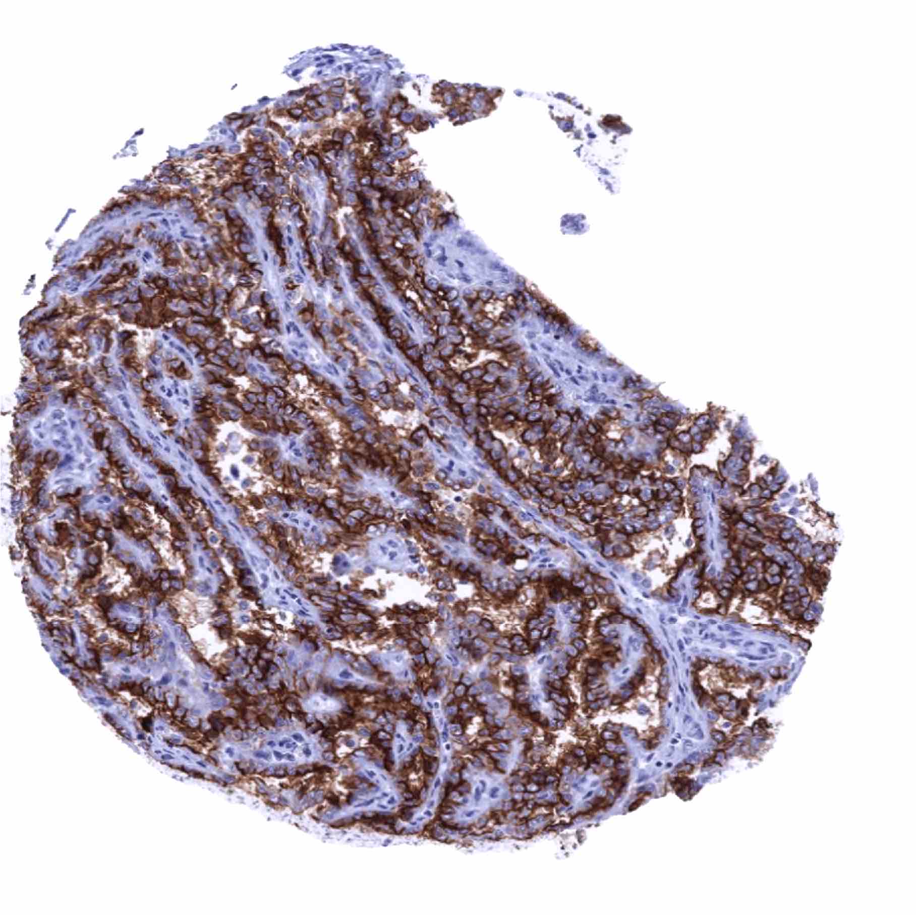
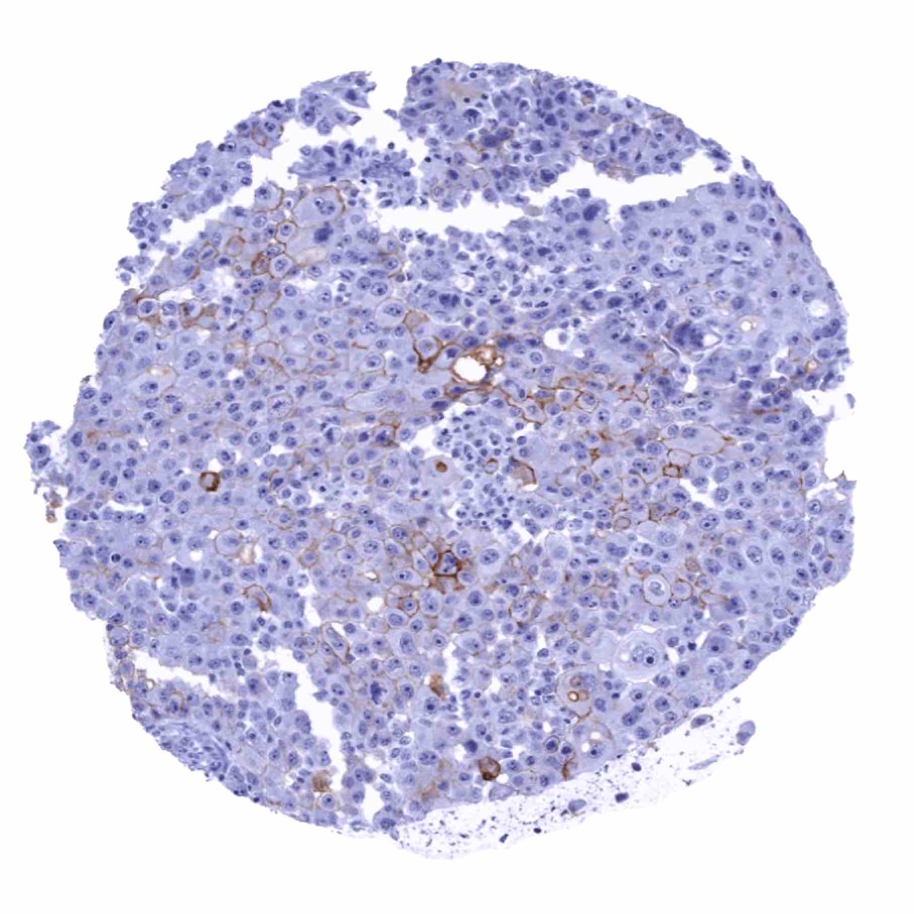
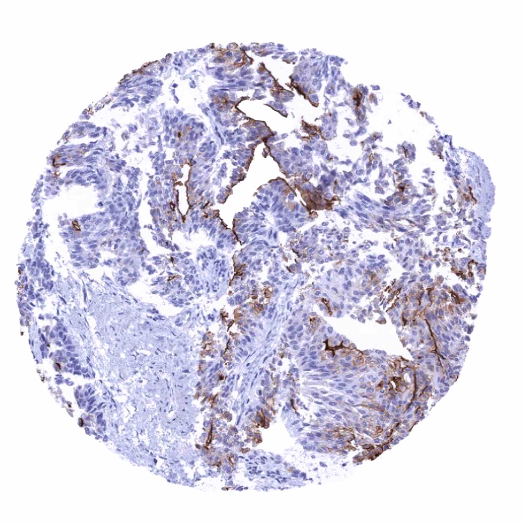
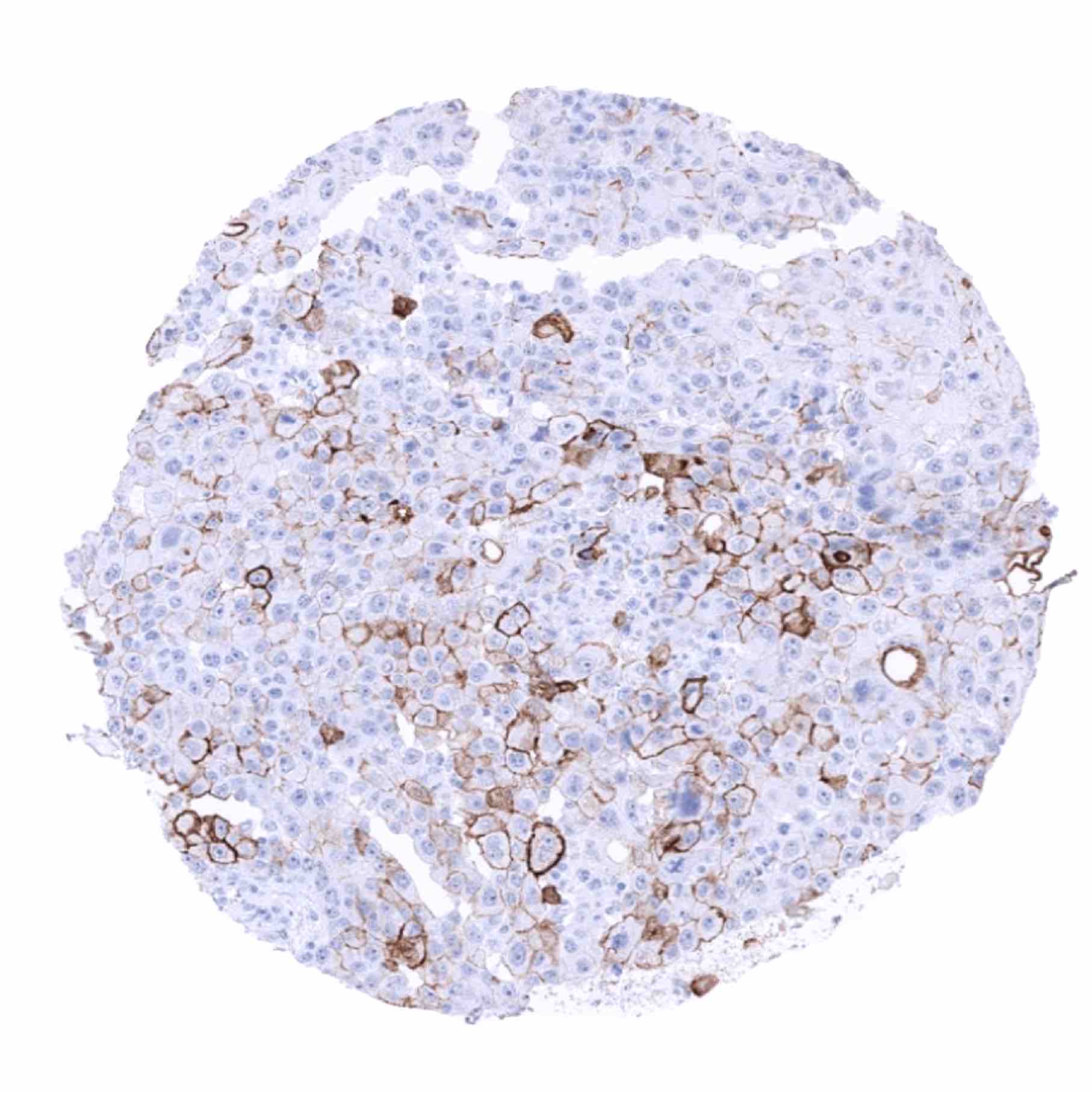
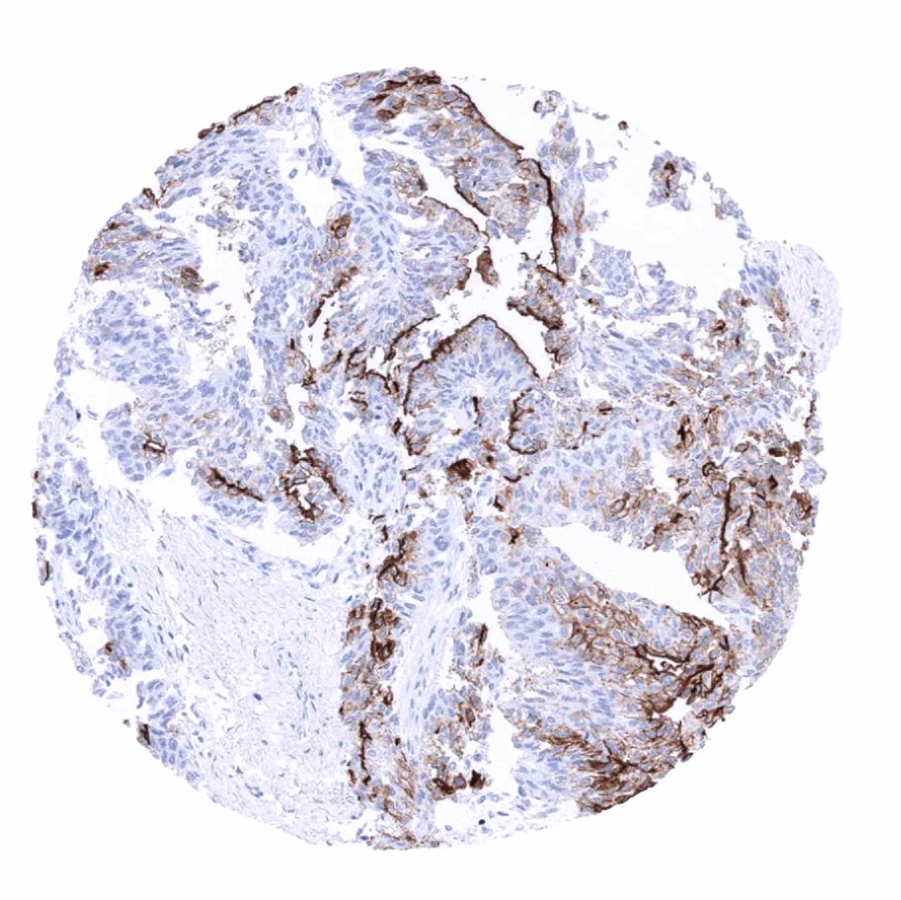
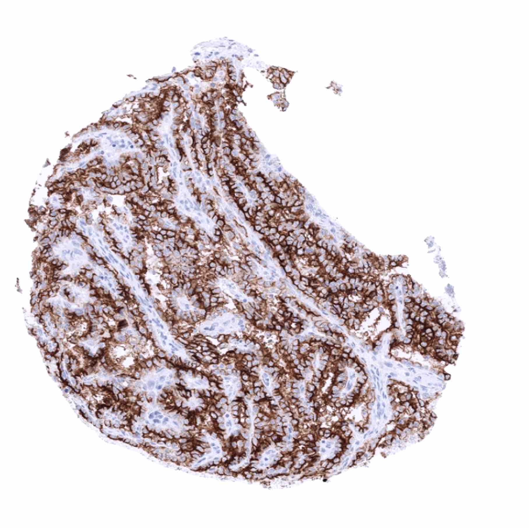
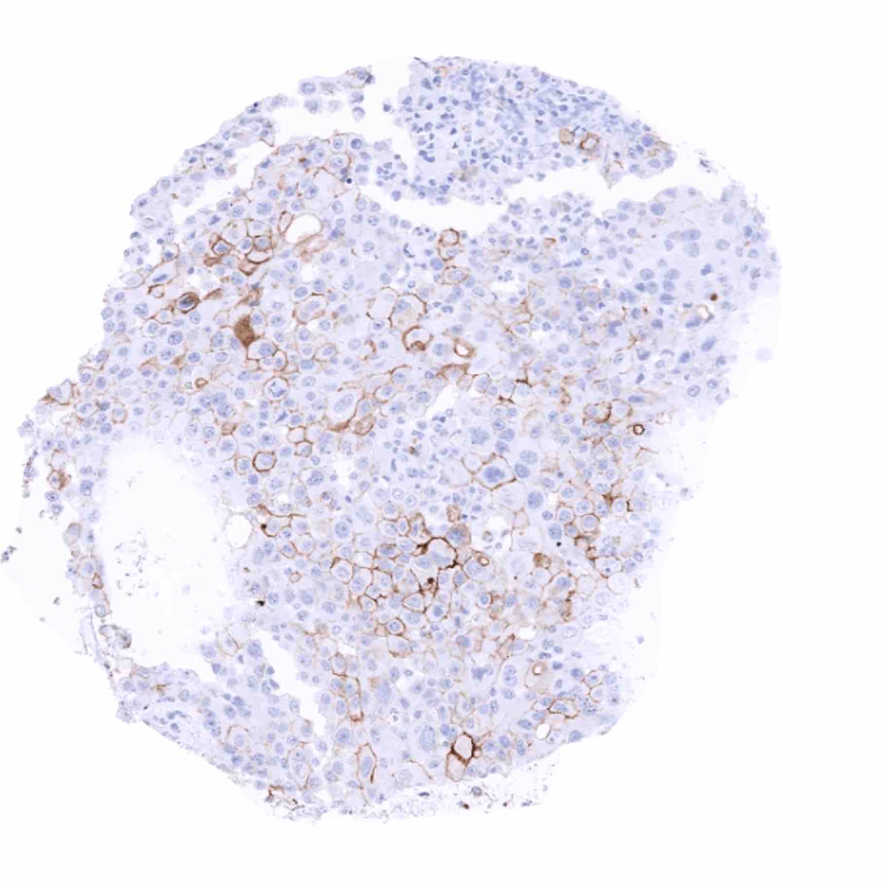
In this study, at least one Upk3b positive case was seen 17 of 151 (11.3%) of tumor types and only 6 (4%) tumor categories included at least one case with strong positivity. Lennartz et al described the highest Upk3b positivity rates in epithelioid (82.1%) and biphasic mesotheliomas (30.8%; positivity always limited to epithelioid cells), followed by various categories of urothelial tumors (10.8–45.7%) including Brenner tumors of the ovary (10.8%) as well as four other subtypes of ovarian cancers (0.9–10.6%). Four additional tumor entities showed a weak to moderate Upk3b positivity in less than 5% of cases. The distribution of positive staining results is shown in an “organ-systematic” and in a “ranking order” figure below (images based on data from Lennartz et al.). Results on possible associations with histopathological and clinical parameters of tumor aggressiveness are also summarized below (table based on data from Lennartz et al.).
Authors conclusions on diagnostic utility of Upk3b immunohistochemistry with respect to the distinction of neoplastic from non-neoplastic tissues (Lennartz et al.):
- Upk3b immunostaining may represent a useful tool in the cytological analysis of pleural effusions because mesothelial cells are selectively stained while most metastatic tumor cells will remain Upk3b negative.
Authors conclusions on diagnostic utility of Upk3b immunohistochemistry with respect to the distinction of different tumor entities (Lennartz et al.):
- Distinction of mesotheliomas (mostly positive) from adenocarcinomas of the lung (negative) and metastatic adenocarcinomas to the lung (negative).
Authors conclusions on prognostic/predictive role of Upk3b expression (Lennartz et al.):
- Absence of Upk3b staining was linked to advanced stage (p=0,0244), nodal metastasis (p=0,0234), and shortened recurrence-free survival (p=0,0084) in muscle-invasive urothelial carcinomas.
- Absence of Upk3b staining was linked to poor grade (p=0,0055) in non-invasive (pTa) urothelial carcinomas.
Figure 1. Uroplakin 3B staining in cancer (“organ-systematic”; according to Lennartz et al.)

Figure 2. Uroplakin 3B staining in cancer (“ranking list”; according to Lennartz et al.)

Protocol Recommendations
IHC users have different preferences on how the stains should look like. Some prefer high staining intensity of the target stain and even accept some background. Others favor absolute specificity and lighter target stains. Factors that invariably lead to more intense staining include higher concentration of the antibody and visualization tools, longer incubation time, higher temperature during incubation, higher temperature and longer duration of the heat induced epitope retrieval (slide pretreatment). The impact of the pH during slide pretreatment has variable effects and depends on the antibody and the target protein.
All images and data shown here and in our image galleries are obtained by the manual protocol described below. Other protocols resulting in equivalent staining are described as well.
Manual protocol
Freshly cut sections should be used (less than 10 days between cutting and staining). Heat-induced antigen retrieval for 5 minutes in an autoclave at 121°C in pH 7,8 Target Retrieval Solution buffer. Apply MSVA-736M at a dilution of 1:150 at 37°C for 60 minutes. Visualization of bound antibody by the EnVision Kit (Dako, Agilent) according to the manufacturer’s directions.
Agilent / Dako – Autostainer Link 48
Pretreatment in PT-Link for 30 minutes at 95°C (pH high); FLEX peroxidase blocking for 5 minutes (room temperature), MSVA-736M 1:150 for 20 minutes (room temperature), FLEX+ mouse/rabbit (LINKER) for 15 minutes (room temperature), horseradish peroxidase (HRP) for 20 minutes (room temperature), FLEX DAB+Sub-Chromo for 10 minutes (room temperature), FLEX hematoxylin for 5 minutes (room temperature).

These images reflect stainings by the protocol described above. It is of note that a comparable staining result can also be obtained by different protocols. In general, a longer pretreatment, a longer incubation time of the primary antibody, a higher antibody concentration, and a longer incubation time of FLEX+LINKER result in stronger staining, potentially at the cost of more background staining. Modifications of the protocol with a strengthening effect on staining intensity in combination with changes of other parameters that result in lower staining intensity can result in a comparable result as shown above.
Leica – BOND RX
Dewax at 72°C for 30 seconds; Pretreatment in Bond Epitope Retrieval Solution (ER2 – EDTA pH9) for 20 minutes at 100°C; Peroxidase blocking for 5 minutes (room temperature), MSVA-736M 1:150 for 15 minutes (room temperature), Post primary (rabbit anti mouse) for 8 minutes (room temperature), Polymer (goat anti rabbit) for 8 minutes (room temperature), mixed DAB refine for 10 minutes (room temperature), hematoxylin for 5 minutes (room temperature).
These images reflect stainings by the protocol described above. It is of note that a comparable staining result can also be obtained by different protocols. In general, a longer pretreatment, a longer incubation time of the primary antibody, a higher antibody concentration, a higher temperature during incubation, and a longer incubation time of Post primary and or the Polymer result in stronger staining, potentially at the cost of more background staining. Modifications of the protocol with a strengthening effect on staining intensity in combination with changes of other parameters that result in lower staining intensity can result in a comparable result as shown above.
Roche – Ventana Discovery ULTRA
Pretreatment for 64 minutes at 100°C (pH 8,4); CM peroxidase blocking for 12 minutes (room temperature), MSVA-736M 1:150 for 20 minutes at 36°C, secondary antibody (anti-mouse HQ) for 12 minutes at 36°C, anti-HQ HRP for 12 minutes at room temperature, DAB at room temperature, hematoxylin II at room temperature for 8 minutes, bluing reagent at room temperature for 4 minutes.

These images depict staining results obtained by the protocol described above. It is of note, that the Ventana machines generally require higher antibody concentrations than other commonly used autostainers because the antibodies are automatically diluted during the procedure. Various other protocols can result in an identical result as shown above. A longer pretreatment, a longer incubation time of the primary antibody, a higher antibody concentration, a higher temperature during incubation, and a longer incubation time of secondary antibody and or the anti-HQ HRP result in stronger staining, potentially at the cost of more background staining.
Potential Research Applications
- The prevalence and clinical significance of Upk3b expression in cancer is unknown.
- The potential diagnostic utility of Upk3b immunostaining needs to be further evaluated.
Evidence for Antibody Specificity in IHC
There are two ways how the specificity of antibodies can be documented for immunohistochemistry on formalin fixed tissues. These are: 1. Comparison with a second independent method for target expression measurement across a large number of different tissue types (orthogonal strategy), and 2. Comparison with one or several independent antibodies for the same target and showing that all positive staining results are also seen with other antibodies for the same target (independent antibody strategy).
Orthogonal validation: For the antibody MSVA-736M considerable discrepancy exist as compared to data from three independent RNA screening studies, including the Human Protein Atlas (HPA) RNA-seq tissue dataset, the FANTOM5 project, and the Genotype-Tissue Expression (GTEx) project, which are all summarized in the Human Protein Atlas (Tissue expression Uroplakin 3B) . In concordance with MSVA-736M IHC data, RNA expression is seen in the urinary bladder, but other tissues with high RNA expression such as esophagus, lung, salivary glands, adipose tissue, testis, vagina, fallopian tube, ovary or the prostate do not show any IHC staining. In addition, MSVA-736M IHC staining observed in amnion cells is not reflected by RNA expression data. This discrepancies are likely to be be due to a contamination of RNA from many organs by (Upk3b expressing) mesothelial cells lining their peritoneal surface of examined tissues. Amnion cells may constitute a too small subpopulation of placenta cells, that Upk3B expression may have been masked by an overwhelming majority of Upk3B negative other cells.
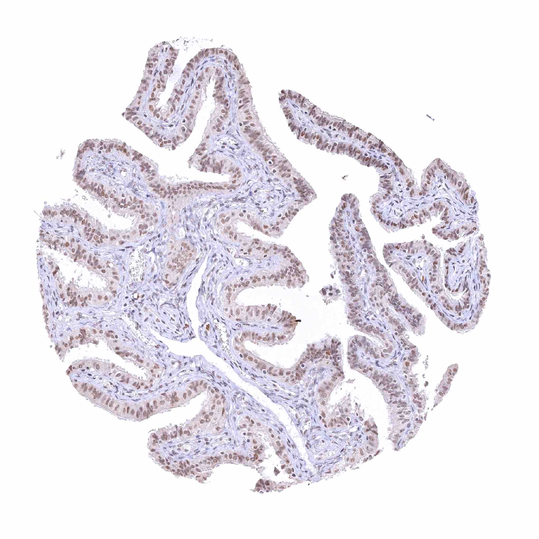
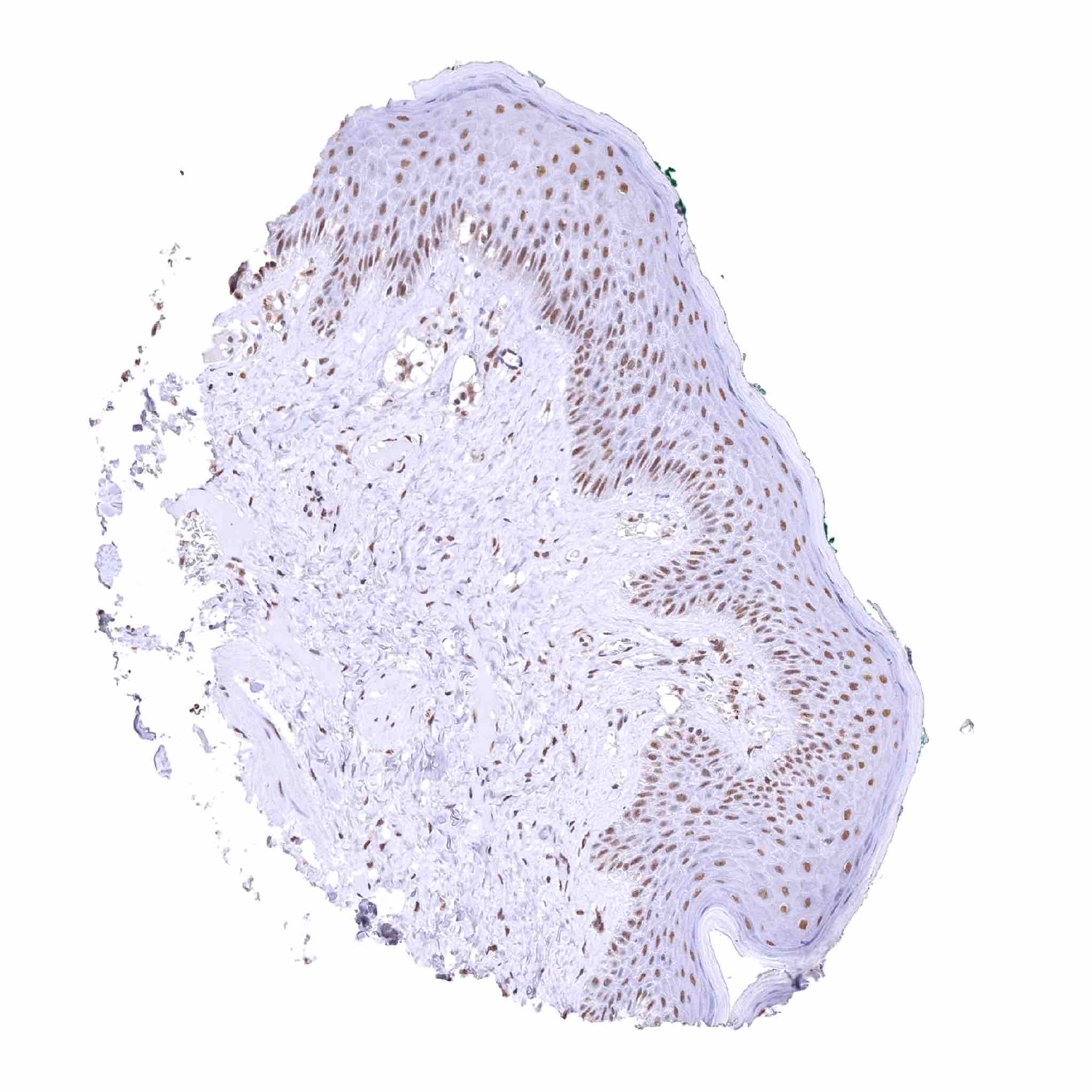
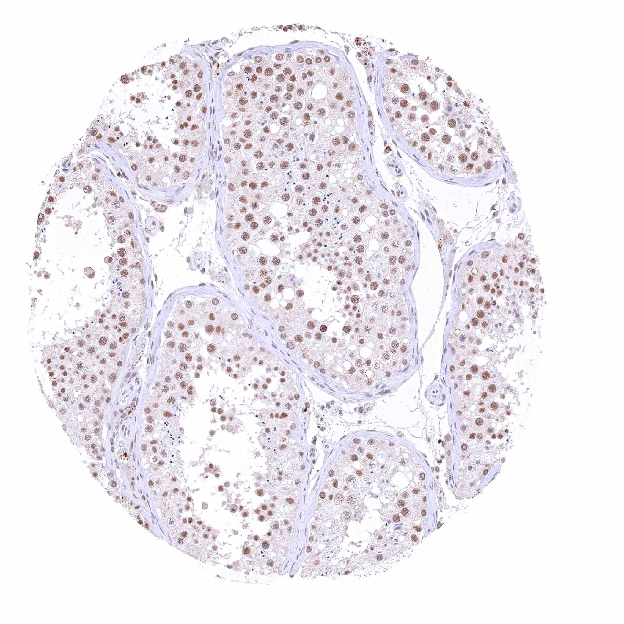
Comparison of antibodies: True expression of Upk3b and correct membranous staining by MSVA-736M of mesothelial, umbrella, and amnion cells is confirmed by comparable staining obtained by a second, independent, commercially available anti-Upk3b antibody, termed “validation antibody”. Independence of the validation antibody is demonstrated by its prominent nuclear staining of numerous tissues such as the skin and the testis, that were not stained by MSVA-736M. These stainings were therefore considered antibody-specific cross-reactivities of the validation antibody.