Product details
Synonyms = H. pylori
Antibody type = Mouse monoclonal / Mouse IgG2b, kappa
Clone = MSVA-466M
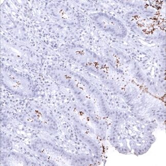
Positive control = Gastric tissue with known helicobacter pylori infection should show a distinct staining of bacteria on the surface and/or within glands.
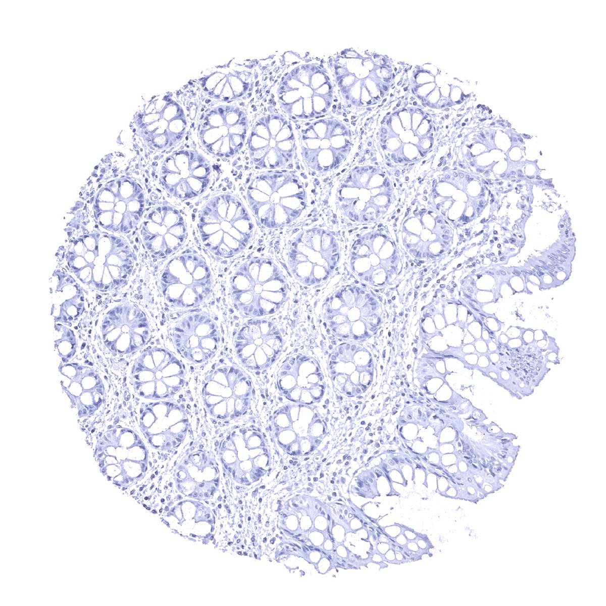
Negative control = Gastric tissue: Normal epithelial and stromal cells should not stain.
Cellular localization = Cell surface. Cytoplasm.
Reactivity = Human
Application = Immunohistochemistry
Dilution = 1:100 – 1:200
Intended Use = Research Use Only
Relevance of Antibody
Marker for Helicobacter pylori.
Biology Behind
Helicobacter pylori is a gram-negative spiral bacterium which can be found in the stomach. H. pylori infection usually does not cause symptoms but sometimes results in inflammation or even ulcers of the stomach or the first part of the small intestine. H. pylori has been associated with gastric cancer and gastric mucosa-associated lymphoid tissue (MALT) lymphoma. In 2015, it was estimated that over 50% of the world’s population were carrying H. pylori in their upper gastrointestinal tracts, a situation which is believed to be more common in developing than in developed countries. The Helicobacter antibody MSVA-466M stains the helicobacter catalase protein.
Staining Pattern in Normal Tissues
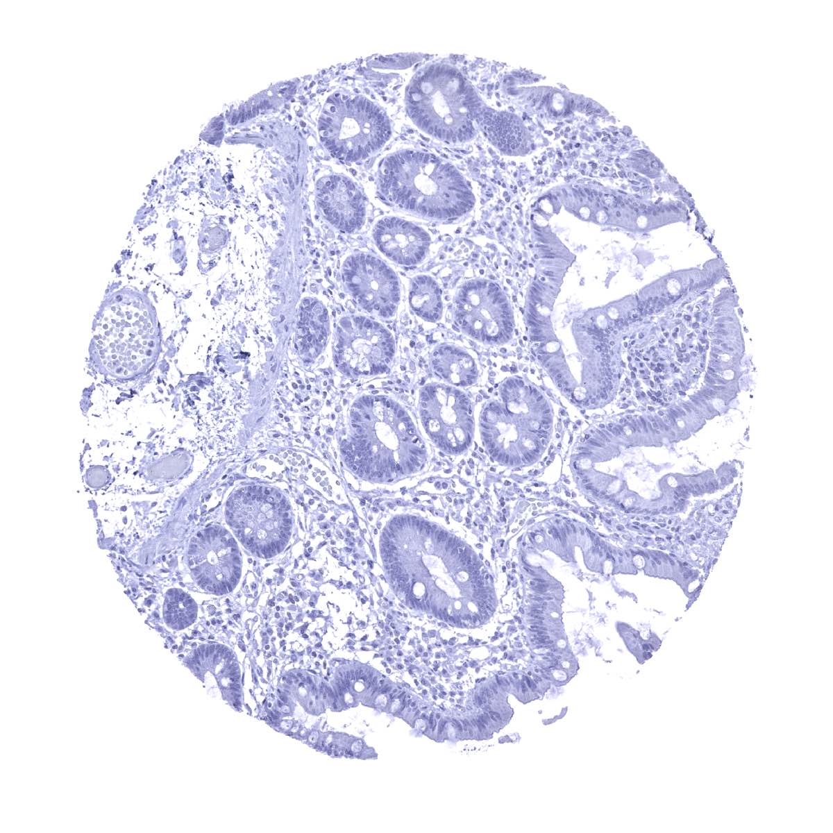
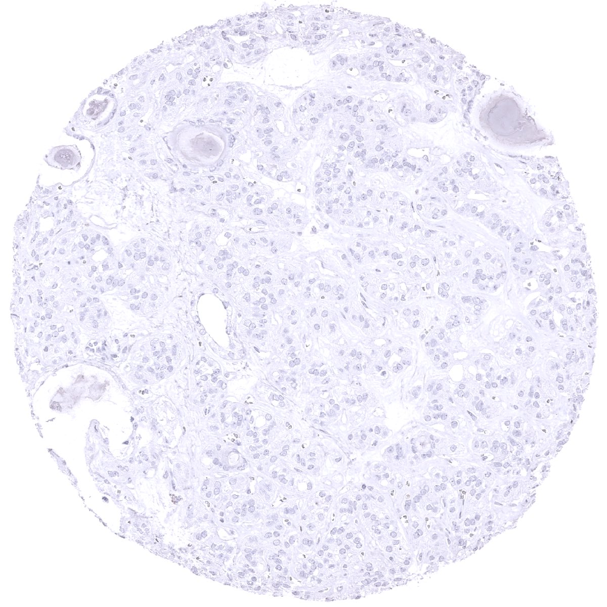
Among normal adult tissues, immunostaining is not observed for the helicobacter specific antibody MSVA-466M.
As MSVA-466M detects a microbial antigen, normal tissue findings cannot be compared with data from the protein atlas.
Positive control = Gastric tissue with known helicobacter pylori infection should show a distinct staining of bacteria on the surface and/or within glands.
Negative control = Gastric tissue: Normal epithelial and stromal cells should not stain.

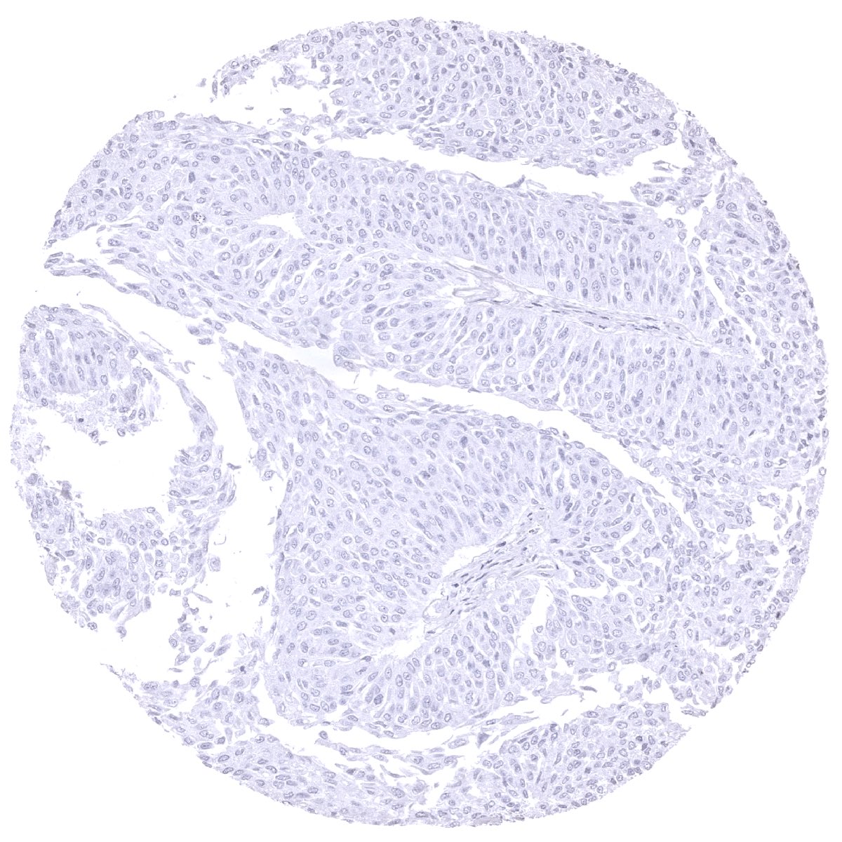
Staining Pattern in Relevant Tumor Types
A positive immunostaining with the antibody MSVA-466M immunostaining is not seen in cancer cells.
Compatibility of Antibodies
No data available at the moment
Protocol Recommendations
IHC users have different preferences on how the stains should look like. Some prefer high staining intensity of the target stain and even accept some background. Others favor absolute specificity and lighter target stains. Factors that invariably lead to more intense staining include higher concentration of the antibody and visualization tools, longer incubation time, higher temperature during incubation, higher temperature and longer duration of the heat induced epitope retrieval (slide pretreatment). The impact of the pH during slide pretreatment has variable effects and depends on the antibody and the target protein.
All images and data shown here and in our image galleries are obtained by the manual protocol described below. Other protocols resulting in equivalent staining are described as well.
Manual protocol
Freshly cut sections should be used (less than 10 days between cutting and staining). Heat-induced antigen retrieval for 5 minutes in an autoclave at 121°C in pH 7,8 Target Retrieval Solution buffer. Apply MSVA-466M at a dilution of 1:150 at 37°C for 60 minutes. Visualization of bound antibody by the EnVision Kit (Dako, Agilent) according to the manufacturer’s directions.
Potential Research Applications
- The role of H.p. in various extra-gastrointestinal diseases is under investigation.
- The risk situation for patients to still suffer from H.p. associated disease even after H.p. eradication needs further assessment.