Product details
Synonyms = Antigen NY-CO-13, BCC7, Cellular Tumor Antigen p53, LFS1, TP53, Transformation Related Protein 53 (TRP53), Tumor Protein p53, Tumor Suppressor p53
Antibody type = Recombinant Rabbit monoclonal / Rabbit IgG
Clone = MSVA-053R
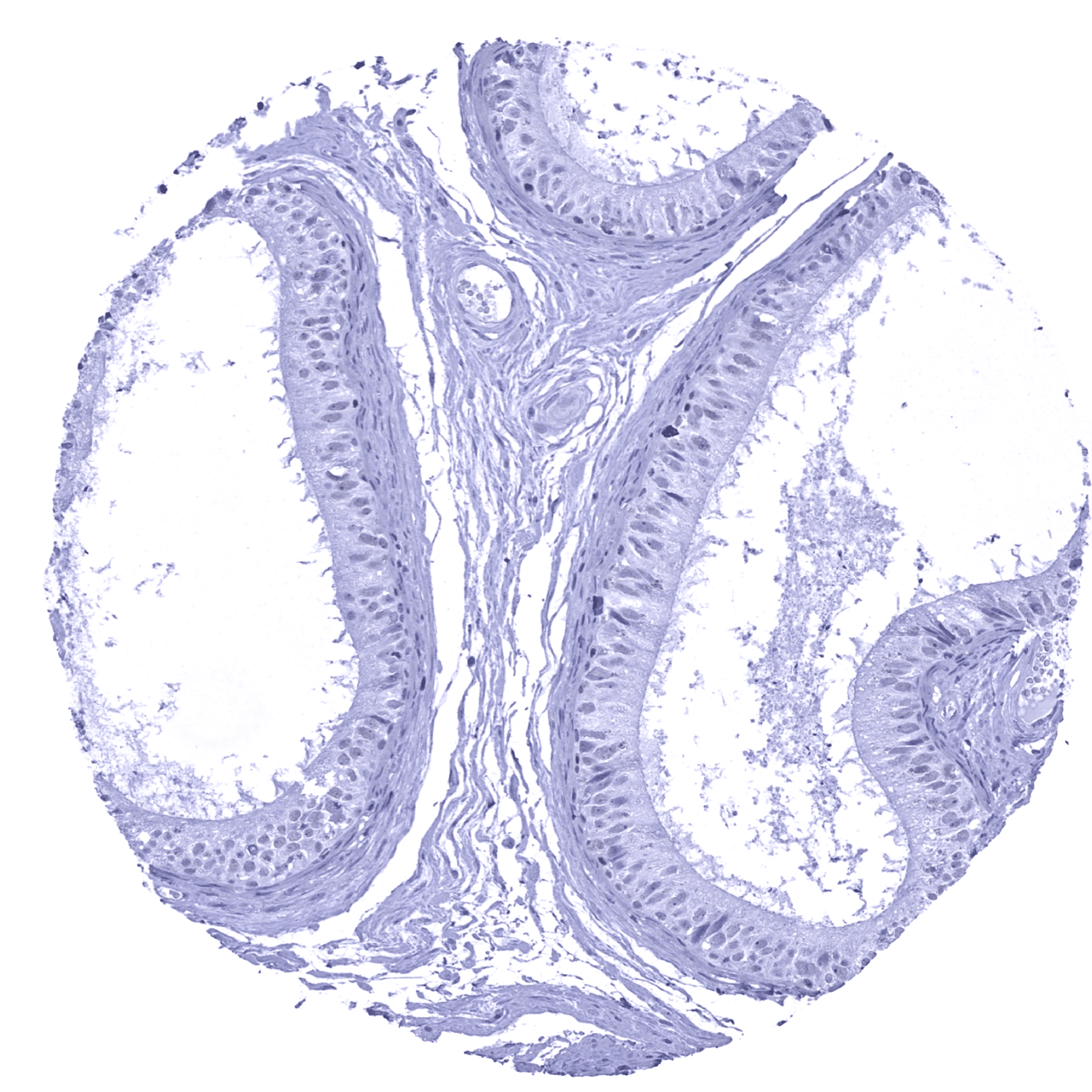
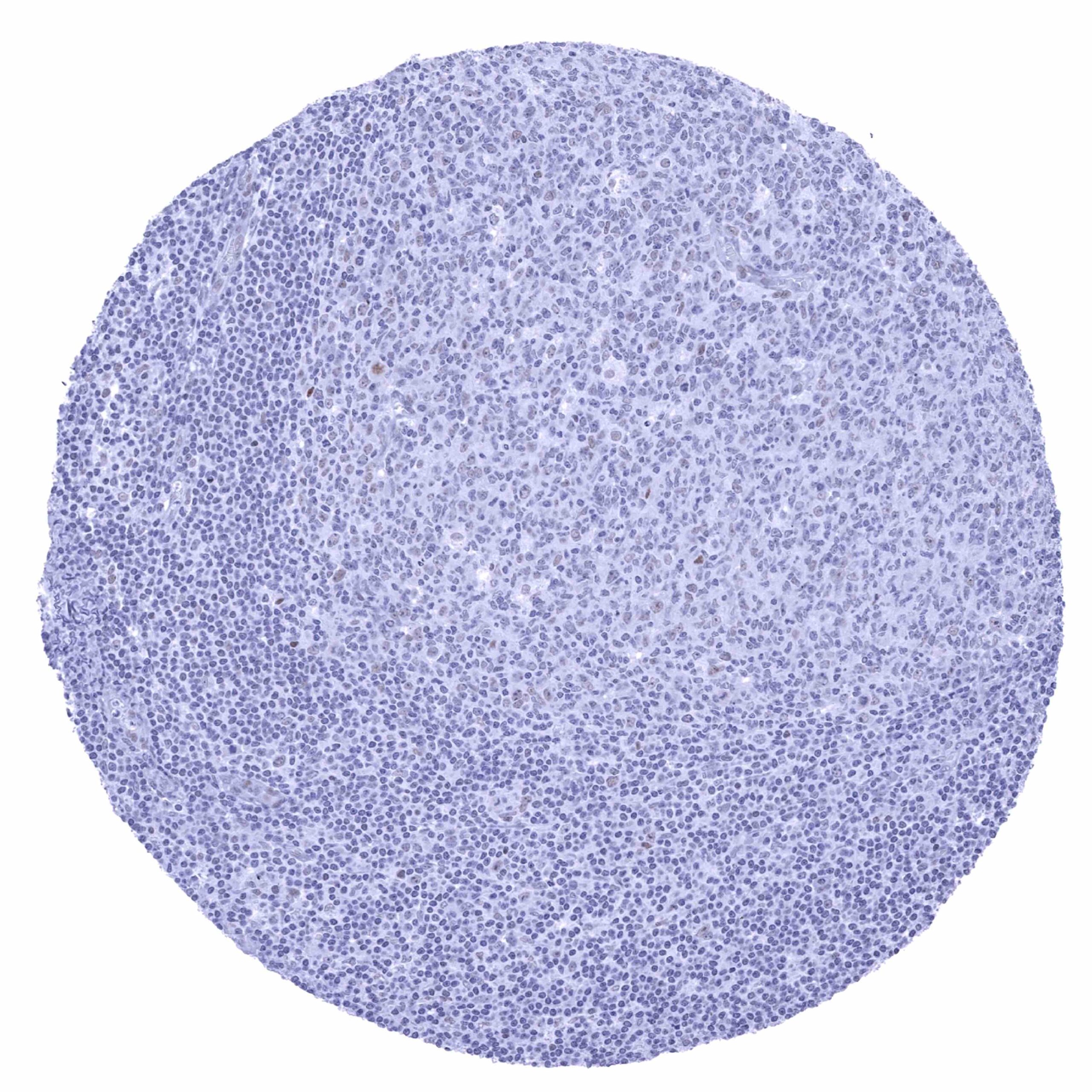
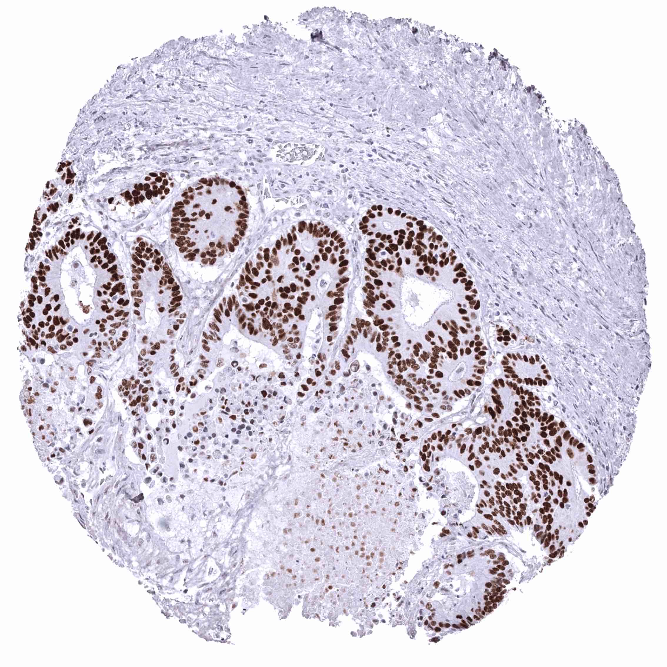
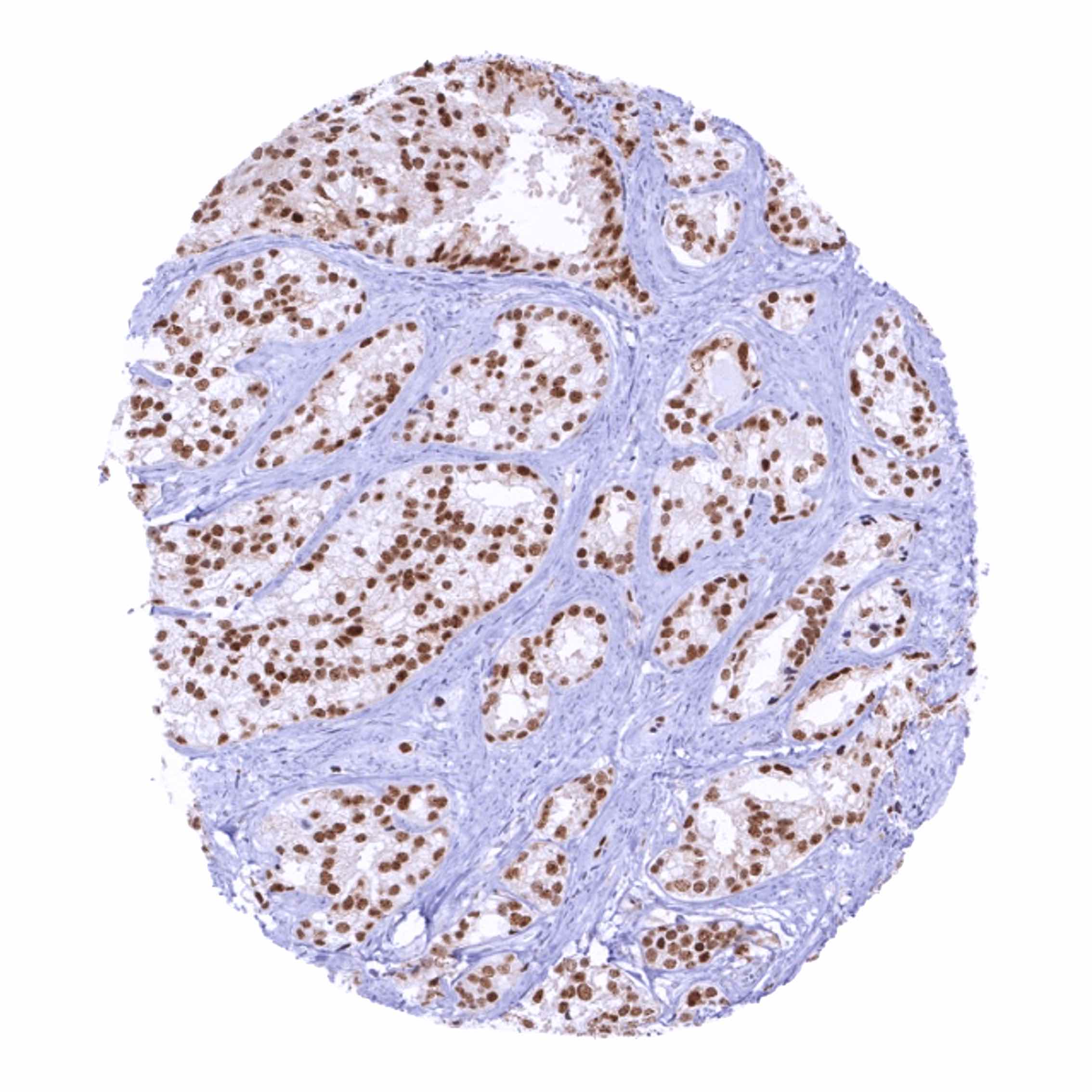
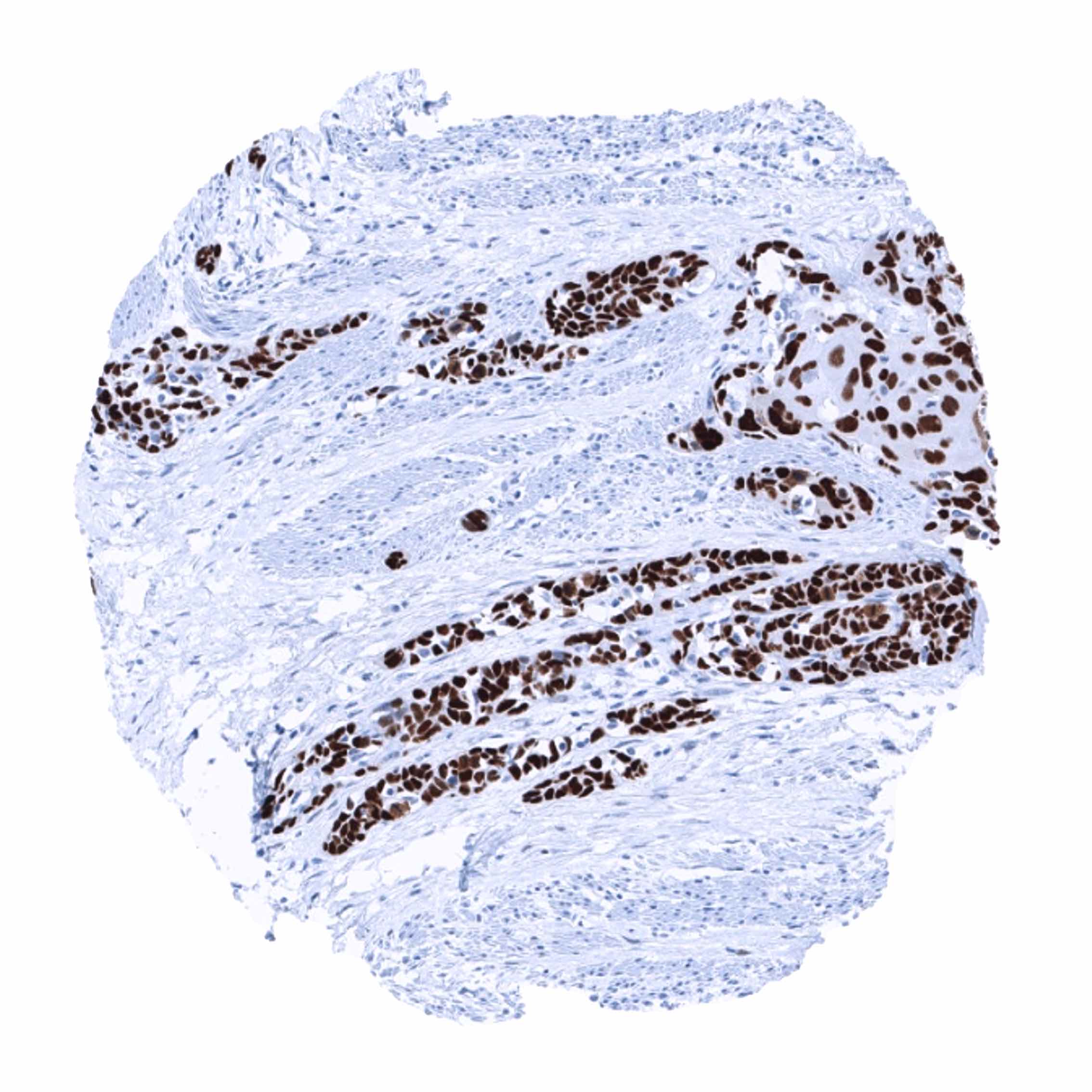
Positive control = Tonsil: More than 20 % of germinal centre B-cells must show a weak to moderate nuclear staining.
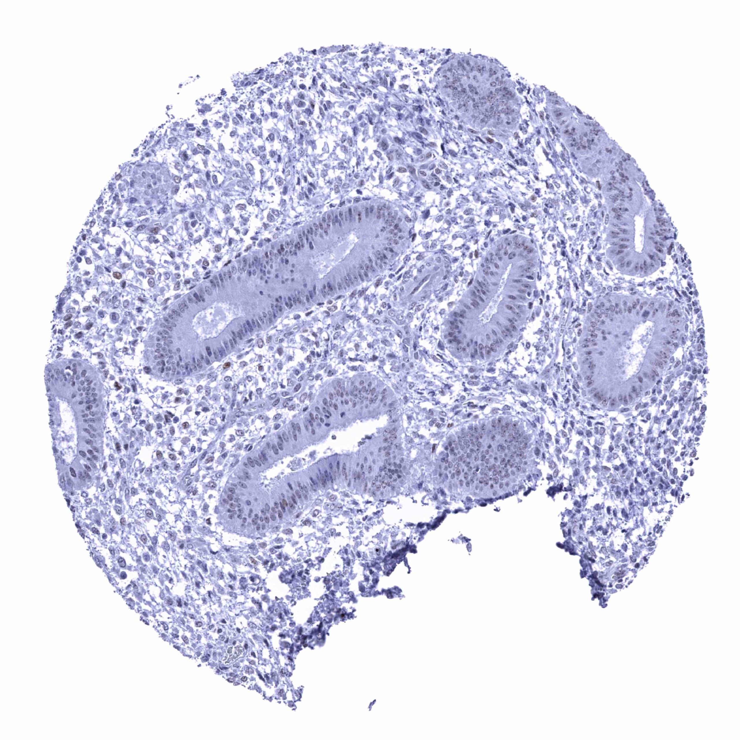
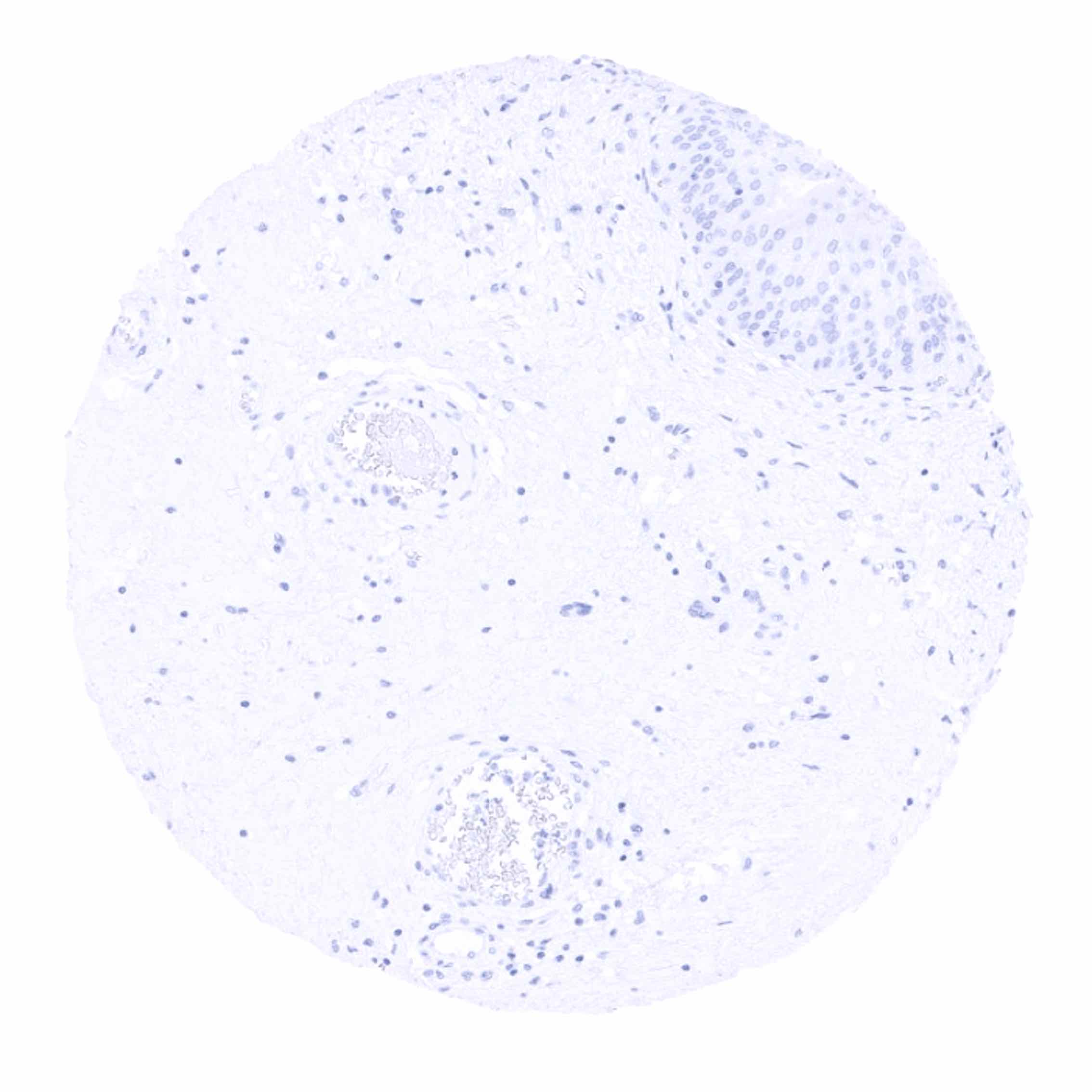
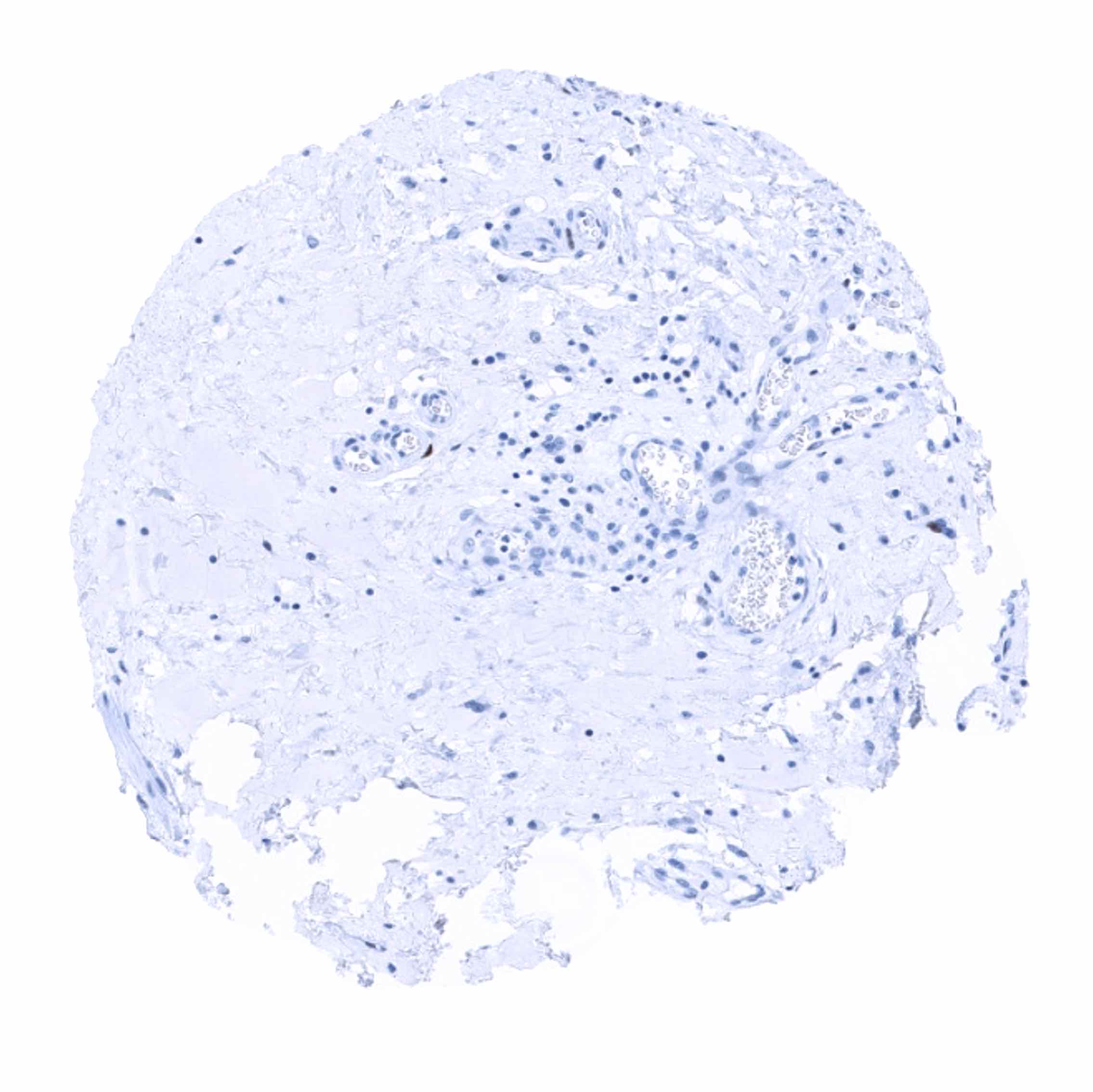
Negative control = Colon: Luminal epithelial cells must remain p53 negative.
Cellular localization = Intracellular
Reactivity = Human
Application = Immunohistochemistry
Dilution = 1:100 – 1:200
Intended Use = Research Use Only
Relevance of Antibody
p53 is the most frequently mutated gene in human cancers.
Biology Behind
The tumor protein p53 is a tumor suppressor protein coded by the TP53 gene on 17p13.1. The protein and its homologs is crucial for preventing cancer formation in vertebrates. Because of its role in preserving DNA stability it has been termed “the guardian of the genome”. In normal tissues, p53 becomes activated in response to various different cellular stress events, including but not limited to DNA damage, oxidative stress, osmotic shock, ribonucleotide depletion, and deregulated oncogene expression. Activation occurs through a prolonged half-life resulting in nuclear accumulation of the protein and conformational change. In case of DNA damage, p53 activation can induce cell cycle arrest, DNA repair and invoke apoptosis if repair is not feasible. P53 activity is regulated by various protein kinases. Interaction with MDM2 leads to p53 inactivation. The p53 gene is the most frequently mutated gene in human cancer. More than 50% of all cancers harbor p53 mutations. Various different mutations exert variable effects on the p53 protein including loss of function and gain of function mutations. A large fraction of mutations result in a nuclear accumulation of altered p53 protein which becomes visible by immunohistochemical analysis. Although gain of function mutations can go along with nuclear p53 accumulation, immunohistochemical p53 positivity is generally viewed as a sign of p53 inactivation.
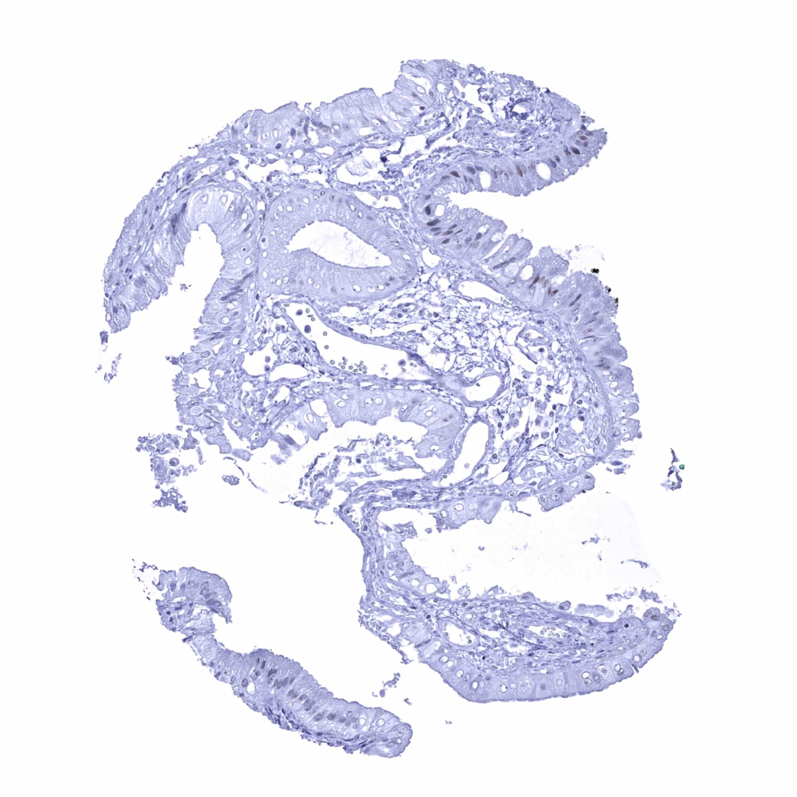
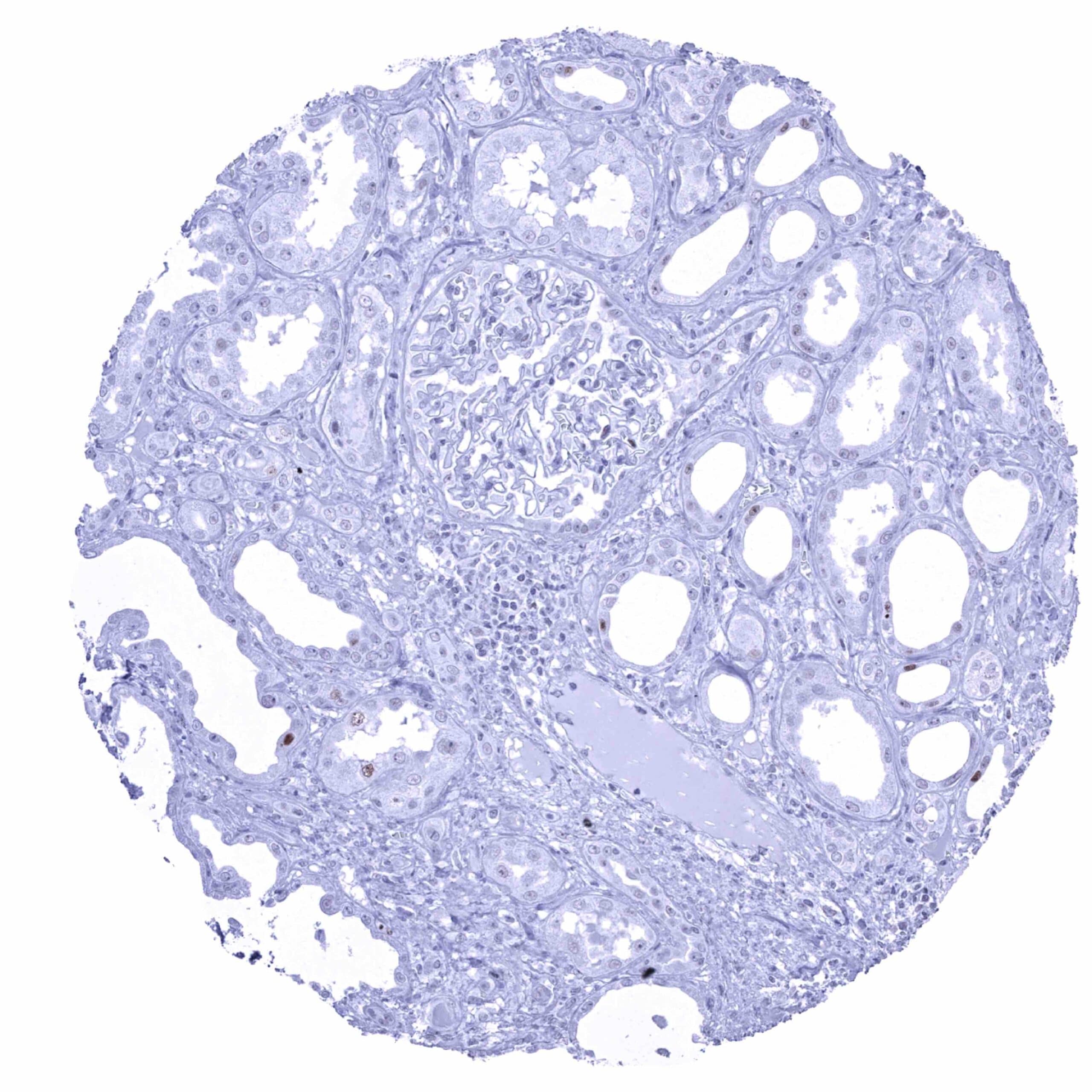
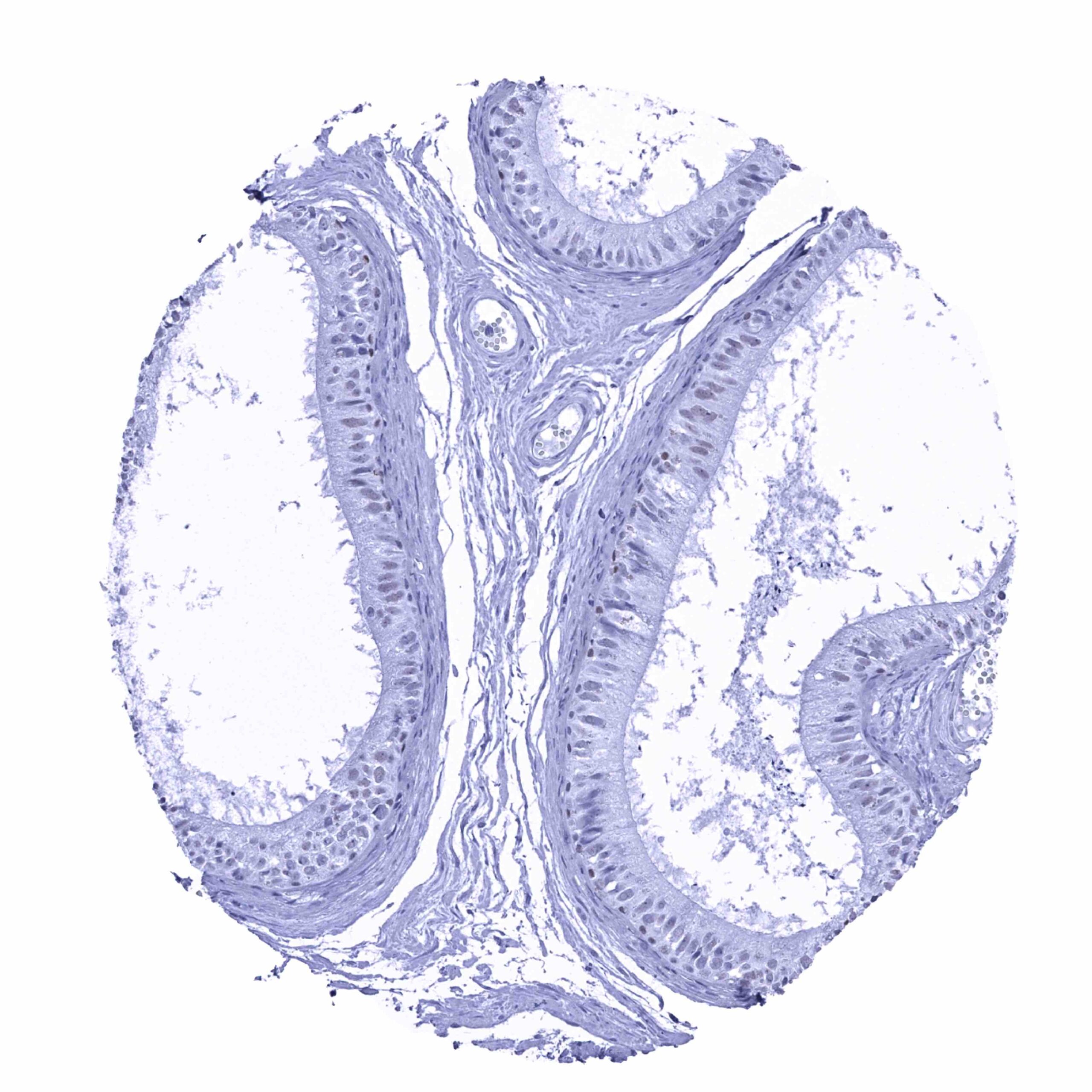
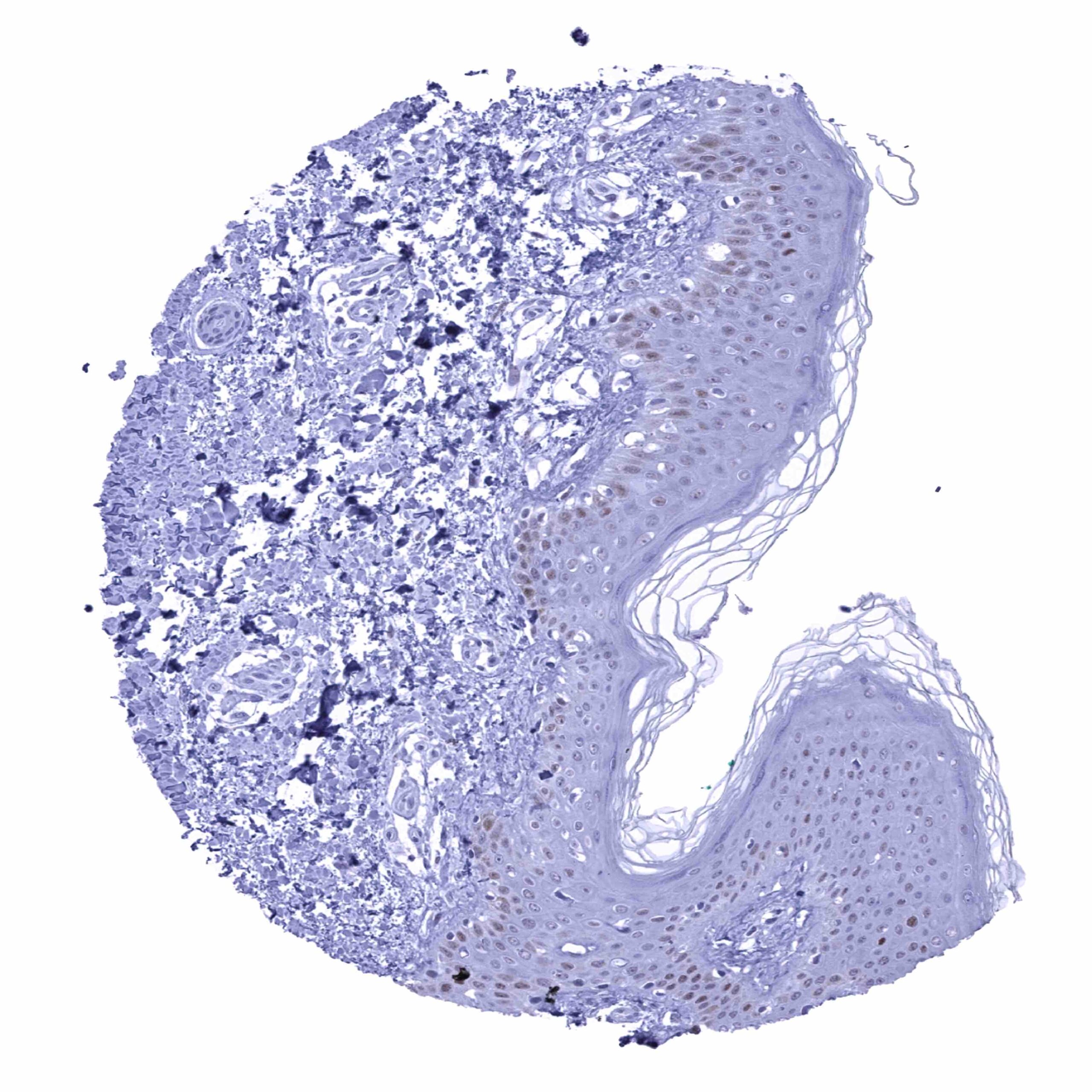
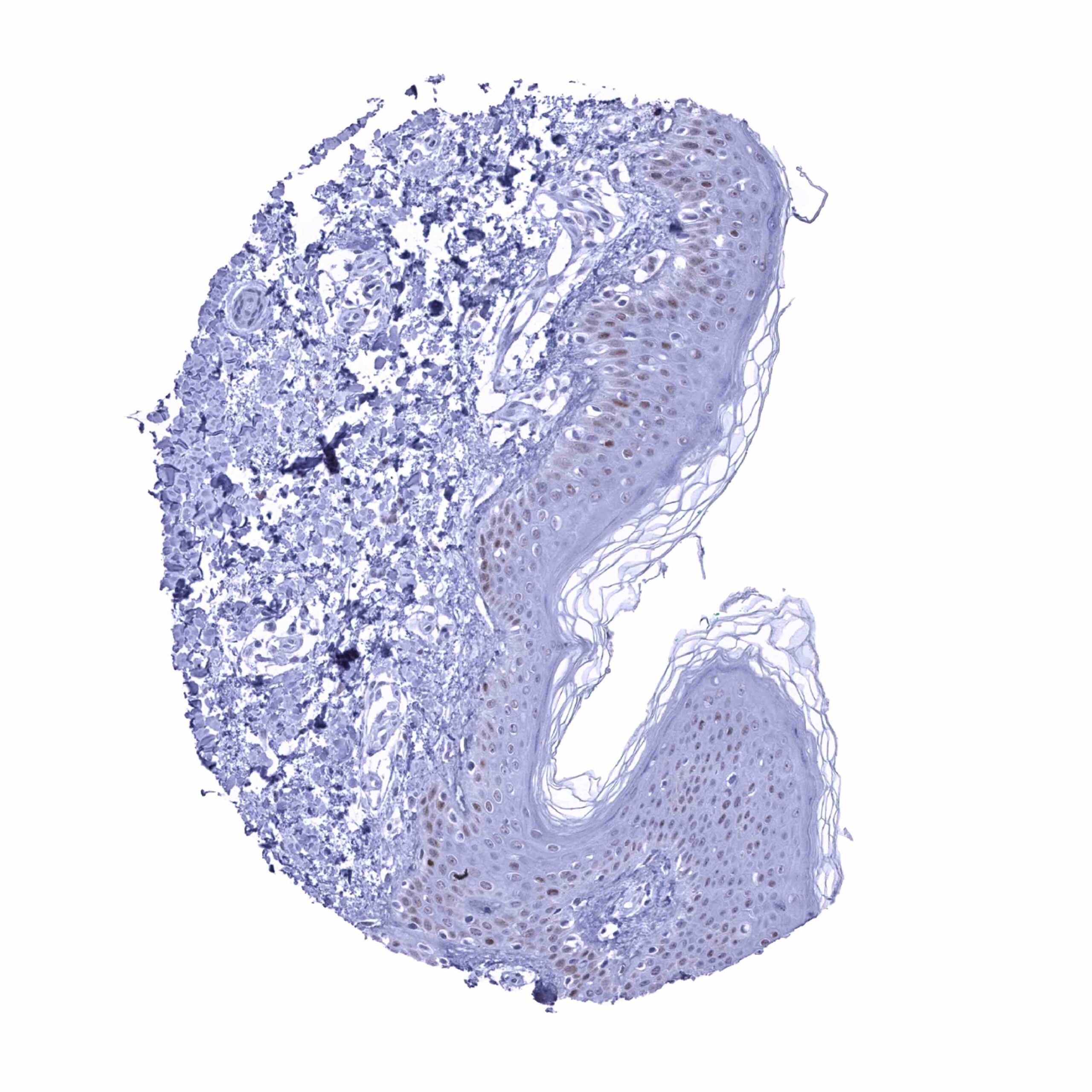
Staining Pattern in Normal Tissues
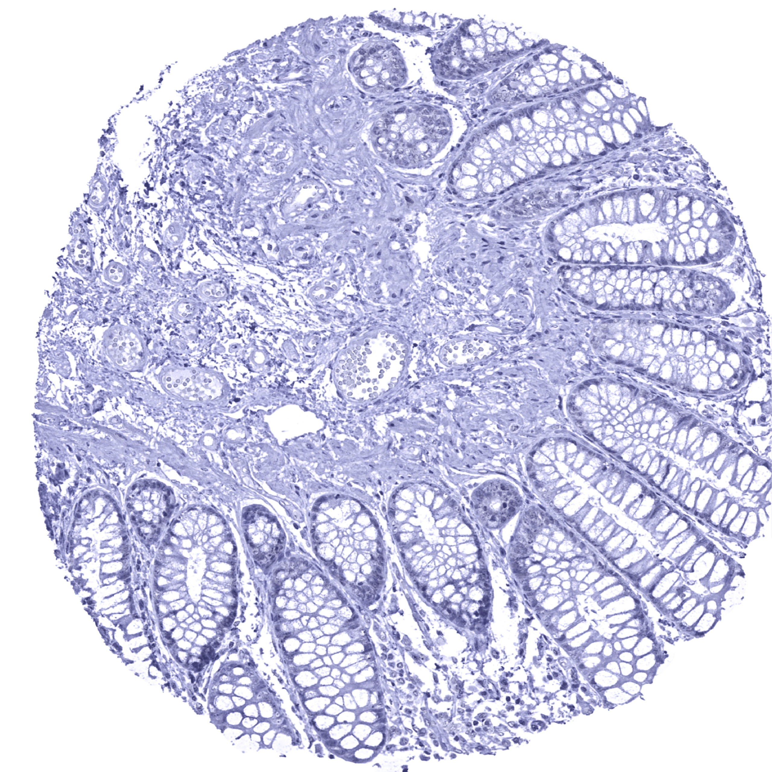
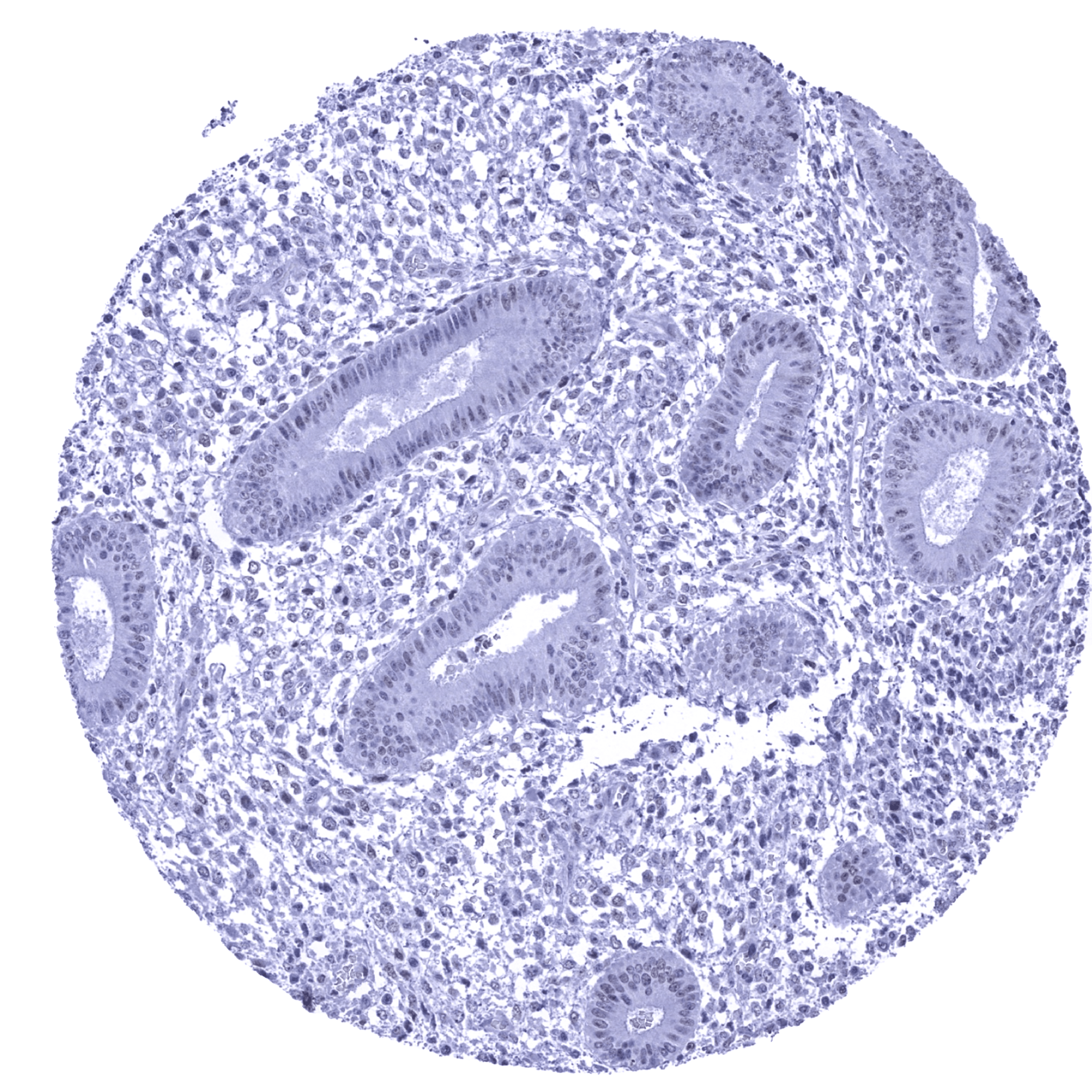
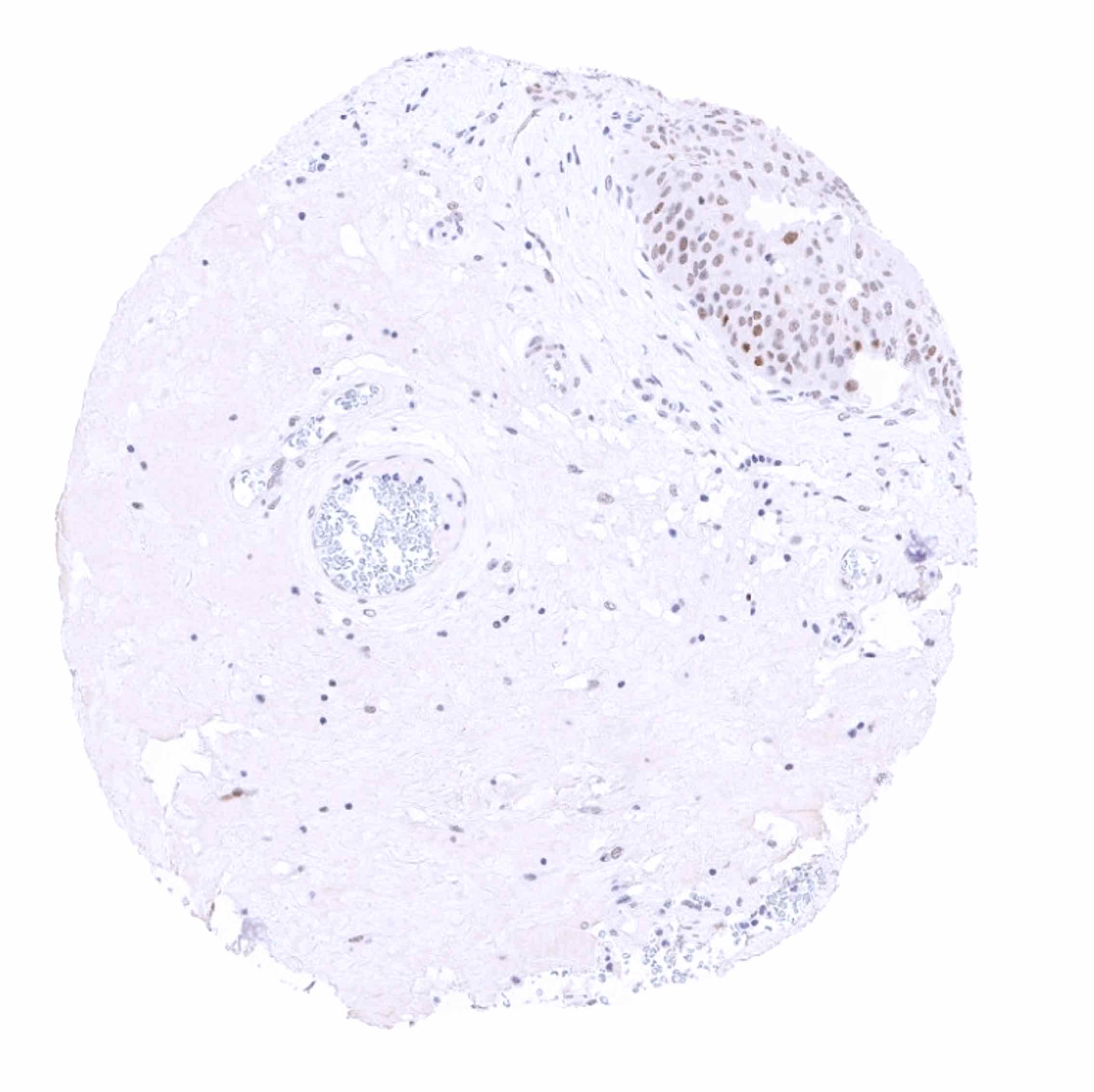
p53 is expressed in the nuclei of all normal cells, but usually not immunohistochemically detectable due to a very short half-life (10-20 min.). A weak to moderate p53 staining can, however, be seen in a subset of (mostly proliferative) cells of various epithelial cell types such as in the colon, gallbladder, basal cells of the prostate, respiratory epithelium, squamous epithelium, and the urothelium. A faint p53 positivity can also be seen in some endothelial cells, spermatogonia, and in a fraction of lymphocytes, especially in germinal centres.
The findings described above are this consistent with the RNA data described in the Human Protein Atlas (Tissue expression p53)
Positive control = Tonsil: More than 20 % of germinal centre B-cells must show a weak to moderate nuclear staining.
Negative control = Colon: Luminal epithelial cells must remain p53 negative.
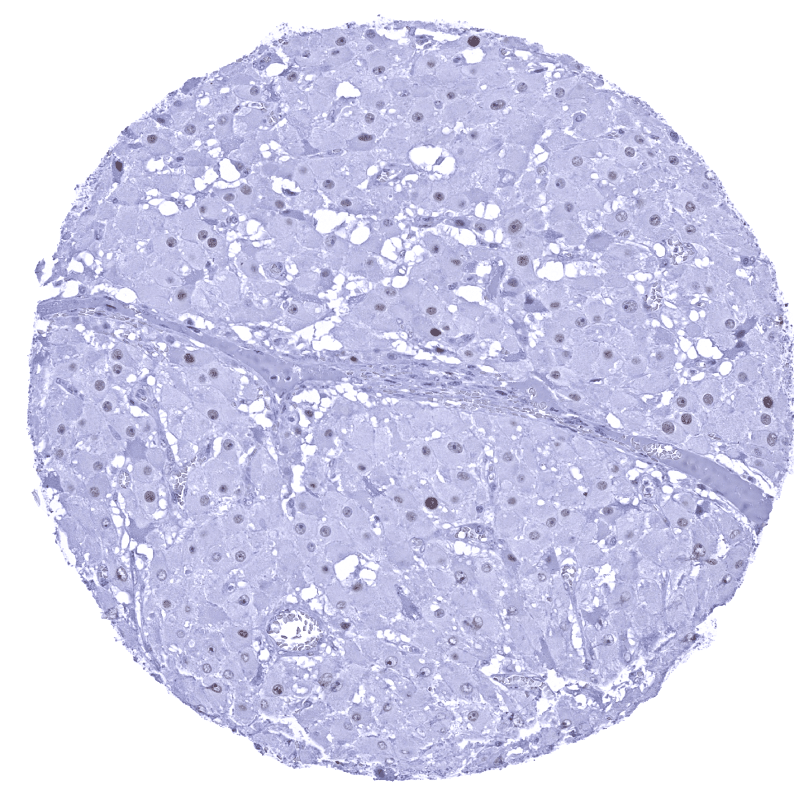
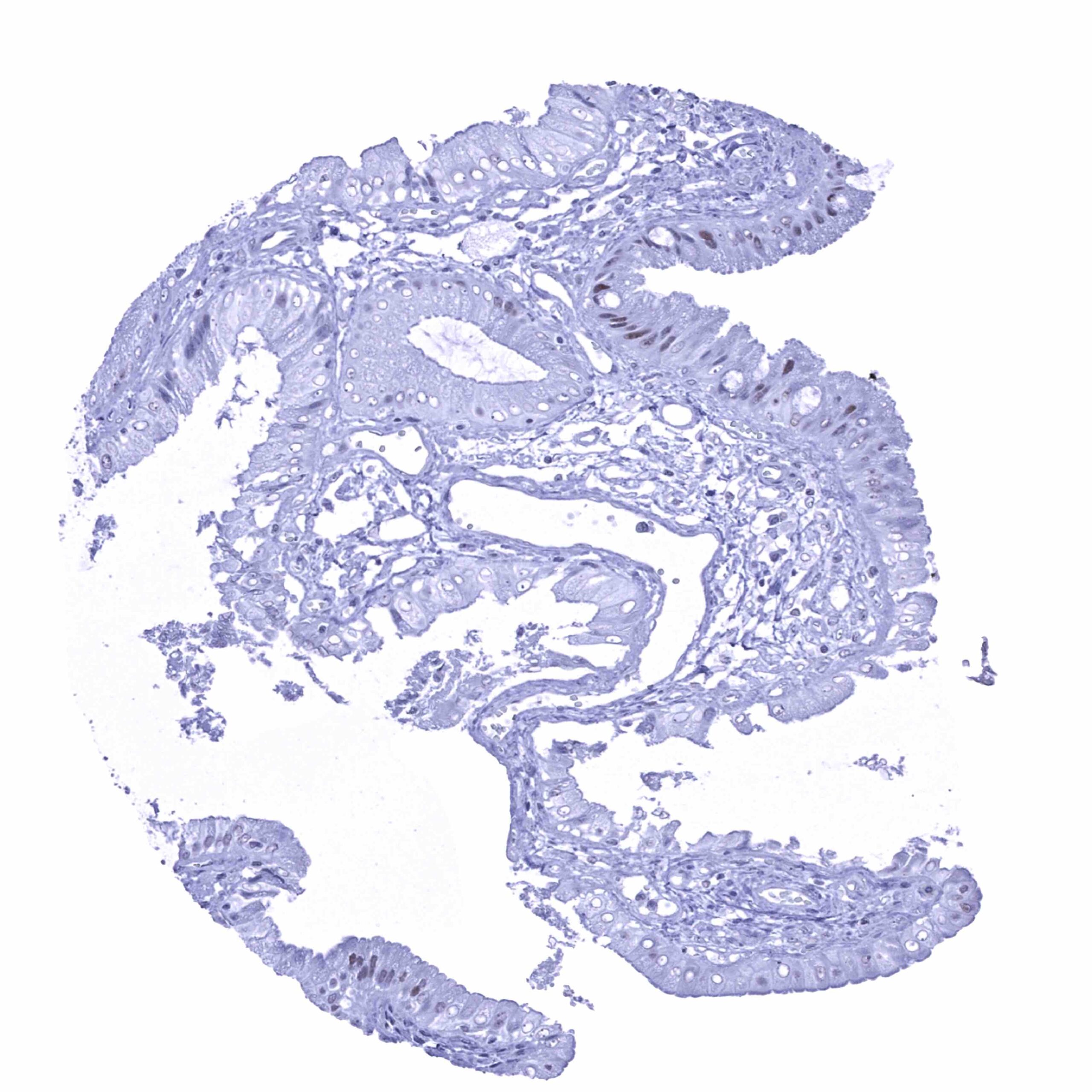
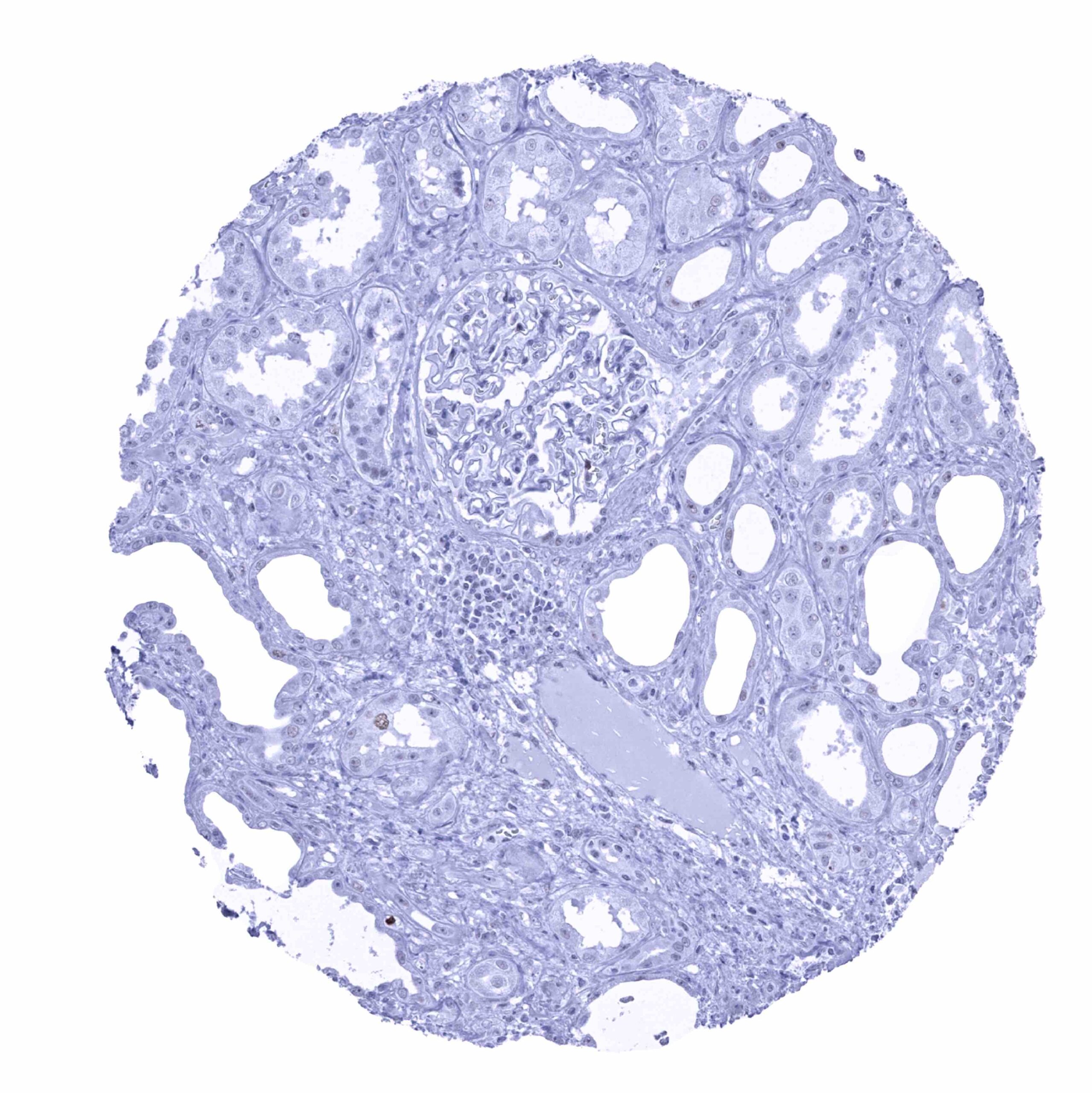
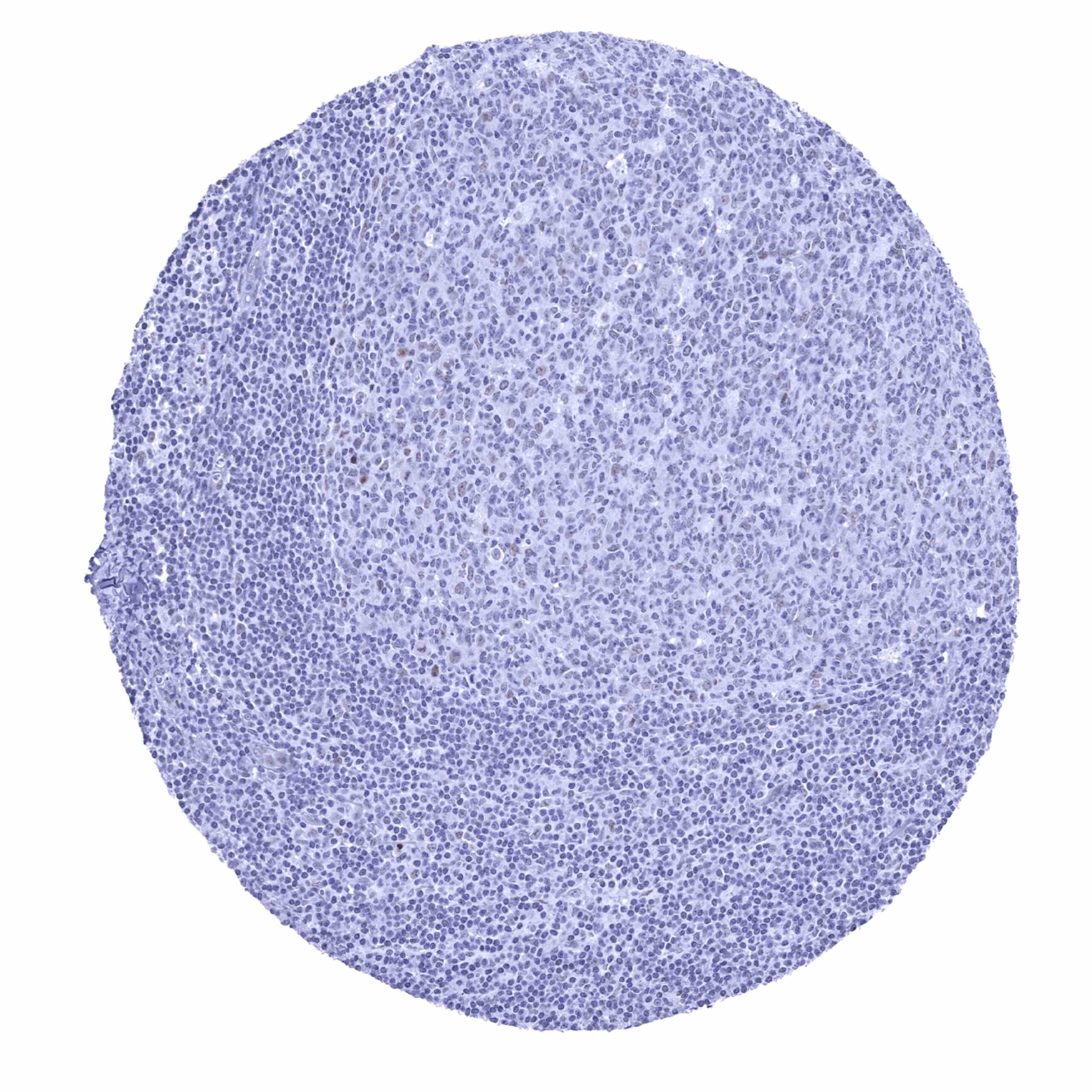
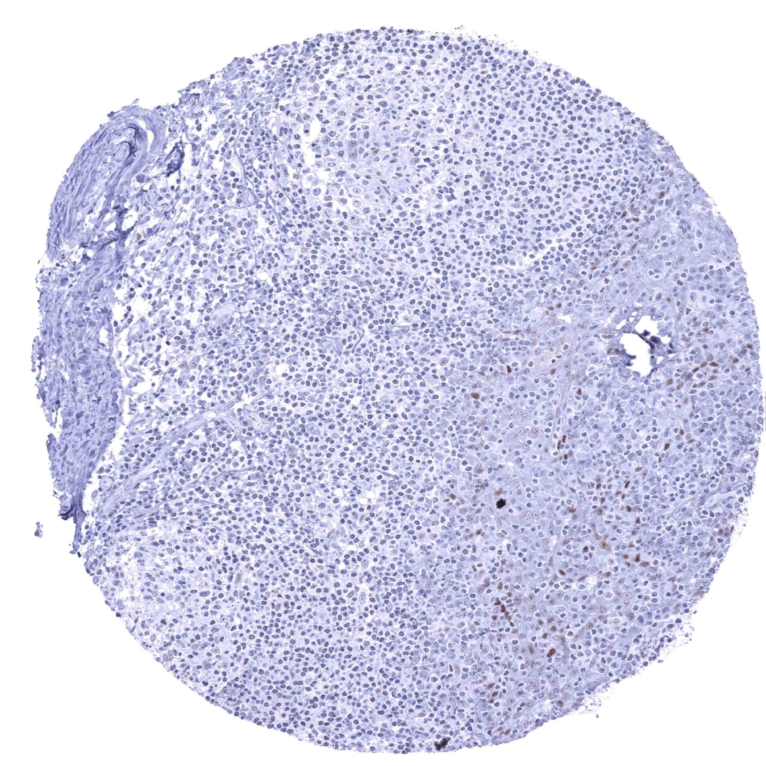
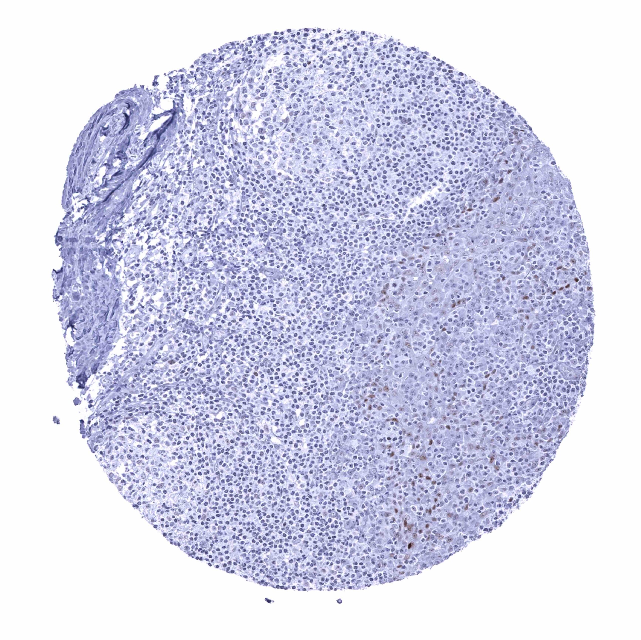
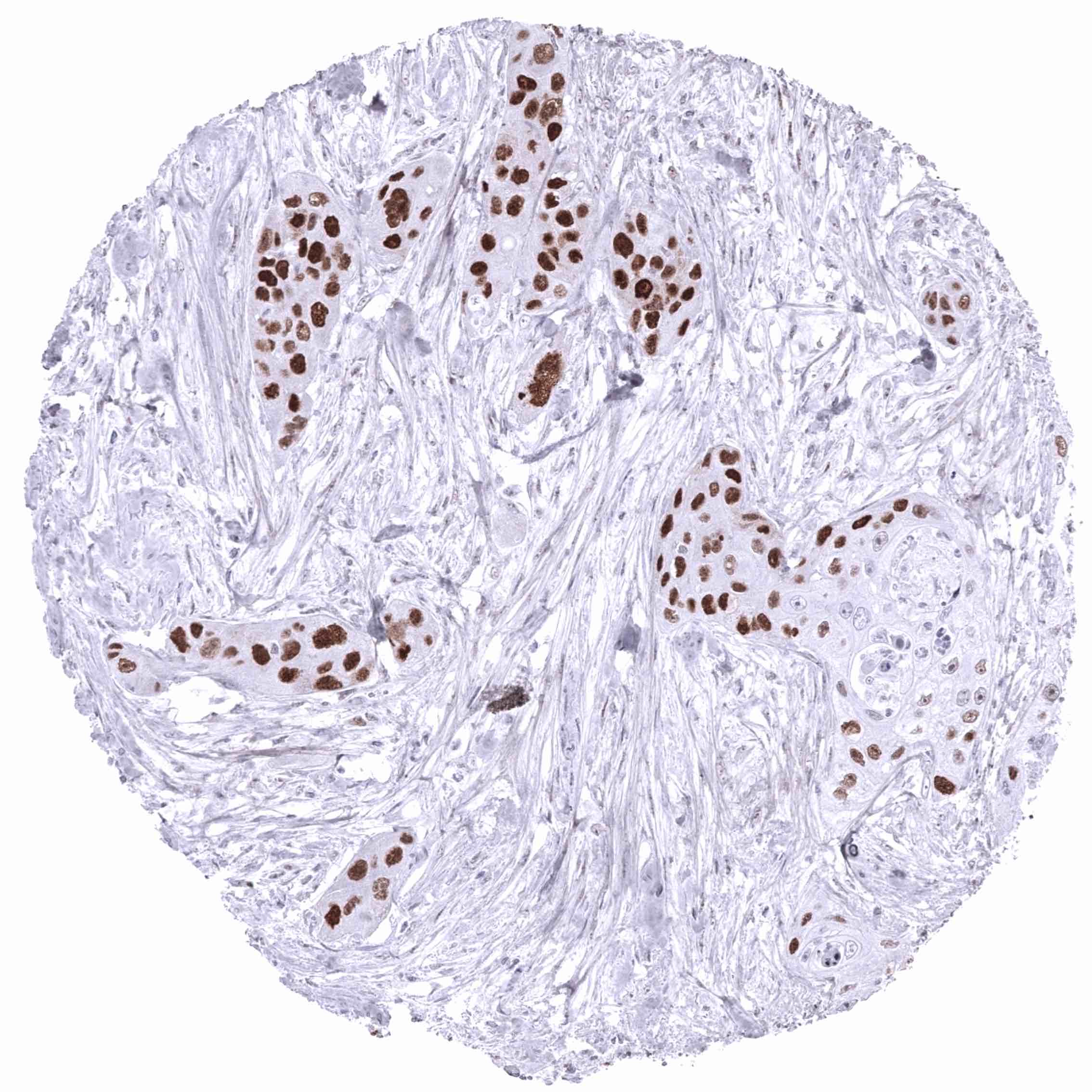
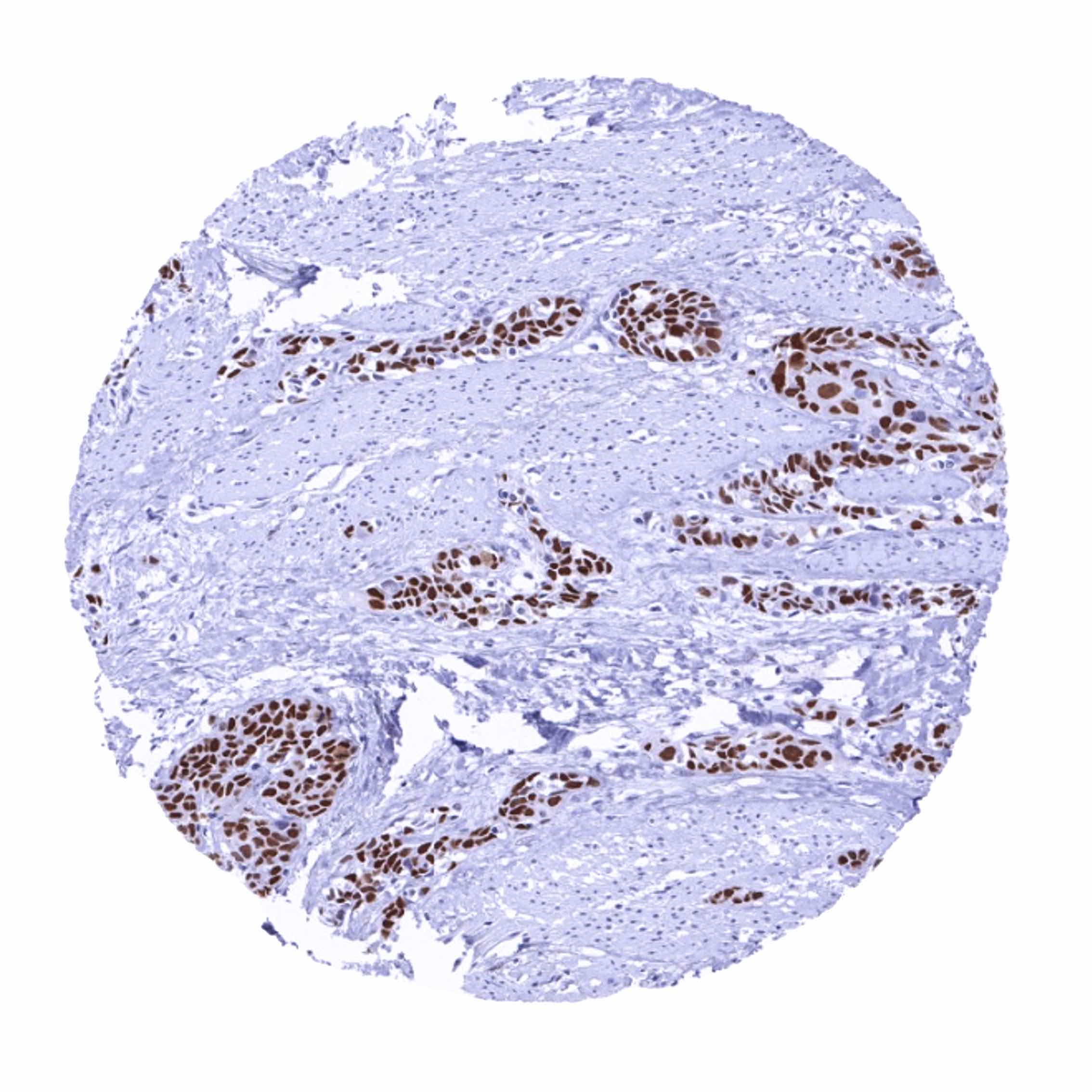
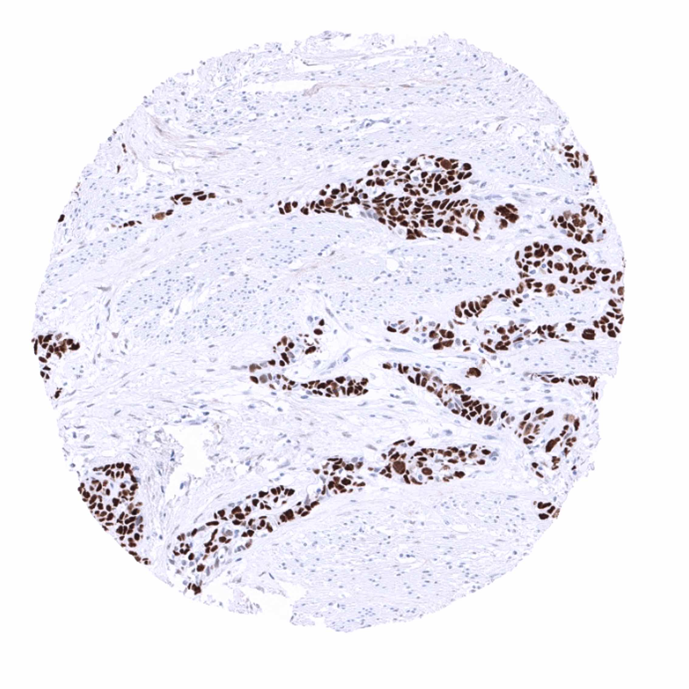
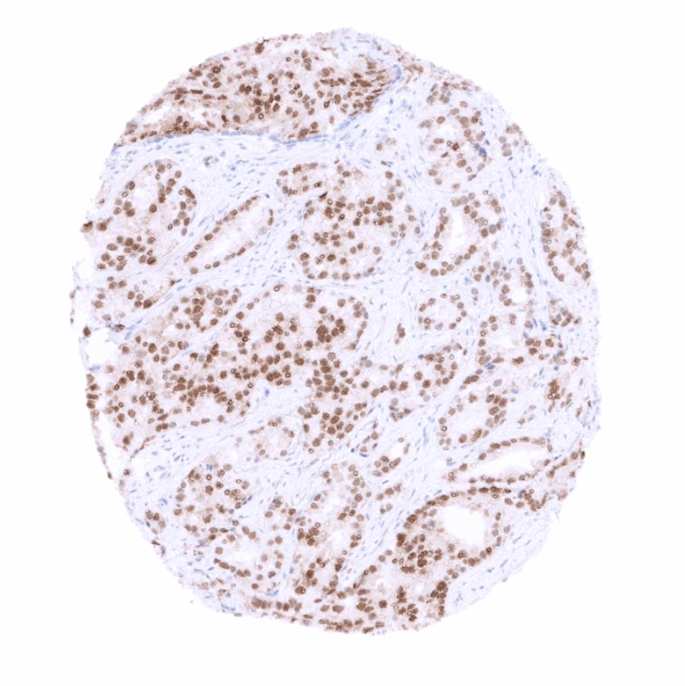
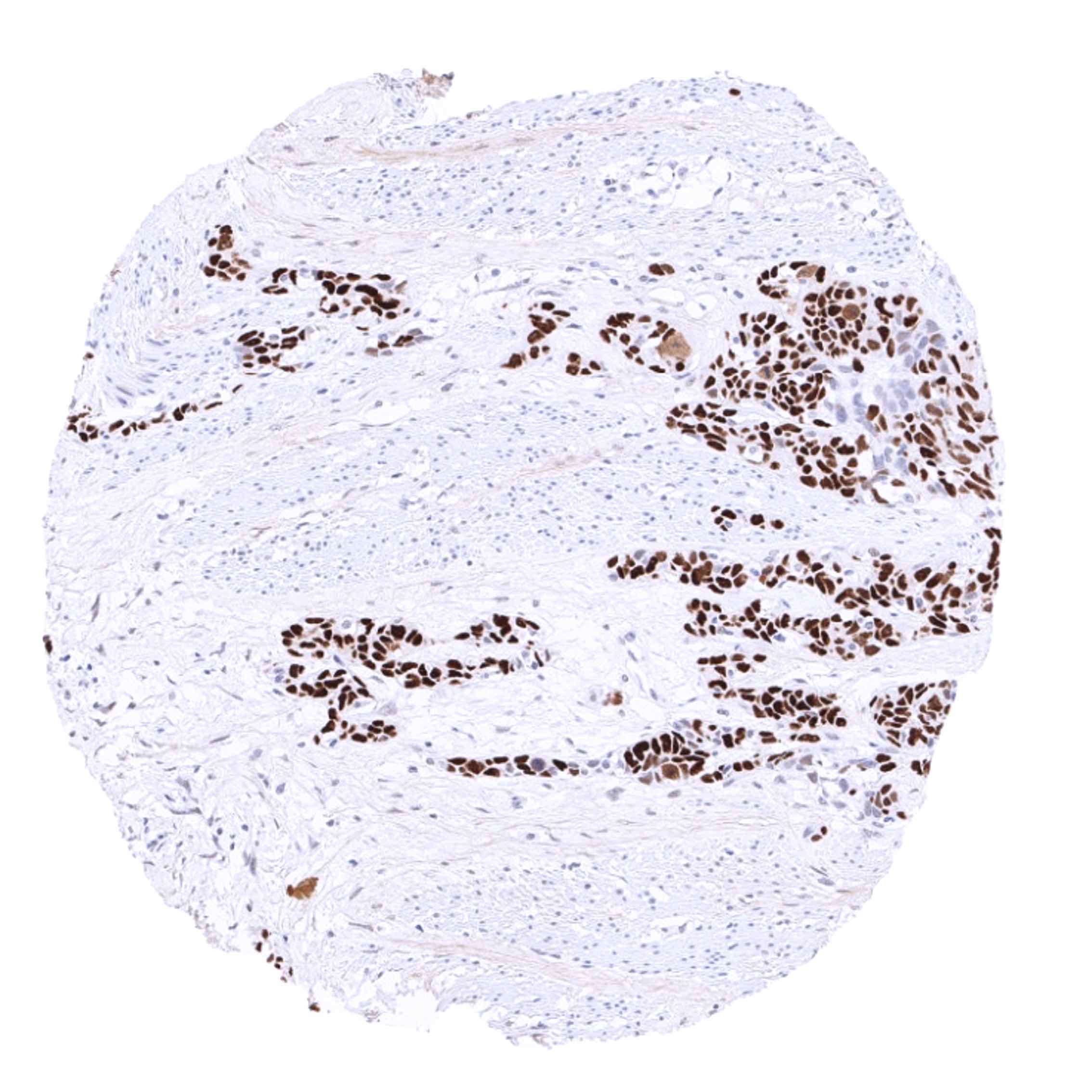
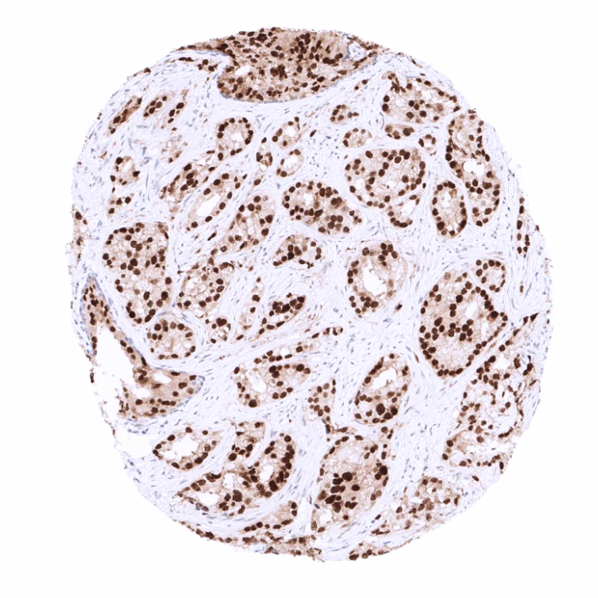
Staining Pattern in Relevant Tumor Types
A positive p53 immunostaining can be seen in many malignant neoplasms from a broad spectrum of different entities.
The TCGA findings on p53 RNA expression in different tumor categories have been summarized in the Human Protein Atlas.
Compatibility of Antibodies
No data available at the moment
Protocol Recommendations
IHC users have different preferences on how the stains should look like. Some prefer high staining intensity of the target stain and even accept some background. Others favor absolute specificity and lighter target stains. Factors that invariably lead to more intense staining include higher concentration of the antibody and visualization tools, longer incubation time, higher temperature during incubation, higher temperature and longer duration of the heat induced epitope retrieval (slide pretreatment). The impact of the pH during slide pretreatment has variable effects and depends on the antibody and the target protein.
All images and data shown here and in our image galleries are obtained by the manual protocol described below. Other protocols resulting in equivalent staining are described as well.
Manual protocol
Freshly cut sections should be used (less than 10 days between cutting and staining). Heat-induced antigen retrieval for 5 minutes in an autoclave at 121°C in pH 7,8 Target Retrieval Solution buffer. Apply MSVA-053R at a dilution of 1:150 at 37°C for 60 minutes. Visualization of bound antibody by the EnVision Kit (Dako, Agilent) according to the manufacturer’s directions.
Agilent / Dako – Autostainer Link 48
Pretreatment in PT-Link for 30 minutes at 95°C (pH high); FLEX peroxidase blocking for 5 minutes (room temperature), MSVA-053R 1:150 for 20 minutes (room temperature), FLEX+ mouse/rabbit (LINKER) for 15 minutes (room temperature), horseradish peroxidase (HRP) for 20 minutes (room temperature), FLEX DAB+Sub-Chromo for 10 minutes (room temperature), FLEX hematoxylin for 5 minutes (room temperature).
These images reflect stainings by the protocol described above. It is of note that a comparable staining result can also be obtained by different protocols. In general, a longer pretreatment, a longer incubation time of the primary antibody, a higher antibody concentration, and a longer incubation time of FLEX+LINKER result in stronger staining, potentially at the cost of more background staining. Modifications of the protocol with a strengthening effect on staining intensity in combination with changes of other parameters that result in lower staining intensity can result in a comparable result as shown above.
Leica – BOND RX
Dewax at 72°C for 30 seconds; Pretreatment in Bond Epitope Retrieval Solution (ER2 – EDTA pH9) for 20 minutes at 100°C; Peroxidase blocking for 5 minutes (room temperature), MSVA-053R 1:150 for 15 minutes (room temperature), Post primary (rabbit anti mouse) for 8 minutes (room temperature), Polymer (goat anti rabbit) for 8 minutes (room temperature), mixed DAB refine for 10 minutes (room temperature), hematoxylin for 5 minutes (room temperature).
These images reflect stainings by the protocol described above. It is of note that a comparable staining result can also be obtained by different protocols. In general, a longer pretreatment, a longer incubation time of the primary antibody, a higher antibody concentration, a higher temperature during incubation, and a longer incubation time of Post primary and or the Polymer result in stronger staining, potentially at the cost of more background staining. Modifications of the protocol with a strengthening effect on staining intensity in combination with changes of other parameters that result in lower staining intensity can result in a comparable result as shown above.
Roche – Ventana Discovery ULTRA
Pretreatment for 64 minutes at 100°C (pH 8,4); CM peroxidase blocking for 12 minutes (room temperature), MSVA-053R 1:150 for 20 minutes at 36°C, secondary antibody (anti-rabbit HQ) for 12 minutes at 36°C, anti-HQ HRP for 12 minutes at room temperature, DAB at room temperature, hematoxylin II at room temperature for 8 minutes, bluing reagent at room temperature for 4 minutes.

These images depict staining results obtained by the protocol described above. It is of note, that the Ventana machines generally require higher antibody concentrations than other commonly used autostainers because the antibodies are automatically diluted during the procedure. Various other protocols can result in an identical result as shown above. A longer pretreatment, a longer incubation time of the primary antibody, a higher antibody concentration, a higher temperature during incubation, and a longer incubation time of secondary antibody and or the anti-HQ HRP result in stronger staining, potentially at the cost of more background staining.
Potential Research Applications
- Despite decades of research, the exact functions of p53 alterations are still not fully understood. This especially applies for gain of function mutations.
- The role of p53 within multiparametric prognostic tests needs to be established.
- The role of p53 null alterations (complete loss of gene function by inactivating mutations and deletions) is unclear in many tumor entities.
Evidence for Antibody Specificity in IHC
There are two ways how the specificity of antibodies can be documented for immunohistochemistry on formalin fixed tissues. These are: 1. Comparison with a second independent method for target expression measurement across a large number of different tissue types (orthogonal strategy), and 2. Comparison with one or several independent antibodies for the same target and showing that all positive staining results are also seen with other antibodies for the same target (independent antibody strategy).
Orthogonal validation: is not well suited for p53 antibodies because p53 expression levels vary more between cell types than between organs. Therefore, available RNA databases identified comparable levels of p53 RNA expression in most organs Human Protein Atlas (Tissue expression p53), and these RNA expression data are not cell type specific.
Comparison of antibodies: For the antibody MSVA-053R specificity is suggested by the strong concordance of its immunostaining data with data obtained by an independent second commercially available antibody (termed “validation antibody”).